Embed Size (px)
Citation preview
*Corresponding author. Department of Mathematical Sciences, New Jersey Institute of Technology,University Heights, Newark, NJ 07102-1982, USA. Tel.: #1-973-596-5835; fax: #1-973-596-5591.E-mail address: [email protected] (V. Booth).
0925-2312/01/$ - see front matter � 2001 Elsevier Science B.V. All rights reserved.PII: S 0 9 2 5 - 2 3 1 2 ( 0 1 ) 0 0 3 8 0 - 0
Neurocomputing 38}40 (2001) 497}504
Regulating "ring rate of networks of pyramidal cells
Victoria Booth*, Amitabha BoseDepartment of Mathematical Sciences, Center for Applied Mathematics and Statistics,
New Jersey Institute of Technology, Newark, NJ 07102-1982, USA
Abstract
In a minimal network model consisting of two pyramidal cells and one interneuron, we showhow excitation and inhibition cooperate to produce "ring rate changes in pyramidal cells thatare consistent with observed place cell "ring in region CA3 of the hippocampus. We show thatinhibition from a common interneuron can synchronize networks of pyramidal cells with nodirect connections. Moreover, recurrent excitation together with common inhibition canmodulate burst pro"les of synchronously "ring cells from complex bursts to bursts withmultiple spike to single spikes. � 2001 Elsevier Science B.V. All rights reserved.
Keywords: Inhibition; Excitation; Place cells
1. Introduction
The "ring patterns of pyramidal cells in region CA3 of the rat hippocampus arebelieved to exhibit both a "ring rate code [4,15] and a phase-based temporal code[6,10] which determines the animal's location in known spatial environments. Thereis active interest in determining the neural mechanisms that underlie the generationand reproduction of these codes, however, much is left to be understood. While theanatomy of the hippocampus is fairly well known, the functional interactions amongpyramidal cells and interneurons has not been fully determined. Thus it is unclearwhether changes in pyramidal cell "ring rate, for example, result from changes inexcitation or inhibition or both.
In this paper, using a minimal network model consisting of two pyramidal cells andone interneuron, we show how excitation and inhibition cooperate to produce "ringrate changes in pyramidal cells that are consistent with the observed "ring patterns ofplace cells as a rat runs on a linear track. In particular, we show how the "ring rate ofplace cells can increase either linearly [4] or in a Gaussian-like manner [3] as the ratpasses through the place "eld. The results that we present depend on a dynamicbalancing act between excitatory and inhibitory input to the pyramidal cells.In previous work (reviewed in the Model section below), using the 2-compartment
pyramidal cell model of Pinsky and Rinzel [7], we showed how inhibition arrivingduring a burst can change the burst pro"le and interburst (spike) interval of a repeti-tively bursting cell. Speci"cally, we showed that as the maximal conductance of fastinhibitory input to the dendrites of the pyramidal cell increases, the pyramidal cell"ring changes from complex bursts, to bursts with multiple spikes to single spikes.Here, we address the question of what e!ect recurrent excitation has on networks of
pyramidal cells which receive inhibition from a common interneuron. Recurrentexcitatory connections in CA3 are anatomically dense, albeit mathematically sparse.Previous modeling studies have shown that fast, recurrent excitation can synchronizeburst "ring of pyramidal cells [7,12]. We show that inhibition from a commoninterneuron can synchronize networks of pyramidal cells with no direct connections.Moreover, recurrent excitation together with common inhibition can modulate burstpro"les of synchronously "ring cells from complex bursts to bursts with multiple spiketo single spikes.Our motivation to study the dual e!ects of excitation and inhibition is to develop
a model for place cell "ring that is consistent with the functional model of associativememory developed by Recce [8] and with our biophysical model of phase precession[2]. Recce [8] proposes that synchronously "ring assemblies of place cells code for thesame spatial location and that recurrent excitation drives the recruitment, and thusthe recall, of the location memory. We have proposed that the timing of inhibitoryinput to place cells controls the phase precession phenomena [2].
2. Model
The minimal network model that we consider consists of two pyramidal cells andone interneuron (Fig. 1). Each of the pyramidal cells is described by the 2-compart-ment model of Pinsky and Rinzel [7], while the excitable interneuron is modeled usingthe Morris-Lecar equations [5]. A pyramidal cell consists of a soma compartment,capable of producing fast sodium spikes, and a dendrite compartment, capable ofproducing a broad calcium-based depolarization. Pinsky and Rinzel [7] show thatdepending on the level of applied current to the soma and the relative strength of theelectrotonic coupling between compartments, the cell can exhibit complex bursts,spike doublets and single spikes. The complex burst results from the interactionbetween the soma and dendrite compartments. Namely, the soma initially "resa sodium spike which back propagates to the dendrite instigating a large scale calciumevent. The dendritic, calcium-based depolarization then supports further depolarized
498 V. Booth, A. Bose / Neurocomputing 38}40 (2001) 497}504
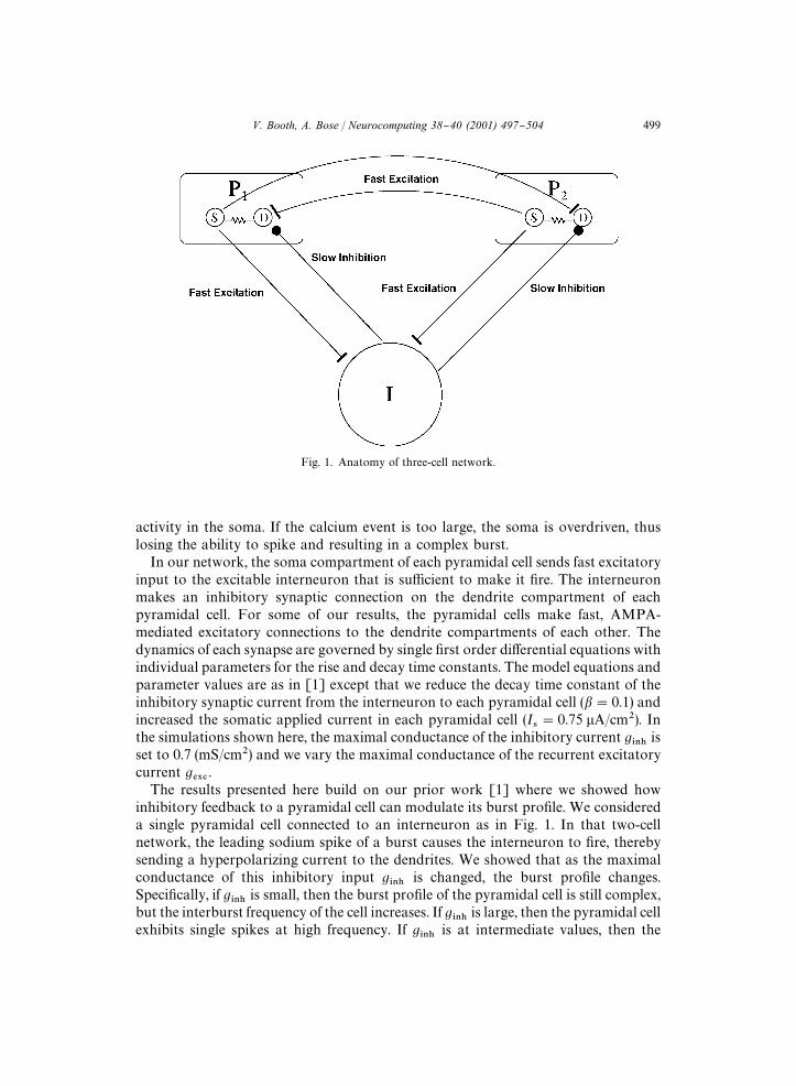
Fig. 1. Anatomy of three-cell network.
activity in the soma. If the calcium event is too large, the soma is overdriven, thuslosing the ability to spike and resulting in a complex burst.In our network, the soma compartment of each pyramidal cell sends fast excitatory
input to the excitable interneuron that is su$cient to make it "re. The interneuronmakes an inhibitory synaptic connection on the dendrite compartment of eachpyramidal cell. For some of our results, the pyramidal cells make fast, AMPA-mediated excitatory connections to the dendrite compartments of each other. Thedynamics of each synapse are governed by single "rst order di!erential equations withindividual parameters for the rise and decay time constants. The model equations andparameter values are as in [1] except that we reduce the decay time constant of theinhibitory synaptic current from the interneuron to each pyramidal cell (�"0.1) andincreased the somatic applied current in each pyramidal cell (I
�"0.75 �A/cm�). In
the simulations shown here, the maximal conductance of the inhibitory current g���is
set to 0.7 (mS/cm�) and we vary the maximal conductance of the recurrent excitatorycurrent g
���.
The results presented here build on our prior work [1] where we showed howinhibitory feedback to a pyramidal cell can modulate its burst pro"le. We considereda single pyramidal cell connected to an interneuron as in Fig. 1. In that two-cellnetwork, the leading sodium spike of a burst causes the interneuron to "re, therebysending a hyperpolarizing current to the dendrites. We showed that as the maximalconductance of this inhibitory input g
���is changed, the burst pro"le changes.
Speci"cally, if g���is small, then the burst pro"le of the pyramidal cell is still complex,
but the interburst frequency of the cell increases. If g���is large, then the pyramidal cell
exhibits single spikes at high frequency. If g���is at intermediate values, then the
V. Booth, A. Bose / Neurocomputing 38}40 (2001) 497}504 499
pyramidal cell has bursts with 4, 3 or 2 spikes (as g���is increased). The reason that
these patterns occur is straightforward. When inhibition is weak (g���small), it is not
strong enough to prevent a full dendritic calcium event, and a complex burst arises.When inhibition is too large, on the other hand, the leading somatic sodium spikebackpropagates to the dendrite, but the full dendritic calcium event is completelysuppressed. Thus, there is not su$cient depolarizing current to initiate any furthersodium spikes. For intermediate values of g
���, the dendritic calcium event is only
partially blocked. This allows the soma to become su$ciently depolarized to havea second sodium spike, which again back propagates to the dendrite. If this secondspike causes enough calcium to enter the cell, then a third sodium spike can begenerated and so on. In [1], we used phase plane methods to show exactly how thisoccurs and additionally to show why there is an increase in interburst frequency withincreasing g
���.
3. Results
3.1. Synchrony induced by common inhibition
With no recurrent excitatory connections between pyramidal cells (g���
"0.0), thecommon inhibition provided by the interneuron can synchronize pyramidal cell "ring.By varying the maximal conductance g
���and the decay rate � of the inhibitory
synaptic current, we obtain synchronous "ring of complex bursts, of bursts withmultiple spikes and of single spikes (Fig. 2a, other simulations not shown). Themodulation of the burst pro"le with varying g
���is obtained as described in theModel
section and in [1]. Synchronization depends on adjusting the decay rate of inhibition.In particular, synchronizing complex bursts depends on weak g
���and small � while
to synchronize single spikes, both g���and � need to be larger. As in other studies
obtaining synchrony with slowly decaying inhibition among cells [11,13,14], in ournetwork the slowly decaying inhibition provides for a time compression of dendriticvoltages during the silent phase of the burst cycle. In [1], we showed that dendriticvoltage needed to increase past a certain level (what we called <H
�) in order for the
soma to "re and initiate a burst. In the present case with two pyramidal cells, theslowly decaying inhibition forces the dendritic voltages to pass through <H
�closely in
time. For complex burst synchronization, where the duration of the silent phase islong, a relatively slower decay rate of inhibition is needed as compared to the singlecell case.Since synchrony is induced during the silent phase of bursting, the same mechanism
synchronizes "ring of the di!erent burst pro"les. The burst dynamics in the activestate are relatively unimportant for synchrony. In contrast, synchrony throughrecurrent excitation is induced during the active state of the neuron. In a network ofPinsky}Rinzel cells, this tends to promote complex burst synchronization, as opposedto singlet or doublet burst synchronization. The reason is that dendritic excitationpromotes the full calcium event which underlies the complex burst, but which needs tobe suppressed for singlet or doublet burst "ring.
500 V. Booth, A. Bose / Neurocomputing 38}40 (2001) 497}504
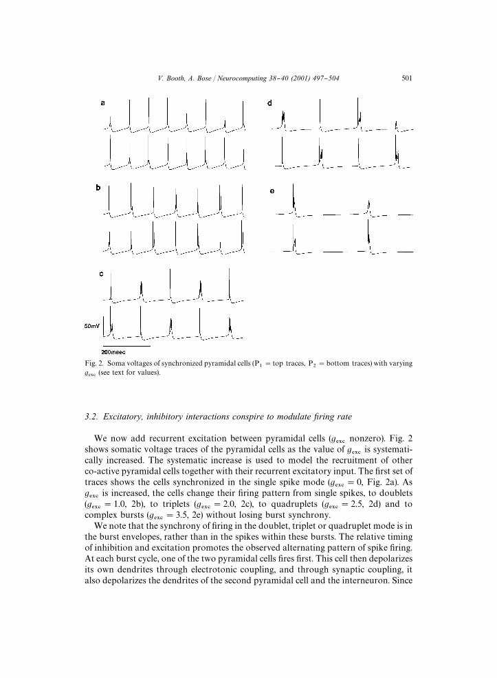
Fig. 2. Soma voltages of synchronized pyramidal cells (P�"top traces, P
�"bottom traces) with varying
g���(see text for values).
3.2. Excitatory, inhibitory interactions conspire to modulate xring rate
We now add recurrent excitation between pyramidal cells (g���nonzero). Fig. 2
shows somatic voltage traces of the pyramidal cells as the value of g���is systemati-
cally increased. The systematic increase is used to model the recruitment of otherco-active pyramidal cells together with their recurrent excitatory input. The "rst set oftraces shows the cells synchronized in the single spike mode (g
���"0, Fig. 2a). As
g���is increased, the cells change their "ring pattern from single spikes, to doublets
(g���
"1.0, 2b), to triplets (g���
"2.0, 2c), to quadruplets (g���
"2.5, 2d) and tocomplex bursts (g
���"3.5, 2e) without losing burst synchrony.
We note that the synchrony of "ring in the doublet, triplet or quadruplet mode is inthe burst envelopes, rather than in the spikes within these bursts. The relative timingof inhibition and excitation promotes the observed alternating pattern of spike "ring.At each burst cycle, one of the two pyramidal cells "res "rst. This cell then depolarizesits own dendrites through electrotonic coupling, and through synaptic coupling, italso depolarizes the dendrites of the second pyramidal cell and the interneuron. Since
V. Booth, A. Bose / Neurocomputing 38}40 (2001) 497}504 501
electrotonic coupling acts much faster than synaptic coupling, the feedback inhibitionfrom the interneuron arrives at this pyramidal cell after the onset of the dendriticcalcium event and, thus, merely modulates it. On the other hand, the inhibition arrivesat the second pyramidal cell concurrently (or within a short time window) of theexcitatory input, and is capable of blocking its dendritic calcium event. Thus, thesecond pyramidal cell "res only one spike. As described in [1], the interburst intervalfollowing a single spike is shorter than that following a complex burst, thus the secondcell becomes the initiator of the next burst.
4. Discussion
We have shown that inhibition can be used to not only synchronize pyramidal cells,but can also work with excitation to shape the "ring patterns of these cells. Theprimary targets of these synaptic currents and the electrotonic current between somaand dendrite compartments are calcium-based mechanisms. The timing of inputs tothe dendrites are also important in determining the "ner structure of spikes withina burst.We note that inhibition is not necessary to evoke changes in "ring rate and
frequency. Indeed in the original Pinsky-Rinzel paper, they show how these changescan occur by changing the applied current to the soma. The reason that we chose tostudy the e!ects of inhibition is that we have used the changing role and timing ofinhibitory input to produce new explanations for the phase precession phenomenafound by O'Keefe and Recce [6] in previous work. In [2], we proposed that the phaseprecession phenomenon of hippocampal place cells could result from changes incontrol of interneuron networks in region CA3. Speci"cally, we showed that ifinterneuron "ring is initiated by theta pacemaker input, then place cells don't "re, or"re without precession. Alternatively, if interneuron "ring is initiated by place cells,then both the interneuron and the place cell phase precess. We also providedmechanisms which could account for the switch in control at the beginning and end ofa place "eld. In [1], we showed that fast decaying inhibition arriving just beforea pyramidal cell burst could delay it by over one theta cycle, and that fast decayingperiodic inhibitory input could completely suppress "ring.Our results suggest neural mechanisms that account for the rate changes observed
in place cell "ring as the corresponding place "eld is crossed. For example, Mehta etal. [4] propose that "ring rate increases linearly as the animal moves through theplace "eld. This can clearly be achieved in our model if inhibition is kept constant andthe net excitatory input to the dendrites of the pyramidal cell increases cycle by cycle.This increase can occur as more co-active place cells are recruited into the "ringpattern. Burgess et al. [3] have alternatively proposed that "ring rate within a place"eld is better described by a Gaussian envelope. In our model this can be achieved, forexample, if both co-active place cells and interneurons are di!erentially recruited cycleby cycle, or if the excitatory synapses among co-active place cells are depressing.In summary, the e!ects described above provide insight into how place cells may
modulate their "ring rate. Together with our prior results on how the timing of
502 V. Booth, A. Bose / Neurocomputing 38}40 (2001) 497}504
inhibitory input can be changed [2,9], a consistent model for the phase precession andthe "ring rate changes of place cells can be obtained. Our results suggest that dynamicbalances between excitatory and inhibitory input may be crucial in understandingcoding schemes in other brain regions.
Acknowledgements
The authors were supported in part by grants from the National Science Founda-tion (�DMS-9973230 AB,VB and�IBN-9722946 VB) and fromNJIT (�421540 ABand �421590 VB).
References
[1] V. Booth, A. Bose, Neural mechanisms for generating rate and temporal codes in model CA3pyramidal cells, J. Neurophysiol., 2000, submitted for publication.
[2] A. Bose, V. Booth, M. Recce, A temporal mechanism for generating the phase precession ofhippocampal place cells, J. Comput. Neurosci. 9 (2000) 5}30.
[3] N. Burgess, M. Recce, J. O'Keefe, A model of hippocampal function, Neural Networks 17 (1994)1065}1081.
[4] M.R. Mehta, M.C. Quirk, M.A. Wilson, Experience-dependent asymmetric shape of hippocampalreceptive "elds, Neuron 25 (2000) 707}715.
[5] C. Morris, H. Lecar, Voltage oscillations in the barnacle giant muscle "ber, Biophys. J. 35 (1981)193}213.
[6] J. O'Keefe, M.L. Recce, Phase relationship between hippocampal place units and the EEG thetarhythm, Hippocampus 3 (1993) 317}330.
[7] P.F. Pinsky, J. Rinzel, Intrinsic and network rhythmogenesis in a reduced Traub model for CA3neurons, J. Comput. Neurosci. 1 (1994) 39}60.
[8] M. Recce, Encoding information in neuronal activity, in: W. Maass, C.M. Bishop (Eds.), PulsedNeural Networks, MIT Press, Cambridge, MA, 1999, pp. 111}131.
[9] M. Recce, A. Bose, V. Booth, Hippocampal place cells and the generation of a temporal code,Neurocomputing 32}33 (2000) 225}234.
[10] W.E. Skaggs, B.L. McNaughton, M.A. Wilson, C.A. Barnes, Theta-phase precession in hippocampalneuronal populations and the compression of temporal sequences, Hippocampus 6 (1996)149}172.
[11] D. Terman, N. Kopell, A. Bose, Dynamics of two mutually coupled slow inhibitory neurons, PhysicaD 117 (1998) 241}275.
[12] R.D. Traub, R. Wong, R. Miles, H. Michelson, A model of a CA3 hippocampal pyramidalneuron incorporating voltage-clamp data on intrinsic conductances, J. Neurophysiol. 66 (1991)635}649.
[13] C. van Vreeswijk, L. Abbott, G.B. Ermentrout, When inhibition, not excitation synchronizes neural"ring, J. Comput. Neurosci. 1 (1994) 313}321.
[14] X.J. Wang, J. Rinzel, Alternating and synchronous rhythms in reciprocally inhibitory neuron models,Neural Comput. 4 (1992) 84}97.
[15] M.A. Wilson, B.L. McNaughton, Dynamics of the hippocampal ensemble code for space, Science 265(1993) 1055}1058.
V. Booth, A. Bose / Neurocomputing 38}40 (2001) 497}504 503

Amitabha Bose received his Ph.D. from Brown University in Applied Mathemat-ics. He currently is an assistant professor in the Department of MathematicalSciences at the New Jersey Institute of Technology. His research interests are inapplications of dynamical systems to neuronal networks and nonlinear waves.
Victoria Booth received her Ph.D. in Applied Mathematics from NorthwesternUniversity. Following a postdoctoral fellowship at the Mathematical ResearchBranch at NIH, she joined the Department of Mathematical Sciences at the NewJersey Institute of Technology as an assistant professor. Her research interests arein biophysical modeling of neuronal networks.
504 V. Booth, A. Bose / Neurocomputing 38}40 (2001) 497}504