Embed Size (px)
Citation preview

Regulated Endoplasmic Reticulum-associated Degradationof a Polytopic Proteinp97 RECRUITS PROTEASOMES TO Insig-1 BEFORE EXTRACTION FROM MEMBRANES*
Received for publication, July 15, 2009, and in revised form, October 6, 2009 Published, JBC Papers in Press, October 8, 2009, DOI 10.1074/jbc.M109.044875
Yukio Ikeda‡, George N. DeMartino§, Michael S. Brown‡1, Joon No Lee‡, Joseph L. Goldstein‡2, and Jin Ye‡3
From the Departments of ‡Molecular Genetics and §Physiology, University of Texas Southwestern Medical Center,Dallas, Texas 75390-9046
Polytopicmembraneproteins subjected to endoplasmic retic-ulum (ER)-associated degradation are extracted from mem-branes and targeted to proteasomes for destruction. The extrac-tionmechanism is poorly understood.One polytopic ERproteinsubjected to ER-associated degradation is Insig-1, a negativeregulator of cholesterol synthesis. Insig-1 is rapidly degraded byproteasomes when cells are depleted of cholesterol, and its deg-radation is inhibited when sterols accumulate in cells. Insig-2, afunctional homologue of Insig-1, is degraded slowly, and itsdegradation is not regulated by sterols. Here, we report that asingle amino acid substitution in Insig-2, Insig-2(L210A), causesInsig-2 to be degraded in an accelerated and sterol-regulatedmanner similar to Insig-1. In seeking an explanation for theaccelerated degradation, we found that proteasomes bind towild type Insig-1 and mutant Insig-2(L210A) but not to wildtype Insig-2, whereas the proteins are still embedded in cellmembranes. This binding depends on at least two factors, ubiq-uitination of Insig and association with the ATPase p97/VCPcomplex. These data suggest that p97 recruits proteasomes topolytopic ER proteins even before they are extracted frommembranes.
Many transmembrane proteins of the endoplasmic reticu-lum (ER)4 are degraded via the ER-associated degradation(ERAD) pathway (1, 2). According to the current model, ERADbegins when selected membrane proteins are recognized and
ubiquitinated by one of several ER-associated E3 ubiquitinligases. Through the action of the ATPase p97/VCP complex(hereafter referred to as p97), these ubiquitinated membraneproteins are extracted from membranes and translocated intothe cytosol, where they are degraded by proteasomes. Themechanism by which polytopic transmembrane proteins areunwound and extracted from membranes is unknown.The 26 S proteasome is a protein degradation machine that
consists of a 20 S catalytic core and a 19 S regulatory portioncomposed of 14 and 19 unique protein subunits, respectively(3, 4). Proteins subjected to ERAD are recognized by protea-somes through polyubiquitin chains that are added to theseproteins by ubiquitin ligases (5). Whether proteasomes bind toubiquitinated membrane proteins before or after their extrac-tion from membranes is unknown.Inmammalian cells ERAD of some proteins can be regulated
by small molecule metabolites. One such regulated protein isInsig-1, an ERmembrane protein with sixmembrane-spanninghelices (6). Insig-1 is an ER anchor protein that mediates feed-back inhibition of cholesterol synthesis. When excess sterolsaccumulate in cells, Insig-1 is induced to bind to either of twoER membrane proteins; that is, Scap or 3-hydroxy-3-methyl-glutaryl coenzyme A (HMG-CoA) reductase (6). Scap is anescort protein that carries sterol regulatory element-bindingproteins (SREBPs) to the Golgi complex (7). SREBPs are mem-brane-bound transcription factors that must be transported tothe Golgi complex for processing to release nuclear fragmentsthat activate all of the genes required for cholesterol synthesisand uptake (8, 9). Certain SREBPs also activate the synthesis ofunsaturated fatty acids (8, 9). In cholesterol-loaded cells Insig-1binds to Scap, and this causes ER retention of Scap, therebypreventing translocation of Scap-bound SREBPs to the Golgi(10). As a result, cholesterol synthesis and uptake are reduced.Sterols also induce binding of Insig-1 to HMG-CoA reductase,which results in ubiquitination and subsequent proteasomaldegradation of the reductase (11, 12). This degradation slowsthe production of mevalonate, a rate-limiting step in choles-terol synthesis (13). The two actions of Insig-1 serve to preventover-accumulation of cholesterol and its sterol precursors.We previously showed that Insig-1 itself is subject to ERAD
in a reaction that is regulated by sterols and unsaturated fattyacids (14, 15). In cholesterol-depleted cells, Insig-1 does notbind to Scap or HMG-CoA reductase. Instead, Insig-1 binds togp78, an E3 ubiquitin ligase (16). This interaction results inubiquitination of Insig-1 (16). When cells are treated with ste-
* This work was supported, in whole or in part, by National Institutes of HealthGrants 2PO1HL20948 and RO1DK46181. This work was also supported bythe Perot Family Foundation.
1 To whom correspondence may be addressed: Dept. of Molecular Genetics,UT Southwestern Medical Center, 5323 Harry Hines Blvd., Dallas, TX 75390-9046. Tel.: 214-648-2141; Fax: 214-648-8804; E-mail: [email protected].
2 To whom correspondence may be addressed: Dept. of Molecular Genet-ics, UT Southwestern Medical Center, 5323 Harry Hines Blvd., Dallas, TX75390-9046. Tel.: 214-648-2141; Fax: 214-648-8804; E-mail: [email protected].
3 Supported by American Heart Association National Scientific DevelopmentGrant 0630029N.
4 The abbreviations used are: ER, endoplasmic reticulum; ERAD, ER-associ-ated degradation; CMV, cytomegalovirus; two-dimensional DIGE, two-di-mensional fluorescence difference gel electrophoresis; FCS, fetal calfserum; 25-HC, 25-hydroxycholesterol; HMG-CoA, 3-hydroxy-3-methylglu-taryl coenzyme A; LPDS, lipoprotein-deficient serum; N-Suc-Leu-Leu-Val-Tyr-AMC, N-succinyl-Leu-Leu-Val-Tyr-7-amido-4-trifluromethylcoumarin;siRNA, small interfering RNA; SREBP, sterol regulatory element-bindingproteins; TK, thymidine kinase; TEV, tobacco etch virus; WT, wild type; HA,hemagglutinin; CHO, Chinese hamster ovary; HSV, herpes simplex virus.
THE JOURNAL OF BIOLOGICAL CHEMISTRY VOL. 284, NO. 50, pp. 34889 –34900, December 11, 2009© 2009 by The American Society for Biochemistry and Molecular Biology, Inc. Printed in the U.S.A.
DECEMBER 11, 2009 • VOLUME 284 • NUMBER 50 JOURNAL OF BIOLOGICAL CHEMISTRY 34889
by guest on July 26, 2018http://w
ww
.jbc.org/D
ownloaded from

rols, Scap binds to Insig-1, displacing gp78 from Insig-1 (16).Consequently, ubiquitination of Insig-1 is inhibited, and Insig-1is stabilized (16). Degradation of ubiquitinated Insig-1 requirestwo other proteins, Ubxd8 and p97 (15). RNAi-mediatedknockdown of either of these proteins does not block ubiquiti-nation of Insig-1, but it does prevent its degradation. Whenunsaturated fatty acids are added to cells, Insig-1 is stabilizedeven in the absence of sterols. Fatty acids do not block Insig-1ubiquitination. Rather, they stabilize ubiquitinated Insig-1 byblocking the interaction between Insig-1 and the Ubxd8�p97complex so that ubiquitinated Insig-1 cannot be extracted fromthe membranes (15).Mammalian cells also contain a close homolog of Insig-1,
namely Insig-2, which functions like Insig-1 to block choles-terol synthesis (17). Unlike Insig-1, Insig-2 does not interactwith gp78 and, therefore, it is not ubiquitinated (16). As a result,Insig-2 has a much longer half-life, and its degradation is notregulated by sterols (18, 19).In the current study we characterize a mutant Insig-2,
Insig-2(L210A), which retains all the functions of Insig-2 butis degraded in a sterol-regulated manner like Insig-1. Weshow that proteasomes bind to ubiquitinated mutant Insig-2(L210A) and wild type Insig-1 (but not to wild type Insig-2),whereas the proteins are still embedded in ER membranes.This binding requires the p97 complex, and it appears toprecede the extraction and degradation of the Insig proteins.These studies raise the possibility that proteasomes togetherwith p97 play a direct role in the extraction of polytopicproteins from ER membranes.
EXPERIMENTAL PROCEDURES
Materials—We obtained hydroxypropyl-�-cyclodextrin fromCyclodextrin Technologies Development, arachidonic acidfrom NU-CHEK-PREP, INC, defatted bovine serum albuminfrom Roche Applied Science, Nonidet P-40 Alternative (Non-idet P-40) and digitonin fromCalbiochem, Fos-choline 13 fromAnatrace, MG-132 from Boston Biochem, N-Succinyl-Leu-Leu-Val-Tyr-7-amido-4-methylcoumarin (N-Suc-Leu-Leu-Val-Tyr-AMC) from Bachem. cycloheximide, sodium salt of ATP,3� FLAG peptide, anti-c-Myc-agarose affinity gel, anti-FLAG-M2-agarose affinity gel, and rabbit polyclonal anti-FLAG fromSigma, IgG-Sepharose 6 Fast Flow, CyDye DIGE Fluorminimallabeling kit, and Deep Purple Total Protein Stain from GEHealthcare, rabbit polyclonal anti-Myc, anti-hemagglutinin(HA), anti-T7, and ReliaBLOT from Bethyl Laboratories,mousemonoclonal anti-HSV IgG and T7-tag antibody-agarosefrom Novagen, mouse monoclonal anti-�1 IgG, anti-�7 IgG,and anti-Rpt6 IgG from Biomol, mouse monoclonal anti-p97IgG from BD Transduction Laboratories, and horseradish per-oxidase-conjugated donkey anti-mouse and anti-rabbit IgGs(affinity-purified) from Jackson ImmunoResearch. Hybridomacells producing IgG-9E10, a mouse monoclonal antibodyagainst Myc tag, were obtained from the American Type Cul-ture Collection (Manassas, VA). IgG-4H4, amousemonoclonalantibody against hamster Scap, was generated by immunizinga mouse with a fusion protein encoding amino acids 1–767 ofScap that was epitope-tagged with 10 histidine residues atthe NH2 terminus (20). Rabbit polyclonal antibodies against
human gp78 (16) and human Rpt5 (21) were reported previ-ously. Lipoprotein-deficient serum (LPDS) (22), delipidatedfetal calf serum (23), sodium oleate (22), sodium mevalonate(24), and compactin (24) were prepared as described in theindicated references. SR-12813 was synthesized by the CoreMedicinal Chemistry Laboratory, University of Texas South-westernMedical Center (25). A stock solution of 10mM sodiumarachidonate-bovine serum albumin in 0.15 M NaCl (final pH7.6) was prepared as previously described (23).Plasmid Constructs—The following plasmids were described
in the indicated references or obtained from the indicatedsources. pTK-Scap and pCMV-Scap encode hamster Scapunder control of the thymidine kinase (TK) and the cytome-galovirus (CMV) promoters, respectively (14). pTK-HSV-SREBP2 encodes HSV-tagged human SREBP-2 under controlof the TK promoter (26). pCMV-HMGCoAR(TM1–8)-T7encodes amino acids 1–346 of hamster HMG-CoA reductasefollowed by three tandem copies of the T7 epitope tag undercontrol of the CMV promoter (11). pCMV-Myc-Ubxd8encodes Myc-tagged human Ubxd8 under control of the CMVpromoter (15). pEF1a-HA-ubiquitin (provided by Dr. ZhijianChen, University of Texas Southwestern Medical Center)encodes amino acids 1–76 of human ubiquitin tagged with HAepitope tag at the NH2 terminus under control of the EF1apromoter (16). pCIneo-gp78 (obtained from Dr. Allan M.Weissman, National Cancer Center) encodes human gp78under control of the CMV promoter (16). pCMV-Insig1-T7and pCMV-Insig2-T7 encode human Insig-1 and Insig-2,respectively, each tagged with three tandem copies of the T7epitope tag at the COOH terminus under control of the CMVpromoter (16). pTK-Insig2-Myc and pCMV-Insig2-Myc en-codewild type human Insig-2, each taggedwith six copies of theMyc epitope tag at the COOH terminus under control of theTK and the CMV promoters, respectively (26). pCMV-FLAG-Insig1 encodes human Insig-1 with two tandem copies of aFLAG epitope tag under control of the CMV promoter (27).pCMV-Insig2-FLAG-TEV-ProteinA encodeswild type humanInsig-2, sequentially tagged at the COOH terminus with threetandem copies of the FLAG epitope tag, one copy of the TEVprotease cleavage site, and two copies of Protein A under con-trol of the CMV promoter. Mutants of Insig-1 or Insig-2 weregenerated with the QuikChange XL site-directed mutagenesiskit (Stratagene) according to the manufacturer’s instructions.The integrity of all plasmidswas confirmedbyDNAsequencingof all open reading frames.Tissue Culture Medium—Medium A is a 1:1 mixture of
Ham’s F-12 medium and Dulbecco’s modified Eagle’s mediumcontaining 100 units/ml penicillin and 100�g/ml streptomycinsulfate. Medium B is medium A containing 5% (v/v) LPDS,50 �M sodium compactin, and 50 �M sodium mevalonate.Medium C is Dulbecco’s modified Eagle’s medium (1 g/literglucose) containing 10% (v/v) fetal calf serum (FCS), 100units/ml penicillin, and 100 �g/ml streptomycin sulfate.Medium D is medium C supplemented with 5 �g/ml choles-terol (added in ethanol; final concentration, 0.05%), 1 mM
sodiummevalonate, and 20 �M sodium oleate. Medium E con-tains IS GRO (Irvine Scientific) supplemented with 10% (v/v)FCS, 10 mM L-glutamine, 100 units/ml penicillin, and 100
p97-mediated Recruitment of Proteasomes to Insigs
34890 JOURNAL OF BIOLOGICAL CHEMISTRY VOLUME 284 • NUMBER 50 • DECEMBER 11, 2009
by guest on July 26, 2018http://w
ww
.jbc.org/D
ownloaded from

�g/ml streptomycin sulfate. Medium F is Dulbecco’s modifiedEagle’s medium (1 g/liter glucose) containing 10% (v/v) delipi-dated FCS, 50 �M sodium compactin, 50 �M sodium meval-onate, 100 units/ml penicillin, and 100 �g/ml streptomycinsulfate.Cell Culture—Human 293S cells, a clone of HEK-293 cells
adapted to suspension culture (28), were grown inmonolayer at37 °C in 5% CO2. HEK-293S/pInsig1 cells is a line of HEK-293Sthat stably expresses pCMV-Insig1-T7-TEV-Protein A (con-taining the gene encoding Zeocin resistance) (15). HEK-293S/pInsig2 and HEK-293S/pInsig2(L210A) cells are lines of HEK-293S cells that stably express pCMV-Insig2-FLAG-TEV-Protein A and pCMV-Insig2(L210A)-FLAG-TEV-Protein A,respectively. They were generated by transfecting HEK-293Scells with pCMV-Insig2-FLAG-TEV-Protein A or pCMV-Insig2(L210A)-FLAG-TEV-Protein A (both plasmids contain-ing the Zeocin resistance gene) followed by selection with 700�g/ml Zeocin. Monolayers of stably transfected HEK-293Scells were grown in 5% CO2 at 37 °C in medium D containing500 �g/ml Zeocin. SRD-14/pTK-Insig1-Myc cells, a line ofmutant CHO cells lacking endogenous Insig-1 that stablyexpress pTK-Insig1-Myc (14), were grown at 37 °C in 8% CO2in medium A supplemented with 5% LPDS and 500 �g/mlG418. SRD-13A cells, a Scap-deficient cell line derived fromCHO-7 cells (29), were grown in monolayer at 37 °C in 8% CO2in medium A supplemented with 5% FCS, 5 �g/ml cholesterol,1 mM sodium mevalonate, and 20 �M sodium oleate. SRD-15cells, an Insig-1/Insig-2-deficient cell line (30), were main-tained in medium A supplemented with 5% LPDS, 10 �M ofSR-12813, and 1�g/ml 25-hydroxycholesterol (25-HC).Mono-layers of SV-589 cells, an immortalized line of human fibro-blasts expressing the SV40 large T antigen (31), were grown at37 °C in 5% CO2 in medium C.Transient Transfection of Cells and Immunoblot Analysis—
SRD-13A, SRD-15, and SV589 cells were transiently trans-fectedwith FuGENE6 reagent (RocheApplied Science) accord-ing to the manufacturer’s protocol. Conditions of incubationafter transfection are described in the figure legends. AfterSDS/PAGE (8 or 10% gels), the proteins were transferred toHybond-C extra nitrocellulose filters (GE Healthcare). The fil-ters were incubated at room temperature with one of the fol-lowing primary antibodies: 0.5 �g/ml anti-HSV IgG; 1 �g/mlIgG-9E10; 0.5 �g/ml rabbit polyclonal anti-Myc, rabbit poly-clonal anti-T7 and rabbit polyclonal anti-HA; 0.16�g/ml rabbitpolyclonal anti-FLAG; 5 �g/ml IgG-4H4; 1 �g/ml rabbit poly-clonal anti-gp78; 1:1000 dilution of mouse monoclonal anti-p97 IgG, anti-�1 IgG, anti-�7 IgG, and anti-Rpt6 IgG; 1:5000dilution of rabbit polyclonal anti-Rpt5. Except for Rpt5 immu-noblots shown in Fig. 6A in which we used ReliaBLOT horse-radish peroxidase conjugate (1:5,000 dilution) as the secondaryantibody, horseradish peroxidase-conjugated donkey anti-mouse and anti-rabbit IgGs (1:5000 dilution) were used as thesecondary antibody in all other immunoblot analyses. Boundantibodies were visualized by chemiluminescence (SuperSignalSubstrate; Pierce) according to themanufacturer’s instructions.Filters were exposed to Kodak X-Omat Blue XB-1 film at roomtemperature for 1–60 s.
RNA Interference (RNAi)—Duplexes of small interferingRNA (siRNA) were synthesized by Dharmacon Research(Lafayette, CO). The siRNAswere designed to target nucleotidepositions 1130–1148 (relative to the codon for the initiatingmethionine) for human gp78 (GenBankTM accession numberNM_001144) and positions 1095–1113 for human p97 (Gen-BankTM accession number NM_007126). SV589 cells weretransfected with 400 pmol of siRNA duplexes/60-mm dishusing OligofectAMINETM reagent (Invitrogen). HEK-293S/pInsig1 cells and HEK-293S/pInsig2(L210A) cells were trans-fected with 400 pmol of siRNA duplexes/100-mm dish usingLipofectamineTMRNAiMAX reagent (Invitrogen). After trans-fection the cells were used for experiments as described in thelegend to Fig. 7.Tandem Affinity Purification and Two-dimensional Fluores-
cence Difference Gel Electrophoresis (Two-dimensional DIGE)—Membrane fractions from 2 liters of HEK-293S/pInsig2 cellsand 4 liters of HEK-293S/pInsig2(L210A) cells were preparedas described previously (10) and solubilized with buffer A (50mM Hepes-KOH (pH 7.6), 150 mM NaCl, and a protease inhib-itor mixture containing 25 �g/ml N-acetyl-leucinal-leucinal-norleucinal, 5 �g/ml pepstatin A, 2 �g/ml aprotinin, and 10�g/ml leupeptin) supplemented with 0.1% (w/v) Nonidet P-40.After incubation for 1 h, the insoluble material was removed bycentrifugation at 100,000� g for 1 h. All subsequent operationswere carried out at 4 °C. The supernatant was incubated in atube containing IgG-Sepharose 6 Fast Flow beads. After 16 hthe beadswere transferred to a Bio-Spin disposable chromatog-raphy column (Bio-Rad) and washed with 20� bead-volume ofbuffer A containing 0.05% Nonidet P-40 and 5 mM sodiumEDTA. The beads were then incubated for 2 h with buffer Acontaining 0.05% Nonidet P-40, 5 mM sodium EDTA, and 50units/ml of AcTEVTM protease (Invitrogen) followed by elu-tion. The resulting eluate (2ml) plus an additional 12ml ofwashwith buffer A containing 0.05% Nonidet P-40 were applied 5times to a column containing anti-FLAG-M2-agarose affinitygel followed by a wash with 20� bead-volumes of buffer Acontaining 0.05% Nonidet P-40. The washed beads were thentransferred to a 2-ml tube and incubated for 2 h with buffer Acontaining 0.05% Nonidet P-40 and 0.3 mg/ml 3� FLAG pep-tide followed by elution. The elution procedure was repeated 3times. The pooled eluateswere concentrated�100-foldwith anAmiconUltra-15Centrifugal FilterDevice (10-kDa cut off;Mil-lipore) and diluted to a final volume of 240 �l in buffer contain-ing 0.5 mMHepes-KOH (pH 7.6), 1.5 mM NaCl, and 0.5% Non-idet P-40. Each sample was precipitated overnight at �80 °Cwith 5 volumes of cold acetone and centrifuged at 20,000� g for30min. The resulting pellet was dried and solubilized in 65�l ofbuffer B (30 mM Tris-HCl (pH 8.8), 2 M thiourea, 7 M urea, 1%Nonidet P-40). Aliquots of the solubilized pellets (20 �l) fromthe HEK-293S/pInsig2 cells and HEK-293S/pInsig2(L210A)cells were each labeled with 200 pmol of the CyDye fluoro-chromes Cy3 and Cy5, respectively. Separate aliquots (10 �l)from each cell line were mixed together and then labeled with200 pmol of Cy2 as the internal standard. Both labelings weredonewith theCyDyeDIGE Fluorminimal labeling kit. Labelingwas performed in the dark with a 30-min incubation, afterwhich the reaction was quenched by the addition of 1 �l of 10
p97-mediated Recruitment of Proteasomes to Insigs
DECEMBER 11, 2009 • VOLUME 284 • NUMBER 50 JOURNAL OF BIOLOGICAL CHEMISTRY 34891
by guest on July 26, 2018http://w
ww
.jbc.org/D
ownloaded from

FIGURE 1. L210A mutation destabilizes Insig-2 but preserves its ability to inhibit SREBP processing and to accelerate HMG-CoA reductase degradation.A, shown is amino acid sequence alignment of human Insig-1 and Insig-2. Identical residues are shown in black. Transmembrane segments (27) are boxed. Lysineresidues that undergo ubiquitination in Insig-1 (14) are shown in green. The site of the L210A mutation of Insig-2 is shown in yellow. B, on day 0 Scap-deficient SRD-13Acells were set up at 3.5�105 cells/60-mm dish as described under “Experimental Procedures.” On day 2 each dish of cells was transfected with 0.3�g of pTK-Insig2-Mycor 0.6 �g of pTK-Insig2(L210A)-Myc with or without 1.5 �g of pTK-Scap in medium A supplemented with 5% FCS. The total amount of DNA was adjusted to 2.1 �g/dishwith pcDNA3 empty vector. After incubation for 16 h at 37 °C, the cells were switched to medium B containing 1% (w/v) hydroxypropyl-�-cyclodextrin for 1 h, afterwhich the cells were washed twice with phosphate-buffered saline and switched to medium B in the absence or presence of 1 �g/ml 25-HC or 10 �M MG-132 asindicated. After incubation for 4 h at 37 °C, the cells were harvested and subjected to SDS/PAGE and immunoblot analysis with IgG-4H4 (against Scap) and anti-Myc(against Insig-2). C, on day 0 Insig-1/Insig-2-deficient SRD-15 cells were set up at a density of 5.0�105 cells/60-mm dish in medium A supplemented with 5% (v/v) LPDS.On day 1 each dish of cells was transfected with 2.5 �g of pTK-HSV-SREBP2 together with either 0.065 �g of pCMV-Insig2-Myc or 0.12 �g of pCMV-Insig2(L210A)-Mycin medium A supplemented with 5% LPDS, 5 �M compactin, and 50 �M sodium mevalonate. The total amount of DNA was adjusted to 2.62 �g/dish with pcDNA3empty vector. After incubation for 16 h at 37 °C, the cells were switched to medium B containing 1% hydroxypropyl-�-cyclodextrin for 1 h, after which the cells werewashed twice with phosphate-buffered saline and switched to medium B in the absence or presence of 1 �g/ml 25-HC. After incubation for 2 h at 37 °C, the cells weretreated with 25 �g/ml N-acetyl-leucinal-leucinal-norleucinal for 2 h and then harvested, fractionated, and subjected to SDS/PAGE and immunoblot analysis withanti-HSV (against SREBP-2) and anti-Myc (against Insig-2). N and P denote the cleaved nuclear and precursor forms of SREBP-2, respectively. D, Insig-1/Insig-2 deficientSRD-15 cells were set up and transfected with 1 �g of pCMV-HMGCoAR(TM1–8)-T7 and the indicated amount of pCMV-Insig2-Myc or pCMV-Insig2(L210A)-Myc asdescribed in D. After incubation for 16 h at 37 °C, the cells were switched to medium B containing 1% hydroxypropyl-�-cyclodextrin for 1 h, after which the cells werewashed twice with phosphate-buffered saline and switched to medium B in the absence or presence of 1 �g/ml 25-HC and 10 mM sodium mevalonate (Mev). Afterincubation for 6 h at 37 °C, the cells were harvested and subjected to SDS/PAGE and immunoblot analysis with anti-T7 (against HMG-CoA reductase (Red.)) andanti-Myc (against Insig-2).
p97-mediated Recruitment of Proteasomes to Insigs
34892 JOURNAL OF BIOLOGICAL CHEMISTRY VOLUME 284 • NUMBER 50 • DECEMBER 11, 2009
by guest on July 26, 2018http://w
ww
.jbc.org/D
ownloaded from

mM lysine. After incubation for 10 min, the labeled proteinswere separated with the Ettan IPGphor 3 IEF System (firstdimension) and 10% SDS/PAGE (second dimension) accordingto the manufacturer’s protocol (GE Healthcare). After the sec-ond dimensional electrophoresis, fluorophore-labeled proteingels were scanned using a Typhoon TRIO� variable modeimager (GE Healthcare) at 100-�m resolution. Spot detectionand quantification were performed using the DeCyder two-di-mensional differential analysis software (Version 6.5; GEHealthcare). The position of the spots that had a �3.5-fold dif-ference in intensity between the eluates from the two cell lineswere recorded. Todetermine the protein identity in these spots,the eluate from 8 liters of HEK-293S/pInsig2(L210A) cells wassubjected to two-dimensional electrophoresis as describedabove and stained with the Deep Purple Total Protein Stain.
Protein spots were picked by EttanSpot Picker (GE Healthcare) andanalyzed by nano-high pressure liq-uid chromatography-tandem massspectrometry at the Protein Chem-istry Core Facility at the Universityof Texas Southwestern MedicalCenter.Immunoprecipitation andProtein
A Affinity Purification—All opera-tions were carried out at 4 °C.Immunoprecipitations of Insig pro-teins fromdetergent-solubilized cellextracts of SRD-14/pTK-Insig1-Myc cells and transiently trans-fected SRD-13A cells were per-formed as previously described (26)except that 1% (w/v) digitonininstead of 0.1% Nonidet P-40 wasused in the experiment shown inFig. 6A.Protein A affinity purification of
Insig proteins from stably trans-fected HEK-293S cells were carriedout as follows. For experiments inFigs. 4,A andB, 5, 7,A andC, pooledpellets were lysed with a Douncehomogenizer in buffer C (50 mM
Hepes-KOH (pH 7.6), 5 mM MgCl2,10 mM KCl, 5 mM sodium EDTA, 5mM EGTA, 1 mM sodium ATP, 250mM sucrose). Cell lysates were cen-trifuged at 10,000 � g for 15 min,and the resulting supernatants werecentrifuged at 100,000 � g for 60min to isolate the membrane frac-tions. Each membrane fraction wassolubilized with buffer D (50 mM
Hepes-KOH (pH 7.6), 20 mM NaCl,5 mM MgCl2, 1 mM sodium ATP,10% (v/v) glycerol) supplementedwith 0.1% Nonidet P-40. For theexperiments in Figs. 7,B andD, cells
were lysed directly in bufferD supplementedwith 0.1%NonidetP-40. After incubation for 1 h, the insoluble material wasremoved by centrifugation at 100,000 � g for 1 h. The superna-tant was applied to a Bio-Spin disposable chromatography col-umn containing IgG-Sepharose 6 Fast Flow beads, and thesuspension of Sepharose beads was rotated for 16 h. The firstflow-through was saved. The beads were then washed with a30� bead-volumes of buffer D containing 0.01% Nonidet P-40after which the bound material was eluted by incubation for1.5 h in buffer D containing 0.01% Nonidet P-40 and 25units/ml of AcTEV protease. The eluate (2 ml) plus an addi-tional 1 ml of wash was concentrated 10 times to a final volumeof 300�l. One aliquot of the concentrated solutionwas dialyzedand used for assay of proteasomal activity (see below). A secondaliquot was used for SDS/PAGE and immunoblot analysis. In
FIGURE 2. Insig-2(L210A) is ubiquitinated by gp78. A, on day 0 Scap-deficient SRD-13A cells were set up at3.5 � 105 cells/60-mm dish as described under “Experimental Procedures.” On day 2 each dish of cells wasswitched to medium A supplemented with 5% FCS and transfected with 0.2 �g of pEF1a-HA-ubiquitintogether with one of the following plasmids: 0.1 �g pCMV-Insig1-T7, 0.3 �g pCMV-Insig2-T7, or 1.2 �g pCMV-Insig2(L210A)-T7. The total amount of DNA was adjusted to 1.4 �g/dish with pcDNA3 empty vector. Afterincubation for 16 h at 37 °C, the cells were incubated with 10 �M MG-132 for 30 min and harvested for prepa-ration of detergent-solubilized cell lysates, which were subjected to immunoprecipitation with anti-T7 toprecipitate Insigs. The immunoprecipitates (IP) were analyzed by immunoblot analysis (IB) with anti-HA(against ubiquitin) and anti-T7 (against Insigs). B, SRD-13A cells were set up and transfected with 0.1 �g ofpCIneo-gp78 and one of the plasmids, 0.15 �g of pCMV-Insig1-T7, 0.7 �g of pCMV-Insig2-T7, and 2.1 �g ofpCMV-Insig2(L210A)-T7, as described in A. The total amount of DNA was adjusted to 2.2 �g/dish with pcDNA3empty vector. After incubation for 16 h at 37 °C, the cells were treated with 10 �M MG-132 for 2 h and thenharvested, lysed, and immunoprecipitated with anti-T7 to precipitate Insigs. Pellets (representing 0.5 dish ofcells) and supernatants (Sup; representing 0.05 dish of cells) were subjected to SDS/PAGE and immunoblotanalysis with anti-T7 (against Insigs) and anti-gp78. C, on day 0 SV589 cells were set up at 2.0 � 105 cells/60-mmdish in medium C. On day 1, cells were transfected with 0.6 �g of pCMV-Insig2(L210A)-Myc. After incubation for4 h at 37 °C, cells were transfected with 400 pmol/dish siRNA duplexes targeting green fluorescent protein orgp78. On day 3 cells were treated with 50 �M cycloheximide (CHX). After the indicated period of time, cells wereharvested, and detergent lysates were subjected to SDS/PAGE followed by immunoblot analysis with anti-Myc(against Insig-2) and anti-gp78.
p97-mediated Recruitment of Proteasomes to Insigs
DECEMBER 11, 2009 • VOLUME 284 • NUMBER 50 JOURNAL OF BIOLOGICAL CHEMISTRY 34893
by guest on July 26, 2018http://w
ww
.jbc.org/D
ownloaded from
some experiments the membranes were washed once by resus-pension in buffer and centrifuged again at 100,000� g for 60min.Purification of Proteasomes—26 S proteasomes were purified
from bovine red blood cells as previously described (32).Proteasome Activity Assay—Each concentrated, solubilized
Protein A precipitate from detergent-solubilized 100,000 � gmembranes (see above)was dialyzed for 44 h at 4 °C in 8 liters ofbuffer E (20 mM Tris-HCl (pH 7.6), 5 mM MgCl2, 1 mM sodiumATP, 1 mM 2-mercaptoethanol, 10% (v/v) glycerol) using aSlide-A-Lyzer dialysis cassette (10-kDa cut off; Pierce). Protea-some activity was assessed by measuring the hydrolysis ofN-Suc-Leu-Leu-Val-Tyr-AMC as previously described (33).Each reaction contained in a final volume of 75 �l 58 mM Tris-
HCl (pH 8.0), 5.6 mM dithiothreitol,0.6 mM 2-mercaptoethanol, 13 mM
MgCl2, 0.7 mM sodium ATP, 6%(v/v) glycerol, 0.1%Nonidet P-40, 50�M N-Suc-Leu-Leu-Val-Tyr-AMC,and a source of proteasomes asdescribed in the legend to Fig. 4C.Ubiquitination of Insig—Immu-
noprecipitation and immunoblotanalysis to determine the ubiquiti-nation of Insig proteins was done aspreviously described (16).
RESULTS
Fig. 1A compares the amino acidsequences of human Insig-1 andInsig-2 with the six putative trans-membrane helices indicated. In apreliminary screen of mutagenizedInsig-2 proteins, we encounteredone point mutation (L210A) thatcaused Insig-2 to become subject tosterol-regulated ERAD, just likeInsig-1. Leu-210 is located in the18-residue cytoplasmic tail ofInsig-2, indicated in yellow in Fig.1A. Fig. 1B shows an experiment inwhich we compared the stabilityand function of wild type Insig-2and Insig-2(L210A).We transfectedcDNAs encoding wild type ormutant Insig-2 into SRD-13A cells,mutant CHO cells deficient in Scap(29). Some of the cells were sub-jected to cotransfection with a plas-mid encoding Scap (Fig. 1B). Thecells were then incubated in thepresence or absence of 25-HC, anoxysterol that inhibits proteolyticactivation of SREBPs by promotingbinding of Insig proteins to Scap(10, 34). The amount of Insig-2 pro-tein in these cells was then deter-mined by immunoblot analysis. Asexpected, wild type Insig-2 was
readily detectable, and its amount was not affected by treat-ment with 25-HC with or without Scap (Fig. 1B, lanes 2, 3, 10,and 11). In the absence of Scap, Insig-2(L210A) was barely vis-ible regardless of treatment with 25-HC (Fig. 1B, lanes 4 and 5).When cells were treated with the proteasome inhibitorMG-132, the amount of Insig-2(L210A) increased to wild typelevels, indicating that the low expression level was caused byaccelerated degradation. When Scap was cotransfected, theamount of Insig-2(L210A) remained low in cells incubated inthe absence of 25-HC, but treatment with the sterol increasedthe amount of Insig-2(L210A) to a level similar to that of wildtype Insig-2 (Fig. 1B, lanes 12 and 13). Thus, unlike wild typeInsig-2 but similar to Insig-1, the rapid degradation of Insig-
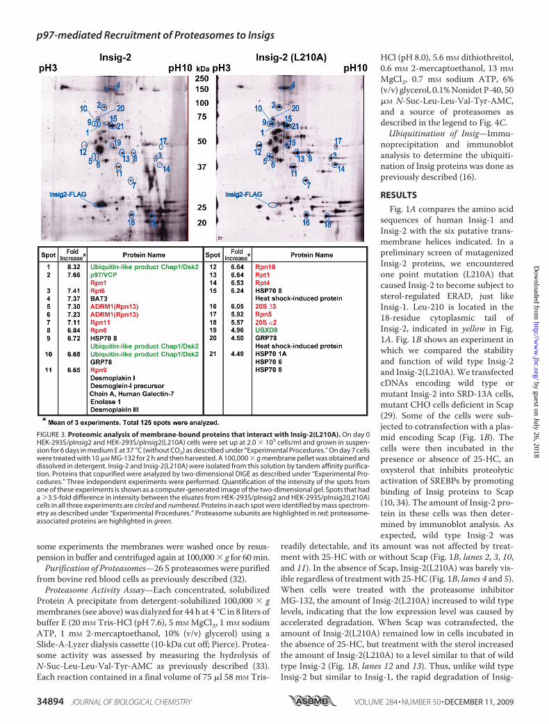
FIGURE 3. Proteomic analysis of membrane-bound proteins that interact with Insig-2(L210A). On day 0HEK-293S/pInsig2 and HEK-293S/pInsig2(L210A) cells were set up at 2.0 � 105 cells/ml and grown in suspen-sion for 6 days in medium E at 37 °C (without CO2) as described under “Experimental Procedures.” On day 7 cellswere treated with 10 �M MG-132 for 2 h and then harvested. A 100,000 � g membrane pellet was obtained anddissolved in detergent. Insig-2 and Insig-2(L210A) were isolated from this solution by tandem affinity purifica-tion. Proteins that copurified were analyzed by two-dimensional DIGE as described under “Experimental Pro-cedures.” Three independent experiments were performed. Quantification of the intensity of the spots fromone of these experiments is shown as a computer-generated image of the two-dimensional gel. Spots that hada �3.5-fold difference in intensity between the eluates from HEK-293S/pInsig2 and HEK-293S/pInsig2(L210A)cells in all three experiments are circled and numbered. Proteins in each spot were identified by mass spectrom-etry as described under “Experimental Procedures.” Proteasome subunits are highlighted in red; proteasome-associated proteins are highlighted in green.
p97-mediated Recruitment of Proteasomes to Insigs
34894 JOURNAL OF BIOLOGICAL CHEMISTRY VOLUME 284 • NUMBER 50 • DECEMBER 11, 2009
by guest on July 26, 2018http://w
ww
.jbc.org/D
ownloaded from

2(L210A) is inhibited by sterols in a Scap-dependent manner.Importantly, the L210A mutation does not affect the ability ofInsig-2 to inhibit SREBP-2 cleavage because expression ofInsig-2(L210A) was as efficient as wild type Insig-2 in restoringthe sterol-induced inhibition of proteolytic activation ofSREBP-2 in mutant CHO cells deficient in both Insig-1 andInsig-2 (30) (Fig. 1C). Similar to wild type Insig-2, Insig-2(L210A) also accelerated the degradation of HMG-CoAreductase in these mutant CHO cells (Fig. 1D).We showed previously that Insig-1 is ubiquitinated by gp78
and rapidly degraded by proteasomes in SRD-13A cells that aredeficient in Scap (14, 16). In contrast to Insig-1, Insig-2 does notbind to gp78 (16), so it is neither ubiquitinated nor rapidlydegraded in these cells (19). Inasmuch as Insig-2(L210A) isdegraded in a way that is similar to Insig-1, we examined
whether the mutant Insig-2 is ubiq-uitinated by gp78 in SRD-13A cells.To determine the ubiquitination ofInsig proteins, we transfected SRD-13A cells with a plasmid encodingubiquitin tagged with an epitopederived from the influenza HA pro-tein together with a plasmid encod-ing epitope-tagged wild type ormutant Insig. The cells were treatedwith MG-132 to block degradationof ubiquitinated Insig. Insig pro-teins were then immunoprecipi-tated from the cell lysates, and theprecipitates were subjected toimmunoblot analysis with anti-HAto detect ubiquitinated proteins.Ubiquitinated Insig-1 and Insig-2(L210A) were visualized as highmolecular weight smears in theanti-HA immunoblot (Fig. 2A, lanes3 and 5). Ubiquitination of wild typeInsig-2 was not detected (Fig. 2A,lane 4).We next examined whether
Insig-2(L210A) interacts with gp78.For this purpose SRD-13A cellswere cotransfected with a plasmidencoding gp78 and a plasmidencoding epitope-tagged wild typeor mutant Insig proteins. Insig pro-teins were then immunoprecipi-tated from cell lysates, and theamount of gp78 coimmunoprecipi-tated was examined by immunoblotanalysis. As expected, significantamounts of gp78 were coimmuno-precipitated with Insig-1 but notInsig-2 (Fig. 2B, lanes 3 and 4). gp78was coimmunoprecipitated withInsig-2(L210A) in an amount thatwas similar to that precipitated withInsig-1 (Fig. 2B, lanes 3 and 5).
To determine whether gp78 is required for degradation ofInsig-2(L210A), we transfected SV589 cells, a line of immortal-ized human fibroblasts (31) with a plasmid encoding themutant Insig-2 protein. The cells were subsequently trans-fected with duplexes of siRNA targeting either gp78 or greenfluorescent protein, a control mRNA not present in the cells.The cells were then treated with cycloheximide to block thesynthesis of proteins. At various times after cycloheximidetreatment, cells were harvested, and the amount of Insig-2(L210A)was determinedby immunoblot analysis. As shown inFig. 2C, Insig-2(L210A) was rapidly degraded in cells trans-fected with the control siRNA (Fig. 2C, lanes 1–3). The trans-fected Insig-2(L210A)was rapidly degraded even thoughwedidnot deplete the cells of cholesterol, presumably because theamount of overexpressed mutant Insig-2 was much more than
FIGURE 4. Membrane-bound Insig-2(L210A) associates with proteasomal subunits. A, on day 0 HEK-293S/pInsig2 and HEK-293S/pInsig2(L210A) cells were set up at 2.0 � 105 cells/ml and grown in suspension for 6 daysin medium E at 37 °C (without CO2). On day 7 cells were treated with 10 �M MG-132 for 2 h and then harvested.A 100,000 � g membrane pellet was prepared from pooled cell pellets of HEK-293S/pInsig2 cells (0.3 liter ofcells) and HEK-293S/pInsig2(L210A) cells (0.6 liter of cells). The membranes were solubilized, and Insig-2 wasisolated by affinity precipitation with IgG-Sepharose beads as described under “Experimental Procedures.”Aliquots of eluates from the beads (representing 16.7% of cells) and the non-absorbed flow-through (FT)fraction (representing 0.42% of cells) were subjected to SDS/PAGE and immunoblot analysis with anti-FLAG(against Insig-2), anti-Rpt6, anti-Rpt5, and anti-�7. IB, immunoblot. B, cells were set up as described in A. 0.4 literof HEK-293S/pInsig2 cells were mixed with 1.6 liter of HEK-293S cells. Cells pelleted from this mixture and from2 liters of HEK-293S/pInsig2(L210A) cells were harvested, and the 100,000 � g membrane fractions wereprepared. After washing once with buffer D, the membranes were solubilized, and Insig-2 was subjected toIgG-Sepharose affinity precipitation (AP) as described above. Precipitated material (0.1 liter of cells) was sub-jected to SDS/PAGE and immunoblot analysis with anti-FLAG (against Insig-2), anti-Rpt5, and anti-�1. Theindicated amount of purified 26 S proteasomes was also subjected to SDS/PAGE and immunoblot analysis withanti-Rpt5 or anti-�1. C, shown is proteasomal activity of material coprecipitating with mutant Insig-2(L210A)but not wild type Insig-2. Each reaction was carried out as described under “Experimental Procedures” in theabsence or presence of 50 �M MG-132 as indicated and one of the following sources of proteasomes; lanes 1and 2, precipitated material from B representing 0.3 liter of the indicated cells; lane 3, 0.47 �g of purified 26 Sproteasome. After incubation for 21 min at 37 °C, the amount of cleaved AMC liberated by proteasomes wasquantified by fluorometry. A blank reading in reactions containing only buffer D and fluorescent substrate(507– 632 fluorescence units (FU)) was subtracted from each value. Each value is the average of duplicatedeterminations. Similar results were obtained in two other independent experiments.
p97-mediated Recruitment of Proteasomes to Insigs
DECEMBER 11, 2009 • VOLUME 284 • NUMBER 50 JOURNAL OF BIOLOGICAL CHEMISTRY 34895
by guest on July 26, 2018http://w
ww
.jbc.org/D
ownloaded from

that of endogenous Scap in SV589 cells. This degradation wassignificantly retarded in cells receiving siRNA targeting gp78(Fig. 2C, lanes 4–6).The results presented above show that the single pointmuta-
tion L210A subjects Insig-2 to sterol-regulated degradation in amanner similar to wild type Insig-1 whilemaintaining the func-tions of Insig-2. To identify proteins that might participate in
this degradation, we used HEK-293S cells, a line of HEK-293 cellsadapted to suspension culture (28).We stably transfected the cells withplasmids encoding Insig-2 andInsig-2(L210A), both fused withtandem Protein A and FLAG epi-tope tags to generate HEK-293S/pInsig2 and HEK-293S/pInsig2-(L210A) cells, respectively. Mem-brane fractions from suspensioncultures of these cells were pre-pared by ultracentrifugation. Wildtype and mutant Insig-2 were thenpurified from detergent extracts ofthe membranes through sequentialaffinity purification, making use ofthe tandem tags. Proteins thatcopurified with Insigs were ana-lyzed by two-dimensional fluores-cence difference gel electrophoresis(Fig. 3). In three separate experi-ments we identified a total of 125spots that copurified with Insig-2.21 of these spots met the stringentcriteria of showing in all threeexperiments a greater than 3.5-foldrelative increase in membranesderived from the HEK-293S/pInsig2(L210A) cells as comparedwith cells expressing wild typeInsig-2 (Fig. 3). Each of these 21 pro-tein spots was eluted and subjectedto sequencing by mass spectrome-try. The majority of the peptides ineach of these 21 spots correspondedto 26 S proteasome subunits (3, 4).Altogether, we detected 12 protea-some subunits or proteins that areknown to associate with protea-somes, including p97 (35) and Dsk2(36) (Fig. 3).To confirm the interaction be-
tween proteasome subunits andmembrane-bound Insig2(L210A),we isolated wild type and mutantInsig-2, respectively, from the100,000 � g pellet of membranesfrom HEK-293S/pInsig2 and HEK-293S/pInsig2(L210A) cells that hadbeen treated with the proteasome
inhibitorMG-132. After resuspending themembrane pellets indetergent, we added IgG-coupled Sepharose beads that bind tothe Protein A tag fused to the Insig proteins. Wild type andmutant Insig-2 were eluted from the beads by incubation withAcTEV protease, which cleaves between the Protein A tag andInsig-2. The elutedmaterial was subjected to immunoblot anal-ysis with antibodies against proteasome subunits Rpt5, Rpt6,
FIGURE 5. Membrane-bound Insig-2(L210A) associates with proteasomal subunits. On day 0 HEK-293S/pInsig2 and HEK-293S/pInsig2(L210A) cells were set up at 2.0 � 105 cells/ml and grown in suspension for 6 daysin medium E at 37 °C (without CO2). On day 7 cells were treated with 10 �M MG-132 for 2 h and harvested.Pooled cell pellets of HEK-293S/pInsig2 cells (1 liter of cells) and HEK-293S/pInsig2(L210A) cells (2 liters of cells)were harvested, and the 100,000 � g membrane pellet was obtained as described under “Experimental Pro-cedures.” The membrane pellet was resuspended in the indicated buffer, incubated at 4 °C for 30 min, andpelleted again at 100,000 � g. The washed membranes were detergent-solubilized and subjected to affinityprecipitation with IgG-Sepharose beads to precipitate Insig-2. The eluate (representing 5.6% of cells) andflow-through (FT) fraction (representing 0.14% of cells) were subjected to SDS/PAGE and immunoblot analysiswith anti-FLAG (against Insig-2), anti-Rpt5, and anti-�7. IB, immunoblot.
FIGURE 6. Sterol depletion and ubiquitinated lysines required for association of Insig-1 with proteaso-mal subunits. A, on day 0 SRD-14/pTK-Insig1-Myc cells were set up at 7.0 � 105 cells/100-mm dish in mediumA supplemented with 5% LPDS. On day 2 cells were switched to medium B. On day 3 cells were incubated with10 �M MG-132 in the absence or presence of 1 �g/ml 25-HC. After incubation for 4 h, cells were harvested andlysed, and Insig-1 was immunoprecipitated from the cell lysate with anti-Myc. Pellets (representing 0.5 dish ofcells for Insig-1 and 6.5 dishes of cells for proteasomal subunits) and supernatants (representing 0.033 dish ofcells for Insig-1 and 0.43 dish of cells for proteasomal subunits) were subjected to SDS/PAGE and immunoblotanalysis with IgG-9E10 (against Insig-1), anti-Rpt5, and anti-�7. IP, immunoprecipitate; IB, immunoblot; Sup,supernatant fraction. B, on day 0 Scap-deficient SRD-13A cells were set up at 1.6 � 105 cells/60-mm dish. On day1 each dish of cells was transfected with 0.2 �g pCMV-Insig1-T7 or pCMV-Insig1(K156R/K158R)-T7 in mediumA supplemented with 5% FCS. On day 4 cells were incubated with 10 �M MG-132 for 2 h and then harvested,lysed, and immunoprecipitated with anti-T7 to precipitate Insig-1. Pellets (representing 0.5 dish of cells forInsig-1 and 7.7 dish of cells for proteasome subunits) and supernatants (representing 0.033 dish of cells forInsig-1 and 0.51 dish of cells for proteasome subunits) were subjected to SDS/PAGE and immunoblot analysiswith anti-T7 (against Insig-1), anti-Rpt5, and anti-�7. The asterisk (*) denotes IgG light chain.
p97-mediated Recruitment of Proteasomes to Insigs
34896 JOURNAL OF BIOLOGICAL CHEMISTRY VOLUME 284 • NUMBER 50 • DECEMBER 11, 2009
by guest on July 26, 2018http://w
ww
.jbc.org/D
ownloaded from

and�7 (Fig. 4A). Bothwild type andmutant Insig-2were boundand efficiently released by TEV protease from the IgG-Sepha-rose beads, as determined by immunoblotting against theFLAG epitope (Fig. 4A, lanes 1–4) (in these stably transfectedcells, mutant Insig-2, but not the wild type protein, showed alowermolecular weight band, which was not seen in transientlytransfected cells (Figs. 1, B–D); its origin is not known). The 19S proteasome subunits Rpt5 and Rpt6 as well as the 20 S pro-teasome subunit �7 were coeluted with Insig-2(L210A) but notwild type Insig-2 (Fig. 4A, lanes 1 and 2). Blotting of the flow-through fractions showed that all of the cell extracts containedequal amounts of the proteasome subunits.The results shown in Figs. 3 and 4A suggest that intact 26 S
proteasomes may associate with Insig-2(L210A) on ER mem-
branes. To confirm that the protea-somes are active, we precipitatedwild type and mutant Insig-2 from100,000 � g pellets of membranesfrom stably transfected HEK-293Scells that had been treated with theproteasome inhibitor MG-132 (Fig.4B). In multiple experiments thelevel of precipitated wild typeInsig-2 protein was consistentlyhigher than that of the mutantInsig-2 even after treatment withMG-132. To equalize the amountsof precipitated wild type andmutant Insig-2, we prepared mem-branes from0.4 liter of cells express-ing wild type Insig-2 and 2 liters ofcells expressing Insig-2(L210A). Toequalize the amounts of total mem-brane protein in the precipitationreactions, we added 1.6 liters ofuntransfected HEK-293S cells tothe 0.4 liter of HEK-293S cellsexpressingwild type Insig-2 to bringthe total amount of cells to 2 liters.Cells were pelleted from the culturemedia, homogenized, and subjectedto ultracentrifugation to isolate a100,000 � g membrane pellet. Sim-ilar amounts of wild type andmutant Insig-2 were precipitatedfrom the solubilized membranes(Fig. 4B, lanes 1 and 2). Rpt5 and�1,which are representative compo-nents of the 19 S and 20 S protea-some, respectively, were precipi-tated with mutant but not wild typeInsig-2 (Fig. 4B, lanes 7 and 8). Cal-ibration with purified 26 S protea-somes indicated that between 0.1and 0.2 �g of proteasomes werecoprecipitated with Insig-2(L210A)for each 0.1 liter of HEK-293S/pInsig2(L210A) cells (Fig. 4B, lanes
4, 5, and 8). After removing MG-132 by dialysis, the affinity-precipitated material from 0.3 liter of cells was assayed for pro-teasomal activity. Proteasomal activity corresponding to about0.47 �g of purified 26 S proteasomes was precipitated withInsig-2(L210A) from 0.3 liter of cells, and this activity wasinhibited byMG-132 in the assay buffer (Fig. 4C, lanes 1 and 3).Within experimental error, this number agrees with the immu-noblots of Rpt-5 and �-1, which indicated that 0.1–0.2 �g ofproteasomes was precipitated with Insig-2(L210A) from 0.1liter of HEK-293S/pInsig2(L210A) cells (Fig. 4B).The results presented in Figs. 3 and 4 show that active pro-
teasomeswere coprecipitatedwith Insig-2(L210A) in detergentlysates prepared frommembrane fractions. We next examinedwhether the proteasomes could be released by washing the
FIGURE 7. p97 is required for the interaction between proteasomes and Insig proteins. A, on day 0 HEK-293S/pInsig1 cells were set up at 5 � 105 cells/100-mm dish in medium C. On day 3 cells were switched tomedium F. On day 4 cells were incubated with 10 �M MG-132 in the absence or presence of 100 �M sodiumarachidonate-albumin. After incubation for 6 h, cells were harvested, and Insig-1 was precipitated from deter-gent-solubilized 100,000 � g membranes as described under “Experimental Procedures.” The eluate (repre-senting 3.2 dishes of cells) and flow-through (FT) fraction (representing 0.21 dish of cells) were subjected toimmunoblot analysis with anti-T7 (against Insig-1), anti-Rpt5, anti-�7, and anti-p97. IB, immunoblot. B, on day0 HEK-293S/pInsig1 cells were set up at 4 � 105 cells/100-mm dish in medium C. On day 1 cells were transfectedwith 400 pmol/dish of the indicated siRNA. After incubation for 4 h, cells were switched to medium F. On day 2dishes in lanes 3-6 received a direct addition of MG-132 at a final concentration of 10 �M. After incubation for6 h, the cells were harvested. Aliquots of cell lysates (5 �g protein) from cells not receiving MG-132 (lanes 1 and2) were subjected to immunoblot analysis with the indicated antibody. Cell lysates from cells receiving MG-132(lanes 3– 6) were subjected to affinity precipitation (AP) of Insig-1 with IgG-Sepharose beads, after which theeluates and flow-through (FT) fractions were subjected to immunoblot analysis with the indicated antibody asdescribed in A. C, on day 0 HEK-293S/pInsig2(L210A) cells were set up 4 � 105 cells/100-mm dish in medium D.The interaction of proteasome subunits with Insig-2(L210A) was determined as described in A. D, on day 0HEK-293S/pInsig2(L210A) cells were set up at 3 � 105 cells/100-mm dish in medium D. The interaction ofproteasome subunits with Insig-2(L210A) was determined as described in B.
p97-mediated Recruitment of Proteasomes to Insigs
DECEMBER 11, 2009 • VOLUME 284 • NUMBER 50 JOURNAL OF BIOLOGICAL CHEMISTRY 34897
by guest on July 26, 2018http://w
ww
.jbc.org/D
ownloaded from

membranes with neutral or alkaline buffers. Proteasome sub-units Rpt5 and �7 were coprecipitated with Insig-2(L210A) butnot wild type Insig-2 whenmembranes were not washed (Fig. 5,lanes 1–4). Washing of membranes with a buffer at neutral pHdid not affect the coprecipitation of Insig-2(L210A) with theproteasome subunits (Fig. 5, lanes 5–8). Washing of mem-branes with alkaline sodium carbonate buffer (pH 11), which isknown to remove peripheral proteins frommembranes, did notreduce the amount of Insig-2 in the precipitates (Fig. 5, lanes 2,6, and 10), consistent with the fact that Insig-2 is an intrinsicmembrane protein. However, the alkaline buffer reduced theamounts of proteasome subunits in both the flow-through frac-tion and the Insig-2(L210A) eluate. This indicates that theinteraction of proteasomes with Insig-2(L210A), as well asother membrane proteins, is mediated by ionic forces.Inasmuch as Insig-2(L210A) is degraded in a regulated man-
ner that is similar to wild type Insig-1, we examined whetherproteasomes also interact with Insig-1. For this purpose, weused SRD-14 cells, a mutant line of CHO cells deficient inInsig-1 (25). We stably transfected the cells with a plasmidencoding Myc epitope-tagged Insig-1 (SRD-14/pTK-Insig1-Myc cells). The amount of exogenous Insig-1 expressed in thesecells is similar to that of endogenous Insig-1 in wild type CHOcells (14). These cells were treatedwithMG-132 in the presenceor absence of 25-HC. Insig-1 was immunoprecipitated withanti-Myc from the cell lysate, and the amount of proteasomesubunits coprecipitated was determined by immunoblot analy-sis. The two bands of Insig-1 represent translational productsinitiated at two different methionines, i.e. residues 1 and 37(10). Proteasome subunits Rpt5 and �7 were coimmunopre-cipitatedwith Insig-1 in cells incubated in the absence of 25-HC(Fig. 6A, lane 1). When cells were treated with 25-HC, the pro-teasome subunits no longer interacted with Insig-1 (Fig. 6A,lane 2). To confirm that ubiquitination is required for protea-somes to bind to Insig-1, we employed Insig-1(K156R/K158R),
amutant Insig-1 that is not ubiquiti-nated due to mutations in the twolysine residues that receive poly-ubiquitin chains (14). We trans-fected a plasmid encoding wild typeInsig-1 or Insig-1(K156R/K158R)into SRD-13A cells. We used SRD-13A cells for this experimentbecause these cells do not expressScap, the inhibitor of Insig-1 ubiq-uitination (14, 16). As shown in Fig.6B, proteasome subunits Rpt5 and�7 were coimmunoprecipitatedwith wild type Insig-1 but notInsig-1(K156R/K158R).We next examined the effect of
unsaturated fatty acids on the inter-action between Insig-1 and protea-somes. Unsaturated fatty acids donot block ubiquitination of Insig-1,but they block its subsequent pro-teasomal degradation (15). Previousstudies showed that unsaturated
fatty acids act by blocking the binding of Insig-1 to Ubxd8,which in turn binds to p97 (15). p97 is an ATPase that interactswith proteasomes and assists in the extraction of ubiquitinatedproteins from ERmembranes (37). As shown in Fig. 7A, lanes 1and 2, the amount of p97 bound to Insig-1 was markedlyreduced when arachidonic acid was added to cells. The amountof the proteasome subunits Rpt5 and �7 that coprecipitatedwith Insig-1 was also markedly reduced in cells treated witharachidonic acid (Fig. 7A, lanes 1 and 2). Similar results wereobtained whenmutant Insig-2(L210A) was used instead of wildtype Insig-1 (Fig. 7C).To test the requirement for p97 directly, we used RNA inter-
ference to reduce the amount of p97. We studied HEK-293S/pInsig1 cells, a line of HEK-293-derived cells stably transfectedwith Insig-1.We transiently transfected the cells with duplexesof siRNA targeting p97 or green fluorescent protein as a con-trol. The cells were cultured in serum from which all lipids hadbeen extracted. Knockdown of p97 increased the amount ofInsig-1 protein (Fig. 7B, lanes 1 and 2). To examine the interac-tion between proteasomes and Insig-1, we treated the cells withMG-132 to equalize the amount of Insig-1 protein, andwe usedimmunoblots to measure the amount of proteasome subunitscoprecipitated with Insig-1. As shown in Fig. 7B, knockdown ofp97 markedly reduced the amount of proteasome subunitsRpt5 and �7 coprecipitated with Insig-1 (Fig. 7B, lanes 3 and 4).Similar results were obtainedwhenmutant Insig-2(L210A) wasused instead of wild type Insig-1 (Fig. 7D).The results from Figs. 6 and 7 indicate that ubiquitination of
Insig-1 (or mutant Insig-2) and recruitment of p97 to Insig-1are both required for proteasomes to bind to Insig-1. However,it remains unclear whether ubiquitination of Insig-1 is requiredfor its interaction with p97. Fig. 8 shows an immunoblot inwhich we compared the interaction between p97 and two ver-sions of Insig-1; that is, wild type and a double lysine mutantthat is not ubiquitinated (K156R/K158R) (14).When cells were
FIGURE 8. Ubiquitination of Insig-1 is not required for its interaction with p97. On day 0 Scap-deficientSRD-13A cells were set up at 6.0 � 105 cells/60-mm dish as described under “Experimental Procedures.” On day1 each dish of cells was transfected with 0.3 �g pCMV-Myc-Ubxd8, pCMV-FLAG-Insig1, or pCMV-FLAG-Insig1(K156R/K158R) as indicated. The total amount of transfected DNA was adjusted to 1.3 �g/dish withpcDNA3 empty vector. After incubation for 6 h each dish of cells was switched to medium A supplementedwith 5% delipidated FCS. On day 2 the cells were treated for 6 h with or without 100 �M sodium arachidonate-albumin in the presence of 10 �M MG-132. The cells were then harvested and lysed with detergent, and Insig-1was immunoprecipitated with anti-FLAG-agarose beads. Pellets (representing 0.1 dish of cells) and superna-tants (representing 0.01 dish) were subjected to SDS-PAGE followed by immunoblot analysis with anti-Myc(against Ubxd8), anti-FLAG (against Insig-1), and anti-p97. IP, immunoprecipitation; IB, immunoblot. Thisexperiment was repeated two more times with similar results. To observe arachidonate-regulated bindingbetween Insig-1 and p97, the amount of transfected plasmids had to be adjusted to prevent excessive over-expression of Insig-1.
p97-mediated Recruitment of Proteasomes to Insigs
34898 JOURNAL OF BIOLOGICAL CHEMISTRY VOLUME 284 • NUMBER 50 • DECEMBER 11, 2009
by guest on July 26, 2018http://w
ww
.jbc.org/D
ownloaded from

cotransfected with plasmids encoding wild type Insig-1 andUbxd8, p97 was coimmunoprecipitated (Fig. 8, lane 4). Simi-larly, themutant version of Insig-1 also interacted with p97 in aUbxd8-dependent fashion (lane 8). With both versions ofInsig-1, the interaction with p97 was disrupted by the additionof arachidonate to the culture medium (lanes 5 and 9). Similarresults were obtained when the corresponding lysinemutant ofInsig-2(L210A), i.e. Insig-2 (K100R; K102R; L210A) was ana-lyzed (data not shown). These results indicate that ubiquitina-tion of Insig-1 is not required for its interaction with p97.
DISCUSSION
Fig. 9 shows a model for sterol- and unsaturated fatty acid-regulated ERAD of Insig-1 and mutant Insig-2(L210A). Thismodel extends our previous model (15) by providing newinsights into how proteasomes are attached to membrane-bound Insig proteins. Themodel is based ondata gathered fromexperiments with either wild type Insig-1 or Insig-2(L210A).For simplicity, we frame the discussion in terms of Insig-1, rec-ognizing that the same process applies to mutant Insig-2(L210A) but not to wild type Insig-2. In sterol- and fatty acid-depleted cells, two proteins bind to membrane-embeddedInsig-1. One of these proteins, gp78, is a ubiquitin E3 ligase thatattaches polyubiquitin chains to lysine 156 and lysine 158 ofInsig-1, both of which are located on the first cytoplasmic loopand both of which are conserved in Insig-2 (colored green, Fig.1A). The second protein, Ubxd8, contains a UBX domain thatbinds to p97 (37). We previously showed that Ubxd8 binds toInsig-1 and recruits p97 (15). The ATPase p97 has been shownby others to play a role in the extraction of membrane proteinsduring ERAD (1, 37). Here, we identify a new function of p97 byshowing that it recruits proteasomes to ubiquitinated Insig-1before the Insig is extracted from ERmembranes, a finding thatlends physiological significance to the recently reported inter-action between p97 and proteasomes (35). Thereafter, the pro-teasomes and p97 act in concert to extract Insig-1 from themembrane in a reaction that is closely coupled to degradation(Fig. 9).ERAD of Insig-1 is regulated by two classes of end-products,
each of which inhibits one limb of the pathway shown in Fig. 9.As previously demonstrated, sterols such as cholesterol or25-hydroxycholesterol cause Insig-1 to bind to Scap, and thisprecludes the binding of gp78, thereby preventing ubiquitina-tion (16). In contrast, sterols do not inhibit binding of theUbxd8�p97 complex to Insig-1 (15), which is consistentwith theobservation that this interaction does not require ubiquitina-tion of Insig-1 (Fig. 8). Unsaturated fatty acids such as oleic acidand arachidonic acid do not block ubiquitination, but they pre-vent the binding of the Ubxd8�p97 complex to Insig-1 (15).Inasmuch as proteasome binding requires both ubiquitinationand p97, either sterols or unsaturated fatty acids can block thedegradative pathway (Fig. 9). Inasmuch as Insig-1 controls theactivation of SREBPs and SREBPs increase the synthesis of bothsterols and unsaturated fatty acids, the regulatory scheme out-lined in Fig. 9 illustrates how both end-products can represstheir own synthesis.Although the current studies suggest that proteasomes play
an active role in extracting Insig-1 from the ERmembrane, they
do not reveal the mechanism. An attractive hypothesis for theextraction of membrane proteins from the ER was advanced byPloegh (38), who suggested that extraction is facilitated by split-ting of the ER bilayer, due to the insertion of nonpolar lipidssuch as triglycerides or even cholesteryl esters. In the case ofInsig-1, it is possible that Ubxd8 somehow facilitates thisbilayer splitting and that arachidonic acid blocks the process byreleasingUbxd8 from the complex. This hypothesis is currentlybeing tested in our laboratory.A major finding in the current study is that ubiquitination of
Insig-1 is not required for its interaction with p97. This isbecause p97 is recruited to Insig-1 by Ubxd8 whose interactionwith Insig-1 does not require ubiquitination (Fig. 8). On theother hand, p97 does require ubiquitination to interact withother proteins subjected to ERAD. This list includes the class Iheavy chains of the major histocompatibility complex whose
FIGURE 9. Model for sterol and unsaturated fatty acid-regulated ERAD ofInsig-1. The sequential steps in the ubiquitination of Insig-1 and its extractionfrom ER membranes by the proteasome are discussed in the text.
p97-mediated Recruitment of Proteasomes to Insigs
DECEMBER 11, 2009 • VOLUME 284 • NUMBER 50 JOURNAL OF BIOLOGICAL CHEMISTRY 34899
by guest on July 26, 2018http://w
ww
.jbc.org/D
ownloaded from

interaction with p97 is mediated through Ufd1, another p97-associated adaptor protein that recognizes ubiquitinated pro-teins (39, 40). Thus, whether or not ubiquitination of a proteinis required for its interaction with p97 depends on the adaptorprotein that mediates the interaction.A paradox in the current studies arises from the observation
that the L210A mutation in the cytoplasmic tail of Insig-2replaces a leucine that is conserved in Insig-1, reducing theoverall sequence conservation in the cytoplasmic tails of thetwo proteins (see Fig. 1A). The paradoxical effect of this muta-tion in Insig-2 may be related to the neighboring insertion offour residues (YECK) that is found in Insig-2 (residues 213–216) as compared with Insig-1. We previously found thatreplacement of glutamic acid with alanine in the YECKsequence in Insig-2 increased its ubiquitination (19). It seemslikely that a conformational aspect of this cytoplasmic tail pro-tects Insig-2 from being recognized by gp78 for ubiquitination.Alterations to this conformation permit ubiquitination ofInsig-2 but only in a sterol-depleted state. According to currentideas about Insig structure, the cytoplasmic tail in Insig-1 isonly 14 residues long (18 residues in Insig-2). Whether thisshort sequence is sufficient to permit a direct interaction withgp78 remains to be determined.
Acknowledgments—We thank Lisa Beatty, Angela Carroll, Sho-manike Head, and Ijeoma Onwuneme for invaluable help with tissueculture, Dorothy Goddard and David Thompson for excellent techni-cal assistance, Ayako Suzuki and Masashi Yanagisawa for help withtwo-dimensional DIGE analysis, and Russell DeBose-Boyd for criticalreview of the manuscript.
REFERENCES1. Vembar, S. S., and Brodsky, J. L. (2008)Nat. Rev.Mol. Cell Biol. 9, 944–9572. Hampton, R. Y., and Garza, R. M. (2009) Chem. Rev. 109, 1561–15743. Demartino, G. N., and Gillette, T. G. (2007) Cell 129, 659–6624. Finley, D. (2009) Annu. Rev. Biochem. 78, 477–5135. Schmidt, M., Hanna, J., Elsasser, S., and Finley, D. (2005) Biol. Chem. 386,
725–7376. Goldstein, J. L., DeBose-Boyd, R. A., and Brown, M. S. (2006) Cell 124,
35–467. Brown, M. S., and Goldstein, J. L. (1997) Cell 89, 331–3408. Horton, J. D., Goldstein, J. L., and Brown, M. S. (2002) J. Clin. Invest. 109,
1125–11319. Horton, J. D., Shah, N. A., Warrington, J. A., Anderson, N. N., Park, S. W.,
Brown, M. S., and Goldstein, J. L. (2003) Proc. Natl. Acad. Sci. U.S.A. 100,12027–12032
10. Yang, T., Espenshade, P. J., Wright, M. E., Yabe, D., Gong, Y., Aebersold,R., Goldstein, J. L., and Brown, M. S. (2002) Cell 110, 489–500
11. Sever, N., Yang, T., Brown, M. S., Goldstein, J. L., and DeBose-Boyd, R. A.
(2003)Mol. Cell 11, 25–3312. Sever, N., Song, B. L., Yabe, D., Goldstein, J. L., Brown,M. S., and DeBose-
Boyd, R. A. (2003) J. Biol. Chem. 278, 52479–5249013. Goldstein, J. L., and Brown, M. S. (1990) Nature 343, 425–43014. Gong, Y., Lee, J. N., Lee, P. C., Goldstein, J. L., Brown, M. S., and Ye, J.
(2006) Cell Metab. 3, 15–2415. Lee, J. N., Zhang, X., Feramisco, J. D., Gong, Y., and Ye, J. (2008) J. Biol.
Chem. 283, 33772–3378316. Lee, J. N., Song, B. L., DeBose-Boyd, R. A., and Ye, J. (2006) J. Biol. Chem.
281, 39308–3931517. Yabe, D., Brown, M. S., and Goldstein, J. L. (2002) Proc. Natl. Acad. Sci.
U.S.A. 99, 12753–1275818. Lee, J. N., and Ye, J. (2004) J. Biol. Chem. 279, 45257–4526519. Lee, J. N., Gong, Y., Zhang, X., andYe, J. (2006)Proc. Natl. Acad. Sci. U.S.A.
103, 4958–496320. Hua, X., Nohturfft, A., Goldstein, J. L., and Brown, M. S. (1996) Cell 87,
415–42621. Wojcik, C., and DeMartino, G. N. (2002) J. Biol. Chem. 277, 6188–619722. Goldstein, J. L., Basu, S. K., and Brown,M. S. (1983)Methods Enzymol. 98,
241–26023. Hannah, V. C., Ou, J., Luong, A., Goldstein, J. L., and Brown, M. S. (2001)
J. Biol. Chem. 276, 4365–437224. Brown, M. S., Faust, J. R., Goldstein, J. L., Kaneko, I., and Endo, A. (1978)
J. Biol. Chem. 253, 1121–112825. Sever, N., Lee, P. C., Song, B. L., Rawson, R. B., and Debose-Boyd, R. A.
(2004) J. Biol. Chem. 279, 43136–4314726. Radhakrishnan, A., Ikeda, Y., Kwon,H. J., Brown,M. S., andGoldstein, J. L.
(2007) Proc. Natl. Acad. Sci. U.S.A. 104, 6511–651827. Feramisco, J. D., Goldstein, J. L., and Brown, M. S. (2004) J. Biol. Chem.
279, 8487–849628. Reeves, P. J., Thurmond, R. L., and Khorana, H. G. (1996) Proc. Natl. Acad.
Sci. U.S.A. 93, 11487–1149229. Rawson, R. B., DeBose-Boyd, R. A., Goldstein, J. L., and Brown, M. S.
(1999) J. Biol. Chem. 274, 28549–2855630. Lee, P. C., Sever, N., and Debose-Boyd, R. A. (2005) J. Biol. Chem. 280,
25242–2524931. Yamamoto, T., Davis, C. G., Brown, M. S., Schneider, W. J., Casey, M. L.,
Goldstein, J. L., and Russell, D. W. (1984) Cell 39, 27–3832. Liu, C. W., Li, X., Thompson, D., Wooding, K., Chang, T. L., Tang, Z., Yu,
H., Thomas, P. J., and DeMartino, G. N. (2006)Mol. Cell 24, 39–5033. Koulich, E., Li, X., and DeMartino, G. N. (2008) Mol. Biol. Cell 19,
1072–108234. Adams, C. M., Reitz, J., De Brabander, J. K., Feramisco, J. D., Li, L., Brown,
M. S., and Goldstein, J. L. (2004) J. Biol. Chem. 279, 52772–5278035. Besche, H. C., Haas, W., Gygi, S. P., and Goldberg, A. L. (2009) Biochem-
istry 48, 2538–254936. Matiuhin, Y., Kirkpatrick, D. S., Ziv, I., Kim, W., Dakshinamurthy, A.,
Kleifeld, O., Gygi, S. P., Reis, N., and Glickman, M. H. (2008)Mol. Cell 32,415–425
37. Halawani, D., and Latterich, M. (2006)Mol. Cell 22, 713–71738. Ploegh, H. L. (2007) Nature 448, 435–43839. Meyer, H. H., Wang, Y., and Warren, G. (2002) EMBO J. 21, 5645–565240. Flierman, D., Ye, Y., Dai, M., Chau, V., and Rapoport, T. A. (2003) J. Biol.
Chem. 278, 34774–34782
p97-mediated Recruitment of Proteasomes to Insigs
34900 JOURNAL OF BIOLOGICAL CHEMISTRY VOLUME 284 • NUMBER 50 • DECEMBER 11, 2009
by guest on July 26, 2018http://w
ww
.jbc.org/D
ownloaded from
Goldstein and Jin YeYukio Ikeda, George N. DeMartino, Michael S. Brown, Joon No Lee, Joseph L.
MEMBRANESp97 RECRUITS PROTEASOMES TO Insig-1 BEFORE EXTRACTION FROM Regulated Endoplasmic Reticulum-associated Degradation of a Polytopic Protein:
doi: 10.1074/jbc.M109.044875 originally published online October 8, 20092009, 284:34889-34900.J. Biol. Chem.
10.1074/jbc.M109.044875Access the most updated version of this article at doi:
Alerts:
When a correction for this article is posted•
When this article is cited•
to choose from all of JBC's e-mail alertsClick here
http://www.jbc.org/content/284/50/34889.full.html#ref-list-1
This article cites 40 references, 20 of which can be accessed free at
by guest on July 26, 2018http://w
ww
.jbc.org/D
ownloaded from


![Lovelock brane cosmology - NTUA · Lovelock brane cosmology The action of the model We consider the following action S[X] = Z d4˘ p g + K + 2 R | {z } First brane Lovelock invariants:](https://img.pdfslide.us/doc/110x75/5e89832ae2dc892b3f37833a/lovelock-brane-cosmology-lovelock-brane-cosmology-the-action-of-the-model-we-consider.jpg)