Embed Size (px)
Citation preview

Preclinical Development
Regression of Human Prostate Cancer Xenografts inMice byAMG 212/BAY2010112, a Novel PSMA/CD3-Bispecific BiTEAntibody Cross-Reactive with Non-Human Primate Antigens
Matthias Friedrich1, Tobias Raum2, Ralf Lutterbuese1, Markus Voelkel1, Petra Deegen1, Doris Rau1,Roman Kischel1, Patrick Hoffmann1, Christian Brandl1, Joachim Schuhmacher2, Peter Mueller1,Ricarda Finnern2, Melanie Fuergut1, Dieter Zopf2, Jerry W. Slootstra3, Patrick A. Baeuerle1, Benno Rattel1, andPeter Kufer1
AbstractFor treatment of patients with prostate cancer (PCa), we developed a novel T cell-engaging (BiTE) antibody
designated AMG 212 or BAY2010112 that is bispecific for prostate-specific membrane antigen (PSMA) and the
CD3 epsilon subunit of the T cell receptor complex. AMG 212/BAY2010112 induced target cell-dependent
activation and cytokine release of T cells, and efficiently redirected T cells for lysis of target cells. In addition to
Chinesehamster ovary cells stably expressinghumanor cynomolgusmonkeyPSMA,T cells redirectedbyAMG
212/BAY2010112 also lysed human PCa cell lines VCaP, 22Rv1,MDAPCa 2b, C4-2, PC-3-huPSMA, andLnCaP
at half maximal BiTE concentrations between 0.1 and 4 ng/mL (1.8–72 pmol/L). No lysis of PSMA-negative
human PCa cell lines PC-3 and DU145 was observed. The subcutaneous (s.c.) formation of tumors from PC-3-
huPSMAcells inNOD/SCIDmicewas significantly preventedbyoncedaily intravenous (i.v.) injection ofAMG
212/BAY2010112 at a dose level as low as 0.005 mg/kg/d. Rapid tumor shrinkage with complete remissions
were observed inNOD/SCIDmice bearingestablished s.c. 22Rv1xenografts after repeateddaily treatmentwith
AMG 212/BAY2010112 by either the i.v. or s.c. route. Of note, 22Rv1 tumors were grown in the absence of
human T cells followed by intraperitoneal injection of T cells 3 days before BiTE treatment. No effects on tumor
growth were observed in the absence of human T cells or AMG 212/BAY2010112. On the basis of these
preclinical results, AMG 212/BAY2010112 appears as a promising new BiTE antibody for the treatment of
patients with PSMA-expressing PCa. Mol Cancer Ther; 11(12); 2664–73. �2012 AACR.
IntroductionProstate cancer (PCa) is themost common typeof cancer
in men in the United States with estimated 192,000 newcases and 27,400 deaths in 2009 (1), and is the secondleading cause of cancer death in men after lung cancer.Relapse after primary therapy by radical prostatectomyremains a common problem in PCa patients because anestimated 30% to 40% of men experience recurrencewithin 10 years (2, 3). There is a critical need for newtargeted agents in this PCa patient population and forthose patients who become hormone refractory andrequire chemotherapy.
Antibodies that target PSMA are a potential addition tothe armentarium of PCa therapies. Developments com-prise naked humanized or human IgG1 antibodies, andantibodies of enhanced activity such as antibody drugconjugates and radioimmunoconjugates (4). PSMA is alsoan established marker for imaging of residual PCa andmetastases using the radiolabeled diagnostic antibodyProstaScint (5). The antigen is expressed in a high percent-age of PCa patients across all stages of the disease (6). Ahighly promising approach to enhance the anticanceractivity of antibodies is the engagement of T cells by usingbispecific antibodies and related constructs. One exampleis blinatumomab, a CD19/CD3-bispecific antibody con-struct of the BiTE (for bispecific T cell engager) class thathas shown high response rates and an acceptable benefit/risk profile in patients with B-cell lineage lymphoma (7)and leukemia (8). The group of Els€asser–Beile was first todesign and characterize PSMA/CD3-bispecific antibodiesof the so-called diabody format (9–11). While such bacte-rially expressed diabodies showed conditional T cell acti-vation, redirected PCa cell lysis and activity against out-growing xenografts in mouse models, their murine natureand highly restricted species cross-reactivity of the CD3bindingmoiety greatly limit nonclinical safety assessment.
Authors' Affiliations: 1Amgen (Munich) Research GmbH, Munich; 2BayerPharma AG, Berlin, Germany; and 3Pepscan Presto, RC Lelystad, TheNetherlands
M. Friedrich, T. Raum, R. Lutterbuese, B. Rattel, and P. Kufer contributedequally to this work.
Corresponding Author: Patrick A. Baeuerle, Amgen Research (Munich)GmbH,Staffelseestr. 2, 81477Munich, Germany. Phone: 49-089-895-277-601; Fax: 49-089-895-27784601; E-mail: [email protected]
doi: 10.1158/1535-7163.MCT-12-0042
�2012 American Association for Cancer Research.
MolecularCancer
Therapeutics
Mol Cancer Ther; 11(12) December 20122664
on January 28, 2020. © 2012 American Association for Cancer Research. mct.aacrjournals.org Downloaded from
Published OnlineFirst October 5, 2012; DOI: 10.1158/1535-7163.MCT-12-0042

Here, we characterized AMG 212/BAY 2010112, thefirst PSMA/CD3-bispecific single-chain antibody con-struct that (i) is very close in its amino acid sequence tohuman germline Ig-V segments, (ii) is cross-reactive tohumanandnon-humanprimatePSMAandCD3antigens,(iii) is highly active against PCa tumor formation aswell asestablished PCa xenograft tumors in a novel mousemodel, and (iv) is meeting essential requirements as aredesired for clinical development of novel antibody-basedtherapeutics.
Materials and MethodsExpression, production, and purification ofAMG 212/BAY2010112AMG 212/BAY2010112 is a single-chain bispecific anti-
body construct comprising of an anti-PSMA and an antiCD3 scFv, both reacting with human and cynomolgustargets. AMG 212/BAY2010112 was constructed byrecombinant DNA technology and produced in super-natants from stably transfected Chinese hamster ovary(CHO) cells. Purification of the monomeric protein wasdone in a 2-step process using Ni chelate chromatogar-aphy followed by gel filtration as described for an EGFreceptor–specific BiTE antibody in ref. 12.
Affinity determination on target positive cell linesusing Scatchard evaluationFor affinity analysis, saturation-binding experiments
were conducted using a monovalent detection system(anti-His Fab/Alexa 488, Micromet GmbH) to deter-mine monovalent binding of AMG 212/BAY2010112to PSMA or CD3 positive cell lines. The human PCa cellline LNCaP, CHO cells transfected with human PSMA(CHO-huPSMA), CHO cells transfectedwith cynomolgusmonkey PSMA (CHO-cyPSMA), the human T cell lineHPB ALL and the rhesus monkey T cell line 4919 LnPx(kindly provided by Prof. Fickenscher, University Erlan-gen-Nuernberg, Germany; ref. 13) were used. Two to5�104 cells of the respective cell line were incubated with50 mL from a 1:3 dilution series (starting at 800 nmol/L)of AMG 212/BAY2010112, followed by 45 minutes ofincubation on ice, and 1 residual washing step. Subse-quently, cells were incubated for 30minutes with 30 mL ofan anti-His Fab/Alexa488 solution (30 mg/mL). After 1washing step, cells were resuspended in 120 mL fluores-cence-activated cell sorting (FACS) buffer and analyzedusing FACS flow or a FACS Canto machine (BectonDickinson), andCell quest software (GraphPad Software).Data are means from 3 experiments from at least 2 dif-ferent sets of experiments.
Epitope determination using linear peptide mappingThePSMA-binding scFv ofAMG212/BAY2010112was
subjected to an epitope mapping approach based onpeptide scanning (Pepscan). 693different 15-mer peptideswere synthesized that span the entire extracellular aminoacid sequence of human PSMA and overlap with each
neighboring 15-mer peptide by 14 amino acids. Thesepeptides were synthesized directly in 455-well peptidearrays. For this experiment, the anti-PSMA scFv of AMG212/BAY 2010112 was produced in Escherichia coli andcrude periplasmic extracts were used for the analysis. ThescFv was incubated on polypropylene cards containingthe covalently linked peptides and specific binding wasdetected and calculated as firstly described in ref. 14.
Cell lines and cell cultureLNCaP, 22Rv1, PC-3, and CHO-dhfr- (CHO) were pur-
chased from DSMZ (Germany), VcaP, and MDA PCa 2bfrom ATCC, and C4-2 from ViroMed Laboratories (Min-netonka, Minnesota), respectively. The cell lines wereobtained before the experiment series and analyzed forauthenticity by the respective cell bank (DNA-fingerprint-ing techniques such as short tandem repeat profiling).No additional identification was conducted in-house.The 22Rv1 and C4-2 were cultured in RPMI 1640 and10% fetal calf serum (FCS; Invitrogen), LNCaP in Iscove’sBasal Medium (Biochrom AG) and 10% FCS, VCaP inDulbecco’s Modified Eagle’s Media (Biochrom AG), and10% FCS. MDA PCa 2b cells were cultured in F-12 K-Nutrient Mixture (Invitrogen) and 20% FCS supple-mentedwith 5mL Insulin-Transferrin-Selenium-Solution(Invitrogen), 10 ng/mL recombinant human EGF (MBL),and 100 pg/mL hydrocortisone (Sigma-Aldrich). Celllines were cultured at 37�C in a 5% CO2 chamber.
PC-3 were stably transfected with the human PSMAcDNA (PC-3-huPSMA) using the eukaryotic expressionvector pEF-puromycin (15). CHO cells stably expressinghuman (CHO-huPSMA) or cynomolgus monkey PSMA(CHO-cyPSMA)were generated by transfecting cellswithplasmids containing the respective cDNAs. Selection ofclones and amplification of expression was conducted inthe presence puromycin (0.8 mg/mL) and methotrexate(20 nmol/L).
PK analysis in miceFemale BALB/c mice received a single intravenous
(i.v.) bolus injection of 0.1, 0.3, or 1 mg/kg AMG 212/BAY2010112 or were subcutaneous (s.c.) injected witha single dose of 0.3 mg/kg AMG 212/BAY2010112.Whole blood samples were collected at the followingtimes: 0 (predose), 0.083, 0.167, 0.5, 1, 2, 4, 7, 24, and48 hours after administration. AMG 212/BAY2010112serum concentrations were determined by an electroche-miluminescence-based assay. A polyclonal goat anti-AMG 212/BAY2010112 serum was coated to a highbinding plate (MSD). After blocking overnight with Dul-becco’s Phosphate Buffered Saline containing 5% bovineserum albumin, plates werewashedwith an ELISAwash-er. Standards, quality-control samples, and study sampleswere added to the carbon plate and were subsequentlyincubated on a rotation shaker for 1 hour at room tempera-ture. After another wash step with the ELISA washer, thedetection anti-penta His-biotin-labeled detection anti-body (GenScript) was added and incubated for 1 hour
PSMA-Specific BiTE Antibody
www.aacrjournals.org Mol Cancer Ther; 11(12) December 2012 2665
on January 28, 2020. © 2012 American Association for Cancer Research. mct.aacrjournals.org Downloaded from
Published OnlineFirst October 5, 2012; DOI: 10.1158/1535-7163.MCT-12-0042

on a rotation shaker at room temperature. StreptavidinSulfoTag (MSD)was added and the plateswere incubatedfor 1 hour at room temperature, followed by anotherwashstep. Finally, reading buffer T (MSD) was added and theplates were analyzed at the Sector Imager 2400 accordingto the manufacturer’s instructions. All samples were de-termined in triplicates. The pharmacokinetic parameterswere calculated from the geometric mean plasma concen-trations by noncompartmental analysis (16, 17) using theKinEx program version 3.0 (Bayer Pharma AG, 2010).
Cytotoxicity, T cell activation, and cytokine releaseRedirected T cell cytotoxicity was assayed by flow
cytometry using human and cynomolgusmonkeyperiph-eral blood mononuclear cells (PBMC) as effector cells andPSMA-positive human PCa cell lines or PSMA-trans-fected CHO cells as targets. PBMCwere isolated by Ficolldensity gradient centrifugation and if applicable enrich-ment for CD3þ T cells was conducted by using the PanT Cell Isolation Kit (Miltenyi Biotech) according to themanufacturer’s instructions. Effector cells were coculti-vated with target cells labeled with Vybrant DiO (Invitro-gen) at effector-to-target (E:T) cell ratios of 5:1 or 10:1 withincreasing concentrations of AMG 212/BAY2010112 for48 hours and cell lysiswasmonitored bynuclear uptake ofpropidium iodide by flow cytometry (12).
Activation of CD4þ and CD8þ T cell subsets was deter-mined by using phycoerythrin (PE)-labeled anti-CD4 andallophycocyanine (APC)-Cy7-labeled anti-CD8 antibo-dies. PE-Cy7-conjugated anti-CD69 and APC-conjugatedanti-CD25 were used to characterize activated T cells. Allfluorescently labeled antibodies were purchased from BDBiosciences (Heidelberg). For staining of cell surface pro-teins, cells were incubated with the appropriate antibodymixtures for 30minutes at 4�Cand sampleswere analyzedby flow cytometry.
Cytokine concentrations were determined in superna-tants of cytotoxicity assays by use of cytometric beadarrays (CBA-kit, BD Biosciences) in accordance to themanufacturer’s protocol.
Animal tumor studiesIn vivo studies to evaluate antitumor activity were
conducted in, NOD.CB17-Prkdcscid/J mice (Charles Riv-er). Mice were housed in individually ventilated cageswith laboratory chow and water available ad libitum. Allexperiments were conducted according to the GermanAnimal Protection Lawwith permission from the respon-sible local authorities.
In the tumor formation study, 5 � 106 PC-3-huPSMAcells were injected s.c. with or without PBMC obtainedfrom a single healthy human donor at an E:T cell ratio of1:2 into the flank of female nonobese diabetic/severecombined immunodeficient (NOD/SCID) mice (n ¼ 8).Mice were treated once daily from the day of tumor/PBMC injection with 5, 0.5, 0.05 and 0.005 mg/kgAMG 212/BAY2010112 BiTE antibody for 10 days by i.v. bolus injection into the lateral tail vein. Controls includ-
ed 2 vehicle-treated groups (tumor cells injected with orwithout human PBMC). Treatment efficacy in terms oftumor formation and growth was determined by externalcaliper measurements, and tumor volumes were calcu-lated using a standard hemi-ellipsoid formula: {length(mm) � width (mm)2}/2.
For the established tumor model, male NOD/SCIDmice were irradiated with a single dose (2 Gy) of 137Csg-ray before s.c. injection of 22Rv1 (5 � 106) tumor cellsdispersed in medium with 50% Matrigel (Cultrex, R&DSystems) before injection. Mice were irradiated to facili-tate engraftment of the injected human T cells. Animals(n ¼ 8) were allocated to treatment groups when tumorshad reached a volume of approximately 200 mm3 andwere treatedwith a single i.v. bolus injection of anti-asialoGM1 rabbit antibody (WAKO) into the lateral tail vein1 day before human T cell injection to deplete murine NKcells. In vitro activated and expanded human T cellsisolated from the blood of a single donor (T cell activa-tion/expansion kit, Miltenyi Biotech) were injected intothe peritoneal cavity on day 11 after tumor cell injection.One control group of animals (n ¼ 5) did not receivehuman T cells. Animals received AMG 212/BAY2010112or vehicle once daily by either s.c., or i.v. bolus injectioninto the lateral tail vein for a total of 28 days starting at day14 posttumor cell inoculation. Growth of tumors wasdetermined as described above.
Statistical analysisData were analyzed by ANOVA, and differences in
tumor volumes of the AMG 212/BAY2010112 BiTE-trea-ted groups to the vehicle control group injected withhuman effector cells were assessedwith a Dunnett’s post-test. P < 0.05 was considered to be statistically significant.All statistical analyses were conducted using GraphPadPrism version 5.00 for Windows (GraphPad Software).
ResultsGeneration of human PSMA/CD3-bispecific BiTEantibody AMG 212/BAY2010112
The PSMA/CD3-bispecific BiTE antibody AMG 212/BAY2010112 as shown in Fig. 1A was purified in largeramounts to apparent homogeneity (Fig. 1B). It showedone major protein species (95.6%) of defined charge uponanalytical high-resolution cation exchange chromatogra-phy (Fig. 1C). Only the monomeric form of AMG 212/BAY2010112 was used for further in vitro and in vivocharacterization. The BiTE antibody showed a high sta-bility in human plasma at 37�Cwhere after incubation for24 hours EC50 values for redirected target cell lysis werenot significantly different to untreated controls.
Bispecific binding of AMG 212/BAY2010112 to CD3and a defined epitope of PSMA
Saturation binding curves of AMG 212/BAY2010112 toCHO cells expressing either human or cynomolgus mon-keyPSMAand toT cells of humanor cynomolgusmonkey
Friedrich et al.
Mol Cancer Ther; 11(12) December 2012 Molecular Cancer Therapeutics2666
on January 28, 2020. © 2012 American Association for Cancer Research. mct.aacrjournals.org Downloaded from
Published OnlineFirst October 5, 2012; DOI: 10.1158/1535-7163.MCT-12-0042

origin were used to determine equilibrium bindingconstants (KD) of AMG 212/BAY2010112. As shownin Fig. 2A, AMG 212/BAY2010112 bound to CD3 and
PSMA of both species. KD values for human and cyno-molgus monkey CD3 were 9.4 � 4.3 nmol/L and 16.3 �2.1 nmol/L, respectively, and 47.0 � 8.1 nmol/L and
Figure 1. Characteristicsof purifiedAMG 212/BAY 2010112. Thedomain arrangement of AMG 212/BAY2010112 is schematicallyshown in A. Purified AMG 212/BAY2010112 was separated bySDS-PAGE and stained withCoomasie Blue (B) and subjected tohigh-resolution cation exchangechromatography (C) to show purityand charge homogeneity. Theelution profile of purified AMG 212/BAY2010112 from the cationexchange column was recorded byabsorption at 280 nm. Thecalculated molecular weight ofAMG 212/BAY2010112 is 55 kDa.mAU, milli absorption units.
High-resolution IEXB CA
60
50
80
80 100
(mA
U)
120 (mL)
SDS-PAGE
-
-
--
-
Mo
l. W
t. (
kDa)
40
30
N
C
Anti-PSMA
Anti-CD3
x6 His
50
40
30
20
10
0
Figure 2. Binding of AMG 212/BAY2010112 to human andcynomolgusmonkeyPSMAandCD3antigens. Increasing concentrationsof monomeric AMG 212/BAY2010112 were incubated with10,000 cells of the indicated celllines. Cell-bound BiTE antibody wasdetected by flow cytometry using amonovalent antihexahistidine Fabantibody fragment covalentlycoupled to ALEXA 488. Bindingsignals are given as median relativefluorescence intensity (RFI). A,hyperbolic binding curves wereplotted and half-maximal bindingconcentrations (KD) were calculatedusing the algorithm by GeorgeScatchard. Exemplary bindingcurves are depicted. KD values withstandard deviations weredetermined from 3 independentexperiments per cell line. B, theepitope on human PSMA recognizedby AMG 212/BAY2010112 wasdetermined by binding of the anti-PSMA single-chain antibody moietyof AMG 212/BAY2010112 to a panelof 15-mer peptides permutated by 1amino acid that spanned the entireextracellular domain of humanPSMA. Binding signals for peptidesin the ELISA assay are given asrelative signal units. C, the position ofthe most reactive peptide ishighlighted in the crystal structure ofhuman PSMA (20).
PSMA-Specific BiTE Antibody
www.aacrjournals.org Mol Cancer Ther; 11(12) December 2012 2667
on January 28, 2020. © 2012 American Association for Cancer Research. mct.aacrjournals.org Downloaded from
Published OnlineFirst October 5, 2012; DOI: 10.1158/1535-7163.MCT-12-0042

212.6 � 29 nmol/L for human and cynomolgus monkeyPSMA, respectively.
To map the binding site of AMG 212/BAY2010112 tohuman PSMA, overlapping peptides of the extracellulardomain of PSMAwere synthesized and tested for bindingof AMG 212/BAY2010112 using a PepBlot (18, 19). Thehighest binding signal of AMG 212/BAY2010112 wasseenwith a peptide corresponding to amino acid residues325 to 348 of human PSMA (Fig. 2B). According to thepublished crystal structure of PSMA (20), this peptidelocalizes to a small loop structure on the apical side of theprotein facing away from the plasmamembrane (Fig. 2C).The sequence of this peptide is identical between humanand cynomolgus monkey PSMA, which explains thecross-species bindingofAMG212/BAY2010112 toPSMA.
Human/cynomolgus monkey cross-reactivity ofAMG212/BAY2010112 for T cell activation, cytokinerelease, and redirected lysis
The pharmacologic effects of AMG 212/BAY2010112on effector T cells and PSMA-expressing target cells wereanalyzed in vitro using cell coculture assays. AMG 212/BAY2010112 activity was compared in both human andcynomolgus monkey cell-based assay systems to investi-gate whether the cynomolgus monkey can serve as rele-vant species for pharmacologic and toxicological safetyassessments.
The dose-dependent activation of CD4þ and CD8þ Tcell populations by AMG 212/BAY2010112 was analyzedusingFACSby the appearance of the immediate-early andlate activation markers CD69 and CD25, respectively,after 48 hours of incubation in the presence of humanLNCaPPCa cells (Fig. 3A).At themaximum,between60%to 80% of CD4þ and CD8þ T cells of both species wereactivated, whereas less than 10% of T cells were foundactivated when freshly isolated. Values for half-maximaleffective concentration (EC50) of T cell activation rangedbetween 3.4 and 6.7 ng/mL of AMG 212/BAY2010112 forhuman and between 13.7 and 21.2 ng/mL for cynomolgusmonkey cell cocultures. The largest difference betweenhuman and cynomolgus monkey T cells was observedwith the CD4þ/CD25þ T cell population where EC50
values differed by 6.2-fold. No T cell activation wasobserved in the absence of PSMA-expressing target cells(data not shown).
Next, the AMG 212/BAY2010112 dose-dependent re-lease of 6 different cytokines by activated T cells in thepresence of target cellswas compared. The cytokine releaseprofiles were very similar in human and cynomolgusmonkey cell coculture systems with absolute amounts ofcytokines reaching approximately 2-fold higher levels inthe human system. Highest concentrations were observedfor interferon-g , which reached 3 to 6 ng/mL in cell culturemedium after 48 hours (Fig. 3B). Intermediate increasesreaching levels of 1 to 1.5 ng/mLwere observed for TNF-a,IL-2 and IL-10. For technical reasons, IL-10 levels could notbedetermined in cynomolgusmonkey cocultures,whereasIL-5 levels were not detected in human cocultures. Little or
no increase was observed for IL-4 and IL-6. As observedpreviously, cytokine release of T cells was observed only inthe presence of target cells (data not shown), which is onehallmark of BiTE antibodies (21).
The dose-dependent redirected lysis of target cells byAMG 212/BAY2010112 was investigated with CHO cells,which expressed either human or cynomolgus monkeyPSMA, tohave the same cellular backgroundof target cells.Overall, potent lysis of transfected CHO cells was observ-ed with EC50 values ranging across species between 0.37and 6.69 ng/mL (Fig. 3C). In homologous assays whereT cells and PSMA from the same species were used, an18-fold higher concentration of AMG 212/BAY2010112was required to reach half maximal lysis in the cynomol-gus monkey system compared with the human system.Notably, target cells expressing human PSMAwere morepotently lysed by either human or cynomolgus monkeyT cells than those expressing cynomolgusmonkey PSMA,which may be attributed to the somewhat lower affinityof AMG 212/BAY2010112 to cynomolgus monkey PSMA(Fig. 2A). Human T cells showed a 5-fold higher potencyof redirected lysis with AMG 212/BAY2010112 thancynomolgus monkey T cells, regardless whether humantumor cells or CHO cells expressing either human orcynomolgus monkey PSMA were used. Taken together,AMG 212/BAY2010112 showed dose-dependent activ-ity for T cell activation, cytokine release and redirectedlysis with human and cynomolgus monkey T cells, andreacted with PSMA antigen from both species.
Redirected lysis of human PCa lines by AMG 212/BAY2010112
To assess the potency of redirected lysis by AMG 212/BAY2010112with target cells naturally expressing PSMA,the 7 humanPCa cell linesVCaP, 22Rv1,MDAPCa 2b,C4-2, PC-3, DU145, and LNCaP were procured. FACS stain-ing showed that, except for PC-3 and DU145 cells, all PCacell lines expressed detectable levels of PSMA. Surfacedensities of PSMA were quantified and ranged from1.2� 104 (VCaP) to 5� 105 (PC-3-huPSMA) PSMAmolec-ules per cell (see Fig. 4B). Target cells were incubated inthe presence of increasing concentrations of AMG 212/BAY2010112 with a 10-fold excess of unstimulated PBMCas effector cells and lysis was measured after 48 hours bynuclear uptake of propidium iodine via FACS. All 5 PCacell lines, which naturally express PSMA as well as PC-3-huPSMA cells (stably expressing a human PSMA cDNA),were lysed with EC50 values ranging between 0.1 and4 ng/mL (i.e., 1.8–64 pmol/L) AMG 212/BAY2010112(Fig. 4A). No lysis of parental PSMA�negative PC-3 cellswas observed. EC50 values for redirected lysis correlatedwith the number of surface expressed PSMA moleculesper cell in a statistically significant manner (Fig. 4B).
Pharmacokinetics of AMG 212/BAY2010112 inBALB/c mice
Pharmacokinetic parameters ofAMG212/BAY2010112were determined after i.v. bolus administration of 0.1,
Friedrich et al.
Mol Cancer Ther; 11(12) December 2012 Molecular Cancer Therapeutics2668
on January 28, 2020. © 2012 American Association for Cancer Research. mct.aacrjournals.org Downloaded from
Published OnlineFirst October 5, 2012; DOI: 10.1158/1535-7163.MCT-12-0042

0.3, and 1 mg/kg to BALB/c mice. A dose proportionalincrease of serum concentrations was observed. Serumclearance was low and amounted to approximately0.3 L/(kg�h) independently of the dose. An intermediatevolume of distribution at steady state of approximately1 L/kg was observed. This indicates that the BiTE anti-body did not only distribute into the extravascular space(as observed for IgGs) but also into tissues,which resultedin a considerably extended terminal half-life of approxi-mately 8 hours.After s.c. administration of 0.3 mg/kg to mice, maxi-
mum serum concentrations of approximately 64 mg/Lwere reached 2 hours after administration. Bioavailabilitywas 18%, indicating significant metabolism duringabsorption via the lymphatic system. Pharmacokinetic
parameters after i.v. and s.c. bolus administration to miceare summarized in Table 1.
Inhibition of tumor growth and tumor regression byAMG 212/BAY2010112-redirected human T cells inimmunodeficient mice
Following the characterization ofAMG212/BAY2010112in vitro, its efficacy on the inhibition of tumor growthwas investigated in mouse xenograft models. AMG 212/BAY2010112 was first studied for its preventive effect in atumor outgrowth model. PC-3-huPSMA cells were mixedwith unstimulated human PBMC shortly before injectionand mixtures were administered s.c. to immunodeficientNOD/SCID mice. Immediately following tumor cell inoc-ulation, mice were treated i.v. with AMG 212/BAY2010112
Figure 3. Effects of AMG 212/BAY2010112 on T cell activation, cytokine release, and redirected lysis in coculture experiments. Cocultures of LNCaP humanPCa cells and isolated human or cynomolgus monkey CD3þ T cells at effector-to-target (E:T) cell ratios of 5:1 (human) or 10:1 (cynomolgus monkey) wereincubatedwith increasing concentrations of AMG212/BAY2010112 for 48 hours. A, T cell activation. The percentage of activatedCD4þ andCD8þT cells wasdetermined by flow cytometry using antigen-specific fluorochrome-conjugated monoclonal antibodies against CD69 and CD25. B, cytokine release.Concentrations of the indicated human and cynomolgus monkey cytokines in the cell culture supernatants were determined using cytometry-based beadarrays. C, redirected lysis of target cells. CHO cells transfected with either human or cynomolgus monkey PSMA or untransfected cells and isolated humanor cynomolgus monkey CD3þ T cells (E:T cell ratio: 10:1) were incubated with increasing concentrations of AMG 212/BAY2010112 for 48 hours. Thepercentage of target cell lysis was monitored by flow cytometric determination of propidium iodine uptake into nuclei of lysed cells. Data points in all assaysrepresent the mean of duplicate wells. Error bars represent standard deviation of the mean. Concentrations that induced half maximal T cell activationand target cell lysis (EC50), respectively, are shown.
PSMA-Specific BiTE Antibody
www.aacrjournals.org Mol Cancer Ther; 11(12) December 2012 2669
on January 28, 2020. © 2012 American Association for Cancer Research. mct.aacrjournals.org Downloaded from
Published OnlineFirst October 5, 2012; DOI: 10.1158/1535-7163.MCT-12-0042

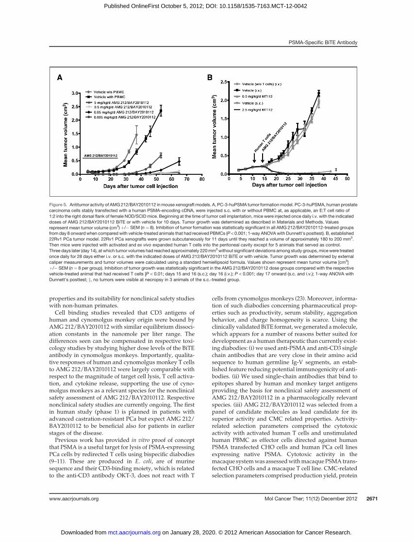
at doses ranging from 5 mg/kg down to 0.005 mg/kg byonce daily injection for 10 consecutive days. Efficacy wasevaluated by monitoring tumor formation. A significantand dose-dependent delay of tumor formation/growthwasobservedwith tumor growth inhibition after amonitor-ing period of 43 days ranging from 86% (0.005 mg/kg/d)to 99% (5 mg/kg/d) compared with the vehicle plusPBMC control (Fig. 5A). The inhibition was statisticallysignificant even at the lowest dose of 0.005 mg/kg, whichcorresponded to only 0.1 ng AMG 212/BAY2010112 permouse/d. AMG 212/BAY2010112 completely preventedtumor formation in 1 and 2 of 8 animals at doses of 0.5 or5 mg/kg/d, respectively, whereas small nodules becamevisible in some animals on Days 46 and 53, respectively.
We next addressed the question whether AMG 212/BAY2010112 could affect established PCa xenografts. Anew study design was established in which human effec-tor T cells were delivered shortly before initiation of BiTEtreatment by intraperitoneal (i.p.) injection into tumor-bearing NOD/SCID mice. Previous experiments haveshown that i.p.-delivered human T cells enter the circu-lation of mice and remain detectable for approximately 2weeks (22). For the present study, human 22Rv1 PCaxenografts were grown s.c. in male NOD/SCID in theabsence of human T cells until they had reached a volumeof180 to200mm3.Then,humanTcells expanded invitro for18 days were injected i.p. and treatment with AMG 212/BAY2010112 was initiated 3 days after the adoptive T celltransfer by once daily i.v. or s.c. injections with doses of 0.5or 2.5 mg/kg, respectively. To achieve similar drug expo-sures for i.v. and s.c. administrations a 5 times higher dosewas given s.c. as based on the 18% bioavailability of AMG212/BAY2010112 after s.c administration (see Table 1).Established tumors began to readily regress after initiationof either i.v. or s.c. treatment and tumor volumes maxi-mally declined within the first week of treatment (Fig. 5B).Complete tumor remissions were observed in 3/8 animalsof the s.c.-treated group after necropsy, whereas smallnodules with sizes less than 50 mm3 were detected in theresidual animals. In the i.v.-treated group small nodules ofless than 50 mm3, most likely representing fibrous tissueremnants, were detected in 3/8 animals, whereas smalltumors remained in the other animals. Unrestricted tumorgrowth occurred in animals treated with human T cellsalone or a vehicle. In summary AMG 212/BAY2010112very potently inhibited growth of PSMA expressinghuman PCa xenografts in mice, which eventually led tocomplete remission in some animals after s.c. application.
DiscussionIn the present study, the anti-PSMA/anti-CD3 single-
chain antibody construct AMG 212/BAY2010112 wasexamined for its in vitro and in vivo pharmacologic
A B
10
30
50
70
90
10–4 10–2 100 102 104 104 105 1060AMG 212 (ng/mL)
Sp
ecif
ic ly
sis
(%)
PC-3
VCaP22Rv1MDA PCa 2bC4-2LNCaPPC3-huPSMA
0.02
0.04
0.06
0.08
0.10
0.12
r ² = 0.31P = <0.0001
PSMA surface molecules per cell
Hal
f m
axim
al ly
sis
(nm
ol/L
)
0
Figure 4. Redirected lysis of human PCa cell lines by AMG 212/BAY2010112 using unstimulated human effector cells. A, the human PCa cell lines VCaP,22Rv1, MDA PCa 2b, C4-2, LNCaP, PC-3-huPSMA, and PC-3 cells and human PBMC (E:T cell ratio: 10:1) were incubated with increasing concentrations ofAMG 212/BAY2010112 for 48 hours. The percentage of tumor cell lysis was measured by uptake of propidium iodide using flow cytometry. Each datapoint represents the mean of duplicate wells. Error bars represent standard error of mean. B, concentrations that induced half maximal target cell lysis (EC50)were plotted against mean number of PSMA molecules on the surface of the respective human PCa cell line.
Table 1. Pharmacokinetic parameters ofAMG 212/BAY2010112 after i.v. and s.c.administration to BALB/C mice
Administration i.v. boluss.c. bolus
Dose (mg/kg) 0.10 0.30 1.0 0.30
AUC (mg h/L) 0.33 0.93 3.3 0.17AUCnorm (kg h/L) 3.3 3.1 3.3 0.55CLmatrix (L/h/kg) 0.30 0.32 0.30 n.a.Cmax norm (kg/L) 10.4 11 9.5 0.21Vss (L/kg) 0.89 1.2 0.94 n.a.t1/2 (h) 6.5 9.7 7.9 11F (bioavailability) (%) 100 100 100 18
NOTE: Serum levels of AMG 212/BAY2010112 after singlebolus i.v. administration of 0.1, 0.3, and 1 mg/kg and aftersingle bolus s.c. administration of 0.3mg/kg to female BALB/cmicewere determined as described inMaterials andMethods.Abbreviations: AUC, area under the curve; AUCnorm; normal-ized AUC; CLmatrix, serum clearance; Cmax norm, maximumserum levels; Vss, volume of distribution at steady state;t1/2, terminal serum half-life (beta); n.a., not applicable.
Friedrich et al.
Mol Cancer Ther; 11(12) December 2012 Molecular Cancer Therapeutics2670
on January 28, 2020. © 2012 American Association for Cancer Research. mct.aacrjournals.org Downloaded from
Published OnlineFirst October 5, 2012; DOI: 10.1158/1535-7163.MCT-12-0042
properties and its suitability for nonclinical safety studieswith non-human primates.Cell binding studies revealed that CD3 antigens of
human and cynomolgus monkey origin were bound byAMG 212/BAY2010112 with similar equilibrium dissoci-ation constants in the nanomole per liter range. Thedifferences seen can be compensated in respective toxi-cology studies by studying higher dose levels of the BiTEantibody in cynomolgus monkeys. Importantly, qualita-tive responses of human and cynomolgus monkey T cellsto AMG 212/BAY2010112 were largely comparable withrespect to the magnitude of target cell lysis, T cell activa-tion, and cytokine release, supporting the use of cyno-molgus monkeys as a relevant species for the nonclinicalsafety assessment of AMG 212/BAY2010112. Respectivenonclinical safety studies are currently ongoing. The firstin human study (phase 1) is planned in patients withadvanced castration-resistant PCa but expect AMG 212/BAY2010112 to be beneficial also for patients in earlierstages of the disease.Previous work has provided in vitro proof of concept
that PSMA is a useful target for lysis of PSMA-expressingPCa cells by redirected T cells using bispecific diabodies(9–11). These are produced in E. coli, are of murinesequence and their CD3-binding moiety, which is relatedto the anti-CD3 antibody OKT-3, does not react with T
cells from cynomolgusmonkeys (23). Moreover, informa-tion of such diabodies concerning pharmaceutical prop-erties such as productivity, serum stability, aggregationbehavior, and charge homogeneity is scarce. Using theclinically validatedBiTE format,wegenerated amolecule,which appears for a number of reasons better suited fordevelopment as a human therapeutic than currently exist-ing diabodies: (i) we used anti-PSMA and anti-CD3 singlechain antibodies that are very close in their amino acidsequence to human germline Ig-V segments, an estab-lished feature reducing potential immunogenicity of anti-bodies. (ii) We used single-chain antibodies that bind toepitopes shared by human and monkey target antigensproviding the basis for nonclinical safety assessment ofAMG 212/BAY2010112 in a pharmacologically relevantspecies. (iii) AMG 212/BAY2010112 was selected from apanel of candidate molecules as lead candidate for itssuperior activity and CMC related properties. Activity-related selection parameters comprised the cytotoxicactivity with activated human T cells and unstimulatedhuman PBMC as effector cells directed against humanPSMA transfected CHO cells and human PCa cell linesexpressing native PSMA. Cytotoxic activity in themacaque systemwas assessedwithmacaquePSMAtrans-fected CHO cells and a macaque T cell line. CMC-relatedselection parameters comprised production yield, protein
Figure 5. Antitumor activity of AMG 212/BAY2010112 inmouse xenograft models. A, PC-3-huPSMA tumor formationmodel. PC-3-huPSMA, human prostatecarcinoma cells stably transfected with a human PSMA-encoding cDNA, were injected s.c. with or without PBMC at, as applicable, an E:T cell ratio of1:2 into the right dorsal flank of female NOD/SCID mice. Beginning at the time of tumor cell implantation, mice were injected once daily i.v. with the indicateddoses of AMG 212/BAY2010112 BiTE or with vehicle for 10 days. Tumor growth was determined as described in Materials and Methods. Valuesrepresent mean tumor volume (cm3) þ/� SEM (n¼ 8). Inhibition of tumor formation was statistically significant in all AMG 212/BAY2010112-treated groupsfrom day 8 onward when compared with vehicle-treated animals that had received PBMCs (P < 0.001; 1-way ANOVAwith Dunnett's posttest). B, established22Rv1 PCa tumor model. 22Rv1 PCa xenografts were grown subcutaneously for 11 days until they reached a volume of approximately 180 to 200 mm3.Then mice were injected with activated and ex vivo expanded human T cells into the peritoneal cavity except for 5 animals that served as control.Three days later (day 14), at which tumor volumes had reached approximately 220mm3without significant deviations among study groups,micewere treatedonce daily for 28 days either i.v. or s.c. with the indicated doses of AMG 212/BAY2010112 BiTE or with vehicle. Tumor growth was determined by externalcaliper measurements and tumor volumes were calculated using a standard hemiellipsoid formula. Values shown represent mean tumor volume [cm3]þ/� SEM (n ¼ 8 per group). Inhibition of tumor growth was statistically significant in the AMG 212/BAY2010112 dose groups compared with the respectivevehicle-treated animal that had received T cells [P < 0.01; days 15 and 16 (s.c.); day 16 (i.v.); P < 0.001; day 17 onward (s.c. and i.v.); 1-way ANOVA withDunnett's posttest; z, no tumors were visible at necropsy in 3 animals of the s.c.-treated group.
PSMA-Specific BiTE Antibody
www.aacrjournals.org Mol Cancer Ther; 11(12) December 2012 2671
on January 28, 2020. © 2012 American Association for Cancer Research. mct.aacrjournals.org Downloaded from
Published OnlineFirst October 5, 2012; DOI: 10.1158/1535-7163.MCT-12-0042

homogeneity in high-resolution ion exchange chromatog-raphy, stability in human plasma, thermal stability, anddimer conversion. Compared with published PSMA/CD3-bispecific diabodies, which have EC50 values for re-directed lysis of approximately 0.25 nmol/L (15 ng/mL;refs. 9, 10), AMG 212/BAY2010112 is considerably morepotent with EC50 values ranging from 1.8 to 72 pmol/L(0.1 and 4 ng/mL) depending on the cell line. Onlyrecently, a newly constructed diabody was publishedwith an EC50 value of approximately 23 pmol/L (1.4ng/mL) using C4-2 target cells (11). (iv) Finally, weobserved that AMG 212/BAY2010112 canmediate remis-sion of 22Rv1 PCa xenografts, whereas published PSMA/CD3-bispecific diabodies were thus far only shown toinhibit tumor formation. On the basis of all of the abovefeatures, we believe that AMG 212/BAY2010112meets allproperties required for development of a potent novelhuman therapeutic.
This is also the first report on a novel in vivo studydesign for analyzing the activity of T cell-engaging anti-bodies against established tumors in mice. Most impor-tantly, tumors were grown s.c. to an average size of 200mm3 in NOD/SCIDmice in the absence of human T cells.Ex vivo expanded human T cells were then injected intothe peritoneal space of mice fromwhere they had to enterthe peripheral blood compartment to reach the s.c.implanted tumor. Three days following adoptive T celltransfer, dosing of AMG 212/BAY2010112 was started,which triggered a rapid onset of tumor regression. This isconsistent with an immediate engagement of peripheral Tcells for redirected tumor lysis. The fast onset is differentfrom vaccines and anti-CTLA-4 antibodies, which requiredays to weeks for mounting a tumor-specific T cellresponse in man (22). Rapid tumor regression also sug-gests that both AMG 212/BAY2010112 and i.p.-deliveredT cells readily gainedaccess to tumor tissue. In ourpresentstudy, the s.c. administration of the BiTE antibodyappeared to be even more efficacious than the i.v. route.
The bioavailability of AMG 212/BAY2010112 in miceafter administration of a single s.c. dose was 18%. Pre-liminary studies indicate that AMG 212/BAY2010112 isbioavailable and pharmacologically active after s.c. appli-cation in cynomolgus monkeys. Additional studies are inprogress to better understand the activity of AMG 212/BAY2010112 after s.c. administration and evaluate itsapplicability for treatment of PCa patients.
Disclosure of Potential Conflicts of InterestM. Friedrich, T. Raum, R. Lutterbuese, M. Voelkel, P. Deegen, D.
Rau, R. Kischel, P. Hoffmann, C. Brandl, P. Mueller, M. Fuergut, P.A.Baeuerle, B. Rattel, and P. Kufer are Amgen employees and havereceived stock. T. Raum, R. Lutterbuese, R. Kischel, P. Hoffmann, P.Kufer have ownership interest (including patents). J. Schuhmacher, R.Finnern, and D. Zopf are Bayer Pharma employees. J.W. Slootstra isemployed by Pepscan Presto.
Authors' ContributionsConception and design: R. Lutterbuese, R. Finnern, P.A. Baeuerle,B. Rattel, P. KuferDevelopment of methodology: C. BrandlAcquisition of data (provided animals, acquired and managed patients,provided facilities, etc.): T. Raum, M. Voelkel, P. Deegen, D. Rau,R. Kischel, C. Brandl, M. Fuergut, J.W. SlootstraAnalysis and interpretation of data (e.g., statistical analysis, biostatis-tics, computational analysis): M. Friedrich, T. Raum, R. Lutterbuese,P. Deegen, P.Hoffmann, J. Schuhmacher, R. Finnern,M. Fuergut, B. Rattel,P. KuferWriting, review, and/or revision of the manuscript: M. Friedrich,T. Raum, R. Lutterbuese, P. Deegen, P. Hoffmann, J. Schuhmacher,P. Mueller, R. Finnern, D. Zopf, P.A. Baeuerle, B. Rattel, P. KuferStudy supervision: M. Friedrich, B. Rattel, P. KuferProject Management Support: P. Mueller
AcknowledgmentsThe authors thank Maren Voges for preparing figures.
The costs of publication of this article were defrayed in part by thepayment of page charges. This article must therefore be hereby markedadvertisement in accordance with 18 U.S.C. Section 1734 solely to indicatethis fact.
Received January 18, 2012; revised September 17, 2012; acceptedOctober1, 2012; published OnlineFirst October 5, 2012.
References1. Jemal A, Siegel R, Ward E, Hao Y, Xu J, Thun MJ. Cancer statistics,
2009. CA Cancer J Clin 2009;59:225–49.2. SimmonsMN,StephensonAJ, Klein EA.Natural history of biochemical
recurrence after radical prostatectomy: risk assessment for secondarytherapy. Eur Urol 2007;51:1175–84.
3. Ward JF, Moul JW. Rising prostate-specific antigen after primaryprostate cancer therapy. Nat Clin Pract Urol 2005;2:174–82.
4. Elsasser-Beile U, Buhler P, Wolf P. Targeted therapies for prostatecancer against the prostate specific membrane antigen. Curr DrugTargets 2009;10:118–25.
5. Mohammed AA, Shergill IS, Vandal MT, Gujral SS. ProstaScint and itsrole in the diagnosis of prostate cancer. Expert Rev Mol Diagn2007;7:345–9.
6. Joniau S, Abrahamsson PA, Bellmunt J, Figdor C, Hamdy F, VerhagenP, et al. Current vaccination strategies for prostate cancer. Eur Urol2012;61:290–306.
7. Bargou R, Leo E, Zugmaier G, Klinger M, Goebeler M, Knop S, et al.Tumor regression in cancer patients by very low doses of a T cell-engaging antibody. Science 2008;321:974–7.
8. ToppMS, Kufer P, Gokbuget N, Goebeler M, Klinger M, Neumann S,et al. Targeted therapy with the T-cell-engaging antibody blinatu-momab of chemotherapy-refractory minimal residual disease in B-lineage acute lymphoblastic leukemia patients results in highresponse rate and prolonged leukemia-free survival. J Clin Oncol2011;29:2493–8.
9. Buhler P, Wolf P, Gierschner D, Schaber I, Katzenwadel A, Schultze-Seemann W, et al. A bispecific diabody directed against prostate-specific membrane antigen and CD3 induces T-cell mediated lysisof prostate cancer cells. Cancer Immunol Immunother 2008;57:43–52.
10. Buhler P, Molnar E, Dopfer EP, Wolf P, Gierschner D, Wetterauer U,et al. Target-dependent T-cell activation by coligation with a PSMA xCD3 diabody induces lysis of prostate cancer cells. J Immunother2009;32:565–73.
11. Fortmuller K, Alt K,GierschnerD,Wolf P, BaumV, FreudenbergN, et al.Effective targeting of prostate cancer by lymphocytes redirected by aPSMA x CD3 bispecific single-chain diabody. Prostate 2011;71:588–96.
Friedrich et al.
Mol Cancer Ther; 11(12) December 2012 Molecular Cancer Therapeutics2672
on January 28, 2020. © 2012 American Association for Cancer Research. mct.aacrjournals.org Downloaded from
Published OnlineFirst October 5, 2012; DOI: 10.1158/1535-7163.MCT-12-0042
12. Lutterbuese R, Raum T, Kischel R, Hoffmann P, Mangold S, Rattel B,et al. T cell-engaging BiTE antibodies specific for EGFR potentlyeliminate KRAS- and BRAF-mutated colorectal cancer cells. Proc NatlAcad Sci U S A 2010;107:12605–10.
13. Knappe A, Feldmann G, Dittmer U, Meinl E, Nisslein T, Wittmann S,et al. Herpesvirus saimiri-transformed macaque T cells are toleratedand do not cause lymphoma after autologous reinfusion. Blood2000;95:3256–61.
14. Slootstra JW, Puijk WC, Ligtvoet GJ, Langeveld JP, Meloen RH.Structural aspects of antibody-antigen interaction revealedthrough small random peptide libraries. Mol Divers 1996;1:87–96.
15. Raum T, Gruber R, Riethmuller G, Kufer P. Anti-self antibodiesselected from a human IgD heavy chain repertoire: a novel approachto generate therapeutic human antibodies against tumor-associat-ed differentiation antigens. Cancer Immunol Immunother 2001;50:141–50.
16. Yamaoka K, Nakagawa T, Uno T. Statistical moments in pharmaco-kinetics. J Pharmacokinet Biopharm 1978;6:547–58.
17. Benet LZ, Galeazzi RL. Noncompartmental determination of thesteady-state volume of distribution. J Pharm Sci 1979;68:1071–4.
18. Bernard J, Harb C, Mortier E, Quemener A, Meloen RH, Vermot-Desroches C, et al. Identification of an interleukin-15alpha receptor-binding site onhuman interleukin-15. JBiolChem2004;279:24313–22.
19. Teeling JL, Mackus WJ, Wiegman LJ, van den Brakel JH, Beers SA,French RR, et al. The biological activity of human CD20 monoclonalantibodies is linked to unique epitopes on CD20. J Immunol2006;177:362–71.
20. Davis MI, Bennett MJ, Thomas LM, Bjorkman PJ. Crystal structure ofprostate-specific membrane antigen, a tumor marker and peptidase.Proc Natl Acad Sci U S A 2005;102:5981–6.
21. Brischwein K, Parr L, Pflanz S, Volkland J, Lumsden J, Klinger M, et al.Strictly target cell-dependent activation of T cells by bispecific single-chain antibody constructs of the BiTE class. J Immunother2007;30:798–807.
22. Hoos A, Ibrahim R, Korman A, Abdallah K, Berman D, Shahabi V, et al.Development of ipilimumab: contribution to a newparadigm for cancerimmunotherapy. Semin Oncol 2010;37:533–46.
23. Kipriyanov SM, Moldenhauer G, Martin AC, Kupriyanova OA, Little M.Two amino acid mutations in an anti-human CD3 single chain Fvantibody fragment that affect the yield on bacterial secretion but notthe affinity. Protein Eng 1997;10:445–53.
PSMA-Specific BiTE Antibody
www.aacrjournals.org Mol Cancer Ther; 11(12) December 2012 2673
on January 28, 2020. © 2012 American Association for Cancer Research. mct.aacrjournals.org Downloaded from
Published OnlineFirst October 5, 2012; DOI: 10.1158/1535-7163.MCT-12-0042

2012;11:2664-2673. Published OnlineFirst October 5, 2012.Mol Cancer Ther Matthias Friedrich, Tobias Raum, Ralf Lutterbuese, et al. Cross-Reactive with Non-Human Primate Antigens212/BAY2010112, a Novel PSMA/CD3-Bispecific BiTE Antibody Regression of Human Prostate Cancer Xenografts in Mice by AMG
Updated version
10.1158/1535-7163.MCT-12-0042doi:
Access the most recent version of this article at:
Cited articles
http://mct.aacrjournals.org/content/11/12/2664.full#ref-list-1
This article cites 23 articles, 7 of which you can access for free at:
Citing articles
http://mct.aacrjournals.org/content/11/12/2664.full#related-urls
This article has been cited by 9 HighWire-hosted articles. Access the articles at:
E-mail alerts related to this article or journal.Sign up to receive free email-alerts
Subscriptions
Reprints and
To order reprints of this article or to subscribe to the journal, contact the AACR Publications Department at
Permissions
Rightslink site. Click on "Request Permissions" which will take you to the Copyright Clearance Center's (CCC)
.http://mct.aacrjournals.org/content/11/12/2664To request permission to re-use all or part of this article, use this link
on January 28, 2020. © 2012 American Association for Cancer Research. mct.aacrjournals.org Downloaded from
Published OnlineFirst October 5, 2012; DOI: 10.1158/1535-7163.MCT-12-0042


![Third-line treatment and 177Lu-PSMA radioligand therapy of ... · refractory adenocarcinomas of the prostate express prostate-specific membrane antigen (PSMA) [13]. 68Ga-PSMA HBED-CC](https://img.pdfslide.us/doc/110x75/5f0256ec7e708231d403c8b9/third-line-treatment-and-177lu-psma-radioligand-therapy-of-refractory-adenocarcinomas.jpg)
![Labelling Efficiency DOTA PSMA Methods - Trasis 68Ga ISRS.pdf · Objectives [68Ga]Ga-HBED-11-PSMA (PSMA) and [68Ga]Ga-DOTA-tate (DOTAtate) are two well established PET tracers for](https://img.pdfslide.us/doc/110x75/5aae60737f8b9a6b308bf490/labelling-efficiency-dota-psma-methods-68ga-isrspdfobjectives-68gaga-hbed-11-psma.jpg)