Embed Size (px)
Citation preview
pISSN 2287-2728 eISSN 2287-285X
http://dx.doi.org/10.3350/cmh.2016.22.1.183Clinical and Molecular Hepatology 2016;22:183-187Case Report
Corresponding author : Byung-Cheol SongDepartment of Internal Medicine, Jeju National University School of Medicine, 15 Aran 13-gil, Jeju 63241, KoreaTel: +82-64-754-7177, Fax: + 82-64-717-1131. E-mail: [email protected]
Abbreviations: ALT, alanine aminotransferase; CHB, chronic hepatitis B, HBeAg, hepatitis B e antigen; HBsAg, hepatitis B surface antigen; HBV, hepatitis B virus; NA, nucleos(t)ide analogues
Received : Apr. 9, 2015 / Revised : May 11, 2015 / Accepted : May 18, 2015
INTRODUCTION
Hepatitis B virus (HBV) infection is a worldwide public health
problem. It is estimated that there are more than 350 million HBV
carriers in the world.1 HBV is the most common cause of chronic
liver disease including liver cirrhosis and hepatocellular carcino-
ma.1 Recently, potent oral nucleos(t)ide analogues (NA), including
entecavir and tenofovir, have been widely used in the treatment
of chronic hepatitis B (CHB).2-5 The potent antiviral treatment in
patients with CHB can suppress the viral replication and prevent
progression to cirrhosis, hepatic failure and development of hepa-
tocellular carcinoma.6-12 Furthermore, recent studies demonstrated
that regression of liver cirrhosis could be achieved in 74-86% of
patients who received NA therapy over 5 years.8,13 However, there
is little data showing that antiviral therapy can regress esopha-
geal varices in patients with HBV-related liver cirrhosis. Herein,
we report a patient with HBV-related liver cirrhosis, whose esoph-
ageal varices regressed during entecavir therapy.
CASE REPORT
A 48-year-old man visited outpatient clinic with abdominal dis-
comfort. He was a hepatitis B surface antigen (HBsAg) carrier for
long time. The physical examination showed shifting dullness and
pitting edema. Laboratory findings were as follows: white blood
cell count of 5,100 /µL (N:4,000-10,000), hemoglobin of 15.3 g/
dL (N: 13-17), platelet count of 1.79×10⁵/µL (N:150,000-450,000),
Regression of esophageal varices during entecavir treatment in patients with hepatitis-B-virus-related liver cirrhosisHye Young Jwa1, Yoo-Kyung Cho1, Eun Kwang Choi1, Heung Up Kim1, Hyun Joo Song1, Soo-Young Na1, Sun-Jin Boo1, Seung Uk Jeong1, Bong Soo Kim2, Byoung-Wook Lee3, and Byung-Cheol Song1
Department of 1Internal Medicine and 2Radiology, Jeju National University School of Medicine; 3Department of Internal Medicine, Yeollin Hospital, Jeju, Korea
Recent studies suggest that liver cirrhosis is reversible after administering oral nucleos(t)ide analogue therapy to patients with hepatitis B virus (HBV) infection. However, few studies have addressed whether esophageal varices can regress after such therapy. We report a case of complete regression of esophageal varices during entecavir therapy in patients with HBV-related liver cirrhosis, suggesting that complications of liver cirrhosis such as esophageal varices can regress after the long-term suppression of HBV replication. (Clin Mol Hepatol 2016;22:183-187)Keywords: Hepatitis B virus; Entecavir; Esophageal varices; Liver cirrhosis
Copyright © 2016 by The Korean Association for the Study of the LiverThis is an Open Access article distributed under the terms of the Creative Commons Attribution Non-Commercial License (http://creativecommons.org/licenses/by-nc/3.0/) which permits unrestricted non-commercial use, distribution, and reproduction in any medium, provided the original work is properly cited.
184 http://www.e-cmh.org
Clin Mol HepatolVolume_22 Number_1 March 2016
http://dx.doi.org/10.3350/cmh.2016.22.1.183
creatinine of 1.2 mg/dL (0.9-1.3), aspartate aminotransferase level
of 442 IU/L (N:8-38), alanine aminotransferase (ALT) level of 414
IU/L (N:4-44), total bilirubin of 1.7 mg/dL (N:0.2-1.2), albumin of
3.3 g/dL (N:3.8-5.3), and prothrombin time of 1.1 international
normalization ratio (INR) (N:0.88-1.2). HBsAg and hepatitis B e
antigen (HBeAg) were positive. Anti-HBs and anti-HCV were neg-
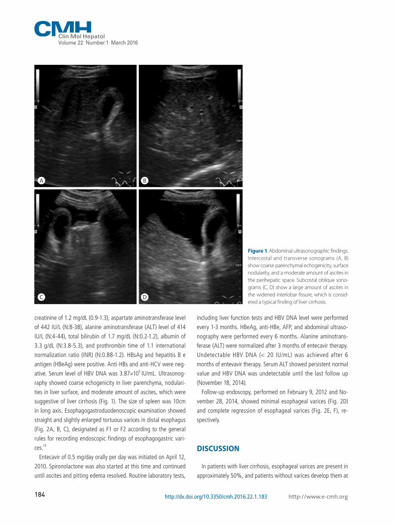
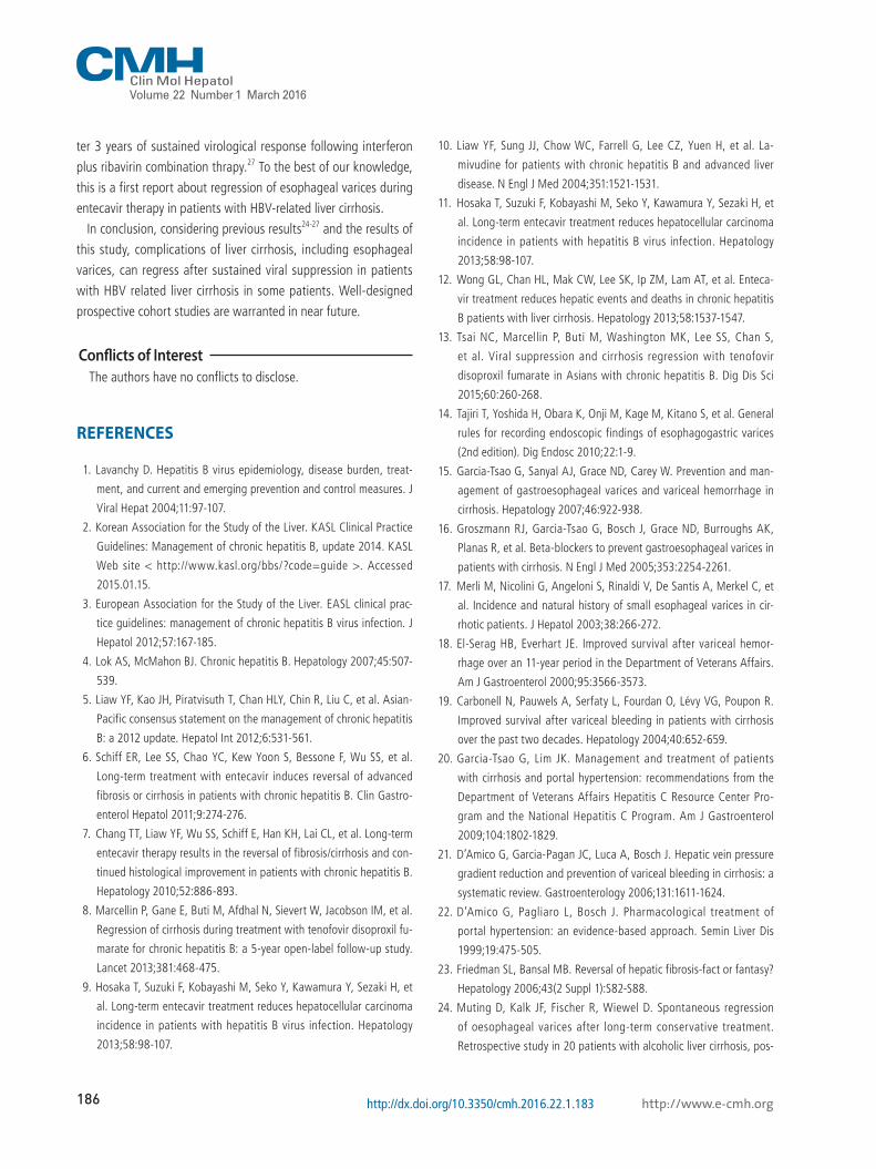
ative. Serum level of HBV DNA was 3.87×10⁵ IU/mL. Ultrasonog-
raphy showed coarse echogenicity in liver parenchyma, nodulari-
ties in liver surface, and moderate amount of ascites, which were
suggestive of liver cirrhosis (Fig. 1). The size of spleen was 10cm
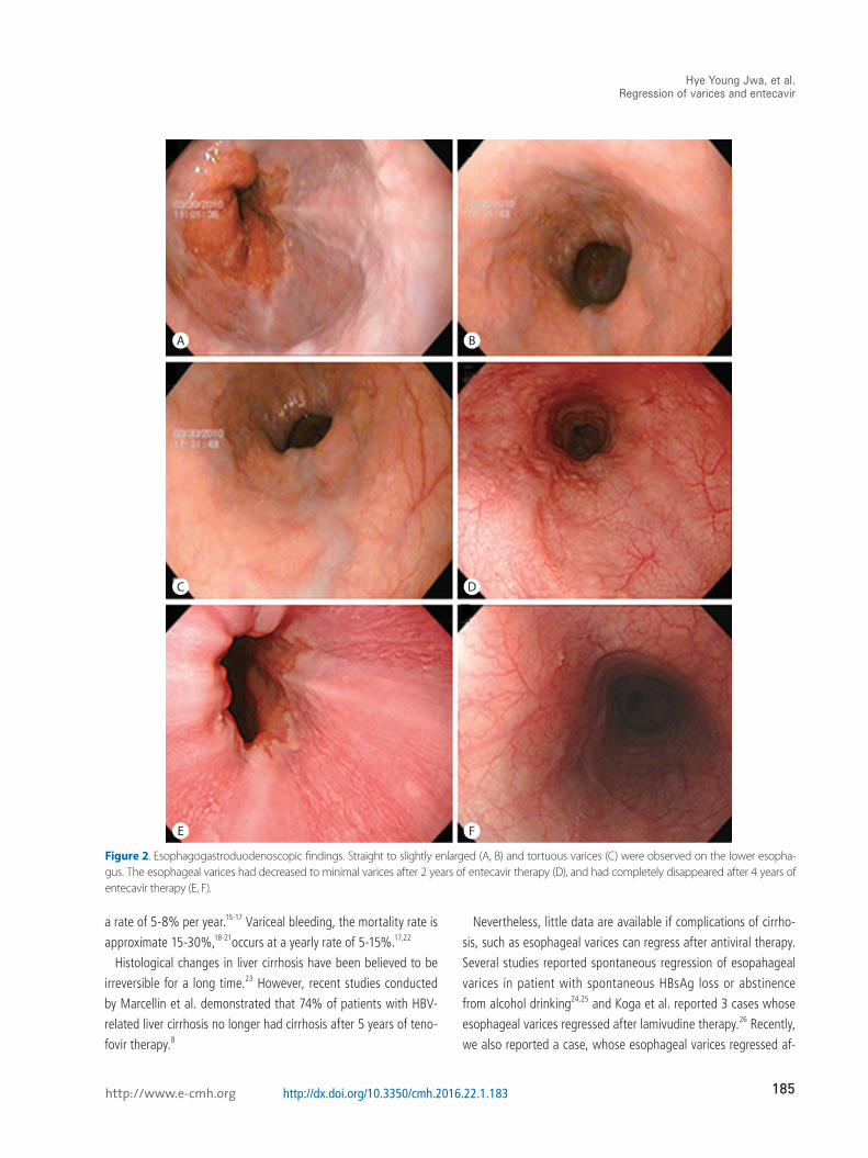
in long axis. Esophagogastroduodenoscopic examination showed
straight and slightly enlarged tortuous varices in distal esophagus
(Fig. 2A, B, C), designated as F1 or F2 according to the general
rules for recording endoscopic findings of esophagogastric vari-
ces.14
Entecavir of 0.5 mg/day orally per day was initiated on April 12,
2010. Spironolactone was also started at this time and continued
until ascites and pitting edema resolved. Routine laboratory tests,
including liver function tests and HBV DNA level were performed
every 1-3 months. HBeAg, anti-HBe, AFP, and abdominal ultraso-
nography were performed every 6 months. Alanine aminotrans-
ferase (ALT) were normalized after 3 months of entecavir therapy.
Undetectable HBV DNA (< 20 IU/mL) was achieved after 6
months of entevavir therapy. Serum ALT showed persistent normal
value and HBV DNA was undetectable until the last follow up
(November 18, 2014).
Follow-up endoscopy, performed on February 9, 2012 and No-
vember 28, 2014, showed minimal esophageal varices (Fig. 2D)
and complete regression of esophageal varices (Fig. 2E, F), re-
spectively.
DISCUSSION
In patients with liver cirrhosis, esophageal varices are present in
approximately 50%, and patients without varices develop them at
Figure 1. Abdominal ultrasonographic findings. Intercostal and transverse sonograms (A, B) show coarse parenchymal echogenicity, surface nodularity, and a moderate amount of ascites in the perihepatic space. Subcostal oblique sono-grams (C, D) show a large amount of ascites in the widened interlobar fissure, which is consid-ered a typical finding of liver cirrhosis.
A
C
B
D
185
Hye Young Jwa, et al. Regression of varices and entecavir
http://www.e-cmh.org http://dx.doi.org/10.3350/cmh.2016.22.1.183
a rate of 5-8% per year.15-17 Variceal bleeding, the mortality rate is
approximate 15-30%,18-21occurs at a yearly rate of 5-15%.17,22
Histological changes in liver cirrhosis have been believed to be
irreversible for a long time.23 However, recent studies conducted
by Marcellin et al. demonstrated that 74% of patients with HBV-
related liver cirrhosis no longer had cirrhosis after 5 years of teno-
fovir therapy.8
Nevertheless, little data are available if complications of cirrho-
sis, such as esophageal varices can regress after antiviral therapy.
Several studies reported spontaneous regression of esopahageal
varices in patient with spontaneous HBsAg loss or abstinence
from alcohol drinking24,25 and Koga et al. reported 3 cases whose
esophageal varices regressed after lamivudine therapy.26 Recently,
we also reported a case, whose esophageal varices regressed af-
A
C
E
B
D
F
Figure 2. Esophagogastroduodenoscopic findings. Straight to slightly enlarged (A, B) and tortuous varices (C) were observed on the lower esopha-gus. The esophageal varices had decreased to minimal varices after 2 years of entecavir therapy (D), and had completely disappeared after 4 years of entecavir therapy (E, F).
186 http://www.e-cmh.org
Clin Mol HepatolVolume_22 Number_1 March 2016
http://dx.doi.org/10.3350/cmh.2016.22.1.183
ter 3 years of sustained virological response following interferon
plus ribavirin combination thrapy.27 To the best of our knowledge,
this is a first report about regression of esophageal varices during
entecavir therapy in patients with HBV-related liver cirrhosis.
In conclusion, considering previous results24-27 and the results of
this study, complications of liver cirrhosis, including esophageal
varices, can regress after sustained viral suppression in patients
with HBV related liver cirrhosis in some patients. Well-designed
prospective cohort studies are warranted in near future.
Conflicts of InterestThe authors have no conflicts to disclose.
REFERENCES
1. Lavanchy D. Hepatitis B virus epidemiology, disease burden, treat-
ment, and current and emerging prevention and control measures. J
Viral Hepat 2004;11:97-107.
2. Korean Association for the Study of the Liver. KASL Clinical Practice
Guidelines: Management of chronic hepatitis B, update 2014. KASL
Web site < http://www.kasl.org/bbs/?code=guide >. Accessed
2015.01.15.
3. European Association for the Study of the Liver. EASL clinical prac-
tice guidelines: management of chronic hepatitis B virus infection. J
Hepatol 2012;57:167-185.
4. Lok AS, McMahon BJ. Chronic hepatitis B. Hepatology 2007;45:507-
539.
5. Liaw YF, Kao JH, Piratvisuth T, Chan HLY, Chin R, Liu C, et al. Asian-
Pacific consensus statement on the management of chronic hepatitis
B: a 2012 update. Hepatol Int 2012;6:531-561.
6. Schiff ER, Lee SS, Chao YC, Kew Yoon S, Bessone F, Wu SS, et al.
Long-term treatment with entecavir induces reversal of advanced
fibrosis or cirrhosis in patients with chronic hepatitis B. Clin Gastro-
enterol Hepatol 2011;9:274-276.
7. Chang TT, Liaw YF, Wu SS, Schiff E, Han KH, Lai CL, et al. Long-term
entecavir therapy results in the reversal of fibrosis/cirrhosis and con-
tinued histological improvement in patients with chronic hepatitis B.
Hepatology 2010;52:886-893.
8. Marcellin P, Gane E, Buti M, Afdhal N, Sievert W, Jacobson IM, et al.
Regression of cirrhosis during treatment with tenofovir disoproxil fu-
marate for chronic hepatitis B: a 5-year open-label follow-up study.
Lancet 2013;381:468-475.
9. Hosaka T, Suzuki F, Kobayashi M, Seko Y, Kawamura Y, Sezaki H, et
al. Long-term entecavir treatment reduces hepatocellular carcinoma
incidence in patients with hepatitis B virus infection. Hepatology
2013;58:98-107.
10. Liaw YF, Sung JJ, Chow WC, Farrell G, Lee CZ, Yuen H, et al. La-
mivudine for patients with chronic hepatitis B and advanced liver
disease. N Engl J Med 2004;351:1521-1531.
11. Hosaka T, Suzuki F, Kobayashi M, Seko Y, Kawamura Y, Sezaki H, et
al. Long-term entecavir treatment reduces hepatocellular carcinoma
incidence in patients with hepatitis B virus infection. Hepatology
2013;58:98-107.
12. Wong GL, Chan HL, Mak CW, Lee SK, Ip ZM, Lam AT, et al. Enteca-
vir treatment reduces hepatic events and deaths in chronic hepatitis
B patients with liver cirrhosis. Hepatology 2013;58:1537-1547.
13. Tsai NC, Marcellin P, Buti M, Washington MK, Lee SS, Chan S,
et al. Viral suppression and cirrhosis regression with tenofovir
disoproxil fumarate in Asians with chronic hepatitis B. Dig Dis Sci
2015;60:260-268.
14. Tajiri T, Yoshida H, Obara K, Onji M, Kage M, Kitano S, et al. General
rules for recording endoscopic findings of esophagogastric varices
(2nd edition). Dig Endosc 2010;22:1-9.
15. Garcia-Tsao G, Sanyal AJ, Grace ND, Carey W. Prevention and man-
agement of gastroesophageal varices and variceal hemorrhage in
cirrhosis. Hepatology 2007;46:922-938.
16. Groszmann RJ, Garcia-Tsao G, Bosch J, Grace ND, Burroughs AK,
Planas R, et al. Beta-blockers to prevent gastroesophageal varices in
patients with cirrhosis. N Engl J Med 2005;353:2254-2261.
17. Merli M, Nicolini G, Angeloni S, Rinaldi V, De Santis A, Merkel C, et
al. Incidence and natural history of small esophageal varices in cir-
rhotic patients. J Hepatol 2003;38:266-272.
18. El-Serag HB, Everhart JE. Improved survival after variceal hemor-
rhage over an 11-year period in the Department of Veterans Affairs.
Am J Gastroenterol 2000;95:3566-3573.
19. Carbonell N, Pauwels A, Serfaty L, Fourdan O, Lévy VG, Poupon R.
Improved survival after variceal bleeding in patients with cirrhosis
over the past two decades. Hepatology 2004;40:652-659.
20. Garcia-Tsao G, Lim JK. Management and treatment of patients
with cirrhosis and portal hypertension: recommendations from the
Department of Veterans Affairs Hepatitis C Resource Center Pro-
gram and the National Hepatitis C Program. Am J Gastroenterol
2009;104:1802-1829.
21. D’Amico G, Garcia-Pagan JC, Luca A, Bosch J. Hepatic vein pressure
gradient reduction and prevention of variceal bleeding in cirrhosis: a
systematic review. Gastroenterology 2006;131:1611-1624.
22. D’Amico G, Pagliaro L, Bosch J. Pharmacological treatment of
portal hypertension: an evidence-based approach. Semin Liver Dis
1999;19:475-505.
23. Friedman SL, Bansal MB. Reversal of hepatic fibrosis-fact or fantasy?
Hepatology 2006;43(2 Suppl 1):S82-S88.
24. Muting D, Kalk JF, Fischer R, Wiewel D. Spontaneous regression
of oesophageal varices after long-term conservative treatment.
Retrospective study in 20 patients with alcoholic liver cirrhosis, pos-

187
Hye Young Jwa, et al. Regression of varices and entecavir
http://www.e-cmh.org http://dx.doi.org/10.3350/cmh.2016.22.1.183
thepatitic cirrhosis and haemochromatosis with cirrhosis. J Hepatol
1990;10:158-162.
25. Calès P, Burtin P, Oberti F. Spontaneous regression of esophageal
varices is a phenomenon that has spontaneously disappeared from
our memory. J Hepatol 1991;12:263-264.
26. Koga H, Ide T, Oho K, Kuwahara R, Hino T, Ogata K, et al. Lamivu-
dine treatment-related morphological changes of esophageal varices
in patients with liver cirrhosis. Hepatol Res 2007;37:503-509.
27. Lee SJ, Song BC, Kim HU, Choi EK, Cho EK, Song HJ, et al. Com-
plete regression of esophageal varices after interferon plus ribavirin
therapy in patients with HCV related liver cirrhosis. J Gastroenterol
Hepatol 2014;29 (Suppl 3):195.