Embed Size (px)
Citation preview
DEVELOPMENTAL BIOLOGY 188, 17–33 (1997)ARTICLE NO. DB978622
Regionalized Transcriptional Domains of MyosinLight Chain 3f Transgenes in the EmbryonicMouse Heart: Morphogenetic Implications
Diego Franco,*,1 Robert Kelly,†,1 Wouter H. Lamers,**Margaret Buckingham,† and Antoon F. M. Moorman*,2
*Department of Anatomy and Embryology, Academic Medical Center, University ofAmsterdam, Amsterdam, The Netherlands; and †Department of Molecular Biology,Pasteur Institute, CNRS, URA 1947, Paris, France
Within the embryonic heart, five segments can be distinguished: two fast-conducting atrial and ventricular compartmentsflanked by slow-conducting segments, the inflow tract, the atrioventricular canal, and the outflow tract. These compart-ments assume morphological identity as a result of looping of the linear heart tube. Subsequently, the formation ofinteratrial, interventricular, and outflow tract septa generates a four-chambered heart. The lack of markers that distinguishright and left compartments within the heart has prevented a precise understanding of these processes. Transgenic micecarrying an nlacZ reporter gene under transcriptional control of regulatory sequences from the MLC1F/3F gene providespecific markers to investigate such regionalization. Our results show that transgene expression is restricted to distinctregions of the myocardium: b-galactosidase activity in 3F-nlacZ-2E mice is confined predominantly to the embryonic rightatrium, atrioventricular canal, and left ventricle, whereas, in 3F-nlacZ-9 mice, the transgene is expressed in both atrialand ventricular segments (right/left) and in the atrioventricular canal, but not in the inflow and outflow tracts. These linesof mice illustrate that distinct embryonic cardiac regions have different transcriptional specificities and provide earlymarkers of myocardial subdivisions. Regional differences in transgene expression are not detected in the linear heart tubebut become apparent as the heart begins to loop. Subsequent regionalization of transgene expression provides new insightsinto later morphogenetic events, including the development of the atrioventricular canal and the fate of the outflow tract.q 1997 Academic Press
INTRODUCTION al., 1989; Van der Loop et al., 1992; Ruzicka and Schwartz,1988; Barton et al., 1988; Lyons et al., 1990; Moorman etal., 1995). In the chicken heart tube an atrial-specific markerIn mammalian embryos the precardiac mesoderm, or car-is already activated specifically in the posterior part of thediogenic plate, occupies a horseshoe-shaped area at the ros-tube, although other chamber-specific markers do not showtral end of the germ disc. The two dorsolateral edges of thethis early compartmentalization (Yutzey et al., 1994; Yut-cardiogenic plate migrate toward the midline of the bodyzey and Bader, 1995); indeed, in vivo labeling techniquesaxis and fuse, in a craniocaudal direction, into a single tubein the chicken suggest that the cardiac tube contains only(see Manasek, 1968; Van Mierop, 1979). As soon as the pre-ventricular primordia (De la Cruz et al., 1989). In mammals,cardiac mesoderm is formed, the cells begin to express mus-it is not clear whether the expression of chamber-specificcle-specific proteins including SERCA2, myosin heavymyocardial genes shows spatial restriction at this stage.chain (MHC), myosin light chain (MLC), actin, tropomyo-
With further development, the primitive cardiac tubesin, and various intermediate filament isoforms (Schaart etbends to the right-hand side and newly formed myocardiumis added at both ends of the tube (De la Cruz et al., 1989).At this stage the atrial and ventricular compartments can1 These two authors contributed equally to the paper.first be distinguished. The cardiac tube is formed of two2 To whom correspondence should be addressed at Departmentlayers, endocardium and myocardium, separated by extra-of Anatomy & Embryology, Academic Medical Center, Meiberg-cellular matrix, the cardiac jelly (Davis, 1927). The myocar-dreef 15, 1105 AZ Amsterdam, The Netherlands. Fax 31-20-
6976177. E-mail: [email protected]. dium is characterized by gradients of gene expression along
17
0012-1606/97 $25.00Copyright q 1997 by Academic PressAll rights of reproduction in any form reserved.
AID DB 8622 / 6x28$$$281 07-16-97 09:46:01 dbal

18 Franco et al.
its length. a-MHC is highly expressed in the inflow tract specific enhancer element (Donoghue et al., 1988; Rosen-thal et al., 1989; Kelly et al., 1995). 3F-nlacZ-9 mice containregion and decreases toward the outflow tract (Wessels et
al., 1991). In contrast, b-MHC shows the opposite pattern 9 kb of sequence upstream from the MLC3F transcriptionalinitiation site and no 3* enhancer. The 9-kb sequence in-of expression, since it is highly expressed in the outflow
and decreases toward the inflow tract (De Groot et al., 1989; cludes a second muscle-specific enhancer, present in thefirst intron of the gene (Kelly et al., 1997). Transgene expres-Wessels et al., 1991).
During further development, the cardiac jelly becomes sion in 3F-nlacZ-2E mice reveals a distinction between leftand right sides of the heart. Expression is mainly confinedconfined to the outflow tract and atrioventricular canal (Ma-
nasek, 1976), and the ventricle acquires a trabeculated mor- to the embryonic right atrium, atrioventricular canal, andleft ventricle, whereas in 3F-nlacZ-9 mice expression is re-phology (Ben-Sahchar et al., 1985). During this process,
changes in the pattern of gene expression occur which are stricted to both atrial and ventricular cavities (right/left)and the atrioventricular canal, but is not observed in thecorrelated with differences in function (De Jong et al., 1992;
Moorman and Lamers, 1994). Five segments can be distin- outflow or inflow tract regions. These lines of mice illus-trate that distinct embryonic cardiac segments acquire dif-guished; two fast-conducting atrial and ventricular com-
partments flanked by slow-conducting segments, the inflow ferent transcriptional specificities as the heart loops andprovide early markers of subdivisions of the myocardiumtract, the atrioventricular canal, and the outflow tract. The
atrial and ventricular compartments are characterized by with implications for cardiac morphogenesis.the expression of specific MHC isoforms, a and b, respec-tively (De Groot et al., 1989; Wessels et al., 1991). Theflanking segments coexpress both isoforms (De Groot et al., MATERIALS AND METHODS1989). The expression of regulatory and alkali MLC iso-forms also becomes confined to distinct compartments (see Transgenic MiceLyons, 1994). MLC1A and MLC2A mRNAs are mostly re-
Two MLC3F transgenic lines containing the nlacZ reporter genestricted to the atrial myocardium (Lyons et al., 1990; Kuba-under the transcriptional control of regulatory elements of thelak et al., 1994) and MLC1V and MLC2V mRNAs to theMLC3F gene were analyzed. Construct 3F-nlacZ-2E (Kelly et al.,ventricular compartment (Lyons et al., 1990; O’Brien et al.,1995) contains 2 kb of upstream sequence in front of the MLC3F
1993). MLC3F mRNA is also present in embryonic cardiac promoter driving expression of an nlacZ reporter gene and the 3*muscle, being more abundant in the atria than the ventri- MLC1F/3F enhancer element placed 3* to the polyadenylation sitecles (Kelly et al., 1995). Changes in the spatial expression (Fig. 1). Construct 3F-nlacZ-9 (Kelly et al., 1997) contains a 9-kbof myocardial genes occur asynchronously during develop- fragment of DNA upstream of the MLC3F transcription initiation
site driving the expression of the nlacZ reporter gene (Fig. 1). De-ment (Lyons et al., 1990).tails of transgene construction and the characterization of skeletalCorrect development of the heart involves the formationmuscle expression during development and in the adult in theseof three different septa that converge in the lesser curvaturetransgenic lines are reported elsewhere (Kelly et al., 1995, 1997).of the heart: the aorticopulmonary septum, the interatrialIn this study data are presented for one line of each transgene con-septum, and the interventricular septum. The process ofstruct (3F-nLacZ-2E, line 1, and 3F-nLacZ-9, line 9a). In both casesseptation is a crucial event during cardiogenesis and manya second transgenic line gave similar results.
abnormalities originate from a failure of this process (Beckerand Anderson, 1983). The outflow tract is initially con-nected with the embryonic right ventricle and the common Embryosatrium is connected with the embryonic left ventricle. With
Heterozygous and homozygous adult specimens and embryosfurther development, the left ventricular chamber obtainsranging from Embryonic Day (E) 7 to E16 for each transgenic linean independent arterial connection. Whereas the specifica-(crossed with nontransgenic C57BL6/J 1 SJL F1 females) were ana-tion of atrial and ventricular compartments has been exten-lyzed. The day of plug was taken as E0.5. Embryos were excised
sively studied (Yutzey and Bader, 1995), little is known from the uterus and the thoracic wall was removed (E12.5 to E16.5),about the origins of right and left ventricular specification exposing the heart to allow maximal penetration of fixatives andand how the new connections are made, i.e., the left arterial reagents. Adult hearts were dissected at the arterial and venousand right atrioventricular connections. The lack of markers pole of the heart, keeping intact the caval and pulmonary veins.
Specimens were briefly fixed in freshly prepared 4% paraformalde-that distinguish right and left cardiac compartments hashyde for in situ hybridization (4 hr) or in methanol:acetone:waterprevented a precise understanding of the septation process.for immunohistochemistry (30 min–4 hr) and rinsed twice in phos-The generation of transgenic mice carrying an nlacZ re-phate-buffered saline (PBS). For in toto b-galactosidase histochem-porter gene under the transcriptional control of regulatoryistry, or in toto b-galactosidase histochemistry followed by immu-sequences of the MLC1F/3F gene provides a tool to unravelnohistochemistry, specimens were directly processed as detailedthese processes.below. Specimens were rinsed in increasing sucrose gradients (10,
We describe here a detailed analysis of the pattern of 20, and 30% in PBS) for 2 hr at each step, embedded in OTC (MilesnlacZ expression during cardiac embryogenesis in two lines Inc., USA), and frozen. Freeze cryotome serial sections of 7–10 mmof MLC3F transgenic mice. 3F-nlacZ-2E mice contain 2 ki- were cut, mounted onto gelatin-coated slides, and stored at 0207Clobases (kb) of DNA sequence upstream of the MLC3F tran- until use. Embryos for in situ hybridization were dehydrated in
increasing concentrations of ethanol and embedded in paraffin. Se-scription initiation site together with a 3* skeletal muscle-
Copyright q 1997 by Academic Press. All rights of reproduction in any form reserved.
AID DB 8622 / 6x28$$$281 07-16-97 09:46:01 dbal

19Transcriptional Domains in the Developing Heart
FIG
.1.
ML
C3F
tran
sgen
eco
nst
ruct
san
dth
epa
tter
nof
card
iac
expr
essi
onat
E10
.Sch
emat
icre
pres
enta
tion
ofth
eM
LC
1F/3
Flo
cus
show
ing
the
tran
scri
ptio
nal
star
tsi
tes
(_),
exon
(j)s
tru
ctu
re,
ML
C1F
(—),
and
ML
C3F
(---
)sp
lici
ng
patt
ern
san
dth
epo
stio
nof
two
skel
etal
mu
scle
enh
ance
rel
emen
ts(g
rey
bars
).M
LC
3Ftr
ansg
ene
con
stru
cts
are
show
nw
ith
tran
sgen
eex
pres
sion
patt
ern
sat
appr
oxim
atel
yE
mbr
yon
icD
ay(E
)10
.B
lue
repr
esen
tstr
ansg
ene
expr
essi
onan
dye
llow
the
endo
card
ial
cush
ion
sof
the
outfl
owtr
act
and
the
atri
oven
tric
ula
rca
nal
.OFT
,ou
tflow
trac
t;A
VC
,at
riov
entr
icu
lar
can
al;
IFT
,in
flow
trac
t;R
A,
righ
tat
riu
m;
LA
,le
ftat
riu
m;
RV
,ri
ght
ven
tric
le;
LV
,le
ftve
ntr
icle
;IV
S,in
terv
entr
icu
lar
sept
um
;SP
,se
ptu
mpr
imu
m.
Copyright q 1997 by Academic Press. All rights of reproduction in any form reserved.
AID DB 8622 / 6x28$$8622 07-16-97 09:46:01 dbal

20 Franco et al.
TABLE 1Transgene Expression in the Developing Mouse Heart in 3F-nLacZ-2E and 3F-nLacZ-9
Embryonic Fetal Adult
Structure Lines: 2E 9 2E 9 2E 9
Outflow tract 0 0 0 0Right ventricle 0 / 0 / 0 /Interventricular septum / / / / / /Left ventricle / / / / / /Atrioventricular canal / /Right atrium / / / / / /Left atrium 0 / 0 / 0 /Septum primum 0 0Septum secundum 0 0Interatrial septum 0 0Sinus horns / /Coronary sinus 0 0 0 0Caval veins 0 0 0 0Pulmonary veins 0 0 0 0
Note. b-Galactosidase expression in the embryonic, fetal, and adult cardiac structures in 3F-nlacZ-2E (2E) and 3F-nlacZ-9 (9) transgenicmice. (0) no expression, (/) positive expression. Where no indication is given, the structure does not exist at the stage in question.
rial sections of 7 mm were cut, mounted onto RNase-free aminopro- cific binding of the primary antibody. After this pretreatment,whole-mount embryos were washed again in PBTMT and incu-pyltriethosixylane-coated slides, and stored at room temperature.bated overnight with primary antibody diluted in PBTMT (1:20). Aspecific primary polyclonal antibody recognizing all myosin heavy
In Toto X-Gal Histochemical Staining chain isoforms was used (Cusella de Angelis et al., 1994) to delin-eate the myocardium. First antibody binding was detected by the
Specimens were briefly fixed in freshly prepared 4% paraformal-uncoupled peroxidase–antiperoxidase method as described else-
dehyde (30 min–1 hr) before histochemical detection of b-galactosi-where (Dent et al., 1987).
dase. Incubation in X-gal solution at 377C was performed for periodsof 30 min to overnight as detailed elsewhere (Sanes et al., 1986).Subsequently, whole-mount embryos and adult hearts were post-
b-Galactosidase Detection andfixed in freshly made 4% paraformaldehyde for 4 hr to overnightImmunohistochemistry in Cryosectionsand conserved in 70% ethanol until analyzed.
Sections were rinsed briefly in PBS and incubated at 377C (1 hr–overnight) in X-gal reagent. After a brief rinse in PBS for 5 min,In Toto b-Galactosidase Staining andalternative sections were washed in bidistilled water for 5 min,Immunohistochemistrycounterstained with azofloxine or eosin (1%) for 5 min, dehydrated,and mounted in Entellan (Merck); other sections were processedThe specimens were first stained with X-gal as described above,
rinsed twice in PBT (PBS containing 0.1% Tween 20) for 5 min, for immunohistochemical detection of cardiac-specific antigens.Specific primary monoclonal antibodies against human a-MHCand then processed for immunohistochemistry. The embryos were
treated for 4–5 hr with hydrogen peroxide (3% in PBT) to reduce and b-MHC (Wessels et al., 1991) were used to visualize atrialand ventricular myocardium, respectively. After treatment withendogenous peroxidase activity. They were then incubated with
PBTMT (1% defatted milk powder in PBT) for 2 hr to avoid nonspe- hydrogen peroxide (3% in PBS, 30 min) to reduce endogenous perox-
FIG. 2. Developmental profile of transgene expression in the 3F-nlacZ-2E mice. In toto localization of b-galactosidase in 3F-nlacZ-3Etransgenic embryos. (a) Labeling is distributed symmetrically (arrowheads) in the precardiac mesoderm (E7.5) and (b) early tubular heartstages (E8). (c) In the looping heart (E8.5), b-galactosidase activity is observed throughout the cardiac tube; however, it is reduced at thepoles (arrowheads). (d) As development procedes (E9.5), expression in the atrium (A) becomes preferentially restricted toward the rightside (arrowheads). (e) Whole-mount immunocytochemistry using an anti-MHC antibody on an E9.5 embryo demonstrates that whereasthe common ventricle (V) is strongly b-galactosidase positive, few b-galactosidase cells are present in the ouflow tract (OFT) which ispositive for the myocardial marker. (f) and (g) correspond to an E10.5 embryo in ventral and dorsal views, respectively. Transgene expressionis confined to the left ventricle (LV) and right atrium (RA) (f), forming a continuum of b-galactosidase cells (arrow) along the atrioventricularcanal (AVC) (g). (h) and (i) correspond to an E14.5 heart in dorsal and ventral views, respectively. The pattern of expression is essentiallyidentical to E10.5. LA, left atrium; RV, right ventricle.
Copyright q 1997 by Academic Press. All rights of reproduction in any form reserved.
AID DB 8622 / 6x28$$$281 07-16-97 09:46:01 dbal

21
AID DB 8622 / 6x28$$8622 07-16-97 09:46:01 dbal

22 Franco et al.
idase activity, an incubation in TENG-T (10 mM Tris, 5 mM EDTA, in Fig. 1 and Table 1. The 3F-nlacZ-2E line shows striking150 mM NaCl, 0.25% gelatin, 0.05% Tween 20, pH 8.0; 30 min) predominance of expression in the left ventricle and rightwas performed to avoid nonspecific binding. The sections were atrium, whereas in the 3F-nLacz-9 line both ventricles andthen incubated overnight with specific primary antibodies. Binding atria express the transgene, but the outflow and inflowof the first antibody was detected using a rabbit anti-mouse immu- tracts are negative. Specific attention is given to transgenenoglobulin, followed by a goat anti-rabbit immunoglobulin and fi-
expression during the processes of atrioventricular canalnally a rabbit peroxidase anti-peroxidase (PAP) complex. Each incu-septation (3F-nlacZ-2E line) and outflow tract septation (3F-bation lasted 2 hr and was followed by three washes in PBS (5 minnlacZ-9 line). The data presented show the expression pat-each). All sera were diluted in PBS. The visualization of the PAPtern of the transgene in heterozygous mice; homozygouscomplex was performed by incubation with 0.5 mg/ml 3,3*-diami-
nobenzidine and 0.02% hydrogen peroxide in 30 mM imidazole, 1 animals displayed similar patterns of expression (data notmM EDTA (pH 7.0) buffer. Sections were dehydrated and mounted shown).in Entellan (Merck).
3F-nlacZ-2E Transgenic MiceIn Situ Hybridization 3F-nlacZ-2E transgenic mice contain nlacZ under tran-
scriptional control of the MLC3F promoter (2 kb of se-Complementary RNA probes against rat a-MHC (Schiaffino etquence upstream from the transcriptional start site) and aal., 1989; Boheler et al., 1992), rat b-MHC (Boheler et al., 1992),
MLC2A (Kubalak et al., 1994), MLC2V (O’Brien et al., 1993), and 3* enhancer element (Kelly et al., 1995). Two 3F-nlacZ-2Eb-galactosidase (Kelly et al., 1995) mRNAs were radiolabeled with transgenic lines show indistinguishable expression pat-[35S]UTP by in vitro transcription according to standard protocols terns. The overall adult cardiac expression in these mice(Melton et al., 1984). Hybridization conditions were as detailed has been reported previously and shows that transcriptionalelsewhere (Moorman et al., 1993, 1995). Briefly, the sections were specificities differ in the right and left ventricular chambersdeparaffined, rinsed in absolute ethanol, and dried in an air stream.
(Fig. 1; Kelly et al., 1995). In this study we follow the devel-Pretreatment of the sections was as follows: 20 min 0.2 N HCl, 5opment of these differences during embryogenesis.min bidistilled water, 20 min 21 SSC (707C), 5 min bidistilled
Expression in the precardiac mesoderm and the primi-water, 2–20 min digestion in 0.1% pepsin dissolved in 0.01 N HCl,tive cardiac tube (E7.5–E9.5). At the cardiogenic plate30 sec in 0.2% glycine/PBS, two 30-sec rinses in PBS, 20 min ofstage (E7.5), a few cells expressing the nlacZ reporter genepostfixation in freshly prepared 4% paraformaldehyde, 5 min in
bidistilled water, 5 min in 10 mM EDTA, 5 min in 10 mM DTT, are observed symmetrically located on both sides of theand finally drying in an air stream. The prehybridization mixture anteroposterior axis (Fig. 2a). These areas fuse in an antero-contained 50% formamide, 10% dextran sulfate, 21 SSC, 21 Den- posterior direction at the midline of the body to form ahardt’s solution, 0.1% Triton X-100, 10 mM DTT, and 200 ng/ml single cardiac tube in which b-galactosidase-positive cellsheat-denatured herring sperm DNA. The sections were hybridized are symmetrically distributed (E8, Fig. 2b). nlacZ expressionovernight at 527C and washed as follows: a rinse in 11 SSC, 30
extends from the embryonic atrium through the atrioven-min at 527C in 50% formamide dissolved in 11 SSC, 10 min in 11tricular junction and common ventricle (Fig. 2c). The ex-SSC, 30 min in RNase A (10 mg/ml), 10 min in 11 SSC, 10 min intremities of the heart tube are initially b-galactosidase posi-0.11 SSC, and dehydration in 50, 70, and 90% ethanol containingtive, expression colocalizing with that of b-MHC and0.3 M ammonium acetate. The sections were then dried and im-SERCA2 mRNAs at the venous and arterial myocardialmersed in nuclear autoradiographic emulsion G5 (Ilford). The expo-
sure times ranged from 7 to 14 days and the development times boundaries, respectively (as assessed by in situ hybridiza-from 4 to 8 min. Photographs were taken with a Zeiss Axiophot tion to serial transverse sections, Figs. 3a and 3b). The firstmicroscope, using Agfa 25ASA films. signs of regionalization are observed at E8.5 as the heart
starts to loop to the right-hand side. Negative myocardio-cytes are observed at the distal arterial pole adjacent to the
RESULTS aortic sac and at the venous pole at the entrance of the sinushorns (Fig. 2c); nlacZ-expressing cells are located in theproximal part of the right and left sinus horns, facing the3F-nlacZ-2E and 3F-nlacZ-9 transgenic mice express a b-
galactosidase reporter gene with a nuclear localization sig- common atrium (Figs. 2c, 3a, and 3b). The b-galactosidase-negative areas at the arterial and venous poles probably rep-nal (nlacZ) in skeletal and cardiac striated muscle. Nuclear
localization improves the resolution of detection at the sin- resent the earliest primordia of the outflow tract and inflowtract, respectively.gle-cell level. This study is restricted to the analysis of
transgene expression in the myocardium, in both embry- At E9–9.5 five functional segments can be distinguished:outflow tract, common ventricle, atrioventricular canal,onic and adult transgenic mice. Cardiac development is con-
sidered over three periods: the first covers the period from common atrium, and inflow tract (Moorman and Lamers,1994). Expression of b-galactosidase is observed in the ven-the precardiac mesoderm stage up to the looping heart (early
embryonic period), the second comprises the development tricle, atrioventricular canal, and common atrium (Fig. 2d),but not in the outflow tract or inflow tract, which are nega-of right/left components by the process of atrioventricular
septation (embryonic period), and the third includes septa- tive for transgene expression although positive on incuba-tion with an anti-myosin antibody (Fig. 2e). Although thetion of the heart and acquisition of the mature cardiac archi-
tecture (fetal and adult period). Our results are summarized common ventricular cavity is b-galactosidase positive,
Copyright q 1997 by Academic Press. All rights of reproduction in any form reserved.
AID DB 8622 / 6x28$$$281 07-16-97 09:46:01 dbal

23Transcriptional Domains in the Developing Heart
FIG. 3. Histological analysis of transgene expression in 3F-nlacZ-2E mice. Histochemical localization of b-galactosidase activity incryostat sections of hearts corresponding to E8.5 (a, b), E11.5 (c, d), and E12.5 (e, f) embryos. (b) Myocardial cells at the venous pole of theheart in an adjacent serial section to (a), as revealed by in situ hybridization to SERCA2 mRNA. At E8.5 all b-galactosidase-positive nuclei(arrows) are myocardial (a), whereas endocardial cells are not labeled (arrowhead) (b). In sections through all four cardiac compartmentsat E11.5, b-galactosidase activity is restricted predominantly to the left ventricle (LV), interventricular septum (IVS), and right atrium(RA) (c, d). At E12.5, b-galactosidase activity is confined to the LV, IVS, and RA up to the entrance of the right superior caval vein (rsc).In the left atrium (LA), the myocardium in close apposition to the atrioventricular junction is also positive for the transgene (arrows) (e).In contrast, structures such as the septum primum (arrowhead) (e), caval veins (arrow) (f), and coronary sinus (arrowheads) (f) do not showb-galactosidase activity.
Copyright q 1997 by Academic Press. All rights of reproduction in any form reserved.
AID DB 8622 / 6x28$$8622 07-16-97 09:46:01 dbal
24 Franco et al.
more positive cells are observed in the left side than the thin rim of myocardial cells around the left semilunar valveis b-galactosidase negative (data not shown). The right ven-right side of the primitive ventricular myocardium (data not
shown). In the atrium at this stage transgene expression is tricular compartment contains a few positive cells adjacentto the apex of the heart (Fig. 2h), whereas the interventricu-higher in the right side than the left (Fig. 2d). Thus, prior
to septation, signs of right–left differentiation are already lar septum and the left ventricle are mostly positive. Theprecise labeling pattern shows some individual variationdetectable in the embryonic heart.
Expression pattern in the early septating heart (E10.5– within the transgenic lines. At the venous pole of the heart,the myocardium surrounding the caval veins, pulmonaryE12.5). From E10.5 onward the common ventricle and
atrium become separated into right and left components, veins, coronary sinus, and the dorsal myocardial wall of theatrium, including the septum primum and the developingpreceding septation of the outflow tract. The outflow tract
(Fig. 2f) and embryonic right ventricle show almost no stain- septum secundum, are b-galactosidase negative (data notshown; see also Fig. 3f).ing, whereas the left ventricle is positive (Figs. 2f, 2g, and
3d), including the interventricular septum (Figs. 3c and 3d). In the adult heart, the left ventricle is mostly positive,whereas transgene expression is excluded from the rightAt the venous pole of the heart, the dorsal wall of the atrium
shows an outgrowth, the septum primum, which first de- ventricle (Kelly et al., 1995). The entire right atrium ex-presses the transgene at a high level, except for the myocar-limits the right and left atrial components (Kauffman, 1992).
Transgene expression is confined to the atrioventricular ca- dium of the caval veins and the coronary sinus, whereasthe left atrium, including the pulmonary myocardium, isnal (Fig. 2g) and the right atrium (Fig. 3d), whereas the left
atrium (Fig. 2g) and the septum primum are negative. In a negative, apart from a few weakly b-galactosidase-positivecells (data not shown).dorsal view it can be seen that b-galactosidase-positive cells
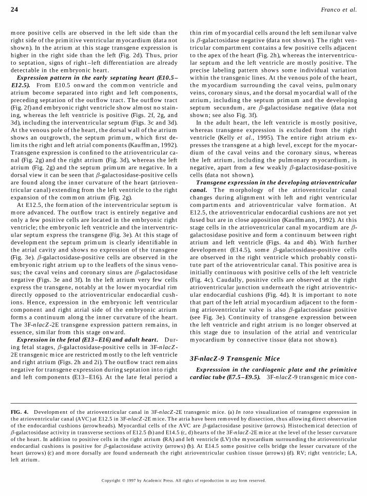
are found along the inner curvature of the heart (atrioven- Transgene expression in the developing atrioventricularcanal. The morphology of the atrioventricular canaltricular canal) extending from the left ventricle to the right
expansion of the common atrium (Fig. 2g). changes during alignment with left and right ventricularcompartments and atrioventricular valve formation. AtAt E12.5, the formation of the interventricular septum is
more advanced. The outflow tract is entirely negative and E12.5, the atrioventricular endocardial cushions are not yetfused but are in close apposition (Kauffmann, 1992). At thisonly a few positive cells are located in the embryonic right
ventricle; the embryonic left ventricle and the interventric- stage cells in the atrioventricular canal myocardium are b-galactosidase positive and form a continuum between rightular septum express the transgene (Fig. 3e). At this stage of
development the septum primum is clearly identifiable in atrium and left ventricle (Figs. 4a and 4b). With furtherdevelopment (E14.5), some b-galactosidase-positive cellsthe atrial cavity and shows no expression of the transgene
(Fig. 3e). b-galactosidase-positive cells are observed in the are observed in the right ventricle which probably consti-tute part of the atrioventricular canal. This positive area isembryonic right atrium up to the leaflets of the sinus veno-
sus; the caval veins and coronary sinus are b-galactosidase initially continuous with positive cells of the left ventricle(Fig. 4c). Caudally, positive cells are observed at the rightnegative (Figs. 3e and 3f). In the left atrium very few cells
express the transgene, notably at the lower myocardial rim atrioventricular junction underneath the right atrioventric-ular endocardial cushions (Fig. 4d). It is important to notedirectly opposed to the atrioventricular endocardial cush-
ions. Hence, expression in the embryonic left ventricular that part of the left atrial myocardium adjacent to the form-ing atrioventricular valve is also b-galactosidase positivecomponent and right atrial side of the embryonic atrium
forms a continuum along the inner curvature of the heart. (see Fig. 3e). Continuity of transgene expression betweenthe left ventricle and right atrium is no longer observed atThe 3F-nlacZ-2E transgene expression pattern remains, in
essence, similar from this stage onward. this stage due to insulation of the atrial and ventricularmyocardium by connective tissue (data not shown).Expression in the fetal (E13–E16) and adult heart. Dur-
ing fetal stages, b-galactosidase-positive cells in 3F-nlacZ-2E transgenic mice are restricted mostly to the left ventricle
3F-nlacZ-9 Transgenic Miceand right atrium (Figs. 2h and 2i). The outflow tract remainsnegative for transgene expression during septation into right Expresssion in the cardiogenic plate and the primitive
cardiac tube (E7.5–E9.5). 3F-nlacZ-9 transgenic mice con-and left components (E13–E16). At the late fetal period a
FIG. 4. Development of the atrioventricular canal in 3F-nlacZ-2E transgenic mice. (a) In toto visualization of transgene expression inthe atrioventricular canal (AVC) at E12.5 in 3F-nlacZ-2E mice. The atria have been removed by dissection, thus allowing direct observationof the endocardial cushions (arrowheads). Myocardial cells of the AVC are b-galactosidase positive (arrows). Histochemical detection ofb-galactosidase activity in transverse sections of E12.5 (b) and E14.5 (c, d) hearts of the 3F-nlacZ-2E mice at the level of the lesser curvatureof the heart. In addition to positive cells in the right atrium (RA) and left ventricle (LV) the myocardium surrounding the atrioventricularendocardial cushions is positive for b-galactosidase activity (arrows) (b). At E14.5 some positive cells bridge the lesser curvature of theheart (arrows) (c) and more dorsally are found underneath the right atrioventricular cushion tissue (arrows) (d). RV; right ventricle; LA,left atrium.
Copyright q 1997 by Academic Press. All rights of reproduction in any form reserved.
AID DB 8622 / 6x28$$$281 07-16-97 09:46:01 dbal

25Transcriptional Domains in the Developing Heart
AID DB 8622 / 6x28$$862207-16-97 09:46:01 dbal

26 Franco et al.
tain 9 kb upstream of the MLC3F transcriptional start site the semilunar valve swellings (Fig. 6a) and the conal ridges(Fig. 6b) is negative for the transgene as is the adjacent tra-fused to nlacZ in the absence of the 3* enhancer element.
Early 3F-nlacZ-9 transgene expression, from the cardiogenic beculated portion of the embryonic right ventricle (Figs. 6cand 6d). The latter area corresponds to the embryonic rightplate stage up to the formation of a primitive cardiac tube,
is similar to that observed in 3F-nlacZ-2E mice (compare infundibulum. The negative area within the right ventriculeincreases in size with development (E13–E16). At the endFigs. 5a and 2c). Once the cardiac tube is formed and looping
initiated (E8.5), cardiomyocytes expressing b-galactosidase of the process of arterial pole septation (E16) an area sur-rounding the left semilunar valve is negative for transgeneare concentrated in the common ventricular region of the
tube (Fig. 5a). As the heart continues to loop (E9–E10), the expression (Fig. 6e), as is the outlet region of the right semi-lunar valve (Fig. 6f). These b-galactosidase-negative myocar-numbers of b-galactosidase-positive cells are reduced at the
arterial pole (Fig. 5b). The first clearly distinguishable differ- dial cells are, however, positive for cardiac troponin ImRNA (Figs. 6g and 6h).ences between these transgenes in the developing heart are
observed after E9. 3F-nlacZ-9 transgene expression in the adult heart.The hearts of 3F-nlacZ-9 mice show expression in both rightTransgene expression in the looped heart delimits the
boundary between the embryonic right ventricle and the and left atria and ventricles. Within the ventricular com-partments, the right ventricle shows a patch of non-nlacZ-outflow tract (E10.5–E12.5). At E10.5 cells expressing the
transgene are observed in the atrial myocardium, the atrio- expressing cells in the distal part of the free wall (Fig. 7a).The outlet septum is also negative (Fig. 7c). In contrast, theventricular canal, and the right and left trabeculated compo-
nents of the embryonic ventricle. The outflow tract of the left ventricular free wall and the interventricular septum areentirely positive. Only a thin rim in the subaortic portion atheart is negative (Fig. 5c) and no labeled cells are seen in
the myocardium of the sinus venosus as demarcated by the level of the semilunar valve is negative (Fig. 7b). At thevenous pole of the heart, myocardial structures such as thecardiac troponin I mRNA expression (data not shown). The
septum primum does not express the transgene. At E12.5, caval veins, pulmonary veins, coronary sinus, interatrialseptum, and dorsal wall of the atria do not express thethe myocardium of the arterial pole of the heart (outflow
tract), which overlies the endocardial cushions, does not transgene (data not shown).express the transgene (Fig. 5d).
As the right and left ventricular and atrial chambers be-come established, the pattern of transgene expression is DISCUSSIONmaintained in both right and left components of the embry-onic ventricular and atrial segments and also in the atrio- Morphogenesis of the Cardiac Compartments:ventricular canal (Fig. 5e). The outflow tract and inflow Transcriptional Regulation in the Early Hearttract regions do not express the transgene; this pattern ismaintained until the outflow tract starts to separate into In this study we show that two nlacZ transgenes with
different regulatory elements from the myosin MLC1F/3Fright and left components (E13 onward).3F-nlacZ-9 transgene expression as a marker to follow gene have distinct expression patterns in the mouse heart.
One (3F-nlacZ-2E) shows striking left/right differences,the process of outflow tract septation (E13–E16). As theoutflow tract undergoes separation into aortic and pulmo- with the right ventricle, left atrium, outflow tract, and in-
flow tract negative for transgene expression, while the sec-nary trunks (E13–15), the distal portion of the right ventric-ular segment shows no expression of nlacZ (E15; Fig. 5f). A ond (3F-nlacZ-9) is expressed more extensively in the atria
and ventricles, but the outflow and inflow tracts are nega-sharp boundary between the b-galactosidase–negative cellsand labeled myocardiocytes of the ventricular compart- tive. A developmental analysis shows that the early heart
tube does not show regionalization of transgene expression;ments is observed. This boundary coincides, ventrally, withthe bulboventricular groove (Fig. 5f). As the outflow tract b-galactosidase-positive cells are similarly distributed
throughout the linear heart tube. It is only after the onsetstarts to ‘‘disappear’’ as a clearly identifiable segment of theheart, the endocardial cushions become fused and separated of looping (E9) that regional differences begin to be apparent.
In the chicken (Yutzey et al., 1994; Yutzey and Bader, 1995)into right and left portions by the formation of the aortico-pulmonary and conal septa. The myocardium surrounding there is evidence from atrial myosin heavy chain gene ex-
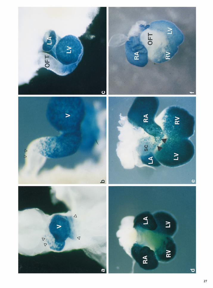
FIG. 5. Developmental profile of transgene expression in 3F-nlacZ-9 mice. In toto localization of b-galactosidase in 3F-nlacZ-9 embryonichearts. (a) At E8.5 the early looping heart shows a profile of transgene expression which is indistinguisable from that of the 3F-nlacZ-2Emice at the same stage (see Fig. 2c). b-Galactosidase expression is weaker at both poles of the cardiac tube (arrowheads) (b). At E9, transgeneexpression is negative in the arterial pole of the heart (arrowheads); and expression is still comparable to that of the 3F-nlacZ-2E transgene.At later stages of development (E10.5 (c); E12.5 (d)), the outflow tract (OFT) is negative for transgene expression, whereas both ventricles(LV, RV) and both atria (LA, RA) are positive. (e) A dorsal view at E12.5 shows that the inflow tract including the superior caval veins(sc) is negative (translucent blue overlying stronger expressing atrial region). Note that the atrioventricular canal (arrowheads) is alsopositive. (f) At E15, the right ventricular infundibulum (OFT) is b-galactosidase negative.
Copyright q 1997 by Academic Press. All rights of reproduction in any form reserved.
AID DB 8622 / 6x28$$$281 07-16-97 09:46:01 dbal
27
AID DB 8622 / 6x28$$8622 07-16-97 09:46:01 dbal

28 Franco et al.
pression that a subpopulation of cells which will contribute in the right ventricle and outflow tract rather than the leftventricle. Again, these differences in expression are clearlyto the atrial (posterior at this stage) compartment is already
distinguishable in the linear heart tube as it forms. In the detected in the embryonic heart, once looping has occurred,although there is a suggestion that the MLC2V transgenemouse, the myosin MLC2V gene has been reported to show
early expression in future ventricular cells (O’Brien et al., may show earlier regionalization (Ross et al., 1996). If thisis the case, it would suggest that the right ventricular com-1993). In the mammalian heart gradients of gene expression
have been detected in the primitive cardiac tube, but it is partment can already be distinguished in the early hearttube. Precise comparison of developmental timing betweenonly after looping of the heart that clearly defined compart-
ment-specific gene expression is detected (Moorman and lines showing right ventricular transgene expression andthose described in this study, however, is necessary to clar-Lamers, 1994). The areas which appear as negative in our
transgenic lines later in development may not be repre- ify this point.Not only transgenes, but also a few endogenous cardiacsented by an extensive cell population in the primitive car-
diac tube. This is probable in the case of the outflow and genes show some left/right differences in transcription.This has been reported for the MCK gene at the time ofinflow tracts and may extend to the adjacent right ventricle
and left atrium, respectively. Regionalization of transcrip- onset of expression in the mouse heart, which is relativelylate (E14) (Lyons, 1994). MCK transcripts are first detectabletional potential, at this level of resolution, on the anterior/
posterior axis of the primitive tube is not yet established. in the right ventricle before extending to the whole myocar-dium. Conversely, the ANF gene, which is initially ex-The results reported here indicate that, once looping is
initiated, different regions of the heart, including left/right pressed throughout the myocardium, is down-regulated inthe ventricules from about E14, where it remains detectablecompartments, have different transcriptional potential. b-
Galactosidase expression correlates with the level of nlacZ at very low levels in the left ventricle (Nemer et al., 1986;Zeller et al., 1987). Recently two sequences, e-HAND andtranscripts (Kelly et al., 1995; this paper), confirming that
these differences are at the transcriptional level. We con- d-HAND, which carry the basic-HLH transcription factormotif have been shown to be present in the early vertebrateclude that regulatory elements in the transgenes, probably
in the extended MLC3F promoter region (Kelly et al., manu- heart (Srivastava et al., 1995). In birds, antisense RNA ex-periments have shown that interference with both e-HANDscript submitted; McGrew et al., 1996), and the intronic
enhancer are activated in different regions of the heart. Pre- and d-HAND arrests cardiac development at the loopingheart tube stage (Srivastava et al., 1995). Interestingly, insumably, elements responsible for regional activation or re-
pression of the transgenes are masked in the context of the the mouse heart the e-HAND gene is not expressed in theright ventricle (Lyons, 1996; C. Biben and R. P. Harvey, un-endogenous gene. We are investigating the significance of
these elements with respect to the expression pattern of the published data); again this restriction is detected as theheart begins to loop. A mutation in Nkx2.5, the vertebrateendogenous gene. That the regulatory elements are con-
tained in the transgenes themselves, rather than coming homologue of the tinman gene which is essential for heart(dorsal vessel) formation in Drosophila (Bodmer, 1993), in-from surrounding DNA at the site of insertion of the
transgene, is suggested by the fact that different transgenic terferes with the looping of the mouse heart and subsequentmorphogenesis (Lyons et al., 1995). In this mutant, e-HANDlines containing the same construct show similar expres-
sion patterns. expression is abolished, suggesting that it may play a rolein the looping phenomenon and related right–left regional-Recently, transgenic lines carrying regulatory sequences
of a number of different myocardial genes have also been ization (see Srivastava and Olson, 1996). Our results suggestthat additional transcriptional factors which show regional-reported to show left/right differences in expression in the
heart. Regulatory sequences from the cardiac a-actin gene ized expression are likely to exist, and their isolation andthe subsequent study of their activation in the early heart(Biben et al., 1996) show a similar pattern of nlacZ transgene
expression to that seen in the 3F-nlacZ-2E line. In contrast should provide more information about the nature and ori-gin of regional differences. Transgenes such as those re-lacZ transgenes carrying regulatory sequences from the
SM22a (Li et al., 1996), desmin (Kuisk et al., 1996), and ported here provide support for the existence of regionaltranscriptional domains and provide the first markers formyosin MLC2V (Ross et al., 1996) genes show expression
FIG. 6. Septation of the outflow tract in 3F-nlacZ-9 mice. Detection of b-galactosidase activity in serial transverse sections of an E14.5heart (a–d). (a) At the distal level of the outflow tract the myocardium surrounding the endocardial cushions is negative for the transgeneexpression (arrowheads). (b) More proximally, where the cushions are not yet fused, the myocardium (arrowheads) surrounding the conalridges (cr) is still negative, whereas the left ventricle (LV), atrioventricular canal (AVC), and atrial myocardium are positive. At the levelof the boundary of the cushions, myocardium within the RV does not express b-galactosidase (c), and even more caudally, trabeculaeshow no transgene expression (arrowheads) (d). In situ hybridization used radiolabeled lacZ cRNA (e, f) and cardiac troponin I cRNA (g,h) probes in serial sections of an E16 heart. lacZ is not expressed in the myocardium surrounding the right semilunar valve or the leftsemilunar valve (arrows), respectively (e, f). Furthermore, the right superior cava vein (rsc) and the left superior cava vein (lsc) are negativefor transgene expression (e, f) but positive for cardiac troponin I mRNA (g, h). RV, right ventricle; RA, right atrium; LA, left atrium; ao,aorta; P, pulmonary trunk.
Copyright q 1997 by Academic Press. All rights of reproduction in any form reserved.
AID DB 8622 / 6x28$$$281 07-16-97 09:46:01 dbal

29Transcriptional Domains in the Developing Heart
07-16-97 09:46:01 dbal

30 Franco et al.
FIG
.7.
Tra
nsg
ene
expr
essi
onin
the
adu
lt3F
-nla
cZ-9
mic
e.In
toto
loca
liza
tion
oftr
ansg
ene
expr
essi
onin
adu
lt3F
-nla
cZ-9
mic
e.(a
)A
ven
tral
view
ofth
eh
eart
show
sth
atth
eri
ght
ven
tric
ula
rin
fun
dubu
lum
isn
egat
ive
for
the
tran
sgen
e.(b
)In
ace
phal
icvi
ewm
yoca
rdiu
mw
hic
his
neg
ativ
efo
rth
etr
ansg
ene
surr
oun
dsth
eao
rtic
valv
ula
ror
ifice
(arr
owh
eads
).(c
)D
isse
ctio
nof
the
adu
lth
eart
thro
ugh
out
the
pulm
onar
yva
lve
show
sth
atth
eou
tlet
sept
um
,i.
e.,
the
sept
um
sepa
rati
ng
the
aort
ican
dth
epu
lmon
ary
outl
ets
(arr
owh
eads
),is
also
neg
ativ
efo
rth
etr
ansg
ene.
RV
,ri
ght
ven
tric
le;
LV
,le
ftve
ntr
icle
;R
A,
righ
tat
riu
m;
LA
,le
ftat
riu
m;
P,
pulm
onar
ytr
un
k;
ao,
aort
a.
Copyright q 1997 by Academic Press. All rights of reproduction in any form reserved.
AID DB 8622 / 6x28$$8622 07-16-97 09:46:01 dbal

31Transcriptional Domains in the Developing Heart
left versus right differences as well as further myocardial atrioventricular valve morphology between these species(Icardo et al., 1993).subdivisions such as regionalization within the right ventri-
cle. Although it is difficult to draw conclusions about possi-ble lineage differences in the cell populations which make Contribution of the Embryonic Outflow Tractup these compartments from observations based on gene to the Adult Ventricular Chambersexpression, since the transcriptional status of the transgene
An important and unsolved issue in cardiac embryologymay fluctuate, the results reported here tend to suggest thatis whether the embryonic outflow tract disappears or isearly heart looping and transcriptional compartmentaliza-partially reabsorbed into the ventricular compartmenttion are linked processes.(Goor et al., 1972; Pexieder, 1995). Similarly, controversystill exists about the developmental process leading to the
Insights into Specific Aspects of Cardiac formation of an independent arterial connection from theMorphogenesis Provided by the Patterns left ventricle (Pexieder, 1995). These questions remainedof MLC3F Transgene Expression obscure because of the lack of markers distinguishing right
and left ventricular compartments from the embryonic out-Septation of the atrioventricular junction. It is well es-flow tract. We present here the first detailed description oftablished that the working atrial myocardium is continuousdifferential gene (transgene) expression between the outflowwith the myocardium of the ventricles through the atrio-tract and the embryonic right ventricle that allows us toventricular junction during early cardiac development (An-approach the questions of outflow tract fate and septation.derson et al., 1978; Wessels et al., 1991). At this stage, theOur data suggest that the embryonic outflow tract contri-atrioventricular junction myocardium shows a slow im-butes mostly to the right ventricular infundibulum, whilepulse propagation, thus substituting for valve function anda minor portion remains surrounding the left semilunarovercoming the necessity for insulation of the atrial andvalve. The outlet septum of the right ventricle also lacksventricular myocardium with fibrous tissue (Moorman andexpression of the transgene, suggesting that this structureLamers, 1994; De Jong et al., 1992, 1993). The insulation ofis derived from the embryonic outflow tract. A complemen-the atrial and ventricular myocardial masses occurs con-tary image (positive outflow tract is provided by anothercomitantly with the development of the atrioventriculartransgenic MLC1V line that we have developed in the labo-valves (Wessels et al., 1996).ratory (R. Kelly, unpublished data). Our data are in agree-In this study we show that the atrioventricular canal isment with observations showing that muscularization ofpositive for the expression of the transgene in the 3F-nlacZ-the endocardial cushions by ingrowth of outflow tract myo-2E line while the left atrium is negative and the right atriumcardial cells contributes to the formation of the outlet sep-is positive. With development, b-galactosidase-positivetum (Lamers et al., 1995; Franco et al., 1996).cells are also observed in the lower rim of the left atrial
Another intriguing aspect of heart formation is the originmyocardium. These findings show for the first time that aand development of the atrial chambers which occurs rela-subpopulation of the atrial myocardium, characterized bytively late during cardiogenesis. In the 3F-nlacZ-9 line thelow expression of connexin 43 (Van Kempen et al., 1996)inflow tract is also b-galactosidase negative. Detailed analy-and thus a slow impulse conduction (De Jong et al., 1987),sis of transgene expression in the developing atrial myocar-has a distinct transcriptional regulation. These data supportdium in relation to the extension of the pericardial cavitythe notion that the former atrioventricular myocardium be-should further our understanding of this aspect of inflowcomes incorporated into the lower rim of the atrial wallstract ontogeny. Diverse aspects of cardiac morphogenesis(Wessels et al., 1996), maintaining a distinct pattern of genecan therefore be reassessed using the transcriptional subdo-expression.mains revealed by these and other transgenic lines.Lamers et al. (1995) have suggested that, in man, the tri-
cuspid valve is derived entirely from the right ventricularmyocardium. Although these authors were capable of dis-
ACKNOWLEDGMENTStinguishing atrial and ventricular myocardial cell subpopu-lations by their distinct expression of either a- or b-MHC
This work was supported by grants from the Pasteur Institute,isozymes, they could not distinguish between right and leftCNRS, AFM, and EC Biotechnology Programme (M.B.). D.F. wasventricular components. In this study we show that b-galac-supported by postdoctoral fellowships (EX-27383239, Ministerio detosidase-positive cells are observed in the lesser curvatureEducacion y Ciencia, Madrid, Spain; and NWO Grant No. 902-16-
of the heart; as development proceeds, these cells underlie 219, The Netherlands) and a short-term EMBO grant (Heidelberg).the fibrous tissue of the right atrioventricular anterior R.K. was supported by a Wellcome Travelling Fellowship and byleaflet. These myocardial cells show expression of the 3F- the EC Biotechnology Programme (Grant PL 950228).nlacZ-2E transgene and b-MHC mRNA (ventricular) butnot a-MHC mRNA (atrial). This suggests that a small popu-lation of cells from the lesser curvature contributes to the REFERENCESmuscular component of the tricuspid valve leaflets. It re-mains to be established whether these observations in mice Anderson, R. H., Becker, A. E., and Wenink, A. C. G. (1978). The
development of the conducting tissues. In ‘‘Cardiac Arrhythmiascan be made in man, since there are differences in the adult
Copyright q 1997 by Academic Press. All rights of reproduction in any form reserved.
AID DB 8622 / 6x28$$$281 07-16-97 09:46:01 dbal

32 Franco et al.
in the Neonate Infant and Child’’ (E. A. Roberts, Ed.). Appleton- tricular valves of the mouse. I. A scanning electron microscopestudy. J. Anat. 182, 87–94.Century Crofts, New York, NY.
Barton, P. J. R., Robert, B., Cohen, A., Garner, I., Sassoon, D., Weyd- Kauffman, M. H. (1992). ‘‘The Atlas of Mouse Development.’’ Aca-demic Press, London, UK.ert, A., and Buckingham, M. (1988). Structure and sequence of
the myosin alkali light chain gene expressed in adult cardiac atria Kelly, R., Alonso, S., Tajbakhsh, S., Cossu, G., and Buckingham,and fetal striated muscle. J. Biol. Chem. 263, 12669–12676. M. (1995). Myosin light chain 3F regulatory sequences confer
regionalised cardiac and skeletal muscle expression in transgenicBecker, A. E., and Anderson, R. H. (1983). ‘‘Cardiac Pathology.’’Churchill Livingstone, Edinburgh. mice. J. Cell. Biol. 192, 383–396.
Kelly, R. G., Zammit, P. S., Schneider, A., Alonso, S., Biben, C.,Ben-Sahchar, G., Arcilla, R. A., Lucas, R. V., and Manasek, F. J.(1985). Ventricular trabeculations in the chick embryo heart and and Buckingham, M. E. (1997). Embryonic and fetal myogenic
programs act through separate enhancers at the MLC1F/3F locus.their contribution to ventricular and muscular septal develop-ment. Circ. Res. 57, 759–766. Dev. Biol., in press.
Kubalak, S. W., Miller-Hance, W. C., O’Brien, T. X., Dyson, E., andBiben, C., Hadchouel, J., Tajbakhsh, S., and Buckingham, M. (1996).Developmental and tissue-specifc regulation of the murine car- Chien, K. R. (1994). Chamber specification of atrial myosin light
chain-2 expression precedes septation during murine cardiogen-diac actin gene in vivo depends on distinct skeletal and cardiacmuscle-specific enhancer elements in addition to the proximal esis. J. Biol. Chem. 269, 1691–1697.promoter. Dev. Biol. 173, 200–212. Kuisk, I. R., Li, H., Tran, D., and Capetanaki, Y. (1996). A single
MEF-2 site governs desmin transcription in both heart and skele-Boheler, K. R., Chassange, C., Martin, X., Wisnewsky, C., andtal muscle during embryogenesis. Dev. Biol. 174, 1–13.Schwartz, K. (1992). Cardiac expression of a and b myosin heavy
Lamers, W. H., Viragh, Sz., Wessels, A., Moorman, A. F. M., andchains and sarcomeric a-actins are regulated through transcrip-Anderson, R. H. (1995). The formation of the tricuspid valve intional mechanisms. J. Biol. Chem. 267, 12979–12985.the human heart. Circulation 91, 111–121.Bodmer, R. (1993). The gene tinman is required for specification of
Li, L., Miano, J. M., Mercer, B., and Olson, E. N. (1996). Expressionthe heart and visceral muscle in Drosophila. Development 118,of the SM22a promoter in transgenic mice provides evidence719–729.for distinct transcriptional regulatory programs in vascular andCussella de Angelis, M. G., Molinari, S., Ledonne, A., Coletta, M.,visceral smooth muscle cells. J. Cell Biol. 132, 8749–8759.Vivarelli, E., Bouche, M., Molinaro, M., Ferrari, S., and Cossu,
Lyons, G. (1994). In situ analysis of the cardiac muscle gene pro-G. (1994). Differential response of embryonic and fetal myoblastsgram during embryogenesis. Trends Cardiovasc. Med. 4, 70–77.to TGFb: A possible regulatory mechanism of skeletal muscle
histogenesis. Development 121, 637–649. Lyons, G. E. (1996). Vertebrate heart development. Curr. OpinionGen. Dev. 6, 454–460.Davis, C. L. (1927) Development of the human heart from its first
appearance to the stage found in embryos of twenty paired so- Lyons, G. E., Schiaffino, S., Sassoon, D., Barton, P., and Bucking-ham, M. (1990). Developmental regulation of myosin expressionmites. Contrib. Embryol. 19, 245.in mouse cardiac muscle. J. Cell Biol. 111, 2427–2437.De Jong, F., Geerts, W. J. C., Lamers, W. H., Los, J. A., and Moor-
man, A. F. M. (1987). Isomyosin expression patterns in tubular Lyons, I., Parsons, L. M., Hartley, L., Li, R., Andrews, J. E., Robb,L., and Harvey, R. P. (1995). Myogenic and morphogenetic defectsstages of chicken heart development: A 3-D immunohistochemi-
cal analysis. Anat. Embryol. 177, 81–90. in the heart tubes of murine embryos lacking the homeobox geneNkx2-5. Genes Dev. 9, 1654–1666.De Jong, F., Ophof, T., Wilde, A. A. M., Janse, M. J., Charles, R.,
Lamers, W. H., and Moorman, A. F. M. (1992). Persisting zones Melton, D. A., Krieg, P. A., Rebagliati, M. R., Maniatis, T., Zinn,K., and Green, M. R. (1984). Efficient in vitro synthesis of biologi-of slow impulse conduction in developing chicken hearts. Circ.
Res. 71, 240–250. cally active RNA and RNA hybridization probes from plasmidscontaining a bacteriophage SP6 promoter. Nucleic Acids Res. 12,De Groot, I. J. M., Lamers, W. H., and Moorman, A. F. M. (1989).7035–7056.Isomyosin expression pattern during heart morphogenesis: An
immunohistochemical study. Anat. Rec. 224, 365–373. Manasek, F. J. (1968). Embryonic development of the heart. I. Alight and electron microscopic study of myocardial developmentDe la Cruz, M. V., Sanchez-Gomez, C., and Palomino, M. A. (1989).in the early chick embryo. J. Morphol. 125, 329–366.The primitive cardiac regions in the straight tube heart (stage 9)
and their anatomical expression in the mature heart: An experi- Manasek, F. J. (1976). Glycoprotein synthesis and tissue interac-tions during establishment of functional embryonic chick heart.mental study in the chick embryo. J. Anat. 165, 121–131.J. Mol. Cell. Cardiol. 8, 389–402.Dent, J. A., Polson, A. G., and Klymkowsky, M. W. (1989). A whole-
mount immunocytochemical analysis of the expression of inter- McGrew, M. J., Bogdanova, N., Hasegawa, K., Hughes, S. H., Kitsis,R. N., and Rosenthal, N. (1996). Distinct gene expression patternsmediate filament vimentin in Xenopus. Development 105, 61–
74. in skeletal and cardiac muscle are dependent on common regula-tory sequences in the MLC1/3 locus. Mol. Cell. Biol. 16, 4524–Donoghue, M., Ernst, H., Wentworth, B., Nadal-Ginard, B., and4534.Rosenthal, N. (1988). A muscle-specific enhancer is located at
the 3* end of the myosin light-chain 1/3 gene locus. Genes Dev. Moorman, A. F. M., DeBoer, P. A., Vermeulen, J. L., and Lamers,W. H. (1993). Practical aspects of radio-isotopic in situ hybridisa-2, 1779–1790.tion on RNA. Histochem. J. 25(4), 251–266.Franco, D., Wagenaar, G. T. M., Ya, J., Moorman, A. F. M., and
Lamers, W. H. (1996). Contribution of the embryonic outflow Moorman, A. F. M., and Lamers, W. H. (1994). Molecular anatomyof the developing heart. Trends Cardiovasc. Med. 4, 257–264.tract myocardium to the adult heart. Eur. Heart J. 17(Suppl.), 40.
[Abstract] Moorman, A. F. M., Vermeulen, J. L. M., Schwartz, K., Lamers,W. H., and Boheler, K. R. (1995). Patterns of expression of sarco-Goor, D. A., Dische, R., and Lillehei, C. W. (1972). The conotrun-
cus. I. Its normal inversion and conus absorption. Circulation plasmic reticulum Ca//-ATPase and phospholamban mRNAduring rat heart development. Circ. Res. 76, 616–625.46, 375–389.
Icardo, J. M., Arrechedera, H., and Colvee, E. (1993). The atrioven- Nemer, M., Lavigne, J. P., Drouin, J., Thibault, G., Gannon, M.,
Copyright q 1997 by Academic Press. All rights of reproduction in any form reserved.
AID DB 8622 / 6x28$$$281 07-16-97 09:46:01 dbal

33Transcriptional Domains in the Developing Heart
and Antakly, T. (1986). Expression of atrial natriuretic factor gene Srivastava, D., Cserjesi, P., and Olson, E. N. (1995). A subclass ofbHLH proteins required for cardiac morphogenesis. Science 270,in heart ventricular tissue. Peptides 7, 1147–1152.1995–1999.O’Brien, T. X., Lee, K. J., and Chien, K. R. (1993). Positional speci-
Van der Loop, F. T. L., Schaart, G., Langmann, W., Raemakers,fication of ventricular myosin light chain 2 expression in theF. C. S., and Viebahn, C. (1992). Expression and organization ofprimitive murine heart tube. Proc. Natl. Acad. Sci. USA 90,the early developmental stages of the rabbit heart. Anat.5157–5161.Embryol. 185, 439–450.Olson, E. N., and Srivastava, D. (1996). Molecular pathways con-
Van Kempen, M. J. A., Vermeulen, J. L. M., Moorman, A. F. M.,trolling heart development. Science 272, 671–676.Gros, D., Paul, D. L., and Lamers, W. H. (1996). DevelopmentalPexieder, T. (1995). Conotruncus and its septation at the advent ofchanges of connexin 40 and connexin 43 mRNA distributionthe molecular biology era. In ‘‘Developmental Mechanisms ofpatterns in the rat heart. Cardiovasc. Res. 32, 886–900.Heart Disease’’ (E. B. Clark, R. R. Markwald, and A. Takao, Eds.),
Van Mierop, L. H. S. (1979). Morphological development of thepp. 227–247. Futura Pub., New York.heart. In ‘‘Handbook of Physiology: The Cardiovascular System.Rosenthal, N., Kornhauser, J. M., Donoghue, M., Rosen, K. M., andI. The Heart’’ (R. M. Berne, Ed.). Bethesda, MD.Merlie, J. P. (1989). Myosin light chain enhancer activates mus-
Wessels, A., Vermeulen, J. L. M., Viragh, S., Kalman, F., Lamers,cle-specific, developmentally regulated gene expression inW. H., and Moorman, A. F. M. (1991). Spatial distribution of ‘‘tis-transgenic mice. Proc. Natl. Acad. Sci. USA 86, 7780–7784.sue-specific’’ antigens in the developing human heart and skele-Ross, R. S., Navankasattusas, S., Harvey, R. P., and Chien, K. R.tal muscle. II. An immunohistochemical analysis of myosin(1996). An HF-1a/HF-1b/MEF-2 combinatorial element confersheavy chain isoforms expression patterns in the embryonic heart.cardiac ventricular specificity and establishes an anterior–poste-Anat. Rec. 229, 335–368.rior gradient of expression. Development 122, 1799–1809.
Wessels, A., Markman, M. W. M., Vermeulen, J. L. M., Anderson,Ruzicka, D. L., and Schwartz, R. J. (1988). Sequential activationR. H., Moorman, A. F. M., and Lamers, W. H. (1996). The devel-during avian cardiogenesis: Vascular smooth muscle a-actin geneopment of the atrioventricular junction in the human heart. Circ.transcripts mark the onset of cardiac differentiation. J. Cell Biol.Res. 78, 110–117.107, 2575–2585.
Yutzey, K. E., Rhee, J. T., and Bader, D. (1994). Expression of theSanes, J. R., Rubenstein, J. L. R., and Nicolas, J. F. (1986). Use of aatrial-specific myosin heavy chain AMCH1 and the establish-recombinat retrovirus to study post-implantation cell lineage inment of anteroposterior polarity in the developing chicken heart.the mouse embryo. EMBO J. 5, 3133–3142.Development 120, 871–883.Schaart, G., Viebahn, C., Langmann, W., and Ramaekers, F. C. S.
Yutzey, K. E., and Bader, D. (1995). Diversification of cardiomyo-(1989). Desmin and titin expression in early postimplantationgenic cell lineages during early heart development. Circ. Res. 77,mouse embryos. Development 107, 585–596.216–219.Schiaffino, S., Samuel, J. L., Sassoon, D., Lompre, A. M., Garner, I.,
Zeller, R., Bloch, K. D., Williams, B. S., Arceci, R. J., and Seidman,Marotte, F., Buckingham, M., Rappapert, L., and Schwartz, K.C. E. (1987). Localized expression of the atrial natriuretic factor(1989). Nonsynchronous accumulation of a-skeletal actin andgene during cardiac embryogenesis. Genes Dev. 1, 693–698.b-myosin heavy chain mRNAs during early stages of pressure-
overload-induced cardiac hypertrophy demostrated by in situ hy- Received for publication February 20, 1997Accepted May 14, 1997bridisation. Circ. Res. 64, 937–948.
Copyright q 1997 by Academic Press. All rights of reproduction in any form reserved.
AID DB 8622 / 6x28$$$281 07-16-97 09:46:01 dbal