Embed Size (px)
Citation preview
REGIONAL EPILEPSY SURGERY CENTRES –
PROGRAM MODEL AND TECHNICAL GUIDE
Epilepsy Implementation Task ForceCritical Care Services Ontario | May 2016

This document is a product of Critical Care Services Ontario (CCSO)
The Regional Epilepsy Surgery Centres – Program Model and Technical Guide is the
result of a collaborative effort between CCSO, the Epilepsy Implementation
Task Force (EITF), and Provincial Neurosurgery Ontario (PNO). The EITF was
established in June 2013 to develop and implement a provincial framework to
maximize value from the system of epilepsy care in Ontario. CCSO supports
the work of the EITF, a subgroup of PNO, as part of its mandate to support
equitable and timely access to neurosurgical care, including epilepsy surgery,
and to help maintain the province’s neurosurgical capacity.

How to Use This Document
This document outlines protocols and a program model for hospitals and their
collaborative interdisciplinary teams that provide care for patients at Regional
Epilepsy Surgery Centres in Ontario. The protocols and program model
are based on current processes and represent expectations for the highest
standards of epilepsy care.
This document provides recommendations only.
For information about this document, please contact:
Critical Care Services Ontario
Phone: 416-340-4800 x 5577
Email: [email protected]
Website: www.criticalcareontario.ca
CCSO is funded by the Government of Ontario.
Version ControlName of document Regional Epilepsy Surgery Centres –
Program Model and Technical Guide
Version 1.0 Created May 2016
Suggested next review May 2018
Approved by The Epilepsy Implementation Task Force and Provincial Neurosurgery Ontario
Disclaimer: The contents of these guidelines may change over time. Clinicians and
hospital administrators should use sound judgment for individual patient encounters.
Critical Care Services Ontario, the Epilepsy Implementation Task Force, and
Provincial Neurosurgery Ontario strongly recommend evidence-based practices.
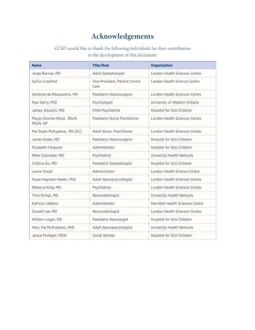
Acknowledgements
CCSO would like to thank the following individuals for their contribution
to the development of this document:
Name Title/Role Organization
Jorge Burneo, MD Adult Epileptologist London Health Sciences Centre
Sylvie Crawford Vice President, Patient Centre Care
London Health Science Centre
Sandrine de Ribaupierre, MD Paediatric Neurosurgeon London Health Sciences Centre
Paul Derry, PhD Psychologist University of Western Ontario
James, Deutsch, MD Child Psychiatrist Hospital for Sick Children
Margo Devries-Rizzo, BScN, MScN, NP
Paediatric Nurse Practitioner London Health Sciences Centre
Pat Doyle-Pettypiece, RN (EC) Adult Nurse Practitioner London Health Sciences Centre
James Drake, MD Paediatric Neurosurgeon Hospital for Sick Children
Elizabeth Ferguson Administrator Hospital for Sick Children
Peter Giacobbe, MD Psychiatrist University Health Network
Cristina Go, MD Paediatric Epileptologist Hospital for Sick Children
Laurie Gould Administrator London Health Science Centre
Susan Hayman-Abello, PhD Adult Neuropsychologist London Health Sciences Centre
Rebecca King, MD Psychiatrist London Health Sciences Centre
Timo Krings, MD Neuroradiologist University Health Network
Kathryn LeBlanc Administrator Hamilton Health Sciences Centre
Donald Lee, MD Neuroradiologist London Health Sciences Centre
William Logan, MD Paediatric Neurologist Hospital for Sick Children
Mary Pat McAndrews, PhD Adult Neuropsychologist University Health Network
Janice Mulligan, MSW Social Worker Hospital for Sick Children
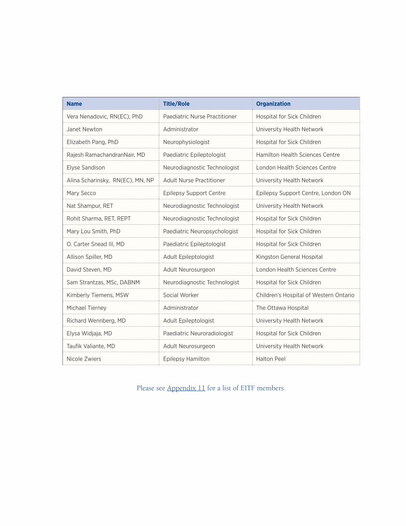
Name Title/Role Organization
Vera Nenadovic, RN(EC), PhD Paediatric Nurse Practitioner Hospital for Sick Children
Janet Newton Administrator University Health Network
Elizabeth Pang, PhD Neurophysiologist Hospital for Sick Children
Rajesh RamachandranNair, MD Paediatric Epileptologist Hamilton Health Sciences Centre
Elyse Sandison Neurodiagnostic Technologist London Health Sciences Centre
Alina Scharinsky, RN(EC), MN, NP Adult Nurse Practitioner University Health Network
Mary Secco Epilepsy Support Centre Epilepsy Support Centre, London ON
Nat Shampur, RET Neurodiagnostic Technologist University Health Network
Rohit Sharma, RET, REPT Neurodiagnostic Technologist Hospital for Sick Children
Mary Lou Smith, PhD Paediatric Neuropsychologist Hospital for Sick Children
O. Carter Snead III, MD Paediatric Epileptologist Hospital for Sick Children
Allison Spiller, MD Adult Epileptologist Kingston General Hospital
David Steven, MD Adult Neurosurgeon London Health Sciences Centre
Sam Strantzas, MSc, DABNM Neurodiagnostic Technologist Hospital for Sick Children
Kimberly Tiemens, MSW Social Worker Children’s Hospital of Western Ontario
Michael Tierney Administrator The Ottawa Hospital
Richard Wennberg, MD Adult Epileptologist University Health Network
Elysa Widjaja, MD Paediatric Neuroradiologist Hospital for Sick Children
Taufik Valiante, MD Adult Neurosurgeon University Health Network
Nicole Zwiers Epilepsy Hamilton Halton Peel
Please see Appendix 11 for a list of EITF members.

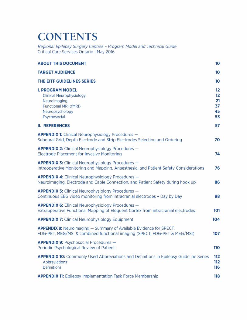
CONTENTS
ABOUT THIS DOCUMENT 10
TARGET AUDIENCE 10
THE EITF GUIDELINES SERIES 10
I. PROGRAM MODEL 12Clinical Neurophysiology 12Neuroimaging 21Functional MRI (fMRI) 37Neuropsychology 45Psychosocial 53
II. REFERENCES 57
APPENDIX 1: Clinical Neurophysiology Procedures — Subdural Grid, Depth Electrode and Strip Electrodes Selection and Ordering 70
APPENDIX 2: Clinical Neurophysiology Procedures — Electrode Placement for Invasive Monitoring 74
APPENDIX 3: Clinical Neurophysiology Procedures — Intraoperative Monitoring and Mapping, Anaesthesia, and Patient Safety Considerations 76
APPENDIX 4: Clinical Neurophysiology Procedures — Neuroimaging, Electrode and Cable Connection, and Patient Safety during hook up 86
APPENDIX 5: Clinical Neurophysiology Procedures — Continuous EEG video monitoring from intracranial electrodes – Day by Day 98
APPENDIX 6: Clinical Neurophysiology Procedures — Extraoperative Functional Mapping of Eloquent Cortex from intracranial electrodes 101
APPENDIX 7: Clinical Neurophysiology Equipment 104
APPENDIX 8: Neuroimaging — Summary of Available Evidence for SPECT, FDG-PET, MEG/MSI & combined functional imaging (SPECT, FDG-PET & MEG/MSI) 107
APPENDIX 9: Psychosocial Procedures — Periodic Psychological Review of Patient 110
APPENDIX 10: Commonly Used Abbreviations and Definitions in Epilepsy Guideline Series 112Abbreviations 112Definitions 116
APPENDIX 11: Epilepsy Implementation Task Force Membership 118
Regional Epilepsy Surgery Centres – Program Model and Technical Guide Critical Care Services Ontario | May 2016
Regional Epilepsy Surgery Centres Program Model and Technical Guide
10 Critical Care Services Ontario • May 2016
About this Document
The EITF has developed this document in an effort to provide guidelines for evidence-based practice for all
health care providers in Ontario who are the primary point of care for patients with epilepsy. This document
presents best practices as a recommended, but not mandatory, program model for hospitals and their
collaborative interdisciplinary teams that provide care for patients at Regional Epilepsy Surgical Centres.
Target Audience
The intended target audience of this document includes, but is not limited to, clinicians and
administrators from District Epilepsy Centres (DECs) and Regional Epilepsy Centres (RESCs).
The EITF Guidelines Series
The EITF is developing a series of guidelines intended to support primary care providers, community
neurologists, and district and regional epilepsy centres. These guidelines aim to increase the awareness of,
and referrals to, appropriate diagnostic assessment and surgical care of patients in Ontario.
For Primary Care Providers:
1. Provincial Guidelines for the Management of Epilepsy in Adults and Children (January 2015)
To support the flow of patients towards appropriate treatment for epilepsy, this document contains
a set of guidelines to help with the diagnosis, treatment, and referral practices from the moment of a
patient’s first seizure.
2. Provincial Guidelines for Epilepsy Surgery Referrals in Ontario (February 2016)
This document provides an approach to referral of medically-refractory epilepsy patients by
defining evidence-based indications to epilepsy surgery in all age groups, with careful consideration
given to age-specific issues ranging from infants to the elderly.
3. Provincial Guidelines for the Management of Medically-Refractory Epilepsy in Adults and Children who are
not Candidates for Epilepsy Surgery (March 2016)
This guideline will provide an approach to the management of the patient with medically intractable
epilepsy in whom surgical treatment is not an option. It will include the use of antiepileptic
medications and non-antiepileptic therapy such as dietary management and neurostimulation.
4. Provincial Guidelines for Transitional Care of Paediatric Epilepsy Programs to Adult (to be released soon)
To ensure uninterrupted quality medical care for adolescent patients with chronic disorders,
this document provides guidelines for paediatric and adult practitioners to assist in the seamless
transition of epilepsy care for adolescents who are departing the paediatric system and entering the
adult health care network.
Regional Epilepsy Surgery Centres Program Model and Technical Guide
Critical Care Services Ontario • May 2016 11
For Providers and Administrators in District and Regional Epilepsy Centres:
5. Provincial Epilepsy Monitoring Unit (EMU) Guidelines for Ontario (January 2014)
This document outlines protocols and provides guidelines for EMUs for diagnostic evaluation for
epilepsy. It can be used as a guide for neurosurgical centres with EMU beds.
6. Provincial Guidelines for Regional Epilepsy Surgical Centres
This document presents guidelines that set out accountabilities for hospitals and their collaborative
interdisciplinary teams that provide care for patients at Regional Epilepsy Surgical Centres.
7. Regional Epilepsy Surgery Centres – Program Model and Technical Guide
This document presents best practices as a recommended, but not mandatory, clinical protocols and
program model for hospitals and their collaborative interdisciplinary teams that provide care for
patients at Regional Epilepsy Surgical Centres.
Regional Epilepsy Surgery Centres Program Model and Technical Guide
12 Critical Care Services Ontario • May 2016
I. Program Model
The following section represents a program model for Regional Epilepsy Surgery Centres in Ontario. It
provides a framework for collaborative interdisciplinary teams and reflects leading practices in epilepsy
surgery and expectations for the highest standards of epilepsy care. This program model can be used as a
guide; RESC program models may differ across Ontario given each Centre’s unique requirements.
Clinical Neurophysiology
Intra-Cranial EEG-Video Monitoring and Intraoperative Functional Mapping
in the Epilepsy Monitoring Unit
Objectives
The neurophysiology section outlines the skill sets required by a Certified/Registered Neurophysiology
Technologist and/or IOM Technologist to adequately perform his/her duties during the planning,
implantation, recording/monitoring and removal of subdural grid, depth and strip electrodes during
epilepsy surgery cases in a Regional Epilepsy Surgery Centre (RESC) in Ontario.
Personnel
Epilepsy Monitoring Technologist (EMU Technologist)
An EMU technologist who does invasive monitoring should be a Registered Electroencephalograph
Technologist (RET) certified by the Canadian Board of Registration of Electroencephalograph
Technologists (CBRET). CBRET is the only national organization that provides a qualifying examination.
Please refer Mizrahi (1999) for further details regarding EMU technologist role and responsibilities.
Additional certifications, such as a Registered Evoked Potentials Technologist (REPT) from the American
Board of Electroencephalographic and Evoked Potential Technologists (ABRET), are an asset (please refer
to www.abret.org).
Regional Epilepsy Surgery Centres Program Model and Technical Guide
Critical Care Services Ontario • May 2016 13
EMU-Invasive Monitoring (EMU-IM) Technologist
EMU-IM technologists should have a minimal of 2 to 3 years of dedicated EMU experience, an additional
6 months of supervised invasive monitoring training, in addition to being an RET. An up-to-date CPR/
BCLS (cardio pulmonary resuscitation/basic cardiac life support) certification is required in paediatric
centres and optional in adult centres.
Electroencephalograph (EEG) Technologists
EEG technologists in Canada are trained either through college-based diploma programs in
electrophysiology or in hospital-based EEG training programs.
Hospital-based programs must include ten (10) hours of structured learning per week for students
in training. They must have at least one (1) CBRET registered technologist, a full-time or major part-
time electroencephalographer who is a MD and a certified member of the Canadian Society of Clinical
Neurophysiologists (CSCN).
College-based diploma programs must include a minimum of 500 hours of EEG instruction (please refer
to www.cbret.org).
Intraoperative Neuromonitoring Technologist (IOM Tech)
IOM technologists are also part of the invasive monitoring team and require additional training and
certification.
IOM technologists should have obtained a Certification in Neurophysiologic Intraoperative (CNIM)
which is offered by the American Board of Registration of Electroencephalographic and Evoked Potential
Technologists (ABRET). Currently, ABRET is the only national organization that provides a qualifying IOM
technologist certification.
Certified IOM Technologists are eligible to write the exam through one of two pathways:
Pathway 1: Employed in intraoperative neuromonitoring with a current R.EEG T (Registered
Electroencephalography Technologist) or REPT (Registered Evoked Potential Technologist) credential, and
have documentation of 150 IOM cases*, and have a current CPR/BCLS certification.
Pathway 2: Employed in intraoperative neuromonitoring with at least a Bachelor’s degree, and have
documentation of 150 IOM cases*, and have a current CPR/BCLS certification.
* Please refer to www.abret.org for requirements of case documentation. For practice guidelines and competencies
for the technologist role, please refer to the National Competency Skill Standards for Performing Intraoperative
Neurophysiologic Monitoring (available on the American Society of Electroneurodiagnostic Technologists (ASET)
website: www.aset.org)
Regional Epilepsy Surgery Centres Program Model and Technical Guide
14 Critical Care Services Ontario • May 2016
Intraoperative Neuromonitoring Neurophysiologist
Intraoperative neuromonitoring neurophysiologists are board certified through the American Board
of Neurophysiologic Monitoring (ABNM) and upon completion of the credentialing process, become
diplomats of the board (D.ABNM). ABNM is currently the only national organization that provides a
qualifying intraoperative neurophysiologist credentialing process.
Requirements for application include:
• Minimum of an earned doctoral degree in a physical science, life science or clinical allied health profession from an accredited institution
• Must submit documentation of a minimum of 300 cases monitored with the primary responsibility for professional interpretation and technical supervision, 100 cases in which the applicant physically performed the majority of the technical aspects of monitoring
• Have at least thirty-six (36) months’ experience in the field of neurophysiologic monitoring as documented in the case log
Please refer to www.abnm.info for the full list of requirements.
For practice guidelines and competencies for the technologist role, please refer to Skinner et al., 2013.
Procedures
Subdural Grid, Depth Electrode and Strip Electrodes Selection and Ordering
Once the patient has been selected to undergo invasive monitoring, the epilepsy monitoring team (MD
and EMU-IM technologist) determines the type, size and number combinations of temporary (<30
days) invasive subdural grid, strip and depth electrodes to be implanted. This is based on preoperative
epilepsy workup data collected from interictal and ictal scalp VEEG (video electroencephalography)
studies, clinical seizure features, magnetic resonance imaging (MRI), functional MRI, and
magnetoencephalography (MEG). Please see Appendix 1 for Figure 2.0 grid placement as well as the
methodological details of electrode selection and ordering.
Electrode Placement for Invasive Monitoring
Please see Appendix 2 for details.
Intraoperative Monitoring and Mapping, Anaesthesia, and Safety Considerations
Please see Appendix 3 for details.
Regional Epilepsy Surgery Centres Program Model and Technical Guide
Critical Care Services Ontario • May 2016 15
Neuroimaging, Electrode and Cable Connection; Patient Safety during hook-up
Please see Appendix 4 for details.
Continuous EEG video monitoring from intracranial electrodes
Please see Appendix 5 for details.
Functional Mapping of Eloquent Cortex from Intracranial electrodes
Please see Appendix 6 for details.
Technical and Computer Back-Up during the Time of EEG Video Monitoring
from Intracranial Electrodes
A dedicated EMU-IM technologist should be assigned to take care of the patient during invasive
monitoring. It is recommended that a technologist be on-call to provide both technical and computer
support to resolve issues during the monitoring session. This support is necessary to prevent data loss
and decreased hardware and software downtime during an invasive monitoring session, due to the time-
sensitive nature of the recording.
VPN (virtual private network) access from home to the EMU-IM system is desirable (using local hospital
clients and guidelines). This enables remote review for troubleshooting should problems arise afterhours.
Data Management- Archival and Pruning EEG and Video Segments for Storage
All EEG and video data is reviewed, marked and annotated by the technologist for
electroencephalographer or clinical neurophysiologist to review.
Following data interpretation, either selected epochs of EEG or the entire EEG file and video segments
are archived to a centralized server as per naming conventions used in the individual institutions. Data
management on recording stations need to be managed as needed (e.g., local copies on acquisition
station hard drives may need to be deleted based on available space) to continue uninterrupted
recording and prevent shortage of storage space on the recording systems local hard drive. Each
EMU-IM acquisition unit must have enough storage space to store data on the local hard drives for a
minimum of seven (7) days.
A minimum of 500 GB of free storage should be available on the local hard drive.
Regional Epilepsy Surgery Centres Program Model and Technical Guide
16 Critical Care Services Ontario • May 2016
Post Invasive Monitoring Procedures and Planning for Epilepsy Surgery
The monitoring phase is one of several stages that a patient undergoes during subdural grid surgery.
Once all required EEG and functional mapping data have been collected from the patient, the invasive
monitoring session is stopped. The decision to stop is determined by the electroencephalographer and the
epilepsy monitoring physician (e.g., once the desired number of seizures has been captured).
The epilepsy team informs EMU-IM technologist to disconnect invasive monitoring electrode cables.
All additional monitoring electrodes – EMG (electromyogram), EKG (electrocardiogram), patient ground
electrode) – are removed.
The patient’s head is re-bandaged once a surgical plan has been established. The patient is then prepared
for the next stage in the surgical process, either removal of all implanted electrodes or cortical excision and
possible further ECoG (electrocorticography) post excision in the operating room.
A cortical excision map is created by the epilepsy monitoring team and electroencephalographer. This
map depicts all mapped eloquent cortices that were mapped on Day 1 in the operating room, optional
Day 2 topographic SEP (somatosensory evoked potentials), Day 3-4 functional mapping and the seizure
focus or foci.
This map can be created using a variety of imaging and photo editing software available either from the EEG
equipment vendor or a third-party visual/digital software manufacturer. If such maps are to be created, it
is recommended that the following software be utilized: Microsoft PowerPoint, Natus/Stellate GridView or
Persyst MagicMarker™ software.
This map is used by the team to discuss the invasive monitoring findings and the surgical plan either with
the patient in adult cases or with the parents in paediatric cases.
The EMU-IM technologist takes the surgical map to the operating room on the day of the grid removal
and possible epilepsy surgery.
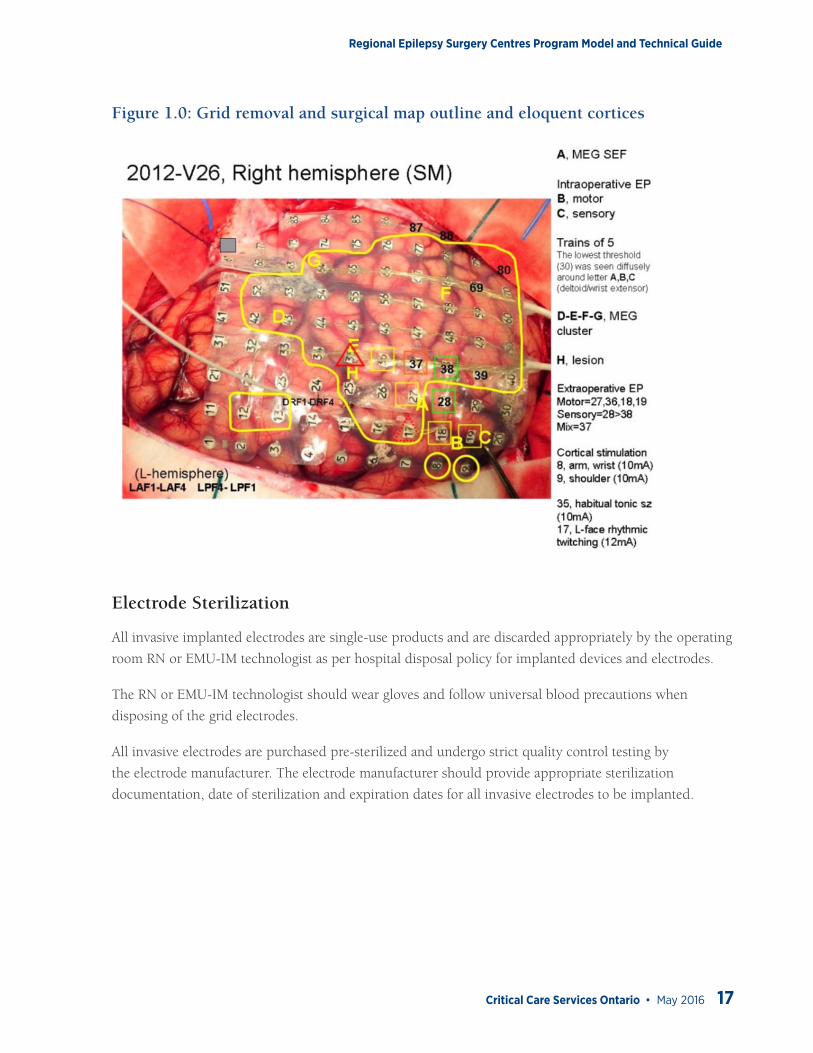
The surgical map also acts as an excision guide for the neurosurgeon. Prior to the removal of the subdural
grid and cortical excision, the epileptologist guides the neurosurgeon in marking the surgical excision
boundaries using the map on the following page:
Regional Epilepsy Surgery Centres Program Model and Technical Guide
Critical Care Services Ontario • May 2016 17
Figure 1.0: Grid removal and surgical map outline and eloquent cortices
Electrode Sterilization
All invasive implanted electrodes are single-use products and are discarded appropriately by the operating
room RN or EMU-IM technologist as per hospital disposal policy for implanted devices and electrodes.
The RN or EMU-IM technologist should wear gloves and follow universal blood precautions when
disposing of the grid electrodes.
All invasive electrodes are purchased pre-sterilized and undergo strict quality control testing by
the electrode manufacturer. The electrode manufacturer should provide appropriate sterilization
documentation, date of sterilization and expiration dates for all invasive electrodes to be implanted.
Regional Epilepsy Surgery Centres Program Model and Technical Guide
18 Critical Care Services Ontario • May 2016
Equipment
Please see Appendix 7 for details, as well as Provincial Epilepsy Monitoring Unit (EMU) Guidelines for Ontario.
Epilepsy Surgery Team
The multidisciplinary epilepsy surgery team is headed by a neurologist and/or neurosurgeon. All decisions
regarding epilepsy surgery candidacy are made by consensus of the epilepsy surgery team in meetings
chaired by the team leader.
Neurologist
The neurologist is an epileptologist with a minimum of two (2) years of formal training in an epilepsy
fellowship program with specific training in epilepsy surgery. The epileptologist should have board
certification in clinical EEG and neurophysiology by the Canadian Society of Clinical Neurophysiologists
(CSCN Diplomate (EEG)) or the US equivalent. There should be at least two (2) such epileptologists at
a Regional Epilepsy Surgery Centre. The epileptologist oversees and is responsible for all facets of
the invasive monitoring procedure, as described above under Clinical Neurophysiology – Procedures and
in Appendices 1 to 5, and is capable of interpretation of EEG, EEG-video, electrocorticography, and iEEG
(intracranial electroencephalography) data. The epileptologist is capable of doing intraoperative functional
mapping of eloquent cortex in conjunction with the neurosurgeon (Appendix 3) and also extraoperative
functional mapping from intracranial electrodes (Appendix 6). Similarly, the epileptologist can interpret
iEEG data and, in conjunction with other members of the epilepsy team, create brain maps showing the
anatomical localization of the epileptogenic zone and eloquent cortex pursuant to surgical decision making
(please see: Clinical Neurophysiology – Procedures: Post Invasive Monitoring Procedures and Planning for
Epilepsy Surgery above).
Neurosurgeon
The Regional Epilepsy Surgery Centre should have at least one neurosurgeon with two (2) or more years
of post-fellowship experience in the following areas:
• Resective epilepsy surgery
• Placement of intracranial electrodes
• Insertion of vagus nerve stimulator
The epilepsy surgeon should have the ability to perform all of the following surgical procedures:
• Emergency or elective neurosurgery
• Management of surgical complications
• Lesional epilepsy surgery
• Corpus callosotomy
Regional Epilepsy Surgery Centres Program Model and Technical Guide
Critical Care Services Ontario • May 2016 19
• Vagal Nerve Stimulation
• Deep Brain Stimulation
• Functional cortical mapping by stimulation of subdural electrodes either extraoperatively or intraoperatively (Appendix 3 and Appendix 6)
• Evoke potential recording capable of being used safely with intracranial electrodes (Appendix 3)
• Intraoperative electrocorticography (Appendix 3)
• Resection of epileptogenic tissue in the absence of structural lesions based on intracranial monitoring data
Nursing and Nurse Practitioner
The role of the Nurse Practitioner (NP) in the epilepsy surgery program is to:
• Provide continuity of care/ care coordination for patients during their perioperative periods in both inpatient and outpatient settings
• Enhance communication within the epilepsy surgery multidisciplinary team
The epilepsy surgery program Nurse Practitioner (NP) is a Master’s prepared nurse practitioner registered
in the extended class RN (EC) with the College of Nurses of Ontario (CNO), the body which regulates the
practice of nurses in Ontario with experience in neuroscience nursing.
The NP role within the epilepsy program spans the perioperative period and is enacted in the outpatient
and inpatient settings. Responsibilities are outlined according to the CNO standards of care for nurse
practitioners and program needs.
Regional Epilepsy Surgery Centres Program Model and Technical Guide
20 Critical Care Services Ontario • May 2016
Responsibilities
• Provide advanced comprehensive holistic health assessments
• Formulate diagnoses
• Identify potential surgical candidates in epilepsy clinic and EMU
• Ensure patient goals of care are addressed/documented
• Provide continuity of care throughout hospital to home continuum
• Coordinate discharge planning
• Ensure patient safety measures are addressed
• Consult appropriate services for comorbid issues
• Act as resource to nursing/medical staff for urgent/acute care issues
• Facilitate communication between patient/family and health care team to achieve collaborative outcomes
• Collaborate with team/allied health members including but not limited to:
o Neurosurgical service
o Neurosurgery NP
o Neuropsychology
o EEG technologists
o Biomedical staff
o PT/OT (physiotherapist/occupational therapist) and dieticians
• Provide education to patients and families regarding epilepsy management, treatment and lifestyle and on surgical management in particular
Note: NPs/RN(EC)s may be authorized to perform controlled acts and activities, such a diagnosing medical
conditions, prescribing medications, ordering investigations, and admitting and discharging patients, depending
on the role defined by the hospital and, in the case of Professional Staff NPs, in accordance with Medical Advisory
Committee or Professional Staff by-laws.
Regional Epilepsy Surgery Centres Program Model and Technical Guide
Critical Care Services Ontario • May 2016 21
Neuroimaging
The imaging modalities covered in the neuroimaging section include:
• Structural imaging, primarily MRI, for the identification of underlying lesion responsible for the epilepsy
• Functional imaging, including SPECT, FDG-PET (Fluoro-2-deoxy-D-glucose-positron emission tomography) and MEG/MSI (magnetoencephalography/ magnetic source imaging), to confirm or clarify the epileptogenic zone
• Functional MRI (fMRI) or MEG/MSI to localize the eloquent cortex
• Diffusion tensor imaging (DTI) to localize the eloquent white matter tracts
The imaging section covers the objectives of neuroimaging in epilepsy and, for each modality, the (i)
personnel, (ii) procedures, and (iii) equipment.
Objectives
• To identify underlying pathologies, such as malformations, tumours, hippocampal sclerosis, granulomas, vascular malformations, traumatic lesions or strokes, that merit specific treatment.
• To aid the formulation of syndromic and etiological diagnoses in order to provide an accurate prognosis for patients, their relatives, and physicians.
In patients with suspected localization-related epilepsy undergoing work-up for epilepsy surgery, the aims
of neuroimaging are:
• To identify underlying etiology responsible for the epilepsy
• To confirm or clarify the location of the epileptogenic zone
• To identify eloquent cortex and white matter tracts so as to minimize functional deficits following surgery
This document provides guidelines for neuroimaging evaluation of patients with suspected localization-
related epilepsy who are worked-up for epilepsy surgery. The following modalities have been included:
• Structural imaging, primarily MRI, for the identification of underlying lesion responsible for the epilepsy
• Functional imaging, including SPECT, FDG-PET and MEG/MSI, to confirm or clarify the epileptogenic zone
• Functional MRI (fMRI) or MEG/MSI to localize the eloquent cortex
• Diffusion tensor imaging (DTI) to localize the eloquent white matter tracts
For each imaging modality, the guideline considers the following:
(i) personnel, (ii) procedures, and (iii) equipment
Regional Epilepsy Surgery Centres Program Model and Technical Guide
22 Critical Care Services Ontario • May 2016
Structural Imaging: MRI
Personnel
• Dedicated neuroradiology training (or paediatric neuroradiology for those reporting paediatric epilepsy cases) for at least twelve (12) months in Canada, US or abroad
• Recognized as a neuroradiologist specialist by the College of Physicians and Surgeons of Ontario (CPSO).
Responsibilities:
• Reporting
o The MRI report should include clinical details, MRI sequences acquired, MRI findings, and interpretation/ conclusion.
o Scans must be interpreted in the context of clinical semiology and EEG findings
o The video EEG data may not be available at the time of reporting. The neuroradiologist with a special interest in epilepsy should review the MRI of those cases that are considered to be normal or have questionable subtle changes, when all the clinical data including video EEG and functional imaging are available.
Procedures
This MRI guideline was developed based on published guidelines from the International League Against
Epilepsy (Commission on Neuroimaging, 1997; Gaillard et al., 2009), National Institute of Health
common data elements (Theodore et al., 1997) and expert opinion. General sequences and some
parameters are listed. Detailed imaging parameters are not listed as they depend on the make of scanner
and magnetic field strength.
The recommendations are to optimize the sequences to provide high resolution (high matrix, thin slices)
imaging with good signal-to-noise and excellent gray-white matter distinction. If a lesion is not seen
on the initial MRI, repeat MRI with higher resolution imaging targeted to the area of concern may be
necessary to identify subtle focal cortical dysplasia.

Regional Epilepsy Surgery Centres Program Model and Technical Guide
Critical Care Services Ontario • May 2016 23
Adults (18 years and older):• Axial FLAIR T2 weighted (slice thickness of 2-4 mm, gap 0-0.5 mm, matrix 256x512), whole brain
• Coronal oblique FLAIR T2 weighted (slice thickness of 3-4 mm, gap 0-0.5 mm), whole brain, acquired orthogonal to the long axis of the hippocampus as visualized on the sagittal scannogram
• High resolution coronal oblique sequence orthogonal to the long axis of the hippocampus as visualized on the sagittal scannogram; the high resolution coronal sequence is targeted for the temporal lobes:
o Inversion recovery sequence (slice thickness of 3 mm, gap 0, matrix 512x512), OR
o Turbo / fast spin echo T2 weighted sequence (slice thickness of 3 mm, gap 0, matrix 512x512)
o Volumetric T1 weighted sequence (isotropic voxels, ≤1 mm) with good gray-white matter contrast and with multiplanar reformats
• The following sequences may be helpful:
o Axial turbo / fast spin echo T2-weighted images (slice thickness of 2-4 mm, skip 0-0.5 mm), whole brain
o Turbo / fast spin echo proton density sequences in axial (slice thickness of 2-4 mm, gap 0-0.5 mm) and coronal (slice thickness of 2-4 mm, gap 0-0.5 mm) planes
o Volumetric T2 FLAIR (slice thickness ≤1.5 mm)
o Gradient echo/ susceptibility weighted imaging (SWI) to look for calcification or old hemorrhage
o Magnetization transfer imaging
o Gadolinium contrast is reserved for circumstances where tumor or vascular malformations arise or are suspected based on review of noncontrast studies.
Children (1-18 years):• Axial FLAIR T2 weighted (slice thickness of 2-3 mm, gap 0-0.5), whole brain
• High resolution coronal oblique turbo / fast spin echo T2 weighted sequence (slice thickness of 3-4 mm, gap 0-0.5), whole brain
• Coronal oblique FLAIR T2 weighted (slice thickness of 3-4 mm, gap 0-0.5 mm)
• All coronal sequences should be acquired in an oblique plane orthogonal to the long axis of the hippocampus
• Axial turbo / fast spin echo T2-weighted images (slice thickness of 2-3 mm, gap 0-0.5 mm), whole brain
• Volumetric T1 weighted sequence (isotropic voxels, ≤1 mm) with good gray-white matter contrast and with multiplanar reformats
• Turbo / fast spin echo proton density sequences (slice thickness of 2-3 mm thick) and 3D T2 FLAIR (slice thickness ≤1.5 mm) can be helpful in detecting subtle focal cortical dysplasia
• High resolution coronal oblique inversion recovery sequence (slice thickness of 3 mm, gap 0) can be helpful in detecting hippocampal sclerosis
Regional Epilepsy Surgery Centres Program Model and Technical Guide
24 Critical Care Services Ontario • May 2016
• Gradient echo/ susceptibility weighted imaging (SWI) may be helpful to look for calcification or old hemorrhage
• Gadolinium contrast is reserved for circumstances where tumor, vascular malformations, Sturge Weber syndrome, inflammation and infectious concerns arise or are suspected based on review of noncontrast studies. Routine administration of gadolinium provides little advantage in children with epilepsy
• The field of view for the sequences should be adjusted based on head size
Infants (< 1 year):• Children younger than one year require special sequences as immature myelination affects the ability
to identify common causes of epilepsy. MR imaging (especially high resolution T2 images) performed early in the first year of life in infants with epilepsy is important to identify malformations of cortical development, including focal cortical dysplasia.
• Axial turbo / fast spin echo T2 weighted images (slice thickness 2-3 mm, gap 0), whole brain
• Coronal turbo / fast spin echo T2 weighted high resolution coronal oblique sequence (maximum slice thickness of 2-3 mm, gap 0 mm), whole brain
• Volumetric T1 weighted sequence (isotropic voxels, ≤1 mm)
• FLAIR T2 weighted and proton density images are less helpful in infants due to lack of myelination
• The field of view for the sequences should be adjusted based on head size
• If MR imaging done before the age of 2 years is normal, and seizures persist, then MRI should be repeated at age 2 to 3 years.
Equipment
• MRI is the structural imaging modality of choice
• 3T MRI is the ideal equipment for structural imaging (Knake et al., 2005; Zijlmans et al., 2009)
• Minimum of 8 channel head coil is recommended
• Regular quality control of the MRI scanner is necessary
• Occasionally, CT may be useful as a complementary imaging technique in the detection of cortical calcifications
Regional Epilepsy Surgery Centres Program Model and Technical Guide
Critical Care Services Ontario • May 2016 25
Functional Imaging (SPECT, FDG-PET and MEG/MSI)
Indications for functional imaging
• Clarification of the extent of the epileptogenic zone
• Patients with normal or equivocal MRI
• Those with discordance between MRI and other data
• Those who may require invasive intracranial electrodes
SPECT, FDG-PET and MEG/MSI may be useful for planning the sites of intracranial electrode placement
for recording ictal onset in temporal and extratemporal epilepsies.
Summary of available evidence
• Functional neuroimaging can provide additional data in seizure patients (Appendix 8, Table 1-3)
• The sensitivity of SPECT for localizing epileptogenic focus increases from interictal, (44-84%) to ictal examinations (59-97%) (Appendix 8, Table 1). The sensitivity is lower in cases of extratemporal partial epilepsy compared to temporal lobe epilepsy, in which only the ictal examination is reliable. Subtraction techniques of the interictal from the ictal study may be helpful; however, the ictal study remains the preferred examination.
• The sensitivity of FDG-PET ranges from 33% to 95% (Appendix 8, Table 1). The sensitivity is lower in extratemporal lobe epilepsy compared to temporal lobe epilepsy. There is no consensus as to whether ictal SPECT is more sensitive than FDG-PET for the localization of epileptogenic foci.
• The sensitivity of MEG/MSI is 20% – 100% (84%), and specificity is 6% – 100% (52%) (Appendix 8, Table 2). MEG/MSI can provide information on the extent of electrode coverage for invasive intracranial EEG monitoring. The sensitivity of MEG/MSI is lower in temporal lobe epilepsy compared to extratemporal lobe epilepsy.
• There are some studies that have compared SPECT with FDG-PET (Appendix 8, Table 1), and few studies that have compared either SPECT with MEG/MSI, or FDG-PET with MEG/MSI (Appendix 8, Table 3). The choice of functional tests depends on local availability and local expertise.
• There are some suggestions that two different combinations of functional tests may be helpful in patients with normal MRI, and those with subtle or non-specific changes on MRI. However, there is insufficient evidence to recommend which two combinations of tests should be done, and which particular functional test should proceed from the other test.

Regional Epilepsy Surgery Centres Program Model and Technical Guide
26 Critical Care Services Ontario • May 2016
Single Photon Emission Computed Tomography (SPECT)
Personnel
A nuclear medicine specialist must have training in nuclear medicine for at least two months in Canada,
US or abroad and be recognized as a nuclear medicine specialist by the College of Physicians and
Surgeons of Ontario (CPSO).
Responsibilities:
• Supervision of the overall SPECT study including radionuclide administration, data acquisition, and processing
• Reporting SPECT study, which should include:
o Indications for the study
o Assessment of the technical quality of the scan (good, adequate, or poor, including presence of patient movement, deviations from usual laboratory protocol or other factors, if relevant)
o Description of abnormalities
o Interpretation
o Ictal scan should be interpreted with knowledge of the relationship of the injection to the onset of electroclinical seizure.
o The interictal SPECT should be compared to the ictal study. On its own, the interictal SPECT is less reliable for localization.
o Interpretation should also be done in the light of other data, including clinical, EEG, and MRI findings.
• In children, in particular infants younger than 2 years of age, interpretation of interictal SPECT needs to be informed by knowledge of normal age-appropriate findings
• Co-registration of ictal scan with MRI optimizes localization data and co-registration of ictal and interictal examinations is valuable. Subtraction ictal SPECT co-registered to MRI (SISCOM) may be used to improve detection of subtle differences in perfusion. SISCOM may be done by a trained technologist or assistant under the supervision of the nuclear medicine specialist.
Regional Epilepsy Surgery Centres Program Model and Technical Guide
Critical Care Services Ontario • May 2016 27
Procedures
Patient preparation
The patient is to avoid caffeine, cola and energy drinks, alcohol or any drugs known to affect cerebral
blood flow. Video-EEG telemetry needs to be established prior to attempting an ictal SPECT study. The
best results from ictal SPECT studies occur with injections performed as near to seizure onset as possible,
preferably within 60 seconds of seizure onset. Delayed postictal injections often give non-diagnostic and
sometimes confusing results. It is necessary to ensure continuous close observation of the patient and the
EEG by skilled personnel who are familiar with the patient’s seizures, so that the tracer may be injected
with minimum delay. Ensure an indwelling intravenous cannula is placed in advance in the upper limb
that is involved less in the seizure. It is important to document the time of injection in relation to the
seizure onset. Following the seizure, the patient is allowed to recover and can be transported to the
nuclear medicine department for imaging within an approximately 2-hour period. Longer delays will
result in degraded images.
The acquired data are processed as follows:
• Filter all studies in 3 dimensions, which can be achieved either by 2-dimensionally pre-filtering the projection data or by applying a 3D post-processing filter to the reconstructed data.
• Low-pass filters should generally be used.
• Reconstruct data at the highest pixel resolution, that is, 1 pixel thick.
• Reformat data into 3 orthogonal planes.
• Ideally, interictal SPECT should be performed using the same camera, after a seizure-free interval of 24 hours or more, for comparison with the ictal images.
• If sedation is required, the sedative medication should be administered at least 5 minutes after tracer injection, preferably starting only a few minutes before data acquisition.
Regional Epilepsy Surgery Centres Program Model and Technical Guide
28 Critical Care Services Ontario • May 2016
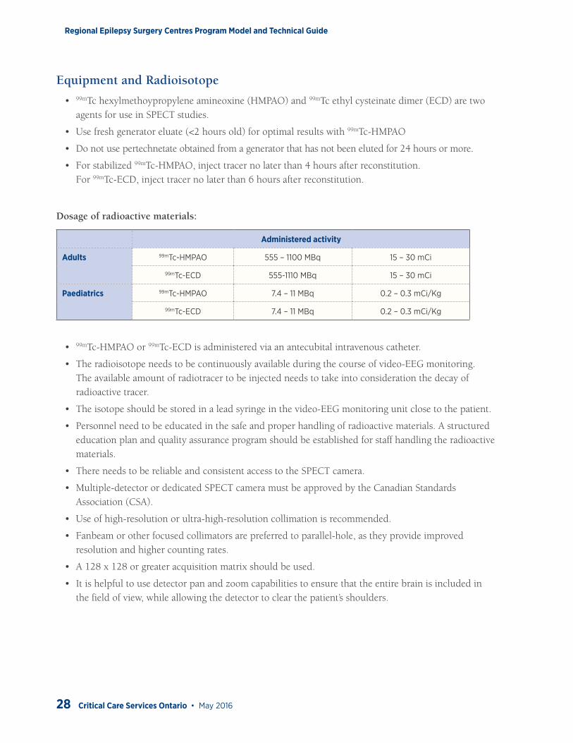
Equipment and Radioisotope
• 99mTc hexylmethoypropylene amineoxine (HMPAO) and 99mTc ethyl cysteinate dimer (ECD) are two agents for use in SPECT studies.
• Use fresh generator eluate (<2 hours old) for optimal results with 99mTc-HMPAO
• Do not use pertechnetate obtained from a generator that has not been eluted for 24 hours or more.
• For stabilized 99mTc-HMPAO, inject tracer no later than 4 hours after reconstitution. For 99mTc-ECD, inject tracer no later than 6 hours after reconstitution.
Dosage of radioactive materials:
Administered activity
Adults 99mTc-HMPAO 555 – 1100 MBq 15 – 30 mCi
99mTc-ECD 555-1110 MBq 15 – 30 mCi
Paediatrics 99mTc-HMPAO 7.4 – 11 MBq 0.2 – 0.3 mCi/Kg
99mTc-ECD 7.4 – 11 MBq 0.2 – 0.3 mCi/Kg
• 99mTc-HMPAO or 99mTc-ECD is administered via an antecubital intravenous catheter.
• The radioisotope needs to be continuously available during the course of video-EEG monitoring. The available amount of radiotracer to be injected needs to take into consideration the decay of radioactive tracer.
• The isotope should be stored in a lead syringe in the video-EEG monitoring unit close to the patient.
• Personnel need to be educated in the safe and proper handling of radioactive materials. A structured education plan and quality assurance program should be established for staff handling the radioactive materials.
• There needs to be reliable and consistent access to the SPECT camera.
• Multiple-detector or dedicated SPECT camera must be approved by the Canadian Standards Association (CSA).
• Use of high-resolution or ultra-high-resolution collimation is recommended.
• Fanbeam or other focused collimators are preferred to parallel-hole, as they provide improved resolution and higher counting rates.
• A 128 x 128 or greater acquisition matrix should be used.
• It is helpful to use detector pan and zoom capabilities to ensure that the entire brain is included in the field of view, while allowing the detector to clear the patient’s shoulders.
Regional Epilepsy Surgery Centres Program Model and Technical Guide
Critical Care Services Ontario • May 2016 29
18F-Fluorodeoxyglucose PET (FDG-PET)
Personnel
A nuclear medicine specialist must have training in nuclear medicine for at least 2 months in Canada, US
or abroad and be recognized as a nuclear medicine specialist by the College of Physicians and Surgeons of
Ontario (CPSO).
Responsibilities
• Supervising the overall FDG-PET study including 18F-FDG administration, data acquisition, and processing
• Reporting the FDG-PET study, which should include (Waxman et al., 2009):
o Clinical information,
o Study technique including dosage of 18F-FDG used,
o Assessment of the technical quality of the study (good, adequate, poor)
o Potential artifact such as patient movement
• Last seizure occurrence
• Interpretation of FDG-PET scan is done in light of all available clinical and MRI information. The initial visual assessment may be done without clinical information, followed by a second visual assessment with clinical and MRI information.
• Co-registration of the PET data to the patient’s MRI improves localization of hypometabolism. This may be done by a trained technologist or assistant under the supervision of the nuclear medicine specialist
• Quantitative analysis of the PET data (e.g., with statistical parametric mapping [SPM]) and co-registered to the patient’s MRI may improve detection of hypometabolism.
• The effects of atrophy and partial volume should be taken into account when interpreting the FDG-PET study.
Regional Epilepsy Surgery Centres Program Model and Technical Guide
30 Critical Care Services Ontario • May 2016
Procedures
Patient preparation (Waxman et al., 2009)
The patient should be fasting for 4 to 6 hours before administration of 18F-FDG to maintain both
blood insulin and glucose levels at a low value. The patient can drink water and take his/her current
medications, but must abstain from juice, carbonated drinks, candies, coffee and any other liquid that
could contain proteins, lipids or sugars. Intravenous hydration must not contain glucose. Blood glucose
should be measured and recorded prior to the injection of 18F-FDG, and must be < 150-200 mg/dL. This
is to ensure that the glucose level is not elevated, which could affect the bio-distribution of 18F-FDG. If the
blood glucose is >150-200 mg/dL, the examination should be re-scheduled for a future time and date when
the patient has better glycemic control. Intravenous access is placed at least 10 min prior to injection.
Ideally, EEG should be recorded to identify any epileptic activity. Monitoring should start 2 hours before
injection and should be maintained at least 20 minutes post-injection. The patient should be closely
observed throughout the study.
FDG-PET imaging should be avoided during periods of frequent seizures (including shortly after
convulsive or nonconvulsive status epilepticus). Seizure frequency and the interval since the last seizure
should be noted.
PET scans should be performed by qualified nuclear medicine technologists under the supervision of
qualified nuclear medicine specialists.
The patient should be in a dark quiet room and resting after 18F-FDG injection and during 18F-FDG
uptake. This period of patient inactivity minimizes unwanted muscular uptake of 18F-FDG, while the 18F-FDG is cleared from the blood and taken up into actively metabolizing tissues.
Imaging (also known as emission scan) should be performed approximately 30 to 60 minutes following 18F-FDG injection.
The emission scan lasts between 10 and 60 minutes, depending on the injected activity, the type of
scanner and acquisition protocol used. A minimum of 10 to 15 minutes emission scan in 3D mode is
recommended.
Attenuation correction is mandatory for 18F-FDG-PET scan and can be done with the following methods:
• In PET-CT systems, a low dose (10 – 30 mAs) non-contrast CT scan should be obtained prior to PET data acquisition to generate an attenuation-correction map.
• In dedicated PET scanner, a transmission scan is acquired prior to the emission scan. Transmission imaging consists of a set of images at a position corresponding to the emission image, which is acquired with an external source of radiation (68Ge or 68Ga or 137Cs source) and with the PET camera itself.
Regional Epilepsy Surgery Centres Program Model and Technical Guide
Critical Care Services Ontario • May 2016 31
Image Processing• Scanner-specific approved reconstruction algorithms should be used. Image reconstruction with 3D
Line of Response (LOR) algorithm or iterative 2D/3D Ordered Subset Expectation-Maximization (OSEM) algorithm are acceptable for all acquisition modes.
• Images are reconstructed in the form of transaxial 128 x 128 or 256 x 256 matrixes.
• Typical pixel size is 2 – 4 mm.
• Depending on the resolution of the PET system, a final image resolution may vary between 2.5 and 10 mm full width half maximum (FWHM).
• Data are then reformatted into 3 orthogonal planes.
• If sedation is used, it is best to give the sedative medication at least 20 minutes after tracer injection, preferably starting only a few minutes before data acquisition.
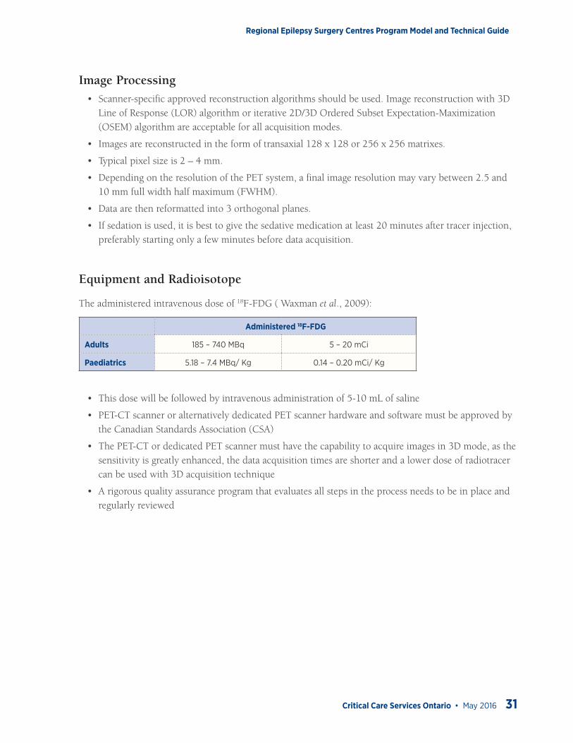
Equipment and Radioisotope
The administered intravenous dose of 18F-FDG ( Waxman et al., 2009):
Administered 18F-FDG
Adults 185 – 740 MBq 5 – 20 mCi
Paediatrics 5.18 – 7.4 MBq/ Kg 0.14 – 0.20 mCi/ Kg
• This dose will be followed by intravenous administration of 5-10 mL of saline
• PET-CT scanner or alternatively dedicated PET scanner hardware and software must be approved by the Canadian Standards Association (CSA)
• The PET-CT or dedicated PET scanner must have the capability to acquire images in 3D mode, as the sensitivity is greatly enhanced, the data acquisition times are shorter and a lower dose of radiotracer can be used with 3D acquisition technique
• A rigorous quality assurance program that evaluates all steps in the process needs to be in place and regularly reviewed

Regional Epilepsy Surgery Centres Program Model and Technical Guide
32 Critical Care Services Ontario • May 2016
Magnetoencephalography/ Magnetic Source Imaging (MEG/MSI)
Personnel
Health care professional reporting the MEG/MSI-EEG studies must have formal training in MEG/MSI in
a specialized centre in Canada, US or abroad, including supervised learning of and practice in clinical
MEG recording, reviewing, and source analysis of clinical MEG. They must also be a member of health
professional organization in neurology, radiology, neurosurgery or psychology. Additional training and
certification for EEG reporting is recommended; please see Provincial Epilepsy Monitoring Unit (EMU)
Guidelines for Ontario for qualifications and training in EEG.
The MRI component of MSI should be reported by a trained Neuroradiologist.
Analysis of spontaneous activity and magnetic evoked fields
Source analysis can be accomplished by a number of methods, including dipole and distributed
source. Equivalent current dipole modeling is the one that is most validated in clinical application,
at least thus far reported in the literature. It is recommended that MEG be analyzed and interpreted
in conjunction with EEG, as the two modalities are complementary and allow optimal resolution of
dipole orientation and temporal evolution of source generators. Non-physician MEG scientists with
a doctoral degree in biological sciences and neurophysiological training, and technologist may assist
with the recording, processing and analysis of MEG. However, only physicians with the appropriate
training and qualifications in MEG/MSI should have the primary responsibility for clinical
interpretation of MEG/MSI.
Regional Epilepsy Surgery Centres Program Model and Technical Guide
Critical Care Services Ontario • May 2016 33
MEG/MSI-EEG Technologist• Trained in EEG and evoked potentials (EP) with a minimum of 2 to 3 years of experience in epilepsy
monitoring, and additional 3 to 6 months’ experience in a clinical MEG centre
• Registered Electroencephalograph Technologist (RET) certified by the Canadian Board of Registration of Electroencephalograph Technologists (www.cbret.org).
• Additional registration in EPs from the American Board of Registered Electroencephalographic and Evoked Potential Technologists (REPT) is an asset (www.abret.org).
Responsibilities:
• EEG electrode placement
• MEG/MSI–EEG recording
• Magnetic evoked fields recordings including somatosensory, motor, auditory, and visual evoked fields
MEG Technologist/ Assistant• Trained in MEG recording, data transfer, fusion of analyzed MEG data to volumetric MRI data
• Understands MEG/MSI study paradigms
Responsibilities:
• Transferring MEG and volumetric T1 MRI data to workstation
• Fusion of the analyzed MEG data to the volumetric T1 MRI data (MSI), and transfer of MSI data to the PACS (picture archiving and communication system)
• Archiving MEG/MSI data and ordering of supplies, including liquid helium, electrodes, fiducials, etc.
• Conducts quality assurance on MEG system
• Sets up service calls and regular maintenance of MEG system
• Some of the above roles may be carried out by personnel from the biomedical engineering or information service
Procedures
MEG lab should follow basic standard clinical epilepsy recording protocols plus additional functional
modalities that can be tailored to each individual patient’s clinical needs. The minimum clinical protocol
should include the following: simultaneous MEG/MSI-EEG and SEF (Sharma et al., 2007).
Regional Epilepsy Surgery Centres Program Model and Technical Guide
34 Critical Care Services Ontario • May 2016
MEG/MSI-EEG• MEG is typically acquired in the interictal state.
• Recording of spikes can be accomplished in awake or sleep states. Achieving sleep aids capture of spikes and increases signal to noise. There is no preference for spikes that occur during awake or sleep states. All patients are sleep deprived to enhance spikes during MEG recording.
• Spontaneous free run EEG or a timed minimum 2 minute epoch of spontaneous MEG data is recorded for a minimum of 10 trials.
• If a spontaneous seizure does occur, and if patient movement does not create error in head positioning / registration, then early ictal discharges (as for interictal spikes and sharp waves) may be analyzed for localization.
• Sedation or general anaesthesia may be necessary, especially for children. Mild oral sedatives such as oral chloral hydrate can be effective in younger children. If general anaesthesia is required, intravenous dexmedetomidine may be used for MEG recording (Konig et al., 2009).
• MEG fiducials are placed in the nasion and preauricular regions, and the site of MEG fiducials is marked. Subsequently MRI contrast markers are placed in exactly the same location to allow accurate image co-registration. MRI is done following MEG acquisition. Due to the requirement for image registration, a volumetric MRI sequence must be used that includes the entire scalp; landmarks such as the nose and ears must be clearly visualized. Ideally voxels are isotropic and close to 1 mm3.
• Fusion of MEG data to the volumetric MRI data, also known as magnetic source imaging (MSI), allows for source localization.
Regional Epilepsy Surgery Centres Program Model and Technical Guide
Critical Care Services Ontario • May 2016 35
Somatosensory Evoked FieldElectrical stimulation of the median nerve and tibial nerve can be used to localize the hand and foot
representation of primary somatosensory cortex. Data from each time point from 18-30 msec are
evaluated with an equivalent dipole model.
Reporting
The MEG/MSI–EEG report should consist of the following principal parts (Bagic et al., 2011):
• Clinical history
• MEG/MSI–EEG acquisition including:
• technical aspects of the recording (type of MEG system, number of channels, types of sensors and number and duration of individual data collection runs)
• patient preparation
• medications used
• specifics of EEG electrode placement
• magnetic evoked fields (specification of stimuli and their presentation, stimulation sites where appropriate, number of averages and number of replications)
Methods of analysis
• All methods used in the analysis of MEG/MSI and of magnetic evoked fields should be clear
• Description of significant MEG/MSI and EEG findings
• Interpretation of findings, including impression regarding its normality or degree of abnormality and conservative correlation of the MEG/MSI-EEG findings with the clinical picture.
Equipment
• The MEG system hardware and software must be approved by the CSA Group and be housed in a magnetically shielded room that meets the operational and patient safety standards specified by the manufacturer as well as jurisdictional health can safety requirements.
• The whole head MEG system must have the ability to record both MEG and electroencephalography (EEG) activity simultaneously.
• The MEG system should have a minimum of 151 channels or greater for adequate recording of magnetic brain activity at a minimum sampling rate of 625 Hz.
Regional Epilepsy Surgery Centres Program Model and Technical Guide
36 Critical Care Services Ontario • May 2016
• The EEG module should have a minimum of 21 channels or greater for simultaneous scalp EEG recording at a minimum sampling rate of 250 Hz. The EEG module should also have a minimum of 6 additional DC inputs to record EKG, EMG and EOG activity if required.
• The MEG lab must be equipped with CSA approved software and hardware to provide time-locked stimulation for recording of the following Evoked Fields:
o Median nerve and posterior tibial nerve somatosensory evoked fields (SEF)
o Upper and lower limb motor evoked fields (MEF)
o Auditory evoked fields (AEF)
o Visual evoked fields (VEF)
o Language evoked magnetic fields from speech comprehension
The MEG lab should be designed and equipped to meet Ontario Ministry of Health and Safety
requirements for both paediatric and adult patients. The MEG lab must be equipped with a seizure
management drug kit, a seizure management protocol and proper EMS supplies. For non-cooperative
patients, CSA approved general anaesthesia monitoring equipment approved for use in paediatric and
adult hospital settings must be used.
For detailed MEG laboratory setup, data acquisition and functional brain mapping using evoked fields
in the pre-surgical mapping of patients with epilepsy, please refer to established guidelines from the
American Clinical Magnetoencephalography Society Clinical Practice Guidelines (Bagic et al., 2011;
Burgess et al., 2012).
For a summary of available evidence regarding the use of SPECT, FDG PET, and MRI/MSI in pre-surgical
evaluation, both alone and in combination, please see Appendix 8.
Regional Epilepsy Surgery Centres Program Model and Technical Guide
Critical Care Services Ontario • May 2016 37
Functional MRI (fMRI)
Localization of Eloquent Cortex
Indications
• If the lesion and/or epileptogenic zone is close to or potentially involve the eloquent cortex (sensory-motor, language, visual and auditory cortex), pre-surgical mapping of the eloquent cortex is advised for purposes of surgical risk assessment and treatment planning.
• For pre-surgical language lateralization, fMRI, Intracarotid Anaesthetic Procedure (or Wada), behavioural testing and MEG may be done.
• For pre-surgical localization of the sensory or sensorimotor cortex, visual or auditory cortex, fMRI or MEG can be used.
• Localization of memory function is currently done as part of research enterprise and is not used for routine patient care.
Summary of available evidence of fMRI
Language lateralization
There is now very good evidence that fMRI is able to reliably determine hemispheric dominance for language
production and comprehension (for reviews, please see Wang et al., 2012; Binder, 2011). For language
lateralization, sensitivity is >90% when compared with inactivation via the Intracarotid Anaesthetic Procedure
(or Wada) (Dym et al., 2011; Woermann et al., 2003; Gaillard et al., 2002; Medina et al., 2007). Although
there is no ‘standard’ activation paradigm, there is a growing consensus for the use of a panel of tasks
that tap both expressive and receptive functions, as combining these improves sensitivity and specificity
(Arora et al., 2009; Gaillard et al., 2004). In addition, there is no consensus at present as to the best metric
for determining hemispheric dominance but reasonably good general guidelines exist for appropriate
standards (Dym et al., 2011; Wilke & Lidzba, 2007; Jones et al., 2011).
Some caveats to consider are that it is important to recognize that engagement of regions does not
indicate their criticality in language functions: regional activation and degree of hemispheric asymmetry
is determined by a number of procedural details (e.g., thresholds for identifying significance, choice
of control tasks, correlated motion, and failure to engage in the task). In addition, tissue with vascular
compromise/lesions (e.g., cavernomas, vascular steal, arterio-venous malformations) may give false
negative findings using the BOLD (blood-oxygen-level dependent) technique. A critical vessel stenosis
may also impair the hemodynamic activation increase.
Regional Epilepsy Surgery Centres Program Model and Technical Guide
38 Critical Care Services Ontario • May 2016
Sensorimotor, visual and auditory cortex
Repetitive movement (compared to rest or other control task) yields robust identification of the
primary motor cortex, and visual checkerboard stimuli (compared with fixation) identify the primary
visual cortex reliably (Turner, 2000; Bernsten, 2008; Gaillard & Berl, 2012). Tones or other sounds
can be used to activate primary auditory cortex. The same caveats for language lateralization above
must be considered.
Personnel
Health care professionals performing fMRI should have experience and formal training in performing
fMRI, and must be a member of regulated health professional organization (in radiology, neurology or
psychology)
Responsibilities
• The health care professional must understand the clinical indications, risks and benefits of the examination, as well as alternative imaging procedures (please see American Society of Functional Neuroradiology, 2007).
• The health care professional supervising the fMRI should have sufficient clinical information from the referring physician (neurologist or neurosurgeon) to determine the appropriate type of fMRI task to be performed prior to the study.
• The health care professional responsible for the examination should supervise patient selection and preparation, and also assess the patient’s ability to comply with the task.
• fMRI reporting (American Society for Functional Neuroradiology, 2011), which should include:
o Clinical indication: Brief statement of the clinical information and reason for obtaining the examination.
o Patient handedness: Right, left or ambidextrous.
o Technique and analysis methods: Brief statement of scan technique and analysis methods.
o Patient training: Brief statement attesting that the patient was trained in the fMRI tasks prior to scanning.
o FMRI tasks (paradigms): Briefly describe each fMRI task employed.
• Data quality analysis: include information for each task, as applicable and available, such as magnetic susceptibility artifact assessment, head motion, direct observations of patient task performance, observations made using “real time” scanning software, accuracy rates and response times, patient comments during post-scan interview regarding performance of covert tasks.
• Interpretation of the BOLD fMRI findings: A summary of the clinically important fMRI task induced BOLD activations and their spatial relationship to pertinent pathology within the brain.
• Relevant activation maps should be included for reference if these are not available on the PACS.
• Impression/ conclusion: Summary of the key findings of the examination.
Regional Epilepsy Surgery Centres Program Model and Technical Guide
Critical Care Services Ontario • May 2016 39
• Anatomic imaging findings: The volumetric T1 imaging should be reported by a neuroradiologist.
• A trained assistant may assist with presenting materials during fMRI, transferring data to workstation and performing the initial post procedure data processing, under the supervision of the health care professional responsible for the fMRI.
Procedures
Patient preparation
Patient competency to undergo the procedure should be assessed in advance of the fMRI scan.
The patient should be given the opportunity to practice the paradigm prior to the fMRI scan.
Scanning procedure
Bold fMRI is typically performed using an echo planar gradient echo (EPI) pulse sequence. Asymmetrical
spin echo pulse sequence can also be used. The sequence should cover the whole brain.
Imaging is typically performed using a well-established block design protocol, although an event related
design could be used. In a block design study, the patients will be presented with 3 to 6 separate blocks
of activation conditions alternating with 3 to 6 rest (or control task) period blocks. For statistical analysis,
a minimum number of data points (e.g., 50 per condition) are crucial, so the task should be designed
so that the combination of block length and repetition (with a 2- or 3-second TR (repetition time)) will
achieve this minimum.
A list of paradigms is provided below. For further details of the paradigms, please refer to the Practice
guideline for the performance of functional magnetic resonance imaging of the brain (American Society of
Functional Neuroradiology, 2007).
For sensorimotor cortex localization, the following tasks may be done:
• Unilateral sequential finger tapping
• Passive hand stimulation
• Lip puckering and tongue movement
• Unilateral foot or ankle movement
Regional Epilepsy Surgery Centres Program Model and Technical Guide
40 Critical Care Services Ontario • May 2016
For language lateralization, the following tasks may be done:
• Adults: covert word generation tasks with stimuli that involve some reading comprehension (e.g., naming to description, sentence completion) are best. If an overt response is required to ensure compliance, decision-based task such as category membership or sentence meaningfulness are also acceptable. In patients with very low literacy or very basic English language skills, passive listening tasks can be done. In each case, appropriate control tasks should eliminate low-level processes (e.g., complex strings of characters for sentence completion to eliminate visual processing differences).
• Paediatrics: Word generation and language comprehension tasks should be performed. These may need to be adapted for cognitively impaired or immature patients.
For auditory cortex localization, the following task may be done:
• Presentation of tones or continuous speech/music compared to rest. Note that sound-cancellation headphones are critical as it is difficult to eliminate background gradient noise.
For visual cortex localization, the following task may be done:
• Presentation of checkerboard or complex visual stimuli compared to rest. Pre-training on the tasks and post-acquisition documentation of patient compliance is necessary to ensure useful data.
Regional Epilepsy Surgery Centres Program Model and Technical Guide
Critical Care Services Ontario • May 2016 41
Post procedure processing
fMRI can be processed using freely available research platform software analysis such as FSL (FMRIB
Software Library), AFNI (Analysis of Functional NeuroImages) or SPM. FMRI software analysis packages
provided by the MRI manufacturer are considered less desirable because they typically lack flexibility.
Initially a 3D image registration routine should be applied to the EPI volumes to realign them with the
first volume of the first series used as a spatial reference. Typically misregistration of voxels less than 2-3
mm is considered acceptable for further analysis. Motion parameters should be regressed out of the data.
All volumes should be spatially smoothed to increase the signal-to-noise ratio and account for residual
intersession differences.
Next individual subject-level statistical analyses should be performed using the general linear model or
other acceptable models. The scans corresponding to the activation condition and the baseline conditions
are typically convolved with a canonical hemodynamic response function.
Contrast maps are obtained by comparing activation versus baseline/control events. A significant
threshold based on spatial extent and cluster probability (please see comments above re: thresholding)
is then applied to the contrast maps to show statistically significant areas of activation. These are
superimposed on a co-registered high resolution T1 image for visualization
Equipment
• fMRI can be done on 1.5T or 3T magnet, but preferably on 3T magnet.
• Minimum of 8 channel head coil is recommended.
• Additional equipment needed for fMRI include: software for presenting the stimuli, MRI compatible goggles for visualizing the stimuli, headphones for presenting auditory stimuli and software for analyzing the fMRI such as FSL, AFNI, SPM etc.
MEG
For MEG evoked fields (somatosensory, motor, language, auditory and visual), to localize or lateralize
eloquent cortex, please refer to the MEG documentation above.
Regional Epilepsy Surgery Centres Program Model and Technical Guide
42 Critical Care Services Ontario • May 2016
Evaluation of Eloquent White Matter Tracts
Diffusion Tensor Imaging (DTI)
If a lesion is close to or potentially involves the eloquent white matter tract such as corticospinal tract,
optic radiation or arcuate fasciculus, pre-surgical mapping of the eloquent white matter tract is indicated.
Mapping of specific white matter fibre tract can be done using diffusion tensor imaging directional color
mapping and/or tractography for purposes of surgical risk assessment and treatment planning.
Summary of available evidence
Diffusion tensor directional colour map and/or tractography may help demonstrate the relation of lesion
to eloquent white matter tracts and therefore help in surgical decision making, predicting postoperative
neurological outcome, and in preoperative counseling of patients undergoing epilepsy surgery
(Radhakrishan et al., 2011; Powell et al., 2005; Nilsson et al., 2007; Winston et al., 2011; Winston et al.,
2012; Zhu et al., 2012; Hayashi et al., 2012; Ohue et al., 2012; Mikuni et al., 2007). Tractography of the
optic radiations may be useful to visualize the Meyer’s loop so as to assess the risk of visual field defect
prior to temporal lobe resection (Powell et al., 2005; Nilsson et al., 2007; Winston et al., 2011; Winston et
al., 2012; Yogarajah et al., 2009). However, physician basing clinical decision on DTI colour map and/or
tractography should be familiar with the limitations and potential pitfalls inherent to the technique.
Personnel
Physicians performing DTI directional color mapping and/or tractography should have experience and
training in DTI, and must be a member of health professional organization in radiology, neurology,
neurosurgery or psychology.
Regional Epilepsy Surgery Centres Program Model and Technical Guide
Critical Care Services Ontario • May 2016 43
Responsibilities
• DTI reporting should include brief statement of the clinical information and reason for obtaining the examination, findings and interpretation.
• The health care professional reporting the DTI colour map and tractography should be aware of the technical limitations of DTI, which may prevent some tract or portions thereof from being visualized. The spatial relationship of the tracts to lesion should be described qualitatively, avoiding specific measurement of physical distance between the lesion and tract margin.
• The health care professional should also be aware that non-visualized tracts may not be absent. Tracts retaining sufficient organization to be visualized on these maps can be presumed intact and most likely functional, but not necessarily uninvolved by disease.
• The “number & density” of fibres should be avoided in the report as these measures have not been validated as a reliable method of tissue characterization for clinical application.
Procedures
Please see guideline from American Society of Functional Neuroradiology (Field et al., 2012).
Scanning Procedure
• Single-shot, spin-echo, echoplanar image acquisition at b value similar to those used for conventional DWI (diffusion weighted imaging) (e.g., b = 1000 s/mm2) is usually performed.
• Voxel dimensions approaching isotropic with no slice gap are generally preferred for fibre tracking.
• Increasing the number of diffusion-encoding directions acquired for DTI trades scan time for more robust fitting of the tensor model in the presence of noise. For most clinical applications, approximately 25-30 directions are recommended.
Regional Epilepsy Surgery Centres Program Model and Technical Guide
44 Critical Care Services Ontario • May 2016
Post processing (Field et al., 2012)
Source images should be inspected for quality assurance as the accuracy of all parameter maps and
tractography ultimately depends on the source images. Corrections of source images for head motion,
susceptibility and eddy current artifacts are recommended.
Directional color maps can be used for viewing tensor orientations to localize specific tracts.
When performing tractography, many post processing parameters such as algorithm, seed location,
stopping criteria, etc. can affect the end result of tractography. There is currently no widely accepted
guideline for the selection of these parameters. Therefore, an open dialog and understanding of these
uncertainties between the health care professional responsible for the tractography and the referring
clinician is essential.
Seeding of tractography utilizing functionally-defined location of eloquent cortex such as from fMRI
or MEG/MSI is preferred over anatomically-defined location of eloquent cortex (Gaetz et al., 2010;
Schonberg et al., 2006).
Some tractography processing packages are capable of exporting tractography to neurosurgical navigation
systems. Clinical use of this capability must be done with the understanding that the accuracy and
precision of tract localization are limited by many factors including image coregistration errors and brain
shift during surgery.
Equipment
• DTI can be done on 1.5T or 3T magnet, but preferably on 3T magnet
• Minimum of 8-channel head coil and parallel imaging acquisition is recommended
• Additional equipment needed for DTI include software for analyzing the DTI
• Regular scanner maintenance and quality assurance, including field homogeneity, gradient performance, etc. is essential
Regional Epilepsy Surgery Centres Program Model and Technical Guide
Critical Care Services Ontario • May 2016 45
Neuropsychology
Objectives
The objective of a neuropsychological assessment is to document a profile of cognitive and
behavioural strengths and weaknesses that can be used to assess a person’s suitability for surgery.
This assessment will identify neuroanatomical regions of function and dysfunction, document the
effects of anti-epileptic drugs, predict and evaluate the consequences of treatments, and identify
needs for and progress in rehabilitation programs (Baker & Goldstein, 2004; Baker, 2001; Cull &
Goldstein, 1997; Helmstaedter & Witt, 2012; Jones-Gotman et al., 2010). The assessment results
also yield information relevant for understanding the patient’s educational and vocational prospects,
interpersonal relations, and quality of life. A neuropsychologist may contribute in many aspects
of the diagnosis and treatment of people with epilepsy; for example, monitoring for side effects of
anticonvulsant medications, diagnostic assessment and management of non-epileptic seizures (where
not performed by a clinical psychologist), assessment of social, educational and vocational variables,
and planning and carrying out cognitive rehabilitation. For the purposes of this document, emphasis
will be on the role of neuropsychology in the care of people who are candidates for, or who have
undergone, epilepsy surgery.
The results of the neuropsychological assessment provide an understanding of the cerebral organization
of cognitive and behavioural skills in the individual and provide insight into how the epileptic process
and any underlying brain dysfunction have impacted on those skills, in terms of the individual’s level of
development or expression. The assessment identifies areas of intact or impaired performance, yielding
information on the functional integrity of the epileptogenic area and on the non-epileptogenic regions of
the brain. The neuropsychologist infers not only the influence of the epileptic process on those functions,
but also whether the usual cerebral organization of function has been altered by the epileptogenic or
structural abnormalities of the brain. In this way, the pre-surgical neuropsychological assessment assists
in the evaluation of the localization and lateralization of the seizure focus. Its unique contribution
to this diagnostic process lies in its ability to detect the functional consequences of epilepsy, and of
epilepsy surgery (Helmstaedter et al., 2008; Helmstaedter, 2013; Sherman et al., 2011; Baxendale, 2008).
Therefore, the follow-up of patients after surgery is a critical component of the neuropsychological care of
such patients.
All patients being considered for epilepsy surgery must undergo a comprehensive assessment with a
neuropsychologist who specializes in epilepsy.
Regional Epilepsy Surgery Centres Program Model and Technical Guide
46 Critical Care Services Ontario • May 2016
The goals of this assessment are to:
• Obtain a baseline level of functioning (a) against which post-surgical change can be measured and (b) for which ability to participate in specialty epilepsy surgery processes (e.g., awake language mapping) can be ascertained
• Determine whether the pattern of cognitive strengths and weaknesses (and concomitant lateralization/localization of focal cerebral dysfunction) is consistent with other information regarding the seizure focus (i.e., EEG, MRI, MEG, seizure semiology) toward helping to predict post-operative seizure relief
• Predict the potential cognitive impact of any planned surgery.
Personnel
Neuropsychologist
The role of the neuropsychologist in a comprehensive epilepsy program is multi-faceted, and contributes
to the assessment, treatment, monitoring and rehabilitation of people with epilepsy. The role
of neuropsychologists in epilepsy has been well recognized in a number of published guidelines for
specialized epilepsy centres, in international consensus clinical practice statements on the treatment of
neuropsychiatric conditions associated with epilepsy, and in reviews on the practice of epilepsy surgery
(Cross et al., 2006; Kerr et al., 2011; Kilpatrick et al., 2003; Labiner et al., 2012; Lee, 2010; Miserocchi
et al., 2013). This document builds and extends on those previously written with a focus on the role of
neuropsychology in epilepsy surgery centres, and provides evidence for the guidelines contained within.
Neuropsychologists working in the field of epilepsy have a long tradition of implementing research into
their practice (Loring, 2010; McAndrews & Cohn, 2012). This research has been invaluable in revealing
relationships between brain, cognition and behaviour (Hamberger, 2007), but is also critical for evaluating
the predictive value of diagnostic procedures and for providing key information on patient outcomes, in
this way contributing to evidence-based care (Helmstaedter et al., 2008; Helmstaedter, 2013; Sherman et
al., 2011; Baxendale, 2008; Smith, Elliott & Lach, 2004; Elliott et al., 2012; Hrabok et al., 2013).
The neuropsychologist provides clinical neuropsychological evaluations for epilepsy surgery patients
who are referred by an epileptologist or neurosurgeon. The referral question and goals of the evaluation
will depend on whether the patient is being considered for potential surgical treatment, diagnostic
clarification, or post-operative monitoring. Information from the neuropsychological evaluation may be
used for:
• Evaluation of general cerebral function
• Localization of dysfunction
• Prediction of risk associated with proposed treatment (e.g., surgery)
• Detection/analysis of cognitive change (e.g., post-operative follow-up)
Regional Epilepsy Surgery Centres Program Model and Technical Guide
Critical Care Services Ontario • May 2016 47
• Screening of relevant emotional and personality variables
• Recommendations for further assessment and management of emotional and psychiatric status, including potential inpatient/outpatient psychological treatment for disorders related to the burden of epilepsy
• Recommendations for rehabilitation service (e.g., speech-language, occupational therapy) referrals related to the burden of epilepsy
Responsibilities
The role of a neuropsychologist is to clarify and elucidate the nature of the referral via chart review and
consultation with referral source in order to design an appropriate evaluation with respect to choosing
testing instruments, timing and logistics of testing (e.g., inpatient versus outpatient; timing within inpatient
admission), and priority of patient testing when multiple referrals are under consideration.
Other responsibilities entail:
• Interview patient and/or patient family members for relevant background information
• Communicate the appropriate testing parameters (e.g., time of testing, test list, likelihood of seizures and appropriate response, etc.) to the psychometrist and supervise psychometrist in subsequent administration of tests
• Interpret the results and communicate findings, in the context of other diagnostic information, to epilepsy team by way of written consultation note and verbal feedback at team meetings, and to the patient and/or family where appropriate
• Make recommendations based on evaluation results for any further testing deemed necessary (e.g., fMRI, IAP (intracarotid anesthetic procedure) testing, language mapping)
• Make recommendations for any treatment or further assessment deemed necessary (e.g., detailed clinical psychological or psychiatric evaluation or treatment; rehabilitation or supportive therapies)
• Attend and contribute to team meetings and conferences discussing inpatient monitoring and treatment recommendations (e.g., patient rounds, surgery planning meetings)
• Conduct IAP testing where necessary, within a multidisciplinary team including relevant team members from neuroradiology, anaesthesiology EEG technology, and neurology
• Conduct, participate in, or liaise with other EMU team members for fMRI, intra- or extraoperative functional cortical mapping, and/or MEG, as necessary (varies by centre)
• Create and oversee database of neuropsychological data and use this information to refine predictions for a variety of epilepsy assessment and treatment variables, including pre-operative diagnostic accuracy and prediction of post-operative change
• Supervise psychometrist (and student trainees) according to standards of professional conduct of the College of Psychologists of Ontario.
Regional Epilepsy Surgery Centres Program Model and Technical Guide
48 Critical Care Services Ontario • May 2016
Qualifications
• PhD in Clinical Psychology with specialization in clinical neuropsychology at the pre-doctoral or post-doctoral level as evidenced by APA (American Psychological Association) or CPA (Canadian Psychological Association) accredited (or equivalent) graduate degree, internship, and work experience.
• Experience in the neuropsychology of epilepsy and epilepsy surgery, including conducting and interpreting specific diagnostic tests such as IAP for language or memory.
• Registered with the College of Psychologists of Ontario
Psychometrist
The psychometrist is responsible for the administration of standardized pre- and post - operative clinical
neuropsychological and psychological tests under the direction and supervision of the neuropsychologist.
Responsibilities
• Liaise with EMU personnel (e.g., nurses, EEG technologists) to coordinate appropriate times for patient testing (Note: for inpatient testing)
• Administer and score standardized neuropsychological tests to inpatients and outpatients as directed and supervised by the clinical neuropsychologist
• Establish and maintain rapport with patients so as to foster patient cooperation and obtain valid data; maintain patient confidentiality
• Observe and record behavioural data, including seizures observed during testing, for communication to the supervising neuropsychologist and team as necessary
• Summarize data in a specified manner, and communicate this information to the neuropsychologist (e.g., through data summary sheets)
• Maintain patient files in an organized manner
• May participate in memory and language testing during special assessment and medical procedures (e.g., IAP testing, cortical mapping of cognitive functions), under supervision of the neuropsychologist
• Perform administrative duties as necessary such as stocking and maintaining forms and supplies
Qualifications
• Bachelor’s or Master’s degree in Psychology
• Demonstrated competence with supervised administration and scoring of neuropsychological test instruments within neurological/medical populations
• Adequate computer skills needed to administer and score certain tests, as well as enter test scores into database, if necessary
• Ability to interact with and motivate patients to put forth good effort on tests
Regional Epilepsy Surgery Centres Program Model and Technical Guide
Critical Care Services Ontario • May 2016 49
Procedures
Pre-operative assessment
The assessment will cover the domains of intellectual functioning, verbal and visuospatial functioning,
memory, attention, processing speed, executive functioning, and sensory-motor abilities.
The relative emphasis on each domain and specific tests used in the assessment will vary depending on
a number of factors (e.g., seizure localization, patient age, English-language competency). Guidelines for
the general approach to diagnostic assessment and recommendations regarding specific tests are available
in the following references: Helmstaedter & Witt, 2012; Cross et al., 2006; Kerr et al., 2011; Djordjevic &
Jones-Gotman, 2011; and Djordjevic & Jones-Gotman, 2012. The rationale for specific measures is also
discussed in the National Institute of Neurological Disorders and Stroke (NINDS) common data elements
for research:
http://www.commondataelements.ninds.nih.gov/epilepsy.aspx#tab=Data_Standards
The vast majority of adult cases presenting for surgical consideration involve temporal-lobe epilepsy, in
whom critical issues are functional adequacy of the epileptogenic temporal lobes and functional reserve
of the rest of the brain for memory. Thus, several tests assessing different aspects of this domain are
necessary (i.e., include tests of verbal and non-verbal multi-trial learning, delayed recall, and recognition).
In addition, language measures should include confrontation naming and verbal fluency to assess the
integrity of dominant temporal neocortex. Similar focus on other specific domains of interest will be
useful when the epileptic onset is known to be elsewhere in the brain.
In children, extratemporal or multilobar excisions are more common; the assessment battery should cover
all domains mentioned above, and may include academic screening tests.
Patients may undergo the evaluation as part of an inpatient EMU stay or on an outpatient basis, to be
determined by the neuropsychologist in the context variables such as logistics (e.g., patient’s distance from
hospital, timeline to potential surgery, availability of personnel and resources) and patient factors (e.g.,
the individual’s cognitive and emotional state during admission vis-à-vis medication changes and seizure
occurrence/recovery). Note that it is best to ensure inpatient assessments take place at a relatively stable
time during the admission (e.g., early, if medication reduction is planned), as achieving a clear diagnostic
profile may be compromised by interference from sub-clinical abnormal activity.
Screening for mood and personality issues may also be undertaken to facilitate referrals to other health
care professionals or community agencies. (Please see the section on Psychosocial Support.)
An assessment may also contribute to treatment planning in other cases (e.g., baseline assessment
for other surgical procedures such as corpus callosotomy, VNS or DBS; identifying side effects of
medications). As these assessments are unlikely to impact surgical planning per se, such cases must be
triaged relative to standard pre-surgical investigations based on available resources.
Regional Epilepsy Surgery Centres Program Model and Technical Guide
50 Critical Care Services Ontario • May 2016
Post-operative assessment
Patients who have undergone a resection to control seizures should be seen for post-operative testing,
approximately 6 to 12 months following surgery (Gleissner et al., 2005), and beyond in further follow-up
where clinical or specific patient variables suggest ongoing investigation (Alpherts et al., 2006).
Given that one of the major and, arguably, unique contributions of neuropsychology in epilepsy surgery is
prediction of cognitive morbidity, follow-up testing is essential for centres to be able to evaluate and refine
such predictions, ensuring a cycle of continuous quality improvement (Baxendale, 2008; Hamberger &
Drake, 2006; and Ives-Delperi & Butler, 2012).
Assessment of cognitive morbidity may also have an important contribution to continued research on
epilepsy surgery, such as evaluating overall risks and benefits of standard temporal lobectomy versus
selective amygdalohippocampectomy (Josephson et al., 2013).
Post-operative assessment also ensures that emergent and unanticipated outcomes of surgery (e.g.,
negative impact on mood or cognition) are tracked and appropriate recommendations are made for
additional testing and/or therapy.
In the interest of efficiency and competing resource demands, testing may be more restricted, focusing
primarily on those domains most likely to be impacted by surgery (e.g., verbal memory and language
for dominant temporal-lobe resection) however, a comprehensive post-operative evaluation may provide
additional useful clinical information.
In children undergoing surgery, it may be particularly important to conduct additional follow-up
assessments at later periods beyond one year, as some cognitive impacts may not be apparent until later
in the developmental trajectory (i.e., ‘developmental hindrance’ (Helmstaedter & Elger, 2009)) and
determinants of such outcomes remain unclear (Miserocchi et al., 2013; van Schooneveld & Braun,
2013; Skirrow et al., 2011). In addition, repeat neuropsychological assessment is critical for ensuring
the provision of appropriate academic resources to address the cognitive deficits and learning needs of
children as they progress through the school system.
Regional Epilepsy Surgery Centres Program Model and Technical Guide
Critical Care Services Ontario • May 2016 51
Intracarotid Anaesthetic Procedure (IAP)
Initially introduced for determination of language dominance, it is now primarily employed in cases where
memory adequacy is at issue (Jones-Gotman et al., 2010; Sharan et al., 2011; Risse, 2012; Jones-Gotman
et al., 2005). That is, it should be considered when the neuropsychological evaluation reveals substantial
impairment in memory function for all material types, including those typically associated with the non-
epileptogenic hemisphere. When other language lateralization measures suggest atypical dominance or
when a ‘reversed’ pattern of deficits (e.g., verbal memory impairment in a case of right temporal-lobe
dysfunction) is observed, IAP can be important to clarify both language and memory results.
Although specific protocols may vary by centre, the procedure involves injection of a short-acting
anaesthetic (at present, etomidate is the only available/approved drug in Ontario) into the internal carotid
artery in order to assess functional competency of the contralateral hemisphere. The neuropsychologist
administers the language and/or memory tests and interprets the results. There are several variants of the
procedure (e.g., different materials, unilateral injections versus both sides) and selected references for
administration and interpretation can be found in Sharan et al., 2011; Risse, 2012; Jones-Gotman et al.,
2005; and Wagner et al., 2012.
As it is an invasive procedure, it should be used only in situations in which there is strong presumptive
evidence of a risk of significant memory morbidity with epilepsy surgery, in cases in which reliable
baseline memory function cannot be ascertained (e.g., low functioning individuals or non-English
speakers), or in cases where language dominance is crucial and cannot otherwise be determined. Recent
research raises questions about its value in predicting memory outcome when there is adequate data
regarding pre-surgical memory performance and structural damage to the affected hippocampus in
patients with clear unilateral findings (Baxendale, 2009; Elshortst et al., 2009), but it may be useful in
cases with evidence or suspicion of bilateral pathology. Thus, neuropsychologists should take the lead in
determining whether IAP is required, in consultation with the surgical team.
Language and Sensory-Motor Mapping
Determining language dominance is important for interpreting neuropsychological data. As seizures may
be arising from areas close to eloquent (language or sensory-motor) regions, it is also critical to determine
localization of those functions in relation to the epileptogenic zone.
Lateralization is frequently determined by dichotic listening tests, but there is increasing use of fMRI
using language activation tasks across North America. Neuropsychologists or neurologists with
specialized training and expertise in this modality can conduct and interpret the fMRI testing. (For further
information, please see the section on Neuroimaging).
If neither dichotic listening or fMRI data are available (e.g., in non-English speakers, patients with
significant developmental delay) and this information is crucial (e.g., consideration of left frontal surgery
in case that will not undergo intracranial recording), the IAP can be conducted for language lateralization.
Regional Epilepsy Surgery Centres Program Model and Technical Guide
52 Critical Care Services Ontario • May 2016
In patients with intracranial grids placed for seizure localization, mapping can be done via cortical
stimulation in which stimulation (below after discharge threshold) can provoke or disrupt a functional
response. This procedure is typically used to tailor the extent of resections when the seizure focus is in
close proximity to eloquent cortex (Hamberger, 2007).
Intraoperative stimulation is also carried out in some cases, although this requires a very cooperative
patient who must be awake during the mapping in order to respond to commands and interact consistently
with the surgical team during potentially anxiety-provoking circumstances (Gallentine & Mikati, 2009).
Equipment
Access to private, quiet, office space with testing table and materials where interviewing, testing,
and patient / family feedback can take place (Note: ideally should be in close proximity to epilepsy
monitoring unit for inpatient testing).
For standardized tests, the initial budget outlay is for purchasing a test battery, with periodic spending for
updated versions of tests and newly supported tests based on research, etc. Test protocols are ongoing per
patient expense.
Computer(s) and related software for test administration, scoring, report writing, fMRI analysis (varies by
centre), database storage and management.
Regional Epilepsy Surgery Centres Program Model and Technical Guide
Critical Care Services Ontario • May 2016 53
Psychosocial
Objectives
To provide patient-centred care in both the hospital and the community that recognizes the complex,
varied and changing psychosocial needs of the person with medically-refractory epilepsy and his/her
family. This care should be provided by an experienced and highly-trained mental health care team who
are skilled in working with children, youth, adults and seniors with epilepsy. The mental health care
team should have the capacity to support the range of physiological, psychological, cognitive and social
needs of the person with epilepsy and his/her family. It is only in this context that risks and needs, as well
as stressors can be adequately characterized in order to achieve optimal quality of life for persons with
epilepsy and their families.
Personnel
Individuals and families living with epilepsy, moving toward or recovering from surgical intervention
require access to an ongoing relationship with a mental health care team consisting of a social worker,
clinical psychologist, psychiatrist and Epilepsy Community Liaison.
It is important that the mental health care team create a relationship with the patient to better
understand the impact of epilepsy on the entire family. Psychosocial factors such as coping style, illness
behaviours, prior experience with chronic illness, availability of social supports, relationship stress,
educational impairment, occupational impairment, inability to drive, stigma, and discrimination can
impact psychiatric and epilepsy symptoms alike.
Social Worker: Medical Social Worker
Responsibilities
Individuals with medically-refractory epilepsy and their families can present with complex histories of
trauma, adverse interactions with the medical system, mental health issues in various family members,
financial stressors, work-related stressors, economic and social precariousness, and sibling relationship
issues. The social worker can address patient and family needs related to trauma and work as a resource to
the interdisciplinary team in responding to challenging patient and family situations.
The social worker can:
• Help the patient understand and accept his/her medical condition
• Support patients and families to help manage feelings of anxiety regarding treatments including diet therapies and surgery, enabling the patient to obtain the greatest benefit from medical treatment.
• Address patient and family needs related to resources, funding and advocacy.
Regional Epilepsy Surgery Centres Program Model and Technical Guide
54 Critical Care Services Ontario • May 2016
Qualifications
• Training and experience in clinical social work relevant to intractable epilepsy
• Master in social work
• Registered as a social worker with the Ontario College of Social Workers and Social Service Workers (OCSWSSW)
Clinical Psychologist
Responsibilities
• Assess and diagnose psychological disorders and/or adjustment issues, and report this to the multidisciplinary epilepsy team
• Diagnose Conversion Disorder or other diagnoses in cases of Psychogenic Non-Epileptic Seizures (PNES)
• Provide short-term treatment while an in-patient, to the patient and/or family to treat mood disorders, other psychological comorbidities, and problems with self-esteem and independence
• Assess and treat patients with psychogenic non-epileptic spells, and facilitate a referral for treatment/ Community Epilepsy Agency for PNES at the time of discharge
Qualifications
• Training in Clinical Psychology
• Doctoral degree in Psychology
• Registered with the College of Psychologists of Ontario (CPO) for the practice of Clinical Psychology
Psychiatrist
Responsibilities
Evaluations by a psychiatrist should be considered in cases where the patient presents with depressive
episodes associated with an increased suicidal risk, with psychotic symptomatology, in depressive
episodes that are part of a bipolar disorder, and in patients whose depressive episode have failed to reach
complete symptom remission after two trials of antidepressant drugs (with a different class of drug) at
optimal doses.
Psychiatrists can:
• Enable the epileptologist and epilepsy surgeon to understand the biopsychosocial contributors to the patient’s presentation as it pertains to his/her epilepsy care.
• Offer recommendations to the epileptologist regarding further assessments, investigations, medication changes/interactions/side effects, or therapy options to optimize patient-centered care.
Regional Epilepsy Surgery Centres Program Model and Technical Guide
Critical Care Services Ontario • May 2016 55
• Provide a biopsychosocial psychiatric evaluation of patients with complex epilepsy who are not surgical candidates.
• Provide psychiatric assessment, psychotherapy, and recommendations for ongoing follow-up options as appropriate for continuity of care.
• Enable the patient and his/her family to understand the biopsychosocial contributors to the patient’s presentation as it pertains to his/her epilepsy care.
Qualifications
• Training and experience in neuropsychiatry, consultation-liaison psychiatry, or psychiatry in those with epilepsy
• Registered as a Fellow of the Royal College of Physicians and Surgeons of Canada (FRCPC)
• Maintains an active license to practice psychiatry from the College of Physicians and Surgeons of Ontario (CPSO)
Procedures
Patients with medically-refractory epilepsy are a population at high-risk for psychiatric morbidity
including major depressive disorders (MDD), anxiety disorders, psychotic disorders, attention deficit
hyperactivity disorders (ADHD), personality changes and substance misuse.
Although assessment for psychiatric morbidity is recommended in all patients with epilepsy, it is extremely
important for those being admitted to an epilepsy monitoring unit (EMU) for assessment for surgery.
A pre-surgical mental health evaluation can play a key role in shaping the epilepsy surgery team’s
understanding of the biopsychosocial context of the patient and his/her capacity for informed consent,
and help inform patient selection.
The screening process should be the start to the development of a longer term relationship between the
epilepsy patient and mental health care team. It is through ongoing relationships at the EMU and in the
community that trust and a more in-depth understanding of patient and family mental health needs occur.
In anticipation of admission or at the onset of admission to an EMU for surgical evaluation/assessment, a
member of the mental health care team should screen for psychiatric symptoms and psychosocial factors.
Elements of the pre-surgical mental health evaluation should include an assessment of past and present:
• Mood
• Anxiety
• Psychosis
• Suicidal behaviour
Regional Epilepsy Surgery Centres Program Model and Technical Guide
56 Critical Care Services Ontario • May 2016
• Family dynamics – including histories of trauma, mental health issues, and adverse experiences with the medical system
• Coping strategies
• Quality of life
• Disruptive behaviours including developmental and learning difficulties
If this screening raises concerns about severe symptoms, safety concerns including suicidal ideation, or
biological factors, a formal psychiatric consultation is recommended. Careful chart review, and collateral
from family or social supports is recommended to confirm the accuracy of the available information, but
may be declined by a capable patient above the age of consent.
Community Epilepsy Liaison
Responsibilities
• Work in collaboration with the EMU nurse and social worker to provide psychosocial support and continuity of care between the hospital and community.
• Help patients/families navigate community resources (disability, medication subsidies, respite, funding for medical devices, workplace and school accommodation, financial support).
• Assist patients/families with support as they adjust to life post-surgery with reintegration into community, school and workplace.
• Provide on-going community based case management to support the social, vocational, educational and mental health needs of individuals with complex, medically-refractory epilepsy who are deemed non-surgical candidates.
• Reduce isolation in persons with epilepsy by providing community based peer to peer linkages, support groups, volunteer opportunities and counseling to patients who have been evaluated in the EMU who are not candidates for surgery.
• Provide case management to persons who are discharged from the EMU with a diagnosis of PNES.
Qualifications
A staff member from the local Community Epilepsy Agency who has specialized training in epilepsy with
knowledge of available community services and the day-to-day psychosocial needs of affected patients/
families.
This person will be a point of contact between the community and local EMU.
Regional Epilepsy Surgery Centres Program Model and Technical Guide
Critical Care Services Ontario • May 2016 57
II. ReferencesAdams SJ, O’Brien TJ, Lloyd J, Kilpatrick CJ, Salzberg MR, Velakoulis D. (2008) Neuropsychiatric morbidity in focal epilepsy. Br J Psychiatry, 192(6):464-9.
AES: American Electroencephalographic Society. (1994) Guideline eleven: Guidelines for intraoperative monitoring of sensory evoked potentials. J Clin Neurophysiol, 11(1):77-87.
Aiba T, Seki Y. (1988) Intraoperative identification of the central sulcus: a practical method. Acta Neurochir Suppl (Wien), 42:22-6.
Alessi R, Vincentiis S, Rzezak P, Valente KD. (2013) Semiology of psychogenic nonepileptic seizures: age-related differences. Epilepsy Behav, 27(2):292-5.
Alpherts WC, Vermeulen J, van Rijen PC, da Silva FH, van Veelen CW. (2006) Dutch Collaborative Epilepsy Surgery Program. Verbal memory decline after temporal epilepsy surgery? A 6-Year multiple assessments follow-up study. Neurology, 67:626-63.
American Association of Neuroscience Nurses (2009) Care of the patient with seizures: AANN Clinical Practice Guideline Series, 2nd edn. Available at: http://www.aann.org/pdf/cpg/aannseizures.pdf
ACNS: American Clinical Neurophysiology Society (2008) Guideline 9D: Guidelines on short-latency somatosensory evoked potentials. Available at: https://www.acns.org/pdf/guidelines/Guideline-9D.pdf
American Society of Functional Neuroradiology. (2007) Practice guideline for the performance of functional magnetic resonance imaging of the brain (fMRI). Available at: http://www.asfnr.org/docs/fMRI_Clinical_Guidelines.pdf
American Society of Functional Neuroradiology. (2011) BOLD fMRI Dictation Guidelines. Available at: www.asfnr.org/docs/BOLD-fMRI-Dictation-Guidelines.pdf
Antkowiak B. (1999) Different actions of general anesthetics on the firing patterns of neocortical neurons mediated by the GABA(A) receptor. Anesthesiology, 91(2):500-11.
Arora J, Pugh K, Westerveld M, Spencer S, Spencer DD, Todd Constable R. (2009) Language lateralization in epilepsy patients: fMRI validated with the Wada procedure. Epilepsia, 50:2225-41.
Bagic AI, Barkley GL, Rose DF, Ebersole JS. (2011) American Clinical Magnetoencephalography Society Clinical Practice Guideline 4: Qualifications of MEG-EEG Personnel. J Clin Neurophysiol, 28:364-36.
Bagic AI, Knowlton RC, Rose DF, Ebersole J. (2011) American Clinical Magnetoencephalography Society Clinical Practice Guideline 1: Recording and analysis of spontaneous cerebral activity. J Clin Neurophysiol, 28:348-354.
Bagic AI, Knowlton RC, Rose DF, Ebersole JS. (2011) American Clinical Magnetoencephalography Society Clinical Practice Guideline 3: MEG–EEG Reporting. J Clin Neurophysiol, 28:362–363.
Baker GA, Goldstein LH. (2004) The dos and don’ts of neuropsychological assessment in epilepsy. Epilepsy & Behavior, 5:S77–S80.
Baker GA, (2001) Psychological and neuropsychological assessment before and after surgery for epilepsy: Implications for the management of learning-disabled people. Epilepsia, 42(Suppl. 1):41-43.
Banoub M, Tetzlaff JE, Schubert A. (2003) Pharmacologic and physiologic influences affecting sensory evoked potentials: implications for perioperative monitoring. Anesthesiology, 99(3):716-37.
Barkovich AJ, Kuzniecky RI, Jackson GD, Guerrini R, Dobyns WB. (2005) A developmental and genetic classification for malformations of cortical development. Neurology, 65:1873-87.
Regional Epilepsy Surgery Centres Program Model and Technical Guide
58 Critical Care Services Ontario • May 2016
Barry JJ, Ettinger AB, Friel P, et al. (2008) Consensus statement: the evaluation and treatment of people with epilepsy and affective disorders. Epilepsy Behav, 13(Suppl. 1):S1-29.
Baxendale S, Thompson P. (2010) Beyond localization: the role of traditional neuropsychological tests in an age of imaging. Epilepsia, 51(11):2225-30.
Baxendale S. (2009) The Wada test. Curr Opin Neurol, 22:185-9.
Baxendale S. (2008) The impact of epilepsy surgery on cognition and behavior. Epilepsy Behav, 12(4):592-9.
Berntsen EM, Samuelsen P, Lagopoulos J, Rasmussen IA, Jr., Haberg AK, Haraldseth O. (2008) Mapping the primary motor cortex in healthy subjects and patients with peri-rolandic brain lesions before neurosurgery. Neurol Res, 30:968-73.
Binder JR. (2011) Functional MRI is a valid noninvasive alternative to Wada testing. Epilepsy Behav, 20:214-22.
Bujarski KA, Hirashima F, Roberts DW, Jobst BC, Gilbert KL, Roth RM, Flashman LA, McDonald BC, Saykin AJ, Scott RC, Dinnerstein E, Preston J, Williamson PD, Thadani VM. (2013) Long-term seizure, cognitive, and psychiatric outcome following trans-middle temporal gyrus amygdalohippocampectomy and standard temporal lobectomy. J Neurosurg, 119(1):16-23.
Bowen JM, Snead OC III, Chandra K, Blackhouse G, Goeree R. (2012) Epilepsy care in Ontario: an economic analysis of increasing access to epilepsy surgery. Ont Health Technol Assess Ser, 12:1-41.
Burgess RC, Funke ME, Bowyer SM, Lewine JD, Kirsch HE, Bagic AI. (2011) American Clinical Magnetoencephalography Society Clinical Practice Guideline 2: Presurgical functional brain mapping using magnetic evoked fields. J Clin Neurophysiol, 28:355-361.
Burke D, Bartley K, Woodforth IJ, Yakoubi A, Stephen JP. (2000) The effects of a volatile anaesthetic on the excitability of human corticospinal axons. Brain, 123(Pt. 5):992-1000.
Calancie B, Harris W, Broton JG, Alexeeva N, Green BA. (1998) “Threshold-level” multipulse transcranial electrical stimulation of motor cortex for intraoperative monitoring of spinal motor tracts: description of method and comparison to somatosensory evoked potential monitoring. J Neurosurg, 88(3):457-70.
Carmichael DW, Thornton JS, Rodionov R, Thornton R, McEvoy, A, Allen PJ, Lemieux, L. (2008) Safety of localizing epilepsy monitoring intracranial electroencephalograph electrodes using MRI: radiofrequency-induced heating. J Magn Reson Imaging, 28(5):1233-44.
Carson AJ, Brown R, David AS, et al. (2012) Functional (conversion) neurological symptoms: research since the millennium. J Neurol Neurosurg Psychiatry, 83(8):842-50.
CAS: Canadian Standards Association. (2009) Z314.3-09: Effective sterilization in healthcare facilities by steam process.
Cascella NG, Schretlen DJ, Sawa A. (2009) Schizophrenia and epilepsy: is there a shared susceptibility? Neurosci Res., 63(4):227-35.
CCSO: Critical Care Services Ontario. (2014) Provincial Epilepsy Monitoring Unit (EMU) Guidelines for Ontario. Available at: https://www.criticalcareontario.ca/EN/Toolbox/PublishingImages/Pages/Libary1/Provincial%20Epilepsy%20Monitoring%20Unit%20(EMU)%20Guidelines%20for%20Ontario%20(2014).pdf
Cedzich C.; Taniguchi, M.; Schafer, S.; and Schramm, J. (1996) Somatosensory evoked potential phase reversal and direct motor cortex stimulation during surgery in and around the central region. Neurosurgery, 38(5):962-70.
Chitoku S, Otsubo H, Harada Y, Jay V, Rutka JT, Weiss SK, Abdoll M, Snead OC III. (2001) Extraoperative cortical stimulation of motor function in children. Pediatr Neurol, 24(5):344-50.
Chung MC, Allen RD, Dennis I. (2013) The impact of self-efficacy, alexithymia and multiple traumas on posttraumatic stress disorder and psychiatric co-morbidity following epileptic seizures: a moderated mediation analysis. Psychiatry Res, 210(3):1033-41.
Regional Epilepsy Surgery Centres Program Model and Technical Guide
Critical Care Services Ontario • May 2016 59
Clapcich AJ, Emerson RG, Roye DP Jr, Xie H, Gallo EJ, Dowling KC, Ramnath B, Heyer EJ. (2004) The effects of propofol, small-dose isoflurane, and nitrous oxide on cortical somatosensory evoked potential and bispectral index monitoring in adolescents undergoing spinal fusion. Anesth Analg, 99(5):1334-40; table of contents.
College of Nurses of Ontario. (2011) Nurse Practitioner Practice Standard. Available at: http://www.cno.org/Global/docs/prac/41038_StrdRnec.pdf
Commission on Neuroimaging of the International League against Epilepsy. (1997) Recommendations for neuroimaging of patients with epilepsy. Epilepsia, 38:1255-6.
Cross JH, Jayakar P, Nordli D, Delalande O, Duchowny M, Wieser HG, Guerrini R, Mathern GW. (2006) International League against Epilepsy, Subcommission for Paediatric Epilepsy Surgery - Commissions of Neurosurgery and Paediatrics: Proposed criteria for referral and evaluation of children for epilepsy surgery: recommendations of the Subcommission for Pediatric Epilepsy Surgery. Epilepsia, 47(6):952-9.
Cull C, Goldstein LH (eds.). (1997) The clinical psychologist’s handbook of epilepsy: assessment and management. London/New York: Routledge.
Devous MD, Sr., Thisted RA, Morgan GF, Leroy RF, Rowe CC. (1998) SPECT brain imaging in epilepsy: a meta-analysis. J Nucl Med, 39:285-93.
Djordjevic J, Jones-Gotman M. (2012) Neuropsychological assessment of memory in patients with epilepsy, in Zeman A, Kapur N, Jones-Gotman M (eds.). Epilepsy and memory, Oxford University Press: Oxford.
Djordjevic J, Jones-Gotman M. (2011) Inquiry on assessments across epilepsy centers in different countries, in Helmstaedter C, Hermann B, Lassonde M, Kahane P, Arzimanoglou A. (eds.). Neuropsychology in the care of people with epilepsy. John Libbey Eurotext: Surrey UK.
Dunn DW, Kronenberger WG. (2005) Childhood epilepsy, attention problems, and ADHD: review and practical considerations. Semin Pediatr Neurol, 12(4):222-8.
Dym RJ, Burns J, Freeman K, Lipton ML. (2011) Is functional MR imaging assessment of hemispheric language dominance as good as the Wada test?: a meta-analysis. Radiology, 261:446-55.
Elliott I, Kadis DS, Lach L, Olds J, McCleary L, Whiting S, Snyder T, Smith ML. (2012) Quality of life in young adults who underwent resective surgery for epilepsy in childhood. Epilepsia, 53(9):1577-86.
Elliott, IM, Lach, L, Kadis, DS, Smith, ML. (2008) Psychosocial outcomes in children two years after epilepsy surgery: has anything changed? Epilepsia, 49(4):634-41.
Elshortst N, Pohlmann-Eden B, Horstmann S, Schulz R, Woermann F, McAndrews MP. (2009) Postoperative memory prediction in left temporal lobe epilepsy: the Wada test is of no added value to preoperative neuropsychological assessment and MRI. Epilepsy & Behavior, 16:335-40.
England, MJ, Liverman, CT, Schultz, AM, Strawbridge, LM. (eds.). (2013) Epilepsy across the spectrum: promoting health and understanding. National Academies Press: Washington.
Fazel S, Wolf A, Långström N, Newton CR, Lichtenstein P. (2013) Premature mortality in epilepsy and the role of psychiatric comorbidity: a total population study. Lancet, 382 (9905):1646-54.
Ferro MA. (2013) Adolescents and young adults with physical illness: a comparative study of psychological distress. Acta Paediatr, 103(1):e32-e37.
Field A, Filippi C, Kalnin A, Lipton M, Mukherjee P, Welker K. (2012) ASFNR guidelines for clinical application of diffusion tensor imaging. Available at: http://www.asfnr.org/docs/ASFNR_Guidelines-for-DTI.pdf
Regional Epilepsy Surgery Centres Program Model and Technical Guide
60 Critical Care Services Ontario • May 2016
Fischer MJ, Scheler G, Stefan H. (2005) Utilization of magnetoencephalography results to obtain favourable outcomes in epilepsy surgery. Brain, 128:153-7.
Fountoulakis KN, Gonda X, Samara M, et al. (2012) Antiepileptic drugs and suicidality. J Psychopharmacol. 26(11):1401-7.
Gaetz W, Scantlebury N, Widjaja E, et al. (2010) Mapping of the cortical spinal tracts using magnetoencephalography and diffusion tensor tractography in pediatric brain tumor patients. Childs Nerv Syst, 26:1639-45.
Gaillard WD, Berl MM. (2012) Functional magnetic resonance imaging: functional mapping. Handb Clin Neurol, 107:387-98.
Gaillard WD, Chiron C, Cross JH, et al. (2009) Guidelines for imaging infants and children with recent-onset epilepsy. Epilepsia, 50:2147-53.
Gaillard WD, Balsamo L, Xu B, et al. (2004) fMRI language task panel improves determination of language dominance. Neurology, 63:1403-8.
Gaillard WD, Balsamo L, Xu B, et al. (2002) Language dominance in partial epilepsy patients identified with an fMRI reading task. Neurology, 59:256-65.
Gallentine WB, Mikati MA. (2009) Intraoperative electrocorticography and cortical stimulation in children. J Clin Neuropsychol, 26(2):95-108.
Gardner G, Chang A, Duffield C. (2007) Making nursing work: breaking through the role confusion of advanced practice nursing. Journal of Advanced Nursing, 57(4):382-391.
Gleissner, U, Sassen, R, Schramm J, Elger CE, Helmstaedter C. (2005) Greater functional recovery after temporal lobe epilepsy surgery in children. Brain, 128:2822-29.
Hamberger MJ. (2007) Cortical language mapping in epilepsy: a critical review. Neuropsychol Rev, 17(4):477-89.
Hamberger MJ, Drake EB. (2006) Cognitive functioning following epilepsy surgery. Curr Neurol Neurosci Rep, 6(4):319-26.
Hamed SA, Elserogy YM, Abd-Elhafeez HA. (2013) Psychopathological and peripheral levels of neurobiological correlates of obsessive-compulsive symptoms in patients with epilepsy: a hospital-based study. Epilepsy Behav, 27(2):409-15.
Hamid H, Devinsky O, Vickrey BG, Berg AT, Bazil CW, Langfitt JT, Walczak TS, Sperling MR, Shinnar S, Spencer SS. (2011) Suicide outcomes after resective epilepsy surgery. Epilepsy Behav, 20(3):462-4.
Hausmann ON, Min K, Boos N, Ruetsch YA, Erni T, Curt A. (2002) Transcranial electrical stimulation: significance of fast versus slow charge delivery for intra-operative monitoring. Clin Neurophysiol, 113(10):1532-5.
Hayashi Y, Kinoshita M, Nakada M, Hamada J. (2012) Correlation between language function and the left arcuate fasciculus detected by diffusion tensor imaging tractography after brain tumor surgery. J Neurosurg, 117:839-43.
Helmstaedter C. (2013) Cognitive outcomes of different surgical approaches in temporal lobe epilepsy. Epileptic Disord, 15(3):1-19.
Helmstaedter C, Witt J-A. (2012) Clinical neuropsychology in epilepsy: theoretical and practical issues, in Stefan H, Theodore WH. (eds.) Handbook of Clinical Neurology: vol. 107: 3rd series: Epilepsy Part 1: Basic principles and diagnosis. Elsevier BV: Amsterdam, 437-459.
Helmstaedter C, Wohlfahrt R, Hammen A, Saar J, Steinhoff BJ et al. (2008) The effects of cognitive rehabilitation on memory outcome after temporal lobe epilepsy surgery. Epilepsy Behav, 12:402-9.
Helmstaedter C, Elger CE. (2009) Chronic temporal lobe epilepsy: a neurodevelopmental or progressively dementing disease? Brain, 132:2822-2830.
Regional Epilepsy Surgery Centres Program Model and Technical Guide
Critical Care Services Ontario • May 2016 61
Hermann BP, Seidenberg M, Bell B. (2000) Psychiatric comorbidity in chronic epilepsy: identification, consequences, and treatment of major depression. Epilepsia, 41(Suppl. 2):S31-41.
HQO: Health Quality Ontario. (2012) OHTAC Recommendation: Care for drug-refractory epilepsy in Ontario. Available at: http://www.hqontario.ca/en/documents/eds/2012/EpilepsyOHTACRec2012.pdf
Hrabok M, Dykeman J, Sherman EMS, Wiebe S. (2013) An evidence-based checklist to assess neuropsychological outcomes of epilepsy surgery: How good is the evidence? Epilepsy & Behavior, 29(3):443-8.
Hurlock-Chorostecki C. (2013) Hospital-based nurse practitioner practice: an exploration of interprofessional teams. Doctoral thesis, University of Western Ontario. Available at: http://ir.lib.uwo.ca/cgi/viewcontent.cgi?article=2478&context=etd
Hurlock-Chorostecki C, Forchuk C, Orchard C, Reeves S, van Soeren M.(2013) The value of the hospital-based nurse practitioner role: development of a team perspective framework. J Interprof Care, 27(6):501-8.
Hurlock-Chorostecki C, Forchuk C, Orchard C, van Soeren M, Reeves S. (2014) Labour saver or building a cohesive interprofessional team? The role of the nurse practitioner within hospitals. J Interprof Care, 28(3):260-6.
Hurlock-Chorostecki C, van Soeren M, Goodwin S. (2008) The acute care nurse practitioner in Ontario: a workforce study. Nurs Leadersh (Toronto, Ont.), 21(4):100-16.
Hylka SC, Beschle JC. (1995) Nurse practitioners, cost savings, and improved patient care in the department of surgery. Nurs Econ, 13(6):349-54.
Ishibashi H, Simos PG, Castillo EM, et al. (2002) Detection and significance of focal, interictal, slow-wave activity visualized by magnetoencephalography for localization of a primary epileptogenic region. J Neurosurg, 96:724-30.
Ives-Deliperi VL, Butler JT. (2012) Naming outcomes of anterior temporal lobectomy in epilepsy patients: a systematic review of the literature. Epilepsy & Behavior, 24(2):194-98.
Iwasaki M, Nakasato N, Shamoto H, et al. (2002) Surgical implications of neuromagnetic spike localization in temporal lobe epilepsy. Epilepsia, 43:415-24.
Jackson MJ, Turkington D. (2005) Depression and anxiety in epilepsy. J Neurol Neurosurg Psychiatry, 76(Suppl. 1):i45-i47.
Jones SE, Mahmoud SY, Phillips MD. (2011) A practical clinical method to quantify language lateralization in fMRI using whole-brain analysis. Neuroimage, 54:2937-49.
Jones-Gotman M, Smith ML, Risse GL, Westerveld M, Swanson SJ, Giovagnoli AR, Lee T, Mader-Joaquim MJ, Piazzini A. (2010) The contribution of neuropsychology to diagnostic assessment in epilepsy. Epilepsy Behav, 18(1-2):3-12.
Jones-Gotman M, Sziklas V, Djordjevic J, Dubeau F, Gotman J, Angle M, Tampieri D, Olivier A, Andermann F. (2005) Etomidate speech and memory test (eSAM): a new drug and improved intracarotid procedure. Neurology, 65(11):1723-29.
Josephson CB, Dykeman J, Fiest KM, Liu X, Sadler RM, Jette N, Wiebe S. (2013) Systematic review and meta-analysis of standard vs selective temporal-lobe epilepsy surgery. Neurology, 80:1669-76.
Journee HR. (2008) Motor EP physiology, risks and specific anesthetic effects, in Nuwer MR (ed.) Intraoperative monitoring of neural function. Elsevier: Amsterdam, 218-234.
Juni JE, Waxman AD, Devous MD, Sr., et al. (2009) Procedure guideline for brain perfusion SPECT using 99mTc Radiopharmaceuticals 3.0. Available at: http://www.snm.org/guidelines
Kakinohana M, Fuchigami T, Nakamura S, Kawabata T, Sugahara K. (2002) Propofol reduces spinal motor neuron excitability in humans. Anesth Analg, 94(6):1586-8, table of contents.
Regional Epilepsy Surgery Centres Program Model and Technical Guide
62 Critical Care Services Ontario • May 2016
Kanner AM, Barry JJ, Gilliam F, Hermann B, Meador KJ. (2010) Anxiety disorders, subsyndromic depressive episodes, and major depressive episodes: do they differ on their impact on the quality of life of patients with epilepsy? Epilepsia, 51(7):1152-8.
Kanner AM, Byrne R, Chicharro A, Wuu J, Frey M. (2009) A lifetime psychiatric history predicts a worse seizure outcome following temporal lobectomy. Neurology, 72(9):793-9.
Kanner AM. (2008) Depression in epilepsy: a complex relation with unexpected consequences. Curr Opin Neurol, 21(2):190-4.
Kanner AM. (2013) The treatment of depressive disorders in epilepsy: what all neurologists should know. Epilepsia, 54(Suppl.1):3-12.
Kerr MP, Mensah S, Besag F, de Toffol B, Ettinger A, Kanemoto K, Kanner A, Kemp S, Krishnamoorthy E, LaFrance WC Jr, Mula M Schmitz B, Tebartz van Elst L Trollor J, Wilson SJ. (2011) International consensus clinical practice statements for the treatment of neuropsychiatric conditions associated with epilepsy. Epilepsia, 52(11):2133–2138.
Kerr MP, Mensah S, Besag F, de Toffol B, Ettinger A, Kanemoto K, Kanner A, Kemp S, Krishnamoorthy E, LaFrance WC Jr, Mula M, Schmitz B, van Elst LT, Trollor J, Wilson SJ. (2011) International League of Epilepsy (ILAE) Commission on the Neuropsychiatric Aspects of Epilepsy: International consensus clinical practice statements for the treatment of neuropsychiatric conditions associated with epilepsy. Epilepsia, 52(11):2133-8.
Kilpatrick K. (2013) How do nurse practitioners in acute care affect perceptions of team effectiveness? J Clin Nurs, 22(17-18):2636-47.
Kilpatrick K, Lavoie-Tremblay M, Ritchie JA, Lamothe L. (2011) Advanced practice nursing, health care teams, and perceptions of team effectiveness, Journal of Trauma Nursing , 21(6):291-299.
Kilpatrick K, Harbman P, Carter N, Martin-Misener R, Bryant-Lukosius D, Donald F, Kaasalainen S, Bourgeault I, DiCenso A. (2010) The acute care nurse practitioner role in Canada. Nurs Leadersh (Toronto, Ont.), 23:114-39.
Kilpatrick C, O’Brien T, Matkovic Z, Cook M, Kaye A. (2003) Preoperative evaluation for temporal lobe surgery. Journal of Clinical Neuroscience, 10(5):535–539.
Kim JT, Bai SJ, Choi KO, et al. (2009) Comparison of various imaging modalities in localization of epileptogenic lesion using epilepsy surgery outcome in pediatric patients. Seizure, 18:504-10.
King RB, Schell GR. (1987) Cortical localization and monitoring during cerebral operations. J Neurosurg, 67(2):210-9.
Klem GH, Luders HO, Jasper HH, Elger C. (1999) The ten-twenty electrode system of the International Federation [of Clinical Neurophysiology]. Electroencephalogr Clin Neurophysiol, 52(Suppl.):3-6.
Knake S, Triantafyllou C, Wald LL, et al. (2005) 3T phased array MRI improves the presurgical evaluation in focal epilepsies: a prospective study. Neurology, 65:1026-31.
Knowlton RC, Elgavish RA, Bartolucci A, et al. (2008) Functional imaging: II. Prediction of epilepsy surgery outcome. Ann Neurol, 64:35-41.
Knowlton RC, Elgavish RA, Limdi N, et al. (2008) Functional imaging: I. Relative predictive value of intracranial electroencephalography. Ann Neurol, 64:25-34.
Knowlton RC, Razdan SN, Limdi N, et al. (2009) Effect of epilepsy magnetic source imaging on intracranial electrode placement. Ann Neurol, 65:716-23.
Koch-Stoecker S, Schmitz B, Kanner AM. (2013) Treatment of postsurgical psychiatric complications. Epilepsia, 54(Suppl. 1):46-52.
Regional Epilepsy Surgery Centres Program Model and Technical Guide
Critical Care Services Ontario • May 2016 63
Kombos T, Suess O, Funk T, Kern BC, Brock M. (2000) Intra-operative mapping of the motor cortex during surgery in and around the motor cortex. Acta Neurochir (Wien), 142(3):263-8.
Konig MW, Mahmoud MA, Fujiwara H, Hemasilpin N, Lee KH, Rose DF. (2009) Influence of anesthetic management on quality of magnetoencephalography scan data in pediatric patients: a case series. Paediatr Anaesth, 19:507-12.
Kothbauer K, Deletis V, Epstein FJ. (1997) Intraoperative spinal cord monitoring for intramedullary surgery: an essential adjunct. Pediatr Neurosurg, 26(5):247-54.
Ku AS, Hu Y, Irwin MG, Chow B, Gunawardene S, Tan EE, Luk KD. (2002) Effect of sevoflurane/nitrous oxide versus propofol anaesthesia on somatosensory evoked potential monitoring of the spinal cord during surgery to correct scoliosis. Br J Anaesth, 88(4):502-7.
Kumar A, Juhasz C, Asano E, Sood S, Muzik O, Chugani HT. (2010) Objective detection of epileptic foci by 18F-FDG PET in children undergoing epilepsy surgery. J Nucl Med, 51:1901-7.
Labiner DM, Cascino GD. (2012) Are neurologists really data driven in selecting epilepsy treatment? Neurology, 78(16):1194-5.
Labiner DM, Bagic AI, Herman ST, Fountain NB, Walczak TS, Gumnit RJ, for the National Association of Epilepsy Centers. (2010) Essential services, personnel, and facilities in specialized epilepsy centers—revised 2010 guidelines. Epilepsia, 51(11):2322–2333.
Labiner DM, Bagic AI, Herman ST, Fountain NB, Walczak TS, Gumnit RJ. (2010) Essential services, personnel, and facilities in specialized epilepsy centers: revised 2010 guidelines. Epilepsia, 51(11):2322-33.
Lach, L, Elliott, I, Giecko, T, Olds, J, Snyder, T, McCleary, L, Whiting, S, Lowe, A, Nimigon, J, Smith, ML. (2010) Patient-reported outcome of pediatric epilepsy surgery: social inclusion or exclusion as young adults? Epilepsia, 51(10):2089-2097.
Ladden C, Keane A. (1995) Perioperative nurse practitioners. AORN J, 61(6):1067-8, 1071.
Lafrance WC Jr., Baker GA, Duncan R, Goldstein LH, Reuber M. (2013) Minimum requirements for the diagnosis of psychogenic nonepileptic seizures: a staged approach: a report from the International League Against Epilepsy Nonepileptic Seizures Task Force. Epilepsia, 54(11):2005-18.
LaFrance WC Jr, Kanner AM, Hermann B. (2008) Psychiatric comorbidities in epilepsy. Int Rev Neurobiol, 83:347-83.
Lau M, Yam D, Burneo JG. (2008) A systematic review on MEG and its use in the presurgical evaluation of localization-related epilepsy. Epilepsy Res, 79:97-104.
Lee GP. (2010) Neuropsychology of epilepsy and epilepsy surgery. New York: Oxford University Press.
Lee JJ, Kang WJ, Lee DS, et al. (2005) Diagnostic performance of 18F-FDG PET and ictal 99mTc-HMPAO SPET in pediatric temporal lobe epilepsy: quantitative analysis by statistical parametric mapping, statistical probabilistic anatomical map, and subtraction ictal SPET. Seizure, 14:213-20.
Lesser RP, Luders H, Klem G, Dinner DS, Morris HH, Hahn J. (1984) Cortical afterdischarge and functional response thresholds: results of extraoperative testing. Epilepsia, 25(5):615-21.
Lewis PJ, Siegel A, Siegel AM, et al. (2000) Does performing image registration and subtraction in ictal brain SPECT help localize neocortical seizures? J Nucl Med, 41:1619-26.
Lieberman JA, Lyon R, Feiner J, Diab M, Gregory GA. (2006) The effect of age on motor evoked potentials in children under propofol/isoflurane anesthesia. Anesth Analg, 103(2):316-21, table of contents.
Lindsay B, Bradley PM. (2010) Care delivery and self-management strategies for children with epilepsy. Cochrane Database Syst Rev, 12.
Regional Epilepsy Surgery Centres Program Model and Technical Guide
64 Critical Care Services Ontario • May 2016
Loring DW. (2010) History of neuropsychology through epilepsy eyes. Arch Clin Neuropsychol, 25(4):259-73.
MacDonald DB. (2002) Safety of intraoperative transcranial electrical stimulation motor evoked potential monitoring. J Clin Neurophysiol, 19(5):416-29.
Maguire MJ, Pulman J, Singh J, Marson AG. (2015) Antidepressants for people with epilepsy and depression. J Neurol Neurosurg Psychiatry, 86(9):e3.
Mallik A, Weir AI. (2005) Nerve conduction studies: essentials and pitfalls in practice. J Neurol Neurosurg Psychiatry, 76(Suppl. 2):ii23-ii31.
Marcangelo MJ, Ovsiew F. (2007) Psychiatric aspects of epilepsy. Psychiatr Clin North Am, 30(4):781-802.
Matsuda H, Matsuda K, Nakamura F, et al. (2009) Contribution of subtraction ictal SPECT coregistered to MRI to epilepsy surgery: a multicenter study. Ann Nucl Med, 23:283-91.
McAndrews MP, Cohn M. (2012) Neuropsychology in temporal lobe epilepsy: influences from cognitive neuroscience and functional neuroimaging. Epilepsy Res Treat, doi:10.1155/2012/925238
McLachlan R, Young B. (1999) Minimal standards for digital/quantitative electroencephalography in Canada. Can J Neurol Sci, 26(2):153.
Medina LS, Bernal B, Ruiz J. (2007) Role of functional MR in determining language dominance in epilepsy and nonepilepsy populations: a Bayesian analysis. Radiology, 242:94-100.
Mikuni N, Okada T, Nishida N, et al. (2007) Comparison between motor evoked potential recording and fiber tracking for estimating pyramidal tracts near brain tumors. J Neurosurg, 106:128-33.
Miserocchi M, Cascardo B, Pirodd I C, Fuschillo D, Cardinale F, Nobili L, Francione S, Lo Russo G, Cossu M. (2013) Surgery for temporal lobe epilepsy in children: relevance of presurgical evaluation and analysis of outcome. J Neurosurg Pediatrics; 11:256-267.
Mizrahi EM. (1999) Pediatric electroencephalographic video monitoring. J Clin Neurophysiol, 16(2):100-10.
Møller AR. (1995) Intraoperative neurophysiologic monitoring. Newark NJ: Harwood Academic Publishers.
Mula M. (2013) The interictal dysphoric disorder of epilepsy: a still open debate. Curr Neurol Neurosci Rep, 13(6):355.
Mula M. (2013) Treatment of anxiety disorders in epilepsy: an evidence-based approach. Epilepsia, 54(Suppl.1):13-8.
Nathan N, Tabaraud F, Lacroix F, Moulies D, Viviand X, Lansade A, Terrier G, Feiss P. (2003) Influence of propofol concentrations on multipulse transcranial motor evoked potentials. Br J Anaesth, 91(4):493-7.
National Association of Epilepsy Centers. (2010) Guidelines for essential services, personnel, and facilities in specialized epilepsy centers. Available at: http://www.naec-epilepsy.org/spec_care/documents/NAEC-FinalGuidelineswithruralcenterrevision.pdf
Neuloh G, Bien CG, Clusmann H, von Lehe M, Schramm J. (2010) Continuous motor monitoring enhances functional preservation and seizure-free outcome in surgery for intractable focal epilepsy. Acta Neurochir (Wien), 152(8):1307-14.
Netherton BL, Stecker MM, Patterson T. (2007) Mechanisms of electrode induced injury. Part 3: Practical concepts and avoidance. Am J Electroneurodiagnostic Technol, 47(4):257-63.
Neuloh G, Pechstein U, Cedzich C, Schramm J. (2007) Motor evoked potential monitoring with supratentorial surgery. Neurosurgery, 61(1):SHC337-SHC348.
Regional Epilepsy Surgery Centres Program Model and Technical Guide
Critical Care Services Ontario • May 2016 65
Neuloh G, Pechstein U, Cedzich C, Schramm J. (2004) Motor evoked potential monitoring with supratentorial surgery. Neurosurgery, 54(5):1061-72.
Neuloh G, Schramm J. (2002) Intraoperative neurophysiological mapping and monitoring for supratentorial procedures, in Deletis V, Shils JL (eds.). Neurophysiology in neurosurgery : a modern intraoperative approach, San Diego: Academic Press, 342-347.
Newton MR, Berkovic SF, Austin MC, Rowe CC, McKay WJ, Bladin PF. (1995) SPECT in the localisation of extratemporal and temporal seizure foci. J Neurol Neurosurg Psychiatry, 59:26-30.
Ng R, Maxwell CJ, Yates EA, Nylen K, Antflick J, Jetté N, Bronskill SE. (2015) Brain disorders in Ontario: prevalence, incidence and costs from health administrative data. Toronto: Institute for Clinical Evaluative Sciences
Ng WH, Ochi A, Rutka JT, Strantzas S, Holmes L, Otsubo H. (2009) Stimulation threshold potentials of intraoperative cortical motor mapping using monopolar trains of five in pediatric epilepsy surgery. Childs Nerv Syst, 26(5):675-9.
Nilsson D, Starck G, Ljungberg M, et al. (2007) Intersubject variability in the anterior extent of the optic radiation assessed by tractography. Epilepsy Res, 77:11-6.
Nossek, E. et al. (2011) Intraoperative mapping and monitoring of the corticospinal tracts with neurophysiological assessment and 3-dimensional ultrasonography-based navigation: clinical article. J Neurosurg, 114(3):738-46.
Nurse Practitioners’ Association of Ontario (2014) Nurse Practitioner practice, integration and outcomes study. Available at: http://npao.org/nurse-practitioners/nurse-practitioner-practice-integration-outcomes-study/#.VNURnebF98H (Fact Sheet: http://npao.org/wp-content/uploads/2014/03/Study-Fact-Sheet-Final.pdf)
Nuwer MR. (2008) Somatosensory evoked potential monitoring with scalp and cervical recording, in Nuwer MR (ed.) Intraoperative monitoring of neural function, Boston, Elsevier, 180-189.
Nuwer MR et al. (1994) IFCN recommended standards for short latency somatosensory evoked potentials: report of an International Federation of Clinical Neurophysiology committee. Electroencephalogr Clin Neurophysiol, 91(1):6-11.
Nuwer MR, Banoczi WR, Cloughesy TF, Hoch DB, Peacock W, Levesque MF, Black KL, Martin NA, Becker DP. (1992) Topographic mapping of somatosensory evoked potentials helps identify motor cortex more quickly in the operating room. Brain Topogr, 5(1):53-8.
Ohue S, Kohno S, Inoue A, et al. (2012) Accuracy of diffusion tensor magnetic resonance imaging-based tractography for surgery of gliomas near the pyramidal tract: a significant correlation between subcortical electrical stimulation and postoperative tractography. Neurosurgery, 70:283-94.
Oishi M, Kameyama S, Masuda H, et al. (2006) Single and multiple clusters of magnetoencephalographic dipoles in neocortical epilepsy: significance in characterizing the epileptogenic zone. Epilepsia, 47:355-64.
Otsubo H, Sharma R, Elliott I, Holowka S, Rutka JT, Snead OC III. (1999) Confirmation of two magnetoencephalographic epileptic foci by invasive monitoring from subdural electrodes in an adolescent with right frontocentral epilepsy. Epilepsia, 40(5):608-13.
Papanicolaou AC, Pataraia E, Billingsley-Marshall R, et al. (2005) Toward the substitution of invasive electroencephalography in epilepsy surgery. J Clin Neurophysiol, 22:231-7.
Patterson T, Stecker MM, Netherton BL. (2007) Mechanisms of electrode induced injury. Part 2: Clinical experience. Am J Electroneurodiagnostic Technol, 47(2):93-113.
Pechstein U, Nadstawek J, Zentner J, Schramm J. (1998) Isoflurane plus nitrous oxide versus propofol for recording of motor evoked potentials after high frequency repetitive electrical stimulation. Electroencephalogr Clin Neurophysiol, 108(2):175-81.
Regional Epilepsy Surgery Centres Program Model and Technical Guide
66 Critical Care Services Ontario • May 2016
Penfield W, Boldrey E. (1937) Somatic motor and sensory representation in the cerebral cortex of man as studied by electrical stimulation. Brain, 60(4):389-443.
PIDAC: Provincial Infectious Diseases Advisory Committee. (2013) Best Practices for cleaning, disinfection and sterilization of medical equipment/devices in all health care settings. 3rd edn. Ontario: Public Health Ontario.
Powell HW, Parker GJ, Alexander DC, et al. (2005) MR tractography predicts visual field defects following temporal lobe resection. Neurology, 65:596-9.
Radhakrishnan A, James JS, Kesavadas C, et al. (2011) Utility of diffusion tensor imaging tractography in decision making for extratemporal resective epilepsy surgery. Epilepsy Res, 97:52-63.
Ramaratnam S, Baker GA, Goldstein LH. (2008) Psychological treatments for epilepsy. Cochrane Database Syst Rev, (3).
Ramasubbu R, Beaulieu S, Taylor VH, Schaffer A, McIntyre RS, Canadian Network for Mood and Anxiety Treatments (CANMAT) Task Force. (2012) The CANMAT task force recommendations for the management of patients with mood disorders and comorbid medical conditions: diagnostic, assessment, and treatment principles. Ann Clin Psychiatry, 24(1):82-90.
Ramasubbu R, Taylor VH, Samaan Z, Sockalingham S, Li M, Patten S, Rodin G, Schaffer A, Beaulieu S, McIntyre RS. (2012) The Canadian Network for Mood and Anxiety Treatments (CANMAT) task force recommendations for the management of patients with mood disorders and select comorbid medical conditions. Ann Clin Psychiatry, (1):91-109.
Rayner G, Wilson SJ. (2012) Psychiatric care in epilepsy surgery: who needs it? Epilepsy Curr, 12(2):46-50.
Ring HA, Moriarty J, Trimble MR. (1998) A prospective study of the early postsurgical psychiatric associations of epilepsy surgery. J Neurol Neurosurg Psychiatry, 64(5):601-4.
Risse GL. (2012) Memory assessment in intracarotid anaesthetic procedures: history and current status, in Zeman A, Kapur N, Jones-Gotman M (eds.). Epilepsy and Memory. Oxford: Oxford University Press.
Roper S. (1995) Implantation of grid and strip electrodes, in Techniques in Neurosurgery, 5-10.
Rutka JT, Otsubo H, Kitano S, Sakamoto H, Shirasawa A, Ochi A, Snead OC III. (1999) Utility of digital camera-derived intraoperative images in the planning of epilepsy surgery for children, Neurosurgery, 45(5):1186-91.
Sala F. (2006) Cortical mapping, subcortical mapping and motor evoked potential monitoring using the monopolar short train technique: advantages and limitations. Riv Medica, 12:33-38.
Sala F, Krzan MJ, Deletis V. (2002) Intraoperative neurophysiological monitoring in pediatric neurosurgery: why, when, how? Childs Nerv Syst, 18(6-7):264-87.
Sarkissian S, Wennberg R. (1999) Effects of the acute care nurse practitioner role on epilepsy monitoring outcomes. Outcomes Manag Nurs Pract, 3(4):161-6.
Sartorius CJ, Wright G. (1997) Intraoperative brain mapping in a community setting: technical considerations. Surg Neurol, 47(4):380-8.
Schonberg T, Pianka P, Hendler T, Pasternak O, Assaf Y. (2006) Characterization of displaced white matter by brain tumors using combined DTI and fMRI. Neuroimage, 30:1100-11.
Schwartz DM, et al. (2011) Transcranial electric motor evoked potential monitoring during spine surgery: is it safe? Spine, 36(13):1046-9.
Regional Epilepsy Surgery Centres Program Model and Technical Guide
Critical Care Services Ontario • May 2016 67
Schwartz DM, Schwartz JA, Pratt RE Jr, Wierzbowski LR, Sestokas AK. (1997) Influence of nitrous oxide on posterior tibial nerve cortical somatosensory evoked potentials. J Spinal Disord, 10(1):80-6.
Seidel K, Beck J, Stieglitz L, Schucht P, Raabe A. (2012) Low-threshold monopolar motor mapping for resection of primary motor cortex tumors. Neurosurgery, 71(1 Suppl Operative):104-15.
Seo JH, Noh BH, Lee JS, et al. (2009) Outcome of surgical treatment in non-lesional intractable childhood epilepsy. Seizure, 18:625-9.
Sharma R, Pang EW, Mohamed I, et al. (2007) Magnetoencephalography in children: routine clinical protocol for intractable epilepsy at the hospital for sick children. International Congress Series, 1300:685-688.
Sherman EMS, Wiebe S, Fay-McClymont TB, Tellez-Zenteno J, Metcalfe A, Hernandez-Ronquillo L, Hader WJ, Jette, N. (2011) Neuropsychological outcomes after epilepsy surgery: systemic review and pooled estimates. Epilepsia, 52(5):857-869.
Simon MV. (2013) Intraoperative neurophysiologic sensorimotor mapping and monitoring in supratentorial surgery. J Clin Neurophysiol, 30(6):571-90.
Skinner SA, Cohen BA, Morledge DE, McAuliffe JJ, Hastings JD, Yingling CD, McCaffrey M. (2013) Practice guidelines for the supervising professional: intraoperative neurophysiological monitoring. J Clin Monit Comput, 28(2):103-111.
Skinner SA, Transfeldt EE, Savik K. (2008) Surface electrodes are not sufficient to detect neurotonic discharges: observations in a porcine model and clinical review of deltoid electromyographic monitoring using multiple electrodes. J Clin Monit Comput, 22(2):131-9.
Skirrow C, Cross JH, Cormack F, Harkness W, Vargha-Khadem F, Baldeweg T. (2011) Long-term intellectual outcome after temporal lobe surgery in childhood. Neurology, 76(15):1330-7.
Sharan A, Ooi UC, Langfitt J, Sperling MR. (2011) Intracarotid amobarbital procedure for epilepsy surgery. Epilepsy & Behavior, 20(2):209-13.
Sloan TB, Heyer EJ. (2002) Anesthesia for intraoperative neurophysiologic monitoring of the spinal cord. J Clin Neurophysiol, 19(5):430-43.
Smith ML, Elliott IM, Lach L. (2006) Memory outcome after pediatric epilepsy surgery: objective and subjective perspectives. Child Neuropsychology, 12(3):151-164.
Smith ML, Elliott IM, Lach L. (2004) Cognitive, psychosocial, and family function one year after pediatric epilepsy surgery. Epilepsia, 45(6):650-60.
Spencer SS. (1994) The relative contributions of MRI, SPECT, and PET imaging in epilepsy. Epilepsia, 35(Suppl 6):S72-89.
Stecker MM, Patterson T, Netherton BL. (2006) Mechanisms of electrode induced injury. Part 1: theory. Am J Electroneurodiagnostic Technol, 46(4):315-42.
Suess O, Suess S, Brock M, Kombos T. (2006) Intraoperative electrocortical stimulation of Brodman area 4: a 10-year analysis of 255 cases. Head Face Med, 2:20.
Sutherling WW, Mamelak AN, Thyerlei D, et al. (2008) Influence of magnetic source imaging for planning intracranial EEG in epilepsy. Neurology, 71:990-6.
Szelenyi A, Bueno de Camargo A, Deletis V. (2003) Neurophysiological evaluation of the corticospinal tract by D-wave recordings in young children. Childs Nerv Syst, 19(1):30-4.
Regional Epilepsy Surgery Centres Program Model and Technical Guide
68 Critical Care Services Ontario • May 2016
Szelenyi A, Hattingen E, Weidauer S, Seifert V, Ziemann U. (2010) Intraoperative motor evoked potential alteration in intracranial tumor surgery and its relation to signal alteration in postoperative magnetic resonance imaging. Neurosurgery, 67(2):302-13.
Szelenyi A, Joksimovic B, Seifert V. (2007) Intraoperative risk of seizures associated with transient direct cortical stimulation in patients with symptomatic epilepsy. J Clin Neurophysiol, 24(1):39-43.
Szelenyi A, Senft C, Jardan M, Forster MT, Franz K, Seifert V, Vatter H. (2011) Intra-operative subcortical electrical stimulation: a comparison of two methods. Clin Neurophysiol, 122(7):1470-5.
Taniguchi M, Cedzich C, Schramm J. (1993) Modification of cortical stimulation for motor evoked potentials under general anesthesia: technical description. Neurosurgery, 32(2): 219-26.
Tellez-Zenteno JF, Patten SB, Jetté N, Williams J, Wiebe S. (2007) Psychiatric comorbidity in epilepsy: a population-based analysis. Epilepsia, 48(12):2336-44.
Theodore WH, Sato S, Kufta CV, Gaillard WD, Kelley K. (1997) FDG-positron emission tomography and invasive EEG: seizure focus detection and surgical outcome. Epilepsia, 38: 81-6.
Toleikis R. (2010) Intraoperative monitoring using somatosensory evoked potentials: a position statement by the American Society of Neurophysiological Monitoring. Journal of Clinical Monitoring and Computing, 19(3):241-258.
Turner R. (2000) fMRI: methodology: sensorimotor function mapping. Adv Neurol, 83:213-20.
Tuxhorn FH. (2005) Impact of epilepsy surgery on developing minds: how do we weigh the consequences? Epilepsia, 46(4):561-567.
Ubags, LH, Kalkman CJ, Been HD, Drummond JC. (1997) Differential effects of nitrous oxide and propofol on myogenic transcranial motor evoked responses during sufentanil anaesthesia. Br J Anaesth, 79(5):590-4.
Van Schooneveld MMJ, Braun KPJ. (2013) Cognitive outcome after epilepsy surgery in children. Brain and Development, 35(8):721-29.
Velasco TR, Wichert-Ana L, Mathern GW, et al. (2011) Utility of ictal single photon emission computed tomography in mesial temporal lobe epilepsy with hippocampal atrophy: a randomized trial. Neurosurgery, 68:431-6.
Wagner K, Hader C, Metternich B, Buschmann F, Schwarzwald R, Schulze-Bonhage A. (2012) Who needs a Wada test? Present clinical indications for amobarbital procedures. J Neruol Neurosurg Psychiatry, 83:503-509.
Wang A, Peters TM, de Ribaupierre S, Mirsattari SM. (2012) Functional magnetic resonance imaging for language mapping in temporal lobe epilepsy. Epilepsy Res Treat, 2012:1981-83.
Waxman AD, Herholz K, Lewis DH, et al. (2009) Society of Nuclear Medicine Procedure Guideline for FDG PET Brain Imaging, version 1.0. http://www.snm.org/guidelines.
Widjaja E, Shammas A, Vali R, et al. (2013) FDG-PET and magnetoencephalography in presurgical workup of children with localization-related nonlesional epilepsy. Epilepsia, 54:691-9.
Wiebe S, Bellhouse DR, Fallahay C, Eliasziw M. (1999) Burden of epilepsy: the Ontario Health Survey. Can J Neurol Sci, 26(4):263-270.
Wiglusz MS, Cubała WJ, Gałuszko-Węgielnik M, Jakuszkowiak-Wojten K, Landowski J. (2012) Mood disorders in epilepsy - diagnostic and methodological considerations. Psychiatr Danub, 24(Suppl.1):S44-50.
Wilke M, Lidzba K. (2007) LI-tool: a new toolbox to assess lateralization in functional MR-data. J Neurosci Methods, 63:128-36.
Regional Epilepsy Surgery Centres Program Model and Technical Guide
Critical Care Services Ontario • May 2016 69
Williamson S, Twelvetree T, Thompson J, Beaver K. (2012) An ethnographic study exploring the role of ward-based Advanced Nurse Practitioners in an acute medical setting. J Adv Nurs, 68(7):1579-88.
Willmann O, Wennberg R, May T, Woermann FG, Pohlmann-Eden B. (2007) The contribution of 18F-FDG PET in preoperative epilepsy surgery evaluation for patients with temporal lobe epilepsy: a meta-analysis. Seizure, 16:509-20.
Winston GP, Daga P, Stretton J, et al. (2012) Optic radiation tractography and vision in anterior temporal lobe resection. Ann Neurol, 71:334-41.
Winston GP, Yogarajah M, Symms MR, McEvoy AW, Micallef C, Duncan JS. (2011) Diffusion tensor imaging tractography to visualize the relationship of the optic radiation to epileptogenic lesions prior to neurosurgery. Epilepsia, 52:1430-8.
Woermann FG, Jokeit H, Luerding R, et al. (2003) Language lateralization by Wada test and fMRI in 100 patients with epilepsy. Neurology, 61:699-701.
Wood CC, Spencer DD, Allison T, McCarthy G, Williamson PD, Goff WR. (1988) Localization of human sensorimotor cortex during surgery by cortical surface recording of somatosensory evoked potentials. J Neurosurg, 68(1):99-111.
Wrench JM, Matsumoto R, Inoue Y, Wilson SJ. (2011) Current challenges in the practice of epilepsy surgery. Epilepsy Behav, 22(1):23-31.
Wrench JM, Rayner G, Wilson SJ. (2011) Profiling the evolution of depression after epilepsy surgery. Epilepsia, 52(5):900-8.
Yatham LN, Kennedy SH, Parikh SV, Schaffer A, Beaulieu S, Alda M, O’Donovan C, Macqueen G, McIntyre RS, Sharma V, Ravindran A, Young LT, Milev R, Bond DJ, Frey BN, Goldstein BI, Lafer B, Birmaher B, Ha K, Nolen WA, Berk M. (2013) Canadian Network for Mood and Anxiety Treatments (CANMAT) and International Society for Bipolar Disorders (ISBD) collaborative update of CANMAT guidelines for the management of patients with bipolar disorder. Bipolar Disord, 15(1):1-44.
Yazici E, Yazici AB, Aydin N, Orhan A, Kirpinar I, Acemoglu H. (2013) Temperament and character traits in patients with epilepsy: epileptic personality. J Nerv Ment Dis, 201(5):365-70.
Yeager S, Shaw KD, Casavant J, Burns SM. (2006) An acute care nurse practitioner model of care for neurosurgical patients. Crit Care Nurse, 26(6):57-64.
Yingling CD, Ojemann S, Dodson B, Harrington MJ, Berger MS. (1999) Identification of motor pathways during tumor surgery facilitated by multichannel electromyographic recording. J Neurosurg, 91(6):922-7.
Yogarajah M, Focke NK, Bonelli S, et al. (2009) Defining Meyer’s loop-temporal lobe resections, visual field deficits and diffusion tensor tractography. Brain, 132:1656-68.
Zaknun JJ, Bal C, Maes A, et al. (2008) Comparative analysis of MR imaging, ictal SPECT and EEG in temporal lobe epilepsy: a prospective IAEA multi-center study. Eur J Nucl Med Mol Imaging, 35:107-15.
Zhu FP, Wu JS, Song YY, et al. (2012) Clinical application of motor pathway mapping using diffusion tensor imaging tractography and intraoperative direct subcortical stimulation in cerebral glioma surgery: a prospective cohort study. Neurosurgery, 71:1170-84.
Zijlmans M, de Kort GA, Witkamp TD, et al. (2009) 3T versus 1.5T phased-array MRI in the presurgical work-up of patients with partial epilepsy of uncertain focus. J Magn Reson Imaging, 30:256-62.
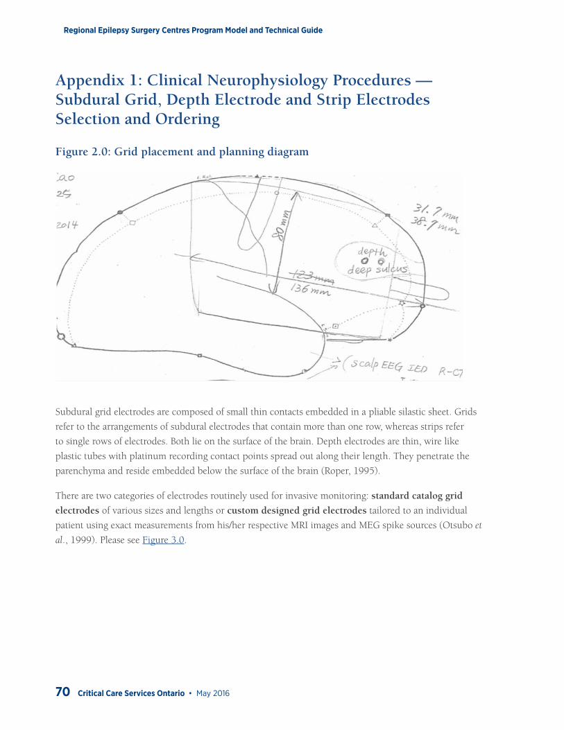
Regional Epilepsy Surgery Centres Program Model and Technical Guide
70 Critical Care Services Ontario • May 2016
Appendix 1: Clinical Neurophysiology Procedures — Subdural Grid, Depth Electrode and Strip Electrodes Selection and Ordering
Figure 2.0: Grid placement and planning diagram
Subdural grid electrodes are composed of small thin contacts embedded in a pliable silastic sheet. Grids
refer to the arrangements of subdural electrodes that contain more than one row, whereas strips refer
to single rows of electrodes. Both lie on the surface of the brain. Depth electrodes are thin, wire like
plastic tubes with platinum recording contact points spread out along their length. They penetrate the
parenchyma and reside embedded below the surface of the brain (Roper, 1995).
There are two categories of electrodes routinely used for invasive monitoring: standard catalog grid
electrodes of various sizes and lengths or custom designed grid electrodes tailored to an individual
patient using exact measurements from his/her respective MRI images and MEG spike sources (Otsubo et
al., 1999). Please see Figure 3.0.
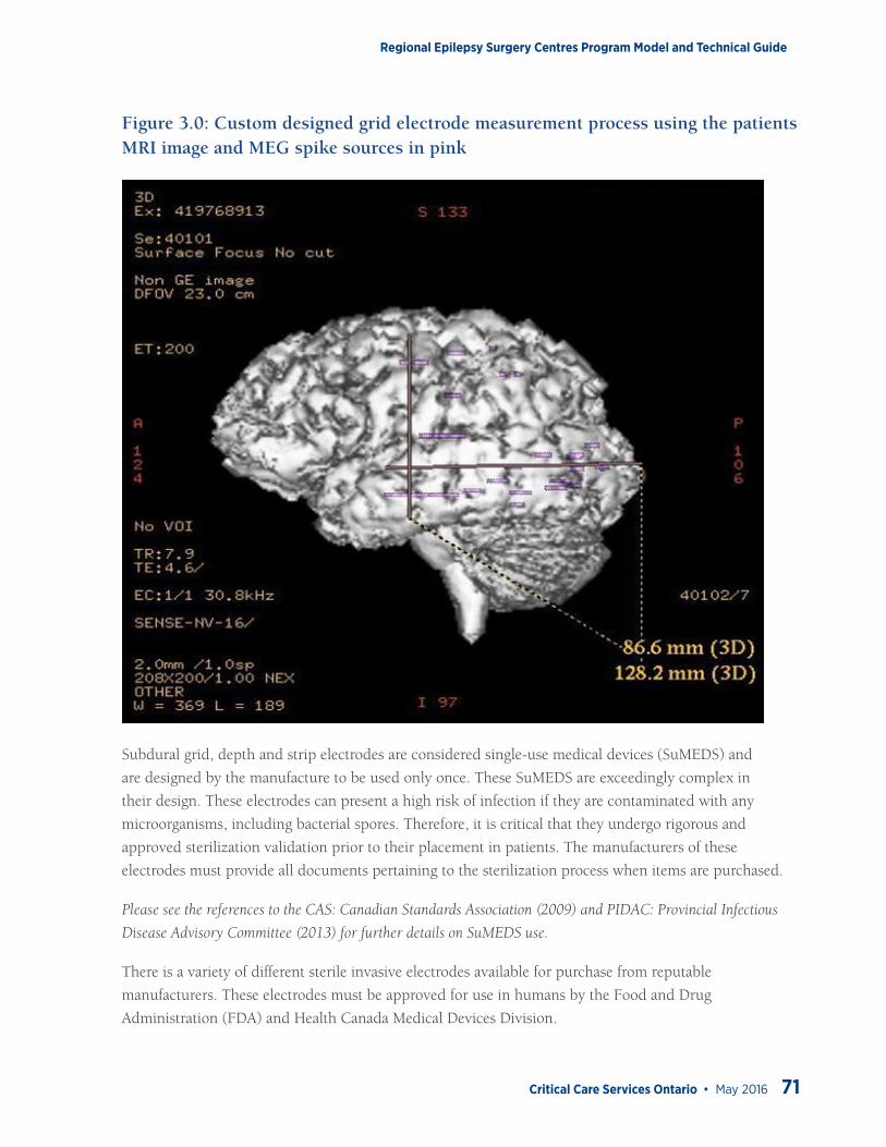
Regional Epilepsy Surgery Centres Program Model and Technical Guide
Critical Care Services Ontario • May 2016 71
Figure 3.0: Custom designed grid electrode measurement process using the patients MRI image and MEG spike sources in pink
Subdural grid, depth and strip electrodes are considered single-use medical devices (SuMEDS) and
are designed by the manufacture to be used only once. These SuMEDS are exceedingly complex in
their design. These electrodes can present a high risk of infection if they are contaminated with any
microorganisms, including bacterial spores. Therefore, it is critical that they undergo rigorous and
approved sterilization validation prior to their placement in patients. The manufacturers of these
electrodes must provide all documents pertaining to the sterilization process when items are purchased.
Please see the references to the CAS: Canadian Standards Association (2009) and PIDAC: Provincial Infectious
Disease Advisory Committee (2013) for further details on SuMEDS use.
There is a variety of different sterile invasive electrodes available for purchase from reputable
manufacturers. These electrodes must be approved for use in humans by the Food and Drug
Administration (FDA) and Health Canada Medical Devices Division.
Regional Epilepsy Surgery Centres Program Model and Technical Guide
72 Critical Care Services Ontario • May 2016
The invasive electrodes range in number of contacts and size: grids (4 to 128) contacts, depths/strips
(1x4, 1x6, 1x8, 1x10, 1x12) contacts or any combination that is required for each individual case. Please
see Figure 4.0.
These electrodes are used in conjunction with EEG and EP equipment for the recording, monitoring and
stimulation of electrical signals on the surface level of the brain.
The electrodes are designed to localize epileptogenic foci and stimulate and map the eloquent cortex.
If using custom designed invasive electrodes, a special access form may need to be completed by
the responsible physician and submitted for approval to Health Canada: Medical Devices Bureau.
Platinum-iridium electrodes that are embedded in a thin (0.5 mm) flexible silastic plate are the best
invasive electrodes available for implantation.
These electrodes are MRI safe, thus allowing MRI scans to be completed once they have been implanted.
The vendor must supply all sterilized invasive electrodes and cable connections ready for use. The
electrodes undergo strict quality control serialization methods by the vendor and are approved by both
FDA and Heath Canada.
Note: All invasive electrodes used are single use items and are discarded according to safe disposal methods by the
operating room medical professionals after their use.
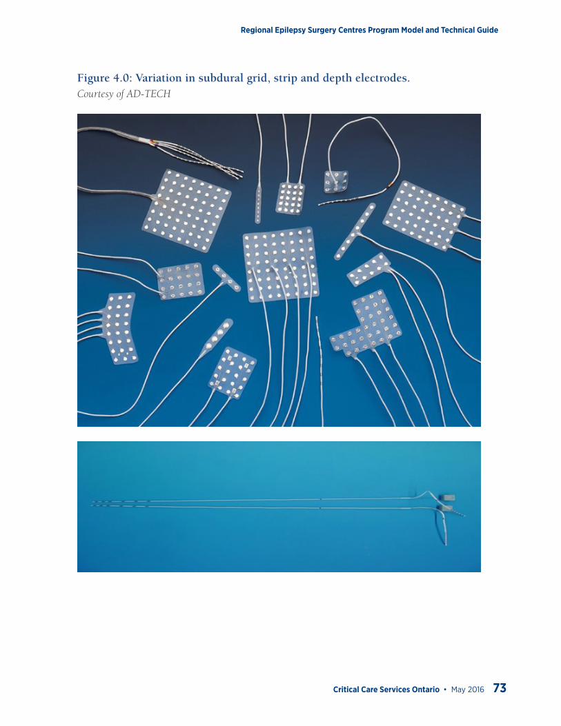
Regional Epilepsy Surgery Centres Program Model and Technical Guide
Critical Care Services Ontario • May 2016 73
Figure 4.0: Variation in subdural grid, strip and depth electrodes. Courtesy of AD-TECH
Regional Epilepsy Surgery Centres Program Model and Technical Guide
74 Critical Care Services Ontario • May 2016
Appendix 2: Clinical Neurophysiology Procedures — Electrode Placement for Invasive Monitoring
Prior to implantation of invasive monitoring electrodes, the epilepsy monitoring teams meets to create a
map of where all the different combinations of invasive electrodes will be placed. This map is based on the
patient’s clinical history, MRI, fMRI, MEG, EEG and EMU data. Please refer to Figure 2.0.
Either the EMU-IM technologist/operating room RN prepares the invasive electrodes and confirms that
they are all sterilized and the pouches have not been damaged. This process may vary between adult and
paediatric centres.
A map of the electrodes to be implanted is taken to the operating room and given to the neurosurgery
team for reference.
The patient is prepared for invasive electrode implantation surgery as per hospital approved surgical
policy and procedures.
Various methods of validation may be used during the invasive electrode implantation process. This
includes image guidance monitoring, intra operative monitoring and ECoG monitoring. The epilepsy
monitoring and neurosurgery teams determine which of these additional modalities and tools to utilize
during electrode implantation.
Prior to the placement of the subdural grid, IOM is performed to localize the somatosensory and motor cortices.
All eloquent cortices are identified and marked with letters along with any significant areas of interest
(spike focus, MEG cluster and lesion). During this process, intraoperative digital photography is utilized
to document and provide a visual reference later for EEG data analysis.
Subdural grid, strips and/or depth electrodes of varying combinations (4, 6, 8, 16, and 64 contacts) are
placed on patient’s cortical area of interest by the neurosurgeon guided by the responsible epileptologist.
The EMU-IM technologist/OR nurse records and keeps track of all subdural grids, strips and/or depth
electrodes used during the implantation process. All items used and their serial numbers are recorded in the
hospital OR database for documentation of all implanted devices used during the surgery.
It is recommended that the EMU-IM technologist also take a sterile 64 contact back-up grid, back-up
strip and depth electrodes to the OR. This is necessary should the electrodes being used for invasive
monitoring become accidently contaminated, fall to the floor or are faulty.
Digital photography images are obtained to mark regions of functional brain tissue with respect to cortical
surface landmarks and subdural grid placement. These images are used later to create a neurosurgical
map to aid the neurosurgeon and the epileptologist with the proposed cortical resections (Rutka et al.,
1999). Please see Figure 5.0.
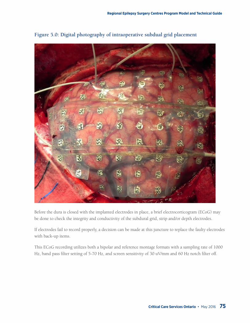
Regional Epilepsy Surgery Centres Program Model and Technical Guide
Critical Care Services Ontario • May 2016 75
Figure 5.0: Digital photography of intraoperative subdual grid placement
Before the dura is closed with the implanted electrodes in place, a brief electrocorticogram (ECoG) may
be done to check the integrity and conductivity of the subdural grid, strip and/or depth electrodes.
If electrodes fail to record properly, a decision can be made at this juncture to replace the faulty electrodes
with back-up items.
This ECoG recording utilizes both a bipolar and reference montage formats with a sampling rate of 1000
Hz, band pass filter setting of 5-70 Hz, and screen sensitivity of 30 uV/mm and 60 Hz notch filter off.
Regional Epilepsy Surgery Centres Program Model and Technical Guide
76 Critical Care Services Ontario • May 2016
Appendix 3: Clinical Neurophysiology Procedures — Intraoperative Monitoring and Mapping, Anaesthesia, and Patient Safety Considerations
Patient Set-up for Intraoperative Mapping/MonitoringPatient set-up is completed in the operating room after the patient is induced and intubated by the IOM
technologist.
The use of sterile, single use, subdermal needle electrodes is recommended for recording somatosensory
evoked potentials (SEP), trans-cranial motor evoked potentials to electrical stimulation (tceMEP). Surface
electrodes have been shown to be insufficient in detecting neurotonic discharges (Skinner et al., 2008).
In addition, surface electrodes can dry out over prolonged periods of time (Nuwer et al., 1992), and
sweat can cause electrodes to detach from the skin, increasing impedance and preventing stable recording
during lengthy procedures.
Surface electrodes may be used for stimulating peripheral nerves. Surface electrodes reduce the charge density
applied to the skin and help prevent burns (Netherton et al. 2007; Patterson et al. 2007; Stecker et al., 2006).
Additional ground, reference and EKG electrode is applied by EMU-IM technologist to the patient.
Technologist can use any of the following types of electrodes: surface gold disc electrodes, single use
needle electrodes or disposable pad electrodes.
These electrodes are utilized for ECoG recordings to confirm the placement and integrity of the invasive
electrode placement prior to the surgeon closing the Dura.
IOM Electrode Application
Scalp electrode application is done using the international 10-20 System of Electrode Placement (Klem et
al., 1999). Modifications to the placement of electrodes may be required as a result of the large incision
site for surgery.
Direct cranial SEP recordings should be obtained with a minimum 1x4 subcortical strip electrode. (1 x
8, 2 x 4, or 2 x 8 strip electrodes may be used if recording channels are available.) The electrodes are
platinum or stainless steel discs embedded in silicone.
tceMEP electrodes are applied in a bipolar montage. The active electrode is placed in or near the belly of
the muscle, and the reference electrode is placed on or near the tendon of the muscle. This placement
results in a maximal amplitude response by increasing common mode rejection and improving the signal
to noise ratio (Mallik & Weir, 2005).
Regional Epilepsy Surgery Centres Program Model and Technical Guide
Critical Care Services Ontario • May 2016 77
It is recommended that, in order to lower impedances, each electrode site should be rubbed with an abrasive
skin preparation and wiped with isopropyl alcohol prior to lead application. The impedances should be at
acceptable levels that are between 1000-5000 ohms. This significantly improves the signal to noise ratio.
Caution: When using skin abrasive products, care must be taken to prevent skin rupture and bleeding.
Test Protocols
Functional Cortical Mapping
Functional mapping of eloquent cortex is executed in the operating room prior to insertion of the
subdural grid. The goals are: 1) to identify the primary motor cortex by stimulating the brain directly and
recording compound muscle action potentials (CMAP), and 2) to identify the central sulcus by recording
phase reversal SEPs. This information is used for preservation of motor and sensory function during
cortical resection.
Phase reversals
Phase reversal SEPs are utilized as an intraoperative monitoring tool to identify the sensory portion of
the sensorimotor cortex (central sulcus identification) (Aiba & Seki, 1988; Cedzich et al., 1996; King &
Schell, 1987; Nuwer et al., 1992; Wood et al., 1988).
Stimulation montage (Nuwer et al., 1992; Nuwer et al., 1994):
• Median n. or ulnar n. at the wrist contralateral to tested hemisphere
• Anode placed 2-3 cm distal to the cathode
• Intensity is supra-maximal at 15-20 ma producing an observable twitch
• Rate is 4-5 Hz and can be lowered to 1-3 Hz for children under 3 years of age
• Pulse duration is 200-300 µs
Recording montage (Nuwer et al., 1992; Nuwer et al., 1994):
• Channel 1: C-spine
• Channel 2-5: 1 x 4 strip electrode
• Channels 1-5 referenced to Fz
• Filter settings at 30-500 Hz. (Reduce high frequency filter to eliminate high frequency artifact)
• Strip electrode positioned with long axis in an anterior-posterior orientation placed over the presumed central sulcus, or previously mapped magnetoencephalography (MEG) somatosensory evoked field (SEF)
• Time base 50 ms
• 100 trials or until waveforms are clearly identifiable
• Test must be reproducible
Regional Epilepsy Surgery Centres Program Model and Technical Guide
78 Critical Care Services Ontario • May 2016
Interpretation of Phase Reversals (Nuwer et al., 1994; Nuwer et al., 1992; Simon, 2013; Wood et al.,
1988)
• Maximal amplitude of the major negative peak at about 20 ms (N20) generated from the postcentral gyrus
• Phase reversal of the same 20 ms potential (P20) across the central sulcus with a 1-2 ms increased latency arising from the motor cortex
• The latency of the principal negative peak can vary with patient height, age and gender
• If no phase reversal is observed, the strip electrode should be repositioned over a different area of the exposed cortex and stimulation re-attempted.
• The strip electrode, on occasion, may have to be rotated and positioned in a more superior-inferior direction.
Direct Cortical Stimulation (DCS)
Two methods of DCS have evolved. The 60 Hz technique applies stimuli with a 50 or 60 Hz frequency
over a period of many seconds. This has been described by Penfield in the 1930s (Penfield & Boldrey,
1937). It requires a much higher number of pulses and, as a result, imparts a greater total charge to the
brain than the high frequency train-of-five (HF-TOF) technique.
A literature review found stimulation associated seizures is reported in 1.2% with the HF-TOF technique
and significantly more frequently in 9.5% with the 60 Hz technique (Szelenyi et al, 2007). The incidence
of stimulation associated seizures with the 60 Hz technique has been reported as high as 24% (Sartorius
& Wright, 1997; Yingling et al., 1999). It has also been shown that there is no increased risk of the
occurrence of stimulation-associated seizures during surgery for patients with symptomatic epilepsy
compared with those patients without (Szelenyi et al., 2007).
The 60 Hz technique may be less sensitive in paediatric patients due to the relative in excitability of the
motor cortex (Sala et al., 2002).
Another advantage of the HF-TOF technique is that, as stimulation intensity is increased in a stepwise
manner, you are able to selectively activate a small portion of fibres and record CMAPs, as opposed to
observing gross motor movements. This improves mapping sensitivity (Taniguchi et al., 1993) and allows
specific regions of the motor homunculus to be identified across much of the entire body (Kombos et al.,
2000; Ng et al., 2009).
Regional Epilepsy Surgery Centres Program Model and Technical Guide
Critical Care Services Ontario • May 2016 79
It is for these reasons that there has been a shift from the 60 Hz technique to the high frequency HF-TOF
technique over the years. Stimulation and recording parameters for the HF-TOF technique is described below.
Stimulation montage:
• Monopolar, 3 mm, ball-tipped probe for mapping
• Monopolar probe plugged into anode (red, positive)
• Anodal stimulation produces responses from contralateral muscle groups
• Needle electrode plugged into cathode (black, negative) 2 cm lateral to Cz on contralateral side of head
• Constant voltage intensity 25-100 V (Kombos et al., 2000; Ng et al., 2009)
• Constant current intensity 3-20 mA (Cedzich et al., 1996; Simon, 2013)
• Rate is 500-900 Hz (interstimulus interval = 1.1 ms - 2.5 ms)
• Number of pulses 5 - 7
• 50 µs square wave pulse for constant voltage
• 200 - 500 µs square wave pulse for constant current
Recording montage (Ng et al., 2009):
• Subdermal needle electrodes in contralateral muscles
• Orbicularis oris
• Deltoid
• Extensor digitorum communis
• First dorsal interosseuos
• Rectus femoris
• Tibialis anterior
• Abductor hallucis
• Time Base 20 -100 ms
• Filters 100 - 3000 Hz
Technical notes:
• Stimulation intensity is increased in a stepwise manner in 5 V, or 3 mA increments until a reproducible CMAP is obtained.
• The lowest stimulation thresholds are recorded for each muscle group or combination of muscle groups (Ng et al., 2009).
• Constant voltage stimulators produce a more rapid charge delivery and require a lower total charge for equivalent stimulation, as compared to constant current (Hausmann et al., 2002).
• The extensor digitorum communis has been found to have the highest recorded rate and requires lowest stimulation intensity vs. other muscle groups (Ng et al., 2009).
• Mapping is more consistent in upper limbs compared to lower limbs and facial muscles (Ng et al., 2009).
Regional Epilepsy Surgery Centres Program Model and Technical Guide
80 Critical Care Services Ontario • May 2016
• Patients with cortical dysplasia/tuberous sclerosis have higher stimulation thresholds (Ng et al., 2009).
• Higher stimulation thresholds are required in children compared to adults, and reliable CMAP responses may not be obtainable in children younger than 6 years (Lieberman et al., 2006).
• Cold irrigation must be available and ready to use in the event of an induced seizure.
Cortical Monitoring
Cortical monitoring is executed intra-operatively prior to, during, and after the cortical resection. The
goal is to reduce the risk of iatrogenic injury during the cortical resection by preserving motor and
sensory function. The monitoring modalities used are SEPs, tceMEPs, DCS using recurrent high frequency
HF-TOF stimulation, and, if needed, subcortical stimulation of white matter.
Somatosensory Evoked Potentials
SEPs are utilized as an intraoperative monitoring tool to assess the functional status of the somatosensory
pathway and reduce the risk of iatrogenic injury during cortical resection. A comprehensive review of the
underlying neuroanatomy and physiology, as well as a detailed review of the stimulation and recording
settings in the neurosurgical setting is readily available in the literature (AES, 1994; Møller, 1995; Neuloh
& Schramm, 2002; Nuwer, 2008; Nuwer et al., 1994; Nuwer et al, 1992; Toleikis, 2010). A brief overview
of the technical components is listed below.
Stimulation montage:
• Median n. or ulnar n. at the wrist contralateral to tested hemisphere
• Posterior tibial nerve at the medial malleolus
• Anode placed 2-3 cm distal to the cathode
• Intensity is supra-maximal at 15-20 ma for the upper extremities and 30-40 mA for the lower extremities with observable twitch
• Rate is 4-5 Hz and can be lowered to 1-3 Hz for children under 3 years of age
• Pulse duration is 200-300 µs
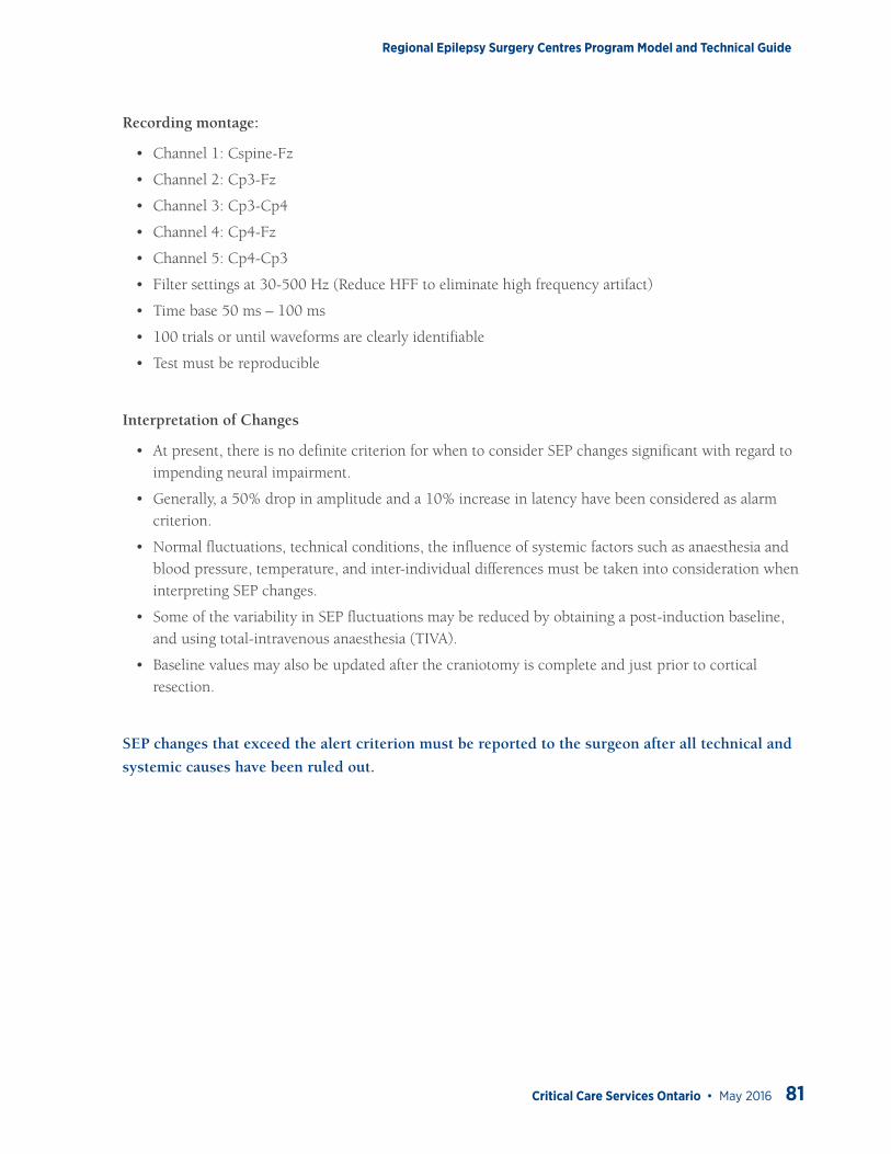
Regional Epilepsy Surgery Centres Program Model and Technical Guide
Critical Care Services Ontario • May 2016 81
Recording montage:
• Channel 1: Cspine-Fz
• Channel 2: Cp3-Fz
• Channel 3: Cp3-Cp4
• Channel 4: Cp4-Fz
• Channel 5: Cp4-Cp3
• Filter settings at 30-500 Hz (Reduce HFF to eliminate high frequency artifact)
• Time base 50 ms – 100 ms
• 100 trials or until waveforms are clearly identifiable
• Test must be reproducible
Interpretation of Changes
• At present, there is no definite criterion for when to consider SEP changes significant with regard to impending neural impairment.
• Generally, a 50% drop in amplitude and a 10% increase in latency have been considered as alarm criterion.
• Normal fluctuations, technical conditions, the influence of systemic factors such as anaesthesia and blood pressure, temperature, and inter-individual differences must be taken into consideration when interpreting SEP changes.
• Some of the variability in SEP fluctuations may be reduced by obtaining a post-induction baseline, and using total-intravenous anaesthesia (TIVA).
• Baseline values may also be updated after the craniotomy is complete and just prior to cortical resection.
SEP changes that exceed the alert criterion must be reported to the surgeon after all technical and
systemic causes have been ruled out.
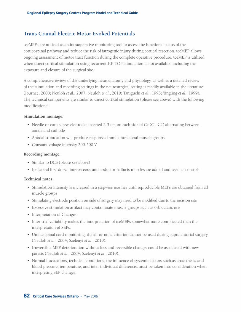
Regional Epilepsy Surgery Centres Program Model and Technical Guide
82 Critical Care Services Ontario • May 2016
Trans Cranial Electric Motor Evoked Potentials
tceMEPs are utilized as an intraoperative monitoring tool to assess the functional status of the
corticospinal pathway and reduce the risk of iatrogenic injury during cortical resection. tceMEP allows
ongoing assessment of motor tract function during the complete operative procedure. tceMEP is utilized
when direct cortical stimulation using recurrent HF-TOF stimulation is not available, including the
exposure and closure of the surgical site.
A comprehensive review of the underlying neuroanatomy and physiology, as well as a detailed review
of the stimulation and recording settings in the neurosurgical setting is readily available in the literature
(Journee, 2008; Neuloh et al., 2007; Neuloh et al., 2010; Taniguchi et al., 1993; Yingling et al., 1999).
The technical components are similar to direct cortical stimulation (please see above) with the following
modifications:
Stimulation montage:
• Needle or cork screw electrodes inserted 2-3 cm on each side of Cz (C1-C2) alternating between anode and cathode
• Anodal stimulation will produce responses from contralateral muscle groups
• Constant voltage intensity 200-500 V
Recording montage:
• Similar to DCS (please see above)
• Ipsilateral first dorsal interosseous and abductor hallucis muscles are added and used as controls
Technical notes:
• Stimulation intensity is increased in a stepwise manner until reproducible MEPs are obtained from all muscle groups
• Stimulating electrode position on side of surgery may need to be modified due to the incision site
• Excessive stimulation artifact may contaminate muscle groups such as orbicularis oris
• Interpretation of Changes:
• Inter-trial variability makes the interpretation of tceMEPs somewhat more complicated than the interpretation of SEPs.
• Unlike spinal cord monitoring, the all-or-none criterion cannot be used during supratentorial surgery (Neuloh et al., 2004; Szelenyi et al., 2010).
• Irreversible MEP deterioration without loss and reversible changes could be associated with new paresis (Neuloh et al., 2004; Szelenyi et al., 2010).
• Normal fluctuations, technical conditions, the influence of systemic factors such as anaesthesia and blood pressure, temperature, and inter-individual differences must be taken into consideration when interpreting SEP changes.
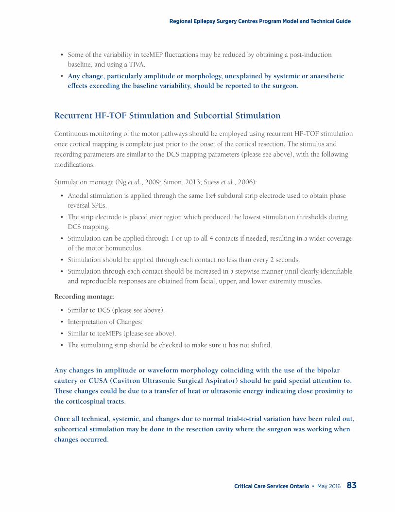
Regional Epilepsy Surgery Centres Program Model and Technical Guide
Critical Care Services Ontario • May 2016 83
• Some of the variability in tceMEP fluctuations may be reduced by obtaining a post-induction baseline, and using a TIVA.
• Any change, particularly amplitude or morphology, unexplained by systemic or anaesthetic effects exceeding the baseline variability, should be reported to the surgeon.
Recurrent HF-TOF Stimulation and Subcortial Stimulation
Continuous monitoring of the motor pathways should be employed using recurrent HF-TOF stimulation
once cortical mapping is complete just prior to the onset of the cortical resection. The stimulus and
recording parameters are similar to the DCS mapping parameters (please see above), with the following
modifications:
Stimulation montage (Ng et al., 2009; Simon, 2013; Suess et al., 2006):
• Anodal stimulation is applied through the same 1x4 subdural strip electrode used to obtain phase reversal SPEs.
• The strip electrode is placed over region which produced the lowest stimulation thresholds during DCS mapping.
• Stimulation can be applied through 1 or up to all 4 contacts if needed, resulting in a wider coverage of the motor homunculus.
• Stimulation should be applied through each contact no less than every 2 seconds.
• Stimulation through each contact should be increased in a stepwise manner until clearly identifiable and reproducible responses are obtained from facial, upper, and lower extremity muscles.
Recording montage:
• Similar to DCS (please see above).
• Interpretation of Changes:
• Similar to tceMEPs (please see above).
• The stimulating strip should be checked to make sure it has not shifted.
Any changes in amplitude or waveform morphology coinciding with the use of the bipolar
cautery or CUSA (Cavitron Ultrasonic Surgical Aspirator) should be paid special attention to.
These changes could be due to a transfer of heat or ultrasonic energy indicating close proximity to
the corticospinal tracts.
Once all technical, systemic, and changes due to normal trial-to-trial variation have been ruled out,
subcortical stimulation may be done in the resection cavity where the surgeon was working when
changes occurred.
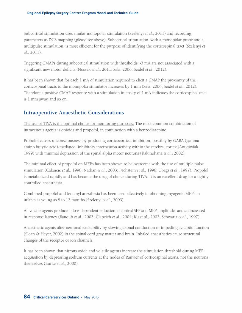
Regional Epilepsy Surgery Centres Program Model and Technical Guide
84 Critical Care Services Ontario • May 2016
Subcortical stimulation uses similar monopolar stimulation (Szelenyi et al., 2011) and recording
parameters as DCS mapping (please see above). Subcortical stimulation, with a monopolar probe and a
multipulse stimulation, is most efficient for the purpose of identifying the corticospinal tract (Szelenyi et
al., 2011).
Triggering CMAPs during subcortical stimulation with thresholds >3 mA are not associated with a
significant new motor deficits (Nossek et al., 2011; Sala, 2006; Seidel et al., 2012).
It has been shown that for each 1 mA of stimulation required to elicit a CMAP the proximity of the
corticospinal tracts to the monopolar stimulator increases by 1 mm (Sala, 2006; Seidel et al., 2012).
Therefore a positive CMAP response with a stimulation intensity of 1 mA indicates the corticospinal tract
is 1 mm away, and so on.
Intraoperative Anaesthetic Considerations
The use of TIVA is the optimal choice for monitoring purposes. The most common combination of
intravenous agents is opioids and propofol, in conjunction with a benzodiazepine.
Propofol causes unconsciousness by producing corticocortical inhibition, possibly by GABA (gamma
amino butyric acid)-mediated inhibitory interneuron activity within the cerebral cortex (Antkowiak,
1999) with minimal depression of the spinal alpha motor neurons (Kakinohana et al., 2002).
The minimal effect of propofol on MEPs has been shown to be overcome with the use of multiple pulse
stimulation (Calancie et al., 1998; Nathan et al., 2003; Pechstein et al., 1998; Ubags et al., 1997). Propofol
is metabolized rapidly and has become the drug of choice during TIVA. It is an excellent drug for a tightly
controlled anaesthesia.
Combined propofol and fentanyl anesthesia has been used effectively in obtaining myogenic MEPs in
infants as young as 8 to 12 months (Szelenyi et al., 2003).
All volatile agents produce a dose-dependent reduction in cortical SEP and MEP amplitudes and an increased
in response latency (Banoub et al., 2003; Clapcich et al., 2004; Ku et al., 2002; Schwartz et al., 1997).
Anaesthetic agents alter neuronal excitability by slowing axonal conduction or impeding synaptic function
(Sloan & Heyer, 2002) in the spinal cord gray matter and brain. Inhaled anaesthetics cause structural
changes of the receptor or ion channels.
It has been shown that nitrous oxide and volatile agents increase the stimulation threshold during MEP
acquisition by depressing sodium currents at the nodes of Ranvier of corticospinal axons, not the neurons
themselves (Burke et al., 2000).
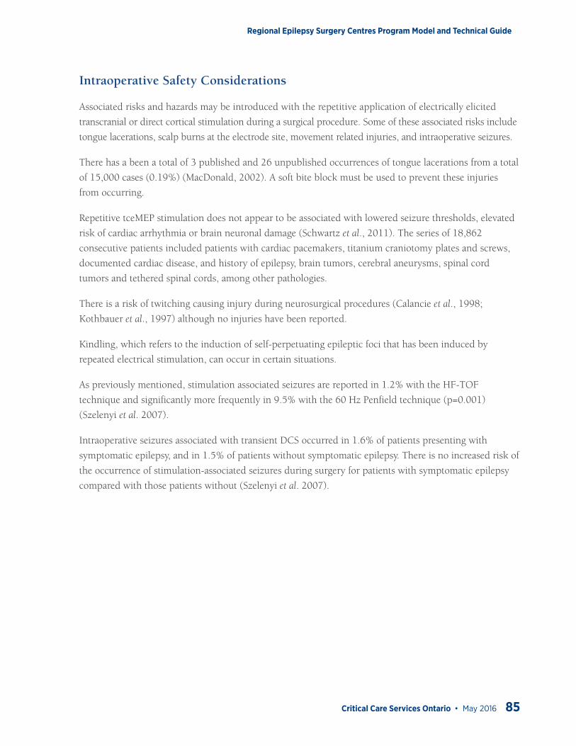
Regional Epilepsy Surgery Centres Program Model and Technical Guide
Critical Care Services Ontario • May 2016 85
Intraoperative Safety Considerations
Associated risks and hazards may be introduced with the repetitive application of electrically elicited
transcranial or direct cortical stimulation during a surgical procedure. Some of these associated risks include
tongue lacerations, scalp burns at the electrode site, movement related injuries, and intraoperative seizures.
There has a been a total of 3 published and 26 unpublished occurrences of tongue lacerations from a total
of 15,000 cases (0.19%) (MacDonald, 2002). A soft bite block must be used to prevent these injuries
from occurring.
Repetitive tceMEP stimulation does not appear to be associated with lowered seizure thresholds, elevated
risk of cardiac arrhythmia or brain neuronal damage (Schwartz et al., 2011). The series of 18,862
consecutive patients included patients with cardiac pacemakers, titanium craniotomy plates and screws,
documented cardiac disease, and history of epilepsy, brain tumors, cerebral aneurysms, spinal cord
tumors and tethered spinal cords, among other pathologies.
There is a risk of twitching causing injury during neurosurgical procedures (Calancie et al., 1998;
Kothbauer et al., 1997) although no injuries have been reported.
Kindling, which refers to the induction of self-perpetuating epileptic foci that has been induced by
repeated electrical stimulation, can occur in certain situations.
As previously mentioned, stimulation associated seizures are reported in 1.2% with the HF-TOF
technique and significantly more frequently in 9.5% with the 60 Hz Penfield technique (p=0.001)
(Szelenyi et al. 2007).
Intraoperative seizures associated with transient DCS occurred in 1.6% of patients presenting with
symptomatic epilepsy, and in 1.5% of patients without symptomatic epilepsy. There is no increased risk of
the occurrence of stimulation-associated seizures during surgery for patients with symptomatic epilepsy
compared with those patients without (Szelenyi et al. 2007).
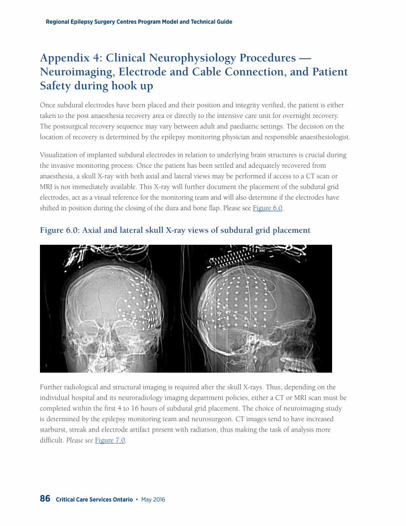
Regional Epilepsy Surgery Centres Program Model and Technical Guide
86 Critical Care Services Ontario • May 2016
Appendix 4: Clinical Neurophysiology Procedures — Neuroimaging, Electrode and Cable Connection, and Patient Safety during hook up
Once subdural electrodes have been placed and their position and integrity verified, the patient is either
taken to the post anaesthesia recovery area or directly to the intensive care unit for overnight recovery.
The postsurgical recovery sequence may vary between adult and paediatric settings. The decision on the
location of recovery is determined by the epilepsy monitoring physician and responsible anaesthesiologist.
Visualization of implanted subdural electrodes in relation to underlying brain structures is crucial during
the invasive monitoring process. Once the patient has been settled and adequately recovered from
anaesthesia, a skull X-ray with both axial and lateral views may be performed if access to a CT scan or
MRI is not immediately available. This X-ray will further document the placement of the subdural grid
electrodes, act as a visual reference for the monitoring team and will also determine if the electrodes have
shifted in position during the closing of the dura and bone flap. Please see Figure 6.0.
Figure 6.0: Axial and lateral skull X-ray views of subdural grid placement
Further radiological and structural imaging is required after the skull X-rays. Thus, depending on the
individual hospital and its neuroradiology imaging department policies, either a CT or MRI scan must be
completed within the first 4 to 16 hours of subdural grid placement. The choice of neuroimaging study
is determined by the epilepsy monitoring team and neurosurgeon. CT images tend to have increased
starburst, streak and electrode artifact present with radiation, thus making the task of analysis more
difficult. Please see Figure 7.0.
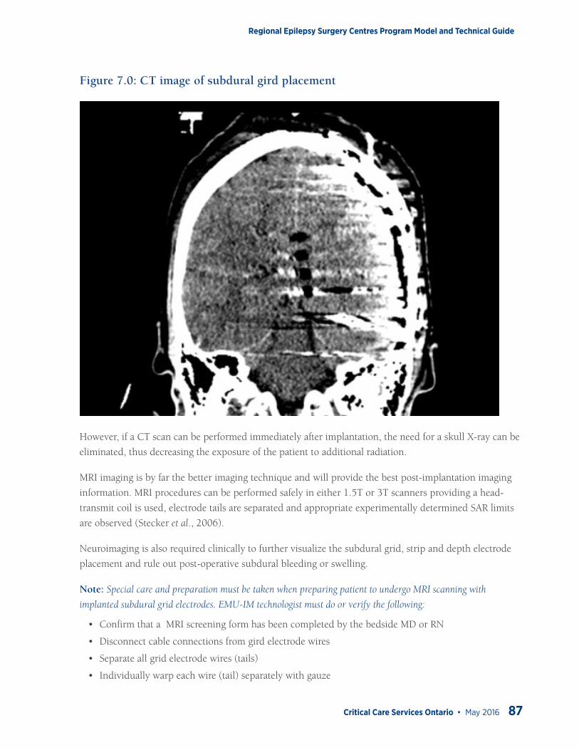
Regional Epilepsy Surgery Centres Program Model and Technical Guide
Critical Care Services Ontario • May 2016 87
Figure 7.0: CT image of subdural gird placement
However, if a CT scan can be performed immediately after implantation, the need for a skull X-ray can be
eliminated, thus decreasing the exposure of the patient to additional radiation.
MRI imaging is by far the better imaging technique and will provide the best post-implantation imaging
information. MRI procedures can be performed safely in either 1.5T or 3T scanners providing a head-
transmit coil is used, electrode tails are separated and appropriate experimentally determined SAR limits
are observed (Stecker et al., 2006).
Neuroimaging is also required clinically to further visualize the subdural grid, strip and depth electrode
placement and rule out post-operative subdural bleeding or swelling.
Note: Special care and preparation must be taken when preparing patient to undergo MRI scanning with
implanted subdural grid electrodes. EMU-IM technologist must do or verify the following:
• Confirm that a MRI screening form has been completed by the bedside MD or RN
• Disconnect cable connections from gird electrode wires
• Separate all grid electrode wires (tails)
• Individually warp each wire (tail) separately with gauze
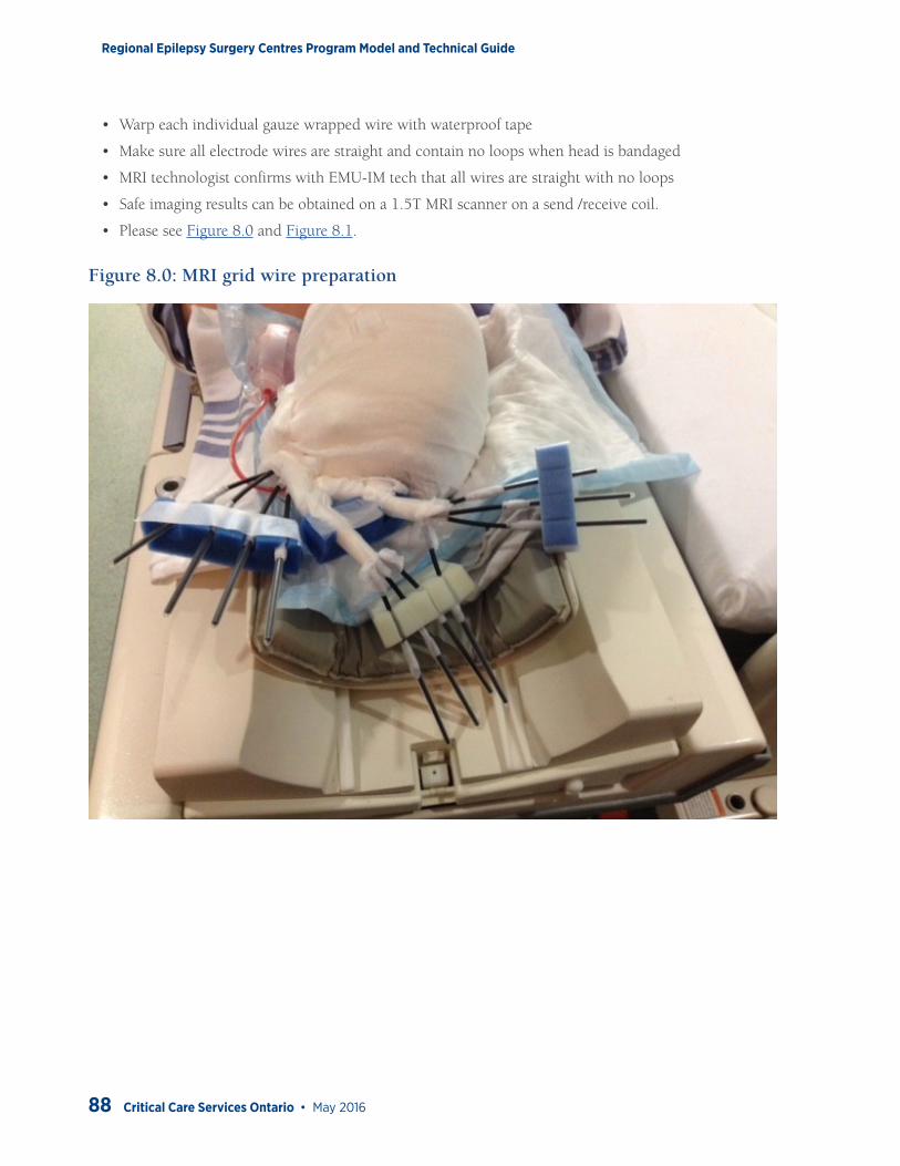
Regional Epilepsy Surgery Centres Program Model and Technical Guide
88 Critical Care Services Ontario • May 2016
• Warp each individual gauze wrapped wire with waterproof tape
• Make sure all electrode wires are straight and contain no loops when head is bandaged
• MRI technologist confirms with EMU-IM tech that all wires are straight with no loops
• Safe imaging results can be obtained on a 1.5T MRI scanner on a send /receive coil.
• Please see Figure 8.0 and Figure 8.1.
Figure 8.0: MRI grid wire preparation
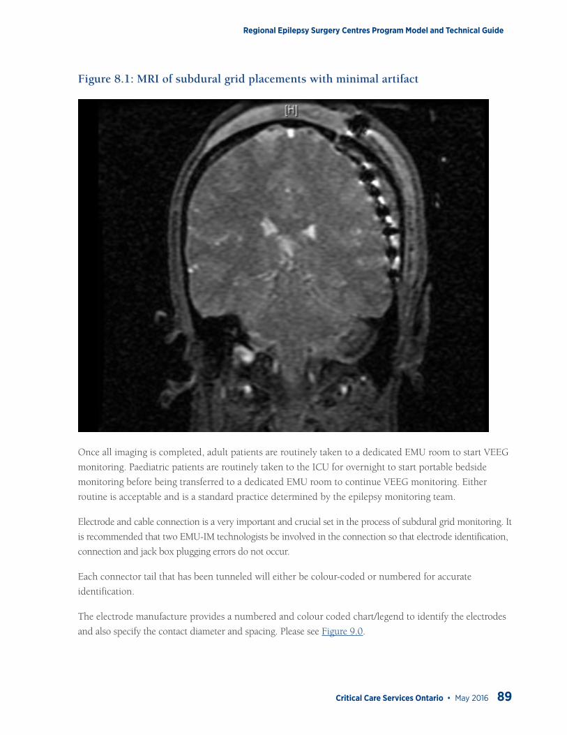
Regional Epilepsy Surgery Centres Program Model and Technical Guide
Critical Care Services Ontario • May 2016 89
Figure 8.1: MRI of subdural grid placements with minimal artifact
Once all imaging is completed, adult patients are routinely taken to a dedicated EMU room to start VEEG
monitoring. Paediatric patients are routinely taken to the ICU for overnight to start portable bedside
monitoring before being transferred to a dedicated EMU room to continue VEEG monitoring. Either
routine is acceptable and is a standard practice determined by the epilepsy monitoring team.
Electrode and cable connection is a very important and crucial set in the process of subdural grid monitoring. It
is recommended that two EMU-IM technologists be involved in the connection so that electrode identification,
connection and jack box plugging errors do not occur.
Each connector tail that has been tunneled will either be colour-coded or numbered for accurate
identification.
The electrode manufacture provides a numbered and colour coded chart/legend to identify the electrodes
and also specify the contact diameter and spacing. Please see Figure 9.0.
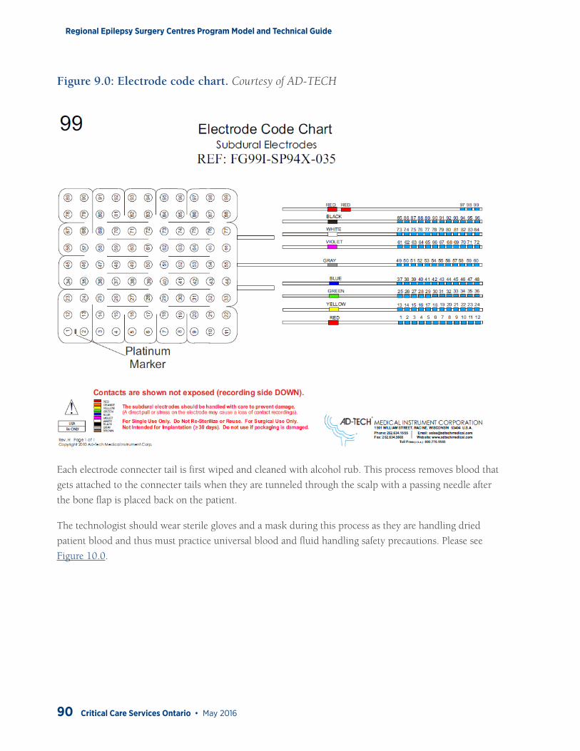
Regional Epilepsy Surgery Centres Program Model and Technical Guide
90 Critical Care Services Ontario • May 2016
Figure 9.0: Electrode code chart. Courtesy of AD-TECH
Each electrode connecter tail is first wiped and cleaned with alcohol rub. This process removes blood that
gets attached to the connecter tails when they are tunneled through the scalp with a passing needle after
the bone flap is placed back on the patient.
The technologist should wear sterile gloves and a mask during this process as they are handling dried
patient blood and thus must practice universal blood and fluid handling safety precautions. Please see
Figure 10.0.
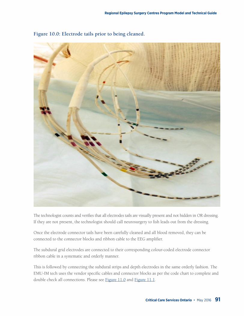
Regional Epilepsy Surgery Centres Program Model and Technical Guide
Critical Care Services Ontario • May 2016 91
Figure 10.0: Electrode tails prior to being cleaned.
The technologist counts and verifies that all electrodes tails are visually present and not hidden in OR dressing.
If they are not present, the technologist should call neurosurgery to fish leads out from the dressing.
Once the electrode connector tails have been carefully cleaned and all blood removed, they can be
connected to the connector blocks and ribbon cable to the EEG amplifier.
The subdural grid electrodes are connected to their corresponding colour-coded electrode connector
ribbon cable in a systematic and orderly manner.
This is followed by connecting the subdural strips and depth electrodes in the same orderly fashion. The
EMU-IM tech uses the vendor specific cables and connector blocks as per the code chart to complete and
double check all connections. Please see Figure 11.0 and Figure 11.1.
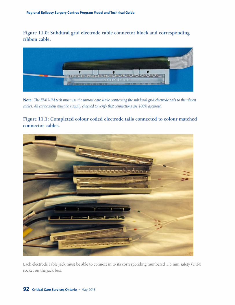
Regional Epilepsy Surgery Centres Program Model and Technical Guide
92 Critical Care Services Ontario • May 2016
Figure 11.0: Subdural grid electrode cable-connector block and corresponding ribbon cable.
Note: The EMU-IM tech must use the utmost care while connecting the subdural grid electrode tails to the ribbon
cables. All connections must be visually checked to verify that connections are 100% accurate.
Figure 11.1: Completed colour coded electrode tails connected to colour matched connector cables.
Each electrode cable jack must be able to connect in to its corresponding numbered 1.5 mm safety (DIN)
socket on the jack box.
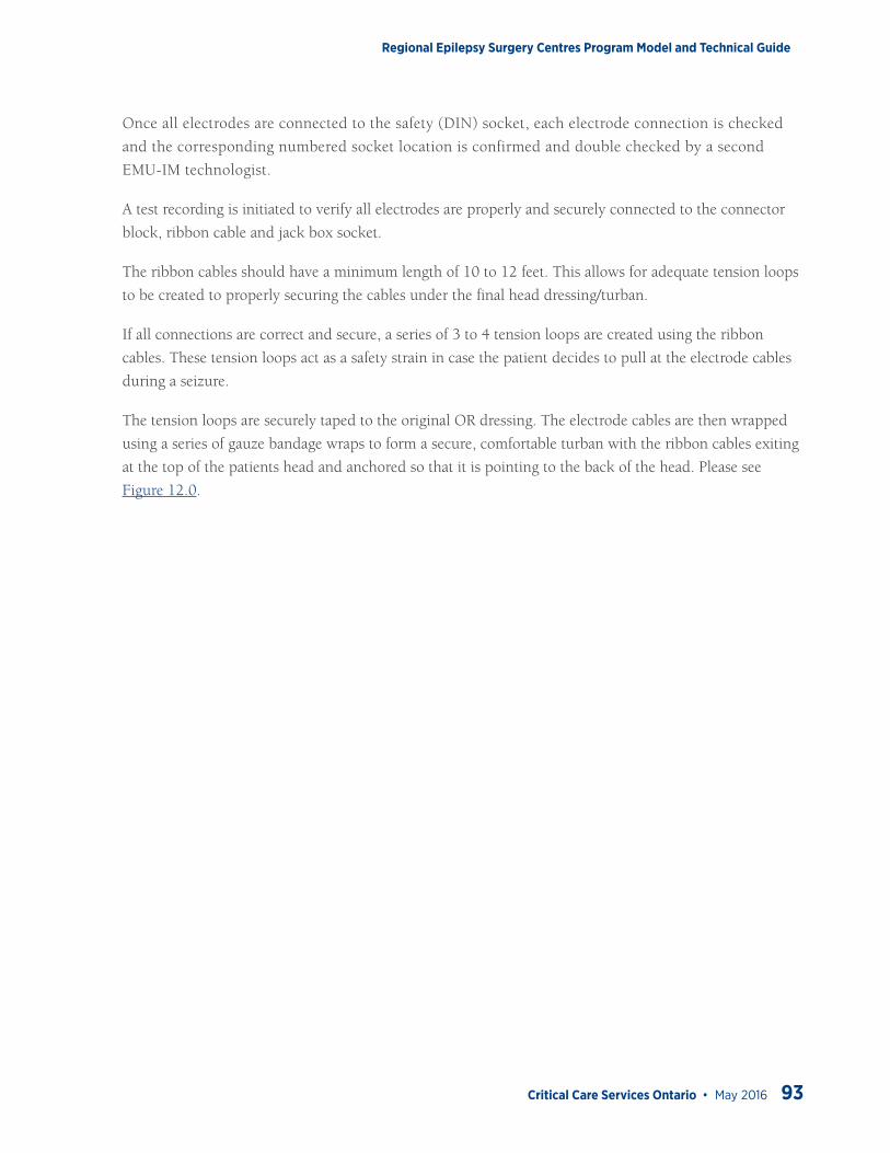
Regional Epilepsy Surgery Centres Program Model and Technical Guide
Critical Care Services Ontario • May 2016 93
Once all electrodes are connected to the safety (DIN) socket, each electrode connection is checked
and the corresponding numbered socket location is confirmed and double checked by a second
EMU-IM technologist.
A test recording is initiated to verify all electrodes are properly and securely connected to the connector
block, ribbon cable and jack box socket.
The ribbon cables should have a minimum length of 10 to 12 feet. This allows for adequate tension loops
to be created to properly securing the cables under the final head dressing/turban.
If all connections are correct and secure, a series of 3 to 4 tension loops are created using the ribbon
cables. These tension loops act as a safety strain in case the patient decides to pull at the electrode cables
during a seizure.
The tension loops are securely taped to the original OR dressing. The electrode cables are then wrapped
using a series of gauze bandage wraps to form a secure, comfortable turban with the ribbon cables exiting
at the top of the patients head and anchored so that it is pointing to the back of the head. Please see
Figure 12.0.
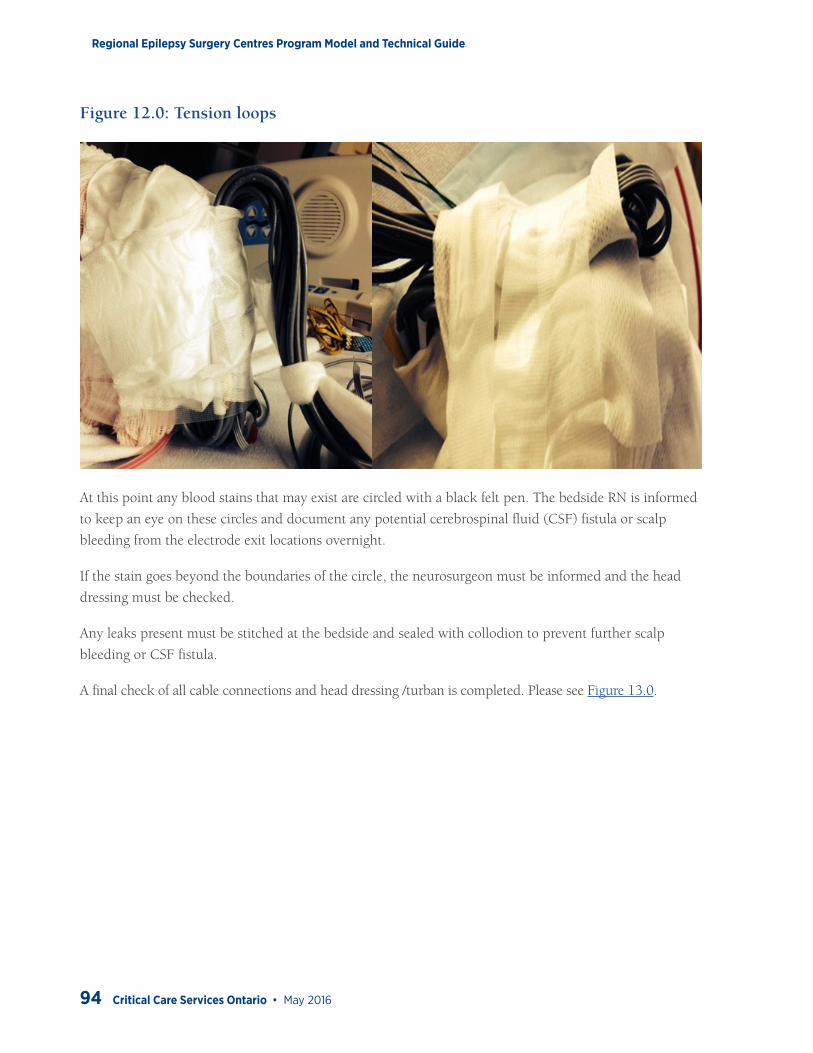
Regional Epilepsy Surgery Centres Program Model and Technical Guide
94 Critical Care Services Ontario • May 2016
Figure 12.0: Tension loops
At this point any blood stains that may exist are circled with a black felt pen. The bedside RN is informed
to keep an eye on these circles and document any potential cerebrospinal fluid (CSF) fistula or scalp
bleeding from the electrode exit locations overnight.
If the stain goes beyond the boundaries of the circle, the neurosurgeon must be informed and the head
dressing must be checked.
Any leaks present must be stitched at the bedside and sealed with collodion to prevent further scalp
bleeding or CSF fistula.
A final check of all cable connections and head dressing /turban is completed. Please see Figure 13.0.
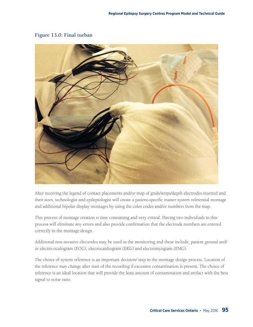
Regional Epilepsy Surgery Centres Program Model and Technical Guide
Critical Care Services Ontario • May 2016 95
Figure 13.0: Final turban
After receiving the legend of contact placements and/or map of grids/strips/depth electrodes inserted and
their sizes, technologist and epileptologist will create a patient-specific master system referential montage
and additional bipolar display montages by using the color codes and/or numbers from the map.
This process of montage creation is time consuming and very critical. Having two individuals in this
process will eliminate any errors and also provide confirmation that the electrode numbers are entered
correctly in the montage design.
Additional non-invasive electrodes may be used in the monitoring and these include, patient ground and/
or electro-oculogram (EOG), electrocardiogram (EKG) and electromyogram (EMG).
The choice of system reference is an important decision/ step in the montage design process. Location of
the reference may change after start of the recording if excessive contamination is present. The choice of
reference is an ideal location that will provide the least amount of contamination and artifact with the best
signal to noise ratio.
Regional Epilepsy Surgery Centres Program Model and Technical Guide
96 Critical Care Services Ontario • May 2016
The system reference and location will vary. The following are suggestions:
• Use of a separate subdural strip inserted away from the active site
• Use of an inactive electrode on the implanted gird away from the active site recording site
• Use of a summated reference consisting of two inactive electrodes on the implanted subdural grid away from the active site
The recoding montage should be simple and follow and sequential number scheme incorporating all the
contacts on the subdual grid. The nomenclature used for the montage design should try and keep within
the guidelines of the 10-20 electrode naming convention (Carmichael et al., 2008). This montage is
designed using the numbered colour coded identification map provided by the electrode manufacturer.
Sample System reference montage and bipolar display montage:
Channel 1: 1-system ref
Channel 2: 2-system ref
Channel 3: 3-system ref
Channel 4: 4-system ref
Channel 5: 5-system ref
Channel 6: 6-system ref
Channel 1: 1-2
Channel 2: 2-3
Channel 3: 3-4
Channel 4: 4-5
Channel 5: 5-6
Channel 6: 6-7
Note: Utmost care must be taken while creating the montage, as the number of contacts is large and it is easy to
confuse the subdural strips names/contact numbers and the connections.
To prevent leakage current entering the patient the application of a ground electrode is a must for EMU
recording. The technologist must ensure that there is only one ground electrode on the patient to avoid
the potential risk of a ground loop.
Please refer to www.caet.org for minimal technical guidelines for EEG.
Note: The implanted electrodes impedances are not documented, as the impedance testing requires sending a
small amount of electrical current to the indwelling electrodes, which might cause local burning. Therefore, for
patient safety reasons it is not performed.
Regional Epilepsy Surgery Centres Program Model and Technical Guide
Critical Care Services Ontario • May 2016 97
Patient Safety• Careful care and precaution must be taken not to pull the excessively hard on the tail ends exiting
out of the bur holes on the scalp.
• The EMU-IM technologist should wear a mask, goggles and gloves during this process. This helps to minimize the secondary infections.
• Again use of limited pressure/force is required when technologist is tracing the tails/ribbon ends of the grids/strips/depth electrodes so that they don’t get tugged or pulled accidently.
• All exiting electrode tails are cleaned with alcohol and all tails are counted and color/number coding must be verified and cross referenced to the electrode code chart.
• The above is done while the patient is seated or lying in bed. Standard precautions to prevent accidental falls and injuries are followed.
• Cables must be connected in a systematic fashion to prevent entanglement utilizing cable management techniques like cable ties, wraps and cable tube covers to prevent accidental strangulation during sleep while being monitored in accordance to hospital’s entanglement policies.
• The electrode amplifier must be properly secured and or wrapped in a heatproof pouch to prevent accidental burn to the patient or accidental disconnection of electrodes during monitoring.
• All EMU-IM beds must be equipped with proper safety bumper pads to protect the patient during seizures.
• Mittens may be used with difficult patients to prevent accidental and/or intentional disconnection of electrodes or tugging of subdural grid ribbon cables exiting from the top of the patients head.
• Use of a sterile head wrap, burn net or conforming bandage to secure electrodes is highly recommended to prevent accidental and/or intentional disconnection of electrodes and infection. Daily checks are required to asses any CSF fistula or scalp bleeding that may arise.
• Patients on whom medication is being tapered during EMU-IM monitoring must have an IV in place along with an EKG and O
2 saturation monitor.
• Age appropriate restraints (if needed) to be used as required following hospital’s restraint policies.
• All rooms must be equipped with a ‘Nurse Call’ button at the bedside, separate from seizure push button marker.
Regional Epilepsy Surgery Centres Program Model and Technical Guide
98 Critical Care Services Ontario • May 2016
Appendix 5: Clinical Neurophysiology Procedures — Continuous EEG video monitoring from intracranial electrodes – Day by Day
Initiate EMU Equipment for Continuous Video EEG Monitoring: Day 1The EMU-IM technologist confirms electrode connections and verification, patient safety check and
confirmation of adequate EEG system CPU hard drive space (500 GB).
Monitoring can be done in either the ICU or dedicated EMU monitoring room using either a dedicated
invasive monitoring portable EEG system or a fixed room unit.
Once the patient is settled in the ICU or EMU, the seizure safety guidelines (CCSO, 2014, p. 18) are
reviewed with the RN and/or caregiver. The RN and/or caregiver are both instructed on the importance of
completing the seizure log sheets for documentation.
The push button alarm is tested by the RN and/or caregiver.
Both the video and infrared camera (including audio) is tested prior to the start of the invasive EMU recording.
The technologist then initiates the EMU equipment for day 1 continuous video EEG monitoring.
Review and Mark Seizures, Push-Button Events and Automatic Seizure and Spike Detections for EMU Epileptologists: Day 2EMU-IM Technologist does the following:
• Checks the recording quality of the invasive monitoring and the patient’s head dressing in the morning and rechecks at end of his/her shift on a daily basis.
• Should any technical problems arise, the EMU-IM technologist is on-call 24/7.
• The EMU-IM technologist reviews patient seizure and push button logs.
• The EMU-IM technologist makes sure the cables and grid cable connectors are securely connected, re-gels and/or reapplies any electrodes (EKG, EMG and patient ground) that may have become loose, detached or are otherwise not functioning properly.
• Verifies with electroencephalographer if the system reference needs to be changed.
• Restarts new dataset and makes backup of previous night’s invasive monitoring data.
Regional Epilepsy Surgery Centres Program Model and Technical Guide
Critical Care Services Ontario • May 2016 99
Data transfer and EEG file back-up maybe very time consuming due to the high sampling rates and
high resolution digital video recording used to record data during invasive monitoring. Therefore, it
is recommended that the hospital network data transfer speed be set at 1 GB or utilize an optic fibre
connection. If this is not available than an encrypted portable external storage drive be used to transfer
data in a fast and efficient manner for review on a reading station. This sequence must be carried out
every day that the patient is being monitored.
It is recommended that the original OR dressing be removed by the 4th or 5th day of monitoring with a
new set of head dressings and padding with the help of the RN and neurosurgery resident.
The EMU-IM technologist must be present during this procedure to make sure grid electrode tails are not
accidently damaged, cut or disconnected to preserve the ongoing recording quality and continuity of the
invasive monitoring..
Documenting the Anatomical Locations of Grids, Strips and Depth ElectrodesThe utilization of MRI images, CT images, MEG spike sources, 3D rendering software, high resolution digital
photography and EEG application software allows the epilepsy team to visually reconstruct the location of
grid, depth and strip electrodes in both adult and paediatric patients undergoing invasive monitoring.
The actual locations of the indwelling grids, strips and depth electrodes are a very important aspect of the
intracranial video-EEG procedure.
These visual aids provide the electroencephalographer with landmarks in order for them to accurately map
the ictal seizure onset as well as the active interictal and irritable zones in the cortical tissues.
One method in which this can accomplished in adults is to use the patient specific pre-MRI scan data and
post implantation CT/ MRI scan data of the patient to make a visual image of the brain.
The EMU-IM technologist or MRI technologist may be trained to import the brain image files in a Digital
Imaging and Communications in Medicine (DICOM) format to vendor specific or third party EEG grid
view application software. This software is designed to reconstruct the MRI signals into a 3D rendering of
the brain and while interpolating the post-insertion electrode positions from either the CT or MRI scans.
The various reconstruction software utilized allows for the fusion of other MRI sequences which may
better demonstrate a patient’s pathology (i.e., cortical dysplasia). The images can be further combined and
fused with DTI and be loaded onto a brain navigation system and used in the operating room. Please see
Figure 14.0.
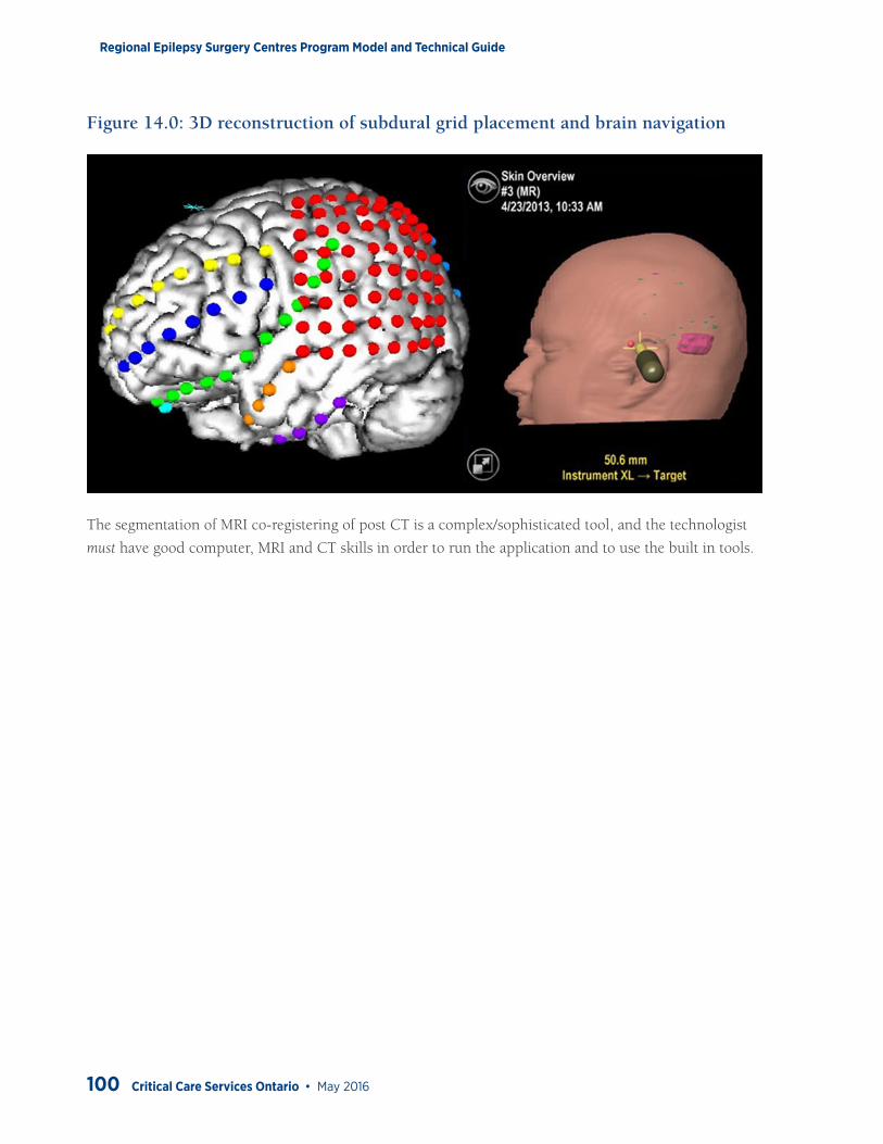
Regional Epilepsy Surgery Centres Program Model and Technical Guide
100 Critical Care Services Ontario • May 2016
Figure 14.0: 3D reconstruction of subdural grid placement and brain navigation
The segmentation of MRI co-registering of post CT is a complex/sophisticated tool, and the technologist
must have good computer, MRI and CT skills in order to run the application and to use the built in tools.
Regional Epilepsy Surgery Centres Program Model and Technical Guide
Critical Care Services Ontario • May 2016 101
Appendix 6: Clinical Neurophysiology Procedures — Extraoperative Functional Mapping of Eloquent Cortex from intracranial electrodes
Topographic Evoked Potentials: Day 2If available, somatosensory evoked potentials (SEP) from median nerve stimulation performed
extraoperatively from grid electrodes is performed. These are acquired with a 32-channel electrode
montage to allow mapping along a longer length of the central sulcus.
Median nerve stimulation is applied according to published neurophysiology guidelines (ACNS, 2008).
Recording parameters are completed according to the published guidelines. The only deviation is that
instead of the typical 4, 8 or 16 channel recordings, a set of 32 channels are chosen. The 32 channels span
the anatomically identified central sulcus; which is then identified by mapping the pattern of phase reversals
of the N20/P22 complex. A negative-going N20 indicates sensory cortex while a positive-going N20
indicates motor cortex.
Eloquent Cortex Mapping: Day 3-4An essential component of invasive monitoring is the mapping of motor, sensory, and language functions
in relation to the ictal epileptogenic zone.
The eloquent cortex mapping is done to delineate the cortical areas of functional importance from the
epileptic focus. This is accomplished by selective electrical stimulation of the grid and strip electrodes
placed on the patient’s cortex using a CSA approved external cortical stimulator in conjunction with a
built in Digital Switch Matrix (DSM) software in the EEG unit.
The DSM allows the technologist to stimulate the cortex by digitally selecting an active and reference
electrode on the subdural grid, thus allowing convenient switching of the electrodes for stimulation,
without having to interrupt the data acquisition process.
Note: Due to variations in age, brain maturation, plasticity and pathology the stimulation process and stimulation
thresholds required to elicit a clinical response will differ between children and adults.
The mapping of the eloquent is performed either on the third or fourth day of invasive monitoring. For
language mapping, the patient needs to be cooperative and fully awake to perform the language tasks
given by the neuropsychologist.
The mapping process requires a delicate balance between stimulation threshold and clinical response
required. Cortical stimulation guidelines must be followed to avoid excessive stimulation that might
provoke atypical focal or generalized seizure (Chitoku et al., 2001).
Regional Epilepsy Surgery Centres Program Model and Technical Guide
102 Critical Care Services Ontario • May 2016
Adult Cortical Mapping Process• The neurosurgeon uses a constant current stimulator which generates the electrical pulses (square) of
different pulse widths with a voltage out-put in steps of 1 .5 mA to max of 17.5 mA.
• The EMU-IM technologist sets up the cortical stimulator to the designated port of the amplifier box as outlined by the EEG equipment manufacturer.
• The EMU-IM technologist opens the DSM application on the acquisition station for the mapping.
• Neurosurgeon then instructs the technologist to activate or open the required grid or strip contact numbers (2 at a time) on the DSM application.
• After the contact numbers are opened, the software will block the EEG signal recordings from the opened contacts and allows the relay of the current to flow through the selected contacts, while the rest of EEG signals are acquired by active channels.
• The neurosurgeon then will start the stimulation of the cortex through the open relay contacts by means of the current stimulator by increasing the current in a stepwise manner of 1.5 mA to 2 mA to a maximum of 17.5 mA, until the required clinical features are elicited.
• The clinical features of interest are of speech arrest, anomia, sensory sensations, and motor manifestations etc. depending on the cortical area of stimulation.
• During this process of increased current stimulation, the technologist will monitor the ongoing EEG signals for After-Discharges (spike, spike and wave complexes) *electrographic seizure(s) which might be induced and communicate this to the neurosurgeon in good time as well documenting the current strength, patient reactions and responses to stimulation.
* At times, the electrographic seizures can evolve into complex partial seizures and progress to generalized tonic-
clonic seizure. Here, the experience of the technologist is of importance to identify these features on EEG as well
on the ongoing video. Suggestion: Keep the video well-focused on the patient to see the clinical features.
Regional Epilepsy Surgery Centres Program Model and Technical Guide
Critical Care Services Ontario • May 2016 103
Paediatric Cortical Mapping ProcessThe EMU-IM technologist sets up the cortical stimulator to the designated port of the amplifier box as
outlined by the EEG equipment manufacturer.
The mapping process is similar to the adult functional mapping process with minor technical differences.
The EMU-IM technologist sets up the extraoperative cortical stimulator. Various FDA and CSA approved
stimulators are available for this process. Stimulations are delivered to the designated active and reference
grid electrodes utilizing the EEG machine DSM using a “distance reference” technique (Lesser et al.,
1984).
The stimulation is delivered using a 50 Hz biphasic pulse (pulse duration of 2 ms) in a single train lasting
15 seconds. Various combinations of distance “reference technique” grid electrodes are first stimulated to
verify /select the best peripheral reference electrode (Szelenyi et al., 2010; Szelenyi et al., 2007).
Stimulation pulses are delivered to the active and reference electrode pair starting at 2 mA, increasing in
intensity by 1-2 mA until the desired functional clinical response is obtained.
All electrode pairs’ names are called out and documented by the EMU-IM technologist to confirm the
correct electrode pairs are being stimulated.
Stimulation “on” and “off” are called out during the mapping process to alert the investigators the start
and stop times of when the stimulation is being delivered.
Push button marker is used by the EMU-IM technologist to mark all clinical responses and seizures
during the mapping process.
The stimulation is stopped immediately after clinical response is captured or a seizure is noted.
Discharges are carefully documented after the stimulation has been stopped.
This procedure is repeated over the cortical area of interest until the neurosurgeon/epileptologist has
obtained all the clinical and electrographic information needed for the resection of the epileptic focus.
After the eloquent cortex mapping is finished, the technologist removes the current stimulator equipment,
cables etc. and makes sure the video-EEG monitoring is going well, the patient condition reconfirmed and
the nurse is updated.
Regional Epilepsy Surgery Centres Program Model and Technical Guide
Appendix 7: Clinical Neurophysiology Equipment
EMU-IM EEG EquipmentThe EEG equipment used for Invasive monitoring should consist of the following and meet the
minimal technical guidelines for EEG recording as mandated by the Canadian Society of Clinical
Neurophysiologists (CSCN) task force.
EEG Amplifier Specifications:• Minimum of 128 with optional 256 channel capability for intracranial EEG recording is
recommended along with a Digital Switch Matrix (DSM) application embedded in the acquisition software (for electrical stimulation mapping). A constant current, biphasic pulse generator is used for eloquent cortex functional mapping.
• To prevent any potential data loss and interruption of data collection during the recording session, it is recommended that the amplifier jack box be equipped with a UPS (uninterruptible power supply) battery back-up with EEG data storage capacity for 20 to 30 minutes. This feature will allow recording to continue while the patient is disconnected from the main acquisition station (e.g., patient going to washroom/for short breaks etc.).
• Minimum of 4 channels of auxiliary inputs for EKG, EOG, EMG and Respiration Monitoring
• Minimum Sampling rate of 1000 Hz or higher (preferably 2000 Hz) for analog to digital conversion is required for invasive monitoring
• Digital Precision of 16 bits or greater, to resolve voltage to 0.5 µV, is recommended.
• Common mode rejection should be 100 dB or greater at each amplifier output. Inter-channel cross-talk must be less than 1%, 40 dB down or less.
• Please refer to McLachlan & Young (1999) for further details regarding EEG amplifier specifications.
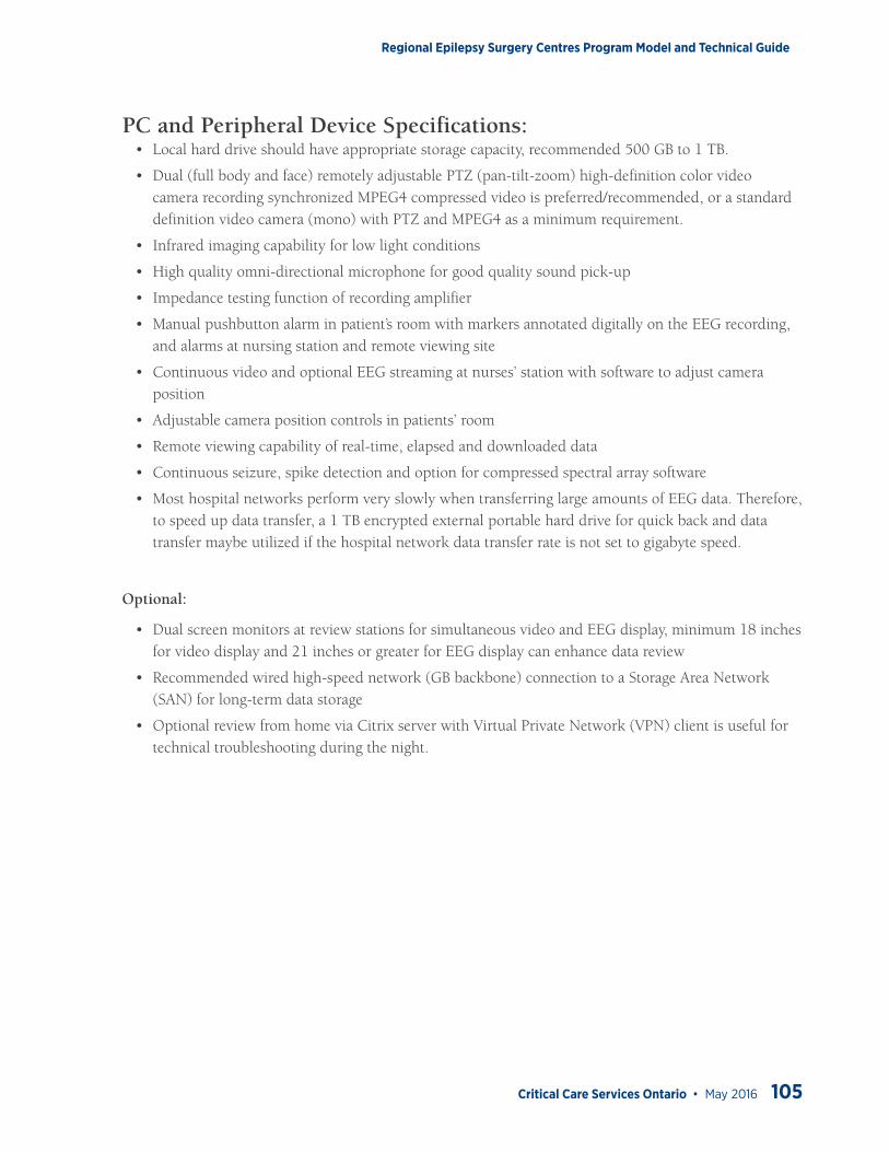
Regional Epilepsy Surgery Centres Program Model and Technical Guide
Critical Care Services Ontario • May 2016 105
PC and Peripheral Device Specifications:• Local hard drive should have appropriate storage capacity, recommended 500 GB to 1 TB.
• Dual (full body and face) remotely adjustable PTZ (pan-tilt-zoom) high-definition color video camera recording synchronized MPEG4 compressed video is preferred/recommended, or a standard definition video camera (mono) with PTZ and MPEG4 as a minimum requirement.
• Infrared imaging capability for low light conditions
• High quality omni-directional microphone for good quality sound pick-up
• Impedance testing function of recording amplifier
• Manual pushbutton alarm in patient’s room with markers annotated digitally on the EEG recording, and alarms at nursing station and remote viewing site
• Continuous video and optional EEG streaming at nurses’ station with software to adjust camera position
• Adjustable camera position controls in patients’ room
• Remote viewing capability of real-time, elapsed and downloaded data
• Continuous seizure, spike detection and option for compressed spectral array software
• Most hospital networks perform very slowly when transferring large amounts of EEG data. Therefore, to speed up data transfer, a 1 TB encrypted external portable hard drive for quick back and data transfer maybe utilized if the hospital network data transfer rate is not set to gigabyte speed.
Optional:
• Dual screen monitors at review stations for simultaneous video and EEG display, minimum 18 inches for video display and 21 inches or greater for EEG display can enhance data review
• Recommended wired high-speed network (GB backbone) connection to a Storage Area Network (SAN) for long-term data storage
• Optional review from home via Citrix server with Virtual Private Network (VPN) client is useful for technical troubleshooting during the night.
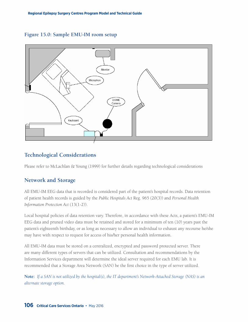
Regional Epilepsy Surgery Centres Program Model and Technical Guide
106 Critical Care Services Ontario • May 2016
Figure 15.0: Sample EMU-IM room setup
Technological Considerations
Please refer to McLachlan & Young (1999) for further details regarding technological considerations
Network and Storage
All EMU-IM EEG data that is recorded is considered part of the patient’s hospital records. Data retention
of patient health records is guided by the Public Hospitals Act Reg. 965 (20(3)) and Personal Health
Information Protection Act (13(1-2)).
Local hospital policies of data retention vary. Therefore, in accordance with these Acts, a patient’s EMU-IM
EEG data and pruned video data must be retained and stored for a minimum of ten (10) years past the
patient’s eighteenth birthday, or as long as necessary to allow an individual to exhaust any recourse he/she
may have with respect to request for access of his/her personal health information.
All EMU-IM data must be stored on a centralized, encrypted and password protected server. There
are many different types of servers that can be utilized. Consultation and recommendations by the
Information Services department will determine the ideal server required for each EMU lab. It is
recommended that a Storage Area Network (SAN) be the first choice in the type of server utilized.
Note: If a SAN is not utilized by the hospital(s), the IT department’s Network-Attached Storage (NAS) is an
alternate storage option.
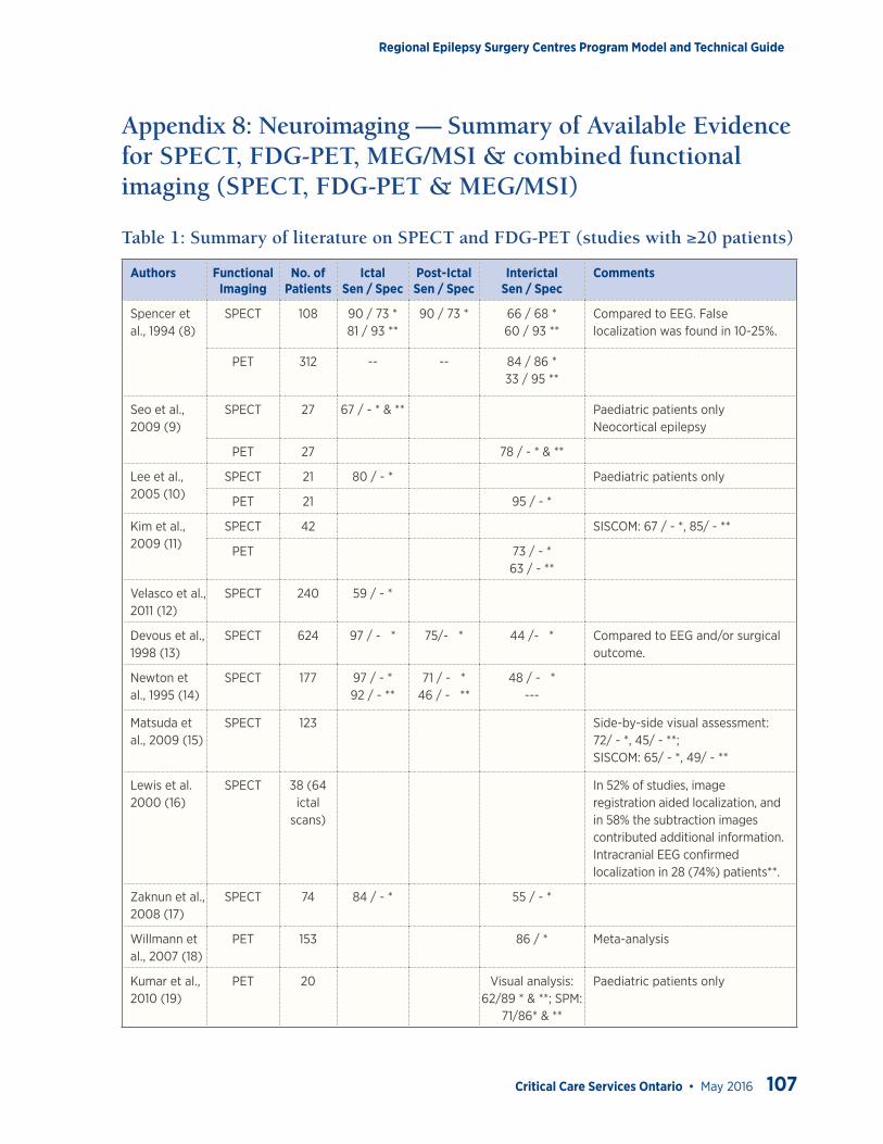
Regional Epilepsy Surgery Centres Program Model and Technical Guide
Critical Care Services Ontario • May 2016 107
Appendix 8: Neuroimaging — Summary of Available Evidence for SPECT, FDG-PET, MEG/MSI & combined functional imaging (SPECT, FDG-PET & MEG/MSI)
Table 1: Summary of literature on SPECT and FDG-PET (studies with ≥20 patients)
Authors Functional Imaging
No. of Patients
Ictal Sen / Spec
Post-Ictal Sen / Spec
Interictal Sen / Spec
Comments
Spencer et al., 1994 (8)
SPECT 108 90 / 73 *81 / 93 **
90 / 73 * 66 / 68 *60 / 93 **
Compared to EEG. False localization was found in 10-25%.
PET 312 -- -- 84 / 86 *33 / 95 **
Seo et al., 2009 (9)
SPECT 27 67 / - * & ** Paediatric patients only Neocortical epilepsy
PET 27 78 / - * & **
Lee et al., 2005 (10)
SPECT 21 80 / - * Paediatric patients only
PET 21 95 / - *
Kim et al., 2009 (11)
SPECT 42 SISCOM: 67 / - *, 85/ - **
PET 73 / - *63 / - **
Velasco et al., 2011 (12)
SPECT 240 59 / - *
Devous et al., 1998 (13)
SPECT 624 97 / - * 75/- * 44 /- * Compared to EEG and/or surgical outcome.
Newton et al., 1995 (14)
SPECT 177 97 / - *92 / - **
71 / - *46 / - **
48 / - *---
Matsuda et al., 2009 (15)
SPECT 123 Side-by-side visual assessment: 72/ - *, 45/ - **;SISCOM: 65/ - *, 49/ - **
Lewis et al. 2000 (16)
SPECT 38 (64 ictal
scans)
In 52% of studies, image registration aided localization, and in 58% the subtraction images contributed additional information. Intracranial EEG confirmed localization in 28 (74%) patients**.
Zaknun et al., 2008 (17)
SPECT 74 84 / - * 55 / - *
Willmann et al., 2007 (18)
PET 153 86 / * Meta-analysis
Kumar et al., 2010 (19)
PET 20 Visual analysis: 62/89 * & **; SPM:
71/86* & **
Paediatric patients only
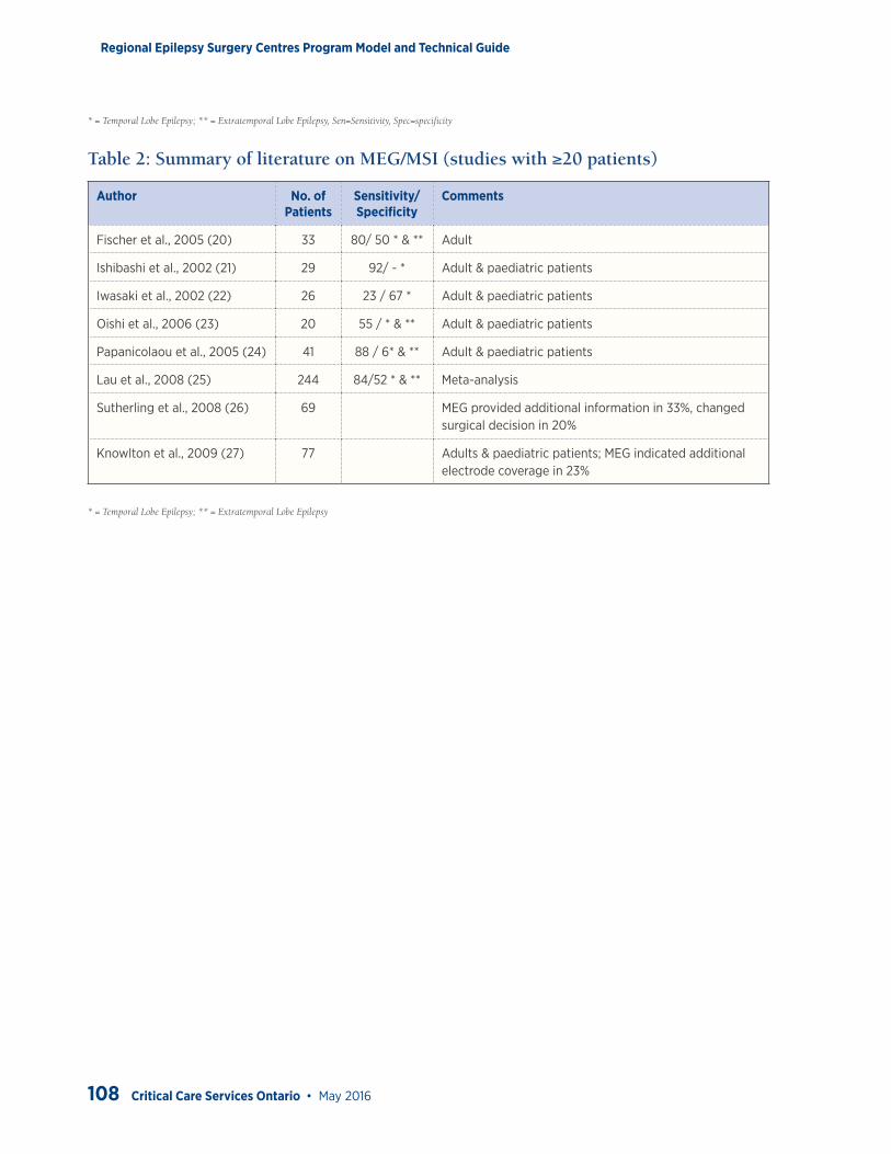
Regional Epilepsy Surgery Centres Program Model and Technical Guide
108 Critical Care Services Ontario • May 2016
* = Temporal Lobe Epilepsy; ** = Extratemporal Lobe Epilepsy, Sen=Sensitivity, Spec=specificity
Table 2: Summary of literature on MEG/MSI (studies with ≥20 patients)
Author No. of Patients
Sensitivity/ Specificity
Comments
Fischer et al., 2005 (20) 33 80/ 50 * & ** Adult
Ishibashi et al., 2002 (21) 29 92/ - * Adult & paediatric patients
Iwasaki et al., 2002 (22) 26 23 / 67 * Adult & paediatric patients
Oishi et al., 2006 (23) 20 55 / * & ** Adult & paediatric patients
Papanicolaou et al., 2005 (24) 41 88 / 6* & ** Adult & paediatric patients
Lau et al., 2008 (25) 244 84/52 * & ** Meta-analysis
Sutherling et al., 2008 (26) 69 MEG provided additional information in 33%, changed surgical decision in 20%
Knowlton et al., 2009 (27) 77 Adults & paediatric patients; MEG indicated additional electrode coverage in 23%
* = Temporal Lobe Epilepsy; ** = Extratemporal Lobe Epilepsy
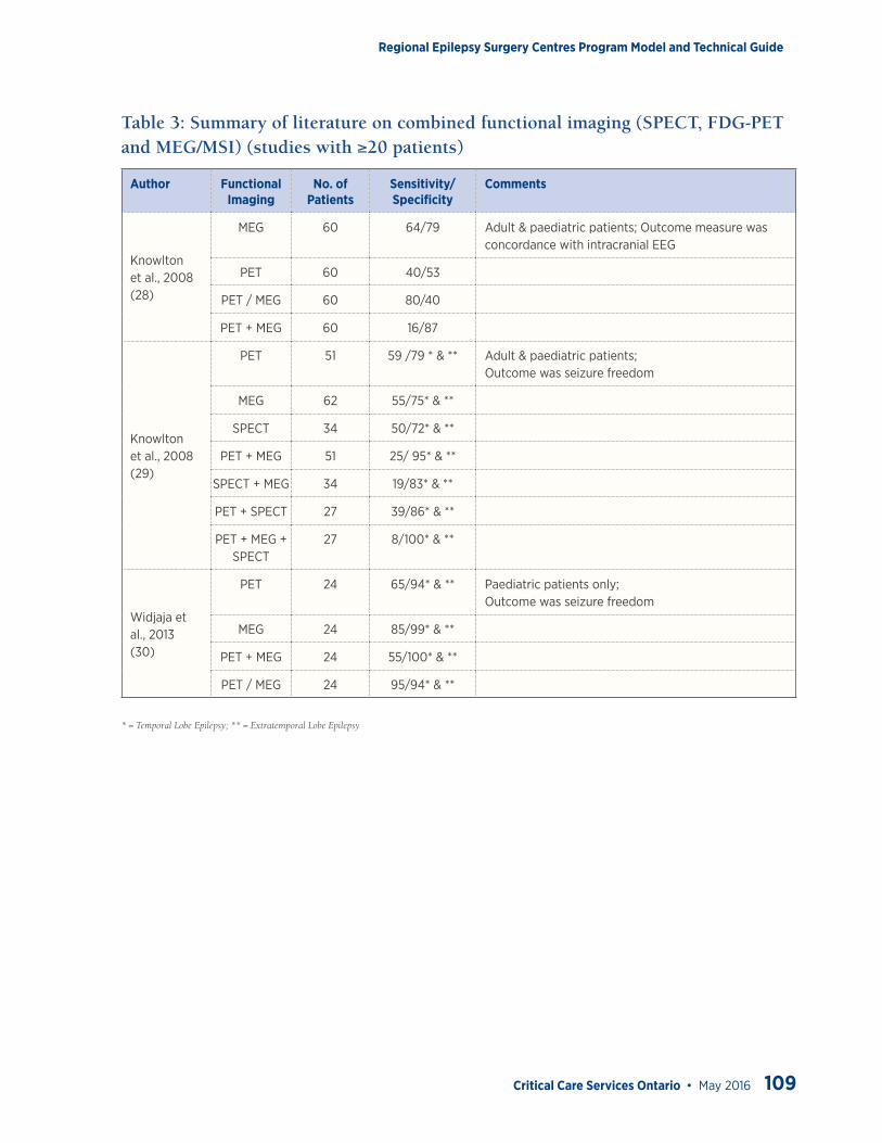
Regional Epilepsy Surgery Centres Program Model and Technical Guide
Critical Care Services Ontario • May 2016 109
Table 3: Summary of literature on combined functional imaging (SPECT, FDG-PET and MEG/MSI) (studies with ≥20 patients)
Author Functional Imaging
No. of Patients
Sensitivity/ Specificity
Comments
Knowlton et al., 2008 (28)
MEG 60 64/79 Adult & paediatric patients; Outcome measure was concordance with intracranial EEG
PET 60 40/53
PET / MEG 60 80/40
PET + MEG 60 16/87
Knowlton et al., 2008 (29)
PET 51 59 /79 * & ** Adult & paediatric patients;Outcome was seizure freedom
MEG 62 55/75* & **
SPECT 34 50/72* & **
PET + MEG 51 25/ 95* & **
SPECT + MEG 34 19/83* & **
PET + SPECT 27 39/86* & **
PET + MEG + SPECT
27 8/100* & **
Widjaja et al., 2013 (30)
PET 24 65/94* & ** Paediatric patients only;Outcome was seizure freedom
MEG 24 85/99* & **
PET + MEG 24 55/100* & **
PET / MEG 24 95/94* & **
* = Temporal Lobe Epilepsy; ** = Extratemporal Lobe Epilepsy
Regional Epilepsy Surgery Centres Program Model and Technical Guide
110 Critical Care Services Ontario • May 2016
Appendix 9: Psychosocial Procedures — Periodic Psychological Review of Patient
Patient is a Surgical CandidatePost-operatively, ongoing screening by a member of the mental health care team is recommended at
approximately 1 month, 3 months, 6 months, 12 months, and 24 months.
Timelines are based on the typical time course for emerging postoperative depressive, anxiety, and
psychotic symptoms. Timelines allow for adjustment and recovery after surgery but also recognize that
significant psychosocial or mental health issues need to be identified in a timely fashion.
Elements of the post-surgical mental health evaluations should include routine assessments of:
• Mood
• Anxiety
• Psychosis
• Suicidal behaviour
• Family dynamics – including histories of trauma, mental health issues, and adverse experiences with the medical system
• Coping strategies
• Quality of life
• Disruptive behaviours including developmental and learning difficulties
Screening for suicidal ideation is mandatory and cannot be effectively identified via screening tools.
Assessments should be conducted with semi-structured interviews, as well as self- and clinician-rated
rating scales that have been validated for use in patients with epilepsy.
An understanding of psychological adjustment and adaptation along with the screening for psychosocial
or mental health issues can be identified at neurology follow up appointments.
Patient should be referred to the Epilepsy Community Agency for ongoing psychosocial support and help
with social adaptation and with transitioning back to school and work.
Patient is NOT a surgical candidateRefer patient to Epilepsy Community Agency for on-going psychosocial support.
Mental health care team to provide individuals diagnosed with PNES with clinical and community based
discharge plan.
Regional Epilepsy Surgery Centres Program Model and Technical Guide
Critical Care Services Ontario • May 2016 111
Recommended Assessment Tools:
Screening Instruments
• *Refers to a measure also used in EpLink Research Projects (Ontario Brain Institute)
• *MINI (MINI International Neuropsychiatric Interview) Version 6.0.0 Sheehan D, Lecrubier Y (2010)
• NDDIE (Neurological Disorders Depression Inventory for Epilepsy).
• Gilliam FG, Barry JJ, Hermann BP, Meador KJ, Vahle V, Kanner AM. Rapid detection of major depression in epilepsy: a multicentre study. Lancet Neurol. (2006) 5:399-405
For Mood and Anxiety:
• *GAD-7 (Generalized Anxiety Disorder 7-item Scale). Spitzer RL, Kroenke K, Williams JB, et al; A brief
measure for assessing generalized anxiety disorder: the GAD-7. Arch Intern Med. (2006) 166:1092-7.
• HADS (Hospital Anxiety and Depression Scale) Zigmond AS, Snaith RP. “The hospital anxiety and depression scale”. Acta Psychiatrica Scandinavica (1983) 67 : 361–370
• HRSD-17 (17-item Hamilton Rating Scale for Depression) Hamilton M. A rating scale for depression. Journal of Neurology, Neurosurgery and Psychiatry. (1960) 23: 56-62
• *MADRS (Montgomery–Åsberg Depression Rating Scale). Montgomery SA, Åsberg M (April 1979). A new depression scale designed to be sensitive to change. British Journal of Psychiatry (1979) 134: 382–89.
• *QIDS-SR (Quick Inventory of Depressive Symptomatology – Self Report). Rush AJ, Trivedi MH, Ibrahim HM, Carmody TJ, Arnow B, Klein DN, Markowitz JC, Ninan PT, Kornstein S, Manber R, Thase ME, Kocsis JH, Keller MB. The 16-item Quick Inventory of Depressive Symptomatology (QIDS) Clinician Rating (QIDS-C) and Self-Report (QIDS-SR): A psychometric evaluation in patients with chronic major depression. Biological Psychiatry (2003) 54:573-583
Global Improvement / Quality of Life:
• *CGI (Clinical Global Impression) Guy W: Clinical Global Impressions (CGI) Scale. Modified From: Rush J, et al.: Psychiatric Measures, APA, Washington DC, 2000.
• QOLIE-31 (Quality of Life in Epilepsy). Cramer JA, Perrine K, Devinsky O, Bryant-Comstock L, Meador K, Hermann B. Development and cross-cultural translations of a 31-item quality of life in epilepsy inventory. Epilepsia. (1998) 39:81-8
• * SDS (Sheehan Disability Scale). Sheehan KH, Sheehan DV: Assessing treatment effects in clinical trials with the
discan metric of the Sheehan Disability Scale. Int Clin Psychopharmacol (2008) 23:70-83
Regional Epilepsy Surgery Centres Program Model and Technical Guide
112 Critical Care Services Ontario • May 2016
Appendix 10: Commonly Used Abbreviations and Definitions in Epilepsy Guideline Series
AbbreviationsABNM American Board of Neurophysiologic Monitoring
ABPN American Board of Psychiatry and Neurology
ADHD attention deficit hyperactivity disorder
AED antiepileptic drug (also known as antiseizure or anticonvulsant drug)
AEF auditory evoked fields
AFNI Analysis of Functional NeuroImages
APA American Psychological Association
ASET American Society of Electroneurodiagnostic Technologists
ASNFR American Society of Functional Neuroradiology
BOLD blood-oxygen-level dependent (context: fMRI findings)
BScN Bachelor of Science in Nursing
CBRET Canadian Board of Registration of Electroencephalograph Technologists
CEP comprehensive epilepsy program
CMAP compound muscle action potentials
CNIM Certification in Neurophysiologic Intraoperative
CNO College of Nurses of Ontario
CPA Canadian Psychological Association
CPO College of Psychologists of Ontario
CPR/BCLS cardio pulmonary resuscitation/basic cardiac life support
CPSO College of Physicians and Surgeons of Ontario
CSF cerebral spinal fluid
CT computerized tomography
CUSA Cavitron Ultrasonic Surgical Aspirator
D.ABNM Diplomat of American Board of Neurophysiologic Monitoring
DBS deep brain stimulation
DCS direct cortical stimulation
DEC district epilepsy centre
DICOM Digital Imaging and Communications in Medicine
DSM Digital Switch Matrix
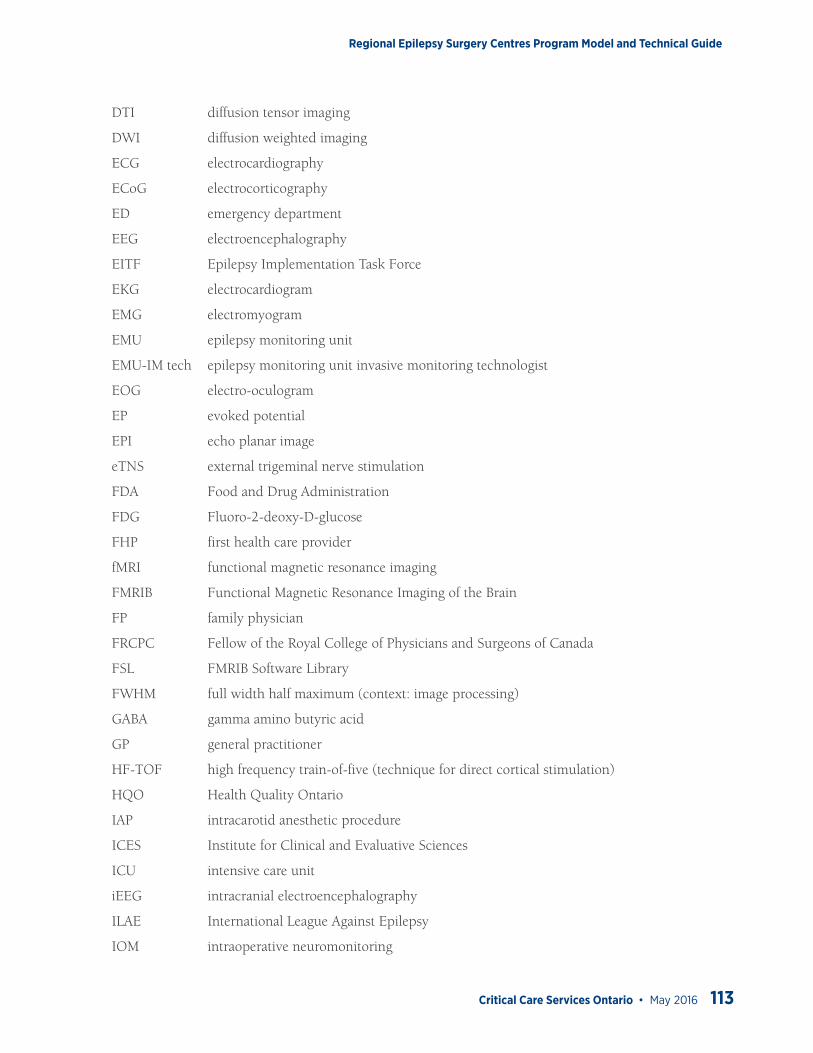
Regional Epilepsy Surgery Centres Program Model and Technical Guide
Critical Care Services Ontario • May 2016 113
DTI diffusion tensor imaging
DWI diffusion weighted imaging
ECG electrocardiography
ECoG electrocorticography
ED emergency department
EEG electroencephalography
EITF Epilepsy Implementation Task Force
EKG electrocardiogram
EMG electromyogram
EMU epilepsy monitoring unit
EMU-IM tech epilepsy monitoring unit invasive monitoring technologist
EOG electro-oculogram
EP evoked potential
EPI echo planar image
eTNS external trigeminal nerve stimulation
FDA Food and Drug Administration
FDG Fluoro-2-deoxy-D-glucose
FHP first health care provider
fMRI functional magnetic resonance imaging
FMRIB Functional Magnetic Resonance Imaging of the Brain
FP family physician
FRCPC Fellow of the Royal College of Physicians and Surgeons of Canada
FSL FMRIB Software Library
FWHM full width half maximum (context: image processing)
GABA gamma amino butyric acid
GP general practitioner
HF-TOF high frequency train-of-five (technique for direct cortical stimulation)
HQO Health Quality Ontario
IAP intracarotid anesthetic procedure
ICES Institute for Clinical and Evaluative Sciences
ICU intensive care unit
iEEG intracranial electroencephalography
ILAE International League Against Epilepsy
IOM intraoperative neuromonitoring
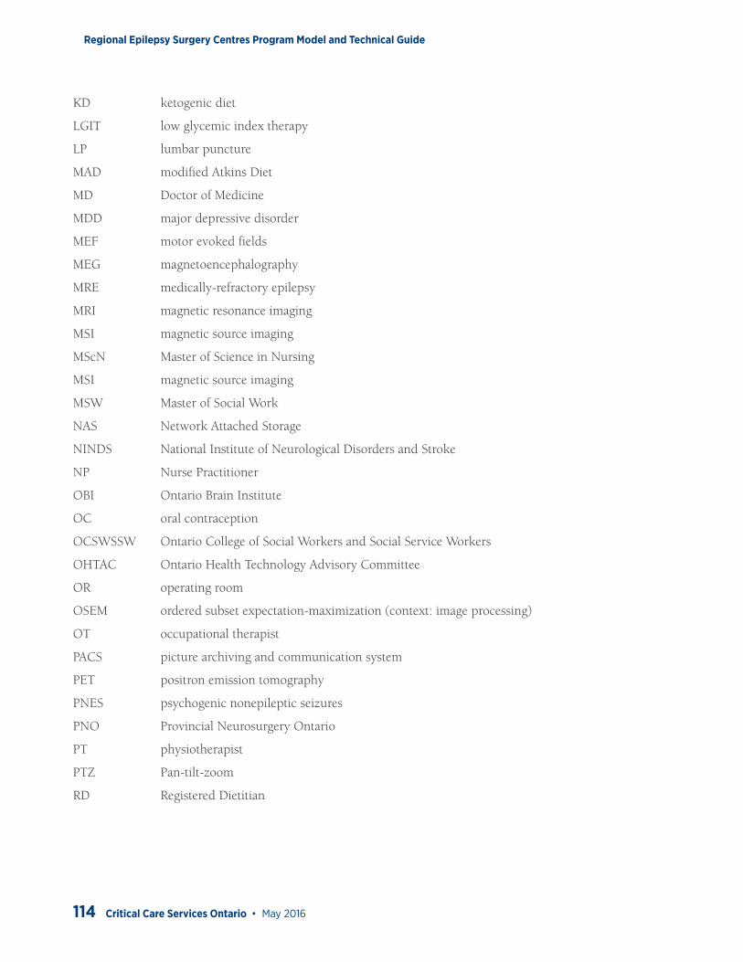
Regional Epilepsy Surgery Centres Program Model and Technical Guide
114 Critical Care Services Ontario • May 2016
KD ketogenic diet
LGIT low glycemic index therapy
LP lumbar puncture
MAD modified Atkins Diet
MD Doctor of Medicine
MDD major depressive disorder
MEF motor evoked fields
MEG magnetoencephalography
MRE medically-refractory epilepsy
MRI magnetic resonance imaging
MSI magnetic source imaging
MScN Master of Science in Nursing
MSI magnetic source imaging
MSW Master of Social Work
NAS Network Attached Storage
NINDS National Institute of Neurological Disorders and Stroke
NP Nurse Practitioner
OBI Ontario Brain Institute
OC oral contraception
OCSWSSW Ontario College of Social Workers and Social Service Workers
OHTAC Ontario Health Technology Advisory Committee
OR operating room
OSEM ordered subset expectation-maximization (context: image processing)
OT occupational therapist
PACS picture archiving and communication system
PET positron emission tomography
PNES psychogenic nonepileptic seizures
PNO Provincial Neurosurgery Ontario
PT physiotherapist
PTZ Pan-tilt-zoom
RD Registered Dietitian
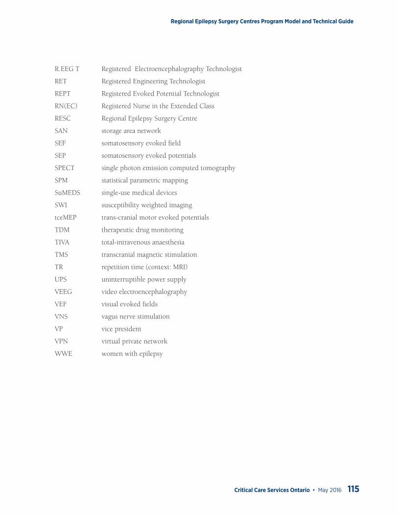
Regional Epilepsy Surgery Centres Program Model and Technical Guide
Critical Care Services Ontario • May 2016 115
R.EEG T Registered Electroencephalography Technologist
RET Registered Engineering Technologist
REPT Registered Evoked Potential Technologist
RN(EC) Registered Nurse in the Extended Class
RESC Regional Epilepsy Surgery Centre
SAN storage area network
SEF somatosensory evoked field
SEP somatosensory evoked potentials
SPECT single photon emission computed tomography
SPM statistical parametric mapping
SuMEDS single-use medical devices
SWI susceptibility weighted imaging
tceMEP trans-cranial motor evoked potentials
TDM therapeutic drug monitoring
TIVA total-intravenous anaesthesia
TMS transcranial magnetic stimulation
TR repetition time (context: MRI)
UPS uninterruptible power supply
VEEG video electroencephalography
VEF visual evoked fields
VNS vagus nerve stimulation
VP vice president
VPN virtual private network
WWE women with epilepsy
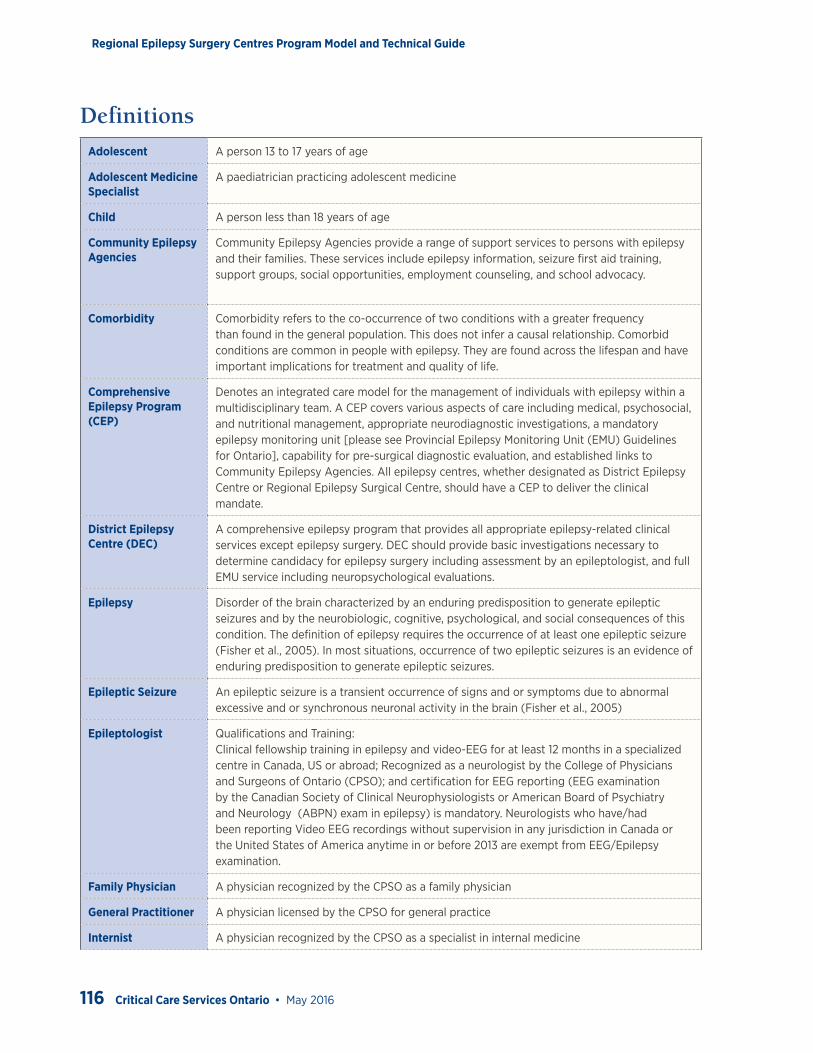
Regional Epilepsy Surgery Centres Program Model and Technical Guide
116 Critical Care Services Ontario • May 2016
DefinitionsAdolescent A person 13 to 17 years of age
Adolescent Medicine Specialist
A paediatrician practicing adolescent medicine
Child A person less than 18 years of age
Community Epilepsy Agencies
Community Epilepsy Agencies provide a range of support services to persons with epilepsy and their families. These services include epilepsy information, seizure first aid training, support groups, social opportunities, employment counseling, and school advocacy.
Comorbidity Comorbidity refers to the co-occurrence of two conditions with a greater frequency than found in the general population. This does not infer a causal relationship. Comorbid conditions are common in people with epilepsy. They are found across the lifespan and have important implications for treatment and quality of life.
Comprehensive Epilepsy Program (CEP)
Denotes an integrated care model for the management of individuals with epilepsy within a multidisciplinary team. A CEP covers various aspects of care including medical, psychosocial, and nutritional management, appropriate neurodiagnostic investigations, a mandatory epilepsy monitoring unit [please see Provincial Epilepsy Monitoring Unit (EMU) Guidelines for Ontario], capability for pre-surgical diagnostic evaluation, and established links to Community Epilepsy Agencies. All epilepsy centres, whether designated as District Epilepsy Centre or Regional Epilepsy Surgical Centre, should have a CEP to deliver the clinical mandate.
District Epilepsy Centre (DEC)
A comprehensive epilepsy program that provides all appropriate epilepsy-related clinical services except epilepsy surgery. DEC should provide basic investigations necessary to determine candidacy for epilepsy surgery including assessment by an epileptologist, and full EMU service including neuropsychological evaluations.
Epilepsy Disorder of the brain characterized by an enduring predisposition to generate epileptic seizures and by the neurobiologic, cognitive, psychological, and social consequences of this condition. The definition of epilepsy requires the occurrence of at least one epileptic seizure (Fisher et al., 2005). In most situations, occurrence of two epileptic seizures is an evidence of enduring predisposition to generate epileptic seizures.
Epileptic Seizure An epileptic seizure is a transient occurrence of signs and or symptoms due to abnormal excessive and or synchronous neuronal activity in the brain (Fisher et al., 2005)
Epileptologist Qualifications and Training:Clinical fellowship training in epilepsy and video-EEG for at least 12 months in a specialized centre in Canada, US or abroad; Recognized as a neurologist by the College of Physicians and Surgeons of Ontario (CPSO); and certification for EEG reporting (EEG examination by the Canadian Society of Clinical Neurophysiologists or American Board of Psychiatry and Neurology (ABPN) exam in epilepsy) is mandatory. Neurologists who have/had been reporting Video EEG recordings without supervision in any jurisdiction in Canada or the United States of America anytime in or before 2013 are exempt from EEG/Epilepsy examination.
Family Physician A physician recognized by the CPSO as a family physician
General Practitioner A physician licensed by the CPSO for general practice
Internist A physician recognized by the CPSO as a specialist in internal medicine
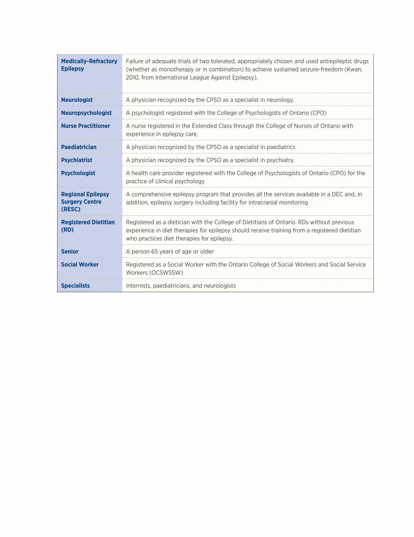
Medically-Refractory Epilepsy
Failure of adequate trials of two tolerated, appropriately chosen and used antiepileptic drugs (whether as monotherapy or in combination) to achieve sustained seizure-freedom (Kwan, 2010, from International League Against Epilepsy).
Neurologist A physician recognized by the CPSO as a specialist in neurology.
Neuropsychologist A psychologist registered with the College of Psychologists of Ontario (CPO)
Nurse Practitioner A nurse registered in the Extended Class through the College of Nurses of Ontario with experience in epilepsy care.
Paediatrician A physician recognized by the CPSO as a specialist in paediatrics
Psychiatrist A physician recognized by the CPSO as a specialist in psychiatry.
Psychologist A health care provider registered with the College of Psychologists of Ontario (CPO) for the practice of clinical psychology
Regional Epilepsy Surgery Centre (RESC)
A comprehensive epilepsy program that provides all the services available in a DEC and, in addition, epilepsy surgery including facility for intracranial monitoring
Registered Dietitian (RD)
Registered as a dietician with the College of Dietitians of Ontario. RDs without previous experience in diet therapies for epilepsy should receive training from a registered dietitian who practices diet therapies for epilepsy.
Senior A person 65 years of age or older
Social Worker Registered as a Social Worker with the Ontario College of Social Workers and Social Service Workers (OCSWSSW)
Specialists Internists, paediatricians, and neurologists
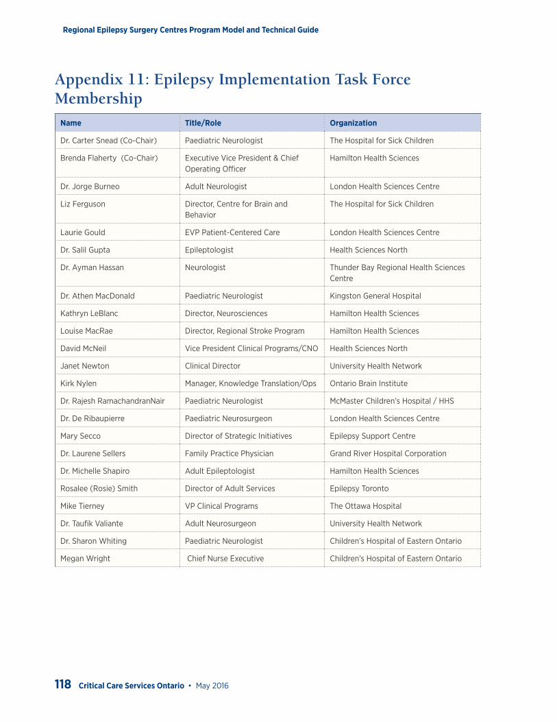
Regional Epilepsy Surgery Centres Program Model and Technical Guide
118 Critical Care Services Ontario • May 2016
Appendix 11: Epilepsy Implementation Task Force Membership
Name Title/Role Organization
Dr. Carter Snead (Co-Chair) Paediatric Neurologist The Hospital for Sick Children
Brenda Flaherty (Co-Chair) Executive Vice President & Chief Operating Officer
Hamilton Health Sciences
Dr. Jorge Burneo Adult Neurologist London Health Sciences Centre
Liz Ferguson Director, Centre for Brain and Behavior
The Hospital for Sick Children
Laurie Gould EVP Patient-Centered Care London Health Sciences Centre
Dr. Salil Gupta Epileptologist Health Sciences North
Dr. Ayman Hassan Neurologist Thunder Bay Regional Health Sciences Centre
Dr. Athen MacDonald Paediatric Neurologist Kingston General Hospital
Kathryn LeBlanc Director, Neurosciences Hamilton Health Sciences
Louise MacRae Director, Regional Stroke Program Hamilton Health Sciences
David McNeil Vice President Clinical Programs/CNO Health Sciences North
Janet Newton Clinical Director University Health Network
Kirk Nylen Manager, Knowledge Translation/Ops Ontario Brain Institute
Dr. Rajesh RamachandranNair Paediatric Neurologist McMaster Children’s Hospital / HHS
Dr. De Ribaupierre Paediatric Neurosurgeon London Health Sciences Centre
Mary Secco Director of Strategic Initiatives Epilepsy Support Centre
Dr. Laurene Sellers Family Practice Physician Grand River Hospital Corporation
Dr. Michelle Shapiro Adult Epileptologist Hamilton Health Sciences
Rosalee (Rosie) Smith Director of Adult Services Epilepsy Toronto
Mike Tierney VP Clinical Programs The Ottawa Hospital
Dr. Taufik Valiante Adult Neurosurgeon University Health Network
Dr. Sharon Whiting Paediatric Neurologist Children’s Hospital of Eastern Ontario
Megan Wright Chief Nurse Executive Children’s Hospital of Eastern Ontario