Embed Size (px)
Citation preview

Biochemical Pharmacology, Vol. 17, pp. 2107-2116. Pergamon Press. 1968. Printed in Great Britain
REGIONAL AND SUBCELLULAR DISTRIBUTION OF ADENYL CYCLASE AND 3’,5’-CYCLIC NUCLEOTIDE
PHOSPHODIESTERASE IN BRAIN AND PINEAL GLAND*t
BENJAMIN WEISS: and E. COSTA:
Departments of Pharmacology and Neurology, College of Physicians and Surgeons, Columbia University,
New York, N.Y., U.S.A.
(Received 27 February 1968; aecepfed 11 April 1968)
Abstract-Adenyl cyclase was assayed in rho by measuring the rate of formation of radioactive cyclic 3’,5’-adenosine monophosphate (cyclic 3’,5’-AMP) from itsi4C-labeled precursor, adenosine triphosphate, and phosphodiesterase activity was determined from the rate of hydrolysis of cyclic 3’,5’-AMP.
Studies of the distribution of these two enzymes in various parts of rat brain revealed that, in general, enzyme activity was higher in those areas containing grey matter (cerebral cortex) than in areas consisting predominantly of white matter (pans, meduha, spinal cord). However, there was no correlation between the relative activities of adenyl cyclase and of phosphodiesterase in the various areas of the brain; the greatest discrepancy was found in the cerebellum, corpora quadrigemina and pineal gland where, relative to other brain areas, there was high adenyl cyclase activity compared with that of phos- phodiesterase. Moreover, the distribution pattern of adenyl cyclase was different from that of the catecholamine concentration in the brain.
Results of differential centrifu~tion of bovine pineal gland and of rat cerebellar homogenates indicated that most of the adenyl cyclase is particulate, being in the mitochondrial and microsomal fractions, whereas the majority of the phosphodiesterase is soluble. The sp. act. of the mitochondrial and microsomal fractions of pineal adenyl cyclase was about four times that of the whole homogenate. No such increase in specific activity was obtained with cerebellar homogenates. The possibility that an activator of adenyl cyclase is present in soluble supernatant fractions of cerebellar homogenates is discussed.
THE NUCLEOTIDE, cyclic 3’,5’-adenosine monophosphate (cyclic 3’,5’-AMP) elicits effects in several tissues similar to those of a wide variety of hormones and transmitters. For example, cyclic 3’,.5’-AMP simulates the glycogenolysis elicited by catecholamines on liver homogenate9 and the lipolysis induced by the amines on fat pad homogenates2 and on intact fat cells.3 In addition, cyclic 3’,5’-AMP induces steroidogenesis in adrenal cortex4 and in corpus luteum,5 thus mimicking the actions of adrenocorticotropic hormone and leuteinizing hormone respectively. Moreover, like serotonin, this nucleotide stimulates phosphofructokinase activity of the liver fluke.6 Since many hormones and transmitters increase tissue concentrations of cyclic 3’,5’-AMP, it has
* A preliminary report of the distribution and subcellular Iocalization of adenyl cycfase and phosphodieste~se was presented at the Seventh ~ntemational Congress of Biochemistry, Tokyo, 1967.
I’ This work was supported in part by the Clinical Research Center for Parkinson’s and Allied Diseases, NB 05184, and the Parkinson Information Center, a part of the National information Network of NINDB under contract PH 43 64 54. iPresent address: Laboratory of Preclinical Pharmacology, National Institute of Mental Health, St. Elizabeth’s Hospital, Washington, DC 20032.
2107

2108 BENJAMIN WEIFS and E. COSTA
been proposed that this cyclic nucleotide mediates the response of these compounds.79 s Increased tissue levels of cyclic 3’,5’-AMP may be caused either by an enhancement
of the activity of adenyl cyclase, the enzyme that catalyzes the conversion of adenosine triphosphate (ATP) to cyclic 3’,5’-AMP,” or by an inhibition of 3’,5’-cyclic nucleotide phosphodiesterase, the enzyme that catalyzes its hydrolysis to 5’-AMP.lo Since these enzymes are present in practically all tissues and their activities are especially high in brain,Y, lo they may play a major role in regulating the cellular responses to a number of hormones and chemical mediators of nerve impulses.
Generally, phosphodiesterase activity is greater than that of adenyl cyclase; despite this discrepancy, cyclic 3’,5’-AMP accumulates in tissues, suggesting that the two enzymes are compartmentalized in distinct subcellular sites.
The subcellular distribution of phosphodiesterase has been studied by several investigators with the following results. Nairll reported that more than half of the phosphodiesterase of dog heart is in the 105,000 g supernatant fluid, and Drummond and Perrott-Yeels found that the phosphodiesterase activity of rabbit brain homo- genates is localized wholly in the 100,OOOg supernatant fraction. However, others have reported13 a microsomal localization of phosphodiesterase activity of rat brain. Since these investigators did not assay adenyl cyclase on the same fraction, it is di~cult to assess the relative subcellular distribution of the two enzymes. In studies of rat cerebral homogenates, we found that about 50 per cent of the adenyl cyclase was in the crude mitochondrial fraction, whereas the majority of the phosphodiesterase was in the supernatant fraction.1” Recently, De Robertis et ~1.~5 reported that in rat brain cortex, about 65 per cent of the phosphodiesterase and essentially all of the adenyl cyclase was particulate.
The present study was undertaken to determine the distribution of adenyl cyclase and phosphodiesterase in discrete areas of rat brain and to examine the subcellular localization of adenyl cyclase and of phosphodiesterase in the cerebellum and pineai gland.
The pineal gland was chosen because pineai adenyl cyclase is stimulated specifically by pharmacologically active catecholamines. 16 Furthermore, in contrast with other areas of the brain, the pineal gland is innervated exclusively by fibers originating from the sympathetic nervous system ;17 thus, this gland is provided with postjunctional sites which are solely adrenergic in character.
METHODS
Male Sprague-Dawley rats (180-220 g) were used in investigations of the distribution of adenyl cyclase and of phosphodiesterase. The animals were sacrificed by decapitation and the brain was removed. Specific brain areas were dissected with the aid of the atlas of Konig and Klippel.l* Bovine pineal glands were obtained from the Allen Meat Packing Co., Elizabeth, N.J. The glands were removed and stored on ice within 30 min after the animals were killed by carotid exsanguination.
Phosphodiesterase activity was determined by the method of Butcher and Sutherland10 with certain modifications.3 Briefly, the tissues were homogenized in Tris hydrochloride buffer (O*OS M), pH 8.0, containing MgSOa (3 * 1O-3 M), and centri- fuged for 10 min at 1OOOg. The supernatant fluid was incubated with cyclic 3’,5’-AMP (10-a M) for I5 min at 30”. The 5’-AMP formed was determined from the amount of inorganic phosphate released under the catalytic influence of alkaline phosphatase

Distribution of adenyl cyclase and phosphodiesterase 2109
added in excess. Phosphate was analyzed according to the procedure of Buell et ~~1.19 Adenyl cyclase was determined by methods previously described.169 2o The assay is
based on measuring the rate of formation of carbon-labeled cyclic 3’,5’-AMP from its radioactive precursor, ATP-8JJC. The tissues were homogenized in Tris-HCI buffer (0.05 M), pH 7.4, and the whole homogenate (equivalent to approximately 100 !Lg protein) was incubated with 1 pc ATP-8-l% (2 9: 10-a M), NaF (1O-2 M), “carrier” cyclic 3’,5’-AMP (3 x 10-s M) or theophylline (10-s M) and Mg-+ 1~ (3 i: 10-a M) in 100 ~1 Tris-HCI buffer (0.05 M), pH 7.4, at 30” for 10 min. Theophylline or “carrier” cyclic 3’,5’-AMP was added to the incubation vessels to prevent the destruction of the newly formed radioactive cyclic 3’,5’-AMP. Theophylline inhibits phospho- diesterase,s, 10 thus preventing the enzymatic hydrolysis of cyclic 3’,5’-AMP. Carrier cyclic 3’,5’-AMP effectively traps the radioactive cyclic 3’,5’-AMP without interfering with its formation. The rate of accumulation of radioactive cyclic 3’,5’-AMP was shown to be similar regardless of which procedure was used to prevent the hydrolysis of the labeled product.16
From the O.D. (260 mp) of the final purified material, the recovery of cyclic 3’,5’-AMP in each sample was determined and appropriate corrections were made. If carrier cyclic 3’,5’-AMP was not added at the beginning of the incubation period, it was added at the end of the incubation period and carried through the purification steps to correct for the recovery of the cyclic nucleotide. The cyclic nucleotide was separated from contaminating products by adsorption onto, and elution from, Dowex 50 ion-exchange columns. Cyclic 3’,5’-AMP was further purified by precipitating any residual trace contaminants with the addition of zinc sulfate and barium hydroxide solutions. These procedures result in the recovery of cyclic 3’,5’- AMP in a chromatographically and crystallographically pure form.“’
In studies of the subcellular distribution of adenyl cyclase and of phosphodiesterase, the tissue was homogenized at 0” for 30 set in 0.32 M sucrose containing magnesium sulfate (3 x 10-s M) with a Dual1 glass tissue grinder and Teflon pestle having a clearance of 0.1 to 0.15 mm and fractionated by differential centrifugation, according to the procedures described by Gray and Whittakeraa and by De Robertis et a1.23 A more detailed description of centrifugation procedures and alternate methods used in various experiments is described in the legends to the tables.
Materials. Adenosine triphosphate (ATP)-8-r4C disodium (crystalline) was pur- chased from Schwarz Bioresearch. Inc.; cyclic 3’,5’-AMP from Calbiochem; alkaline phosphatase, type 111, from Sigma Chemical Co. Other chemicals and reagents were purchased from general commercial sources.
RESULTS
Distribution of adeny cyclase atldphosphodiesterase itI rat brain. Table 1 shows the distribution of adenyl cyclase and of phosphodiesterase in various parts of rat brain. Relatively high adenyl cyclase activity was found in the cerebral cortex, corpora quadrigemina, cerebellum, olfactory bulb, thalamus and pineal gland, whereas the pans, medulla and spinal cord exhibited relatively low enzyme activity. Areas of high phosphodiesterase activity were cerebral cortex, caudate nucleus and septal region, while low activity was found in the corpora quadrigemina, cerebellum, pons, spinal cord and pineal gland. Thus, the areas of high adenyl cyclase activity do not necessarily correspond to the areas of high phosphodiesterase activity; the greatest discrepancy

2110 BENJAMIN WEISS and E. COSTA
was found in the corpora quadrigemina, cerebellum and pineal gland where, relative to other brain areas, adenyl cycfase activity was high compared with that of phos- phodiesterase. It should be noted that Table 1 presents adenyl cyclase activity of the whole homogenate and phosphodiesterase activity only of the high-speed supernatant fraction (which contains about half of the total activity). Taking into account the differences in specific activity of the latter enzyme in various subcellular fractions (see Table 2), these measurements in ~.itro revealed that in most of the brain areas studied the pIlosphodiesterase activity exceeded that of adenyl cyclase by almost IOO-fold.
TABLE I. DISTRIBUTION OF ADENYL CYCLASE AND PHOSPHODIESTERASE
IN RAT BRAIN
Tissue Adenyl cycIase* Phosphodiesteraset
Parietai cortex Corpora quadrigenl Olfactory bulb Thalamus Pineal gland Hippocampus Cerebellum Olfactory tuber& Septum and fornix Hypothalamus Caudate nucleus Medulla Pons Spinal cord
028 k 0.06 49 r 2 0.26 -L 0.04 Ii c 1 0.25 z 0.01 19i, 2 0.24 i: 0.06 24 :r 2 0.21 .c 0.02 10 :k 2 0.20 ._t 0.02 48 _?. 3 0.20 ..t 0.04 7 _t 1 0.19 :i. 0.01 25 rir 4 0.16 -i 0.03 30 + 3 @ I 5 :+ 0@4 25 :* 1 0.14 -.k 0.03 32 r> 2 0.13 jr 0.02 11 ck I 0.10 .k 0.02 11 -A I 0.08 rt. 0.01 8’1
* Adenyl cyclase, mpmoles cyclic 3’,5’-AMP formed/mg protein/min + SE. Tissues were homogenized in Tris-HCl buffer (@05 M), pH 7.4, containing MgSOs (3 y. 10d3 M). The whole homogenate was assayed for adenyl cyclase activity by incubation with ATP-8-W (2 j, lO-3 M), sodium fluoride (1 x lo-” M), theophylline (1 x 10M2 M) and MgS04 (3 ‘K IO-3 M) in Tris-HCI buffer (0.05 M) as described in Methods. Each figure represents the mean of 5 experiments.
7 Phosphodiesterase, mpmoles cyclic 3’,5’-AMP hydrolyzed/ mg protei~/min + S.E. Tissues were homogenized in Tris-HC1 buffer (0.05 M), pH 8.0, containing MgSOs (3 x IO -3 M). The homogenate was centrifuged at 1000 g for 20 min. The super- natant fluid was assayed for phosphodiesterase activity as described in Methods. Each figure represents the mean of 6 experiments.
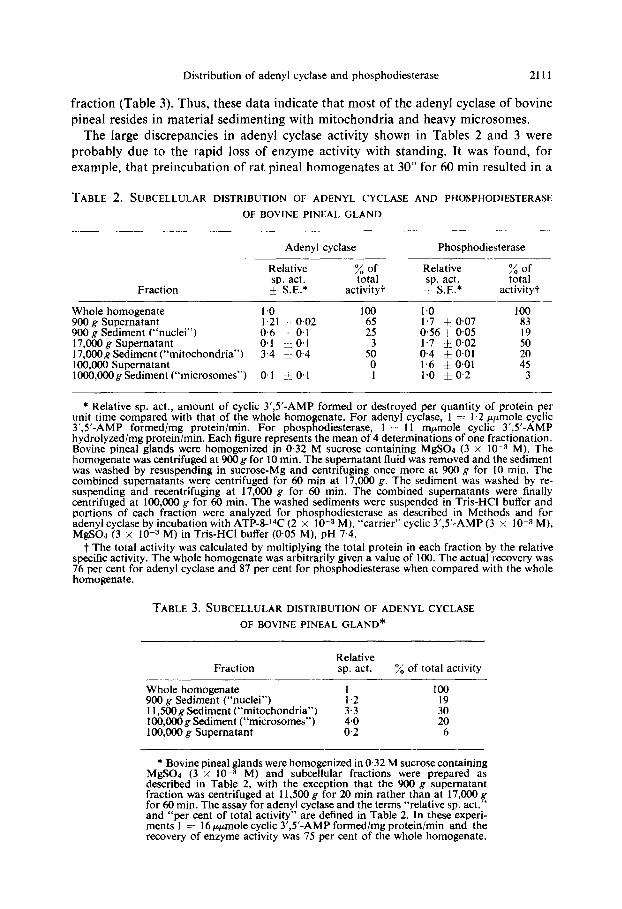
Subcellular d~str~buti~il of adenyl q&use arld of p~losp~lod~est~r~~~~ in pineai gkwd. Studies of the subcelIular distribution of adenyl cyclase and of phosphodiesterase in the bovine pineal gland showed that the majority of phosphodiesterase activity is in the soluble supernatant fraction of a 100,000 g centrifugation, whereas most of the adenyl cyclase activity resides in the crude mitochondrial fraction (Table 2). The specific activity of adenyl cyclase in the crude mitochondrial fractions was more than three times that of the whole homogenate, and the specific activity of phosphodiesterase in the soIuble fraction was about two times that of the whole homogenate.
When bovine pineal homogenates were fractionated at centrifugations of 900 g, 11,500 g and 10,000 g rather than at 900 g, 17,000 g and 100,000 g, the increase in specific activity of adenyl cyclase in the “mitochondrial” and “microsomal” fractions was similar and about 20 per cent of the total activity sedimented with the “microsomal”
Distribution of adenyl cyclase and phosphodiesterase 2111
fraction (Table 3). Thus, these data indicate that most of the adenyl cyclase of bovine
pineal resides in material sedimenting with mitochondria and heavy microsomes. The large discrepancies in adenyl cyclase activity shown in Tables 2 and 3 were
probably due to the rapid loss of enzyme activity with standing. It was found, for example, that preincubation of rat pineal homogenates at 30” for 60 min resulted in a
TABLE 2. SUBCELLULAR DISTRIBUTION OF ADENYL CYCLASE AND PHOSPHODIESTERASE
OF BOVINE PINEAL GLAND
Fraction
Adenyl cyclase Phosphodiesterase
Relative sp. act. I SE.*
% of total
activity? -
Whole homogenate 900 g Supematant 900 g Sediment (“nuclei”) 17,000 g Supematant 17,OOOg Sediment (“mitochondria”) 100,000 Supematant lOOO,OOOg Sediment (“microsomes”)
I.0 100 I.21 -I- 0.02 0.6 -I: 0.1 ;:
0.1 $ 0.1 3.4 * 0.4 5: 0
0.1 ‘: 0.1 I
Relative sp. act. -+ SE.’
Zt:: activity?
I.0 100 I.7 5 0.07 83 0.56 + 0.05 19 1.7 IO.02 0.4 * 0.01 :: 1.6 & 0.01 45 1.0 * 0.2 3
* Relative sp. act., amount of cyclic 3’,5’-AMP formed or destroyed per quantity of protein per unit time compared with that of the whole homogenate. For adenyl cyclase, 1 = 1.2 ppmole cyclic 3’,5’-AMP formed/mg proteimmin. For phosphodiesterase, 1 = 11 mrmole cyclic 3’,,5’-AMP hydrolyzedimg proteinimin. Each figure represents the mean of 4 determinations of one fracttonation. Bovine pineal glands were homogenized in 0.32 M sucrose containing MgSOr (3 x 10m3 M). The homogenate was centrifuged at 900g for 10 min. The supematant fluid was removed and the sediment was washed by resuspending in sucrose-Mg and centrifuging once more at 900 g for 10 min. The combined supematants were centrifuged for 60 min at 17,000 g. The sediment was washed by re- suspending and recentrifuging at 17,000 g for 60 min. The combined supematants were finally centrifuged at 100,000 g for 60 min. The washed sediments were suspended in Tris-HCI buffer and portions of each fraction were analyzed for phosphodiesterase as described in Methods and for adenyl cyclase by incubation with ATP-8-l‘C (2 x 10e3 M), “carrier” cyclic 3’,5’-AMP (3 x lO-3 M), MgS04 (3 x 1O-3 M) in Tris-HCI buffer (005 M), pH 7.4.
t The total activity was calculated by multiplying the total protein in each fraction by the relative specific activity. The whole homogenate was arbitrarily given a value of 100. The actual recovery was 76 per cent for adenyl cyclase and 87 per cent for phosphodiesterase when compared with the whole homogenate.
TABLE 3. SUBCELLULAR DISTRIBUTIONOFADENYLCYCLASE
OF BOVINE PINEAL GLAND*
Fraction Relative sp. act. % of total activity
Whole homogenate 100 900 g Sediment (“nuclei”) t.2 I9 11,SOOg Sediment (“mitochondria”) 100,000 g Sediment (“microsomes”) ::; :“o 100,000 g Supematant 0.2 6
* Bovine pineal glands were homogenized in 0.32 M sucrose containing MgS04 (3 x 1O-3 M) and subcellular fractions were prepared as described in Table 2, with the exception that the 900 g supematant fraction was centrifuged at 11,500 g for 20 min rather than at 17,000 g for 60 min. The assay for adenyl cyclase and the terms “relative sp. act.” and “per cent of total activity” are defined in Table 2. In these experi- ments I = 16 ppmole cyclic 3’,5’-AMP formed/mg protein/min and the recovery of enzyme activity was 75 per cent of the whole homogenate.

2112 BENJAMIN WEISS and E. COSTA
75 per cent loss of adenyl cyclase activity (Weiss, unpublished). It was difficult to control the interval between the time the steers were sacrificed and the time the pineals were removed, transported back to the laboratory, fractionated and assayed. Processing and assay of rat pineal glands could be conducted more rapidly (within 30 set) and controlled more readily, thus accounting for the considerably greater enzyme activity of rat pineals (see Table 1).
Subcellular distribution qf‘ adeny cyclase qf rat cerebelhm. Table 4 shows that adenyl cyclase of subcellular fractions of rat cerebellum was distributed most13
TABLE 4. SUBCELLULAR DISTRIBUTION OF ADENYL. CYCI.ASI:
OF RAT CEREBELLUM”
Fraction Sp. act.? 7, of total activity
Whole homogenate 193 j_ 14 100 900 g Sediment (“nuclei”) 133 + 12 41 I 1,500~ Sediment (“mitochondria”) 134+ 2 21 17,OOOg Sediment (“mitochondria
and heavy microsomes”) 98 _i_ I 7 100,000~ Sediment (“microsomes”) 613. 1 ? 100,000 g Supernatant 0 0
* Rat cerebella were homogenized in0’32 M sucrose containing MgS04 (3 x 1O-3 M). The homogenates were centrifuged and the following fractions were sedimented, washed and assayed for adenyl cyclase activity: 900 g for 10 min; 11,500 g for 20 min; 17,000 g for 60 min; 100,OOOg for 60 min; 100,000g supernatant fluid. Adenyl cyclase activity was determined as described in Methods. Each value is the mean of 4 determinations of one fractionation. The recovery of adenyl cyclase was 71 per cent of the whole homogenate.
i Cyclic 3’,5’-AMP formed (~~moles/mg protein/min) _t S.E.
between the “nuclear” and “mitochondrial” fractions. In contrast with the bovine pineal gland, fractionation of the cerebellum resulted in a decrease in the specific activity of the enzyme. The possibility that this decrease was due to the removal of a soluble activator of the enzyme was examined. The experiment was repeated, adding a portion of the soluble supernatant fraction to each of the other subcellular fractions. The results shown in Table 5 indicate that the soluble supernatant material, which itself had no adenyl cyclase activity, significantly enhanced the adenyl cyclase activity of the 11,500 g (mitochondrial) fraction. However, adenyl cyclase activity of other fractions was not increased, and the specific activity of the mitochondrial fraction was
still less than that of the whole homogenate.
DISCUSSION
Investigations of the distribution of adenyl cyclase in various regions of the rat brain showed that enzyme activity was high in cerebral cortex, thalamus, corpora quadrigemina, olfactory bulb, cerebellum and pineal gland, and relatively low in the spinal cord, medulla and pons. Since it has been proposed that this enzyme is involved in the mediation of responses of various cells to catecholamines,7* 34 it is of interest to compare the distribution of adenyl cyclase with that of the catecholamines in various brain areas. No correlation was found between enzyme activity and catechol- amine content. For example, the concentration of norepinephrine is about ten times

Distribution of adenyl cyclase and phosphodiesterase 2113
higher in the rat hypothalamus than in the cerebellum or cortex,2s whereas, as can be seen from Table 1, there is more adenyl cyclase activity in the cortex and cerebellum than in the hypothalamus. However, a lack of positive correlation does not necessarily negate the proposal that the amines have a functional role in stimulating adenyl cyclase in brain. The catecholamines have been shown to enhance enzyme activity ilz t+tro in cerebellums6 and in pineal gland. 16, 20. Moreover, a low concentration of
TABLE 5. EFFECT OF SOLUBLE SUPERNATANT FRACTION ON ADENYL CYCLASE ACTIVITY OF
SUBCELLULAR FRACTIONS OF RAT CEREBELLAR HOMOGENATES*
Cyclic 3’,5’-AMP formed (ppmoles/mg protein/min i S.E.)
Fraction
Without 100,000 g
supematant
With 100,~ g
supernatantt
Whole homogenate 427 i 19 347 + 52 900 g Sediment (“nuclei”) 329 + 11 339 + 22 11,500 g Sediment (“mitochondria”) 145i 9 232 h 22: 17,OOOg Sediment (“mitochondria
and heavy microsomes”) 278 f 4 251 & 26 100,000 g Sediment (“microsomes”) 150* 4 130 + 12 100,000 g Supematant 0 0
* Rat cerebella were homogenized and fractionated as described in Table 4. Adenyl cyclase activity was determined as described in Methods. Each value is the mean of 4 determinations of one fractionation. The total recovery of adenyl cyclase activity was 55 per cent of the whole homogenate.
t The supematant fluid of a 100,000 g centrifugation, equivalent to 90 pg protein, was added to each of the other fractions. The amount of protein contributed by the supematant fraction was not considered in calculations of the specific activity of adenyl cyclase.
$ The addition of the soluble supernatant fraction significantly (P i 0.05) increased adenyl cyclase activity of the I 1,500 g sediment.
catecholamines does not necessarily indicate a lack of function for the amines, since (a) turnover rather than amine concentration is a more reliable index of catecholamine functions7 (b) catecholamines may be highly concentrated in nerve endings close to receptor sites, and since nerve endings constitute only a small fraction of the tissue weight, the concentration of catecholamines in tissue will not give a true indication of the concentrations of amines at these sites. There is mounting evidence that adrenergic receptor sites may involve adenyl cyclase. For example, adenyl cyclase is concentrated in fractions containing synaptic membranes,‘” and in the pineal gland most of the enzyme is situated at non-neuronal sites rather than in nerve endings20 and its activity is stimulated selectively by pharmacologically active catecholamines.16 Thus, it has several characteristics similar to those of the adrenergic receptor. On the other hand, the inability of catecholamines to enhance adenyl cyclase activity in many areas of brain (Weiss, unpublished observations) indicates that there are other compounds, as yet unidentified, which may be the physiologic stimulants of brain adenyl cyclase. The evidence that adenyl cyclase of different tissues is stimulated by such a wide variety of hormone+ favors this proposal.
Our findings on the distribution of phosphodiesterase in rat brain agree with the results of Drummond and Perrott-Yee ,12 who found that in rabbit brain the activity of
2114 BENJAMIN WEISS and E. COSTA
cerebral cortex was much higher than that of the pons, medulla or cerebellum. However, our results show further that phosphodiesterase activity is as high in the hippocampus as it is in the cerebral cortex, and activities almost as great were found in the caudate nucleus and septal nuclei. Thus, there is a gradation of phosphodiesterase activity in brain, being relatively low in structures which predominate in white matter and high in areas rich in grey matter. In this regard, the histochemical studies of Shanta et al.ss are particularly instructive. They showed that phosphodiesterase activity was low in neurons but high in the molecular layer of the cerebellum and in the plexiform layer of the cerebral cortex.
An examination of the relative distribution of adenyl cyclase and of phospho- diesterase activities in various brain areas showed no correlation between the two enzymes. The greatest discrepancy in enzyme activities was in the cerebellum, corpora quadrigemina and the pineal gland where the ratio of adenyl cyclase activity to phosphodiesterase activity was high relative to that of other brain areas.
Studies of the subcellular distribution of adenyl cyclase and of phosphodiesterase in the bovine pineal gland showed a different distribution of the two enzymes. Adenyl cyclase of the pineal gland, like that of brain cerebrum,14 cerebral cortex15 and skeletal muscle,“9 is almost completely particulate, being concentrated in the mitochondrial and microsomal fractions. However, most of the phosphodiesterase is soluble. The high percentage of phosphodiesterase found in the soluble fraction relative to the particulate is similar to the results of Drummond and Perrott-Yeets on brain phos- phodiesterase and to those of Nairii on the enzyme from dog heart. The latter author has shown further that repeated freezing of the tissue results in more phosphodiesterase activity in the soluble relative to the particulate fractions. On the other hand, Cheung and Salganicoffra demonstrated that most of the phosphodiesterase activity of brain was in particulate subfractioIls if Triton X-100 was added to the enzyme preparations. According to these workers, this treatment revealved the latent activity of the enzyme resulting in higher phosphodiesterase activity in the particulate than in the soluble fractions. Moreover, they showed that increased activity was not produced by mechanical disruption or by repeated freezing and thawing. Thus. considering all the data, it is apparent that phosphodiesterase is partly soluble and partly particulate; the activity of the particulate fraction can be solubilized by freezing and thawing and can be increased by the addition of the detergent Triton X- 100. It should be mentioned that the addition of Triton X-100 to preparations of adenyl cyclase results in its solubilization but causes a great loss of enzyme activity.Q It is possible that these phenomena are related. In the assay of adenyl cyclase, the formation of the cyclic 3’,5’-AMP is used as the measure of enzyme activity. Since Triton X-100 enhances phosphodiesterase activity, it may cause an increased destruction of cyclic 3’,5’-AMP
resulting in an apparent decrease in adenyl cyclase activity. A comparison of the regional and subcellular distribution of cyclic 3’,S’-nucleotide
phosphodiesterase activity, described herein, with that of 2’,3’-cyclic nucleotide
3’-phosphohydrolase, the enzyme which catalyzes the hydrolysis of cyclic 2’,3’-AMP to 2’-AMP, reveals that the two enzymes are clearly distinguishable. Kurihara and Tsukadaa” recently reported that the activity of the latter enzyme is much greater in cerebral white matter (e.g. corpus callosum) than in cerebral cortex and that cerebrum and cerebellum had approximately the same activity. Moreover, they found that more than 90 per cent of the activity was particulate. In contrast, the present results show
Distribution of adenyl cyclase and phosphodiesterase 211.5
that cyclic 3’,5’-nucleotide phosphodiesterase activity is almost ten times greater in cerebral cortex than in the cerebellum and that more than half of the activity is soluble.
It should be pointed out that there was nearly a 4-fold increase in the specific activity of adenyl cyclase in the mitochondrial and microsomal fractions of pineal gland, but no such increase in specific activity of any cerebellar fraction. This may be a reflection of the greater homogeneity of the nervous elements and, therefore, of the postjunctional receptor sites in pineal compared with that of cerebellum; it has been shown, for example, that catecholamines enhance adenyl cyclase activity of the pineal gland about 3-fold,16 but show little or no stimulation of cerebellar adenyl cyclase.31 On the other hand, the decrease in specific activity of subcellular fractions of cerebellar homogenates may indicate that some essential requirement or activator of the enzyme is removed by the fractionation procedures. Indeed, the addition of the soluble supernatant material, which itself had no adenyl cyclase activity, significantly increased enzyme activity when added to the mitochondrial fraction. The soluble fraction did not, however, enhance adenyl cyclase activity of other fractions. This may indicate either that the adenyl cyclases of various subfractions are different or that the soluble supernatant fluid contains a substance that antagonizes an inhibitor present in the mitochondrial fraction. A clarification of this mechanism must await isolation and further purification of the enzyme.
AckrzO~~ledgement-The authors gratefully acknowledge the excellent technical assistance of Mr.
Richard Lehne.
REFERENCES
1. T. W. RALL and E. W. SUTHERLAND, J. biol. Chem. 232, 1065 (1958).
2. M. A. RIZACK, J. biol. C’hem. 239, 392 (1964). 3. B. WEISS, J. I. DAVIES and B. B. BRODIE, Biochem. Pharmac. 15, 1553 (1966). 4. R. C. HA~NES, JR., S. B. KORITZ and F. G. PERON, J. biol. Chem. 234, 1421 (1959).
5. J. M. MARSH and K. SAVARD, Steroids 8, 133 (1966).
6. D. B. STONE and T. E. MANSOUR, Molec. Pharmac. 3, 117 (1967).
7. E. W. SUTHERLAND and T. W. RALL, Pharmac. Rev. 12, 265 (1960).
8. E. W. SUTHERLAND and G. A. ROBI~~N, Pharmac. Rev. 18, 145 (1966).
9. E. W. SUTHERLAND, T. W. RALL and T. MENON, J. biol. Chem. 237, 1220 (1962). 10. R. W. BUTCHER and E. W. SUTHERLAND, J. biol. Chrm. 237, 1244 (1962). 11. K. G. NAIR, Biochemistry, N. Y. 5. 150 (1966). 12. G. I. DRUMMOND and S. PEKKO~-YEE, J. biol. Chem. 236, 1126 (1961).
13. W. Y. CHEUNG and L. SALGANICOFF, Nature, Lond. 214.90 (1967). 14. B. WEISS and E. COSTA, Sevenfh In:. Congr. Biochem., abstr. J-373, Tokyo, 1040 (1967). 15. E. DE ROBERTIS, G. RODRIGUEZ DE LORES ARNAIZ, M. ALBERICI, R. W. BUTCHER and E. W.
SUTHERLAND, J. biol. Chem. 242, 3487 (1967). 16. B. WEISS and E. COSTA, J. Pharmac. exp. Ther. 161, 310 (1968).
17. J. A. KAPPERS, Z. Zellforsch. mikrosk. Anat. 52, 163 (1960). 18. J. F. R. KONIG and R. A. KLIPPEL, The Raf Brain. Williams & Wilkins, Baltimore, Md. (1963). 19. M. V. BUELL, 0. H. LOWRY, N. R. ROBERTS, M. W. CHANG and J. I. KAPPHAHN, J. biol. Chem.
232, 979 (1958).
20. B. WEISS and E. COSTA, Science 156, 1750 (1967). 21. G. KRISHNA, B. WEISS and B. B. BRODIE, J. Pharmac. exp. Ther., in press. 22. E. G. GRAY and V. P. WHITTAKER, J. An&. 96, 79 (1962). 23. E. DE ROBERTIS, A. PELLEGRINO DE IRALDI, G. RODRIGUEZ DE LORES ARNAIZ and L. SALGANICOFF.
J. Neurochem. 9, 23 (1962). 24. G. A. ROBISON, R. W. BUTCHER and E. W. SUTHERLAND, Ann. N. Y. Acad. Sri. 139, 703 (1967). 25. J. GLOWINSKI and L. L. IVERSEN, J. Neurochem. 13, 655 (1966).

2116 BENJAMIN WEISS and E. COSTA
26. L. M. KLAINER, Y.-M. CHI, S. L. FREIDBERG, T. W. RALL and E. W. SUTHERLAND, J. biol. Chem. 237, 1239 (1962).
27. B. B. BRODIE, E. COSTA, A. DLABAC, N. H. NEFF and H. H. SMOOKLER, /. Pharmac. exp. Ther, 154,493 (1966).
28. T. R. SHANTA, W. D. WOODS, M. B. WAITZMAN and G. H. BOURNE, Histochemie 7, 177 (1966). 29. M. RABINOWITZ, L. DESALLES, J. MEISLER and L. LORAND, Biochim. biophys. Acta 97, 29 (1965). 30. T. KURIHARA and Y. TSUKADA, J. Neurochem. 14, 1167 (1967). 31. B. WEISS, Fedn Proc. 27, 752 (1968).