Embed Size (px)
Citation preview
This is a repository copy of Regenerative Endodontic Technique using a combination of Amoxicillin and Metronidazole. A review and report of two cases.
White Rose Research Online URL for this paper:http://eprints.whiterose.ac.uk/92474/
Version: Accepted Version
Article:
Nazzal Abudiak, H, Rajan, S, Karagianni, AP et al. (1 more author) (2013) Regenerative Endodontic Technique using a combination of Amoxicillin and Metronidazole. A review and report of two cases. Quintessenz, 64 (3). 1 - 11. ISSN 0033-6580
[email protected]://eprints.whiterose.ac.uk/
Reuse
Unless indicated otherwise, fulltext items are protected by copyright with all rights reserved. The copyright exception in section 29 of the Copyright, Designs and Patents Act 1988 allows the making of a single copy solely for the purpose of non-commercial research or private study within the limits of fair dealing. The publisher or other rights-holder may allow further reproduction and re-use of this version - refer to the White Rose Research Online record for this item. Where records identify the publisher as the copyright holder, users can verify any specific terms of use on the publisher’s website.
Takedown
If you consider content in White Rose Research Online to be in breach of UK law, please notify us by emailing [email protected] including the URL of the record and the reason for the withdrawal request.
Title: Regenerative Endodontic Technique using a combination of Amoxicillin and Metronidazole. A review and discussion of two cases.
Authors: Hani Abudiak1, Sadna Rajan2, Anthoula P. Karagianni3, Monty S Duggal4 1NIHR clinical lecturer, Department of Paediatric Dentistry, Leeds Dental Institute, Clarendon Way, LS2 9LU 2 Postgraduate student, Department of Paediatric Dentistry, Leeds Dental Institute, Clarendon Way, LS2 9LU 3 Postgraduate student, Department of Paediatric Dentistry, Leeds Dental Institute, Clarendon Way, LS2 9LU 4 Professor and consultant in Paediatric Dentistry, Leeds Dental Institute, Clarendon Way, LS2 9LU
Key words: Regenerative Endodontic Technique, non vital immature permanent incisors, bi-antibiotic paste.
Short Summary:
The management of non vital anterior permanent teeth in children is a challenge for the clinicians. Once the tooth becomes non vital, the root development ceases, which renders the tooth weak and unable to withstand the physiological forces of mastication. This results in a high fracture rate, and therefore a poor prognosis in the medium to long term. Recently there has been a paradigm shift in the approach to this clinical problem through the use of regenerative endodontics. Despite many case reports, and a few case series that have been reported, the procedure is still shrouded in uncertainty as differing interventions have been used, though broadly based on similar principles. In the Department of Paediatric Dentistry at the Leeds Dental Institute we have used a particular regenerative endodontic technique (RET), using a mixture of two antibiotics (Amoxicillin and Metronidazole). In this paper, we aim to present the rationale for this technique and present two cases successfully treated and followed for up to two years.
Background:
The management of anterior permanent teeth in children that have become non vital as a
result of a traumatic injury has always been one of the biggest challenges that has faced the
clinicians. Once the tooth becomes non vital, the root development ceases at that point and
the lack of further root development renders the tooth weak and unable to withstand the
physiological forces of mastication, which results in a high fracture rate, and therefore a poor
prognosis in the medium to long term. Traditional endodontic treatment approaches for these
teeth have concentrated on achieving disinfection followed by the creation of an apical
barrier against which the root filling can be condensed. This has been achieved using either
an apexification approach with the use of calcium hydroxide, or more recently with the use of
Mineral Trioxide Aggregate (MTA) to physically create a barrier against which the root canal
can be obturated with a root filling material, such as gutta percha.
Calcium hydroxide had been the material of choice in apexification since it was first used by
Kaiser in 1964 . Despite the wide use of this material, it has several limitations. Calcium
hydroxide apexification requires frequent applications over several months (Finucane and
Kinirons, 1999) hence requiring several appointments. Although the material is relatively
inexpensive, the technique requires multiple appointments adding to the overall cost of
managing these cases. The barrier formed, on the other hand, is often porous and not
continuous or compact. Therefore the approach requires obturation of the canal using a
technique that does not involve lateral condensation in order to prevent fracture of the thin
canal walls (Shah et al., 2008). The long term use of Ca(OH)2 has been found to be associated
with changes to the mechanical properties of dentine, which could later contribute to root
fractures (Twati et al., 2009). It has been shown (Cvek, 1992, Al -Jundi, 2004) that teeth
treated with apexification have an increases risk of root fractures. This is thought to be due to
the high alkalinity of calcium hydroxide (mostly used with apexification) which denatures the
collegen of the dentin, specifically by interfering with the phosphate and the carboxylate
groups within the dentinal proteins (Twati et al., 2009). Cvek et al (1992) reported fractures
ranging from 77% of the most immature teeth to 28% of the most fully developed teeth when
using Ca(OH)2 apexification. Finally, Ca(OH)2 has a high pH which is toxic to vital cells
(Spangberg, 1969) and hence might damage the cells in its contact at the apex, which have
regenerative capacity to heal periapical tissues.
The formation of an artificial apical plug could be achieved using different materials such as
Ca(OH)2 powder (Kaiser, 1964), MTA (Torabinejad et al., 1995) and several other materials
as summarised by Al Ansary (2006). The most common material used in artificial apical
plug formation is MTA. Using MTA has several advantages over Ca(OH)2 apexification in
that it is a biocompatible material, has osteoinductive properties, sets in the presence of
moisture, and the treatment can be completed in a single visit (Shah et al., 2008). On the
other hand, using MTA does not strengthen the remaining tooth structure. Furthermore,
MTA has other disadvantages that include discoloration potential especially for grey MTA,
difficult handling characteristics such as long setting time, expensive, and the difficulty in
removal after setting (Parirokh and Torabinejad, 2010) , in case of failure of the endodontic
treatment. The use of MTA in the last decade has improved the outcomes (Bakland and
Andreasen, 2012), but MTA itself is highly alkaline and is thought to make the teeth brittle
and more prone to root fractures (Twati et al., 2009, Parirokh and Torabinejad, 2010).
Both these techniques have a fundamental problem in that although they allow root canal
obturation, they do not contribute to any qualitative or qualitative increase in root
dimensions, and the tooth remains predisposed to fracture (Cvek, 1992, Andreason et al.,
2002, Al -Jundi, 2004, Twati et al., 2009), with over half suffering root fractures and lost
within the first 5-10 years of treatment, leaving the child with a treatment burden for the rest
of their lives.
More recently there has been a paradigm shift in the approach to this intractable clinical
problem through the use of regenerative endodontics. In order to achieve any quantitative or
qualitative increase in root dimensions, it would be essential to somehow restore the blood
supply to the tooth that was disrupted at the time of the traumatic injury. Only with a
restored blood supply further deposition of dentin and cementum with viable odontoblasts
and cementoblasts is possible. A better understanding of tissue engineering of the pulp-dentin
complex has made it possible to design techniques and technologies in order to achieve
regeneration of pulp-dentin complex, even long after the tooth has become non vital.
It is now accepted that the tissues surrounding the apex of an immature permanent incisor
tooth in children is rich in stem cells and these stem cells have now been characterised as
stem cells from apical papilla (SCAP), which are similar to dental pulp progenitor cells
(Huang et al., 2008, Sonoyama et al., 2008). These cells are shown to be able to undergo
odontoblastic/osteogenic, adipogenic, or neurogenic differentiation (Sedgley and Botero,
2012).
In order to harness the regenerative potential of these cells into forming a new pulp-dentin
complex within a non vital tooth, few techniques have been proposed in the literature
(Murray et al., 2007). Murray et al. (2007) reviewed these different techniques and
summarised them as follows: (a) root canal revascularization via blood clotting, (b) postnatal
stem cell therapy, (c) pulp implantation, (d) scaffold implantation, (e) injectable scaffold
delivery, (f) three-dimensional cell printing, and (g) gene delivery.
These techniques are designed on the accepted principles of tissue engineering, and aim to
harness the potential of stem cells in the apical papilla through the provision of a sterile
environment and a scaffold within the root canal of the non-vital tooth (Huang, 2008). The
use of advanced tissue engineering techniques for dental tissue regeneration, although
promising, is still at the laboratory or animal trial stage (Huang, 2008).
On the other hand, several case reports/series had been published showing successful use of
the conservative treatment approach with root canal disinfection using triple antibiotic paste
and revascularisation via blood clotting in managing immature teeth with necrotic root canal
systems (table 1). Several case reports/series (table 1) have been published and have shown
the technique to have good predictable outcome and continued root development that would
indicate that regeneration of the pulp-dentin complex has occurred. On the other hand, only
one retrospective study has been published that has assessed the quantitative increase in
radiographic measurement of root lengths and dentinal wall widths of immature permanent
teeth treated using a regenerative approach (Bose et al., 2009). The authors collected post
treatment radiographs of treated teeth from around the globe (54 cases) and divided them
according to the intracanal medicament used into three test groups (Ca(OH)2, Triple
Antibiotic , and Formocresol). The authors also used post treatment radiographs of two
control groups, MTA and non surgical root canal treatment (NSRCT)). The results of their
analysis showed the Ca(OH)2 and triple antibiotic groups had significantly greater increases
in root lengths compared to either MTA or NSRCT. They also showed the triple antibiotic
group produced significantly greater increases in dentin wall thickness compared with the
other.

However, despite the plethora of case reports, there are no data based on a systematic
evaluation of these techniques, and many case series that have been reported have all used
differing interventions, though broadly based on similar principles.
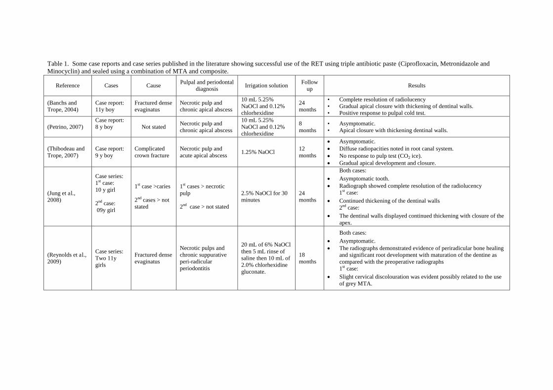
Table 1. Some case reports and case series published in the literature showing successful use of the RET using triple antibiotic paste (Ciprofloxacin, Metronidazole and Minocyclin) and sealed using a combination of MTA and composite.
Reference Cases Cause Pulpal and periodontal
diagnosis Irrigation solution
Follow up
Results
(Banchs and Trope, 2004)
Case report: 11y boy
Fractured dense evaginatus
Necrotic pulp and chronic apical abscess
10 mL 5.25% NaOCl and 0.12% chlorhexidine
24 months
• Complete resolution of radiolucency • Gradual apical closure with thickening of dentinal walls. • Positive response to pulpal cold test.
(Petrino, 2007) Case report: 8 y boy
Not stated Necrotic pulp and chronic apical abscess
10 mL 5.25% NaOCl and 0.12% chlorhexidine
8 months
• Asymptomatic. • Apical closure with thickening dentinal walls.
(Thibodeau and Trope, 2007)
Case report: 9 y boy
Complicated crown fracture
Necrotic pulp and acute apical abscess
1.25% NaOCl 12 months
Asymptomatic. Diffuse radiopacities noted in root canal system. No response to pulp test (CO2 ice). Gradual apical development and closure.
(Jung et al., 2008)
Case series: 1st case: 10 y girl 2nd case: 09y girl
1st case >caries 2nd cases > not stated
1st cases > necrotic pulp 2nd case > not stated
2.5% NaOCl for 30 minutes
24 months
Both cases: Asymptomatic tooth. Radiograph showed complete resolution of the radiolucency
1st case: Continued thickening of the dentinal walls
2nd case: The dentinal walls displayed continued thickening with closure of the
apex.
(Reynolds et al., 2009)
Case series: Two 11y girls
Fractured dense evaginatus
Necrotic pulps and chronic suppurative peri-radicular periodontitis
20 mL of 6% NaOCl then 5 mL rinse of saline then 10 mL of 2.0% chlorhexidine gluconate.
18 months
Both cases: Asymptomatic. The radiographs demonstrated evidence of periradicular bone healing
and significant root development with maturation of the dentine as compared with the preoperative radiographs 1st case:
Slight cervical discolouration was evident possibly related to the use of grey MTA.
The Regenerative Endodontic Technique
In the Department of Paediatric Dentistry at the Leeds Dental Institute we have used a
particular treatment technique based on these published principles (Duggal and Twati, 2011).
This technique involves disinfection followed by induction of a blood clot in the root canal
system and finally a hermitically good coronal seal.
Disinfection in our technique is achieved through minimal instrumentation, irrigation with
0.5% Hypochlorite followed by using an intracanal medicament of a mixture of antibiotics
left in the root canal system for two weeks. The aim of this disinfection technique is to
preserve the thin dentinal walls and any viable tissues containing stem cells such as SCAP
(Sonoyama et al., 2008) that may remain in the canal system. This is followed by the
creation of a blood clot within the canal through inducing of bleeding from the apical stem
cell rich areas into the root canal system using either sterile endodontic files or a sterile
needle. The blood clot serves as a scaffold which the stem cells would use in order to
repopulate the root canal system. A series of cases using this technique have been
successfully treated and reported within our peer group internationally (Karagianni and
Duggal, 2012).
One issue that yet remains to be resolved regards the choice of antibiotics. Many published
case series promote a mixture of three antibiotics, namely Ciprofloxicillin, Metronidazole and
Monocycline. Indeed, a triple antibiotic pre mixed paste is also commercially available. In
our opinion, it is unwise to use these pre-mixed pastes as there is little evidence in the
stability of such mixtures and hence their antimicrobial efficacy. Within our team we have
used this combination, but not in a pre-mixed form. We created a fresh mix of the three
antibiotics just before its application in the root canal. Microbiological research within the
Department of Paediatric Dentistry on the suitability and efficacy of the three antibiotics that
have been suggested for use in the literature has been carried out against the most common
pathogens that are found within the root canals of teeth with necrotic pulps (Twati et al.,
2011). We have found that a combination of two antibiotics (Ciprofloxacin and
Metronidazole) is equally effective as when Minocycline is included in the mixture. Our
initial evaluation of our cases has also shown that some had yellowish discoloration of the
crowns due to the use of Minocycline, as Tetracyclines related antibiotics (Reynolds et al.,
2009, Kim et al., 2010 ) are known to cause tooth discoloration. This finding, together with
our microbiological data, has led us to exclude Minocycline from these treatments. We have
also used a mixture of Amoxicillin and Metronidazole with similar efficacy. More research
is required on the correct choice of the antibiotics that should be used within the root canal
that would have a maximum efficacy and minimal side effects such as discolouration of the
crowns.
Therefore, the Leeds Dental Institute regenerative Endodontic technique involves two dental
visits as follows:
First Treatment Visit
The tooth is first isolated using dry dam (Directa, Upplands Visby, Sweden).
The tooth is then accessed and the pulp extirpated. The root canal system is then irrigated
with copious amounts of 0.5% sodium hypochlorite.
The canal is then negotiated with minimal or no filing to prevent further weakening of the
existing dentinal walls.
The canal is then dried using paper points.
Amoxicillin capsule 250mg) is then mixed with 1ml of 200 mg/5ml Metronidazole
suspension in a dappen dish. An alternative could be mixing Metronidazole (100mg) and
Ciprofloxacin (100mg) capsules (TriBioDent, Pharmacy production unit, Royal Victoria
Infirmary, Newcastle, UK).
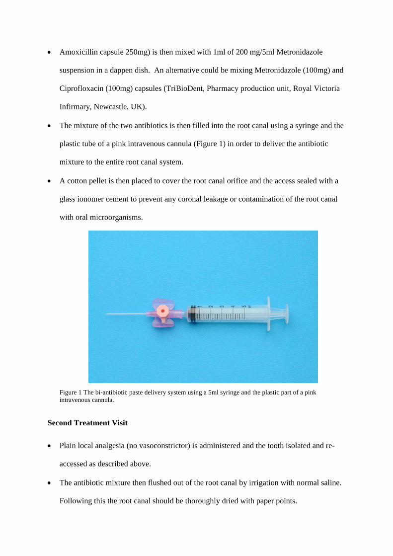
The mixture of the two antibiotics is then filled into the root canal using a syringe and the
plastic tube of a pink intravenous cannula (Figure 1) in order to deliver the antibiotic
mixture to the entire root canal system.
A cotton pellet is then placed to cover the root canal orifice and the access sealed with a
glass ionomer cement to prevent any coronal leakage or contamination of the root canal
with oral microorganisms.
Figure 1 The bi-antibiotic paste delivery system using a 5ml syringe and the plastic part of a pink intravenous cannula.
Second Treatment Visit
Plain local analgesia (no vasoconstrictor) is administered and the tooth isolated and re-
accessed as described above.
The antibiotic mixture then flushed out of the root canal by irrigation with normal saline.
Following this the root canal should be thoroughly dried with paper points.
This is then followed by insertion of a sterile 23-gauge needle or a finger spreader with a
length of 2 mm beyond the working length, past the confines of the root canal, into the
periapical tissues to intentionally induce bleeding into the root canal. The bleeding is then
allowed to fill the root canal.
Once the root canal is filled with blood, a cotton pledget is placed in the pulp chamber
and a clot allowed forming in the root canal.
Once the clot has formed the pulp chamber in the coronal part is thoroughly cleaned to
remove any remnants of the blood, which could cause discolouration in the future.
The access cavity is then hermetically sealed with three layers of material to prevent
coronal leakage and contamination; Portland cement, followed by glass ionomer and then
composite resin.
The following is a description of two cases treated at our department where two non vital
immature permanent incisors were treated using RET. The procedure involved the use of a
mixture of two antibiotics, Amoxicillin and Metronidazole.
Case 1
A healthy 10 year old boy referred by his dentist for management of a traumatised upper left
first permanent incisor UL1. The patient reported colliding against another child at school
playground 6 month prior to that appointment. Our initial clinical and radiographic
examination revealed enamel/dentine fracture of the UL1, restored by the dentist, and no
signs of pulpal inflammation or necrosis. On reviewing the patient a year and a half later, a
negative response to vitality testing of the UL1 was found and a diagnosis of necrotic pulp
canal system was made. Radiographic examination of UL1 revealed incomplete root
formation with parallel dentinal walls and chronic periapical radiolucency suggestive of
chronic periapical abscess (Figure 2,a) . After discussing treatment options with the parents, a
decision was made to treat the tooth using RET. Therefore an information leaflet was given to
the legal guardian and an appointment was arranged for consent and the first stage of RET
treatment. This allowed the legal guardian time to consider the information given and
therefore achieving informed consent.
At the next appointment, the UL1 was isolated using dry dam, accessed, and gentle
extirpation was done. The canal was then irrigated using 0.5% sodium hypochlorite and dried
using paper points. The tooth was then dressed with a mixture of Metronidazole (200 mg/5
ml suspension) and Amoxicillin (250mg capsule). The canal was then temporarily sealed
with a cotton pledget and Fuji IX LC (GC America Inc, IL).
The patient was then seen 10 days later when the tooth was isolated and re-accessed under
local analgesia. The antibiotic mixture was then flushed out of the root canal using normal
saline and then thoroughly dried with paper points. This was then followed by insertion of a
sterile 23-gauge needle with a length of 2 mm beyond the working length and past the
confines of the root canal into the periapical tissues inducing bleeding into the root canal,
allowing the blood to fill the root canal. Once the root canal was filled with blood, a cotton
pledget was placed in the pulp chamber and a clot was allowed to form in the root canal.
Once the clot had formed the pulp chamber in the coronal part had been thoroughly cleaned
to remove any remnants of the blood, in order to prevent discolouration of the crown.
The access cavity was then hermetically sealed with three layers of material to prevent
coronal leakage and contamination; MTA, followed by glass ionomer and then composite
resin.
Three months following RET treatment; no signs of infection, tenderness or discoloration
were evident. Radiographic examination revealed reduction in the size of the periapical lesion
and evidence of continued root development (Figure 2,b).
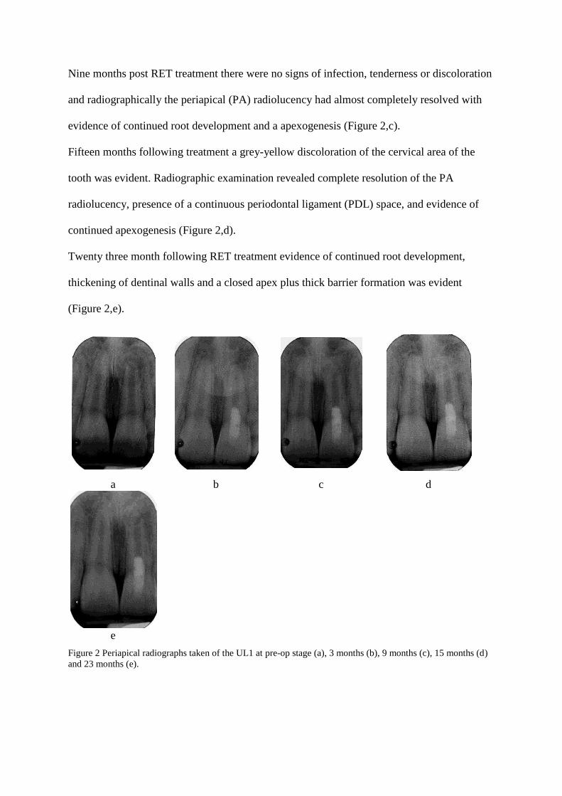
Nine months post RET treatment there were no signs of infection, tenderness or discoloration
and radiographically the periapical (PA) radiolucency had almost completely resolved with
evidence of continued root development and a apexogenesis (Figure 2,c).
Fifteen months following treatment a grey-yellow discoloration of the cervical area of the
tooth was evident. Radiographic examination revealed complete resolution of the PA
radiolucency, presence of a continuous periodontal ligament (PDL) space, and evidence of
continued apexogenesis (Figure 2,d).
Twenty three month following RET treatment evidence of continued root development,
thickening of dentinal walls and a closed apex plus thick barrier formation was evident
(Figure 2,e).
a b c d
e
Figure 2 Periapical radiographs taken of the UL1 at pre-op stage (a), 3 months (b), 9 months (c), 15 months (d) and 23 months (e).
Case 2
A 7 year old girl with developmental delay and poor balance presented to her dentist in
March 2009 with a fractured UR1. She sustained the injury by stumbling and falling face first
onto a concrete floor. No other associated trauma was reported. The next day, her dentist had
restored UR1 with composite restoration. However, over a span of a year, the restorations
needed replacement twice. Subsequently, the child presented with an abscess associated with
the UR1. Antibiotic (amoxicillin 250mg tds) was prescribed and child referred to our
department for further management of non-vital immature UR1.
On presentation, intra-oral examination showed the UR1 to be tender to percussion, had
normal mobility and an associated labial sinus. Periapical radiographic examination indicated
that both UR1 and UL1 had open apices (Figure 3,a). Therefore, the UR1 was diagnosed with
pulp necrosis and periapical periodontitis. In addition, UR1 had an uncomplicated enamel
dentine fracture and dens invaginatus.
At the first visit, local anaesthetic was administered using the Wand local anaesthesia system
(Milestone Scientific, Deerfield, IL). The tooth was isolated with rubber dam, root canal was
accessed and pulp extirpated. Bleeding and suppuration was present in the canal. Canal was
gently irrigated with 0.5% sodium hypochloriteL. The working length was confirmed with a
periapical radiograph. The canal was irrigated with normal saline and dried using paper
points. An antibiotic mixture of Metronidazole (200 mg/5 ml suspension) and Amoxicillin
(250mg capsule) was syringed into the canal. A moist cotton pledget was placed and access
cavity was temporised with Fuji IX LC (GC America Inc, IL).
At the second visit (21 days later), the patient had no complaints. The labial sinus associated
to UR1 was resolving and surrounding soft tissues appeared normal. UR1 was anaesthetised
and tooth isolated with rubber dam. The canal was accessed and irrigated with NaOCL
followed by normal saline and dried. Clinical examination confirmed that the canal was free
of infection and discharge. Finger spreader sized medium was used to induce fresh bleeding
into the canal. This was difficult and slow probably due to the vasoconstrictors in the local
anaesthetic agent used. However, once the canal had filled with fresh blood, a moist cotton
pledget was used to seal access cavity and left for several minutes to clot. White MTA was
placed and access cavity was temporised with Fuji IX LC (GC America Inc, IL). Baseline
periapical radiograph was then taken (Figure 3,b).
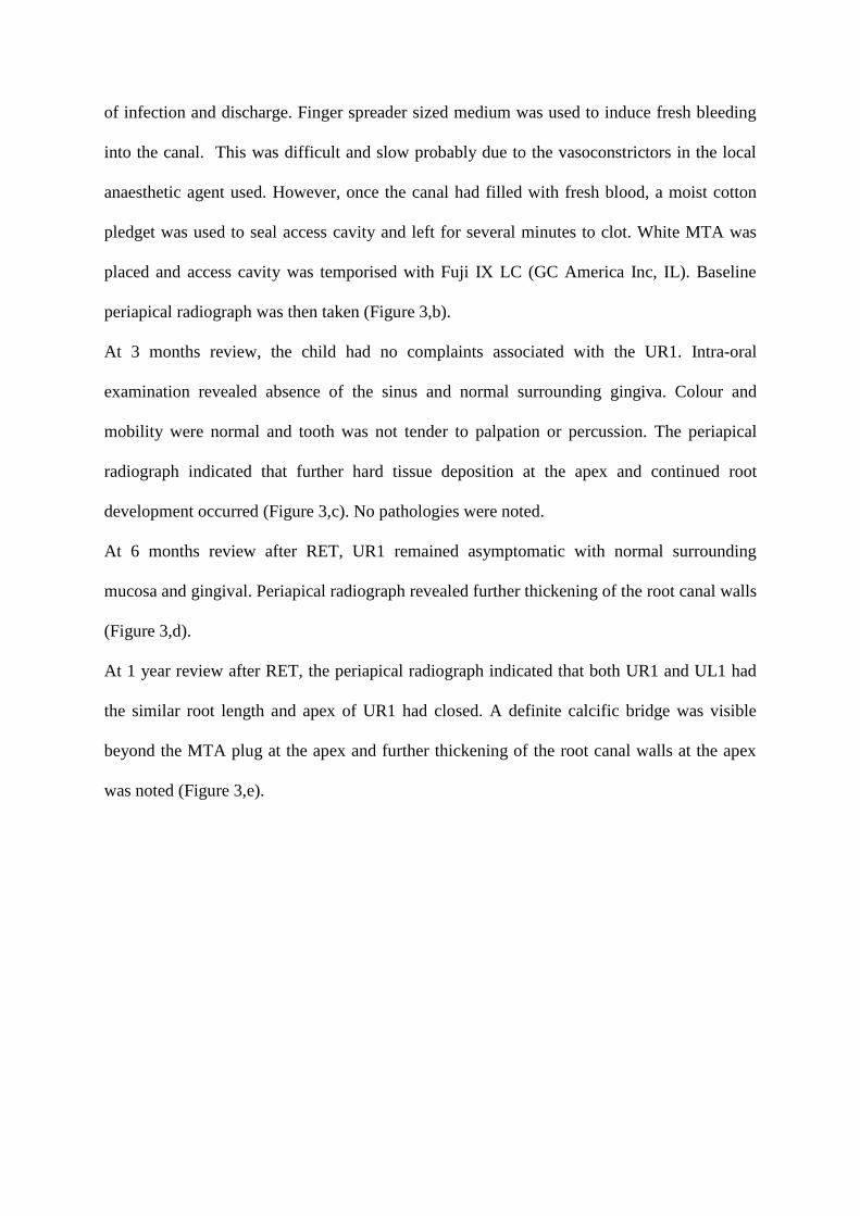
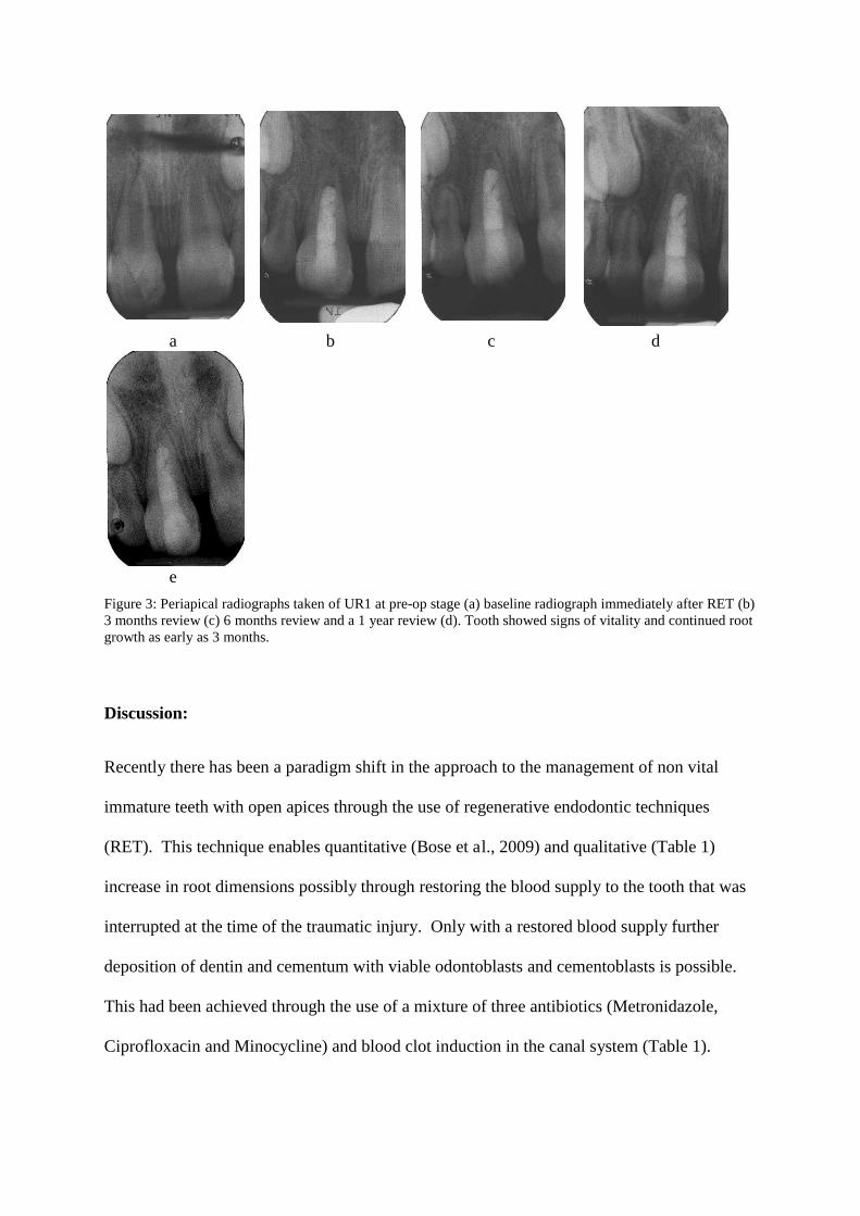
At 3 months review, the child had no complaints associated with the UR1. Intra-oral
examination revealed absence of the sinus and normal surrounding gingiva. Colour and
mobility were normal and tooth was not tender to palpation or percussion. The periapical
radiograph indicated that further hard tissue deposition at the apex and continued root
development occurred (Figure 3,c). No pathologies were noted.
At 6 months review after RET, UR1 remained asymptomatic with normal surrounding
mucosa and gingival. Periapical radiograph revealed further thickening of the root canal walls
(Figure 3,d).
At 1 year review after RET, the periapical radiograph indicated that both UR1 and UL1 had
the similar root length and apex of UR1 had closed. A definite calcific bridge was visible
beyond the MTA plug at the apex and further thickening of the root canal walls at the apex
was noted (Figure 3,e).
a b c d
e
Figure 3: Periapical radiographs taken of UR1 at pre-op stage (a) baseline radiograph immediately after RET (b) 3 months review (c) 6 months review and a 1 year review (d). Tooth showed signs of vitality and continued root growth as early as 3 months.
Discussion:
Recently there has been a paradigm shift in the approach to the management of non vital
immature teeth with open apices through the use of regenerative endodontic techniques
(RET). This technique enables quantitative (Bose et al., 2009) and qualitative (Table 1)
increase in root dimensions possibly through restoring the blood supply to the tooth that was
interrupted at the time of the traumatic injury. Only with a restored blood supply further
deposition of dentin and cementum with viable odontoblasts and cementoblasts is possible.
This had been achieved through the use of a mixture of three antibiotics (Metronidazole,
Ciprofloxacin and Minocycline) and blood clot induction in the canal system (Table 1).
Using this triple antibiotic paste on some of the cases at Leeds Dental Institute, the authors
observed yellowish crown discoloration which was attributed to the use of Minocycline. The
use of triple antibiotic paste was found in an in vitro study to cause more discoloration to
extracted bovine teeth compared to different endodontic medicaments such as Ledermix and
MTA (Lenherr et al., 2012). Therefore an in vitro microbiological study, conducted at our
department, lead us into excluding Minocycline from any future RET treatments (Twati et al.,
2011). We now use a combination of Amoxicillin and Metronidazole in our management as
we have shown in the cases presented here. In case 1, this patient was reviewed for twenty
three months following his RET treatment and showed favourable outcomes such as evidence
of continued root development, thickening of dentinal walls and a closed apex plus thick
apical barrier formation (Figure 2,e). On the other hand, fifteen months following RET
treatment; a grey-yellow discoloration of the cervical area of the tooth was evident. We
attributed this to the use of grey MTA which is associated with tooth discoloration (Parirokh
and Torabinejad, 2010, Lenherr et al., 2012).
This led us to use white MTA in treating case 2 which after 1 year review following RET,
showed evidence of continued root formation and closed apex. A definite calcific bridge was
visible beyond the MTA plug at the apex and further thickening of the root canal walls at the
apex was noted (Figure 3,e). No crown discoloration was noted in this case. However, even
white MTA tends to discolour in an anaerobic environment, as it still contains Bismuth for
radiopacity. We now use Portland cement in our protocols which does not suffer from the
same disadvantage,
Lenherr et al. (2012) conducted an in vitro study comparing the discoloration potential of
different endodontic materials including grey MTA, Grey MTA with blood, White MTA,
White MTA with blood, Portland cement and Portland cement with blood on extracted
bovine incisors in comparison to no medicament use. Colour assessment was performed
using a spectrophotometer (VITA Easyshade compact; VITA Zahnfabrik, Bad Sa¨ckingen,
Germany). The authors reported the following descending sequence of discolouration
potential: Grey MTA, Grey MTA with blood, Portland cement with blood, white MTA with
blood, white MTA and finally Portland cement. However, these materials were applied
directly onto dentine-enamel cuboid blocks rather than into the roots of the teeth at a distance
from the cemento-enamel junction.
Case 2 also showed the need to use plain local analgesia (no vasoconstrictor) on the second
treatment visit as that would help when inducing bleeding into the tooth. This is important as
in some cases it is not easy to induce enough bleeding into the root canal.
Conclusion:
The use of regenerative endodontics has caught the imagination of the dental profession, but
its long term outcomes are far from clear. Systematic studies using adequate control and
randomisation are required before it is recommended for general adoption for the dental
profession. Although the technique is based on the general principles of tissue bio-
engineering, it could be argued that the technique of injuring the apical tissue, and inducing
bleeding , cannot control what tissues might grow into the root canal. Therefore, it is not
possible at the moment to say with certainty whether the tissue that grows into the root canal
is regenerative tissue, repair related tissue, or revascularisation. In the future, it might be
possible to use growth factors in the root canal to introduce a degree of control on what
tissues are regenerated into the root canal system. Many groups are currently working to
understand this technique, and refine it. It is hoped that in the next decade we will be able to
recommend this technique as a treatment of choice for all non vital teeth with immature roots
in children and adolescents. For now we recommend that this technique be considered in
those cases where the root development is so incomplete that the clinicians feel that the

prognosis would be poor with the use of MTA. In such cases if further root development or
more hard tissue deposition is obtained with the use of regenerative technique, this would
improve their medium to long term prognosis.
References: Al -Jundi, S. Type of treatment, prognosis, and estimation of time spent to manage dental
trauma in late presentation cases at a dental teaching hospital: a longitudinal and retrospective study. Dental Traumatology, 2004. 20, 1-5.
Al Ansary, M. 2006. Interventions for treating traumatised non-vital immature permanent front teeth. MDentSci, University of Leeds
Andreason, J, Farik, B & Munksgaard, E. Long-term calcium hydroxide as a root canal dressing may increase risk of root fracture. . Dental Traumatology, 2002. 18, 134-137.
Bakland, LK & Andreasen, JO. Will mineral trioxide aggregate replace calcium hydroxide in treating pulpal and periodontal healing complications subsequent to dental trauma? A review. Dental Traumatology, 2012. 28, 25-32.
Banchs, F & Trope, M. Revascularization of immature permanent teeth with apical periodontitis: new treatment protocol? Journal of Endodontics, 2004. 30, 196-200.
Bose, R, Nummikoski, P & Hargreaves, K. A retrospective evaluation of radiographic outcomes in immature teeth with necrotic root canal systems treated with regenerative endodontic procedures. Journal of Endodontics, 2009. 35, 1343-9.
Cvek, M. Prognosis of luxated non-vital maxillary incisors treated with calcium hydroxide and filled with gutta-percha. A retrospective clinical study. Dental Traumatology, 1992. 8, 45-55.
Duggal, M & Twati, W 2011. Endodontic management of non-vital anterior teeth with incomplete root development. In: SPLIETH, C. H. (ed.) Revolutions in Pediatric Dentistry. Quintessenz Verlags-GmbH.
Finucane, D & Kinirons, MJ. Non-vital immature permanent incisors: factors that may infIuence treatment outcome. Dental Traumatology, 1999. 15, 273-277.
Huang, G, Sonoyama, W, Liu, Y, Liu, H, Wang, S & Shi, S. The hidden treasure in apical papilla: the potential role in pulp/dentin regeneration and bioroot engineering. Journal of Endodontics, 2008. 34, 645-651.
Huang, GTJ. A paradigm shift in endodontic management of immature teeth: Conservation of stem cells for regeneration. Journal of Dentistry, 2008. 36, 379-386.
Jung, I, Lee, S & Hargreaves, K. Biologically based treatment of immature permanent teeth with pulpal necrosis: A case series. Journal of Endodontics, 2008. 34, 876-87.
Kaiser, H. Management of wideopen canals with calcium hydroxide. Read before the Americal Association of Endodontics 1964 Washington.
Karagianni, A & Duggal, M. Regenerative Endodontic Therapy (RET) for management of non vital immature permanent teeth. 11th Congress of the European Academy of Paediatric dentistry OPD125. Place 2012. Published.
Kim, J, Kim, Y, Shin, S, Park, J & Jung, I. Tooth discoloration of immature permanent incisor associated with triple antibiotic therapy: a case report. Journal of Endodontics, 2010 36, 86-91.
Lenherr, P, Allgayer, N, Weiger, R, Filippi, A, Attin, T & Krastl, G. Tooth discoloration induced by endodontic materials: a laboratory study. International Endodontic Journal, 2012. no-no.
Murray, PE, Garcia-Godoy, F & Hargreaves, KM. Regenerative endodontics: a review of current status and a call for action. Journal of Endodontics, 2007. 33, 377-90.
Parirokh, M & Torabinejad, M. Mineral Trioxide Aggregate: A Comprehensive Literature Review—Part III: Clinical Applications, Drawbacks, and Mechanism of Action. Journal of Endodontics, 2010. 36, 400-413.
Petrino, JA. Revascularization of necrotic pulp of immature teeth with apical periodontitis. Northwest Dentistry, 2007. 86, 33-5.
Reynolds, K, Johnson, J & Cohenca, N. Pulp revascularization of necrotic bilateral bicuspids using a modified novel technique to eliminate potential coronal discolouration: a case report. International Endodontic Journal, 2009. 42, 84–92.
Sedgley, C & Botero, T. Dental Stem Cells and Their Sources. Dental Clinics of North America 2012. In press (available online http://www.sciencedirect.com/science/article/pii/S0011853212000365).
Shah, N, Logani, A, Bhaskar, U & Aggarwal, V. Efficacy of revasularization to induce apexification ⁄ apexogenesis in infected, nonvital, immature teeth: a pilot clinical study. Journal of Endodontics, 2008. 34, 919-925.
Sonoyama, W, Liu, Y, Yamaza, T, Tuan, R, Wang, S, Shi, S & Huang, G. Characterization of the apical papilla and its residing stem cells from human immature permanent teeth: a pilot study. Journal of Endodontics, 2008. 34.
Thibodeau, B & Trope, M. Pulp revascularization of a necrotic infected immature permanent tooth: case report and review of the literature. Pediatric Dentistry, 2007. 29, 47-50.
Torabinejad, M, Hong, CU, McDonald, F & Pitt Ford, TR. Physical and chemical properties of a new root-end filling material. Journal of Endodontics, 1995. 21, 349-353.
Twati, W, Percival, R & Duggal, M. The antimicrobial effect of various root canal medicaments used in management of immature non vital anterior teeth. International Journal of Paediatric Dentistry, 2011. 21 (S1) Abstract O12-101, 35.
Twati, W, Wood, D, Liskiewicz, T & Duggal, M. Effect of non-setting calcium hydroxide and MTA on human dentine following long term application. International Journal of Paediatric Dentistry, 2009. 19:S1(Abstract O16-117), 43.