Embed Size (px)
Citation preview

Refractory Gout: An overview of pathogenesis and treatment
Gordon K. Lam, MD, FACR
NorthEast RheumatologyMedical Director, Northern Region Research CenterCarolinas Healthcare System
Disclosures
1. Horizon Pharma, Plc: Research; Speaker Bureau
2. Takeda Pharmaceuticals USA, Inc.: Speaker Bureau; Advisory Board

Objectives
• To review the etiopathogenesis of gout as a chronic, progressive, inflammatory arthritis
• To differentiate the treatment of acute gout flares vs. chronic gouty arthropathy
• To discuss the management of refractory gout
• To facilitate collaboration between podiatrists and rheumatologists in the management of gout patients
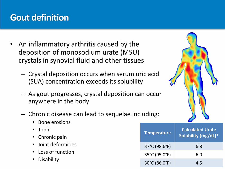
Gout definition
• An inflammatory arthritis caused by the deposition of monosodium urate (MSU) crystals in synovial fluid and other tissues
– Crystal deposition occurs when serum uric acid (SUA) concentration exceeds its solubility
– As gout progresses, crystal deposition can occur anywhere in the body
– Chronic disease can lead to sequelae including:• Bone erosions
• Tophi
• Chronic pain
• Joint deformities
• Loss of function
• Disability
TemperatureCalculated Urate
Solubility (mg/dL)*
37°C (98.6°F) 6.8
35°C (95.0°F) 6.0
30°C (86.0°F) 4.5
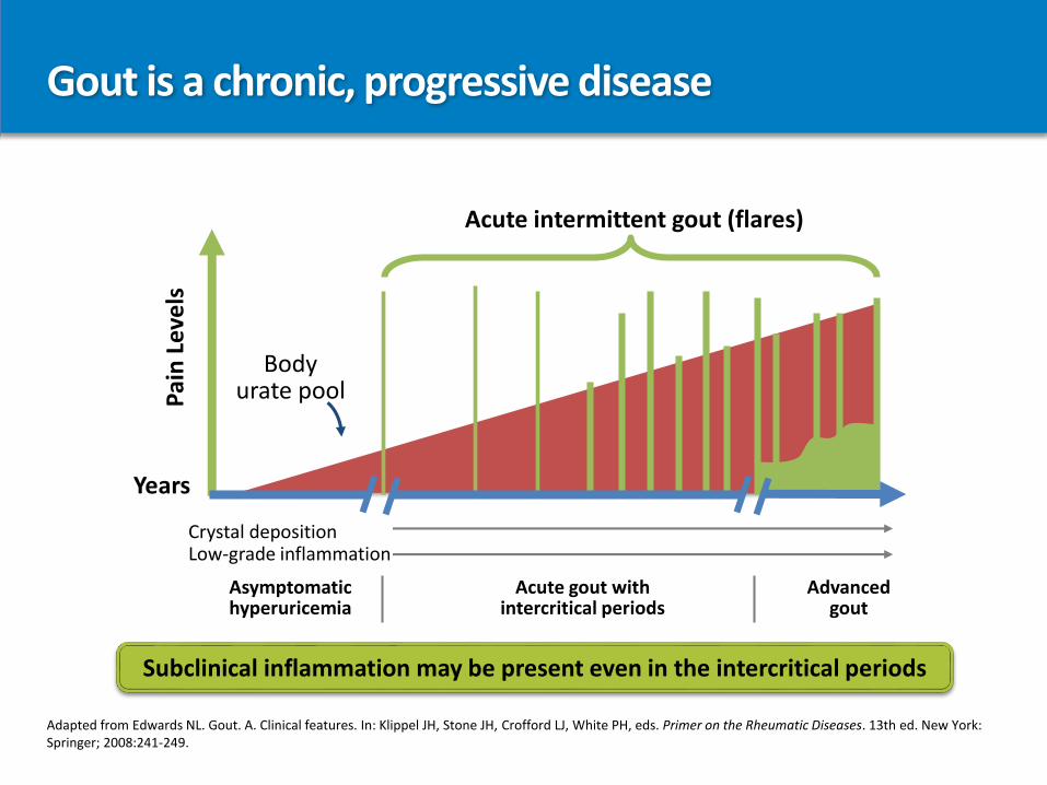
Gout is a chronic, progressive disease
Adapted from Edwards NL. Gout. A. Clinical features. In: Klippel JH, Stone JH, Crofford LJ, White PH, eds. Primer on the Rheumatic Diseases. 13th ed. New York: Springer; 2008:241-249.
Acute intermittent gout (flares)
Crystal depositionLow-grade inflammation
Acute gout withintercritical periods
Asymptomatichyperuricemia
Advancedgout
Years
Pai
n L
eve
ls
Bodyurate pool
Subclinical inflammation may be present even in the intercritical periods
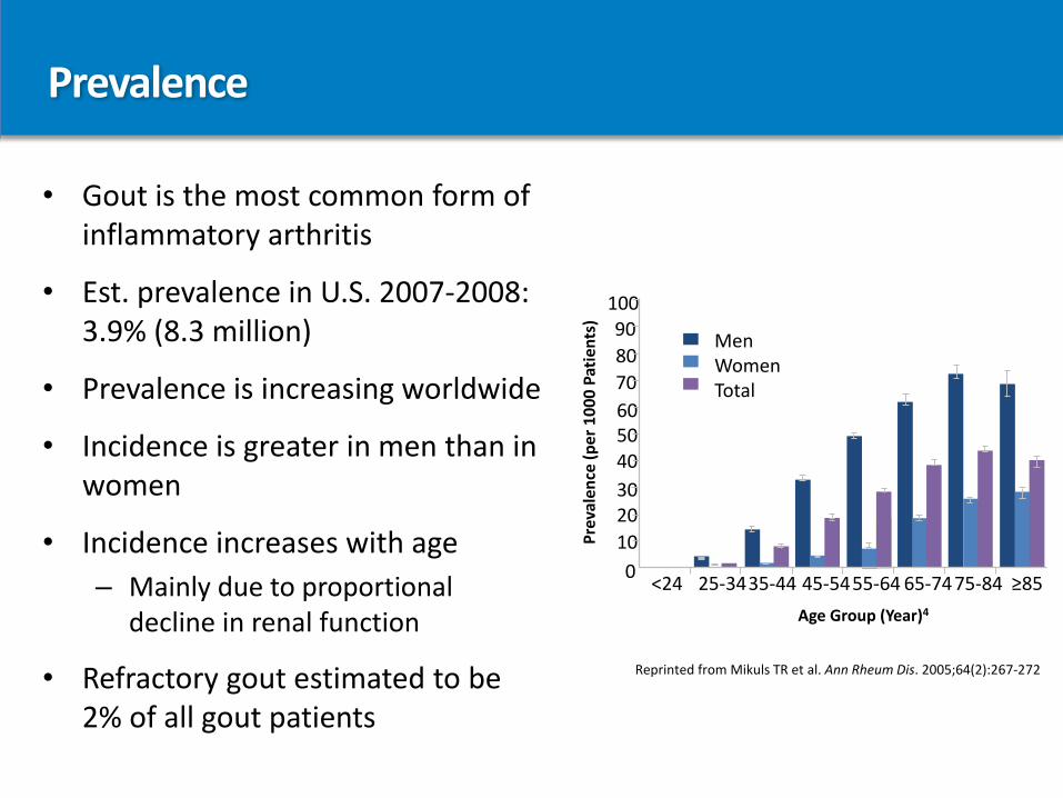
Prevalence
• Gout is the most common form of inflammatory arthritis
• Est. prevalence in U.S. 2007-2008: 3.9% (8.3 million)
• Prevalence is increasing worldwide
• Incidence is greater in men than in women
• Incidence increases with age
– Mainly due to proportional decline in renal function
• Refractory gout estimated to be 2% of all gout patients
10090
80
70
605040
3020
10
0<24 45-5455-64 65-7475-84 ≥8535-4425-34
Age Group (Year)4
Pre
vale
nce
(p
er
10
00
Pat
ien
ts)
MenWomenTotal
Reprinted from Mikuls TR et al. Ann Rheum Dis. 2005;64(2):267-272
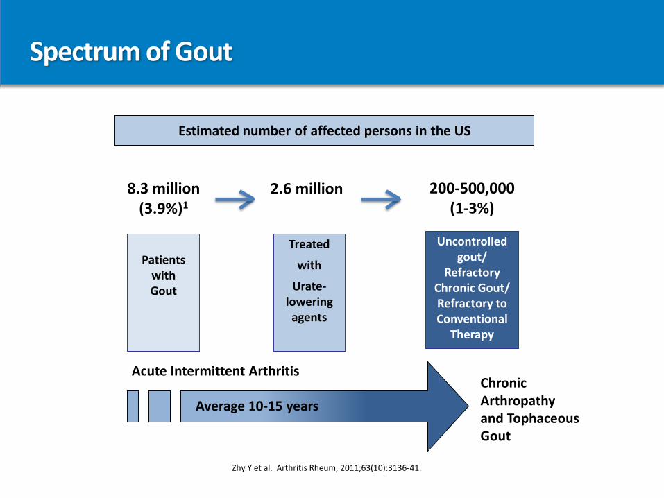
Spectrum of Gout
Zhy Y et al. Arthritis Rheum, 2011;63(10):3136-41.
Estimated number of affected persons in the US
Acute Intermittent Arthritis
Patientswith Gout
Treated
with
Urate-lowering agents
Uncontrolled gout/
Refractory Chronic Gout/ Refractory to Conventional
Therapy
Chronic Arthropathy and Tophaceous Gout
Average 10-15 years
2.6 million 200-500,000 (1-3%)
8.3 million (3.9%)1
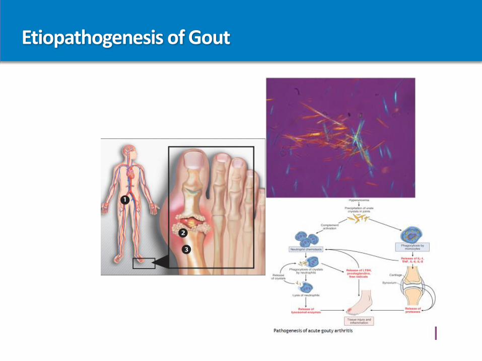
Etiopathogenesis of Gout
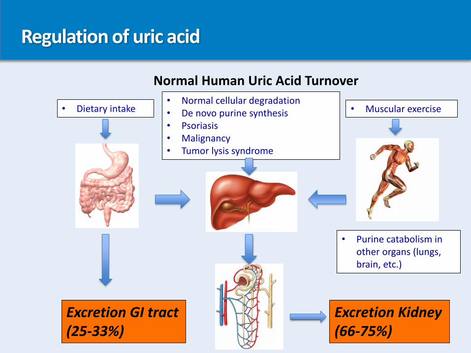
Regulation of uric acid
Normal Human Uric Acid Turnover
• Dietary intake • Muscular exercise• Normal cellular degradation• De novo purine synthesis• Psoriasis• Malignancy• Tumor lysis syndrome
• Purine catabolism in other organs (lungs, brain, etc.)
Excretion GI tract(25-33%)
Excretion Kidney(66-75%)
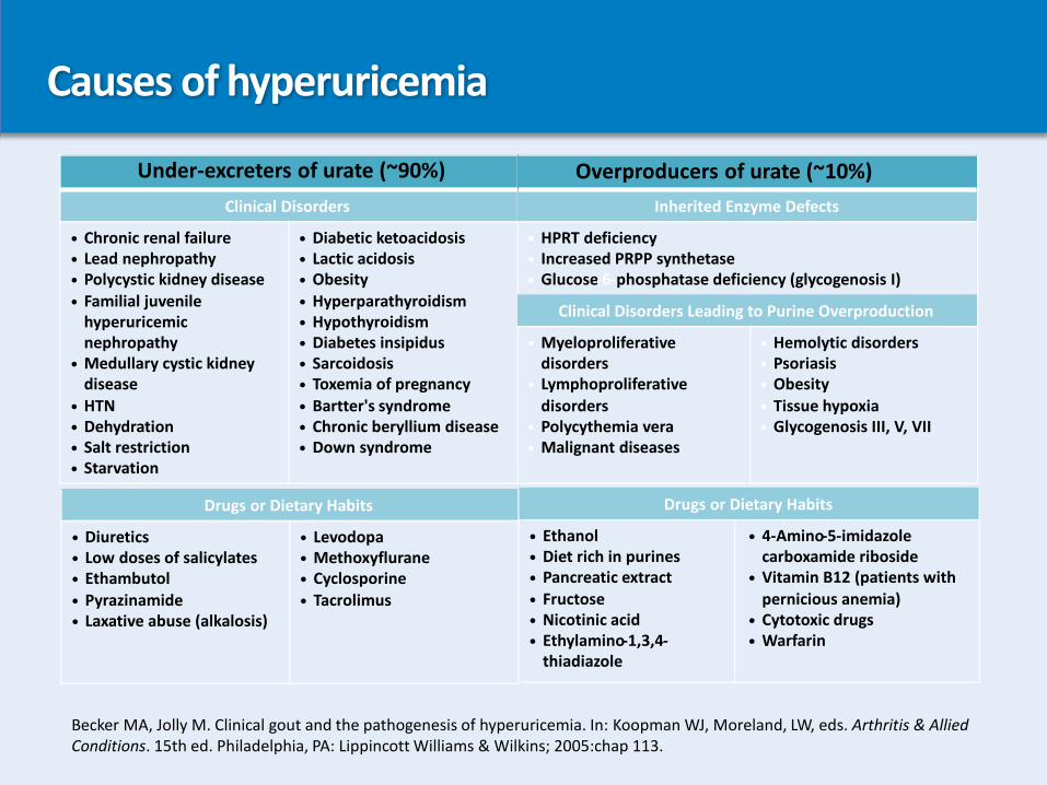
Causes of hyperuricemia
Under-excreters of urate (~90%)
Clinical Disorders
• Chronic renal failure
• Lead nephropathy
• Polycystic kidney disease
• Familial juvenile hyperuricemicnephropathy
• Medullary cystic kidney disease
• HTN
• Dehydration
• Salt restriction
• Starvation
• Diabetic ketoacidosis
• Lactic acidosis
• Obesity
• Hyperparathyroidism
• Hypothyroidism
• Diabetes insipidus
• Sarcoidosis
• Toxemia of pregnancy
• Bartter's syndrome
• Chronic beryllium disease
• Down syndrome
Overproducers of urate (~10%)
Inherited Enzyme Defects
• HPRT deficiency
• Increased PRPP synthetase
• Glucose-6-phosphatase deficiency (glycogenosis I)
Clinical Disorders Leading to Purine Overproduction
• Myeloproliferative disorders
• Lymphoproliferative disorders
• Polycythemia vera
• Malignant diseases
• Hemolytic disorders
• Psoriasis
• Obesity
• Tissue hypoxia
• Glycogenosis III, V, VII
Drugs or Dietary Habits
• Diuretics
• Low doses of salicylates
• Ethambutol
• Pyrazinamide
• Laxative abuse (alkalosis)
• Levodopa
• Methoxyflurane
• Cyclosporine
• Tacrolimus
Drugs or Dietary Habits
• Ethanol
• Diet rich in purines
• Pancreatic extract
• Fructose
• Nicotinic acid
• Ethylamino-1,3,4-thiadiazole
• 4-Amino-5-imidazole carboxamide riboside
• Vitamin B12 (patients with pernicious anemia)
• Cytotoxic drugs
• Warfarin
Becker MA, Jolly M. Clinical gout and the pathogenesis of hyperuricemia. In: Koopman WJ, Moreland, LW, eds. Arthritis & Allied Conditions. 15th ed. Philadelphia, PA: Lippincott Williams & Wilkins; 2005:chap 113.
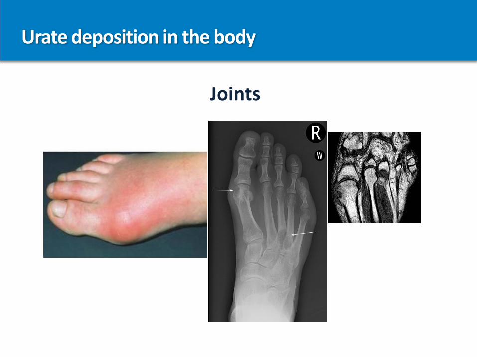
Urate deposition in the body
Joints
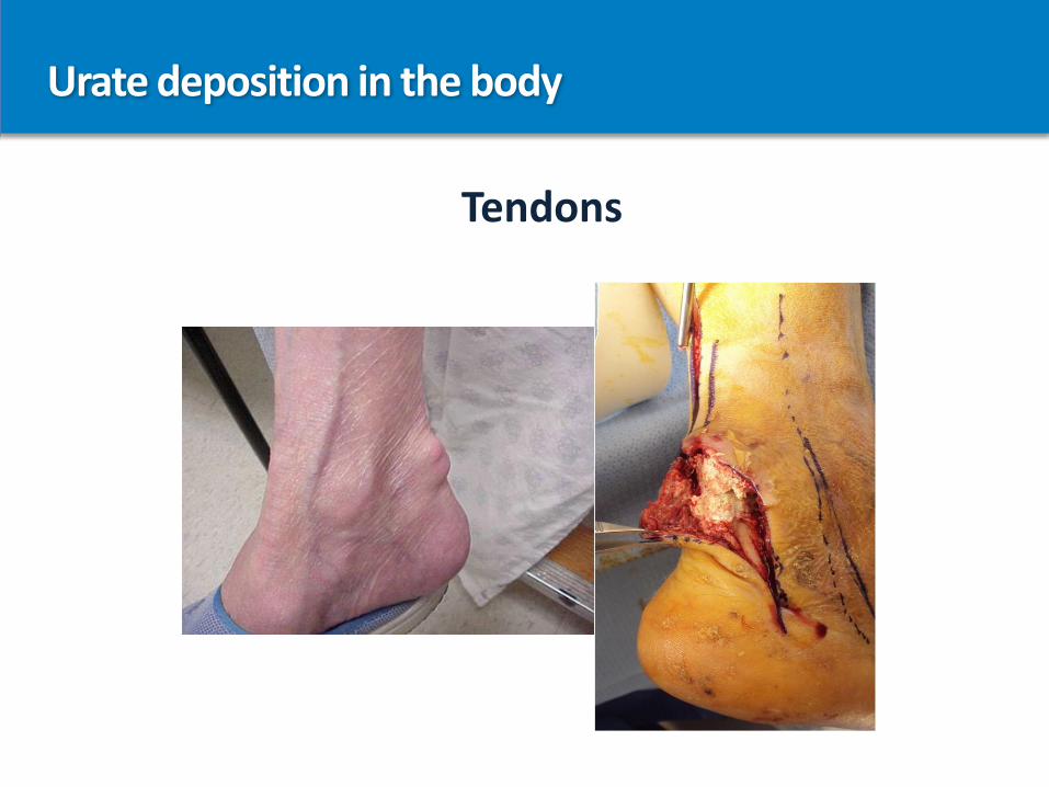
Urate deposition in the body
Tendons
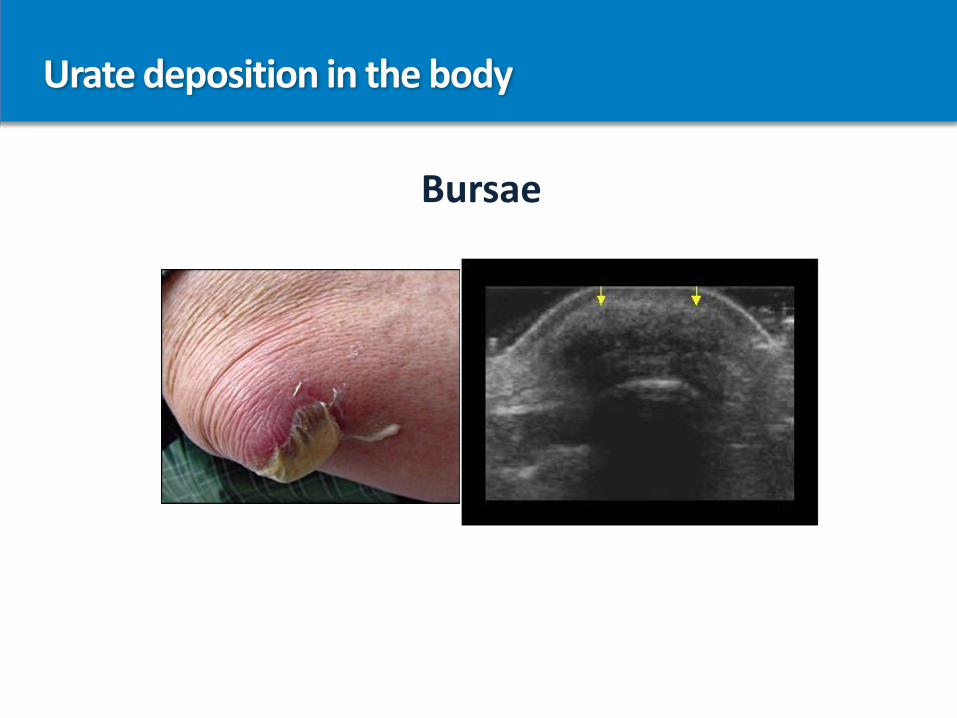
Urate deposition in the body
Bursae
Urate deposition in the body
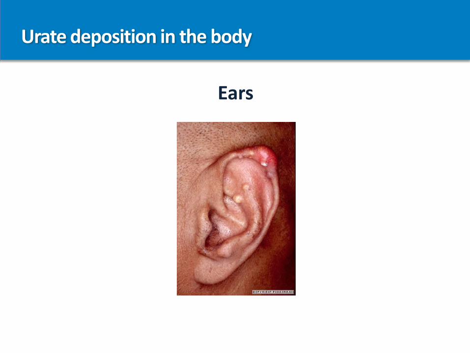
Ears
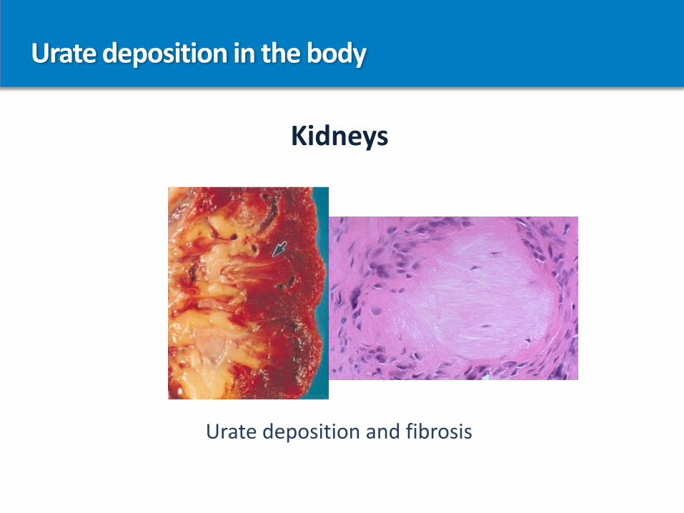
Urate deposition in the body
Kidneys
Urate deposition and fibrosis
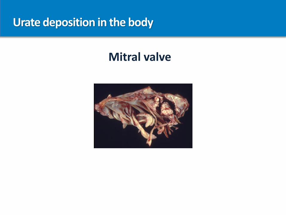
Urate deposition in the body
Mitral valve
Urate deposition in the body
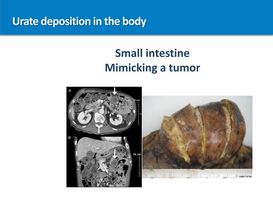
Small intestineMimicking a tumor
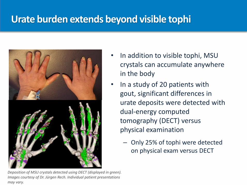
Urate burden extends beyond visible tophi
• In addition to visible tophi, MSU crystals can accumulate anywhere in the body
• In a study of 20 patients with gout, significant differences in urate deposits were detected with dual-energy computed tomography (DECT) versus physical examination
– Only 25% of tophi were detected on physical exam versus DECT
Deposition of MSU crystals detected using DECT (displayed in green). Images courtesy of Dr. Jürgen Rech. Individual patient presentations may vary.
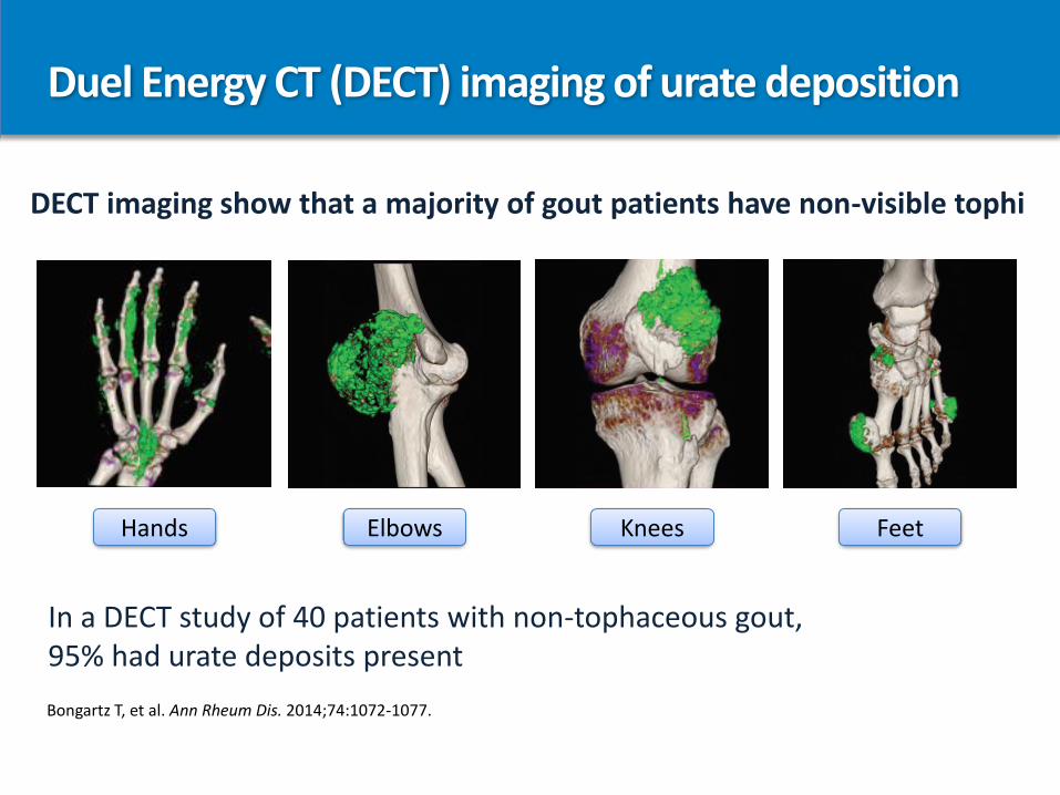
Duel Energy CT (DECT) imaging of urate deposition
DECT imaging show that a majority of gout patients have non-visible tophi
In a DECT study of 40 patients with non-tophaceous gout, 95% had urate deposits present
Hands Elbows Knees Feet
Bongartz T, et al. Ann Rheum Dis. 2014;74:1072-1077.
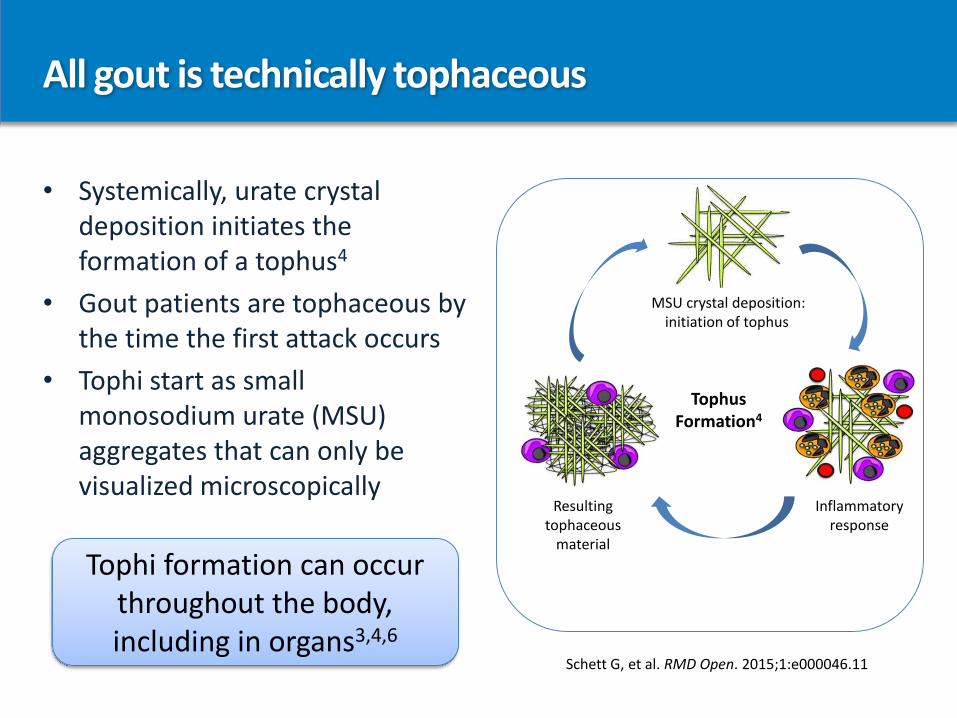
All gout is technically tophaceous
MSU crystal deposition:initiation of tophus
Inflammatory response
TophusFormation4
Resulting tophaceous
material
• Systemically, urate crystal deposition initiates the formation of a tophus4
• Gout patients are tophaceous by the time the first attack occurs
• Tophi start as small monosodium urate (MSU) aggregates that can only be visualized microscopically
Tophi formation can occur throughout the body, including in organs3,4,6
Schett G, et al. RMD Open. 2015;1:e000046.11
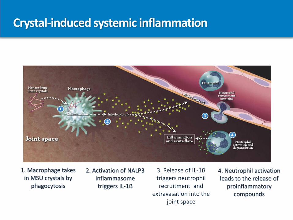
Crystal-induced systemic inflammation
1. Macrophage takes in MSU crystals by
phagocytosis
2. Activation of NALP3 Inflammasometriggers IL-1ẞ
3. Release of IL-1ẞ triggers neutrophil recruitment and
extravasation into the joint space
4. Neutrophil activation leads to the release of
proinflammatory compounds
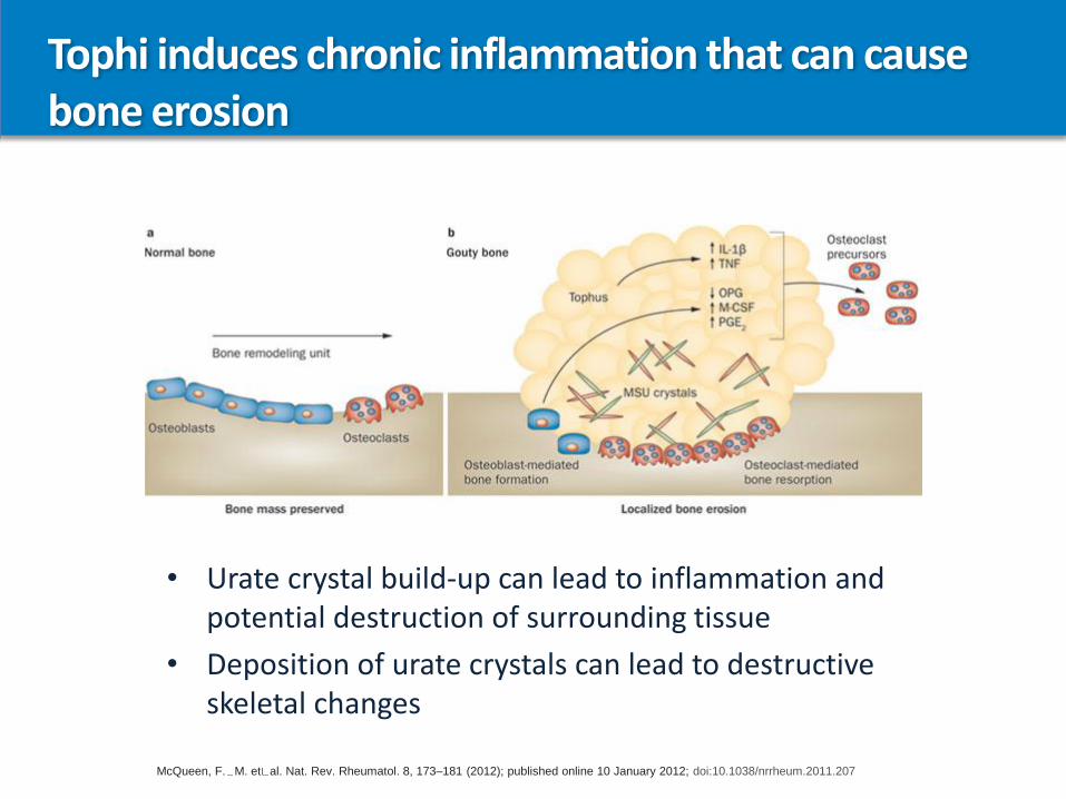
Tophi induces chronic inflammation that can cause bone erosion
• Urate crystal build-up can lead to inflammation and potential destruction of surrounding tissue
• Deposition of urate crystals can lead to destructive skeletal changes
McQueen, F. M. et al. Nat. Rev. Rheumatol. 8, 173–181 (2012); published online 10 January 2012; doi:10.1038/nrrheum.2011.207
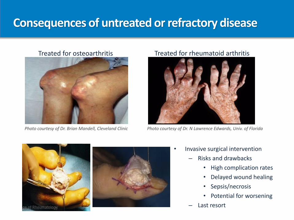
Consequences of untreated or refractory disease
Treated for osteoarthritis Treated for rheumatoid arthritis
Photo courtesy of Dr. Brian Mandell, Cleveland Clinic Photo courtesy of Dr. N Lawrence Edwards, Univ. of Florida
• Invasive surgical intervention
– Risks and drawbacks
• High complication rates
• Delayed wound healing
• Sepsis/necrosis
• Potential for worsening
– Last resort
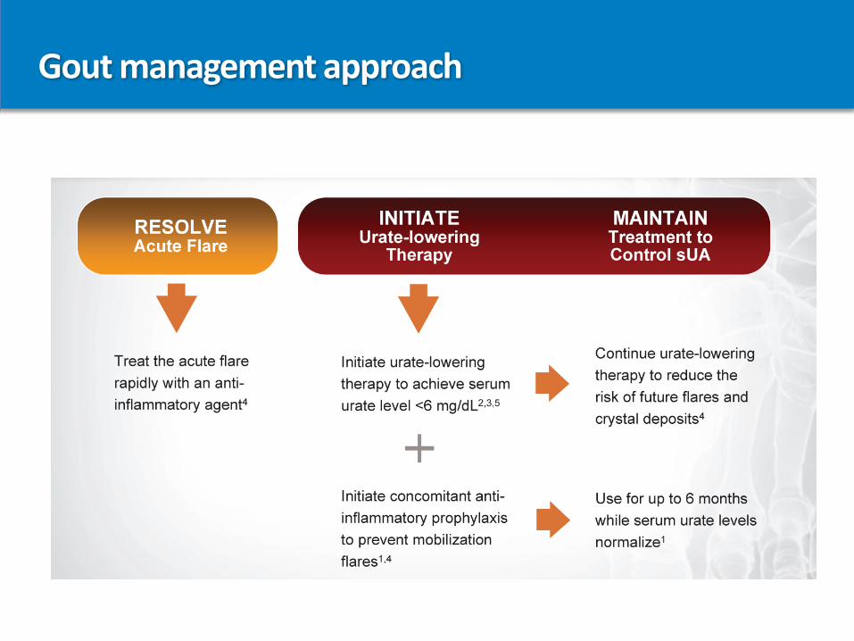
Gout management approach
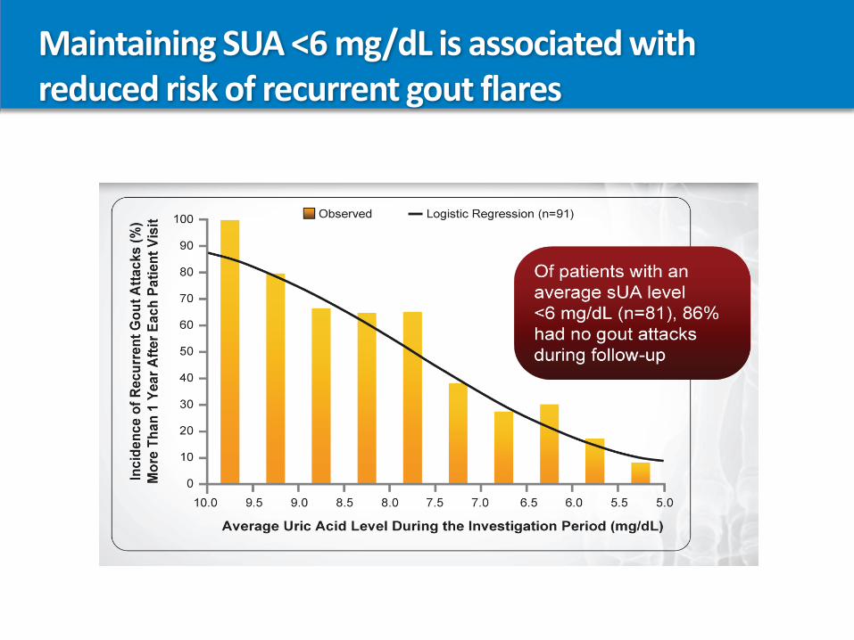
Maintaining SUA <6 mg/dL is associated with reduced risk of recurrent gout flares
Treatment of gout
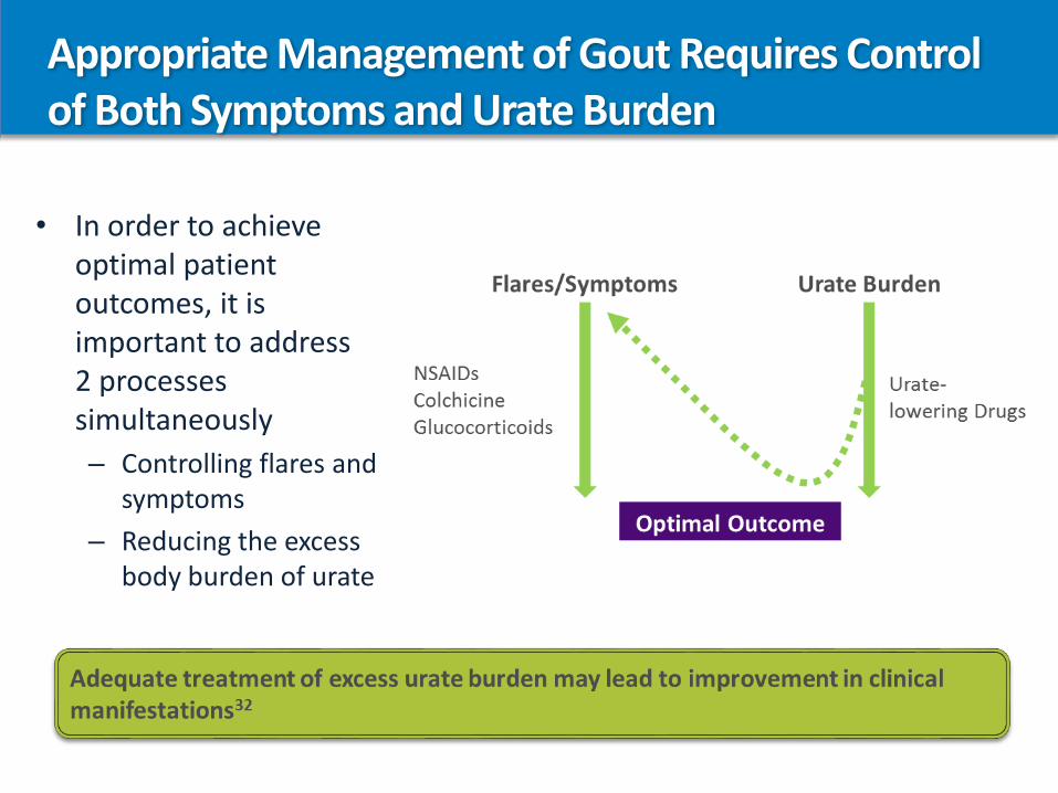
Appropriate Management of Gout Requires Control of Both Symptoms and Urate Burden
• In order to achieve optimal patient outcomes, it is important to address 2 processes simultaneously
– Controlling flares and symptoms
– Reducing the excess body burden of urate
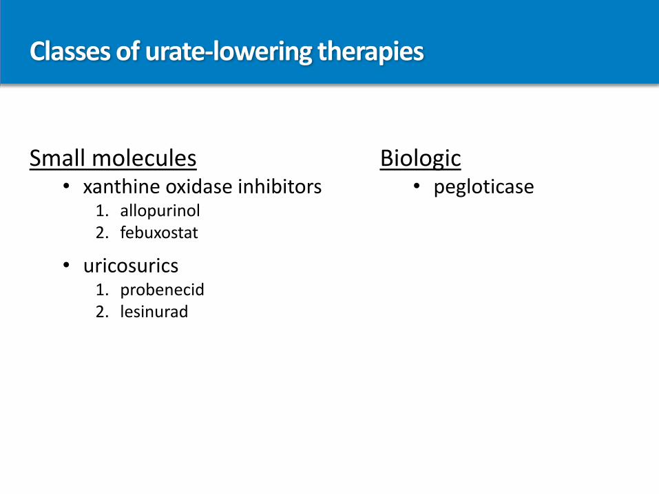
Classes of urate-lowering therapies
Small molecules• xanthine oxidase inhibitors
1. allopurinol2. febuxostat
• uricosurics1. probenecid2. lesinurad
Biologic• pegloticase

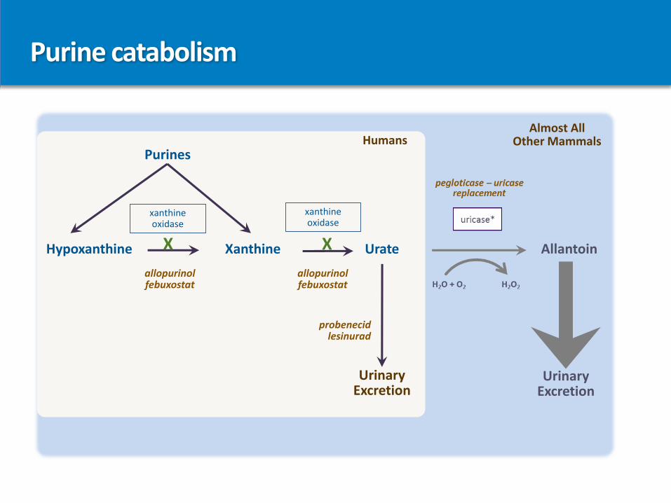
C
UrinaryExcretion
Allantoin
Almost All Other Mammals
Purine catabolism
UrinaryExcretion
xanthineoxidase
allopurinolfebuxostat
allopurinolfebuxostat
probenecidlesinurad
XX
xanthineoxidase
Hypoxanthine Xanthine
Purines
Urate
Humans
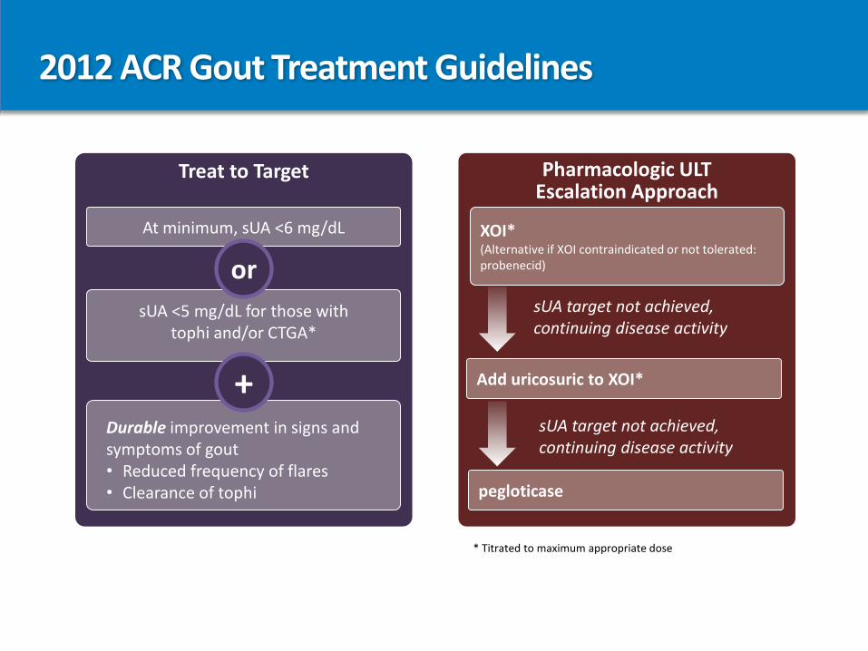
2012 ACR Gout Treatment Guidelines
Treat to Target
Durable improvement in signs and symptoms of gout • Reduced frequency of flares• Clearance of tophi
sUA <5 mg/dL for those with tophi and/or CTGA*
+
At minimum, sUA <6 mg/dL
or
Pharmacologic ULT Escalation Approach
sUA target not achieved, continuing disease activity
sUA target not achieved, continuing disease activity
pegloticase
Add uricosuric to XOI*
XOI*(Alternative if XOI contraindicated or not tolerated: probenecid)
* Titrated to maximum appropriate dose
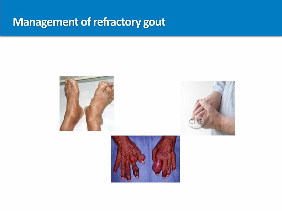
Management of refractory gout

Definition of refractory gout
• Symptomatic gout in which conventional urate-lowering therapies are contraindicated or the maximum medically appropriate dosage of these therapies does not control hyperuricemia
• Recurrent and disabling gout flares• Chronic gout arthropathy with or without bony erosions• Visible progressive tophi• Progressive physical disability• Poor health-related quality of life
The combination of severe gout, high burden of comorbidities, and polypharmacy can make refractory gout challenging to manage
Patients With Refractory Gout Fail to Achieve Target SUA Levels With Oral ULTs
• In about 200,000 gout patients, conventional oral urate-lowering agents fail to achieve target uric acid levels
Sundy JS, et al. JAMA. 2011;306(7):711-720
• Becker, MA, et al. N Engl J Med. 2005;353:2450-2461:
• 79% of patients (n=251) on 300 mg allopurinol/day did not meet target sUA <6.0 mg/dL
• 47% of patients (n=255) on 80 mg febuxostat/day for 52 weeks did not meet target sUA<6.0 mg/dL
Treatment options for Refractory Gout
• Dose escalation of conventional urate lowering therapies:
• allopurinol to 800 mg daily in divided doses
• febuxostat to 160 – 240 mg daily
• probenecid to 1000 mg daily in divided doses
• lesinurad to 200 mg daily
• Combination therapy: xanthine oxidase inhibitor + uricosuric
• Lifestyle modifications– diet - vitamin C - avoidance of high fructose corn syrup
– exercise - losartan for diuretics - low fat dairy products
– cherry extract - fenofibrate for niacin
• Biologic therapy– pegloticase
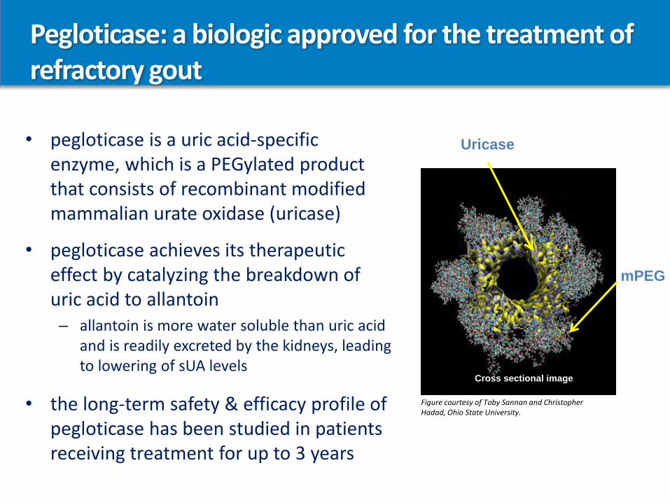
Pegloticase: a biologic approved for the treatment of refractory gout
Figure courtesy of Toby Sannan and Christopher Hadad, Ohio State University.
mPEG
Cross sectional image
Uricase• pegloticase is a uric acid-specific enzyme, which is a PEGylated product that consists of recombinant modified mammalian urate oxidase (uricase)
• pegloticase achieves its therapeutic effect by catalyzing the breakdown of uric acid to allantoin– allantoin is more water soluble than uric acid
and is readily excreted by the kidneys, leading to lowering of sUA levels
• the long-term safety & efficacy profile of pegloticase has been studied in patients receiving treatment for up to 3 years
C
UrinaryExcretion
Allantoin
Almost All Other Mammals
Purine catabolism
UrinaryExcretion
xanthineoxidase
allopurinolfebuxostat
allopurinolfebuxostat
probenecidlesinurad
XX
xanthineoxidase
Hypoxanthine Xanthine
Purines
Urate
Humans
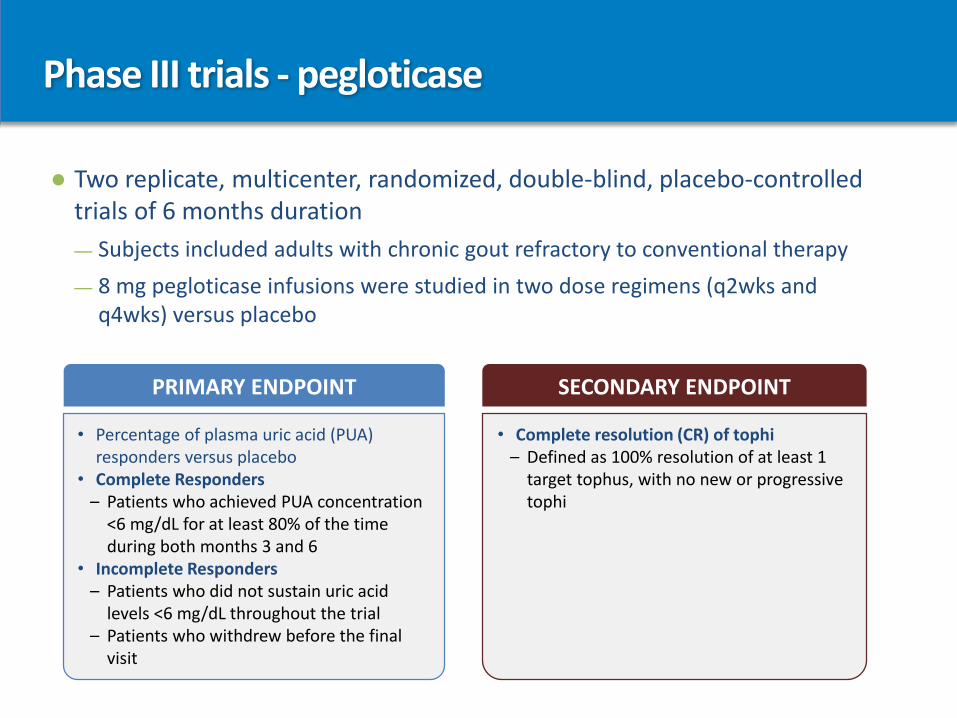
Phase III trials - pegloticase
● Two replicate, multicenter, randomized, double-blind, placebo-controlled trials of 6 months duration
— Subjects included adults with chronic gout refractory to conventional therapy
— 8 mg pegloticase infusions were studied in two dose regimens (q2wks and q4wks) versus placebo
• Percentage of plasma uric acid (PUA) responders versus placebo
• Complete Responders– Patients who achieved PUA concentration
<6 mg/dL for at least 80% of the time during both months 3 and 6
• Incomplete Responders– Patients who did not sustain uric acid
levels <6 mg/dL throughout the trial– Patients who withdrew before the final
visit
• Complete resolution (CR) of tophi – Defined as 100% resolution of at least 1
target tophus, with no new or progressive tophi
PRIMARY ENDPOINT SECONDARY ENDPOINT
Phase III trials –Baseline characteristics
• Patient characteristics
– Mean age: 55 (23-89)
– Predominantly male (82%)
– Mean BMI: 33 kg/m
• Patient disease characteristics
– Mean disease duration: 15 years
– Mean baseline sUA: 10 mg/dL
– Mean flares: 10 in prior 18 months (7 in past year)
• 63% described flares as severe/crippling
– 71% with visible tophi
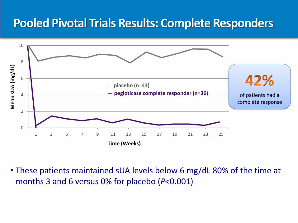
Pooled Pivotal Trials Results: Complete RespondersM
ean
sU
A(m
g/d
L)
Time (Weeks)
0
2
4
6
8
10
1 3 5 7 9 11 13 15 17 19 21 23 25
of patients had a complete response
-— placebo (n=43)
— pegloticase complete responder (n=36)
• These patients maintained sUA levels below 6 mg/dL 80% of the time at months 3 and 6 versus 0% for placebo (P<0.001)
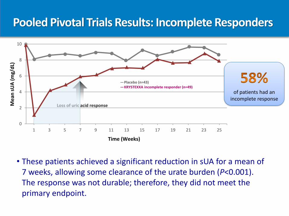
Pooled Pivotal Trials Results: Incomplete Responders
0
2
4
6
8
10
1 3 5 7 9 11 13 15 17 19 21 23 25
Loss of uric acid responseMe
an s
UA
(mg/
dL)
Time (Weeks)
—Placebo (n=43)—KRYSTEXXA incomplete responder (n=49)
of patients had an incomplete response
• These patients achieved a significant reduction in sUA for a mean of 7 weeks, allowing some clearance of the urate burden (P<0.001). The response was not durable; therefore, they did not meet the primary endpoint.
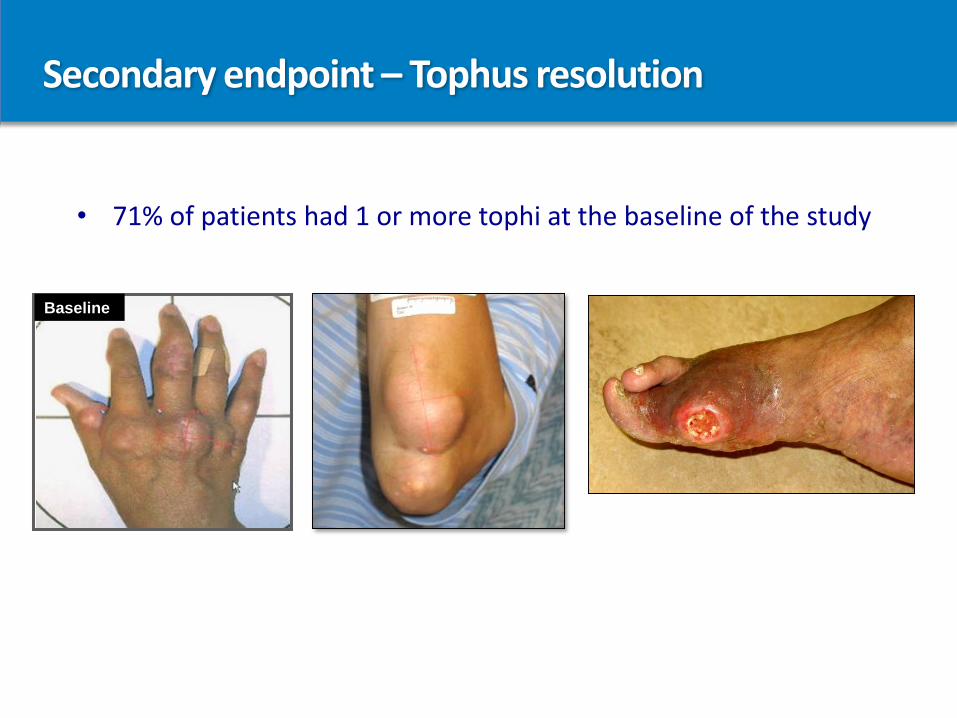
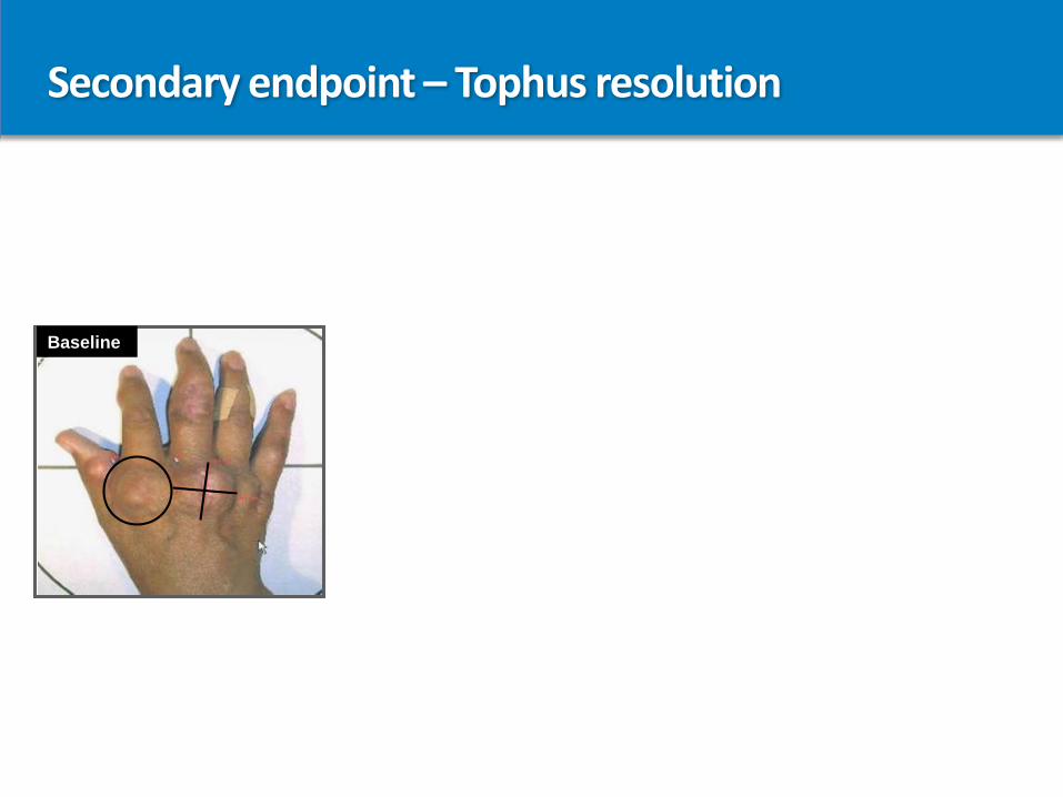
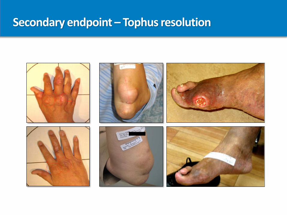
Secondary endpoint – Tophus resolution
• 71% of patients had 1 or more tophi at the baseline of the study
Baseline
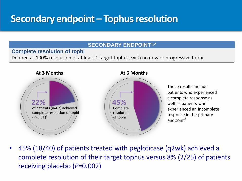
Secondary endpoint – Tophus resolution
Complete resolution of tophi Defined as 100% resolution of at least 1 target tophus, with no new or progressive tophi
SECONDARY ENDPOINT1,2
At 6 Months
45%Complete resolution of tophi
These results include patients who experienced a complete response as well as patients who experienced an incomplete response in the primary endpoint3
At 3 Months
22%of patients (n=62) achieved complete resolution of tophi (P=0.01)3
• 45% (18/40) of patients treated with pegloticase (q2wk) achieved a complete resolution of their target tophus versus 8% (2/25) of patients receiving placebo (P=0.002)
Secondary endpoint – Tophus resolution
Baseline
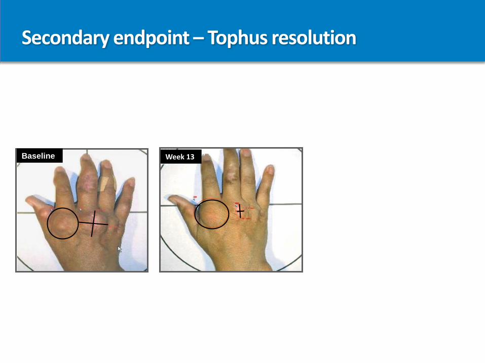
Secondary endpoint – Tophus resolution
Baseline Week 13
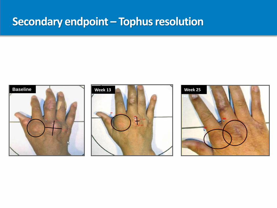
Secondary endpoint – Tophus resolution
Baseline Week 13 Week 25
Secondary endpoint – Tophus resolution
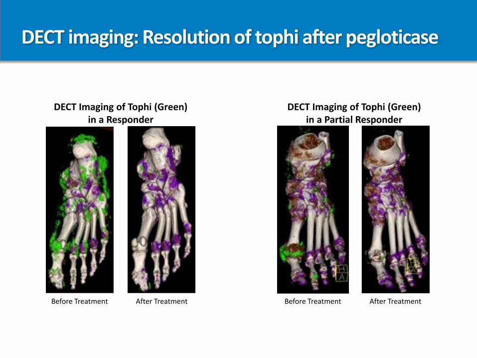
DECT imaging: Resolution of tophi after pegloticase
Before Treatment After Treatment
DECT Imaging of Tophi (Green)in a Responder
Before Treatment After Treatment
DECT Imaging of Tophi (Green)in a Partial Responder
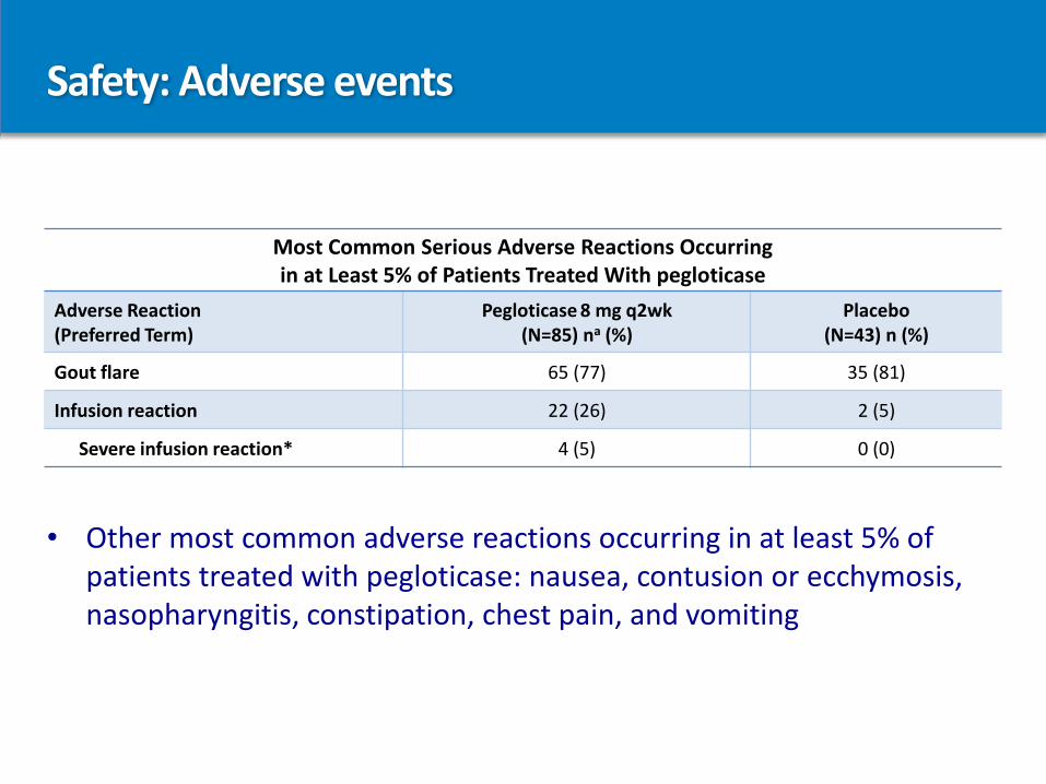
Safety: Adverse events
Most Common Serious Adverse Reactions Occurring in at Least 5% of Patients Treated With pegloticase
Adverse Reaction(Preferred Term)
Pegloticase 8 mg q2wk(N=85) na (%)
Placebo(N=43) n (%)
Gout flare 65 (77) 35 (81)
Infusion reaction 22 (26) 2 (5)
Severe infusion reaction* 4 (5) 0 (0)
• Other most common adverse reactions occurring in at least 5% of patients treated with pegloticase: nausea, contusion or ecchymosis, nasopharyngitis, constipation, chest pain, and vomiting
Safety: Adverse events
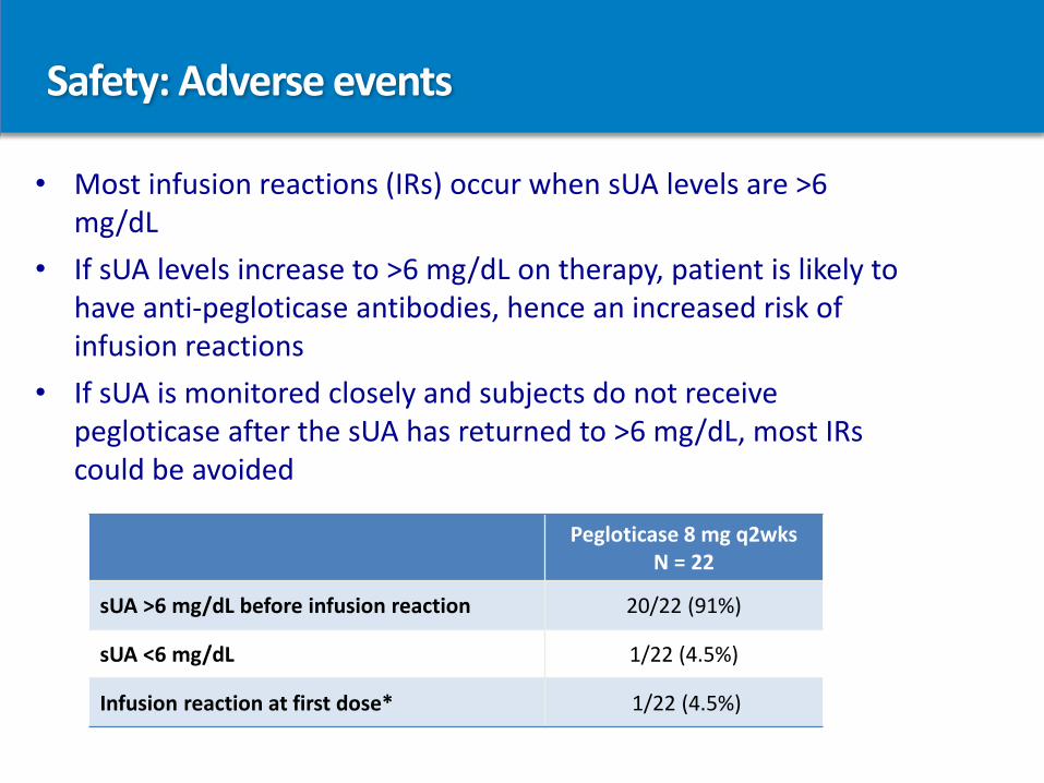
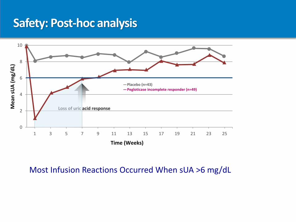
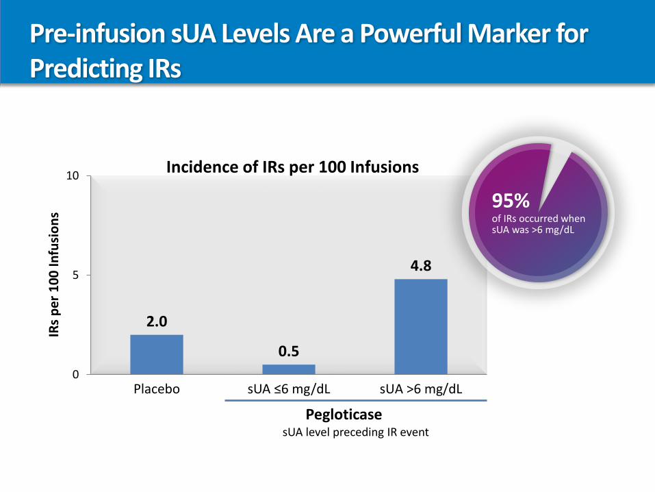
• Most infusion reactions (IRs) occur when sUA levels are >6 mg/dL
• If sUA levels increase to >6 mg/dL on therapy, patient is likely to have anti-pegloticase antibodies, hence an increased risk of infusion reactions
• If sUA is monitored closely and subjects do not receive pegloticase after the sUA has returned to >6 mg/dL, most IRs could be avoided
Pegloticase 8 mg q2wksN = 22
sUA >6 mg/dL before infusion reaction 20/22 (91%)
sUA <6 mg/dL 1/22 (4.5%)
Infusion reaction at first dose* 1/22 (4.5%)
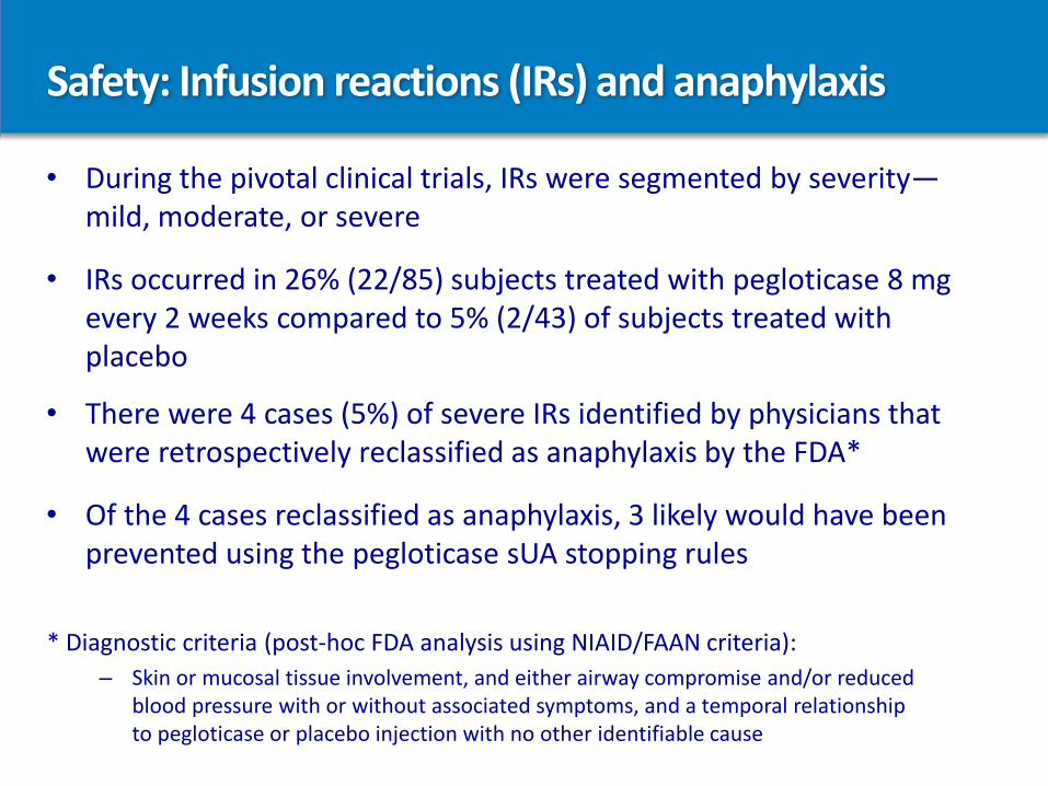
Safety: Infusion reactions (IRs) and anaphylaxis
• During the pivotal clinical trials, IRs were segmented by severity—mild, moderate, or severe
• IRs occurred in 26% (22/85) subjects treated with pegloticase 8 mg every 2 weeks compared to 5% (2/43) of subjects treated with placebo
• There were 4 cases (5%) of severe IRs identified by physicians that were retrospectively reclassified as anaphylaxis by the FDA*
• Of the 4 cases reclassified as anaphylaxis, 3 likely would have been prevented using the pegloticase sUA stopping rules
* Diagnostic criteria (post-hoc FDA analysis using NIAID/FAAN criteria):
– Skin or mucosal tissue involvement, and either airway compromise and/or reduced blood pressure with or without associated symptoms, and a temporal relationship to pegloticase or placebo injection with no other identifiable cause
Safety:
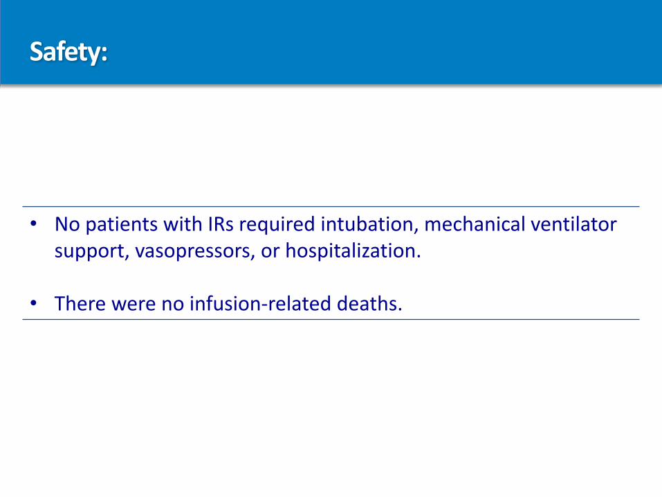
• No patients with IRs required intubation, mechanical ventilator support, vasopressors, or hospitalization.
• There were no infusion-related deaths.
Safety: Post-hoc analysis
0
2
4
6
8
10
1 3 5 7 9 11 13 15 17 19 21 23 25
Loss of uric acid responseMe
an s
UA
(mg/
dL)
Time (Weeks)
—Placebo (n=43)—Pegloticase incomplete responder (n=49)
Most Infusion Reactions Occurred When sUA >6 mg/dL
Pre-infusion sUA Levels Are a Powerful Marker forPredicting IRs
2.0
0.5
4.8
0
5
10
Placebo sUA ≤6 mg/dL sUA >6 mg/dL
IRs
pe
r 1
00
In
fusi
on
s
PegloticasesUA level preceding IR event
Incidence of IRs per 100 Infusions
95%of IRs occurred when sUA was >6 mg/dL
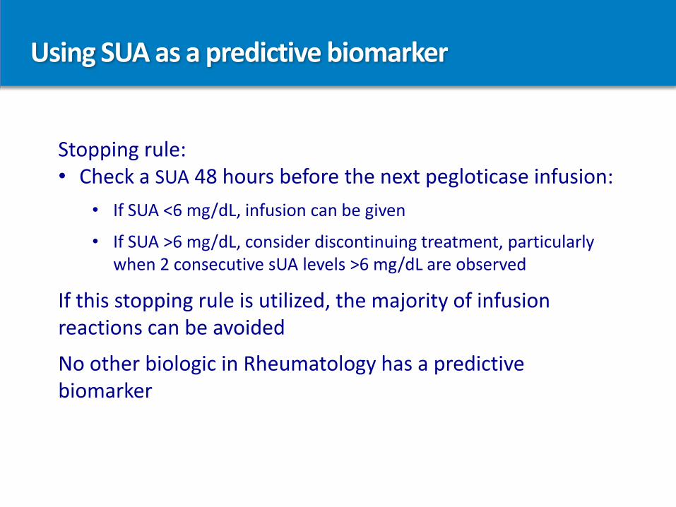
Using SUA as a predictive biomarker
Stopping rule:• Check a SUA 48 hours before the next pegloticase infusion:
• If SUA <6 mg/dL, infusion can be given
• If SUA >6 mg/dL, consider discontinuing treatment, particularly when 2 consecutive sUA levels >6 mg/dL are observed
If this stopping rule is utilized, the majority of infusion reactions can be avoided
No other biologic in Rheumatology has a predictive biomarker
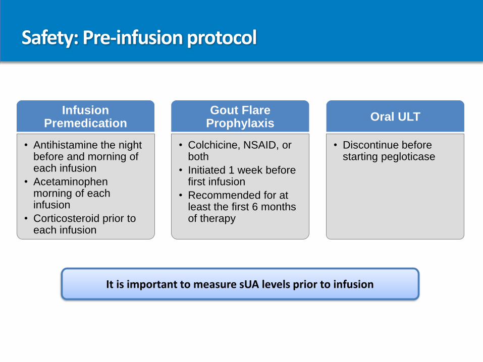
Safety: Pre-infusion protocol
InfusionPremedication
• Antihistamine the night before and morning of each infusion
• Acetaminophen morning of each infusion
• Corticosteroid prior to each infusion
Gout Flare Prophylaxis
• Colchicine, NSAID, or both
• Initiated 1 week before first infusion
• Recommended for at least the first 6 months of therapy
Oral ULT
• Discontinue before starting pegloticase
It is important to measure sUA levels prior to infusion

Collaboration between podiatrists & rheumatologists
• Latest treatment options mark a watershed moment in the management of gout
• Podiatrists role in gout management today is now more critical than ever
• Increased emphasis on comprehensive, collaborative, and correlated care amongst healthcare providers
• “First responders” of gout flares
• Surgical management of refractory tophaceous gout
• Missing link between a patient’s PCP and rheumatologist
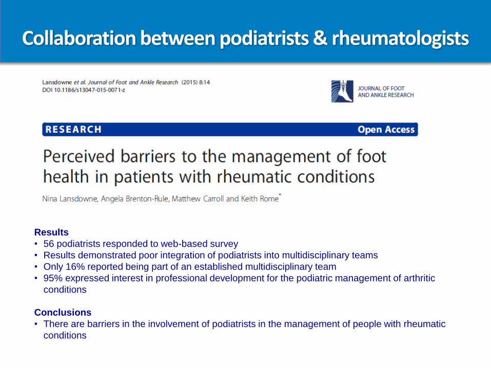
Collaboration between podiatrists & rheumatologists
Results
• 56 podiatrists responded to web-based survey
• Results demonstrated poor integration of podiatrists into multidisciplinary teams
• Only 16% reported being part of an established multidisciplinary team
• 95% expressed interest in professional development for the podiatric management of arthritic
conditions
Conclusions
• There are barriers in the involvement of podiatrists in the management of people with rheumatic
conditions

Overcoming barriers between podiatrists and rheumatologists in the care of gout patients
• Identify a local rheumatologist who has a common interest in gout
• Foster collaborative relationship
• Direct contact for referrals and timely consultations
• Dual podiatry-rheumatology clinics
• Develop co-management strategy• Delineation of roles• Identification of common ground
• Joint community outreach
Summary
• Gout is a chronic, progressive arthritis caused by hyperuricemia with associated chronic inflammation
• Body urate burden extends beyond clinically and physically apparent tophi
• Gout can be difficult to treat, beyond management with xanthine oxidase inhibitors and uricosurics
• Pegloticase is the first biologic and only FDA-approved treatment option for patients with chronic refractory gout
• Pegloticase can be an effective option for patients with chronic refractory gout

Thank you