Embed Size (px)
Citation preview

Otology & Neurotology39:S81–S94 � 2018, Otology & Neurotology, Inc.
Reflections on the Last 25 Years of the American OtologicalSociety and Thoughts on its Future
�D. Bradley Welling
and yRobert K. Jackler�Harvard Department of Otolaryngology, Massachusetts Eye and Ear Infirmary, Boston, Massachusetts; and yDepartment ofOtolaryngology Head and Neck Surgery, Stanford University, Stanford, California
Copyright ©
Address correspondencM.D., Ph.D., Harvard DeEye and Ear Infirmary, 2E-mail: Brad_Welling@M
The authors have no reDOI: 10.1097/MAO.0
ontributions of the American Otologi-ver the most recent quarter century
physics, and evidence based medicinbenefitted the practice of otology o
Purpose: To review ccal Society (AOS) o(1993–2018) and to comment on possible future evolution ofthe field during the quarter century to come.Methods: Retrospective review of selected topics from theAOS transactions, distinguished lectureships over the past25 years, and selective reflection by the authors. Speculationon potential advances of the next quarter century derivedfrom emerging topics in the current literature and foreseeabletrends in science and technology are also proffered forconsideration (and possible future ridicule).Results: Integration of multiple disciplines including bioen-gineering, medical imaging, genetics, molecular biology,
2018 Otology & Neurotology, Inc. Unaut
e and reprint requests to D. Bradley Welling,partment of Otolaryngology, Massachusetts43 Charles Street, Boston, MA 02114;
EEI.HARVARD.EDUlevant conflicts of interest000000000001760
S81
e have substantiallyver the past quarter
century. The impact of the contributions of members of theAOS in these developments cannot be over estimated.Conclusions: Further scientific advancement will certainlyaccelerate change in the practice of otologic surgery andmedicine over the coming decade in ways that will bemarvelous to behold. Key Words: Future—History ofotology—Sesquicentennial.
Otol Neurotol 39:S81–S94, 2018.
On the occasion the 150th anniversary of the AmericanOtological Society (AOS), it is appropriate to reflect onthe changes to the field of otology. This manuscriptfocuses on the most recent quarter of a century. Muchhas changed in the practice of otology since eight inau-gural members gathered in the beautiful new OceanHouse Hotel in Newport, Rhode Island on July 22,1868 to establish the American Otological Societyand, in fact, a great deal of that change has occurredin the past 25 years. Much of the progress in otology hasbeen made possible by the application of basic sciencediscovery to clinical medicine. Although by no means acomprehensive review, a few of the important advancesin otology for the last quarter century from the authors’opinions are highlighted. The programs from the AnnualAOS meetings were reviewed for trends and progress,with particular attention paid to lectures from the Guestsof Honor and our Scientific Lectures at the annualmeetings such as the Saumil Nalin Merchant MemorialScientific Lecturers (Table 1).
ADVANCES IN OTOLOGY OVER THE QUARTERCENTURY
First, a few cursory observations in reviewing theprograms from the Annual Meeting of the AOS from1993 to 2017 are preffered. Remarkably, the earlierprograms from 1993 to 2004 have no financial disclo-sures in the program whatsoever. These have certainlyproliferated in recent years. Financial disclosures firstappeared in 2005. Soon they occupied more space in theprogram than the program itself along with ‘‘Identifica-tion of Professional Practice Gaps,’’ ‘‘Goals & Objec-tives,’’ ‘‘Learning Objectives,’’ ‘‘Desired Results,’’ and‘‘Full Disclosures’’ from all authors on all presentations.The growing administrative burden of meeting the regu-latory requirements is clearly evident and emblematic ofmany similar encumbrances on the time of the membersof the AOS which detract from time spent in patient care,research, and teaching. It also burdens those who admin-ister the AOS educational programs.
Certainly of much greater significance, through thepast 25 years there is strong evidence of increasingintersections of clinical and basic science at the AnnualAOS meeting (Table 1). Such collaborations have greatlyaccelerated the acquisition of key knowledge to pushclinical treatments forward. The Guest of Honor in 1993,Cesar Fernandez, spoke on ‘‘The Need for Research in
horized reproduction of this article is prohibited.

Copyright © 2018 Otology & Neurotology, Inc. Unauthorized reproduction of this article is prohibited.
TABLE 1. Twenty five years of special lectures at the American Society of Otological Society
Year Guest of Honor Lecture TitleScientific/Merchant
Lecture Lecture Title
1993 D. Thane R. Cody, M.D. Remarks None
1994 Cesar Fernandez, M.D. The need for research inOtology
None
1995 Richard R. Gacek, M.D. The periodicity of theprofessional career
None
1996 James L. Sheehy, M.D. Tinnitus: a few thoughts None
1997 Mansfield F. W. Smith, M.D. The heritage and duty of theAmerican Otological Society
None
1998 Robert A. Jahrsdoerfer, M.D. You’ve come a long way baby None
1999 Barbara A. Bohne, Ph.D. Degeneration of theCochlea after noise damage:primary versus secondayevents
None
2000 Dearld E. Brackmann, M.D. Balancing the satisfaction of thepractice of medicine withpersonal and family life
None
2001 James B. Snow, Jr., M.D. Progress in the prevention ofotitis media throughimmunization
NONE
2002 David J. Lim, M.D. None
2003 James F. Battey, Jr., M.D., Ph.D. Remarks None
2004 Ugo Fisch, M.D. Surgical management oftemporal paragangliomas: along-term review
None
2005 George A. Gates, M.D. Science in Otology: past,present and future
None
2006 Richard A. Chole, M.D., Ph.D. Bacterial biofilms: the source oftissue destruction incholesteatomas?
Bradford J. May, Ph.D.Beverly Wright, Ph.D.Charles Limb, M.D.
Basic science seminar how wehear, how we listen
2007 Fred H. Linthicum, Jr., M.D. Bob Shannon, Ph.D. Speech understanding fromimplants: cochlear, brainstemand midbrain
2008 H. Richard Harnsberger, M.D. Decision support in the 21stcentury
Richard D. Rabbitt, Ph.D. Pathological semicircular canalafferent signals transmitted tothe brain during benignpositional vertigo and theirbiomechanical origins
2009 Robert J. Ruben, M.D. The Promise of Otology Alec N. Salt, Ph.D. Opportunities and techniques forlocal drug delivery to theinner ear
Scott Plotkin, M.D., Ph.D. The new frontier: targetedtherapies for NF2-relatedvestibular schwannomas
2010 Edwin W. Rubel, Ph.D. Toward a new era of hearingHabilitation
Jay T. Rubinstein, M.D.,Ph.D.
Characterization of theelectrically-evoked compoundaction potential of thevestibular nerve
2011 Richard A. Miyamoto, M.D. Cochlear implants: past, presentand future?
Kirk Aleck, M.D. Patterns of inheritance asillustrated by disorders ofhearing
2012 Vincente Honrubia, M.D. Vestibular testing, after 50 yearsstill a challenge
Carol Bauer, M.D. The neuroscience of tinnitus-implications for treatment
2013 Bruce J. Gantz, M.D. Electric þ acoustic speechprocessing: what have welearned about the auditorysystem
Neil Segil, Ph.D. Can we restore lost hearing?Molecular control of cell fateand cell division in thedevelopment and regenerationof the inner ear
2014 David A. Moffat, Ph.D. Ethical dilemmas in otology Josef P. Rauschecker, Ph.D. The gray area—tinnitus and thebrain
2015 Joseph B. Nadol, Jr., M.D. An imperative for otology M. Charles Liberman, Ph.D.a Hidden hearing loss: permanentcochlear nerve loss aftertemporary noise-inducedthreshold shift
S82 D. B. WELLING AND R. K. JACKLER
Otology & Neurotology, Vol. 39, No. 4S, 2018
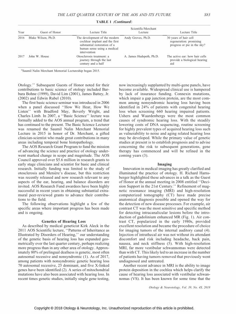
TABLE 1 (Continued)
Year Guest of Honor Lecture TitleScientific/Merchant
Lecture Lecture Title
2016 Blake Wilson, Ph.D. The development of the moderncochlear implant and the firstsubstantial restoration of ahuman sense using a medicalintervention
Andy Groves, Ph.D. 30 years of hair cellregeneration: promisingprogress or pie in the sky?
2017 John W. House Otosclerosis treatment: ajourney through the lastcentury and a half
A. James Hudspeth, Ph.D. The active ear: how hair cellsprovide a biological hearingaid
aSaumil Nalin Merchant Memorial Lectureship began 2015.
THE LAST QUARTER CENTURY OF THE AOS AND ITS FUTURE S83
Otology.’’ Subsequent Guests of Honor noted for theircontributions to basic science of otology included Bar-bara Bohne (1999), David Lim (2001), James Battey, Jr.(2002) and Edwin Rubel (2010).
The first basic science seminar was introduced in 2006when a panel discussed ‘‘How We Hear, How WeListen’’ with Bradford May, Beverly Wright, andCharles Limb. In 2007, a ‘‘Basic Science’’ lecture wasformally added to the AOS annual program, a trend thathas continued to the present. The Basic Science Lecturerwas renamed the Saumil Nalin Merchant MemorialLecture in 2015 in honor of Dr. Merchant, a giftedclinician-scientist who made great contributions in manyareas including temporal bone histopathology.
The AOS Research Grant Program to fund the missionof advancing the science and practice of otology under-went marked change in scope and magnitude. The AOSCouncil approved over $5.6 million in research grants toearly stage clinicians and scientist for basic and clinicalresearch. Initially funding was limited to the study ofotosclerosis and Meniere’s disease, but this restrictionwas recently released and now research relevant to anyaspects of the ear, hearing, and balance disorders areinvited. AOS Research Fund awardees have been highlysuccessful in recent years in obtaining substantial extra-mural peer-reviewed grants to advance their contribu-tions to the field.
The following observations highlight a few of thespecific areas where important progress has been madeand is ongoing.
Genetics of Hearing LossAs described by medical geneticist Kirk Aleck in the
2011 AOS Scientific lecture, ‘‘Patterns of Inheritance asIllustrated by Disorders of Hearing,’’ our understandingof the genetic basis of hearing loss has expanded geo-metrically over the last quarter century, perhaps realizingmore progress than in any other area of otology. Approx-imately 80% of prelingual deafness is genetic, most oftenautosomal recessive and nonsyndromic (1). As of 2017,among patients with nonsyndromic genetic hearing loss70 autosomal recessive, 25 dominant, and five X-linkedgenes have been identified (2). A series of mitochondrialmutations have also been associated with hearing loss. Inrecent times genetic studies, initially single gene testing,
Copyright © 2018 Otology & Neurotology, Inc. Unaut
now increasingly supplanted by multi-gene panels, havebecome available. Widespread clinical use is hamperedby lack of insurance funding. Connexin mutations,which impair a gap junction protein, are the most com-mon among nonsyndromic hearing loss having beenidentified in 24% of patients with congenital hearingloss when screening 660 hearing impaired patients.Ushers and Waardenbergs were the most commoncauses of syndromic hearing loss. With the steadilylowering costs of DNA sequencing, routine screeningfor highly prevalent types of acquired hearing loss suchas vulnerability to noise and aging related hearing lossmay be developed. While the primary value of geneticstudies at present is to establish prognosis and to adviseconcerning the risk to subsequent generations, genetherapy has commenced and will be refined in thecoming years (3).
ImagingInnovation in medical imaging has greatly clarified and
illuminated the practice of otology. H. Richard Harns-berger highlighted these advances in a talk as the Guestof Honor at the annual meeting in 2008 entitled ‘‘Deci-sion Support in the 21st Century.’’ Refinement of mag-netic resonance imaging (MRI) and high-resolutioncomputerized tomography (CT) have made preciseanatomical diagnosis possible and opened the way forthe detection of new disease processes. For example, aircontrast CT was the most sensitive and specific methodfor detecting intracanalicular lesions before the intro-duction of gadolinium enhanced MR (Fig. 1). Air con-trast CT, popularized in the early 1980s, providedexcellent resolution and became the procedure of choicefor imaging tumors of the internal auditory canal (4).Injection of intrathecal air was not without its attendantdiscomfort and risk including headache, back pain,nausea, and neck stiffness (5). With high-resolutionMRI, far more vestibular schwannomas were detectedthan with CT. This likely led to an increase in the numberof patients having tumors removed that previously wentundiagnosed and untreated.
Another recent advance in MRI is the ability to imageprotein deposition in the cochlea which helps clarify thecause of hearing loss associated with vestibular schwan-nomas (VS). It has been known for some time that the
horized reproduction of this article is prohibited.
Otology & Neurotology, Vol. 39, No. 4S, 2018

FIG. 1. A, Air-contrast CT demonstrating a small mass in the right internal auditory canal (IAC) (black arrow). B, T2-weighted magneticresonance image of a small intracanalicular vestibular schwannoma in the left IAC. C, T1-weighted image with contrast with clearenhancement in the right IAC. CT indicates computerized tomography.
S84 D. B. WELLING AND R. K. JACKLER
size of VS was not directly correlated with the hearingloss. Holliday et al. (6) confirmed this finding andobserved that elevated intralabyrinthine protein demon-strated on MRI FLAIR (fluid-attenuated inversion recov-ery sequences) images were correlated decreased pure-tone audiometric averages (Fig. 2). Increased protein inthe cochlea likely correlates with histopathologic find-ings which showed an acidophilic precipitate in the scalamedia of patients with VS (Fig. 3). Characterizing theseproteins may help explain why some tumors causehearing loss and others don’t, regardless of size. To thisend Dilwali et al. (7) have identified secreted proteinsfrom VS, some which are otoprotective of hearing(FGF2) and some which are associated with poorerhearing (TNF-a). Their direct link to the scala mediaprotein imaged, if any, is yet to be discovered.
Further refinement of MRI has led to diffusion tensorimaging, which can differentiate cranial nerves from theadjacent and compressing tumors (8) (Fig. 4). Lookingforward, nuclear magnetic resonance (NMR) in combi-nation with MRI may allow the detection of the chemicalcomposition of tumors, thus reducing the need for surgi-cal biopsy to make a certain pathologic diagnosis. Fur-thermore, precise knowledge of the molecular makeup ofdiscrete tumors in the future may allow prediction of
Copyright © 2018 Otology & Neurotology, Inc. Unauthorized
FIG. 2. A, MRI of an intracanalicular vestibular schwannoma on T1-webright signal in the cochlea likely representing protein deposition. C, T2displaces it in the lateral internal auditory canal. From Holliday et al. (6). Fresonance imaging.
Otology & Neurotology, Vol. 39, No. 4S, 2018
tumor growth and thus guide treatment timing andoptions.
More Selective Treatment of Cranial-base TumorsIn the past 25 years, there has been a shift in the
treatment of vestibular schwannomas (VS). Combiningstereotactic localization for radiation and better imagingtechniques has allowed the inclusion of radiation orobservation as treatment options. Clearly a higher per-centage of patients with VS are being observed for tumorgrowth before intervention than two decades ago (9).Stereotactic radiation has increasingly been selected as atreatment option in the same period. Stereotactic radia-tion is more likely to be recommended to the elderly ormedically infirm with documented tumor growth, butpatients of all ages are considering the relative merits ofeach approach. Why are patients and practitioners select-ing a conservative observational approach in recentyears? MRI can accurately detect growth, therefore,observing for non-growth is the least aggressive initialtreatment option. One argument for early intervention insmaller tumors has been pointed toward the possibility ofhearing preservation. Success may be in part dependentupon a distinct cleavage plane between the VS and thecochlear nerve (Fig. 5), but some VS invade the cochlear
reproduction of this article is prohibited.
ighted image with contrast. B, FLAIR sequence imaging showing-weighted image showing loss of CSF bright signal where tumorLAIR indicates fluid-attenuated inversion recovery; MRI, magnetic

FIG. 3. Acidophilic protein filling the scala media of a patient withneurofibromatosis type 2. Photo courtesy of Dr. Alicia Quesnel.
THE LAST QUARTER CENTURY OF THE AOS AND ITS FUTURE S85
nerve and have a poor cleavage plane as pointed out in1984 by AOS member, Neely (10). Figure 6 shows grossinfiltration of the 8th cranial nerve with no distinctcleavage plane in one tumor and a clearly definedseparation in another. Hearing preservation operationshave not been as successful as would be desired, leadingpatients to a more conservative initial approach in tumorswhere brainstem compression is not an immediateconcern.
The first successful medical intervention for VS waspresented by Plotkin et al. (11), the Basic Science Lecturein 2009. Surprisingly, he and his colleagues demon-strated improved sensorineural hearing in 50% of neuro-fibromatosis type 2 (NF2) patients treated with thevascular endothelial growth factor inhibitor bevacizumab(Fig. 7). Additionally, over 50% of NF2 associated VSshowed a decline in tumor volume when so treated(Fig. 8). Clarification of the tumor biology leads patientsever closer to targeted drug options.
Another major shift has been in the treatment ofglomus jugulare tumors. Twenty-five years ago themajority of patients were treated surgically, but nowsurgical resection is seldom employed, as stereotacticradiation has greatly decreased the number of tumors that
Copyright © 2018 Otology & Neurotology, Inc. Unaut
FIG. 4. Cleavage plane between vestibular schwannoma and cochlearJennifer O’Malley.
require surgery. Cranial nerve sparing with radiationrepresents a significant advantage over surgical resectionin many cases (13–15).
Meniere’s SyndromeThe treatment of Meniere’s syndrome has also shifted.
When conservative measures such as diuretics and dietfail, otologists have largely adopted intratympanic treat-ment including intratympanic steroids or intratympanicaminoglycosides, at least as a second line therapy. Thelatter have been shown effective in limiting Tumarkincrisis and both have resulted in significant control ofvertigo. Hearing preservation is still problematic how-ever (16–19).
Inner ear imaging can now demonstrate endolym-phatic hydrops (20). Dilute gadolinium in the middleear via transtympanic injection has shown apparenthydrops on T2-FLAIR weighted images in the scalamedia. This may eventually play a role in more preciseunderstanding of the underlying causes of Meniere’ssyndrome, and may be useful for determining treatmentoptions as we go forward, although the exact relationshipof hydrops and the symptom complex is not completelyunderstood yet. The genetics of familial Meniere’s dis-ease is also not yet elucidated, but segregation in differentpopulations and various potential genes have been impli-cated. When identified, it will hopefully help unlock themystery of its pathogenesis.
The ability to measure vestibular function has evolvedfrom the measurement of only one of the five sensoryelements of the vestibular system, typically the horizontalsemicircular canal with caloric stimulation, to the additionof measurements of the saccule and utricle with vestibularevoked myogenic potentials (VEMP) (21,22). The pres-ence of cervical VEMP response in Meniere’s syndromepatients has been associated with Tumarkin crisis and maypredict the onset of Meniere’s in the second ear (23).
In the 1980s and 1990s surgical procedures for therelief of vertigo were undertaken much more frequentlythan today. Procedures such as endolymphatic sacdecompression or shunting and vestibular neurectomywere major topics during AOS meetings both in presen-tations and the subject of innumerable panel discussions.
horized reproduction of this article is prohibited.
nerve A, H&E Stain, B, anti-neurofilament stain. Image courtesy of
Otology & Neurotology, Vol. 39, No. 4S, 2018

FIG. 5. Lack of distinct cleavage plane between vestibular schwannoma and cochlear nerve infiltration and distortion A, H&E stain, B, anti-neurofilament stain. Image courtesy of Jennifer O’Malley.
S86 D. B. WELLING AND R. K. JACKLER
These have become much less fashionable today largelydue to the rise of less invasive intratympanic drugtherapies with corticosteroids and aminoglycosides.
Migraine Related DizzinessThe most common cause of episodic vertigo has been
discovered to be migraine-related, another significantchange in the past 25 years. Migraine related episodicvertigo is five times more common than vertigo associ-ated with Meniere’s syndrome and affects children aswell as adults (24–26). Separating migraine from otherforms of episodic vertigo is not always readily accom-plished by history or other laboratory measurements.Recently, however, Murdin and Schilder demonstratedthat migraine sufferers have decreased thresholds forseveral test batteries measured on the platform chair,which provides motion in all rotational and translationalaxes (24). This objective data, the coming ‘‘vestibulo-gram,’’ further helps distinguish migrainous vertigophysiologically from Meniere’s disease. Not yet clini-cally available, this type of test holds promise for morediscreet diagnosis in the near future (27). Recognition ofthe role of migraine in vertigo has led to more aggressivetreatment options including adjustments in life style,diet, and prophylactic and acute pharmacologic controlsmeasures.
Copyright © 2018 Otology & Neurotology, Inc. Unauthorized
FIG. 6. Change in pure-tone average and speech discriminationof patients with NF2 treated with bevacizumab. Data extractedfrom Plotkin et al. (11).
Otology & Neurotology, Vol. 39, No. 4S, 2018
Benign Paroxysmal Positional VertigoThe management of benign paroxysmal positional
vertigo (BPPV) was altered radically when Parnes andMcClure (28) described the underlying pathophysiologyin 1992 with the demonstration of free floating particu-late matter within the membranous duct of the posteriorsemicircular canal (PSSC). Confirmation by scanningelectron microscopy showed otoconia within the lumenof the endolymphatic compartment of the PSSC (29)(Fig. 9). The particle repositioning maneuver, initiallydescribed by Epley (31) in 1980, was not widely adopteduntil the underlying pathophysiology was clarified.This work fundamentally changed the way BPPV istreated today.
Dehiscent Superior Semicircular CanalMinor et al. (32) published a landmark article in 1998
identifying the underlying the association of the dehis-cent superior semicircular canal (DSSC) with the symp-tom complex marked by autophony, disequilibrium,aural fullness, Tullio phenomena, pulse-synchronousoscillopsia, hyperacusis, and low-frequency conductivehearing loss (Fig. 10). Previously, this symptom complex
reproduction of this article is prohibited.
FIG. 7. Waterfall plot demonstrating the percent change investibular schwannoma tumor volumes following treatment withbevacizumab. Plotkin et al. (11).

FIG. 8. Otoconia demonstrated in the posterior semicircularcanal on scanning electron microscopy in a patient with intractableBPPV. Scale bar 2.5 mm. Images courtesy of Parnes and Chole(12). BPPV indicates benign paroxysmal positional vertigo.
FIG. 10. Dehiscence (arrow) of the superior semicircular canalon CT as initially described by Minor et al. (32). CT indicatescomputerized tomography.
THE LAST QUARTER CENTURY OF THE AOS AND ITS FUTURE S87
was poorly understood. Symptoms are frequentlyimproved with canal plugging or resurfacing techniques,but the underlying pathogenesis of the dehiscence is stillunknown. Even with plugging techniques all symptomsare not yet completely resolved and BPPV may occurposttreatment, however, overall serious complicationshave been few with plugging techniques (33). Interest-ingly, recent data suggest that near-dehiscence of thesuperior semicircular canal is associated with symptomssimilar to complete dehiscence and that plugging a nearlydehiscent canal also results in improved symptoms (34).
Copyright © 2018 Otology & Neurotology, Inc. Unaut
FIG. 9. Particle repositioning maneuver described by Epley in 1980. (Barnes (30)).
Cochlear ImplantsThroughout the past 25 years, the program for the
Annual AOS meeting has been filled with advances incochlear implant technology and application. Mostrecently, Wilson (35), was the 2016 Guest of Honor atthe 149th Annual AOS annual meeting and spoke on thetopic ‘‘The Development of the Modern CochlearImplant and the First Substantial Restoration of a HumanSense Using a Medical Intervention.’’ Cochlear implantsare a product of the merger of bioengineering and clinicalotology. Many key contributors have been leaders of theAOS over the years. Wilson proposed that of all positivechanges to the field of otology over the past quartercentury, the greatest accomplishment is the cochlear
horized reproduction of this article is prohibited.
Image Courtesy of Chicago Dizzy Clinic as shown in Welling and
Otology & Neurotology, Vol. 39, No. 4S, 2018

S88 D. B. WELLING AND R. K. JACKLER
implant. The ability of a profoundly deaf patient to gainopen-set speech discrimination is a modern medicalaccomplishment unparalleled in bioengineering to date.The impact this has on deaf patients is transforming,especially for deaf children who had poor prospects ofgaining communication skills which would allow inter-action with the hearing world. There have been over12,000 articles published on cochlear implants in the past25 years. To date, approximately 220,000 patients world-wide have received cochlear implants.
Ongoing areas of CI study include modification andrelaxation of eligibility requirements, hearing sparingelectrodes to allow potential electric–acoustic hybridstimulation and optimizing fitting paradigms. Bruce J.Gantz (AOS president 2010) was the guest of honor forthe annual AOS meeting in 2013 where he summarizedthis ongoing work in a talk entitled ‘‘Acoustic þ ElectricSpeech Processing: What Have We Learned about theAuditory System.’’ He noted that basic science questionsare being answered through clinical applications, such asthe gradual shift in frequency response to shorter hybridimplants (36,37).
Whether or not cochlear implants should be employedfor single-sided deafness and tinnitus suppression isactively being studied in a number of institutions(38,39). The optimal timing of bilateral cochlear implan-tation as contrasted with a period of bimodal stimulationis an ongoing debate with solid data needed to furtherclarify these options (40).
Future understanding of auditory cortex plasticity mayallow pharmacologic intervention to habilitate the con-genitally deafened adult who did not receive early audi-tory stimulation (41,42). The ability to process soundsignals into speech understanding may be followed byfurther voice development and integration into thehearing world.
The field of optogenetics allows for selective nervestimulation with optical sources may further refine ourability to discretely stimulate the auditory and vestibularpathways in the future or even the auditory cortexdirectly (43,44).
Implantable Hearing AidsMuch excitement was generated around active
implantable middle ear hearing aids in the past quartercentury. Fully implantable and partially implantabledevices have been studied. Patient’s rationalizationsfor avoiding conventional hearing aids are well knownincluding cosmesis, irritation of the ear canal, activitylimitation, and poor sound quality, particularly in noise.On the other side of the ledger, challenges have beenmany including battery technology, implantable micro-phone fibrosis, unreimbursed cost in excess of conven-tional aids, long-term viability of the mechanical devicesin the biologic environment leading to device failure andremoval or replacement, decline in unaided hearing as aresult of implantation, and MR incompatibility. Failureto clearly demonstrate objective improvement in per-formance when compared with appropriately fit
Copyright © 2018 Otology & Neurotology, Inc. Unauthorized
Otology & Neurotology, Vol. 39, No. 4S, 2018
conventional hearing aids on a variety of audiologic testshas perhaps been the major deterrent to wide-spreadacceptance of these devices (45,46).
Osseointegrated Implantable Hearing DevicesAnother area of marked progress has been in osseoin-
tegrated bone conduction hearing devices, particularlyfor applications in congenital aural atresias and patientswith severe eczema of the external auditory canal. Thepatient with a chronically draining middle ear is also acandidate. Implantation procedures have been simplified,but irritation and granulation around transcutaneouslyimplanted devices has not been completely overcome.Osseo-integrated devices for single-sided deafness, whencompared with conventional CROS aids, have not beenshown to be clearly superior (47).
Endoscopic Ear SurgeryWithin a few decades following the introduction of the
operating microscope in the 1920s, nearly all ear surgeryinvolved microsurgery. While endoscopes have beenused as adjuncts to the microscope in ear surgery forquite some time, in recent years fully endoscopic earsurgery is increasingly popular (48). Even the mostdelicate of ear surgery, stapedectomy, has been per-formed endoscopically in a few centers (49). Advantagesare greater visualization and illumination of recessessuch as the sinus tympani and the ability to peer intothe epitympanum without removal of the scutum. Dis-advantages which have deterred many otologists includethe need for one handed surgery, a limitation likely to beovercome by future technological advances.
WHAT WAS IN VOGUE 25 YEARS AGO?
Perilymphatic FistulaeA number of years ago a presentation was given in a
national meeting on the topic of perilymphatic fistulae(PLF) in which a map of the prevalence of PLF in theUnited States was flashed briefly. The speaker stated thatthe prevalence of spontaneous PLF seemed to segregatemuch like religion in the country with strong geographicpredilection. The speaker then quickly proceeded to thebody of the presentation. A recent retrospective survey ofover 1,000 patients evaluated for vertigo concluded thatless than 1% of cases were attributed to PLF (50). It is theauthor’s suspicion that the discovery of dehiscent supe-rior semicircular canals resulted in fewer explorations ofthe middle ear for PLF. It is conceivable that patching theround and oval windows did help decrease the symptomsassociated with the third window effect created by DSSC.Recent modeling indicates otherwise however (51).There appears to be a good deal of interest on the topicfrom Japan as a recent national study examined for thepresence of an inner ear specific, Cochlin tomo-protein(CTP), in middle ear lavage from suspected PLF patients.Only 20% of patients with suspected PLF showed CTP, ifthere was no associated physical trauma, lesion of themiddle ear, or recent stapes surgery. Patients with acute
reproduction of this article is prohibited.

THE LAST QUARTER CENTURY OF THE AOS AND ITS FUTURE S89
trauma who waited longer than 30 days for middle earexploration were significantly less likely to find positiveCTP presence (52). The usefulness of CTP may helpclarify the true incidence of PLF going forward.
Decompression of Vascular Loops for DisequilibriumMcCabe and Harker (53) proposed vascular loops as a
cause of incapacitating disequilibrium in 1983 anddecompression of the same was recommended for thecontrol of disabling positional vertigo in 1984 by Jannetta(54). A prolonged I–III interval on ABR was proposed asa result of significant vascular compression of thecochlear nerve. Several case series presented good out-comes from various decompression techniques (55,56).Although this condition may still occur, in the author’sexperience, lack of symptoms in patients with vascularloops found contacting the 8th nerve complex on routineMRI are so prevalent, it has led to a substantial decline indecompressions for vascular loops. A detailed investiga-tion of the relationship between cochleovestibular symp-toms and the type of vascular compression showed norelationship. Sirikci et al. (57) concluded that diagnosisof vascular conflict should not be based on imagingfindings alone.
SPECULATIONS ON FUTURE ADVANCES INOTOLOGY OVER THE NEXT QUARTER
CENTURY
Application of Molecular Biological TechniquesLooking forward to the next decades in our field brings
a great deal of excitement and anticipation. This willoccur in many ways, but most likely through continuedmerger of scientific disciplines. In the inaugural SaumilNalin Merchant Memorial Lectureship, M. Charles Lib-erman delivered a talk which gave an example of theadvances being seen today entitled ‘‘Hidden HearingLoss: Permanent Cochlear Nerve Loss after TemporaryNoise-Induced Threshold Shift.’’ Cochlear synaptopathyresulted from cochlear nerve degeneration after ‘‘tempo-rary’’ noise induced hearing loss (58). This condition ischaracterized by pure-tone thresholds returned to normal,but synapses with the inner ear hair cells were lost atlevels of acoustic trauma below those necessary to inducepermanent hair cell damage and permanent sensorineuralloss. Kujawa and Liberman (59) demonstrated furtherthat Neurotrophin-3, when applied to animal models ofcochlear synaptopathy demonstrate the regeneration ofneurite outgrowth to reconnect with the inner hair cellswith concomitant improved hearing thresholds.
The 2016 Merchant lecturer was Andy Groves whospoke on the topic of hair cell regeneration in hisscholarly presentation ‘‘30 Years of Hair Cell Regen-eration: Promising Progress or Pie in the Sky?’’ Herelated characterization of the changes in the tran-scriptome of neonatal mouse cochlear supporting haircells between 1- and 6-day old mice (60). The impor-tance of the Notch pathway inhibition was demon-strated corroborating the work of Edge and others in
Copyright © 2018 Otology & Neurotology, Inc. Unaut
unlocking the insights in the mechanism of regenera-tion of mammalian hair cells (61).
Lustig led a panel of experts at the 2016 meeting on‘‘Hurdles to Human Gene Therapy.’’ He previouslyshowed restoration of hearing in the VGLUT3 knockoutmouse using virally mediated gene therapy (62).Staecker, another distinguished panelist discussed howhis team knocked down a significant hurdle by deliveringatonal (CGF166) via an adenoviral vector to the livehuman inner ear with the intent of regeneration (63). Thisstudy is ongoing in phase I/II. An ophthalmologist on thepanel, Pierce, described their work in vision restorationusing adeno-associated viral (AAV2) mediated correc-tion of an inherited retinal dystrophy in children whichshowed efficacy in both eyes out to 3-year follow up (64).
The high interest and importance of this areas of studywas highlighted by the address of Neil Segil at the 146thannual meeting entitled ‘‘Can We Restore Lost Hearing?Molecular Control of Cell Fate and Cell Division in theDevelopment and Regeneration of the Inner Ear’’ (65).Other important advances demonstrating restoration ofhearing in young mouse models such as TMC1 and UsherType 2c (66,67). Shibata et al. (68) demonstrated thefeasibility of RNA-interference-mediated suppressiondelivered via a viral vector to slow progression of hearingloss in autosomal-dominant nonsyndromic hearing loss.
As gene editing becomes more widely applicable,specific defects may be selectively corrected in variousmutation affecting hearing. Major challenges with trans-lating gene therapy from bench to bedside are improvingefficiency of targeted delivery without causing furthertrauma or off-target editing. Specialized viral vectorssuch as Ancestral 80 have beautiful distribution through-out the inner and outer hair cells from base to apex in themouse model while minimizing immunogenicity (69).
Many congenital lesions causing pediatric hearing lossare present at birth with the absence of normal anatomicstructure development. Very early intervention, evenprenatal intrauterine intervention, may be necessary toallow critical structural development. Recent delivery tothe amniotic fluid in utero of antisense oligonucleotides(ASO), with subsequent rescue of hearing and balancephenotypes in a mouse model of Ushers syndrome (type1), was shown by the Brigande lab (70). The deliveredASO targeted a causal splice site mutation and showed itcorrected gene expression in the therapeutically relevantinner ear target tissues. Recent major advances in ASOtherapies include ‘‘improved specificity, potency, stabil-ity, delivery, and biodistribution and toxic effects havebeen minimized’’ according to the authors. This maybring a whole new realm of intervention.
As with ASOs, other gene editing systems are dramat-ically increasing genome engineering activities forresearch and eventually therapeutic purposes. Clusteredregularly interspaced short palindromic repeats(CRISPR)-associated Cas9 endonucleases have madegenome editing much more directed and efficient thanolder homologous recombination techniques, potentiallyrevolutionizing gene editing. Improved specificity
horized reproduction of this article is prohibited.
Otology & Neurotology, Vol. 39, No. 4S, 2018

S90 D. B. WELLING AND R. K. JACKLER
limiting off-target activity is crucial but seems to beadvancing (71–73). The application to otologic disease iseminent and very exciting.
Tissue RegenerationGrowth factor stimulated repair of tympanic mem-
brane (TM) perforations has been successfully exploredin animal models as early as the 1980s (74–76). Recentmanufacturing of clinical grade growth factors led tosuccessful human trials in Japan (77,78). Tissue engi-neering for TM repair is evolving quickly and will lead toa significant change in the way that perforated TMs aretreated in the near future—opined that ‘‘a regenerativemethod of tympanic membrane repair could be the great-est advance in otology since the cochlear implant’’ (79).It could simplify the traditional myringoplasty and tym-panoplasty by making it an office procedure.
Precision DiagnosticsIt is most probable that future members of the AOS
will not speak of ‘‘sensorineural hearing loss’’ as gener-ality covering lesions from the cochlea to the cortex.More discrete diagnostic testing will become common-place allowing discrete treatment paradigms. We willspeak of inner or outer hair cell dysfunction, cochlearsynaptopathy, cochlear nerve dyssynchrony, brainstemlesions of the dorsal cochlear nucleus afferents, or failureof efferent feedback. Importantly human temporal bonehistologic findings will be necessary in deciphering thediscrete underlying pathology necessary and cannot beneglected, as was so elegantly described by the Guest ofHonor in 2016, Joseph Nadol (AOS president 2009).Diagnostic imaging will help us decipher delayed audi-tory cortex development and methods then devised toimprove the natural language development of the deaf.
The need for similarly improved diagnostic testing ofthe vestibular system was highlighted by Vincente Hon-rubia in 2013, when as the Guest of Honor be presentedhis thesis on ‘‘Vestibular Testing, after 50 Years Still aChallenge.’’ We might predict that in the near future wewill have access to a simplified clinical ‘‘vestibulogram’’which will give discrete information from all 10 vestib-ular sensory end organs. The central nervous systemadvances will also be additive.
Vestibular ProsthesisAnother exciting development which follows from the
highly successful cochlear implant is the development ofthe vestibular implant for patients impaired by severebilateral vestibular dysfunction. Della Santina, Lewis,Rubinstein, and others have made important progress onthe development of a device to will resupply vestibularafferent function to the profoundly vestibulopathic sys-tem (80–83). Further refinement of multichannel stimu-lating paradigms, reduction of post implantationvariation, and channel interference will likely lead to asuccessful human vestibular prosthesis within the rela-tively near future. Given the aging of the population andthe high cost of falls among the elderly, sensor based fall
Copyright © 2018 Otology & Neurotology, Inc. Unauthorized
Otology & Neurotology, Vol. 39, No. 4S, 2018
reduction technologies are likely to enter widespread usein the coming years (84).
Tinnitus InterventionSeveral decades ago when a patient would ask ‘‘what
shall I do about the ringing in my ear, doctor? A well-respected otologist (Harold Schuknecht, AOS President1977) would answer, ‘‘what size shoe do you wear.’’When informed, he would instruct the patient to buy apair two sizes smaller, and then their tinnitus would notbother them so much. He would promptly exit the room.
What strides have we made in understanding andtreating tinnitus in the last quarter century? Other thanbeing more capable of ruling out tumors of the cerebel-lopontine angle, vascular malformations, and intracranialhypertension, it could be argued that we have not madesubstantial progress in terms of treatments. Masking isnot a new concept, but still useful. Tinnitus retraining hasbeen shown to be more effective than standard supportivetherapy in a recent blinded controlled study by Baueret al. (85) when combined with hearing aids. Effectivepharmacologic agents are yet to be proven.
Auditory neuroscience, however, has progressedsubstantially recently in understanding the pathophys-iology of tinnitus. Carol Bauer’s Basic Science Lecturein 2012 ‘‘The Neuroscience of Tinnitus-Implicationsfor Treatment’’ was outstanding (86). Rauscheckeret al. (87), the scientific lecturer in 2014, presented‘‘The Gray Area – Tinnitus and the Brain’’ to bring usa look at the advances in understanding of tinnitus.Advancing neuroscience certainly gives hope thatunderstanding the generators of abnormal spontaneousactivity in the auditory pathways (dorsal cochlear andventral cochlear nucleus, the inferior colliculus, andthe auditory cortex) or a lack of suppression of spon-taneous activity may lead to the eventual successfultreatment of this symptom. Modulation of the auditorycortex which appears to be hyperactive in tinnitus, maybe another treatment option. While auditory input isdecreased from the damaged cochlea in the region ofthe auditory cortex due to hearing impairment, theoutput from the cortex remains intact to communicatewith other parts of the brain. This persistent outputwhich does not correlate with input may be interpretedas the presence of tinnitus. (See Roberts et al. (88) foran excellent review).
Keeping the hyper-excitable theories in mind, a top-down approach to cortical or deep brain stimulation fortinnitus suppression may provide new treatment options(89,90). Pharmacologic control becomes possible withbetter understanding of the neural modulation of thesehyper-excitability-related signals (91).
Deep brain or cortical stimulation directly may alsoplay a role. Early application in human tinnitus sufferersis equivocal (92,93). The usefulness of transcranial mag-netic stimulation is also not clearly determined and maybe further explored (94). Directed extracranial electricalsuppression is being developed now and may becomerelevant.
reproduction of this article is prohibited.
THE LAST QUARTER CENTURY OF THE AOS AND ITS FUTURE S91
Surely with excellent collaborative efforts, tinnitustreatments should advance significantly past the‘‘smaller shoe-size’’ paradigm.
Eustachian TuboplastyChronic Eustachian tube (ET) dysfunction has been
treated with tympanostomy tubes for decades. The resultsfrom a recent multicenter controlled study evaluatingballoon dilation of the ET for chronic ET dysfunction byPoe (95) may change the way we intervene in the future.The study compared tympanogram normalization inpatients treated with topical steroids alone to patientswith steroids and eustachian tuboplasty. The favorableresults for the eustachian tuboplasty group caused theFDA to recommend early termination of the study and theprocedure was FDA approved for adults. Pediatric stud-ies will soon follow. Replacement of tympanostomytubes with ET dilation would be a major paradigm shift.Long-term sustainability is yet unknowns. Likewise, theapplicability to the pediatric population, and ultimatelythe cost/benefit ratio need to be clarified, but this couldbe a great paradigm shift in the field of a verycommon problem.
Hearing AidsDisruptive innovation is upon us in the hearing aid
versus personal sound amplification units (PSAPs) arenaas comparative studies and devices appear in greaternumbers. A recent report tested hearing in noise withnine PSAPs against a conventional hearing aid, at about1/10th the cost. Of the nine, the best five were selectedand three showed similar benefit to the more expensivetraditional hearing aid. At least one device showed worsediscrimination than no device at all (96). The audiolo-gist’s professional role in guiding patients through thismaze of new devices will accelerate quickly from thispoint. As only 20% of patients with mild to moderatehearing loss currently use hearing devices, there shouldbe an increased role for the audiology professional incounseling patients regarding hearing devices with amodel where the professional counseling is unbundledfrom the sale of a hearing device. This will benefit bothour colleagues in audiology and a growing numberof patients.
There has long been an unjustified stigma associatedwith wearing a hearing aid. The widespread cultural biasthat the wearer is older and less intellectually acute (i.e.,‘‘deaf and dumb’’) has in the past limited adoption ofthese devices among the hearing impaired. This is inmarked contrast to eye glasses which culturally areaccepted as stylish and a mark of intelligence. In thefuture, wearing of an ear device may be as universal asusing a cell phone is today. Led by youth proud to adoptthe latest devices, the current Bluetooth ear-piece revo-lution is a forerunner of what is likely to come. Thesedevices will interface with computers and phones, be aconveyer of information and entertainment, and serve asa telemetry system for continuous biometric monitoringof health. Future digital ear devices may enhance signal
Copyright © 2018 Otology & Neurotology, Inc. Unaut
to noise ratios in adverse listening situations, such asnoisy restaurants, thereby improving the sense of hearingeven among the normal hearing population. Connectedwith high-speed cloud based computers, they will trans-late across all languages in real time. Such highly capabledevices can readily incorporate an ability to adjust theiroutput to accommodate for hearing loss. Importantly, ashearing devices become widely used, consumer elec-tronic devices cost will plummet from their unreasonablyhigh cost of today just as technological capabilities soar.As this transition occurs, the stigma associatedwith hearing devices can be expected to fade and amuch higher fraction of hearing loss patients will adopttheir use (97).
Surgery Within the Living CochleaEarly 21st Century surgeons can operate within the
brain, heart, liver, kidney, and eye while sustaining oreven improving the organ’s native function. The abilityto perform procedures within a functioning, but diseasedcochlea remains impossible with today’s technology. It isthe only organ in the body which remains inaccessible tosurgical intervention for functional gain of its ordinaryphysiological function. Because of the organ’s extremefragility, new methods need to be developed whichenable intervention while preserving Organ of Cortihomeostasis. Fundamental is atraumatic creation of a‘‘cochleaport’’ which affords temporary access andcan be effectively resealed to restore cochlear wallintegrity. As the cochlea is both minute and mechanicallydelicate, internal procedures are beyond the ability of theunaided human hand. Robotic micromanipulators of thetype used in basic research which step down larger handmotions into microscopic scale and extinguish tremorwill be needed. Miniature, steerable endoscopes, andlight sources will also be needed to assist therapies suchas targeted placement of cells and drugs or, e.g., use of alaser to reduce endolymph production in hydrops.
Hearing TestingIn the 20th century, automation alleviated workers of
repetitive mechanical tasks in factories. In the 21stcentury, any process which can be explained as analgorithm can potentially be automated, even complexand sophisticated tasks typically done by highly educatedworkers (98,99). The impact of advances in artificialintelligence and computer image analysis are just nowbeing felt in medicine. It can be foreseen that advancedcomputer image analysis may 1 day greatly enhance thediagnostic ability of radiologists to interpret images (e.g.,CT, MRI) and for pathologists to be supplanted in themicroscopic diagnosis and molecular diagnosis of dis-ease. In hearing health care it seems inevitable thatartificial intelligence systems should be able to readilyreplace human audiologists for most routine hearingtesting. With the reduced burden of diagnostic studies,audiologist will evolve to have a greater emphasis uponthe rehabilitative aspects such as counseling and hearingdevice fitting. With regard to oto-surgical practice,
horized reproduction of this article is prohibited.
Otology & Neurotology, Vol. 39, No. 4S, 2018

S92 D. B. WELLING AND R. K. JACKLER
robotic and image guided surgery is likely to be anadjunct to surgical craft for the foreseeable future ratherthan a replacement. Office practice of otology, withits human interaction is likely to be less impacted byautomation. It will be a long time before computerswill be able to communicate empathy and showcompassion (98,99).
Otologic EducationFinally, just a word about where otologic education
may head in the near future. Immediate access to theworld’s body of published science makes our traineestoday light years ahead of our where our senior member-ship was at the same level of training (at least in thepresent authors’ case). Surgical training is moving to avirtual world with very realistic simulators that will shapethe skills of our young surgeons before they engage in thesurgical theater (100,101). This has been enabled bytechnological advances in immersive learning and isespecially important due to the increasing difficulty ofobtaining sufficient anatomical material for traditionaltemporal bone dissection courses. Automated testing forboard certification of surgical skills may be administeredvirtually in the future. It may be anticipated that fellow-ship-trained neurotologists, who focus their clinical prac-tice on diseases of the ear and lateral cranial base, will beincreasingly called on to provide inner ear surgery andmedicine including stapedectomies, cochlear implants,and gene infusions. The team approach to science andpatient care is evolving which improves the results for all.
SUMMARY
Predicting the future is always fraught with danger, butit is not inconceivable that in the next decade the disci-pline of otology will see application of molecular andgene transfer techniques to significantly change the waywe deal with various maladies including sensorineuralhearing loss and tinnitus. Specific targets and idealdelivery mechanisms are the subjects of intense interest.The biotechnology industry’s interest and investment isrising with the growing population of baby boomersworld-wide who need hearing restoration, balance reha-bilitations, and tinnitus suppression.
In the 1950s, it was said that otology was a declining asa field because most surgeries were done to drain infec-tions and antibiotics were greatly reducing these. Stape-dectomy was the major innovation of this time and itreinvigorated the field. Looking forward, a 0.5 to 1%deafness rate with this procedure should no longer beacceptable as it was in the era of analog hearing aids.Stapes footplate surgery is conducted right at the marginof what a human surgeon’s hand-eye coordination cansafely perform. Technical refinements such as use ofhighly precise robotic tools may reduce the incidence ofsensory loss to that of refractive eye surgery or, withadvances in hearing aid technology, indications for thisprocedure may decline. As biological therapies andtechnological advances provide safer alternatives to
Copyright © 2018 Otology & Neurotology, Inc. Unauthorized
Otology & Neurotology, Vol. 39, No. 4S, 2018
surgery, otologic surgeons may well become much morefocused on the implantation of devices.
Advances in wearable digital technology will almostcertainly lead to routine coupling of man and machine inthe population at large with the ear likely to featureprominently in placement of biosensors as well as com-munication devices. As leading experts in this interface,future otologists may be occupied with designing andmanaging these connections and adopting their use toaccommodate for hearing impairment.
The future contributions of the members of the AOS inteam-science with our colleagues from many disciplineswill surely see even more rapid advances for the welfareof our patients in the coming decades. The growth ofinternational science opens new avenues of collaborationas does the rapid sharing of knowledge. A whole newstory will surely be told when the bicentennial is cele-brated in 2068. Perhaps the larger question then will bewhen scientific advances allow all to hear, will we havemade any significant progress in the human ability tolisten. Brian F. McCabe (AOS President, 1986) wouldoften say ‘‘the proof is in the pudding.’’ The scientificfuture is indeed bright!
REFERENCES
1. Mehta D, Noon SE, Schwartz E, et al. Outcomes of evaluation andtesting of 660 individuals with hearing loss in a pediatric geneticsof hearing loss clinic. Am J Med Genet 2016;170A:2523–30.
2. Shearer AE, Hildebrand MS, Smith RJH. Hereditary hearing lossand deafness overview. In: Adam MP, Ardinger HH, Pagon RA,Wallace SE, Bean LJH, Mefford HC, Stephens K, Amemiya A,Ledbetter N, editors. GeneReviews1 [Internet]. Seattle, WA:University of Washington, Seattle; 1993–2017 . [updated 2017Jul 27].
3. Ahmed H, Shubina-Oleinik O, Holt JR. Emerging gene therapiesfor genetic hearing loss. J Assoc Res Otolaryngol 2017;18:649–70.
4. Sortland O. Computed tomography combined with gas cisternog-raphy for the diagnosis of expanding lesions in the cerebellopon-tine angle. Neuroradiology 1979;18:19–22.
5. Greenberger R, Khangure MS, Chakera MH. The morbidity of CTair meatography: a follow-up of 84 patients. Clin Radiol1987;38:535–6.
6. Holliday MA, Kim HJ, Zalewski CK, et al. Audiovestibularcharacteristics of small cochleovestibular schwannomas in neuro-fibromatosis type 2. Otolaryngol Head Neck Surg 2014;151:117–24.
7. Dilwali S, Landegger LD, Soares VY, Deschler DG, StankovicKM. Secreted factors from human vestibular schwannomas cancause cochlear damage. Sci Rep 2015;22:18559.
8. Hilly O, Chen JM, Birch J, et al. Diffusion tensor imagingtractography of the facial nerve in patients with cerebellopontineangle tumors. Otol Neurotol 2016;37:388–93.
9. Carlson ML, Habermann EB, Wagie AE, et al. The changinglandscape of vestibular schwannoma management in the UnitedStates—a shift toward conservatism. Otolaryngol Head Neck Surg2015;153:440–6.
10. Neely JG. Is it possible to totally resect an acoustic tumor andconserve hearing? Otolaryngol Head Neck Surg 1984;92:162–7.
11. Plotkin SR, Merker VL, Halpin C, et al. Bevacizumab for progres-sive vestibular schwannoma in neurofibromatosis type 2: a retro-spective review of 31 patients. Otol Neurotol 2012;33:1046–52.
12. Kao WT, Parnes LS, Chole RA. Otoconia and otolithic membranefragments within the posterior semicircular canal in benign par-oxysmal positional vertigo. Laryngoscope 2017;127:709–14.
reproduction of this article is prohibited.

13. Dobberpuhl MR, Maxwell S, Feddock J, St Clair W, Bush ML. 35. Wilson BS. The modern cochlear implant: a triumph of biomedical
THE LAST QUARTER CENTURY OF THE AOS AND ITS FUTURE S93
Treatment outcomes for single modality management of glomusjugulare tumors with stereotactic radiosurgery. Otol Neurotol2016;37:1406–10.
14. Jacob JT, Pollock BE, Carlson ML, Driscoll CL, Link MJ.Stereotactic radiosurgery in the management of vestibular schwan-noma and glomus jugulare: indications, techniques, and results.Otolaryngol Clin North Am 2015;48:515–26.
15. Wanna GB, Sweeney AD, Haynes DS, Carlson ML. Contempo-rary management of jugular paragangliomas. Otolaryngol ClinNorth Am 2015;48:331–41.
16. Silverstein H, Isaacson JE, Olds MJ, Rowan PT, Rosenberg S.Dexamethasone inner ear perfusion for the treatment of Meniere’sdisease: a prospective, randomized, double-blind, crossover trial.Am J Otol 1998;19:196–201.
17. Garduno-Anaya MA, Couthino De Toledo H, Hinojosa-GonzalezR, Pane-Pianese C, Rıos-Castaneda LC. Dexamethasone inner earperfusion by intratympanic injection in unilateral Meniere’s dis-ease: a two-year prospective, placebo-controlled, double-blind,randomized trial. Otolaryngol Head Neck Surg 2005;133:285–94.
18. Phillips JS, Westerberg B. Intratympanic steroids for Meniere’s diseaseor syndrome. Cochrane Database Syst Rev (7):2011;CD008514.
19. Viana LM, Bahmad F Jr, Rauch SD. Intratympanic gentamicin asa treatment for drop attacks in patients with Meniere’s disease.Laryngoscope 2014;124:2151–4.
20. Nakashima T, Naganawa S, Sugiura M, et al. Visualization ofendolymphatic hydrops in patients with Meniere’s disease. Laryn-goscope 2007;117:415–20.
21. Colebatch JG, Halmagyi GM, Skuse NF. Myogenic potentialsgenerated by a click-evoked vestibulocollic reflex. J NeurolNeurosurg Psychiatry 1994;57:190–7.
22. McCue MP, Guinan JJ Jr. Acoustically responsive fibers in thevestibular nerve of the cat. J Neurosci 1994;14:6058–70.
23. Timmer FC, Zhou G, Guinan JJ, Kujawa SG, Herrmann BS, RauchSD. Vestibular evoked myogenic potential (VEMP) in patients withMeniere’s disease with drop attacks. Laryngoscope 2006;116:776–9.
24. Murdin L, Schilder AG. Epidemiology of balance symptoms anddisorders in the community: a systematic review. Otol Neurotol2015;36:387–92.
25. Dieterich M, Obermann M, Celebisoy N. Vestibular migraine: themost frequent entity of episodic vertigo. J Neurol 2016;263(Suppl):S82–9.
26. Lewis D, Winner P, Saper J, et al. Randomized double-blind,placebo-controlled study to evaluate the efficacy and safety oftopiramate for migraine prevention in pediatric subjects 12 to17 years of age. Pediatrics 2009;123:924–34.
27. Wang J, Lewis RF. Abnormal tilt perception during centrifugationin patients with vestibular migraine. J Assoc Res Otolaryngol2016;17:253–8.
28. Parnes LS, McClure JA. Free-floating endolymph particles: a newoperative finding during posterior semicircular canal occlusion.Laryngoscope 1992;102:988–92.
29. Welling DB, Parnes LS, O’Brien B, Bakaletz LO, Brackmann DE,Hinojosa R. Particulate matter in the posterior semicircular canal.Laryngoscope 1997;107:90–4.
30. Welling DB, Barnes DE. Particle repositioning maneuver forbenign paroxysmal positional vertigo. Laryngoscope 1994;104(8 Pt 1):946–9.
31. Epley JM. The canalith repositioning procedure: for treatment ofbenign paroxysmal positional vertigo. Otolaryngol Head NeckSurg 1992;107:399–404.
32. Minor LB, Solomon D, Zinreich JS, Zee DS. Sound- and/orpressure-induced vertigo due to bone dehiscence of the superi-orsemicircular canal. Arch Otolaryngol Head Neck Surg1998;124:249–58.
33. Xie Y, Sharon JD, Pross SE, et al. Surgical complications fromsuperior canal dehiscence syndrome repair: two decades of expe-rience. Otolaryngol Head Neck Surg 2017;157:273–80.
34. Ward BK, Wenzel A, Ritzl EK, et al. Near-dehiscence: clinicalfindings in patients with thin bone over the superior semicircularcanal. Otol Neurotol 2013;34:1421–8.
Copyright © 2018 Otology & Neurotology, Inc. Unaut
engineering and the first substantial restoration of human senseusing a medical intervention. IEEE Pulse 2017;8:29–32.
36. Gantz BJ, Dunn CC, Oleson J, Hansen MR. Acoustic plus electricspeech processing: Long-term results. Laryngoscope 2018;128:473–81.
37. Scheperle RA, Tejani VD, Omtvedt JK, et al. Delayed changes inauditory status in cochlear implant users with preserved acoustichearing. Hear Res 2017;350:45–57.
38. Arts RA, George EL, Griessner A, Zierhofer C, Stokroos RJ.Tinnitus suppression by intracochlear electrical stimulation insingle-sided deafness: a prospective clinical trial—part I. AudiolNeurotol 2015;20:294–313.
39. Sladen DP, Carlson ML, Dowling BP, et al. Early outcomes aftercochlear implantation for adults and children with unilateralhearing loss. Laryngoscope 2017;127:1683–8.
40. Moberly AC, Lowenstein JH, Nittrouer S. Early bimodal stimula-tion benefits language acquisition for children with cochlearimplants. Otol Neurotol 2016;37:24–30.
41. Takesian AE, Kotak VC, Sanes DH. Presynaptic GABA(B)receptors regulate experience-dependent development of inhibi-tory short-term plasticity. J Neurosci 2010;30:2716–27.
42. Weis T, Puschmann S, Brechmann A, Thiel CM. Effects of L-dopaduring auditory instrumental learning in humans. PLoS One2012;7:e52504.
43. Guo W, Hight AE, Chen JX, et al. Hearing the light: neural andperceptual encoding of optogenetic stimulation in the centralauditory pathway. Sci Rep 2015;5:10319.
44. Kozin ED, Darrow KN, Hight AE, et al. Direct visualization of themurine dorsal cochlear nucleus for optogenetic stimulation of theauditory pathway. J Vis Exp 2015;52426.
45. Uhler K, Anderson MC, Jenkins HA. Long-term outcome data inpatients following one year’s use of a fully implantable activemiddle ear implant. Audiol Neurootol 2016;21:105–12.
46. Zwartenkot JW, Mulder JJ, Snik AF, Cremers CW, Mylanus EA.Active middle ear implantation: long-term medical and technicalfollow-up, implant survival, and complications. Otol Neurotol2016;37:513–9.
47. Crowson MG, Tucci DL. Mini review of the cost-effectivenessof unilateral osseointegrated implants in adults: possibly cost-effective for the correct indication. Audiol Neurootol 2016;21:69–71.
48. Kiringoda R, Kozin ED, Lee DJ. Outcomes in endoscopic earsurgery. Otolaryngol Clin North Am 2016;49:1271–90.
49. Hunter JB, Rivas A. Outcomes following endoscopic stapessurgery. Otolaryngol Clin North Am 2016;49:1215–25.
50. Patnaik U, Srivastava A, Sikka K, Thakar A. Surgery for vertigo:10-year audit from a contemporary vertigo clinic. J Laryngol Otol2015;129:1182–7.
51. Niesten ME, Stieger C, Lee DJ, et al. Assessment of the effects ofsuperior canal dehiscence location and size on intracochlear soundpressures. Audiol Neurootol 2015;20:62–71.
52. Matsuda H, Sakamoto K, Matsumura T, et al. A nationwidemulticenter study of the Cochlin tomo-protein detection test:clinical characteristics of perilymphatic fistula cases. ActaOtolaryngol 2017;137 (suppl):S53–9.
53. McCabe BF, Harker LA. Vascular loop as a cause of vertigo. AnnOtol Rhinol Laryngol 1983;92:542–3.
54. Jannetta PJ, Møller MB, Møller AR. Disabling positional vertigo.N Engl J Med 1984;310:1700–5.
55. McCabe BF, Gantz BJ. Vascular loop as a cause of incapacitatingdizziness. Am J Otol 1989;10:117–20.
56. Brackmann DE, Kesser BW, Day JD. Microvascular decompres-sion of the vestibulocochlear nerve for disabling positional ver-tigo: the House Ear Clinic experience. Otol Neurotol 2001;22:882–7.
57. Sirikci A, Bayazit Y, Ozer E, et al. Magnetic resonance imagingbased classification of anatomic relationship between the cochle-ovestibular nerve and anterior inferior cerebellar artery in patientswith non-specific neuro-otologic symptoms. Surg Radiol Anat2005;27:531–5.
horized reproduction of this article is prohibited.
Otology & Neurotology, Vol. 39, No. 4S, 2018

58. Kujawa SG, Liberman MC. Adding insult to injury: cochlear nerve 80. Hageman KN, Kalayjian ZK, Tejada F, et al. A CMOS neural
S94 D. B. WELLING AND R. K. JACKLER
degeneration after ‘‘temporary’’ noise-induced hearing loss. JNeurosci 2009;29:14077–85.
59. Kujawa SG, Liberman MC. Synaptopathy in the noise-exposedand aging cochlea: primary neural degeneration in acquired sen-sorineural hearing loss. Hear Res 2015;330 (Pt B):191–9.
60. Maass JC, Gu R, Cai T, et al. Transcriptomic analysis of mousecochlear supporting cell maturation reveals large-scale changes innotch responsiveness prior to the onset of hearing. PLoS One2016;11:e0167286.
61. Mizutari K, Fujioka M, Hosoya M, et al. Notch inhibition inducescochlear hair cell regeneration and recovery of hearing afteracoustic trauma. Neuron 2013;77:58–69.
62. Akil O, Seal RP, Burke K, et al. Restoration of hearing in theVGLUT3 knockout mouse using virally mediated gene therapy.Neuron 2012;75:283–93.
63. Safety, Tolerability and Efficacy for CGF166 in Patients WithBilateral Severe-to-profound Hearing Loss. ClinicalTrials.Gov.
64. Bennett J, Wellman J, Marshall KA, et al. Safety and durability ofeffect of contralateral-eye administration of AAV2 gene therapy inpatients with childhood-onset blindness caused by RPE65 muta-tions: a follow-on phase 1 trial. Lancet 2016;388:661–72.
65. Kwan T, White PM, Segil N. Development and regeneration of theinner ear. Ann N Y Acad Sci 2009;1170:28–33.
66. Askew C, Rochat C, Pan B, et al. Tmc gene therapy restoresauditory function in deaf mice. Sci Transl Med 2015;7:295ra108.
67. Pan B, Askew C, Galvin A, et al. Gene therapy restores auditoryand vestibular function in a mouse model of Usher syndrome type1c. Nat Biotechnol 2017;35:264–72.
68. Shibata SB, Ranum PT, Moteki H, et al. RNA interferenceprevents autosomal-dominant hearing loss. Am J Hum Genet2016;98:1101–13.
69. Landegger LD, Pan B, Askew C, et al. A synthetic AAV vectorenables safe and efficient gene transfer to the mammalian innerear. Nat Biotechnol 2017;35:280–4.
70. Depreux FF, Wang L, Jiang H, et al. Antisense oligonucleotidesdelivered to the amniotic cavity in utero modulate gene expressionin the postnatal mouse. Nucleic Acids Res 2016;44:9519–29.
71. Pattanayak V, Guilinger JP, Liu DR. Determining the specificitiesof TALENs, Cas9, and other genome-editing enzymes. MethodsEnzymol 2014;546:47–78.
72. Cong L, Ran FA, Cox D, et al. Multiplex genome engineeringusing CRISPR/Cas systems. Science 2013;339:819–23.
73. Mali P, Yang L, Esvelt KM, et al. RNA-guided human genomeengineering via Cas9. Science 2013;339:823–6.
74. Kato M, Jackler RK. Repair of chronic tympanic membraneperforations with fibroblast growth factor. Otolaryngol Head NeckSurg 1996;115:538–47.
75. Dvorak DW, Abbas G, Ali T, Stevenson S, Welling DB. Repair ofchronic tympanic membrane perforations with long-term epider-mal growth factor. Laryngoscope 1995;105 (12 Pt 1):1300–4.
76. Santa Maria PL, Weierich K, Kim S, Yang YP. Heparin bindingepidermal growth factor-like growth factor heals chronic tympanicmembrane perforations with advantage over fibroblast growthfactor 2 and epidermal growth factor in an animal model. OtolNeurotol 2015;36:1279–83.
77. Hakuba N, Iwanaga M, Tanaka S, et al. Basic fibroblast growthfactor combined with atelocollagen for closing chronic tympanicmembraneperforations in 87 patients. Otol Neurotol 2010;31:118–21.
78. Kanemaru S, Umeda H, Kitani Y, Nakamura T, Hirano S, Ito J.Regenerative treatment for tympanic membrane perforation. OtolNeurotol 2011;32:1218–23.
79. Jackler RK. A regenerative method of tympanic membrane repaircould be the greatest advance in otology since the cochlearimplant. Otol Neurotol 2012;33:289.
Copyright © 2018 Otology & Neurotology, Inc. Unauthorized
Otology & Neurotology, Vol. 39, No. 4S, 2018
interface for a multichannel vestibular prosthesis. IEEE TransBiomed Circuits Syst 2016;10:269–79.
81. Valentin NS, Hageman KN, Dai C, Della Santina CC, FridmanGY. Development of a multichannel vestibular prosthesisprototype by modification of a commercially availablecochlear implant. IEEE Trans Neural Syst Rehabil Eng2013;21:830–9.
82. Lewis RF. Vestibular implants studied in animal models: clinicaland scientific implications. J Neurophysiol 2016;116:2777–88.
83. Phillips JO, Ling L, Nie K, et al. Vestibular implantation andlongitudinal electrical stimulation of the semicircular canal affer-ents in human subjects. J Neurophysiol 2015;113:3866–92.
84. Kosse NM, Brands K, Bauer JM, Hortobagyi T, Lamoth CJ.Sensor technologies aiming at fall prevention in institutionalizedold adults: a synthesis of current knowledge. Int J Med Inform2013;82:743–52.
85. Bauer CA, Berry JL, Brozoski TJ. The effect of tinnitus retrainingtherapy on chronic tinnitus: a controlled trial. LaryngoscopeInvestig Otolaryngol 2017;2:166–77.
86. Ryan D, Bauer CA. Neuroscience of tinnitus. Neuroimaging ClinN Am 2016;26:187–96.
87. Rauschecker JP, Leaver AM, Muhlau M. Tuning out the noise:limbic-auditory interactions in tinnitus. Neuron 2010;66:819–26.
88. Roberts LE, Eggermont JJ, Caspary DM, Shore SE, Melcher JR,Kaltenbach JA. Ringing ears: the neuroscience of tinnitus. JNeurosci 2010;30:14972–9.
89. Chambers AR, Resnik J, Yuan Y, et al. Central gain restoresauditory processing following near-complete cochlear denerva-tion. Neuron 2016;89:867–79.
90. Knudson IM, Shera CA, Melcher JR. Increased contralateralsuppression of otoacoustic emissions indicates a hyperresponsivemedial olivocochlear system in humans with tinnitus and hyper-acusis. J Neurophysiol 2014;112:3197–208.
91. Kumar M, Reed N, Liu R, Aizenman E, Wipf P, Tzounopoulos T.Synthesis and evaluation of potent KCNQ2/3-specific channelactivators. Mol Pharmacol 2016;89:667–77.
92. Shi Y, Burchiel KJ, Anderson VC, Martin WH. Deep brainstimulation effects in patients with tinnitus. Otolaryngol HeadNeck Surg 2009;141:285–7.
93. De Ridder D, Vanneste S, van der Loo E, Plazier M, Menovsky T,van de Heyning P. Burst stimulation of the auditory cortex: a newform of neurostimulation for noise-like tinnitus suppression. JNeurosurg 2010;112:1289–94.
94. Roland LT, Peelle JE, Kallogjeri D, Nicklaus J, Piccirillo JF. Theeffect of noninvasive brain stimulation on neural connectivity inTinnitus: a randomized trial. Laryngoscope 2016;126:1201–6.
95. Poe D. Study of Safety and Efficacy of Balloon dilation of theEustachian tube. ClinicalTrials.gov Identifier: NCT02087150.
96. Reed NS, Betz J, Kenkig N, Korczak M, Lin FR. Personal soundamplification products vs a conventional hearing aid for speechunderstanding in noise. JAMA 2017;318:89–90.
97. Jackler RK. The impending end to the stigma of wearing eardevices and its revolutionary implications. Otol Neurotol2006;27:299–300.
98. Ford M. The Rise of the Robots: Technology and the Threat of aJobless Future. New York: Basic Books; 2015.
99. Ross A. The Industries of the Future. New York: Simon &Schuster; 2016.
100. Locketz GD, Lui JT, Chan S, et al. Anatomy-specific virtualreality simulation in temporal bone dissection: perceived utilityand impact on surgeon confidence. Otolaryngol Head Neck Surg2017;156:1142–9.
101. Wiet GJ, Stredney D, Kerwin T, et al. Virtual temporal bonedissection system: OSU virtual temporal bone system: develop-ment and testing. Laryngoscope 2012;122 (Suppl):S1–2.
reproduction of this article is prohibited.


![Reflections on American Constitutionalism · HeinOnline -- 38 Am. J. Comp. L. Supp. 423 1990 1990] REFLECTIONS ON AMERICAN CONSTITUTIONALISM 423 several important ways, some of which](https://img.pdfslide.us/doc/110x75/5e1a4029fc26dc706136dbca/reflections-on-american-constitutionalism-heinonline-38-am-j-comp-l-supp.jpg)