Embed Size (px)
DESCRIPTION
referred pain
Citation preview

8 Practical PAIN MANAGEMENT, Nov/Dec 2003
REFERRED PAINVS.ORIGIN OF PAINPATHOLOGY
Understanding the organic
and physiological patterns
of referred pain helps to
identify the true origin of
pathology and inform
proper treatment.by James Woessner, MD, PhD
REFERRED PAINVS.ORIGIN OF PAINPATHOLOGY

Referred pain, as defined by An-derson, is “pain felt at a site dif-ferent from the injured or diseased
organ or body part.”1 Radiating pain,however, is not defined by Anderson; ra-diating pain is more commonly used inconnection with pain perceived in somat-ic nerve and spinal nerve root distribu-tions (i.e. the dermatomes that all physi-cians learn early in their training).Merskey and Bogduk specify that “re-ferred pain is pain perceived in a regionthat has a nerve supply different from thatof the source of pain,”2 which indicatesthat radiating pain is completely differ-ent (the author does not find that ex-cluding radiating pain from referred painuseful; radiating pain is just a subcatego-ry of referred pain).
Bellenir adds “Antidromic” into thedefinition, noting that visceral and so-matic nerve cells may synapse on the sameneuron at the spinal cord.3 With chronicstimulation, “the impulse will spill over. .. . into the somatic nerve.” Warfield andFausett also calls it “heterotopic” pain andstate that “referred pain is a phenomenonthat is frequently encountered and is mostbaffling.”4 Added meaning is conveyed byKhalsa, who defines referred pain as“pain that exists in a location other thanthe immediate area of the spasm”5 with-out defining limits, or specific distribu-tions. However, according to Khalsa, therange of the main pain should not be larg-er than the receptive field, which variesin size depending on the area of the body
It has been said by the IASP Subcom-mittee on Classification that “Pain is al-ways subjective. . .”2 Yet if the cliniciandoes not understand a presenting painpattern, where the pain is already con-sidered “subjective,” the chances of justlyhandling and treating the patient are lim-ited. Indeed, if psychogenic (eg. “subjec-tive”) pain and referred pain become syn-onymous, then the physician may stoplooking for the originating pathology andnot provide proper treatment or anytreatment at all. The patient is likely toslip into a downward spiral of “doctorshopping.”
However, it must be said that all pain“is always real.” Thus, diagnosing painpathology — in the face of referred painthat may be perceived as worse than theorigin of the pain — becomes a dauntingchallenge. An understanding of the painpathophysiology with familiarity of re-ferred pain possibilities, coupled with a
thorough history and physical examina-tion, is essential in making an appropri-ate and potentially correct diagnosis.
Referred Pain CharacteristicsThe best known referred pain patternsoriginate from viscera and myofascialtrigger points. Each type is presented inmore detail below.
Ombregt has provided more preciseprinciples limiting and defining referredpain.6 These principles are paraphrasedas follows:
1. radiation is related to spinal seg-mental,
2. perceived pain site and pathology areon the same side of midline,
3. usually felt deeply,4. referred distally within a dermatome,
but not necessarily throughout the wholedermatome (the author has agreed withthis interpretation above),
5. may be contiguous with or may beseparated from pain origin.
The author proposes a sixth principle:namely that the site of perceived pain isnot tender, whereas the site of pathologyis tender. Central pain phenomena do notnecessarily fit completely within these cri-teria, but it is still useful to understandthe similarities.
Kosek and Hansson have specificallyfound that, “referred pain is most likely aconsequence of misinterpretation [by the
perceptron] of the origin of input fromthe stimulated focal pain area, due to ex-citation of neurons somewhere along theneuroaxis with projected fields in the re-ferred pain area. . . . [and] suggests thatthe divergence of the input is not recip-rocally arranged.”7
Before enumerating and describing thevarious known referred pain patterns, thecomplexity of pain generation and prop-agation needs to be reviewed.
Pain GeneratorsThe author, in a prior article,8 gives a de-tailed description of nociceptive, neuro-pathic and central pain and the neuralpathways involved. For nociceptive pain,stimulation must occur at the free nerveendings with various types of signalsbeing transmitted along several basicnerve fiber types. Neuropathic pain, onthe other hand, is generated by the dys-functioning pain nerves themselves. Cen-tral (“perceptron”) pain describes dys-functional perception of pain by neuronsin the spinal cord and/or brain. One su-perficially easy way to distinguish noci-ceptive and local neuropathic pain frompsychosomatic, central, and referred painis local tenderness, hyperalgesia and/orallodynia.
Above and beyond their identity, thereare some basic principles of nerve distri-bution and anatomy that must be under-
R e f e r r e d P a i n v s . O r i g i n o f P a i n P a t h o l o g y
9Practical PAIN MANAGEMENT, Nov/Dec 2003
FIGURE 1. Note that the pain pathways from the skin surface and from an internal organpass very close to each other at the dorsal horn. Via ephatic transmission (analogous to anelectrical short) causes the brain to mistake pain from the internal organ for pain from theskin (Smith, 2000).13 Reprinted with permission.

stood to follow the concepts presentedhere. To understand the generation ofnociceptive pain, one must first identifythe location of free nerve endings of thesympathetic C-fibers and the A-deltafibers, since the associated free nerveendings are really the only places werenociceptive pain is generated. Radicu-lopathy is a special case where sensoryand motor nerve dysfunction may occur,but we are only concerned with the sen-sory portion of the radicular dysfunctionthat presents pain.
Butler presented photographic proofof myriad distribution of fine nerves onthe spinal dura.9 Such evidence stronglyimplies similar patterns of different den-sities of these nerves occurring on manytissue planes or interfaces throughout thebody. Distribution of nerve fibers on thespinal dura does not specifically assure usthat free nerve endings also occur there.On the other hand, it is highly likely thatsuch free nerve endings do, in fact, occurin this potential space, or more generi-cally this tissue plane, as they do in tissueplanes throughout the body.
Therefore, it is reasonable to expectthat insults (mechanical, chemical orthermal) to the free nerve endings in tis-sue planes throughout the body may re-sult in pain patterns that are completelyconsistent with the specific location of theimpact on those nerve endings and,therefore, considered to be anatomicallyand physiologically valid.
There must be an origin of pain pathol-ogy before referred pain can be perceived.Some of these patterns of referred painare well recognized, while others seemrather esoteric.
Factors Favoring Referred PainOmbregt, in describing “factors favouringreference of pain,” concluded that, frompooled experience, stronger centraland/or proximal deep (vs. superficial)stimuli more likely cause the perceptionof pain beyond the pathology.6 Sclero-tomal referred pain is more likely to occurthan myotomal referred pain, and muchmore likely than bone pain to occur. Thisorder of occurrence may be generally in-versely related to intensity and pain-re-lated dysfunction.
Marcus adds that “tenacious” pain stim-ulation is more likely to be referred, su-perficial pain is more likely to be localiz-able (less likely referred), deep (excludingbone) is more likely referred, soft tissue
referred pain is less localizable (i.e. morelikely referred), and distal pathology ismore localizable than proximal.10
Although the author, in the course ofhis practice, has encountered patientswith specifically localized central pain,the general rule is that as the pathologyis more proximal — progressing from pe-ripheral nerve to nerve trunk; to nerveroot; to spinal cord; to brain — the painis perceived as more generalized, espe-cially as duration increases (i.e. becomesmore chronic).
Referred Pain MechanismsVarious authors (Ombregt, Marcus, Rach-lin, etc.) discuss the embryologic basis forreferred pain.6,10,11 Certainly, the referredpain mechanisms must have a relation-ship to nerve pathways and networks.These pathways and network are geo-metrically and positionally related towhere the precursor structures occurredin the embryo and how these structuresmigrated during growth, developmentand maturation. Thus, referred pain pat-terns have an evolutionarily ancient anddevelopmentally individual relationshipto dermatomes, myotomes, sclerotomesand viscerotomes (the “-tomes” are dis-cussed in more detail in subsequent sec-tions below). Perceptron pathway and net-work pathology can also be better under-
stood in the same way.Rachlin11 refers to Selzer and Spencer,
who suggested five mechanisms for re-ferred pain:12
1. “Convergence-Projection” describesone neuron receiving impulses from twosources, i.e. peripheral neurons, resultingin the central pathways not being able todistinguish between the sources.
2. “Peripheral Branching of PrimaryAfferent Nociceptors” points out that sin-gle neurons are very long narrow tubesthat may have various branches comingfrom different peripheral sources, againmaking it impossible for central painpathways to distinguish the source.
3. “Convergence-Facilitation” is best il-lustrated by Figure 1, where ephatic trans-mission (analogous to electrical “short-ing” between two proximate wires) occurswhen nerves from two different body areasare in close proximity, resulting in signalsfrom the viscera being transmitted alongan associated spinothalamic tract to beperceived in the brain as coming from theskin.
4. “Sympathetic Nervous System Activ-ity” suggests that either restricted bloodflow to an area, due to increase efferentC-fiber transmission, causes pain in thatarea or causes the release of substancesthat sensitize nerve endings in an area ofperceived pain so that hyperesthesia or al-lodynia occur (this is repeated here forcompleteness, but the author does notfind that this possibility makes muchsense; if it did, then tenderness shouldoccur in the area of referred pain withoutother cause).
5. “Convergence or Image Projection atthe Supraspinal Level” describes proxim-ity of neurons in central locations (ratherthan at the dorsal root) via ephatic trans-mission or some similar mechanism sothat pain is perceived in one area whilethe stimulation comes from another.
The following are additional possibili-ties of pain-referral mechanisms:
1. phantom pain; this phenomenon isdiscussed in the labeled section below.
2. embryologic relationship of the in-ternal organs to spinal levels, which isthen directly related to sympathetic chainlevels. The importance of the embry-ologic levels may reflect organization inthe central nervous system. In addition,the main nerve fiber type of the sympa-thetic nerve system is the C-fiber (i.e. aprimitive, unmyelinated pain fiber).
R e f e r r e d P a i n v s . O r i g i n o f P a i n P a t h o l o g y
10 Practical PAIN MANAGEMENT, Nov/Dec 2003
...(for) patients with
specifically localized
central pain, the general
rule is that as the
pathology is more proxi-
mal — progressing from
peripheral nerve to nerve
trunk; to nerve root; to
spinal cord; to brain —
the pain is perceived as
more generalized...

3. along these pathways, neuropathicpain can also be referred and, in somecases, may indicate that the nerve is try-ing to normalize, to heal. Certainly, deadneurons do not transmit pain or any otherimpulse.
4. central pain syndromes could verywell fit into the same category as phan-tom pain. Both central hypersensitizationsyndrome and deafferent pain syndromeare consistent with total amputation, andrepresent pain syndromes with and with-out, respectively, nerve impulses of anysort coming from the periphery. In otherwords, the pathology or dysfunction maybe in the neurons of the central nervoussystem, not necessarily just in the brain;collectively, the author calls this systemthe perceptron.
5. wide dynamic range neurons and in-terneurons of the spinal cord representneuropathic dysfunction that could, byspecific complex mechanisms, end withthe perception of pain where there is nopathology; the pathology, in this case,would be in the spinal cord.
6. sympathetic chain pathology is thesame as the spinal cord pathology. We mayeventually identify Wide Dynamic Range(WDR) neurons of the sympathetic
chains; we will probably come up with adifferent name.
7. patchy brain modulation of pain, i.e.antinociception, could well leave thebrain appreciating pain, where there is nopain with or without a reason, i.e. nerveimpulses of any kind coming from else-where.
Healing nerves and tissue may alsocause pain through the following mecha-nisms:
1. inflammation is part of the healingprocess and the natural chemicals in-volved are caustic to pain nerve endings.The dilemma here is if you stop the pain,specifically with anti-inflammatory med-ications, do you stop the healing?
2. muscle spasms or cramping musclesmay decrease circulation; ischemia caus-es pain by promoting a caustic microen-vironment around nerve endings. In ad-dition, the spasming/cramping musclesmay create pressure on the A-delta and C-fibers nerve endings that exist in the my-ofascial tissue planes.
3. improper healing of any tissue canreasonably contort it and cause nerve dys-function. For example, nociceptive paincould come from pressure on the nerve
endings by various configurations of scartissue, while neuropathic pain could comefrom the changed anatomy/physiologythat result in changes in the chemicalmicro-environment, or by changes in theanatomy of the long, skinny tube that isthe peripheral neuron.
Referred Pain PatchinessIn addition to the complex referral pat-terns implied in the above sections, if thenociceptive pathology is patchy or com-plex, we can expect that the pain refer-ral patterns would be made further com-plex by the complexity of the motherpain.
The various plexuses of the body, e.g.brachial plexus, may be the best to illus-trate the patchiness of tissue plane ad-hesions that can complicate the anatom-ic and/or physiologic mechanisms caus-ing the focal pain patterns and the con-sequent referred pain patterns. If we vi-sualize spreading white glue over theweave of the brachial plexus and then tugand push the surrounding tissue, we canimagine the free pain nerve endingsbeing stimulated at least mechanicallyand making complex patterns of adhe-sions that result in more complex pain
R e f e r r e d P a i n v s . O r i g i n o f P a i n P a t h o l o g y
12 Practical PAIN MANAGEMENT, Nov/Dec 2003
ReferralPattern
Underlying Organic/Physiologic Distribution Suggested sources for pain referral mappings
Dermatomes pain nerves at spinal nerve roots 1. Moore, 1999 — dematomes depicted next to peripheral nerve distributions.14
2. Kopf-Maier, 2001 — depictions of dermatomes.15
3. Bonic & Loeser, 2001 — specific mapping of the sensory distributions ofnerves from spinal segments and anatomic locations of the innervatingnerves.16
4. Brass & Dingle, 1983 — when compared to some of the others, thedermatome distribution can appear to be whole nerve root level off.17
Myotomes pain nerves myofascial tissue planes 1. Coda & Bonica — mappings of referred pain from muscle intentionally inject-ed with an experimental substance known to cause pain.18
Sclerotomes pain nerve at the attachment points of ten-dons, ligaments, cartilage on bone, tosome only at the spinal facet joints
1. Hackett, 1958 — mapped pain referred from ligamentous and tendonattachments.19
2. Fischer, 2002 — ligamentous trigger point referral patterns.20
Viscerotomes pain nerves lining internal organs refer toother structures possibly by “shorting” ofnerves via ephatic transmission as theypass in close proximity at the dorsal horn
1. Coda & Bonica, 2002 — a complete depiction of the referral patters of internalorgans.18
2. Hardy & Naftel, 1997 — each is, to some degree different.21
3. Andersen, 2002 — each is, to some degree different.
Thermotomes referred pain patterns related to the circu-latory distribution of sympathetic nerves,which transmit pain signals afferently andautoregulate circulation efferently
1. Hooshmand, 2000 — unique, but shows generalized patterns that have beenpreviously difficult to interpret.22
TABLE 1. Pain referral patterns and identification of underlying organic/physiologic distributions.

R e f e r r e d P a i n v s . O r i g i n o f P a i n P a t h o l o g y
13Practical PAIN MANAGEMENT, Nov/Dec 2003
patterns and ultimately even more com-plex referred pain.
Because the small pain nerves (i.e. A-delta and C-fibers that coat the nerveroots, plexuses, nerve trunks, cords) divi-sions and axon accumulations of periph-eral nerves are in close proximity to thefibers more distal down that distribution,the brain, by mechanisms mentionedelsewhere in this article, can be fooledinto thinking that the origin of the painis indeed more distal.
Other Diagnostic ConsiderationsMappings of referred pain are, by neces-sity, averages of numerous individualvariations in the way small nerve branch-es grow and develop. This can result inthe general boundary between distribu-tions being millimeters or even centime-ters different between individuals. Notonly do the borders become more errat-ic, but these overlapping distributionsalso makes the borders fuzzy. Further, withtissue damage and adaptations, it is easyto imagine that these borders wouldchange over an individual’s life time.
It is easy to see with interdigitating pe-ripheral nerve distributions that slightdifferences in position and/or functioncould easily result in great specific dif-ferences between individuals. Just as thereferred visceral pain patterns varyamong different individuals, referredpain patterns logically vary between in-dividuals depending on the exact anato-my of the nerve pathways. Because thereare also several little-known patterns ofreferred pain, it is not surprising thatmost practitioners do not know or arecompletely unaware of the number ofpatterns in which referred pain can man-ifest itself.
As neuropathic processes illustratedabove, referred pain can result fromneuropathology anywhere along theneural pathway, in the peripheral nervesproximal to the pathology, at the nerveroots, along spinal tracts, and also prob-ably in the sympathetic chains and in thebrain.
Referred Pain PatternsPain referral patterns have been mappedby various authors and identified as“dermatomes,” “myotomes,” “sclero-tomes,” “viscerotomes,” and “thermo-tomes,” depending on the underlying or-ganic/physiologic origin of pain (seeTable 1). The following sections describe
these patterns in greater detail.
DermatomesTrained and licensed healthcare profes-sional are aware of the meaning of der-matomes as distributions of the so-matosensory fibers that come from spe-cific nerve roots. Most are also cognizantthat every individual may have differentspecific distributions. It is observed thatthe peripheral somatosensory innervatedareas do not exactly overlap with the der-matomes, suggesting that axons of a dis-tal peripheral nerve probably come frommore than one nerve root.
Recognizing that published dermatomemaps are representations of average or
common distributions, one example is theC8 nerve root innervation of the lateralaspect of the fourth digit (the ring finger)is in the median nerve distribution, whichis mostly made up of axons from the C6nerve root.
Most physicians can usually determinethe presence of a pure acute radiculopa-thy. A plexopathy or peripheral mono-neuropathy distribution of altered nervefunction may occur, and the majority ofphysicians suspect common plexus andperipheral nerve injuries. On the otherhand, plexopathy and peripheral nerveinjury distributions of symptoms can showup in complex patterns and have complexpatterns of referred pain, which few physi-
FIGURE 2. Fischer (2002) shows ligamentous trigger point referral patterns overlain on lowerextremity dermatomes.20 The similarities are interesting, but probably not directly related in thesense that dermatomes are somatic sensory nerve distributions and trigger point pain referralpatterns are more related to sympathetic C-fiber distributions. Reprinted with permission.
cians can readily understand. The radiating component of radicular
pain is technically “referred pain.” Thistype of “referred pain” is not a nocicep-tive process, it is neuropathic, even if mo-mentary. Pain with such a specific distri-bution seems unlikely to even be central.Radicular pain also typically radiatesalong a dermatome, and therefore, couldalso be called dermatomal pain. Der-matomal pain suggests nerve root in-volvement from a herniated disc or otherphysical or chemical pathology at thenerve root exit from the spinal canal.
Sometimes myofascial pain referralpatterns may follow dermatomes to somedegree as shown in Figure 2. Fischer hasnicely diagrammed the overlap of my-ofascial trigger pont pain referral pat-terns with typical dermatomal patterns20
(see Figure 2). Variations from these typ-ical patterns can be expected due topatchy pathology and specific anatomicdifferences between unique individuals.There is much to be investigated and con-sidered before an integrated theory real-ly useful to Pain Management can be ad-vanced.
Radicular pain is, by definition, painthat originates at the cervical, thoracic,lumbar or sacral nerve roots. Theoreti-cally, pain down the extremity would notbe necessary in order for low back to beradicular; on the other hand, tradition
dictates that there be a radiating compo-nent associated with a diagnosis of radicu-lopathy. As shown by Fischer (see Figure2), myofascial trigger point referral painpatterns may be the remarkably similar.20
Distinguishing these possibilities re-quires a physical examination by a knowl-edgeable practitioner to get the correctdiagnoses and include all origins of painpathology. In the author’s opinion, pres-sure on the free pain nerve endingsaround the nerve root should be enoughfor discogenic pain to be radicular. My-ofascial trigger point pain can be detect-ed by looking for classic myofascial trig-ger points, as per Travell and Simons.23
MyotomesAlong with viscerotomes, myofascial trig-ger point referral patterns are very com-mon and have been mapped by Drs. Trav-ell and Simons.23,24 As stated above, Fisch-er has tried to fit these patterns into tra-ditional dermatomes.20
The author’s personal experience withreferred myofascial trigger point pain oc-curred about seven years ago. He even-tually discovered that his left middle sca-lene muscle trigger point was referringpain to the extensor muscles in his leftforearm. The perceived pain in his fore-arm did not change to the better or worsewith rubbing and massaging those exten-sor muscles; in other words, these muscle
seemed to hurt, but were not tender. With-in seconds of doing ischemic compressionto that scalene trigger point with his leftthumb, the pain went completely away.Frequent retreatment was necessary overthe first few months; now once a monthor so is sufficient.
Trigger points may develop from directimpact on the tissue itself or may devel-op as a secondary response to referredpain.25 Considering the embryologic rela-tionships of myotomes and neural path-way compensation can help one under-stand why myofascial trigger points occurat sites of soft tissue pathology.
Note that chronic myofascial pain andfibromyalgia may occur simultaneously,or one may grade into the other. Thesediseases are completely different at thegestalt level (focal vs. systemic), as well asmicroscopically. It also been establishedthat referred pain does not occur in clas-sic fibromyalgia,11 where tender points donot refer or radiate.
SclerotomesAccording to Rachlin, sclerotomes arepain referral patterns from sites of en-thesopathy, i.e. pathology of the collage-nous attachments (tendons, ligaments,cartilage, etc.) to bones generated by neu-rogenic inflammation.11 Neurogenic in-flammation occurs locally, when an-tidromic nerve signals cause the releaseof imflammatory chemicals.
Referral of pain from pathology at facetjoints, where collagenous tissue is at-tached to the bones of the facet joints, isa specific subtype of sclerotomal referredpain. Cox26 indicates that Lora and Loynotes this specific referred pain patternby artificially stimulating facet joints.27
While Rachlin11 emphasizes spinal seg-ment sensitization, this phenomenon canbe better understood by rememberingthat the sympathetic C-fiber networks areinvolved and result in a more widespreadand fuzzy picture, much like the ther-matomes22 or Butler’s9 representation ofdura-generated pain patterns.
ViscerotomesVisceral referred pain is probably themost widely recognized, while still beinglittle understood of all of the referred painpatterns. Lingappa & Farey, in fact, de-scribe “referred pain” as “the phenome-non in which injury to internal organscauses pain that localizes, in part, to sur-face structures or other organs clearly dis-
R e f e r r e d P a i n v s . O r i g i n o f P a i n P a t h o l o g y
14 Practical PAIN MANAGEMENT, Nov/Dec 2003
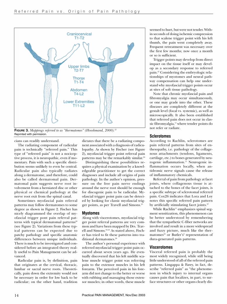
FIGURE 3. Mappings referred to as “thermatomes” (Hooshmand, 2000).22
Reprinted with permission.

tinct from the site of primary injury.”“Typically, the pain is referred to otherstructures that have the same embryonicorigin.”28 While traditional meanings of“referred pain” are restricted to visceralpain, technically the definitions above fitseveral other pain conditions, as indicat-ed elsewhere throughout this article.
Cousins refers to these patterns as “vis-cerotomes.”29 Visceral pain is difficult forthe human brain to locate, because thepain is “referred” to the skin via ephatictransmission (analogous to an electricalshort) and/or that “many different affer-ent sensory nociceptive neurons synapsewith the same ascending fibers in thespinal cord” causes the brain to mistakepain from the internal organ for painfrom the skin and/or nearby subcutaneoustissues and possibly deeper structures.28
Lingappa & Farey also suggest that thebrain generally will have more recentmemory of surface/subcutaneous painand will “ignore” deep pain until an in-citing event occurs.28 Angina with pain re-ferred to the left arm is a classic, well-known example.
While “activation of visceral pain re-ceptors does not always give rise to a sen-sation of pain,”30 the norm, in this con-text, is to at least expect pain, and some-times expect pain referral patterns, thatcan be misinterpreted if not recognized.Pain that becomes rapidly generalizedimplies perforation and leakage of fluidinto the peritoneal cavity. Biliary pain canradiate from the right inferior scapula.Pancreatic and abdominal aneurismalpain may radiate to the back. Ureteralcolic classically is referred to the groin andthigh.
Following is a more complete list ofsome referred visceral pain patterns witha brief description of the respective painreferral patterns. It is assumed there is nodextroposition of the internal organs.Note that one must expect that each pa-tient will display variations on these gen-eralizations.
• Lungs — pain is referred in a collar-like band completely around the neckfrom about the C6 to T3 levels.
• Diaphragm — pain is referred in apattern similar to the lungs.
• Heart — pain can be referred to thearea around the mouth, but is more com-monly referred over the left chest andcontiguously down the anterior left armand directly to the mid-back between thescapula from T4 to T7.
• Gallbladder — pain is referred to su-perior and lateral right shoulder, offsetsuperior similar in size and circular shapeto the superficial distribution of the axil-lary nerve.
• Liver — pain is referred in a similarpattern to the heart, but only on the righthemi-body.
• Stomach — pain is referred just tothe right of midline in the epigastric areaand to the mid-back, just below the re-ferred angina from T7 to T9.
• Ovaries — pain is referred to the skinarea immediately over the ovaries anteri-orly and directly posteriorly, but more lat-eral.
• Appendix — pain is referred toMcburney’s point in the right hypogastricarea.
• Kidneys — pain is referred to the skinarea somewhat below the kidneys, poste-riorly only, and medial to the posterior re-ferred ovarian pain; there is also an areahalf way down the right lateral thigh, theright chest just to the right of the lowersternum.
• Ureters — pain is referred to an an-terior band across the pelvis, includingthe groin and the genitals, but not ex-tending around to the back.
• Bladder — pain is referred to a con-tinuous area encompassing the sacrumfrom S2 down to the upper medial thighs.
Drewes has described some of the dy-namics of viscerotomes (visceral referredpain patterns). His observations can besummarized as follows:31
1) referred pain does not have to be sep-arated from the area of pathology,
2) referred pain spreads over time to amaximum, and
3) there is a great deal of variation be-tween individuals.
Drewes also indicates that the site ofpain stimulation, as well as the referredpain, are perceived to rise and fall almostcoincidentally in intensity over a few min-utes. During the first half of this timecourse, the referred pain is actually per-ceived as worse than the primary pain.31
The origin of pain in classic visceral re-ferred pain patterns can be explained bythe distribution of small pain nerves overthe viscera of the body.
ThermotomesHooshmand coined the term “ther-matomes” to describe pain patterns re-lated to the circulatory distribution ofsympathetic nerves. These relatively
amorphous distributions (see Figure 3)are consistent with the observation thatthese C-fiber nerve pathways end up see-ing pain “through fogged glass.”22
Referred muscular painRachlin discusses “referred pain zones.”11
He states that “referred pain is a mani-festation of spinal segmental sensitiza-tion.” This spinal segmental sensitizationobservation does make mechanistic senseand fits into the broad category of beinga neuropathic phenomenon. The sensi-tized neurons at the nerve roots are dys-functional pain neurons. While the authorknows of no direct evidence, deductivereasoning suggests that impulses viaephatic or similar means are transmittedfrom the site of nociception to the neu-rons innervating the area of referred pain.
“Myotomal” pain involves the myofas-cial tissue planes in and around muscles
R e f e r r e d P a i n v s . O r i g i n o f P a i n P a t h o l o g y
15Practical PAIN MANAGEMENT, Nov/Dec 2003
FIGURE 4. Pain referred from the spinal durais reminiscent of the thermatomes in beingdiffuse, but these referral patterns are unique(from Butler, 1991).9 Reprinted with permission.

groups. While “myotomal” may not be thecorrect description, when muscles wereinjected with hypertonic saline, which isan experimental substance known to pro-duce pain, the above-mapped patterns ofreferred pain emerged.18 We would expectthat these would be the same referredpain patterns as myofascial trigger points.Strangely, gross inspections reveals noclear congruence or overlap, possibly in-dicating that myofascial trigger point re-ferral patterns operate by some differentmechanism.
Dura-Generated PainButler9 has used Cyriax’ map of referredpain from the spinal dura,32 which is alsoprobably related to stimulation/irritationof the sympathetic C-fibers on the dura(illustrated in Figure 4), and is reminis-cent of the thermatones, but these are mu-tually distinct and unique patterns (com-pare Figures 3 & 4). These patterns arefar removed from the spinal segmentedpatterns of the other -tomes related pat-terns. This figure certainly illustrates theconcept that the C-fiber pain is seen bythe brain through “fogged glass.”
Not only are these pain referral pat-terns poorly accepted, but the origin ofpain pathology as being the spinal durais even less recognized. A physician couldreasonably consider this referred painpattern as “non-physiologic” withoutknowledge of this possibility. Certainly,with this pain origin and referral patternas a possibility, the physician must nottake such a presentation lightly, nor writethe patient off as having a “psychogenic”pain problem.
If we think of the possible evolutionaryorigin of the sympathetic chains, which inlower animals transmit all efferent and af-ferent nerve impulses, those pathways(i.e. the sympathetic chains) may very wellbe able to reestablish transmission path-ways in compensation when normal path-ways are lost, much like the developmentof collateral circulation in strokes.
Head and Facial PainPains around the head and neck are com-monly referred, however, these are sel-dom appreciated as such, probably be-cause of the short distances involved. Par-ticularly in migraine headaches, phe-nomena similar to referred pain occurs inaddition to the referred pain, i.e. visualand other sensations that are perceivedwithout a distal initiating stimulus.28
Guyton & Hall observed that:• nasal sinus and eye headaches radi-
ate to a wide area around the eyes frombelow the nose and up to mid-forehead.
• cerebral vault headaches occurfrontally to parietally at the ear.
• brainstem and cerebellar vaultheadaches occur from the ear through theentire occiput.33
As for the referred pain component, theorigin is probably around vessels that arevasodilating and vasoconstricting on themeninges, and subsequently the pain ra-diates to behind the eyes, usually unilat-erally. Throbbing is, by definition, relat-
ed to the heart beat, thus, stimulating andradiating in synchrony with the beating ofthe heart.
Phantom PainPhantom sensations and pain merelymean that the brain perceives the exis-tence of a body part from which no nerveimpulses could possibly be emanating,such as from an amputated limb, and is awell-described phenomena. In a sense,phantom pain is the ultimate “referredpain.” Perception of the pain is obviouslynot where the pain is originating, sincethere cannot be peripheral pain nervestimulation. The author has even had apatient with phantom coccygis of a re-
sected tailbone. Note that, as with all pains with central
components, non-existence of the per-ceived origin of pain pathology makes nodifference in the perception of pain,which is certainly true of all referredpains. However, it does not necessarilyfollow that all referred pain has a centraldysfunctional component. Further, stumpand neuroma pains post-amputation arenot referred pains, and therefore, shouldnot be mistaken for phantom pain. Thereseems to be surprising confusion aboutthese pains versus phantom pain.
Repeating the above meaning, it maybe possible to have phantom pain of abody part that is not missing as evidencedby abdominal pain in spinal cord injuredpatients. However, there is also the possi-bility that this pain may be “real” and ac-tual pain from the perceived site of pain,where pain nerve impulses pass throughsome other continuous pathways to thecentral nervous system, such as throughthe sympathetic chains.
Central Hypersensitization vs.Deafferent PainCentral hypersensitization syndromemerely describes the situation in with cen-tral neurons are sensitized such that nor-mally sub-threshold pain impulses areperceived as pain in widespread regionsof the body. From the author’s own expe-rience with small fiber,34 it is clear thatthese perceptions can be patchy. Painneed not be perceived as coming fromeverywhere. Central hypersensitizationcan also be described as widespread hy-peralgesia or allodynia, i.e. the patient isvery tender, more in some places thanothers. Specific mechanisms for this be-havior are reviewed by Rachlin.11 Rachlinalso presents evidence that fibromyalgiafits into this rather wide category, basedon current knowledge.
Likewise, deafferent pain syndromeneed not be manifest as “whole body”pain; it can be patchy. The contrast hereis, like phantom pain, no peripheral inputis theoretically necessary in a pure deaf-ferent pain situation. These patients arenon-tender, or not remarkably tender.They seem to be detached from the world,but preoccupied by their pain. This typeof pain, on the other hand, can be veryspecific and focal. This syndrome, in theauthor’s opinion, describes precisely“phantom pain” of a body part that is stillphysically present, but not sending pe-
R e f e r r e d P a i n v s . O r i g i n o f P a i n P a t h o l o g y
16 Practical PAIN MANAGEMENT, Nov/Dec 2003
In a sense, phantom
pain is the ultimate
“referred pain.”
Perception of the pain
is obviously not where
the pain is originat-
ing, since there cannot
be peripheral pain
nerve stimulation.

ripheral pain signals to the central nerv-ous system.
ConclusionUnderstanding referred pain is impor-tant in determining the true origin ofpain pathology so that proper diagnosisand treatment occurs. The complexitiesof different referred and radiating painpatterns presented in this article demon-strate that understanding pain requiresspecialized and diverse knowledge andwide experience. Suggesting that com-plaints are “non-anatomic” or “non-phys-iologic” may very well be a clear indica-tion of the diagnostician’s limitationsrather than a true reflection of a patient’spathology or psychological state.
In the defense of these well-meaningand intelligent health care providers, itreally does require a specially trainedphysician to artfully and effectively decidethe primary cause of a patient’s pain prob-lem and to pick the best and most effec-tive treatment early in their care. This di-agnostic exercise is the essential first stepin deciding on a theoretically-based andpragmatically-possible and effective treat-ment plan.
Interestingly enough, these differentpain etiologies and patterns are most di-rectly helpful in dealing with nociceptivepain. In other words, these pain sourcesand referral patterns basically representnormal neurophysiologic functioningand, by and large, provide the patient andthe physician with wonderful informationfor determining a good working diagno-sis and treatment modalities for nocicep-tive pain. However, reality is much morecomplex.
Based on available data and experi-ence, the author concludes that referredpain is neuropathologic and not nocicep-tive. The referred pain phenomenon it-self is apparently a “dyna-”neuropathicprocess — that is, pain nerve dysfunction.This conclusion, then, categorizes re-ferred pain as a problem with thewires/wiring or it can also be central, i.e.the “perceptron” as described in the au-thor’s prior article.8 �
Dr. James Woessner holds a doctorate in bio-logical sciences in conjunction with a medicaldegree. His professional medical training in-cludes neurology and physiatry. Dr. Woessnerhas written numerous articles about pain andother subjects in Physical Medicine. Dr. Woess-ner may be reached at Advanced Physical Med-
icine, 1188 Bishop Street, #1204, Honolulu,Hawaii 98613.
References1. Anderson DM (Chief Lexicographer). 2002.Mosby’s Medical Distionary. Sixth Edition. Mosby: AHarcourt Health Sciences Company, St. Louis. 1867pp.
2. Merskey H and Bogduk N (eds.). Classification ofchronic pain. Descriptions of chronic pain syndromesand definitions of pain terms, 2nd Edition Seattle:IASP Press, 1994. 240 pp.
3. Bellenir K (ed). 2002. Pain Sourcebook. HealthReference Series. Omnigraphics, Detroit. 670 pp.
4. Warfield CA and Fausett HJ (eds). Manual of PainManagement, 2nd Edition Lippincott, Williams &Wilkins, Philadelphia. 2002.
5. Khalsa DS. The Pain Cure: The Proven MedicalProgram that Helps End Your Chronic Pain. Warner
Books, New York. 1999. 418 pp.
6. Ombregt L. A System of Orthopaedic Medicine,2nd Edition. Churchill Livingston. 2003. 1360 pp.
7. Kosek E and Hansson P. Perceptual integration ofintramuscular electrical stimulation in the focal andthe referred pain area in healthy humans. Pain. 2003.105:125-131.
8. Woessner J. A Conceptual Model of Pain. Practi-cal Pain Management. PPM Communications. 2002.2:5, 8-16, 37.
9. Butler DS. Mobilisation of the Nervous System.Churchill Livingstone, New York. 1991. 265 pp.
10. Marcus A. Musculoskeletal Disorders: HealingMethods From Chinese Medicine, Orthopaedic Medi-cine and Osteopathy. North Atlantic Books. Berkeley.1998. 719 pp.
11. Rachlin ES (ed.) Myofascial Pain and Fibromyal-gia: Trigger Point Management, 2nd Edition Mosby.2002. 624pp.
12. Selzer M and Spenser WA. Convergence of vis-ceral and cutaneous afferent pathways in the lum-baar spinal cord. Brain Res. Jul. 1969. 14(2);331-348.13. Smith, CUM. Biology of Sensory Systems. JohnWiley & Sons, New York. 2000. 445 pp. 14. Moore KL and Dalley AF. Clinically OrientedAnatomy. Fourth Edition Lippincott Williams & Wilkins,Philadelphia. 1999. pg. 683.15. Kopf-Maier P (ed). Wolf-Heidegger’s Atlas ofHuman Anatomy. Volume 1: Systemic Anatomy, BodyWall, Upper and Lower Limbs, 5th Edition Karger,New York. 2001. 319 pp.16. Bonica JJ and Loeser JD. Applied Anatomy Rele-vant to Pain. In Loeser, et al. (eds.). Bonica’s Man-agement of Pain, 3rd Edition. Lippincott, Williams &Wilkins, Philadelphia. 2001. pp. 196-221.17. Brass A and Dingle RV Nervous System: Anatomyand Physiology. Volume 1, Part 1. The Ciba Collectionof Medical Illustrations. CIBA-GEIGY, West Caldwell,NJ. 1983. 237 pp.18. Coda BA and Bonica JJ. General Considerationsof Acute Pain. In Loeser, et al. (eds.). Bonica’s Man-agement of Pain, 3rd Edition Lippincott, Williams &Wilkins, Philadelphia. 2001. pp. 222-240.19. Hackett GS. Ligament and Tendon Relaxation(Skeletal Disability) Treated by Prolotherapy (Fibro-Os-seous Proliferation), 3rd Edition Charles C. Thomas,Springfield, IL. 1958. 151 pp.20. Fischer AA. Segmental Neuromyotherapy: A NewConcept in the Diagnosis and Management of Neu-romusculoskeletal Pain taken from Chapters 7 & 13.Rachlin, E.S. & I.S. (eds.) 2002. Myofascial Pain andFibromyalgia: Trigger Point Management, 2nd Edition.Mosby.21. Hardy SGP and Naftel JP. Viscerosensory Path-ways. In Haines, D.E. (ed.). Fundamental Neuro-science. Churchill Livingstone, New York. 1997. pp.255-263.22. Hooshmand H. Chronic Pain: Reflex SympatheticDystrophy, Prevention and Management. CRC Press,Boca Raton, FL. 2000. 202 pp.23. Travell JG and Simons DG. Myofascial Pain andDysfunction: The Trigger Point Manual Vol II (Lowerextremities) Williams & Wilkins. Baltimore, MD. 199224. Simons D, Travell J, and Simons L. MyofascialPain and Dysfunction: The Trigger Point Manual, Vol1(2), 2nd ed. Williams & Wilkins. Baltimore, MD. 1999. 25. Hubbard DR, Jr. Chronic and Recurrent MusclePain: Pathophysiology and Treatment, and Review ofPharmacologic Studies. Journal of MuculoskeletalPain. 1996. 4(1/2):123-143.26. Cox JM. Low Back Pain: Mechanism, Diagnosis,and Treatment. Williams & Wilkins. Baltimore, MD.1999. 735 pp.27. Lora J & Long D. So-called facet denervation inthe management of intractable back pain. Spine.1976. 1(2):121-126.28. Lingappa F and Farey K. Physiological Medicine.McGraw-Hill, New York. 2000. 1008pp.29. Cousins MJ. Visceral pain. In Andersson S, et al.(eds). Chronic Non-Cancer Pain: Assessment andPractical Management. MTP Press. Lancaster. 1987. 30. Haist SA and Robbins JB. . Internal Medicine OnCall. Third Edition. Lange Medical Books/McGraw-Hill. New York. 2002. 697 pp.31. Drewes AM, et al. Gut pain and hyperalgesia in-duced by capsaicin: a human experimental model.Pain. 2003. 104:333-341.32. Cyriax J. Textbook of Orthopaedic Medicine. Vol-ume 1, 8th Edition Baillere Tindall. 1982. 33. GuytGuyton AC and Hall JE (eds.) Textbook ofMedical Physiology. Ninth Edition. WB Saunders.Philadelphia. 1996. 1148 pp.34. Woessner J. A Conceptual Model of Pain. Practi-cal Pain Management. PPM Communications. 2002.2:6, 27-35.
R e f e r r e d P a i n v s . O r i g i n o f P a i n P a t h o l o g y
19Practical PAIN MANAGEMENT, Nov/Dec 2003
The complexities of
different referred and
radiating pain patterns
presented in this article
demonstrate that
understanding pain
requires specialized
and diverse knowledge
and wide experience.


![Elbow Pain - Welcome To | HealthSharehealthshare.org.uk/HS_leaflets/Elbow_Pain.pdf[ 3 ] Elbow Pain Lateral epicondylitis What is lateral epicondylitis? This is often referred to as](https://img.pdfslide.us/doc/110x75/5ac8bc037f8b9a51678c93c8/elbow-pain-welcome-to-he-3-elbow-pain-lateral-epicondylitis-what-is-lateral.jpg)