Embed Size (px)
Citation preview
governmental organizations. International researchers from severalcountries delivered talks on selected subjects.
Researchers, public health authorities, as well as authorities fromthe legislative, executive and judiciary governmental bodies fromBrazil and other South American countries were also present.
Site: www.ufrgs.br/ppgee/rni.htm
After the event, the Porto Alegre Resolution was approved by thescientists frommany countries and participants who have understoodthat the health protection, the well-being and the environmentrequire the immediate adoption of the Precautionary Principle andsome precautionary practices.
Site: http://www.icems.eu/docs/resolutions/Porto_Alegre_Resolution.pdf
5. Conclusion
This research showed the existence of a spatial correlationbetween cases of death by neoplasia and the locations of the BSs, inthe Belo Horizonte municipality from 1996 to 2006.
The mortality rates and the relative risk were higher for theresidents inside a radius of 500 m from the BS, compared to theaverage mortality rate of the entire city, and a decreased dose–response gradient was observed for residents who lived farther awayfrom the BS. The major antenna concentration was located in theCentral-Southern SD of the city, which also had the largestaccumulated incidence (5.83/1000 inhabitants).
The measured values of the EMF, determined in 2008 and 2003,were substantially below the values allowed by the Brazilian federallaw nr. 11934, May 5, 2009. Nevertheless, the values encountered inthis study surpassed the limits of human exposure adopted by manyother countries and cities in the world, including Italy (10 !W/cm2);China (6.6 !W/cm2); Switzerland (4.2 !W/cm2); Paris, France (1 !W/cm2); Salzburg, Austria (0.1 !W/cm2); and Porto Alegre, Brazil(4.2 !W/cm2).
New epidemiological studies must explore this issue with moretimely and appropriate methodology to provide evidence that maycon!rm the relationship between risk and hazard at an individuallevel. Meanwhile, we strongly suggest the adoption of the Precau-tionary Principle until the limits of human exposure, as established inBrazilian Federal Law, can be re-evaluated.
Supplementarymaterials related to this article can be found onlineat doi:10.1016/j.scitotenv.2011.05.051.
Acknowledgements
We would like to thank Márcia Salvador Géo, Medical Doctor;Helvécio Miranda Magalhães Júnior, Medical Doctor; Luciano AssírioBossi, Telecommunications Engineer; Graziella Lage Oliveira, Psycholo-gist; Lívia Daniella Pereira Dode, Production Engineer; and Aline DayrrelFerreira Sales, Epidemiologist for their kind and ever-present support.
References
Alanko T, Hietanen M, von Nadelstadh P. Occupational exposure to RF !elds from basestation antennas on rooftops. Annals of telecommunications. From the issue entitled“Health protection and RF exposure assessment: engineering aspects, 63. ; 2008.p. 125–32.. Numbers 1–2, DOI: 01007/s12243-007-0001-6.
ANATEL. [online] Available from: bwww.anatel.gov.brN 21st April, 2011.ANATEL — “Agência Nacional de Telecomunicação”: ‘Telecommunications National
Agency. Resolução no. 303 de 2 de julho de 2002 — Aprova o Regulamento sobreExposição a Campos Elétricos, Magnéticos e Eletromagnéticos na Faixa de Radio-freqüências entre 9 kHz e 300GHz. (Resolution nº. 303, July 2nd, 2002—Approves theRegulation on Exposure to Electric, Magnetic and Electromagnetic Fields in theRadiofrequencies Band from 9 kHz to 300 GHz.); 2000a. [online]. Available from:bwww.anatel.gov.brN.
ANATEL — “Agência Nacional de Telecomunicações”: ‘Telecommunications NationalAgency’.. Anexo à Resolução no. 303 de 2 de julho de 2002 — Regulamento sobreLimitaçãodaExposição aCamposElétricos,Magnéticos eEletromagnéticosnaFaixadeRadiofreqüências entre 9 kHz e 300 GHz. (Annex to Resolution nº. 303, July 2nd,2002—Regulation onExposure to Electric,Magnetic and Electromagnetic Fields in theRadiofrequencies Band from 9 kHz to 300 GHz.); 2000b. [online]. Available from:www.anatel.gov.br.
Beall C, Delzell E, Cole P, Brill I. Brain tumors among electronics industry workers.Epidemiology 1996;7:125–30.
Beniashvili D, Avinoach I, BaazovD, Zusman I.Household electromagnetic!elds andbreastcancer in elderly women. May–Jun;In Vivo 2005;19(3):563–6. 2005. [online].Available from: bhttp://www.ncbi.nlm.nih.gov/pubmed/15875777N. May 2nd, 2008.
BIOINITIATIVE REPORT: A rationale for a biologically-based public exposure standardfor electromagnetic !elds (ELF and RF). [online]. 2007. Available from: bhttp://www.bioinitiative.orgN, in 2010.
BRAZIL. Ministry of Health. [online]. Available from www.saude.gov.br. 2011.Cherry N. Scienti!c evidence of the risk of adverse health effects from chronic exposure to
low-level electromagnetic radiation— EMRAA, Electromagnetic Radiation Alliance ofAustralia, E-mail: [email protected], Sept. 1999.
Cherry N. Health effects associated with mobile base stations in communities: the needfor health studies. New Zealand: Lincoln University— Environmental Managementand Design Division, 8 June 2000; 2007.. [online]. Available on Dec. 2006. Sep.
Dode, Adilza Condessa. Poluição ambiental e exposição humana a campos eletromagné-ticos: estudo de casos no município de Belo Horizonte com ênfase nas estaçõesradiobase de telefonia celular. (Environmental pollution and human exposure toelectromagnetic !elds: a case study in Belo Horizonte municipality emphasizingmobile phone base stations). Dissertação: Mestrado em Saneamento, Meio Ambientee Recursos Hídricos — Escola de Engenharia, Universidade Federal de Minas Gerais,Belo Horizonte. (Thesis: Master Degree in Sanitation, Environment and HydricResources — Engineering School, Federal University of Minas Gerais, Belo HorizonteCity, Brazil). p175. 2003.
Eger H, Jahn M. Speci!c health symptoms and cell phone radiation in Selbitz (Bavaria,Germany)— evidence of a dose–response relationship. Umwelt-Medizin-Gesellschaft2010;23:2.
1107
1929
1324
985
781
462
328 259
12 4
0
200
400
600
800
1000
1200
1400
1600
1800
2000Number of
deaths by
neoplasia
0 <1 1<2 2<3 3<4 4<5 5<6 6<7 7<8 8<9 9<10Years
Fig. 16. Distribution of the number of deaths by neoplasia versus duration of exposure since the date that the !rst antenna in each analyzed CT came into operation.
3664 A.C. Dode et al. / Science of the Total Environment 409 (2011) 3649–3665
Belo Horizonte, Brazil (2011)
Death rates peaked during the second year of exposure.
Fig. 16. Distribution of the number of deaths by neoplasia versus duration of exposure since the date that the first antenna in each analyzed CT came into operation.
Dode AC, Leao MM, Tejo Fde A et al. Mortality by neoplasia and cellular telephone base stations in the Belo Horizonte municipality, Minas Gerais state, Brazil. Sci Total Environ (2011); 409(19):3649-3665.
Effects of Microwave RF Exposure on
Fertility
Comments on Notice of Inquiry, ET Docket No. 13-84

Impaired Fertility in Fruit Flies
Insects are remarkably resistant to ionizing radiation and radioactivity.They appear to be much more sensitive to the effects of microwave radio frequency exposures.
In a recent study, fruit flies were exposed to 10 μW/cm2 of GSM 900 MHz or 1800 MHz digital RF. This exposure level is 100 times lower than the FCC Guidelines of 1000 μW/cm2
Exposures were for one single exposure intervals per day for five days, ranging from 1 to 21 minutes per day.
Panagopoulos DJ, Margaritis LH. The effect of exposure duration on the biological activity of mobile telephony radiation. Mutat Res (2010); 699(1-2):17-22.
20 D.J. Panagopoulos, L.H. Margaritis / Mutation Research 699 (2010) 17–22
Table 2Effect of different exposure durations of DCS 1800 MHz radiation on the reproductivecapacity of Drosophila melanogaster.
Experimentno.
Groups (dailyexposure durationfor five consecutivedays)
Mean number ofF1 pupae permaternal fly
Deviation fromsham-exposed(SE) groups
2.1 SE (0 min) 11.7E1 (1 min) 7.8 !33.3%E2 (6 min) 7.7 !34.2%E3 (11 min) 6.2 !47.0%E4 (16 min) 4.9 !58.1%E5 (21 min) 3.9 !66.7%
2.2 SE (0 min) 12.9E1 (1 min) 8.6 !33.3%E2 (6 min) 8.3 !35.7%E3 (11 min) 6.9 !46.5%E4 (16 min) 5.9 !54.3%E5 (21 min) 5.0 !61.2%
2.3 SE (0 min) 14E1 (1 min) 9.0 !35.7%E2 (6 min) 7.3 !47.9%E3 (11 min) 7.1 !49.3%E4 (16 min) 6.4 !54.3%E5 (21 min) 5.7 !59.3%
2.4 SE (0 min) 14.3E1 (1 min) 9.3 !35.0%E2 (6 min) 8.4 !41.3%E3 (11 min) 7.5 !47.5%E4 (16 min) 6.7 !53.1%E5 (21 min) 5.5 !61.5%
2.5 SE (0 min) 12.9E1 (1 min) 8.2 !36.4%E2 (6 min) 7.2 !44.2%E3 (11 min) 6.3 !51.2%E4 (16 min) 5.7 !55.8%E5 (21 min) 4.9 !62.0%
2.6 SE (0 min) 12.5E1 (1 min) 7.4 !40.8%E2 (6 min) 6.7 !46.4%E3 (11 min) 5.9 !52.8%E4 (16 min) 5.0 !60.0%E5 (21 min) 4.4 !64.8%
Average ± SD SE (0 min) 13.05 ± 0.96E1 (1 min) 8.38 ± 0.72 !35.8%E2 (6 min) 7.60 ± 0.66 !41.8%E3 (11 min) 6.65 ± 0.61 !49.0%E4 (16 min) 5.77 ± 0.73 !55.8%E5 (21 min) 4.90 ± 0.67 !62.4%
Fig. 1. Reproductive capacity (mean number of F1 pupae per maternal fly) of groupsexposed to GSM 900 MHz radiation for different daily exposure durations (1, 6, 11,16, and 21 min) for five consecutive days, and of sham-exposed groups (no expo-sure).
Fig. 2. Reproductive capacity (mean number of F1 pupae per maternal fly) of groupsexposed to DCS 1800 MHz radiation for different daily exposure durations (1, 6,11, 16, and 21 min) for five consecutive days, and of sham-exposed groups (noexposure).
that differ 10 min in exposure duration between them (e.g. E1–E3,E2–E4, etc.) is in all cases P < 10!2.
We did not detect any temperature increases within the glassvials during the exposures, for all the different exposure durationstested.
4. Discussion
Our results show once again that both types of mobile tele-phony radiation decrease considerably the reproductive capacityof the flies, for all the periods of daily exposure duration tested,from 1 to 21 min, although the intensity of radiation was only10 !W/cm2, which is much lower than the established ICNIRPlimits of 450 !W/cm2 and 900 !W/cm2 for 900 and 1800 MHz,respectively [41].
The statistical analysis clearly shows that the exposedDrosophila groups differ in offspring production between them-selves and compared with the sham-exposed groups, and thisdifference is not due to random variations but to the effect of theGSM/DCS fields. The reason why differences between groups dif-fering only 5 min in daily exposure duration were not as strong(P < 0.07 for GSM 900 and P < 0.08 for DCS 1800) as between groupsdiffering 10 min or more in exposure duration (P < 10!2), is onlythat a 5-min difference in daily exposure duration was not enoughto show a large difference in reproductive capacity, since all theexposures (even the shortest of 1 min daily) produced a significanteffect and all the exposed groups were significantly different fromthe sham-exposed groups (P < 10!5). When the difference in expo-sure duration is increased to 10 min or more, then the differencein reproductive capacity between exposed groups becomes highlysignificant.
The effect of both types of digital mobile telephony radiation onthe reproductive capacity seems to increase almost linearly withincreasing exposure duration from 1 to 21 min, suggesting thatshort-term exposures to this radiation have cumulative effects onliving organisms.
We do not know whether longer short-term exposures than21 min would result in an even higher decrease of reproductivecapacity, or whether the effect would saturate after a certainexposure duration. This is left for future experiments. Our resultsrepresent a first indication that this radiation can have cumulativeeffects.
It is unknown whether long-term exposures of people oranimals residing in areas exposed to radiation by base-stationantennas also have cumulative effects, but this is possible as the
Impaired Fertility in Fruit Flies
0 = control group, with no exposure.
Even at one minute of exposure per day, a significant decrease in fertility is seen.
Fig. 2. Reproductive capacity (mean number of F1 pupae per maternal fly) of groups exposed to DCS 1800MHz radiation for different daily exposure durations (1, 6, 11, 16, and 21min) for five consecutive days, and of sham-exposed groups (no exposure).
Panagopoulos DJ, Margaritis LH. The effect of exposure duration on the biological activity of mobile telephony radiation. Mutat Res (2010); 699(1-2):17-22.
Comments on Notice of Inquiry, ET Docket No. 13-84
Impaired Fertility in Mammals
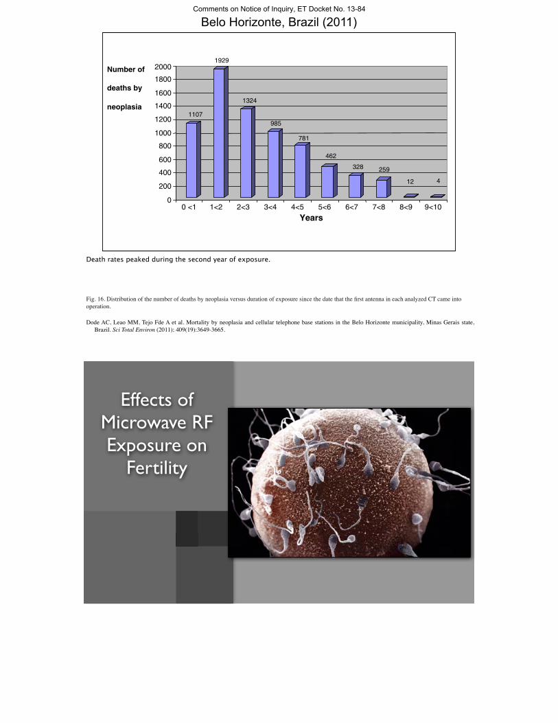
This is a Wistar rat.
A great deal of research has been done on the effects of microwave RF on laboratory animals.
RF-Radiation-Induced Changes in Mice 457
tion i), far from the ‘‘antenna park’’ in a more or lessfree-of-RF radiation environment.
It was extremely difficult to use RF-free controlsat the mountain sites, because it was almost impossibleto make ‘‘electromagnetically screened cages.’’ Sucha cage should ideally provide high (of the order of 30dB) screening at the frequency range between 88.5 and950MHz (Commercial Radio FM band, UHF TV band,and Mobile Communication band), and thereforewould require a very dense and well-grounded, highly Fig. 2. Comparison of the mean values { standard deviation of
number of newborns per dam and mating from all experimentalconductive external metal grid. Obviously, mice couldgroups.hardly survive in such cages for about 5 months.
The litter was considered to be the experimentalunit for the analysis of data. We measured the crown-rump length, the body weight, the number of the poste- some thymol crystals as contamination prevention).
The stained newborns were inspected for skeletal de-rior (lumbar, sacral, and coccygeal) vertebrae, the con-genital malformations, and the ossification of the fects as well as for the degree of ossification of their
bones. The ossification of the skeleton and particularlyskeleton.The RF power was measured in each position, of the vertebrae is an excellent and creditable indicator
of the prenatal exposure to noxious agents and can beusing an electric field meter and a low gain (4 dB)wide-band (80–900 MHz) log-periodic antenna and a measure of development delay.spectrum analyser. To obtain comparable results the‘‘IEEE std. C95.3.1991’’ was used. On the third floor RESULTSof the public school, where the mice were situated, a360 degree integration was also performed, due to the The RF power levels measured, although below
the limits proposed by the ‘‘ENV50166-2’’ and thedirectivity of the measuring antenna together with theclose proximity of the walls and metal furniture. Wher- ‘‘IEEE C95.1.1991’’ standards, are high and well
above the power levels that are likely to be measuredever iron bars or metal screens existed in front of thewindows, two series of measurements were carried out; in other European or U.S. residential areas. In fact, on
the third floor of the public primary school (positionone on each side of the screen.The collected newborns were killed for examina- h), an average power density of 1.053 mW/cm2 was
found, equivalent to a specific absorption rate of 1.935tion. Their crown-rump length was measured, and theywere weighed and inspected under the dissecting mi- mW/kg. In the Hypaithrios Life Refuge (position d)
the average power density in which the mice werecroscope for external congenital malformations. Thenthey were fixed and subsequently cleared and stained located was of the order of 168 nW/cm2. This reduced
level was due to the screening effect of the iron barsin toto by a double staining of their skeleton [Peters,1977]. The procedure was lightly modified as follows: in front of the windows, which gave an 8–10 dB RF-
power decrease. The average power density levels inThe newborns were fixed with alcohol 86% for3 days; their skin, eyes, and viscera were removed; position i (Laboratory of Anatomy, School of Veteri-
nary Medicine, University of Thessaloniki), where thethen they were immersed for 3 days in alcohol 100%and for 4 days in a mixture of alcohol 100% and ether controls were placed and the fifth experimental matings
were performed, was 40 dB weaker.1:1. They were stained for 1–2 days with blue alcyancoloration [alcohol 86% 80 ml, acetic acid 20 ml, al- The number of the littered newborns by the exper-
imental dams of groups A and B were, compared withcyan blue 20 mg] until the nonmineralised carti-lagenous parts of the bones became blue. They were those littered by the controls, progressively reduced
from the first to the fifth pregnancy. This reduction isimmersed in alcohol 100% for 4 days. Then they werestained for 12–24 days with red alizarin coloration more evident in group B and is clearly shown in Table
2 and in Figure 2.[KOH 1 g, H2O 100 ml, alizarin solution (alcohol 86%saturated with alizarin red S) 0.1 ml] until the ossified On the other hand, the rest of the four measured
parameters, i.e., the crown rump length and the weightparts of the bones became red. They were immersedin solution Mall I (KOH 1 g, distilled water 80 ml, and the number of the lumbar, sacral, and coccygeal
vertebrae increased in the newborns from groups A andglycerine 20 ml) until the transparency of their bodywas completed. Finally, they were stored in a conserva- B compared with the controls. This was more evident in
group A than in group B (Table 2 and Fig. 3). Ation solution (distilled water and glycerine 1:1, with
833D/ 8507$$833d 06-27-97 09:08:37 bema W: BEM
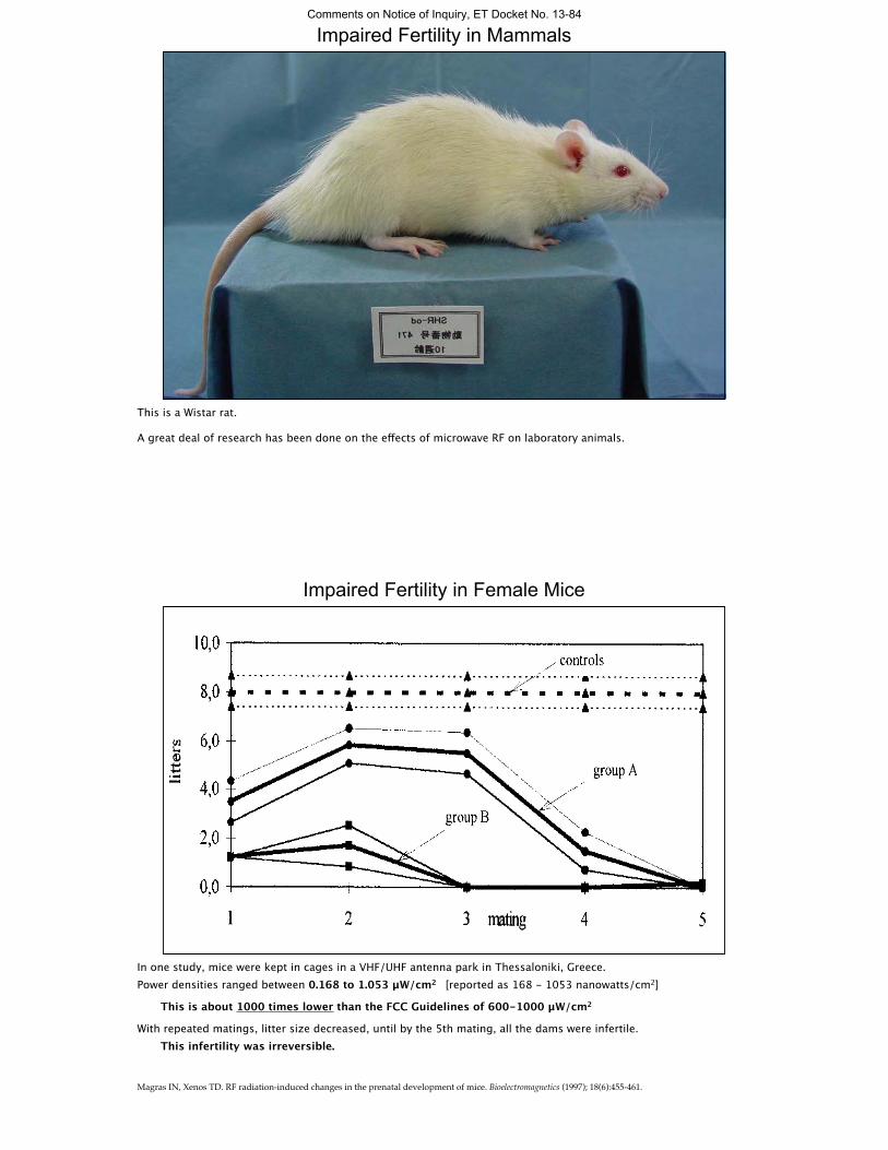
Impaired Fertility in Female Mice
In one study, mice were kept in cages in a VHF/UHF antenna park in Thessaloniki, Greece.Power densities ranged between 0.168 to 1.053 μW/cm2 [reported as 168 - 1053 nanowatts/cm2]
This is about 1000 times lower than the FCC Guidelines of 600-1000 μW/cm2
With repeated matings, litter size decreased, until by the 5th mating, all the dams were infertile. This infertility was irreversible.
Magras IN, Xenos TD. RF radiation-induced changes in the prenatal development of mice. Bioelectromagnetics (1997); 18(6):455-461.
Comments on Notice of Inquiry, ET Docket No. 13-84

Impaired Fertility in Male Rats
0.21 mW/cm2 - 2 hrs/D x 45 DSham exposure
Reduced sperm production in male Wistar rats exposed to 10 GHz microwave RF.0.21 mW/cm2 = one fifth of the FCC Guidelines of 1 mW/cm2
OTHER EFFECTS: Increases in reactive oxygen species, increased free radical formation, decreased activity of glutathione peroxidase and superoxide dismutase, DNA strand breakage, increased apoptosis (cell death) in sperm cells, distortion of sperm structure, reduced testosterone levels, shrinkage of seminiferous tubules and testicular size, decreased number and weight of progeny.
Kesari KK, Kumar S, Behari J. Effects of radiofrequency electromagnetic wave exposure from cellular phones on the reproductive pattern in male Wistar rats. Appl Biochem Biotechnol (2011); 164(4):546-559.
Kesari KK, Kumar S, Behari J. Pathophysiology of microwave radiation: effect on rat brain. Appl Biochem Biotechnol (2012); 166(2):379-388.Kumar S, Kesari KK, Behari J. Influence of microwave exposure on fertility of male rats. Fertil Steril (2011); 95(4):1500-1502.Kumar S, Behari J, Sisodia R. Influence of electromagnetic fields on reproductive system of male rats. Int J Radiat Biol (2012); epub Nov 13:1-8
(p = 0.008) (p = 0.021) (p = 0.036) (p = 0.009)(p = 0.003)
WiFi Exposure Damages Sperm With Oxidant Stress.
0
2.5
5
7.5
10
Serum 8-OH-20-dG TBS GPX (IU/g) Catalase (IU/mg)
ControlExposed for 20 weeks
0
75
150
225
300
HSCORE
*
*
*
* *
The rats were exposed to a Standard WiFi gateway, 24 hours a day for 20 days.
HSCORE = histological staining in testes for 8-OH-20-dG [8-hydroxy-20-deoxyguanosine, byproduct of DNA damage]
Serum 8-OH-20-dG (ng/ml) [byproduct of DNA damage]
TBS = testicular biopsy score 9 = Much spermatogenesis, but germinal epithelium disorganized with marked sloughing or obliteration of lumen
GPX = glutathione peroxidase, an antioxidant (consumed by oxidative stress in exposed rats).
Atasoy HI, Gunal MY, Atasoy P, Elgun S, Bugdayci G. Immunohistopathologic demonstration of deleterious effects on growing rat testes of radiofrequency waves emitted from conventional Wi-Fi devices. J Pediatr Urol (2012); March 30.
Comments on Notice of Inquiry, ET Docket No. 13-84

Impaired Fertility in Birds
In Valladido, Spain, a study compared the productivity of storks nesting closer and farther from a cell phone tower site.30 nests within 200 meters of the antennae, were compared with 30 nests greater than 300 meters from the antennae
Balmori A. Possible Effects of Electromagnetic Fields from Phone Masts on a Population of White Stork. Electromagn Biol Med (2005); 24(2):109-119.
Impaired Fertility in Birds
0
3.75
7.5
11.25
15
Field intensity (V/m) Total productivity Partial productivity Nests without young
Distance to cell tower > 300 mDistance to cell tower ≤ 200 m
*
*
(p = 0.001) (p = 0.26)
Productivity was significantly reduced in birds in the high exposure group.
Average electric field intensity on nests within 200m = 2.36±0.82V/m (~ 1.48 μW/cm2)
This is more than 400 times less than the FCC Guidelines of 600-1000 μW/cm2
Average electric field intensity on nests further than 300m = 0.53 ± 0.82 V/m (~ 0.07 μW/cm2).
Balmori A. Possible Effects of Electromagnetic Fields from Phone Masts on a Population of White Stork. Electromagn Biol Med (2005); 24(2):109-119.
Comments on Notice of Inquiry, ET Docket No. 13-84

Impaired Fertility in Amphibians
Eggs and tadpoles of the European common frog (Rana temporaria) were exposed to RF/EFM from several cell towers located at a distance of 140 meters.
Duration of exposure was 2 months (from egg phase to advanced tadpole stage).
Control groups were placed in same conditions, but contained in a faraday cage that shielded the eggs from RF exposure.
Balmori A. Mobile phone mast effects on common frog (Rana temporaria) tadpoles: the city turned into a laboratory. Electromagn Biol Med (2010a); 29(1-2):31-35.
(EF Field Strength ~ 0.8-3.2 μW/cm2)
Impaired Fertility in Amphibians
0
22.5
45
67.5
90
Mortality (%)
Control (in Faraday cage) Exposed
Exposure intensity 1.8 to 3.5 V/m (~ 0.8-3.2 μW/cm2).
This is 200 times less than the FCC Guidelines of 600-1000 μW/cm2
[In the exposed group (n = 70), low coordination of movements and asynchronous growth was observed in living specimens, resulting in both big and small tadpoles. In the control group (n = 70), growth was normal.]
Balmori A. Mobile phone mast effects on common frog (Rana temporaria) tadpoles: the city turned into a laboratory. Electromagn Biol Med (2010a); 29(1-2):31-35.
Comments on Notice of Inquiry, ET Docket No. 13-84
NZMJ 12 December2008, Vol 121 No 1287; ISSN 1175 8716 Page 52 URL: http://www.nzma.org.nz/journal/121-1287/3416/ ©NZMA
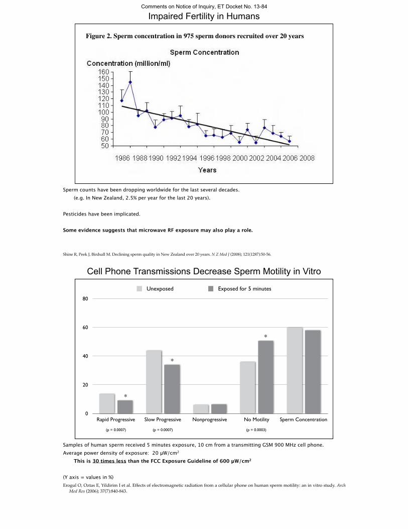
Figure 1. Semen volume in 975 sperm donors recruited over 20 years
!
Figure 2. Sperm concentration in 975 sperm donors recruited over 20 years
Impaired Fertility in Humans
Sperm counts have been dropping worldwide for the last several decades. (e.g. In New Zealand, 2.5% per year for the last 20 years).
Pesticides have been implicated.
Some evidence suggests that microwave RF exposure may also play a role.
Shine R, Peek J, Birdsall M. Declining sperm quality in New Zealand over 20 years. N Z Med J (2008); 121(1287):50-56.
(p = 0.0007)(p = 0.0007) (p = 0.0003)
Cell Phone Transmissions Decrease Sperm Motility in Vitro
0
20
40
60
80
Rapid Progressive Slow Progressive Nonprogressive No Motility Sperm Concentration Untitled 1
Unexposed Exposed for 5 minutes
*
*
*
Samples of human sperm received 5 minutes exposure, 10 cm from a transmitting GSM 900 MHz cell phone. Average power density of exposure: 20 μW/cm2
This is 30 times less than the FCC Exposure Guideline of 600 μW/cm2
(Y axis = values in %)Erogul O, Oztas E, Yildirim I et al. Effects of electromagnetic radiation from a cellular phone on human sperm motility: an in vitro study. Arch
Med Res (2006); 37(7):840-843.
Comments on Notice of Inquiry, ET Docket No. 13-84
(p < 0.01)(p < 0.01)
Cell Phone Use Decreases Sperm Motility in Vivo
0
20
40
60
80
Rapid Progressive Slow Progressive Nonprogressive No Motility Sperm Concentration BLANK
Transmit < 15 minutes per day Transmit > 60 minutes per day
*
*
Semen analysis performed on 371 men at a university clinic.Health questionnaire included query of cell phone use habits.(Y axis = values in %)
Fejes I, Zavaczki Z, Szollosi J et al. Is there a relationship between cell phone use and semen quality? Arch Androl (2005); 51(5):385-393.
different countries and continents. Exposure of radiofre-quency energy depends upon the frequency of the cellularphone. Analog phones operate at 450–900 MHz, digitalphones (Global System for Mobile Communications[GSM]) at 850–1900 MHz, and third-generation phones atapproximately 2000MHz (15). For years the cell phone com-panies have assured people that cell phones are perfectly safe.For assessing exposure from transmitters located near thebody, the most useful quantity is the specific absorptionrate (SAR), the amount of radiofrequency energy absorbedfrom the phone into the local tissues. The SAR of cell phonesvaries from 0.12 to 1.6 W/kg body weight depending uponthe model. In the United States, the upper limit of SAR al-lowed is 1.6 W/kg (16).
We studied the sperm parameters of 361 males attendingan infertility clinic after segregating them into four differentgroups based on their daily active use of cell phone.We foundthat most of the comparisons of four sperm parameters:sperm count, motility, viability, and normal morphology be-tween all the cell phone user groups were significantly differ-ent. This led us to suggest that the use of cell phones mayadversely affect the quality of semen by decreasing the spermcounts, motility, viability, and morphology, which might con-tribute to male infertility. However, these four sperm para-meters showed significant positive correlation among eachother. Therefore, the decrease in value of one sperm parame-ter is bound to reduce the other parameter also. Anothersignificant finding of our study is the decline in the qualityof semen based on the active cell phone usage time. The lab-oratory values of the four sperm parameters were lower in the
FIGURE 1
Sperm parameter profile for cell phone use groups.The x-axis lists eight sperm parameters: 1! volume;2 ! liquefaction time; 3 ! pH; 4 ! viscosity;5 ! sperm count; 6 ! motility; 7 ! viability; and8 ! percent normal morphology. The y-axis depictsthe mean value of the corresponding spermparameters for each cell phone use group.
Agarwal. Cell phone usage and male infertility. Fertil Steril 2008.
TABL
E2
Simultane
ousco
nfide
nceintervalsof
differen
cesbe
twee
nce
llph
oneus
egrou
psev
alua
tingeigh
tspe
rmpa
rameters.
Param
eters
Group
sA&B
Group
sA&C
Group
sA&D
Group
sB&C
Group
sB&D
Group
sC
&D
Volume(m
L)"0
.091
to0.07
2"0
.084
to0.08
3"0
.102
to0.05
8"0
.389
to0.05
3"0
.046
to0.04
0"0
.063
to0.02
6Liqu
efac
tiontim
e(m
in)
"2.27to
2.18
"3.10to
1.40
"2.60to
1.87
"2.47to
0.87
"1.96to
1.27
"1.19to
2.10
pH"0
.115
to0.10
5"0
.209
to0.01
4"0
.223
to"0
.004
"0.175
to"0
.009
a"0
.189
to"0
.02a
"0.098
to"0
.065
Visc
osity
"0.66to
0.70
"0.80to
0.58
"0.63to
0.72
"0.64to
0.38
"0.47to
0.52
"0.35to
0.66
Spe
rmco
unt(#1
06/m
L)"1
.67to
4.40
1.29
to7.49
a4.05
to10
.03a
"0.85to
2.57
0.60
to3.81
a"1
.35to
1.97
Motility
(%)
"6.16to
45.83
12.86to
65.11a
22.71to
74.49a
11.04to
56.09a
22.21to
66.52a
6.65
to51
.36a
Viab
ility
(%)
"5.55to
48.84
14.04to
68.7
0a24
.42to
78.59a
11.69to
58.82a
23.55to
69.92a
7.29
to54
.07a
WHO
morph
olog
y(%
norm
al)
0.12
to1.11
a2.65
to3.66
a4.14
to5.12
a0.70
to1.26
a1.60
to2.12
a"0
.12to
0.41
Note:
Group
A:n
ous
e(n
!40
);grou
pB:<
2h/da
y(n
!10
7);g
roup
C:2
–4h/da
y(n
!10
0);a
ndgrou
pD:>
4h/da
y(n
!11
4).M
eans
andSDwereba
sedon
data
onthe
original
scale;
alla
nalyse
sweredo
newith
approp
riately
tran
sformed
data.
aSignific
ant(P<.05)
usingmultiv
ariate
analys
isof
varia
ncean
dBon
ferron
isim
ultane
ousco
nfide
nceintervals.
Agarwal.C
ellp
hone
usageandmaleinfertility.F
ertil
Steril2008.
126 Agarwal et al. Cell phone usage and male infertility Vol. 89, No. 1, January 2008
Cell Phone Use Degrades Sperm Quality in Vivo
** *
*
* *
*
*
*
* (p < 0.05)
SpermCount
Motility Viability % NormalMorphology
Volume LiquificationTime
pH Viscosity
Three hundred sixty-one men undergoing infertility evaluation were divided into four groups according to their active cell phone use: group A: no use; group B: <2 h/day; group C: 2–4 h/day; and group D: >4 h/day.
With greater than two hours a day of reported talk time, significant reduction in sperm count, motility, viability, and % normal morphology were observed.
[One can assume that with texting rather than talking, the data might be even worse . . . as the phone antenna will be closer to the testes.]
Agarwal A, Deepinder F, Sharma RK, Ranga G, Li J. Effect of cell phone usage on semen analysis in men attending infertility clinic: an observational study. Fertil Steril (2008); 89(1):124-128.
Comments on Notice of Inquiry, ET Docket No. 13-84
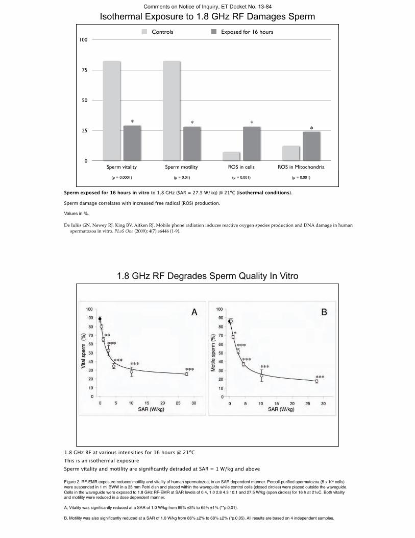
Isothermal Exposure to 1.8 GHz RF Damages Sperm
0
25
50
75
100
Sperm vitality Sperm motility ROS in cells ROS in Mitochondria Blank
Controls Exposed for 16 hours
* * **
(p = 0.0001) (p = 0.01) (p = 0.001) (p = 0.001)
Sperm exposed for 16 hours in vitro to 1.8 GHz (SAR = 27.5 W/kg) @ 21ºC (isothermal conditions).
Sperm damage correlates with increased free radical (ROS) production.
Values in %.
De Iuliis GN, Newey RJ, King BV, Aitken RJ. Mobile phone radiation induces reactive oxygen species production and DNA damage in human spermatozoa in vitro. PLoS One (2009); 4(7):e6446 (1-9).
Discussion
While a high proportion of the male population suffers frominfertility associated with defective sperm function [17], theetiology of this condition remains largely unresolved. Notwith-standing the general paucity of information in this area, recentstudies have highlighted the interesting finding that male infertilitypatients are frequently characterized by high levels of DNAdamage to their spermatozoa [18]. In light of these data, we havehypothesized that the disruption of sperm fertilizing potential andthe concomitant presence of high levels of DNA damage in thesperm nucleus involves a common causative mechanism in theform of oxidative stress [19].Oxidative stress has been known for some time to limit the
fertilizing potential of human spermatozoa through the inductionof peroxidative damage to the sperm plasma membrane [13,20].Oxidative stress is also known to be associated with DNA damagein human spermatozoa [21]. Furthermore, the source of the freeradicals responsible for generating such stress appears to be themitochondria [15]. However, the factors responsible for inducingthe mitochondria to leak electrons and propagate the productionof ROS have not been elucidated. The research described in thisarticle suggests that one of the key environmental factors involvedin the stimulation of sperm mitochondria to produce high levels ofROS, might be excess exposure to RF-EMR from sources such asmobile phones.In a pilot study, human spermatozoa were found to respond to
RF-EMR (at 1.8 GHz with a SAR of 27.5 W/kg) with a range ofnegative changes including dramatic declines in both spermvitality and motility. We also observed significant increases in bothcytoplasmic ROS levels (DHE) as well as mitochondrial ROSlevels (MSR) after RF-EMR exposure. We have previously shownthat the chemical induction of mitochondrial ROS productionwith rotenone can precipitate a state of oxidative stress leading to
high levels of lipid peroxidation and a loss of sperm motility [15].Therefore, these data highlight the particular vulnerability ofhuman spermatozoa to oxidative attack and the potentialsignificance of sperm mitochondria in the generation of freeradicals.To assess whether similar effects could be observed at lower
power densities, closer to the SAR values associated with mobilephones (0.5–1.5 W/kg) a dose-dependent analysis was conducted.In addition to the conventional assessments of motility and vitality,assays were included to assess the potential for RF-EMR to inducesperm DNA damage and further, whether the DNA damage wasoxidative in nature. Confirmation of the detrimental effects of RF-EMR on human sperm was again observed. Over the powerdensity range employed, a significant (P,0.001) dose-dependentresponse for all sperm parameters was observed, includingmotility, vitality, ROS generation by the whole cell, ROSgeneration by the mitochondria, oxidative DNA damage andDNA fragmentation. Furthermore, the profiles of all the observedeffects with respect to SAR were intriguingly similar, suggesting acommon underlying mechanism.Specifically, all of the responses examined showed an extremely
rapid change at low SAR exposures that then reached a plateau ata point where around 30% of the sperm population was affected.This suggests that while we were careful to use only Percoll-purified, high quality spermatozoa in this analysis, there existswithin this cell population, a cohort of spermatozoa that areparticularly vulnerable to the induction of oxidative stress by RF-EMR. These spermatozoa may have compromised mitochondria,poorly remodeled chromatin or a combination of such factors[15,22]. Heterogeneity within the sperm population is a feature ofthe human condition. However, this does not mean that a majorityof spermatozoa would not, ultimately, be affected by RF-EMR invivo; much would depend on the duration of exposure. In vitro, weare limited by the inability of human spermatozoa to survive for
Figure 2. RF-EMR exposure reduces motility and vitality of human spermatozoa, in an SAR dependent manner. Percoll-purifiedspermatozoa (56106 cells) were suspended in 1 ml BWW in a 35 mm Petri dish and placed within the waveguide while control cells (closed circles)were placed outside the waveguide. Cells in the waveguide were exposed to 1.8 GHz RF-EMR at SAR levels of 0.4, 1.0 2.8 4.3 10.1 and 27.5 W/kg(open circles) for 16 h at 21uC. Both vitality and motility were reduced in a dose dependent manner. A, Vitality was significantly reduced at a SAR of1.0 W/kg from 89%63% to 65%61% (**p,0.01). B, Motility was also significantly reduced at a SAR of 1.0 W/kg from 86%62% to 68%62%(*p,0.05). All results are based on 4 independent samples.doi:10.1371/journal.pone.0006446.g002
RF-EMR and Sperm In Vitro
PLoS ONE | www.plosone.org 4 July 2009 | Volume 4 | Issue 7 | e6446
1.8 GHz RF Degrades Sperm Quality In Vitro
1.8 GHz RF at various intensities for 16 hours @ 21ºCThis is an isothermal exposureSperm vitality and motility are significantly detraded at SAR = 1 W/kg and above
Figure 2. RF-EMR exposure reduces motility and vitality of human spermatozoa, in an SAR dependent manner. Percoll-purified spermatozoa (5 x 10⁶ cells) were suspended in 1 ml BWW in a 35 mm Petri dish and placed within the waveguide while control cells (closed circles) were placed outside the waveguide. Cells in the waveguide were exposed to 1.8 GHz RF-EMR at SAR levels of 0.4, 1.0 2.8 4.3 10.1 and 27.5 W/kg (open circles) for 16 h at 21uC. Both vitality and motility were reduced in a dose dependent manner.
A, Vitality was significantly reduced at a SAR of 1.0 W/kg from 89% ±3% to 65% ±1% (**p,0.01).
B, Motility was also significantly reduced at a SAR of 1.0 W/kg from 86% ±2% to 68% ±2% (*p,0.05). All results are based on 4 independent samples.
Comments on Notice of Inquiry, ET Docket No. 13-84

more than 24 hours in a simple defined culture medium. In vivo,spermatozoa may take up to a week to move from the seminiferoustubules in the testes to the cauda epididymis and during the wholeof this time they would be vulnerable to RF-EMR exposure [23].We recognize that these studies were conducted using spermatozoa
suspended in a simple defined culture medium rather than theepididymal plasma in which they would be suspended in vivo.
Nevertheless the fact that effects on sperm quality have previouslybeen observed in both whole animal radiation experiments [3] and inepidemiological studies of human subjects exposed to various levels ofmobile phone radiation [5,7,24], emphasizes the biological andclinical relevance of these findings. Moreover, another recent studyhas found that exposing human spermatozoa to mobile phoneradiation for 1 hour leads to significant declines in motility and
Figure 3. RF-EMR induces ROS generation in human spermatozoa, in an SAR-dependent manner unrelated to thermal effects.Percoll-purified spermatozoa (56106 cells) were suspended in 1 ml BWW in a 35 mm Petri dish and placed within the waveguide while control cellsplaced outside the waveguide (closed circles). Cells in the waveguide were exposed to 1.8 GHz RF-EMR at SAR levels between 0.4 and 27.5 W/kg(open circles) for 16 h at 21uC. Also, purified sperm cells were subjected to incubation temperatures ranging from 21uC–50uC for 2 h. As the powerlevels were increased, the cellular generation of ROS increased in a dose-dependent manner. ROS levels were also observed to increase as a result ofincubation temperature, but such results were not significant until the temperature exceeded 40uC. A, ROS generation (DHE response) wassignificantly increased from control levels after exposure to 1.0 W/kg (*p,0.05) and above (***p,0.001). B, RF-EMR induces ROS generation by thesperm mitochondria as monitored by MSR; significant increases were observed at SAR values of 2.8 W/kg (***p,0.001) and above. All results arebased on 4 independent samples. C, In order to control for thermal effects, the impact of temperature of cellular ROS generation was monitored; asignificant increase in ROS generation was observed as temperatures rose above 40uC (p,0.001). D, Across the entire data set, the total level of ROSgeneration by human spermatozoa (DHE positive cells) was highly correlated with the level of ROS generation by the mitochondria (MSR positivecells: R2 = 0.823).doi:10.1371/journal.pone.0006446.g003
RF-EMR and Sperm In Vitro
PLoS ONE | www.plosone.org 5 July 2009 | Volume 4 | Issue 7 | e6446
ROS Production – RF versus Thermal
more than 24 hours in a simple defined culture medium. In vivo,spermatozoa may take up to a week to move from the seminiferoustubules in the testes to the cauda epididymis and during the wholeof this time they would be vulnerable to RF-EMR exposure [23].We recognize that these studies were conducted using spermatozoa
suspended in a simple defined culture medium rather than theepididymal plasma in which they would be suspended in vivo.
Nevertheless the fact that effects on sperm quality have previouslybeen observed in both whole animal radiation experiments [3] and inepidemiological studies of human subjects exposed to various levels ofmobile phone radiation [5,7,24], emphasizes the biological andclinical relevance of these findings. Moreover, another recent studyhas found that exposing human spermatozoa to mobile phoneradiation for 1 hour leads to significant declines in motility and
Figure 3. RF-EMR induces ROS generation in human spermatozoa, in an SAR-dependent manner unrelated to thermal effects.Percoll-purified spermatozoa (56106 cells) were suspended in 1 ml BWW in a 35 mm Petri dish and placed within the waveguide while control cellsplaced outside the waveguide (closed circles). Cells in the waveguide were exposed to 1.8 GHz RF-EMR at SAR levels between 0.4 and 27.5 W/kg(open circles) for 16 h at 21uC. Also, purified sperm cells were subjected to incubation temperatures ranging from 21uC–50uC for 2 h. As the powerlevels were increased, the cellular generation of ROS increased in a dose-dependent manner. ROS levels were also observed to increase as a result ofincubation temperature, but such results were not significant until the temperature exceeded 40uC. A, ROS generation (DHE response) wassignificantly increased from control levels after exposure to 1.0 W/kg (*p,0.05) and above (***p,0.001). B, RF-EMR induces ROS generation by thesperm mitochondria as monitored by MSR; significant increases were observed at SAR values of 2.8 W/kg (***p,0.001) and above. All results arebased on 4 independent samples. C, In order to control for thermal effects, the impact of temperature of cellular ROS generation was monitored; asignificant increase in ROS generation was observed as temperatures rose above 40uC (p,0.001). D, Across the entire data set, the total level of ROSgeneration by human spermatozoa (DHE positive cells) was highly correlated with the level of ROS generation by the mitochondria (MSR positivecells: R2 = 0.823).doi:10.1371/journal.pone.0006446.g003
RF-EMR and Sperm In Vitro
PLoS ONE | www.plosone.org 5 July 2009 | Volume 4 | Issue 7 | e6446
1.8 GHz RF x 16 hours Heat x 2 hours
*
**
*
A. ROS generation (DHE response) was significantly increased from control levels after exposure to 1.0 W/kg (*p,0.05) and above (***p,0.001).
C. In order to control for thermal effects, the impact of temperature of cellular ROS generation was monitored; a significant increase in ROS generation was observed as temperatures rose above 40ºC (p,0.001).
Figure 3. RF-EMR induces ROS generation in human spermatozoa, in an SAR-dependent manner unrelated to thermal effects.
De Iuliis GN, Newey RJ, King BV, Aitken RJ. Mobile phone radiation induces reactive oxygen species production and DNA damage in human spermatozoa in vitro. PLoS One (2009); 4(7):e6446 (1-9).
vitality in concert with an increase in cellular reactive oxygen speciesgeneration [25]. The levels of RFEMR exposure were not quantifiedin this study nor were the sources of ROS identified. Nevertheless,these findings reinforce the general conclusions generated in thispaper, particularly with respect to central role played by oxidativestress. The ever-increasing prevalence of mobile communicationstechnology means that humans are now exposed to higher amountsof RF-EMR than ever before. Mobile phones are commonly carriedin bags or in pockets in very close proximity to the body. In additionto this, these devices can be stored adjacent to the same part of thebody for extended periods of time. In this context, exposure of themale reproductive system to RF-EMR is clearly a significant issue.The particular significance of the present study is that it not only
demonstrates a direct effect of RF-EMR on sperm motility, vitalityand DNA integrity but also identifies a potential causativemechanism involving electron leakage from the mitochondrialelectron transport chain and the induction of oxidative DNAdamage. In part, these mechanistic insights have been achievedbecause the cell type used in these studies, the humanspermatozoon, has an extremely simple cellular architecture,lacking significant cytosol and possessing few cellular organellesother than the sperm nucleus, flagellum and mitochondria. Oneconsequence of this structure is that these cells are uniquelyvulnerable to oxidative stress. Moreover, such stress is alreadyknown to induce the functional and structural lesions observed inthis study including both a loss of motility mediated byperoxidative damage to the sperm plasma membrane, as well asthe formation of DNA base adducts in the sperm nucleus thatultimately lead to DNA fragmentation [26,27].Notwithstanding the specialized nature of mammalian sperma-
tozoa, the mechanisms suggested by this study may also apply to
RF-EMR-mediated damage in other cell types. The RF-EMRused for communications, including mobile phone networks, is notof high enough power to be classed as ionizing radiation. Thelatter has sufficient energy to pull away electrons, dramaticallyaltering the properties of affected molecules and typically creatingextremely reactive radical species. RF-EMR does not containsufficient energy for these processes. Nevertheless, this form ofradiation may have other effects on larger scale systems such ascells and organelles, which stem from the perturbation of chargedmolecules and the disruption of electron flow [28,29]. Mitochon-dria have one of the largest standing membrane potentials in thebody and their energetic functions are entirely dependent on theregulated movement of electrons and protons within the innermitochondrion membrane. Theoretically, such fluxes might besusceptible to disruptions in local electric fields induced by RF-EMR, offering a potential link between this form of radiation andthe non-thermal biological effects observed in this study.This study clearly demonstrates that RF-EMR can damage
sperm function via mechanisms that involve the leakage of electronsfrom the mitochondria and the creation of oxidative stress. Thesefindings have immediate implications for the high rates of maleinfertility seen in our species, a majority of which is idiopathic.Furthermore, the fact that sperm DNA is damaged by this form ofradiation has additional implications for the health and wellbeing ofchildren born to fathers who have experienced high levels ofoccupational or environmental exposure to RF-EMR around thetime of conception. Overall, these finding raise a number of relatedhealth policy and patient management issues that deserve ourimmediate attention. Specifically we recommend that men ofreproductive age who engage in high levels of mobile phone use, donot keep their phones in receiving mode below waist level.
Figure 4. RF-EMR induces oxidative DNA damage in human spermatozoa. Following Percoll fractionation, 56106 high density, spermatozoawere suspended in 1 ml BWW. The cells were placed in 35 mm Petri dishes and placed inside a waveguide. 56106 cells in 1 ml BWW were placedoutside the waveguide as a control (closed circle). The cells in the waveguide were exposed to 1.8 GHz RF-EMR at SAR levels between 0.4 and 27.5 W/kg (open circles) and all samples were incubated for 16 h at 21uC. Following incubation, Fe2+ and H2O2 was added to cells to act as a positive control,incubated for 1 h, then 100 ml 2 mM DTT/BWW solution was added and incubated for 45 min at 37uC. Cells were fixed and labeled with 100 mlcharcoal purified anti-8-OH-dG, FITC tagged antibody at a dilution of 1:50, incubated at 21uC for 1 h, washed and then assessed by flow cytometry. A,As the power levels were increased, the amount of oxidative DNA damage expressed also increased. A significant amount of oxidative DNA damagewas observed in cells exposed to 2.8 W/kg (*p,0.05) RF-EMR and above (**p,0.01; ***p,0.001). Results are based on 4 independent samples. B, Thelevels of 8-OH-dG expression were positively correlated with the levels of ROS generation by the mitochondria (R2 = 0.727).doi:10.1371/journal.pone.0006446.g004
RF-EMR and Sperm In Vitro
PLoS ONE | www.plosone.org 6 July 2009 | Volume 4 | Issue 7 | e6446
Oxidative Damage To Sperm DNA From 1.8 GHz RF Exposure
1.8 GHz RF x 16 hours @ 21ºC isothermal.A) As the power levels were increased, the amount of oxidative DNA damage expressed also increased. A significant amount of oxidative DNA damage was observed in cells exposed to 2.8 W/kg (*p,0.05) RF-EMR and above (**p,0.01; ***p,0.001).
B) The levels of 8-OH-dG expression were positively correlated with the levels of ROS generation by the mitochondria (R2 = 0.727).
Figure 4. RF-EMR induces oxidative DNA damage in human spermatozoa.
De Iuliis GN, Newey RJ, King BV, Aitken RJ. Mobile phone radiation induces reactive oxygen species production and DNA damage in human spermatozoa in vitro. PLoS One (2009); 4(7):e6446 (1-9).
Comments on Notice of Inquiry, ET Docket No. 13-84

Methods
Ethics StatementThis study was conducted according to the principles expressed
in the Declaration of Helsinki. The study was approved by theUniversity of Newcastle (H-712-0799). All patients providedwritten informed consent for the collection of samples andsubsequent analysis.
Reagents and SolutionsAll chemicals and reagents used in this research were obtained
from Sigma Aldrich (Sigma Chemical Co., St. Louis, MO) unlessstated otherwise. All reagents used were of research grade. Allfluorescent probes were purchased from Molecular Probes Inc.(Eugene, OR). Biggers, Whitten and Whittingham (BWW) mediasupplemented with 1 mg/ml polyvinyl alcohol (PVA) was used inall experiments [30]. It was prepared fresh as required and kept at37uC with an osmolarity in the range of 290–310 mOsm/kg.
Human spermatozoaInstitutional and State Government ethical approval was
secured for the use of human semen samples for this research.The donors were students from the University of Newcastle donorprogram who had no known prior male reproductive pathologiesincluding varicocele and infection. From this pool, 22 normo-zoospermic donors were used in this study. The average (6SEM)age of these donors was 24.161.1 y. After allowing at least 30 minfor liquefaction to occur, spermatozoa were separated fromseminal plasma on a discontinuous two-step Percoll gradient, asdescribed [16]. The isolated spermatozoa were washed with 10 mlBWW, centrifuged at 6006 g for 15 min and finally resuspendedin HEPES-buffered BWW at a concentration of 206 106/mlsupplemented with 1 mg/ml PVA. After acquiring each spermfraction, the vitality, motility and cell density of the spermatozoawere evaluated. Vitality was determined by transferring 5 ml ofeach cell fraction onto a microscope slide followed by the additionof 5 ml of 0.5% eosin; the percentage of non-viable cells stainingpink was then assessed by light microscopy. Motility was assessedby transferring 6 ml of the same sample onto a slide which wasthen covered with a coverslip and examined by phase contrastmicroscopy. For both the vitality and motility assessments, 100cells were counted and the results expressed as a percentage.
Radio Frequency Electromagnetic Radiation andWaveguideIn this study, a cylindrical waveguide copied from the design by
Gajda et al [31] was constructed such that 1.8 GHz radiation could
Figure 5. RF-EMR induces DNA fragmentation in humanspermatozoa. Following Percoll fractionation, 56106 high densityspermatozoa were resuspended in 1 ml BWW, pipetted into 35 mmPetri dishes and placed inside a waveguide. 56106 cells in 1 ml BWWwere placed outside the waveguide as a control (closed circle). The cellsin the waveguide were exposed to 1.8 GHz RF-EMR at SAR levelsbetween 0.4 and 27.5 W/kg (open circles) and all samples wereincubated for 16 h at 21uC. Following incubation, cells were fixed;DNase-I was used as a positive control. After 1 h incubation at 37uC,50 ml of label and enzyme master mixes were added to the cells andincubated for 1 h at 37uC. Cells were then washed and assessed by flowcytometry. A, Significant levels of DNA fragmentation was observed inexposed spermatozoa at 2.8 W/kg (*p,0.05) and above (***p,0.001).B, DNA fragmentation was positively correlated with ROS productionby the mitochondria as monitored by MSR (R2 = 0.861). C, 8-OH-dG wasalso positively correlated with DNA fragmentation (R2 = 0.725). Resultsare based on 4 independent samples.
RF-EMR and Sperm In Vitro
PLoS ONE | www.plosone.org 7 July 2009 | Volume 4 | Issue 7 | e6446
RF Damages Sperm by Increasing Oxidative Stress Methods
Ethics StatementThis study was conducted according to the principles expressed
in the Declaration of Helsinki. The study was approved by theUniversity of Newcastle (H-712-0799). All patients providedwritten informed consent for the collection of samples andsubsequent analysis.
Reagents and SolutionsAll chemicals and reagents used in this research were obtained
from Sigma Aldrich (Sigma Chemical Co., St. Louis, MO) unlessstated otherwise. All reagents used were of research grade. Allfluorescent probes were purchased from Molecular Probes Inc.(Eugene, OR). Biggers, Whitten and Whittingham (BWW) mediasupplemented with 1 mg/ml polyvinyl alcohol (PVA) was used inall experiments [30]. It was prepared fresh as required and kept at37uC with an osmolarity in the range of 290–310 mOsm/kg.
Human spermatozoaInstitutional and State Government ethical approval was
secured for the use of human semen samples for this research.The donors were students from the University of Newcastle donorprogram who had no known prior male reproductive pathologiesincluding varicocele and infection. From this pool, 22 normo-zoospermic donors were used in this study. The average (6SEM)age of these donors was 24.161.1 y. After allowing at least 30 minfor liquefaction to occur, spermatozoa were separated fromseminal plasma on a discontinuous two-step Percoll gradient, asdescribed [16]. The isolated spermatozoa were washed with 10 mlBWW, centrifuged at 6006 g for 15 min and finally resuspendedin HEPES-buffered BWW at a concentration of 206 106/mlsupplemented with 1 mg/ml PVA. After acquiring each spermfraction, the vitality, motility and cell density of the spermatozoawere evaluated. Vitality was determined by transferring 5 ml ofeach cell fraction onto a microscope slide followed by the additionof 5 ml of 0.5% eosin; the percentage of non-viable cells stainingpink was then assessed by light microscopy. Motility was assessedby transferring 6 ml of the same sample onto a slide which wasthen covered with a coverslip and examined by phase contrastmicroscopy. For both the vitality and motility assessments, 100cells were counted and the results expressed as a percentage.
Radio Frequency Electromagnetic Radiation andWaveguideIn this study, a cylindrical waveguide copied from the design by
Gajda et al [31] was constructed such that 1.8 GHz radiation could
Figure 5. RF-EMR induces DNA fragmentation in humanspermatozoa. Following Percoll fractionation, 56106 high densityspermatozoa were resuspended in 1 ml BWW, pipetted into 35 mmPetri dishes and placed inside a waveguide. 56106 cells in 1 ml BWWwere placed outside the waveguide as a control (closed circle). The cellsin the waveguide were exposed to 1.8 GHz RF-EMR at SAR levelsbetween 0.4 and 27.5 W/kg (open circles) and all samples wereincubated for 16 h at 21uC. Following incubation, cells were fixed;DNase-I was used as a positive control. After 1 h incubation at 37uC,50 ml of label and enzyme master mixes were added to the cells andincubated for 1 h at 37uC. Cells were then washed and assessed by flowcytometry. A, Significant levels of DNA fragmentation was observed inexposed spermatozoa at 2.8 W/kg (*p,0.05) and above (***p,0.001).B, DNA fragmentation was positively correlated with ROS productionby the mitochondria as monitored by MSR (R2 = 0.861). C, 8-OH-dG wasalso positively correlated with DNA fragmentation (R2 = 0.725). Resultsare based on 4 independent samples.
RF-EMR and Sperm In Vitro
PLoS ONE | www.plosone.org 7 July 2009 | Volume 4 | Issue 7 | e6446
A) Significant levels of DNA fragmentation was observed in exposed spermatozoa at 2.8 W/kg (*p,0.05) and above (***p,0.001).
B) DNA fragmentation was positively correlated with ROS production by the mitochondria as monitored by MSR. (R2 = 0.861).
Figure 5. RF-EMR induces DNA fragmentation in human spermatozoa.
De Iuliis GN, Newey RJ, King BV, Aitken RJ. Mobile phone radiation induces reactive oxygen species production and DNA damage in human spermatozoa in vitro. PLoS One (2009); 4(7):e6446 (1-9).
Comments on Notice of Inquiry, ET Docket No. 13-84

Sperm Damage From Laptop WiFi
Motile spermatozoa in semen were incubated at room temperature, 3 cm below laptop computer (e.g. lap distance) 4 hours of exposure.
Control incubated in similar conditions, without presence of the computer.
Avendano C, Mata A, Sanchez Sarmiento CA, Doncel GF. Use of laptop computers connected to internet through Wi-Fi decreases human sperm motility and increases sperm DNA fragmentation. Fertil Steril (2012); 97(1):39-45.
(p < 0.01) (p < 0.01) (p < 0.01)
Sperm Damage From Laptop WiFi
0
22.5
45
67.5
90
Dead Progressive Nonprogressive Immobile Fragmented DNA
Unexposed Exposed for 4 hours
*
*
*
Power density ranged 0.45 to 1.05 μW/cm2
[This is roughly 1000 times less than the FCC exposure limit of 1000 μW/cm2]
Avendano C, Mata A, Sanchez Sarmiento CA, Doncel GF. Use of laptop computers connected to internet through Wi-Fi decreases human sperm motility and increases sperm DNA fragmentation. Fertil Steril (2012); 97(1):39-45.
Comments on Notice of Inquiry, ET Docket No. 13-84