Embed Size (px)
Citation preview
REFERENCE ONLY
Mi'
Organising Committee
Prof. (Dr) Mohan Joseph Modayil Director, CMFRI
Course Director
Dr. K. C. George Principal Scientist,
Division of Physiology, Nutrition and Pathology
Co-ordinators
DP. R. Paul Raj, Head, P N P Division
Dr. P. C. Thomas, Principal Scientist
Shpi. N.K. Sanil, Scientist (Sr. Scale)
Dr. (Mrs.) K.S. Sobhana, Scientist (Sr. Scale)
Winter School on
'RECENT ADVANCES IN DIAGNOSIS AND
MANAGEMENT OF DISEASES IN MARICULTURE'
jth Q̂ 27'^ November, 2002
Course Manual
I C A R
Indian Council of Agricultural Research Central ^Aarine Fisheries Research Institute P B. No. 1603, Tatapuram P.O., Cochin 682 014
-gH4.|ffaT Library
-r^mt
Qc^l^nf Mf,r,nc r ^hcncs Rcso.rcn l,r
• ^
tifirte
Technical paper - 3
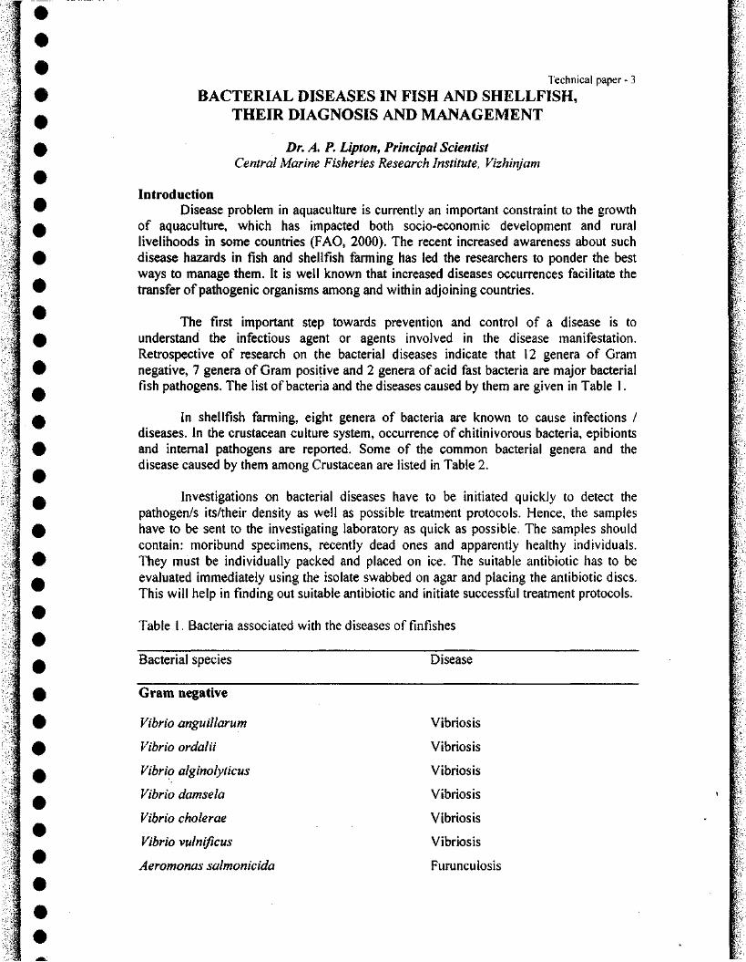
BACTERIAL DISEASES IN FISH AND SHELLFISH, THEIR DIAGNOSIS AND MANAGEMENT
Dr. A. P. Upton, Principal Scientist Central Marine Fisheries Research Institute, Vizhinjam
Introduction Disease problem in aquaculture is currently an important constraint to the growth
of aquaculture, which has impacted both socio-economic development and rural livelihoods in some countries (FAO, 2000). The recent increased awareness about such disease hazards in fish and shellfish farming has led the researchers to ponder the best ways to manage them. It is well known that increased diseases occurrences facilitate the transfer of pathogenic organisms among and within adjoining countries.
The first important step towards prevention and control of a disease is to understand the infectious agent or agents involved in the disease manifestation. Retrospective of research on the bacterial diseases indicate that 12 genera of Gram negative, 7 genera of Gram positive and 2 genera of acid fast bacteria are major bacterial fish pathogens. The list of bacteria and the diseases caused by them are given in Table 1.
In shellfish farming, eight genera of bacteria are known to cause infections / diseases. In the crustacean culture system, occurrence of chitinivorous bacteria, epibionts and internal pathogens are reported. Some of the common bacterial genera and the disease caused by them among Crustacean are listed in Table 2.
Investigations on bacterial diseases have to be initiated quickly to detect the pathogen/s its/their density as well as possible treatment protocols. Hence, the samples have to be sent to the investigating laboratory as quick as possible. The samples should contain: moribund specimens, recently dead ones and apparently healthy individuals. They must be individually packed and placed on ice. The suitable antibiotic has to be evaluated immediately using the isolate swabbed on agar and placing the antibiotic discs. This will help in finding out suitable antibiotic and initiate successful treatment protocols.
Table 1. Bacteria associated with the diseases of finfishes
Bacterial species Disease
Gram negative
Vibrio anguillarum Vibriosis
Vibrio ordalii Vibriosis
Vibrio alginolyticus Vibriosis
Vibrio damsela Vibriosis
Vibrio cholerae Vibriosis
Vibrio vulnificus Vibriosis
Aeromonas salmonicida Furunculosis
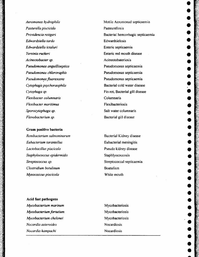
Aeromonas hydrophila
Pasturella piscicida
Providencia rettgeri
Edwardsiella tarda
Edwardsiella ictaluri
Yersinia ruckeri
Acinectobacter sp.
Pseudomonas anguilliseptica
Pseudomonas chlororaphis
Pseudomonas fluorescens
Cytophagd psychoraophila
Cytophaga sp.
Flexibacter columnaris
Flexibacter maritimus
Sporocytophaga sp.
Flavobacterium sp.
Motile Aeromonad septicaemia
Pasteurellosis
Bacterial hemorrhagic septicaemia
Edwardsielosis
Enteric septicaemia
Enteric red mouth disease
Acinectobateriosis
Pseudomonas septicaemia
Pseudomonas septicaemia
Pseudomonas septicaemia
Bacterial cold water disease
Fin rot, Bacterial gill disease
Columnaris
Flexibacteriosis
Salt water columnaris
Bacterial gill disease
Gram positive bacteria
Renibacterium salmoninarum
Euhacterium tarantellus
Lactobacillus piscicola
Staphylococcus epidermidis
Streptococcus sp.
Clostridium botulinum
Myxococcus piscicola
Bacterial Kidney disease
Eubacterial meningitis
Pseudo kidney disease
Staphlyococcosis
Streptococcal septicaemia
Boatulism
White mouth
Acid fast pathogens
Mycobacterium marinum
Mycobacterium fortutium
Mycobacterium chelonei
Nocardia asteroides
Nocardia kampachi
Mycobacteriosis
Mycobacteriosis
Mycobacteriosis
Nocardiosis
Nocardiosis
!l
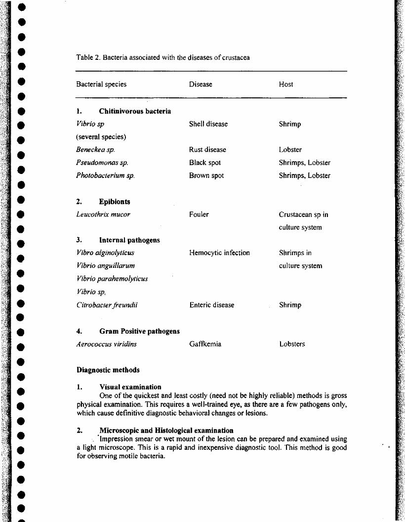
Table 2. Bacteria associated with the diseases of Crustacea
Bacterial species Disease Host
1. Chitinivorous bacteria
Vibrio sp
(several species)
Beneckea sp.
Pseudomonas sp.
Photobacterium sp.
2. Epibionts
Leucothrix mucor
3. Internal pathogens
Vibro alginolyticus
Vibrio anguillarum
Vibrio parahemolyticus
Vibrio sp,
Citrobacter freundii
Shell disease
Rust disease
Black spot
Brown spot
Fouler
Hemocytic infection
Shrimp
Lobster
Shrimps, Lobster
Shrimps, Lobster
Crustacean sp in
culture system
Shrimps in
culture system
Enteric disease Shrimp
4. Gram Positive pathogens
Aerococcus viridins Gaffkemia Lobsters
Diagnostic methods
1 Visual examination One of the quickest and least costly (need not be highly reliable) methods is gross
physical examination. This requires a well-trained eye, as there are a few pathogens only, which cause definitive diagnostic behavioral changes or lesions.
2. Microscopic and Histological examination . Impression smear or wet mount of the lesion can be prepared and examined using
a light microscope. This is a rapid and inexpensive diagnostic tool. This method is good for observing motile bacteria.
The examination of fixed tissue (tissue that has been preserved, embedded in wax and sectioned thin) using microscopy can provide a great deal of valuable information. ™ More specific information can also be obtained from this histological method when ^ special stains are applied to the tissue sections. Although this method does not provide a wealth of information, it is slow and expensive, it also requires a trained technician, and | | \ some times fails to yield a definitive diagnosis. |
• 1 3. Bacterial isolation F
For the diagnosis of bacterial infections, artificial media often can be used. A 1: sample is taken and either streaked on agar - based medium. Alternatively, the samples ^ f can be introduced into a liquid broth containing a mixture of specially designed i ingredients. The composition of the medium is formulated to enhance the growth of | P i certain types of bacteria. Some media are also designed to allow the selective growth of [ certain bacteria from a potential mixture of pathogenic and non-pathogenic bacteria. w | General morphologic classification of the bacteria can be made based on the colony size, ^ ^ shape, color and smell. For more exact identification, biochemical characterization is |-often used. In this, a single purified colony is assayed for its ability to metabolize a 1^ -̂variety of different minerals, chemicals and food sources. This is a very specific f diagnostic method. However, it requires days or even weeks obtaining the results, and not | P "̂ all bacteria will grow on defined medium. ,_ ^
Rapid diagnostic tests for detecting fish pathogens t In order to prevent outbreaks, minimize the presence of pathogens and to reduce | ^ C
the use of antimicrobial compounds, rapid detection of pathogen is essential. They are ?• further advantageous since the tests are: a. speedy, sensitive and accurate, b. presumptive W i and/or confirmatory, c. be micro-modified for inexpensive handling of large number of ^ > • individuals and small volume samples, d. require non-destructive samples, e. yield | qualitative and quantitative results. The results obtained from such tests can be correlated ^ 'i with the other clinical symptoms of the fish. Some of the important rapid diagnostic tests ' f are given below: lH t
# I Rapid immune diagnostics ^ |
Monoclonal versus Polyclonal antibodies I Immuno-diagnostic assays take advantage of the natural specificity of antibodies ^ |
toward foreign objects. For this, polyclonal or monoclonal antibodies in a variety of | formats can be used. This provides rapid detection of infectious agents. Among them, " 1. monoclonal antibodies are considered better to their increased specificity. Monoclonal ^ * antibodies can be produced by somatic cell fusion of chosen cell lines followed by I selection ofdesired clones of the hybrid ceils and thereby production of antibodies from ^ ^ the selected clones. I'
• i Direct fluorescent antibody test (D-FAT) ^ |.
The D-FAT procedure has gained widespread use in llnfish culture, it uses ^ I antibody prepared against the pathogen of interest and then conjugate it with a ^ f fluorescing dye (Fluorochorme). Either an impression smear, bacterial culture or I" specially prepared tissue sections on a microscope slide can be examined for specific HI I-pathogens using fluorescent labelled antibodies. Wherever, the antibodies are attaching ^ p to its target, the target glows when viewed through the fluorescence microscope. Though " ^
!l
this technique has the advantage of visually pin pointing the pathogen, and its location within the tissue, it requires the use of an expensive fluorescence microscope and suffers from expertise (subjective) interpretation of the results.
Enzyme-linked immunosorbant assay (ELISA) Enzyme immuno assays (ElAs) or ELISA are most widely u.sed as a serological
diagnostic technique. In this technique, the antibody molecules are linked to enzymes either directly or indirectly. The direct ELISA detects the antigens: while the indirect ELISA detects the antibodies. In the direct method, the enzyme is conjugated (linked) to a portion of the antibody molecule that does not bind to the antigen. This is referred to as "back end". In the indirect method, a second step is required. Here, a carrier or second antibody is linked with the enzyme. The amount of enzyme is important in producing measurable signal when the primary antibody binds to its target. The antibody is applied to tissue sections on microscope slides. This permits the antigen for pinpointing within a tissue using a normal light microscope. Apart from the diagnostic applications, this technique also helps in studying how the pathogen spreads within the organism and causes disease.
The ELISA detects specific substances in a complex mixture by binding them to antigen or antibody-coated substances. It is also capable of detecting viruses, bacteria, drugs, hormones, toxins and carcinogens, depending on the nature of ELISA. Once binding has occurred, other reagents are added that allow the captured substances to be linked to indicators or enzymes, which can be quantified.
J ^ i ^ Dot immunobinding assay
In the Dot immunobinding assay, the antigen is attached in a series of dots to the nitrocellulose paper. The method is similar to that of ELISA, except for the use of the nitrocellulose paper. The dot-ELISA is advantageous due to its rapidity, sensitivity, less sophistication and its permanent record characteristics.
Western blotting In the Western Blotting technique, the proteins separated from the SDS-PAGE gel
are transferred to a sheet of nitrocellulose. The proteins thus immobilized on the nitrocellulose paper can be used to detect their specific antibodies. Being sensitive, this technique helps to detect the specific antibodies in a serum of low titre.
Latex agglutination assay The Latex agglutination assay was initially devised to detect the furunculosis
disease of salmonids, caused by Aeromonas salmomcida. In this method, the optimum dilution of the specific bacterial globulin could be detected by titrating double dilutions in glycine-bufferred saline by shaking each dilution with an equal volume of latex particle suspension and incubating at 37"C. The coated particles can be stored for more than six months. This method has advantages since the formalin fixed and deep frozen samples can also be analysed apart from the fresh tissues.
DNA-based diagnostics The cloning and manipulating of genetic material has led to the development of
extremely sensitive and specific diagno.stic systems. The DNA probe is created by the recombinant DNA technology. After purifying the infectious agent of interest and
isolating its nucleic acid, an exact copy of the DNA or a portion of the DNA is made by ^ the cloning process. This copy or probe will bind to the original DNA of the pathogen " whenever the two come into contact. In order to accomplish this, the DNA strands of the a^ pathogen and the probe must be separated (by heating). After the strands have been separated, one of the strands of the probe can bind to its complementary strand from the 0 pathogen. By attaching a non-radioactive reporter molecule, the hybrid DNA can be identified and measured. H
Polymerise chain reaction (PCR) A In PCR, the DNA, to be amplified, is denatured by heating the sample in the
presence of DNA polymerase and excess dNTPs. The oligonucleotides that hybridize ^ specifically to the target sequence can prime new DNA synthesis. A product of indeterminate length characterizes the first cycle; however, the second cycle produces the W discrete "short product" which accumulates exponentially with each successive round of ^ amplification. This can lead to many million-fold amplification of the discrete fragment ^ over the course of 20 to 30 cycles. For example 30 cycles can result in 2 X 28 fold (270 n million fold) amplification of the discrete product.
Management of bacterial diseases Application of antibiotics and other chemicals in aquaculture has its own intricate w
problems. For example, regular use of antibiotics in fish or shrimp hatchery or grow out ^ system may> lead to development of not only antibiotic resistant fish/shrimp bacteria, but ^ also huma:n bacteria. Information is still lacking on the absorption and distribution of ^ antibiotics in fish and shrimp and persistence of residues or effects of them in the environment. Hence, promoting the holistic systems approach in managing fish/shellfish 0 health problems needs special attention. .
# Considering the potential threat of diseases on the one hand and the environmental g |
issues on the other hand, disease management aspects should concentrate on environment friendly biotechnological methods like: prophylaxis (vaccines, immunostimulants, H bioremediation of environment - including the methods of administration) and disease treatment protocols. 9
m Bacterins/vaccines
A vaccine could be defined as a substance that causes a specific immune 0 response. Vaccination as a part of standard fish culture programme is relatively new although the impact of vaccination is found to be dramatic. For example, the culture of • salmon in brackish water and marine environment was made possible by usage of the ^ Vibriosis vaccine, which led to a great expansion in pen-rearing of Atlantic salmon in " Norway and Chile. Several studies have been undertaken on the possibility of using live m attenuated vaccines. Considering the importance of vaccination, biotechnologists are f' trying to develop subunit vaccines, i.e., vaccines consisting of the major protective ^ antigens of the pathogen. The sub-unit vaccines have evident advantages: The important advantage is that the vaccine contains only a component of the pathogen and is therefore, ^ more chemically defined and likely to be more stable. The other advantage is that the ^ vaccine can be produced by direct synthesis or recombinant DNA technology. Thus these vaccines may be genetically engineered to express further protective antigens from other ^ fish pathogens and thereby yield multivalent vaccines.
\
•»
Techniques in administering vaccines, bacterins and immunostimuiants The route of exposure of the immunizing antigen has a direct impact on the levels
and types of protective immunity that develops. Currently four methods are commonly used to deliver antigens: a. Injection (intraperitoneal, intramuscular or subcutaneous) induces highest levels
of protection; but is very labour-intensive and stressful. Semi-automated devices, which can immunize 4000 fish per hour, have been developed which reduce the stress on fish and risk of exposure to the worker.
b. Vaccination by immersion is perhaps the most widely used method. In this method, fish are dipped for 20 seconds in a well-aerated vaccine suspension. Thus a litre of vaccine can be used to vaccinate 10,OOOX10g fish. Dip vaccination would be stressful, but the problem is overcome by bath vaccination where fish are vaccinated by being exposed to higher dilutions of vaccines (e.g. 1:100) for periods ranging from 20 minutes to several hours. Vaccine can be added directly
I ^ to hatchery troughs or transport bags. c. Spray vaccination is another modification of direct immersion but in this method
fish must be handled thus making it stressful. The level of protection, though variable has been reported to be comparable to immersion.
d. Immunization by oral route by incorporating the bacterin in the feed is a potentially useful method.
Immunostimuiants Immunostimuiants are substances, which elicit non-specific defence mechanisms
and enhance the barrier of infections against pathogens. They are isolated from natural sources and then synthesized chemically. (Ex: cell wall preparations from bacteria, fungi, mushroom). Most of the research on immunostimuiants has been directed towards treatment of cancer in humans. In fish, the non-specific defense system is activated by the immunostimuiants. The first line of defense - i.e., non specific humoral defense or proteases, lysins and agglutinins in mucous cell secretion; The second line of defense provided by the mucosal lining cells and the third line of defense achieved by blood cells, especially granulocytes and monocytes which destroy microbes present in the circulation are activated. Endocytically active cells such as endothelial cells, macrophages and granulocytes in organs and tissues, which degrade microbes or microbial products, take
1 ^ up the final defence. The final endocytic and degradation process strongly depend on the i effectiveness of reticulo endothelial system, which consist of endothelial cells, and I ^ macrophages, which line the small blood vessels (sinusoids and ellipsoids). The central I cells in the production of antimicrobial substances are macrophages and granulocytes, I 9 which are activated by the immune enhancers. The immunostimuiants have several
advantages:
1. Being natural products, there is no environmental hazard. 2. Unlike vaccines, which give protection to a specific pathogen,
immunostimuiants provide a wide range of protection against several pathogens.
3. Most of the immunostimuiants can be synthesized and the problem of residual effect on shrimps or fish is not encountered.
4. Fish depend more heavily on non-specific defense mechanisms than mammals and therefore immunostimuiants have a significant role in health management strategies in aquaculture.
5. When glucans were administered along with Aeromonas hydrophila vaccine, I
.t
the response was even more enhanced, suggesting that yeast glucans have important role in disease management in warm water aquaculture. HI |
Bioremediation of the farming environment ^ | One of the important prophylactic measures against the disease management in n f
aquaculture is proper water management. In culture conditions, the disease problems are t linked to the stress factors arising out of inadequate physico-chemical and ^ i microbiological quality of water. Ammonia and hydrogen sulfide are two important \ factors of great significance to the well being of the cultivable species. As the culture W i progresses and biomass increases, the water quality deteriorates due to accumulation of ^ f metabolic waste of cultured organisms, decomposition of unutilized feed and decay of ^ ^ other biotic materials. It is reported that many of the pathogens isolated from diseased ^ I-shrimp were the normal flora of the culture system, which become opportunistic | pathogens. For the eco-friendly environment and disease management, the concept of 0 ^ 'probiotics' is gaining importance. |
Presently a variety of commercial products of water additive probiotics are ^ f available. The 'probiotic organisms" work on the principle of competitive exclusion. This < | ecological process modifies the microbial species composition of the host and its | ^ L environment. The prohiotic application also acts as a "bio control", through which ? pathogens can be killed or reduced in number in the aquatic environment. Thus the % \ concept of "bioremediation" is initiated, when microbes are used to treat pollutants or ^ waste, which break down undesirable substances. ^ \
• L 5
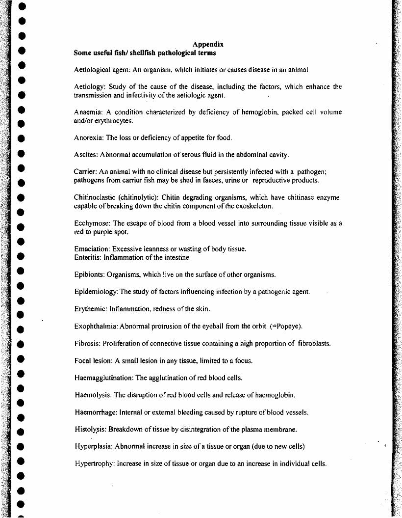
Appendix i 9 Some useful fish/ shellfish pathological terms
I Aetiological agent: An organism, which initiates or causes disease in an animal
> Aetiology; Study of the cause of the disease, including the factors, which enhance the j % transmission and infectivity of the aetiologic agent.
w Anaemia: A condition characterized by deficiency of hemoglobin, packed cell volume
^ and/or erythrocytes.
0 Anorexia: The loss or deficiency of appetite for food.
% Ascites: Abnormal accumulation of serous fluid in the abdominal cavity.
^ Carrier: An animal with no clinical disease but persistently infected with a pathogen; H pathogens from carrier fish may be shed in faeces, urine or reproductive products. % Chitinoclastic (chitinolytic): Chitin degrading organisms, which have chitinase enzyme
capable of breaking down the chitin component of the exoskeleton.
^ Ecchymose: The escape of blood from a blood vessel into surrounding tissue visible as a red to purple spot.
Emaciation; Excessive leanness or wasting of body tissue. 0 Enteritis: Inflammation of the intestine.
^ Epibionts: Organisms, which live on the surface of other organisms.
Epidemiology: The study of factors influencing infection by a pathogenic agent.
Erythemic: Inflammation, redness of the skin.
A Exophthalmia; Abnormal protrusion of the eyeball from the orbit. (=Popeye).
H Fibrosis: Proliferation of connective tissue containing a high proportion of fibroblasts.
; 9 Focal lesion: A small lesion in any tissue, limited to a focus.
^ Haemagglutination: The agglutination of red blood cells.
Haemolysis; The disruption of red blood cells and release of haemoglobin.
^ Haemorrhage: Internal or external bleeding caused by rupture of blood vessels.
^ Histolysis: Breakdown of tissue by disintegration of the plasma membrane.
i % Hyperplasia: Abnoitnal increase in size of a tissue or organ (due to new cells)
k 9 Hypertrophy: Increase in size of tissue or organ due to an increase in individual cells. 4
•
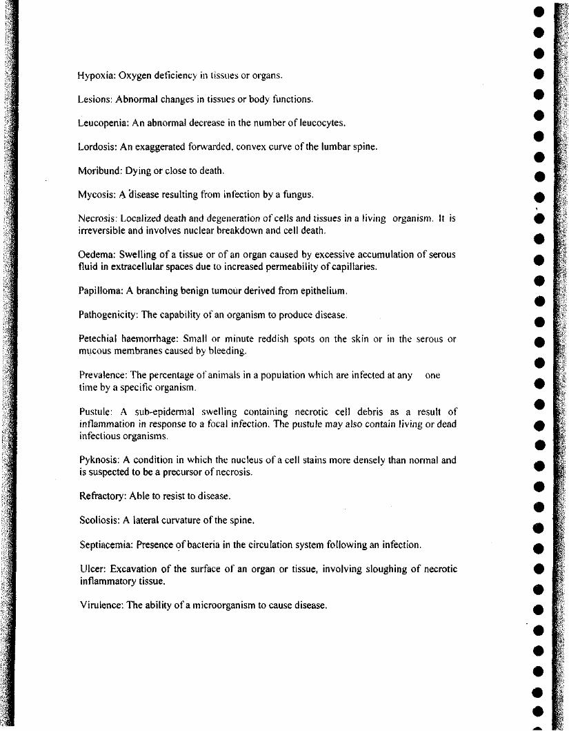
Hypoxia: Oxygen deficiency in tissues or organs. W
Lesions: Abnormal changes in tissues or body functions. ^
Leucopenia: An abnormal decrease in the number of leucocytes.
Lordosis: An exaggerated forwarded, convex curve of the lumbar spine.
Moribund: Dying or close to death. ^ f
I Mycosis: A disease resulting from infection by a fungus. m '
• f Necrosis: Localized death and degeneration of cells and tissues in a living organism. It is 0 L irreversible and involves nuclear breakdown and cell death. ^ [•'
• I Oedema: Swelling of a tissue or of an organ caused by excessive accumulation of serous fluid in extracellular spaces due to increased permeability of capillaries.
Papilloma: A branching benign tumour derived from epithelium.
Pathogenicity: The capability of an organism to produce disease.
Petechial haemorrhage: Small or minute reddish spots on the skin or in the serous or mucous membranes caused by bleeding.
Ulcer: Excavation of the surface of an organ or tissue, involving sloughing of necrotic inflammatory tissue.
Virulence: The ability of a microorganism to cause disease.
• i • i
Prevalence: The percentage of animals in a population which are infected at any one | time by a specific organism. • |
Pustule: A sub-epidermal swelling containing necrotic cell debris as a result of I inflammation in response to a focal infection. The pustule may also contain living or dead (^ J infectious organisms. I
• I Pyknosis: A condition in which the nucleus of a cell stains more densely than normal and ^ -' is suspected to be a precursor of necrosis. ^ |"
• i Refractory: Able to resist to disease. i-• i Scoliosis: A lateral curvature of the spine. _ r-Septiacemia: Presence of bacteria in the circulation system following an infection. ^ f
I i r
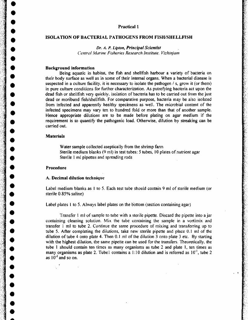
Practical 1
ISOLATION OF BACTERIAL PATHOGENS FROM FISH/SHELLFISH
Dr. A. P. Lipton, Principal Scientist Central Marine Fisheries Research Institute, Vizhinjam
Background information Being aquatic in habitat, the fish and shellfish harbour a variety of bacteria on
their body surface as well as in some of their internal organs. When a bacterial disease is suspected in a culture facility, it is necessary to isolate the pathogen / s, grow it (or them) in pure culture conditions for further characterization. As putrefying bacteria act upon the dead fish or shellfish very quickly, isolation of bacteria has to be carried out from the just dead or moribund fish/shellfish. For comparative purpose, bacteria may be also isolated from infected and apparently healthy specimens as well. The microbial content of the infected specimens may vary ten to hundred fold or more than that of another sample. Hence appropriate dilutions are to be made before plating on agar medium if the requirement is to quantify the pathogenic load. Otherwise, dilution by streaking can be carried out.
Materials
Water sample collected aseptically from the shrimp farm Sterile medium blanks (9 ml) in test tubes: 5 tubes, 10 plates of nutrient agar Sterile 1 ml pipettes and spreading rods
Procedure
A. Decimal dilution technique
Label medium blanks as 1 to 5. Each test tube should contain 9 ml of sterile medium (or sterile 0.85% saline)
Label plates I to 5. Always label plates on the bottom (section containing agar)
Transfer 1 ml of sample to tube with a sterile pipette. Discard the pipette into ajar containing cleaning solution, Mix the tube containing the sample in a vortimix and transfer 1 ml to tube 2. Continue the same procedure of mixing and transferring up to tube 5. After completing the dilutions, take new sterile pipette and place 0.1 ml of the dilution of tube 4 onto plate 4. Then 0.1 ml of the dilution 3 onto plate 3 etc. By starting with the highest dilution, the same pipette can be used for the transfers. Theoretically, the tube 1 should contain ten times as many organisms as tube 2 and plate 1, ten times as many organisms as plate 2. Tubel contains a 1:10 dilution and is referred as 10"', tube 2 as 10'̂ and soon.

The steps involved in streaking for isolation include:
streaks a single time before commencing the next area, or dilution.
Keep for incubation
Record the cultural characteristics of the bacteria isolated.
B. Spread pl̂ te technique
Sterile spreading rods are used to disperse 0.1 ml of each dilution over plate's surface. The same rod can be used on all plates if one proceeds in the order of greatest to ^ ^ least dilution of organisms (Ex: 10"'*-> 10''). A •
i.
i
After allowing the plates to dry, incubate them at room temperature in inverted i position. A dish with solid agar is always inverted to prevent condensation droplets from falling on agar surface and causing colonies to smear. • t
C. Streaking to obtain single colonies
Streaking is done in a variety of ways to obtain isolating individual colonies in order that they may be transferred to other media for further studies.
1. Make the initial streak after the loop is removed from broth or contact with \ bacterial colony, i^ \
2. Flame loop. 3. Make next set of streaks, ? 4. Flame loop, 5. Make third set of streaks. • \
In each case, the partially cooled, flamed loop is allowed to cross one of the last \ ! ; •
t
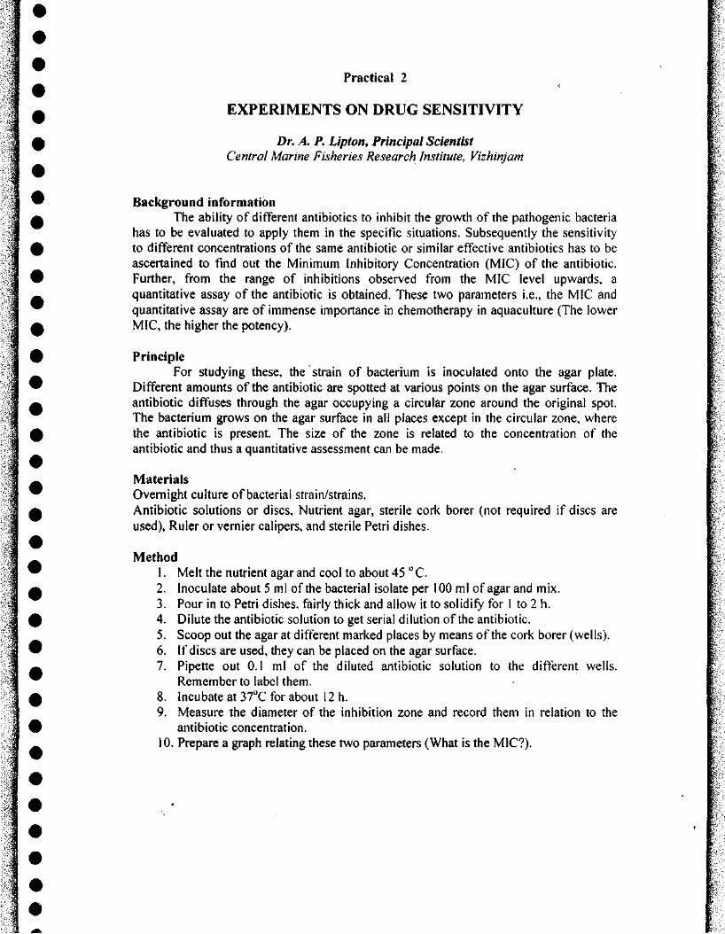
Practical 2
EXPERIMENTS ON DRUG SENSITIVITY
Dr. A. P. Lipton, Principal Scientist Central Marine Fisheries Research Institute, Vizhinjam
Bacl^round information The ability of different antibiotics to inhibit the growth of the pathogenic bacteria
has to be evaluated to apply them in the specific situations. Subsequently the sensitivity to different concentrations of the same antibiotic or similar effective antibiotics has to be ascertained to find out the Minimum Inhibitory Concentration (MIC) of the antibiotic. Further, from the range of inhibitions observed from the MIC level upwards, a quantitative assay of the antibiotic is obtained. These two parameters i.e., the MIC and quantitative assay are of immense importance in chemotherapy in aquaculture (The lower MIC, the higher the potency).
Principle [ For studying these, the strain of bacterium Is inoculated onto the agar plate. [
Different amounts of the antibiotic are spotted at various points on the agar surface. The f antibiotic diffuses through the agar occupying a circular zone around the original spot. f The bacterium grows on the agar surface in all places except in the circular zone, where \ the antibiotic is present. The size of the zone is related to the concentration of the ? antibiotic and thus a quantitative assessment can be made. %
Materials ; Overnight culture of bacterial strain/strains. | Antibiotic solutions or discs, Nutrient agar, sterile cork borer (not required if discs are | used). Ruler or vernier calipers, and sterile Petri dishes. i
Method \ 1. Melt the nutrient agar and cool to about 45 " C. \ 2. Inoculate about 5 ml of the bacterial isolate per 100 ml of agar and mix. |-3. Pour in to Petri dishes, fairly thick and allow it to solidify for 1 to 2 h. | 4. Dilute the antibiotic solution to get serial dilution of the antibiotic. •• 5. Scoop out the agar at different marked places by means of the cork borer (wells). t 6. If discs are used, they can be placed on the agar surface. •• 7. Pipette out 0.1 ml of the diluted antibiotic solution to the different wells. :
Remember to label them. •• 8. Incubate at ST̂ C for about 12 h. \ 9. Measure the diameter of the inhibition zone and record them in relation to the |
antibiotic concentration. 1 10. Prepare a graph relating these two parameters (What is the MIC?). 1
i r
! 1
Practical 3
GUIDELINES FOR DISEASE TREATMENT
Dr. A. P. Upton, Principal Scientist, Central Marine Fisheries Research Institute, Vizhinjam
Methods 1. Dip treatment:
5. by injection;
The required chemical / substance is administered as intra peritoneal, intra muscular or intra dermal routes using syringes fitted with appropriate needles.
Exercise I
Treatment for bacterial infection in shrimp
Example 1:
I t
• i • i
Take known volume of water. Dissolve the recommended antibiotic or antiseptic , \ or other material in this known volume of water. Remove and keep the fish/shellfish to be ^ j-treated in net. Immerse the fish kept in net in the dissolved contents for a specific period ^ \ of time (or still the fish/shellfish show discomfort - depending on the intensity of ^ £ infection). A modified advanced method is the 'Hyper osmotic infiltration technique'. In # ^ this method, the required substances can diffuse through gills or other portals when they | are subjected to mild osmotic changes. % \
2. Flush technique: " % t
• -The required chemical / substance is added to the unit at the water inflow and s
allowed to flow through. 0 » 3. Continuous and Prolonged exposure method: # i
n In this method, the required chemical / substance is added to the system ^ ^
continuously. g | J 4. Feed: % \
I.
The required chemical / substance is mixed (or sometimes sprayed on the pellets) ^ |> with the feed and administered. ^ V
• I Calculation of the recommended dose of oxytetracycline (OTC) antibiotic in feed @ 50- <9 I 55mg/kg of shrimp: I
^
^
Assumptions:
1. An average 20g size shrimp will consume 3.2% of its body weight.
2. Diseased shrimp consume about 25% less (-2.6% body wt,).
3. Hence 100kg of shrimp consumes 2.6 kg of feed.
4. Assuming 50% loss due to leaching effect of OTC in water, a compensation for the antibiotic has to be added (100-1 lOmg).
5. Therefore for 100kg shrimp 100.000 to 110,000 mg of OTC to be added.
6. In 2.6 kg feed incorporate about 4 g of the antibiotic. This will give a value of 10,400 mg per kilogram of the shrimp. Other wise it is 104 mg per kg of shrimp body weight. Assuming 50% leaching loss, it is anticipated that an average 50.2 mg of OTC per kg of shrimp will be administered.


![Publisher - CMFRI Repositoryeprints.cmfri.org.in/9658/1/IJMS_2013[1].pdfP.R. Jayachandran, S. Bijoy Nandan, ... Andi Rezky Puspita Ayu ... Noverita Dian Takarina, Sharfina Tammy Aryanti,](https://img.pdfslide.us/doc/110x75/5aa4e8187f8b9a1d728c777b/publisher-cmfri-1pdfpr-jayachandran-s-bijoy-nandan-andi-rezky-puspita.jpg)