Embed Size (px)
Citation preview

Acta Biomaterialia 8 (2012) 4304–4313
Contents lists available at SciVerse ScienceDirect
Acta Biomaterialia
journal homepage: www.elsevier .com/locate /ac tabiomat
Reducing cytotoxicity while improving anti-cancer drug loading capacityof polypropylenimine dendrimers by surface acetylation
Fei Wang a, Xiaopan Cai b,c, Yunzhang Su a, Jingjing Hu d, Q. Wu e, Hongfeng Zhang a,⇑,Jianru Xiao b,⇑, Yiyun Cheng a,⇑a Shanghai Key Laboratory of Regulatory Biology and School of Life Sciences, East China Normal University, Shanghai 200241, Chinab Department of Orthopedic Oncology, Changzheng Hospital, The Second Military Medical University, Shanghai 200003, Chinac Department of Orthopedics, The third Affiliated Hospital of Anhui Medical University, Hefeis 230000, Chinad Department of Chemistry, University of Science and Technology of China, Hefei, Anhui 230026, Chinae Division of Life Sciences, Hongkong University of Science and Technology, Hongkong, China
a r t i c l e i n f o a b s t r a c t
Article history:Received 6 April 2012Received in revised form 15 July 2012Accepted 20 July 2012Available online 27 July 2012
Keywords:DendrimerAcetylationPPISustained releaseAnticancer drug
1742-7061/$ - see front matter � 2012 Acta Materialhttp://dx.doi.org/10.1016/j.actbio.2012.07.031
⇑ Corresponding authors. Tel.: +86 021 54342935 (E-mail addresses: [email protected] (H. Zh
(J. Xiao), [email protected] (Y. Cheng).
Polypropylenimine (PPI) dendrimers have been widely used as effective delivery vehicles for drugs andnucleic acids during the past decade. However, biomedical applications of PPI dendrimers were limitedbecause of their serious cytotoxicity and low drug loading capacity. In the present study, acetylatedPPI dendrimers with different degrees of acetylation ranging from 14.2% to 94.3% were synthesizedand used to encapsulate drugs, including methotrexate sodium, sodium deoxycholate and doxorubicin.Acetylated PPI dendrimers with a degree of acetylation >80% showed a significantly decreased cytotox-icity (>90% cell viability) on MCF-7 and A549 cells. The drug loading capacity of acetylated PPI dendrimersincreased proportionally with the degree of acetylation on the dendrimer surface. In addition, 94.3% acet-ylated PPI dendrimers exhibited a pH-responsive release profile of anticancer drugs loaded within thenanoparticles. The cytotoxicities of methotrexate sodium and doxorubicin on MCF-7 and A549 cells weresignificantly reduced when they were complexed with acetylated PPI dendrimers with high degrees ofacetylation (>80%), owing to sustained drug release from the dendrimers. The results suggest that surfaceacetylation can reduce the cytotoxicity and improve the anticancer drug loading capacity of cationic den-drimers, and that acetylated PPI dendrimers are promising vehicles for anticancer drugs in clinical trials.
� 2012 Acta Materialia Inc. Published by Elsevier Ltd. All rights reserved.
1. Introduction
Polypropylenimine (PPI) dendrimers are the first commerciallyavailable dendrimers synthesized by a divergent strategy usingdiaminobutane as the central core and propylene imine as repeatunits [1]. They are widely used as templates for the synthesis ofdendrimer-encapsulated nanoparticles and as scaffolds for theconstruction of magnetic resonance imaging contrast agents, espe-cially in drug and gene delivery [2–4]. PPI dendrimers have excel-lent aqueous solubility, and therefore a large number ofhydrophobic cavities in their interior can effectively improve thesolubility and stability of various hydrophobic drugs [5]. In addi-tion, PPI dendrimers with a high density of active groups on theirsurface can be easily functionalized with therapeutic agents, tar-geting moieties, solubilizing ligands and imaging units for targetedcancer diagnosis and therapy [6]. However, PPI dendrimers, espe-cially those with a cationic surface, are not ideal candidates for bio-
ia Inc. Published by Elsevier Ltd. A
Y. Cheng).ang), [email protected]
medical applications, owing to their serious toxicity [7–9]. Forinstance, G5 amine-terminated PPI dendrimer at a low concentra-tion of 1 lg ml�1 caused 83.2% and 76.9% cell death on HepG2 andCOS-7 cells, respectively [7]. Also, G5 cationic PPI dendrimershowed significant decreases in red blood cell count, hemoglobincontent and mean corpuscular hemoglobin value, as well as a sub-stantial increase in white blood cell count [7]. Exposing macro-phages to G2 or G3 cationic PPI dendrimers caused dramaticchanges in macrophage cell size and significant fluctuation inmitochondrial membrane potential [10]. Cationic PPI dendrimersshowed rapid clearance from the blood circulation system afterintravenous or intraperitoneal injection, leading to low bioavail-ability of the administered drugs [11]. Administration of G4 cat-ionic PPI dendrimer caused obvious changes in the behavior ofanimals, such as decreased food and water consumption, and lowerrate of gain in body weight [12].
Besides the non-negligible in vitro and in vivo toxicity, PPI den-drimers have extremely low drug loading capacity for a list ofhydrophobic drugs [13]. PPI dendrimers have smaller molecularsize and interior cavities compared with polyamidoamine (PA-MAM) dendrimers [14]. Though PPI dendrimer with a more hydro-
ll rights reserved.

Scheme 1. (a) Synthetic route of acetylated G4 PPI dendrimer, (b) repeated unit of acetylated PPI dendrimer with proton labeling, and molecular structure of (c) methotrexatesodium and (d) doxorubicin.
F. Wang et al. / Acta Biomaterialia 8 (2012) 4304–4313 4305
phobic interior more easily encapsulate hydrophobic compounds,the yielding PPI/drug complexes are not as stable as PAMAM/drugcomplexes in water and may precipitate from the aqueous solutionwhen PPI dendrimers are loaded with a large number of drug mol-ecules [13]. These defects of PPI dendrimers prevent the develop-ment of PPI dendrimer-based drug formulations and diagnosticagents for biomedical purpose [9].
To decrease the cytotoxicity and improve the drug loadingcapacity of dendrimers, surface engineering of the dendrimer sur-face by PEGylation [15–18], acetylation [19–21], glycosylation [9]and amino acid or peptide modification [9] was proposed by sev-eral groups. Among these strategies, PEGylation and acetylationwere considered to be the most efficient ones in reducing dendri-mer cytotoxicity and improving their aqueous stability in physio-logical conditions [22]. Compared with PEGylation, acetylation ispreferred for the following reasons: (1) acetylation is more facileand highly efficient, and the degree of acetylation on the dendri-mer surface can be easily tailored by choosing proper stoichiome-try of acetic anhydride and dendrimer [23,24]; (2) modification ofPEG chains with a larger molecular size than acetyl groups on thedendrimer surface will cause significant steric hindrance andthereby affect other functional groups such as targeting moieties
[25]; and (3) acetylated dendrimers can maintain the high penetra-tion ability of cationic dendrimers across cell membranes, whilePEGylated dendrimers show much reduced cellular uptake [26].Generally, acetylation can effectively increase the aqueous solubil-ity of dendrimer–drug conjugates, improve their biocompatibility,and optimize their in vivo pharmacokinetic behavior [9].
Though acetylated dendrimers showed several promisingadvantages in previous studies, the following questions are stillunknown: (1) What is the least degree of acetylation on dendrimersurface that can meet the need of biomedical applications such asdrug delivery, gene delivery and disease diagnosis? (2) How willthe degree of acetylation influence the drug loading capacity ofacetylated dendrimers? (3) Can the acetylated dendrimers withhigh degrees of acetylation be used directly as drug vehicles foranticancer drugs? The present study addressed these questionsusing methotrexate sodium, sodium deoxycholate and doxorubicinas model drugs. Sodium deoxycholate was used because it is awidely used amphiphilic guest for dendrimers. G4 PPI dendrimerswith different degrees of acetylation ranging from 14.2% to 94.3%were synthesized, characterized by nuclear magnetic resonance(NMR) techniques, and used to encapsulate three model drugs.The drug loading capacity and cytotoxicity of these acetylated
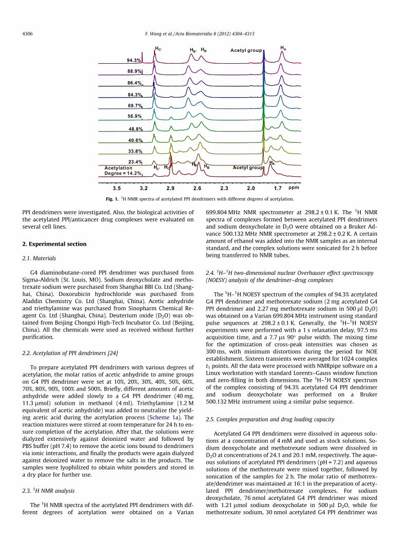
Fig. 1. 1H NMR spectra of acetylated PPI dendrimers with different degrees of acetylation.
4306 F. Wang et al. / Acta Biomaterialia 8 (2012) 4304–4313
PPI dendrimers were investigated. Also, the biological activities ofthe acetylated PPI/anticancer drug complexes were evaluated onseveral cell lines.
2. Experimental section
2.1. Materials
G4 diaminobutane-cored PPI dendrimer was purchased fromSigma-Aldrich (St. Louis, MO). Sodium deoxycholate and metho-trexate sodium were purchased from Shanghai BBI Co. Ltd (Shang-hai, China). Doxorubicin hydrochloride was purchased fromAladdin Chemistry Co. Ltd (Shanghai, China). Acetic anhydrideand triethylamine was purchased from Sinopharm Chemical Re-agent Co. Ltd (Shanghai, China). Deuterium oxide (D2O) was ob-tained from Beijing Chongxi High-Tech Incubator Co. Ltd (Beijing,China). All the chemicals were used as received without furtherpurification.
2.2. Acetylation of PPI dendrimers [24]
To prepare acetylated PPI dendrimers with various degrees ofacetylation, the molar ratios of acetic anhydride to amine groupson G4 PPI dendrimer were set at 10%, 20%, 30%, 40%, 50%, 60%,70%, 80%, 90%, 100% and 500%. Briefly, different amounts of aceticanhydride were added slowly to a G4 PPI dendrimer (40 mg,11.3 lmol) solution in methanol (4 ml). Triethylamine (1.2 Mequivalent of acetic anhydride) was added to neutralize the yield-ing acetic acid during the acetylation process (Scheme 1a). Thereaction mixtures were stirred at room temperature for 24 h to en-sure completion of the acetylation. After that, the solutions weredialyzed extensively against deionized water and followed byPBS buffer (pH 7.4) to remove the acetic ions bound to dendrimersvia ionic interactions, and finally the products were again dialyzedagainst deionized water to remove the salts in the products. Thesamples were lyophilized to obtain white powders and stored ina dry place for further use.
2.3. 1H NMR analysis
The 1H NMR spectra of the acetylated PPI dendrimers with dif-ferent degrees of acetylation were obtained on a Varian
699.804 MHz NMR spectrometer at 298.2 ± 0.1 K. The 1H NMRspectra of complexes formed between acetylated PPI dendrimersand sodium deoxycholate in D2O were obtained on a Bruker Ad-vance 500.132 MHz NMR spectrometer at 298.2 ± 0.2 K. A certainamount of ethanol was added into the NMR samples as an internalstandard, and the complex solutions were sonicated for 2 h beforebeing transferred to NMR tubes.
2.4. 1H–1H two-dimensional nuclear Overhauser effect spectroscopy(NOESY) analysis of the dendrimer–drug complexes
The 1H–1H NOESY spectrum of the complex of 94.3% acetylatedG4 PPI dendrimer and methotrexate sodium (2 mg acetylated G4PPI dendrimer and 2.27 mg methotrexate sodium in 500 ll D2O)was obtained on a Varian 699.804 MHz instrument using standardpulse sequences at 298.2 ± 0.1 K. Generally, the 1H–1H NOESYexperiments were performed with a 1 s relaxation delay, 97.5 msacquisition time, and a 7.7 ls 90� pulse width. The mixing timefor the optimization of cross-peak intensities was chosen as300 ms, with minimum distortions during the period for NOEestablishment. Sixteen transients were averaged for 1024 complext1 points. All the data were processed with NMRpipe software on aLinux workstation with standard Lorents–Gauss window functionand zero-filling in both dimensions. The 1H–1H NOESY spectrumof the complex consisting of 94.3% acetylated G4 PPI dendrimerand sodium deoxycholate was performed on a Bruker500.132 MHz instrument using a similar pulse sequence.
2.5. Complex preparation and drug loading capacity
Acetylated G4 PPI dendrimers were dissolved in aqueous solu-tions at a concentration of 4 mM and used as stock solutions. So-dium deoxycholate and methotrexate sodium were dissolved inD2O at concentrations of 24.1 and 20.1 mM, respectively. The aque-ous solutions of acetylated PPI dendrimers (pH = 7.2) and aqueoussolutions of the methotrexate were mixed together, followed bysonication of the samples for 2 h. The molar ratio of methotrex-ate/dendrimer was maintained at 16:1 in the preparation of acety-lated PPI dendrimer/methotrexate complexes. For sodiumdeoxycholate, 76 nmol acetylated G4 PPI dendrimer was mixedwith 1.21 lmol sodium deoxycholate in 500 ll D2O, while formethotrexate sodium, 30 nmol acetylated G4 PPI dendrimer was

Fig. 2. 1H NMR spectra of acetylated PPI dendrimer/sodium deoxycholate complexes.
F. Wang et al. / Acta Biomaterialia 8 (2012) 4304–4313 4307
mixed with 0.48 lmol sodium deoxycholate in 200 ll deionizedwater. The amount of sodium deoxycholate in the G4 PPI dendri-mer solution was monitored by 1H NMR spectra, while the com-plexes of methotrexate sodium and acetylated PPI dendrimerwere centrifuged at 10000 rpm for 5 min to remove the precipi-tates, and the amount of methotrexate sodium in the supernatantwas measured by a high-performance liquid chromatography(HPLC) method. The preparation of acetylated G4 PPI dendrimer/doxorubicin complexes was conducted according to the methodadopted in the preparation of sodium deoxycholate and metho-trexate sodium complexes, using different amounts of acetylateddendrimers and doxorubicin.
Fig. 3. Drug loading capacities of acetylated PPI dendrimers with different degreesof acetylation using methotrexate sodium as a model drug.
2.6. HPLC analysis
The HPLC experiments were conducted on a reverse HPLCinstrument (Agilent1200, USA) equipped with a C18 column(4.6 mm in diameter, 150 mm long, 5 lm particle size, ZORBAXEclipse XDB, Agilent, USA). The mobile phase was methanol and2% acetic acid at a volume ratio of 20:80 and a flow rate of1.0 ml min�1. A 10 ll sample was injected, and the drug was de-tected at 313 nm. The retention time of methotrexate sodium was7.5 ± 0.2 min. The standard curve for methotrexate sodium is:area = 19,181C � 6.3535, C in mg ml�1; R2 = 0.9984 and the applica-ble methotrexate concentration ranges from 10�4 to 10�2 mg ml�1.
2.7. In vitro release studies
The release rate of methotrexate sodium from acetylated G4 PPIdendrimer with a 94.3% degree of acetylation was measured by anequilibrium dialysis method. Acetylated G4 PPI/methotrexate com-plexes were prepared as described above at a drug/dendrimer mo-lar ratio of 5:1. The complex solutions were transferred into a

Fig. 4. 1H–1H NOESY spectrum of (a) acetylated PPI dendrimer (degree of acetylation 94.3%)/methotrexate and (b) acetylated PPI dendrimer (degree of acetylation 94.3%)/doxycholate complexes in D2O at a mixing time of 300 ms.
4308 F. Wang et al. / Acta Biomaterialia 8 (2012) 4304–4313
dialysis bag (Spectrum, America) with a molecular weight cut off of1000 Da, and immersed into 50 ml aqueous solution. The outerphase of the dialysis bag was stirred during the release experiment.At scheduled intervals, 100 ll of the outer phase solutions waswithdrawn and replenished with an equal volume of fresh water.The cumulative amount of methotrexate sodium released out ofthe dialysis bag was measured by the HPLC method. To investigatethe effect of environmental pH condition on the release rate ofmethotrexate sodium, the pH values of outer phase solutions wereadjusted to 4.0, 5.0 and 7.0 by 0.1 M HCl and NaOH.
2.8. Cytotoxicity of acetylated G4 PPI dendrimers and their complexeswith anticancer drugs
MCF-7 (human breast adenocarcinoma cell line, ATCC) andA549 cells (human lung adenocarcinoma epithelial cell line, ATCC)were incubated at 37 �C in 5% CO2 and Dulbecco’s modified Eagle’smedium (DMEM; Gibco Inc.) supplemented with streptomycin,penicillin sulfate and 10% heat-inactived fetal calf serum (GibcoInc.). Cytotoxicities of acetylated G4 PPI dendrimers (10–20 lM)with different degrees of acetylation and their complexes withtwo anticancer drugs methotrexate sodium (10 lM) and doxorubi-cin (0.2–2.5 lM) on MCF-7 and A549 cells were evaluated by a 3-(4,5-dimethylthiazol-2-yl)-2,5-diphenyltetrazolium bromide(MTT) assay. The acetylated PPI dendrimer/anticancer drug com-plexes were sonicated for 2 h after sample preparation. MCF-7and A549 cells were seeded in 96-well plates 48 h before the cyto-
toxicity experiment and treated with acetylated G4 PPI dendrimersor dendrimer/drug complexes for another 48 h. The cells were thenwashed with PBS buffer and further incubated with MTT reagentfor 3 h, and the formazan crystals yielded were dissolved by DMSO.Absorbances of the DMSO solutions in each well were measured at570 nm using a microplate reader (MQX200R, BioTeK Inc.). Six re-peats were conducted for each sample, and the results are shownas mean ± standard deviation. The data were analyzed by two-tailed, unpaired Student’s t-tests. To visualize the uptake of doxo-rubicin and acetylated PPI/doxorubicin complexes by MCF-7 cells,the cells were incubated with 5 lM doxorubicin or doxorubicin/dendrimer complexes for 3 h, 6 h and 12 h, and washed with PBSbuffer. Then the nuclei of MCF-7 cells were stained with 2-(4-ami-dinophenyl)-6-indolecarbamidine dihydrochloride (DAPI). Thestained cells were then imaged by a fluorescence microscope (Lei-ka DMI4000 B, Germany).
3. Results and discussion
3.1. Acetylation of G4 PPI dendrimers
As shown in Scheme 1b and Fig. 1, cationic G4 PPI dendrimerhas five peaks: HA at 1.77 ppm, HB at 2.52 ppm, HB0 at 2.57 ppm,and HC at 2.88 ppm. The peak for protons HD located at the centralcore of G4 PPI dendrimer is weak and overlaps with that of HA afteracetylation [27]. Acetylation of PPI dendrimer generates a newpeak at 1.85 ppm, corresponding to methyl protons in the acetyl

Scheme 2. Proposed inclusion structures of acetylated G4 PPI dendrimer/drug complexes: (a) methotrexate sodium; (b) sodium deoxycholate.
Fig. 5. Viabilities of MCF-7 and A549 cells incubated with 0.02 mM acetylated PPIdendrimer with different degrees of acetylation.
F. Wang et al. / Acta Biomaterialia 8 (2012) 4304–4313 4309
group. In addition, a new peak appears at 3.07 ppm, correspondingto the methylene protons HC adjacent to the added acetyl groups[24]. The intensities of peaks at 1.85 ppm and 3.07 ppm increaseproportionally with increasing molar ratios of acetyl anhydrideand G4 PPI dendrimer, suggesting an increased degree of acetyla-tion on the PPI surface. The decreased intensity of the peak at2.88 ppm suggests a decreased amount of cationic amine groupson the dendrimer surface. At the same time, peaks for protons HB
and HB0 shift to downfield, owing to the decreased electron densityaround these protons after acetylation.
The degrees of acetylation of G4 PPI dendrimer were calculatedfrom the integration areas of the peaks corresponding to PPI scaf-fold protons and the added acetyl groups in 1H NMR spectra. PeaksHA, HB, HB0 and HC of G4 PPI dendrimer represent 368 protons,while the peak at 1.85 ppm corresponds to the methyl protons inthe added acetyl groups. By comparing the integrated areas ofthese peaks, the average number of acetyl groups conjugated toeach G4 PPI dendrimer can be calculated, and the degrees of acet-ylation are shown in Fig. 1 [28–30]. As expected, the acetylateddendrimers are obtained in high yield (>95%), and the calculateddegree of acetylation is in direct proportion to the added amountof acetyl anhydride. Surprisingly, it can be found that the calcu-lated degree of acetylation on G4 PPI dendrimer is slightly larger
than the theoretical value at acetyl anhydride/amine molar ratios<50%, probably as a result of ionic attachment of acetic acid onthe dendrimer surface during the acetylation process [24] and/orerrors during the integration of 1H NMR spectra.
3.2. Drug loading capacities of acetylated PPI dendrimers withdifferent degrees of acetylation
When sodium deoxycholate was added to solutions of non-modified PPI dendrimers or acetylated PPI dendrimers with lowdegrees of acetylation, white precipitates were observed in thecomplex solutions. As shown in Fig. 2, strong NMR signals ofdeoxycholate (0.573 ppm, 0.785 ppm and 3.919 ppm) were ob-served in the 1H NMR spectrum of unmodified PPI/deoxycholatecomplex because the unmodified PPI dendrimer/deoxycholate pre-cipitate consists of a high percentage of PPI dendrimer and a lowpercentage of deoxycholate. Convincing evidence is the disappear-ance of PPI peaks when the degree of acetylation is 0%, while in thepresence of higher degree of acetylation PPI dendrimers, e.g., 88.9%and 94.3%, the appearance of strong NMR peaks for deoxycholate isdue to the stability of acetylated PPI/deoxycholate complex inaqueous solution, and both the NMR peaks of PPI and deoxycholateare therefore observed in Fig. 2. For acetylated PPI dendrimers withdegrees of acetylation 14.3–69.7%, the acetylated PPI dendrimer/deoxycholate precipitates have a high percentage of deoxycholateand a relatively low percentage of PPI dendrimer. As a result,deoxycholate signals were observed only in the unmodified PPIdendrimer and the almost fully acetylated dendrimer complexsolutions. Acetylated PPI dendrimers with degree of acetylation>88.9% can prevent the precipitation of dendrimer/deoxycholatecomplexes. As demonstrated in previous studies, unmodified G4PPI dendrimer failed to significantly enhance the aqueous solubil-ity of phenylbutazone [13]. Though NOESY studies revealed theencapsulations of phenylbutazone molecules within the cavitiesof G4 PPI dendrimer, phenylbutazone loaded within PPI dendrimerabove a critical concentration causes the precipitation of the PPIdendrimer/phenylbutazone complexes. This is similar to the phe-nomenon of PPI dendrimer/deoxycholate complexes in Fig. 2.
Similarly, the drug loading capacities of acetylated PPI dendri-mers were evaluated using methotrexate sodium (Scheme 1c) asa model drug. The molar ratio of methotrexate/dendrimer is kept

Fig. 6. Cytotoxicities of 10 lM methotrexate sodium on (a) MCF-7 and (b) A549 cells in the absence and presence of acetylated PPI dendrimers with different degrees ofacetylation.
Fig. 7. In vitro release rate of methotrexate sodium from acetylated PPI dendrimer(degree of acetylation 94.3%) in water at different pH conditions.
4310 F. Wang et al. / Acta Biomaterialia 8 (2012) 4304–4313
at a constant 16 throughout the drug loading experiment. Asshown in Fig. 3, the concentrations of methotrexate sodium in den-drimer/methotrexate complex increase linearly with degree ofacetylation of G4 PPI dendrimer until it reaches 88.9%. At higherdegrees of acetylation, drug concentrations in the complex solu-tions are slightly changed, which is in accordance with the resultsof acetylated PPI dendrimer/deoxycholate complexes. In previousstudies, methotrexate was found to form precipitates with cationicPAMAM dendrimers owing to the presence of two carboxyl groupsin the methotrexate molecules [31]. Here, the unmodified PPI den-drimer has a nature similar to that of cationic PAMAM dendrimers,and only a low concentration of methotrexate is recovered in theunmodified PPI/methotrexate complex solution. Therefore,unmodified PPI dendrimers are not suitable for the delivery of so-dium deoxycholate (Fig. 2) and methotrexate sodium (Fig. 3), ow-ing to the low drug loading capacity of this cationic carrier and theinstability of their drug complexes.
To confirm the encapsulation of methotrexate sodium and so-dium deoxycholate within the interior cavities of acetylated PPIdendrimers, 1H–1H NOESY spectra of 94.3% acetylated dendri-mer/methotrexate complex was conducted. The presence of NOEcross-peaks between two protons in the NOESY spectrum indicatesspatial proximity of these protons [32–34], The cross-peak inten-sity decreases with the spatial distance between the protons, andincreases with the number of molecules involved in the cross-peak[33]. As shown in Fig. 4a, strong cross-peaks between methotrex-ate protons (H1, H4–8) and the interior protons of acetylated PPIdendrimer (HA, HB, HB0, HC) were observed, proving the formationof inclusion structures of methotrexate with acetylated PPI dendri-mer. Further in vitro release studies revealed that �5% of the meth-otrexate in the complex solutions is free drugs, and most of themethotrexate forms stable complexes with acetylated PPI dendri-mer. Similarly, cross-peaks between sodium deoxycholate andacetylated PPI dendrimers are observed in the 1H–1H NOESY spec-tra of acetylated PPI dendrimer (94.3%)/deoxycholate complex(Fig. 4b). These results confirmed that acetylated G4 PPI dendri-mers with a degree of acetylation >84.3% can effectively loadamphiphilic and hydrophobic drug molecules and form stableinclusion complexes with these drugs (Scheme 2).
3.3. Cytotoxicities of acetylated PPI dendrimers with different degreesof acetylation
Fig. 5 shows that the cytotoxicities of acetylated PPI dendrimersdepend on their degree of acetylation. The viabilities of cells incu-
bated with acetylated PPI dendrimers increase gradually with de-gree of acetylation on both MCF-7 and A549 cells. In thepresence of 0.02 mM unmodified G4 PPI dendrimer, the viabilitiesof MCF-7 and A549 cells are 53.8% and 18.4%, respectively. 14.2–23.4% acetylation of G4 PPI can slightly decrease cytotoxicity ofthe unmodified cationic dendrimer, while acetylated PPI dendri-mers with higher degrees of acetylation exhibit significantly re-duced toxicity on both cells. As the degree of acetylation ofdendrimer increases from 33.8% to 40.6%, the viability of MCF-7cells increases from 64.2% to 89.5%, suggesting that acetylatedPPI dendrimer with a degree of acetylation >40.6% is relatively bio-compatible on MCF-7 cells at 0.02 mM. Also, it is found that thecytotoxicities of acetylated PPI dendrimers on MCF-7 and A549cells are scarcely changed above the degree of acetylation of 84.3%.
Dendrimer biocompatibility is of central importance in the de-sign of dendrimer-based drug delivery systems [9]. A large numberof researchers have reported the in vitro and in vivo toxicity of PPIdendrimers [7,8]. To decrease the toxicity of PPI dendrimer on nor-mal cells and reduce their hemolytic activities on blood cells, acet-ylation is widely used as an efficient method to solve the issue ofthe toxicity of cationic dendrimers [22,26]. Acetylation of PPI den-drimers can neutralize their surface cationic charges and decreasetheir binding affinities with amphiphilic phospholipids as well asbiomacromolecules, maintaining the integrity of the cell mem-brane and preventing the leakage of intracellular components [22].

Fig. 8. Cytotoxicities of doxorubicin on (a) MCF-7 and (b) A549 cells in the absence and presence of acetylated PPI dendrimers with different degrees of acetylation.
Fig. 9. Images of MCF-7 cells incubated with doxorubicin and acetylated PPI dendrimer/doxorubicin complexes for 12 h; the nuclei of the MCF-7 cells were stained by DAPI.
F. Wang et al. / Acta Biomaterialia 8 (2012) 4304–4313 4311
Here, the effect of degree of acetylation on the cytotoxicity ofacetylated PPI dendrimers on MCF-7 and A549 cells was systemat-ically investigated. Cell viabilities of the dendrimers with high de-
grees of acetylation (>84.3%) are >95%. These dendrimers can beconsidered non-toxic biomaterials in drug delivery. Thus, acety-lated PPI dendrimers with degrees of acetylation of 84.3%, 86.4%,

4312 F. Wang et al. / Acta Biomaterialia 8 (2012) 4304–4313
88.9% and 94.3% were employed in further anticancer drug loadingand cytotoxicity evaluation experiments.
3.4. Anticancer drug delivery efficiencies of acetylated PPI dendrimers
The obstacle of most anticancer drugs in cancer chemotherapyis their serious side effects. To reduce the side effects of anticancerdrugs on healthy tissues, drug delivery systems are proposed as aneffective strategy in clinical trials [30]. Anticancer drug moleculesconjugated to or encapsulated within a macromolecular carriercan achieve sustained drug release and thus diminish the side ef-fects of anticancer drugs [35]. Recently, dendrimers and dendriticpolymers have become hot research topics in this area and have at-tracted increasing attention from miscellaneous fields [36,37].
Here, four acetylated PPI dendrimers with degrees of acetyla-tion >84.3% were used as biocompatible carriers to deliver antican-cer drugs such as methotrexate sodium and doxorubicin to MCF-7and A549 cells. As shown in Fig. 6a, methotrexate sodium at a con-centration of 10 lM killed 50.6% of the MCF-7 cells, while the sameamount of methotrexate encapsulated within acetylated PPI den-drimer only killed 19.1–24.5% MCF-7 cells, depending on the de-gree of acetylation of the PPI dendrimer. Similar results wereobtained on A549 cells (Fig. 6b), in which 10 lM free methotrexateshowed a cell viability of 34.6%, while the same amount of metho-trexate loaded within acetylated PPI dendrimers exhibited cellviabilities of 64.5–70.0%, depending on the degree of acetylation.
To confirm the sustained release of methotrexate sodium fromacetylated PPI dendrimers, release experiments for anticancerdrugs from the complexes at different pH conditions were con-ducted. As shown in Fig. 7, the release rate of methotrexate fromacetylated PPI dendrimer with an acetylation ratio of 94.3% indeionized water is extremely low, and only 5.96% of the drugs re-leased out of the dialysis bag after 12 h. Previous studies havedemonstrated that PPI dendrimer have hydrophobic cavities andcan encapsulate a number of hydrophobic compounds in its inte-rior [38]. Methotrexate sodium, sodium deoxycholate and doxoru-bicin molecules in this study are all amphiphilic compounds withhydrophobic regions, and thus can form stable complexes withthe acetylated PPI dendrimer. In addition, surface acetylation pre-vents the ionic adsorption of drugs on the dendrimer surface.Therefore, the release of drug molecules from acetylated PPI den-dritic matrix is much slower than that from unmodified PPI dendri-mers in previous studies. Fig. 7 also revealed that the release ofmethotrexate from acetylated PPI dendrimer can be tailored bychanging the pH condition of the release system. An acidic environ-ment can stimulate the release of methotrexate molecules. Thedrug release rate is much faster at pH 4.0. The higher release rateof methotrexate in an acidic environment is probably due to thequaternization of tertiary amine groups in the interior pockets ofPPI dendrimer. The quaternization process decreases the hydro-phobicity of PPI pockets and increases the dendrimer volume,which accelerate the release of encapsulated anticancer drugs[39]. This pH-responsive release behavior is very important fordendrimer-based drug delivery systems. Dendrimers and dendri-mer/drug complexes localize mainly in endosomes after cellularuptake [40]. The pH values of endosomes can drop below pH 5.0[41], which stimulates the release of encapsulated anticancerdrugs.
In addition, another anticancer drug doxorubicin (Scheme 1d)was used to evaluate the anticancer drug delivery efficiency ofacetylated PPI dendrimers. As shown in Fig. 8a and b, doxorubicinloaded within acetylated PPI dendrimer showed much reducedcytotoxicity on MCF-7 and A549 cells compared with free doxoru-bicin. It is worth noting that doxorubicin is much more sensitive onA549 cells than on MCF-7 cells. Therefore, different doses of doxo-rubicin were used on these cells.
To confirm that the anticancer drugs loaded within acetylatedPPI dendrimers can be released in cancer cells, MCF-7 cells wereincubated with doxorubicin and acetylated PPI dendrimer/doxoru-bicin complexes and imaged by fluorescence microscopy. Asshown in Fig. 9, high efficient uptake of free doxorubicin byMCF-7 cells and co-localization of doxorubicin and the cellular nu-clei are observed after 12 h of incubation. In the case of acetylatedPPI/doxorubicin complexes, most of the cells are observed with redfluorescence, and a high percentage of the cells show pink nuclei,suggesting the effective delivery of doxorubicin into the nuclei ofMCF-7 cells by acetylated PPI dendrimers. These results suggestthat acetylated PPI dendrimers with degrees of acetylation of84.3%, 86.4%, 88.9% and 94.3% are beneficial in the delivery of anti-cancer drugs such as methotrexate and doxorubicin.
4. Conclusions
Acetylated G4 PPI dendrimers with different degrees of acetyla-tion were synthesized and characterized by NMR techniques. Thedrug loading capacity and cytotoxicity of acetylated PPI dendri-mers depend much on the degree of acetylation. Acetylated PPIdendrimers with high degrees of acetylation >84.3% can effectivelydecrease the dendrimer cytotoxicity and improve their drug load-ing capacity. Anticancer drugs loaded within acetylated PPI dendri-mers exhibited sustained and pH-dependent drug release behavior.Anticancer drugs loaded within the acetylated PPI dendrimers withdegrees of acetylation >84.3% can effectively decrease their cyto-toxicities on MCF-7 and A549 cells. This study provides new in-sights into the design of biocompatible dendrimers for anticancerdrug delivery. Acetylated PPI dendrimers with high degrees ofacetylation are promising drug carriers with low cytotoxicity andhigh drug loading efficiency.
Acknowledgments
The authors thank the Talent Program of East China NormalUniversity (No.77202201), the ‘‘Dawn’’ Program of Shanghai Edu-cation Commission (No.10SG27), and the Innovation Program ofShanghai Municipal Education Commission (No.12ZZ044) forfinancial support on this project.
Appendix A. Figures with essential colour discrimination
Certain figures in this article, particularly Figs 1, 2, 4, 7 and 9and Schemes 1 and 2 are difficult to interpret in black and white.The full colour images can be found in the on-line version, athttp://dx.doi.org/10.1016/j.actbio.2012.07.031.
References
[1] de Brabander-van den Berg EMM, Meijer EW. Poly(propylene imine)dendrimers: large-scale synthesis by hetereogeneously catalyzedhydrogenations. Angew Chem 1993;32:1308–11.
[2] Gajbhiye V, Jain NK. The treatment of glioblastoma xenografts by surfactantconjugated dendritic nanoconjugates. Biomaterials 2011;32:6213–25.
[3] Nir I, Aserin A, Libster D, Garti N. Solubilization of a dendrimer into amicroemulsion. J Phys Chem B 2010;114:16723–30.
[4] Taratula O et al. Surface-engineered targeted PPI dendrimer for efficientintracellular and intratumoral siRNA delivery. J Control Release2009;140:284–93.
[5] Gupta U, Agashe HB, Asthana A, Jain NK. Dendrimers: novel polymericnanoarchitectures for solubility enhancement. Biomacromolecules2006;7:649–58.
[6] Kesharwani P, Tekade RK, Gajbhiye V, Jain K, Jain NK. Cancer targetingpotential of some ligand-anchored poly(propylene imine) dendrimers: acomparison. Nanomedicine 2011;7:295–304.
[7] Jain K, Kesharwani P, Gupta U, Jain NK. Dendrimer toxicity: let’s meet thechallenge. Int J Pharm 2010;394:122–42.
[8] Duncan R, Izzo L. Dendrimer biocompatibility and toxicity. Adv Drug Deliv Rev2005;57:2215–37.
F. Wang et al. / Acta Biomaterialia 8 (2012) 4304–4313 4313
[9] Cheng YY, Zhao LB, Li YW, Xu TW. Design of biocompatible dendrimers forcancer diagnosis and therapy: current status and future perspectives. ChemSoc Rev 2011;40:2673–703.
[10] Kuo JS, Jan MS, Lin Y. Interactions between U-937 human macrophages andpoly(propyleneimine) dendrimers. J Control Release 2007;120:51–9.
[11] Wijagkanalan W, Kawakami S, Hashida M. Designing dendrimers for drugdelivery and imaging: pharmacokinetic considerations. Pharm Res2011;28:1500–19.
[12] Ziemba B et al. In vivo toxicity of poly(propyleneimine) dendrimers. J BiomedMater Res A 2011;99:261–8.
[13] Shao NM, Su YZ, Hu JJ, Zhang JH, Zhang HF, Cheng YY. Polyamidoamine(PAMAM) dendrimer versus polypropylenimine (PPI) dendrimer on drugloading, complex structure, release behavior, and biocompatibility. Int JNanomed 2011;6:3361–72.
[14] Richter-Egger DL, Tesfai A, Tucker SA. Spectroscopic investigations ofpoly(propyleneimine) dendrimers using the solvatochromic probe phenolblue and comparisons to poly(amidoamine) dendrimers. Anal Chem2001;73:5743–51.
[15] He H et al. PEGylated Poly(amidoamine) dendrimer-based dual-targetingcarrier for treating brain tumors. Biomaterials 2010;32:478–87.
[16] Kojima C, Umeda Y, Ogawa M, Harada A, Magata Y, Kono K. X-ray computedtomography contrast agents prepared by seeded growth of gold nanoparticlesin PEGylated dendrimer. Nanotechnology 2010;21:245104.
[17] Guillaudeu SJ, Fox ME, Haidar YM, Dy EE, Szoka FC, Fréchet JMJ. PEGylateddendrimers with core functionality for biological applications. Bioconjug Chem2008;19:461–9.
[18] Kojima C, Kono K, Maruyama K, Takagishi T. Synthesis of polyamidoaminedendrimers having poly(ethylene glycol) grafts and their ability to encapsulateanticancer drugs. Bioconjug Chem 2000;11:910–7.
[19] Mullen DG et al. Effect of mass transport in the synthesis of partially acetylateddendrimer: implications for functional ligand–nanoparticle distributions.Macromolecules 2010;43:6577–87.
[20] Yang K et al. Host–guest chemistry of dendrimer–drug complexes. 6. Fullyacetylated dendrimers as biocompatible drug vehicles using dexamethasone21-phosphate as a model drug. J Phys Chem B 2011;115:2185–95.
[21] Wang Y, Guo R, Cao X, Shen M, Shi XY. Encapsulation of 2-methoxyestradiolwithin multifunctional poly(amidoamine) dendrimers for targeted cancertherapy. Biomaterials 2011;32:3322–9.
[22] Fant K, Esbjorner EK, Jenkins A, Grossel MC, Lincoln P, Norden B. Effects ofPEGylation and acetylation of PAMAM dendrimers on DNA binding,cytotoxicity and in vitro transfection efficiency. Mol Pharm 2010;7:1734–46.
[23] Wang H et al. Computed tomography imaging of cancer cells using acetylateddendrimer-entrapped gold nanoparticles. Biomaterials 2011;32:2979–88.
[24] Majoros IJ, Keszler B, Woehler S, Bull T, Baker Jr JR. Acetylation ofpoly(amidoamine) dendrimers. Macromolecules 2003;36:5526–9.
[25] Zhang YQ et al. Synthesis, biodistribution, and microsingle photon emissioncomputed tomography (SPECT) imaging study of technetium-99m labeled
PEGylated dendrimer poly(amidoamine) (PAMAM)–folic acid conjugates. JMed Chem 2010;53:3262–72.
[26] Kolhatkar RB, Kitchens KM, Swaan PW, Ghandehari H. Surface acetylation ofpolyamidoamine (PAMAM) dendrimers decreases cytotoxicity whilemaintaining membrane permeability. Bioconjug Chem 2007;18:2054–60.
[27] Boisselier E, Liang L, Dalko-Csiba M, Ruiz J, Astruc D. Interactions andencapsulations vitamins C, B3, and B6 with dendrimer in water. Chem Eur J2010;16:6056–68.
[28] Majoros IJ, Thomas TP, Mehta CB, Baker JJR. Poly(amidoamine) dendrimer-based multifunctional engineered nanodevice for cancer therapy. J Med Chem2005;48:5892–9.
[29] Majoros IJ, Myc A, Thomas T, Mehta CB, Baker JJR. PAMAM dendrimer-basedmultifunctional conjugate for cancer therapy: synthesis, characterization, andfunctionality. Biomacromolecules 2006;7:572–9.
[30] Thomas TP et al. Targeting and inhibition of cell growth by an engineereddendritic nanodevice. J Med Chem 2005;48:3729–35.
[31] Zhao LB, Cheng YY, Hu JJ, Wu QL, Xu TW. Host–guest chemistry of dendrimer–drug complexes. 3. Competitive binding of multiple drugs by a singledendrimer for combination therapy. J Phys Chem B 2009;113:14172–9.
[32] Zhao LB, Wu QL, Cheng YY, Zhang JH, Wu JH, Xu TW. High-throughputscreening of dendrimer-binding drugs. J Am Chem Soc 2010;132:13182–4.
[33] Cheng YY, Li YW, Wu QL, Xu TW. New insights into the interactions betweendendrimers and surfactants by two dimensional NOE NMR spectroscopy. JPhys Chem B 2008;112:12674–80.
[34] Hu JJ, Fang M, Cheng YY, Zhang JH, Wu QL, Xu TW. Host–guest chemistry ofdendrimer–drug complexes. 4. An in-depth look into the binding/encapsulation of guanosine monophosphate by dendrimers. J Phys Chem B2010;114:7148–57.
[35] Baker Jr JR. Dendrimer-based nanoparticles for cancer therapy. Hematol AmSoc Hematol Educ Program 2009;2009:708–19.
[36] Mintzer MA, Grinstaff MW. Biomedical applications of dendrimers: a tutorial.Chem Soc Rev 2011;40:173–90.
[37] Astruc D, Boisselier E, Ornelas C. Dendrimers designed for functions: fromphysical, photophysical, and supramolecular properties to applications insensing, catalysis, molecular electronics, photonics, and nanomedicine. ChemRev 2010;110:1857–959.
[38] Kannaiyan D, Imae T. PH-dependent encapsulation of pyrene in PPI-core:PAMAM-shell dendrimers. Langmuir 2009;25:5282–5.
[39] Hu JJ, Cheng YY, Ma YR, Wu QL, Xu TW. Host–guest chemistry and physico-chemical properties of dendrimer–mycophenolic acid complexes. J Phys ChemB 2009;113:64–74.
[40] Saovapakhiran A, D’Emanuele A, Attwood D, Penny J. Surface modification ofPAMAM dendrimers modulates the mechanism of cellular internalization.Bioconjug Chem 2009;20:693–701.
[41] Marchetti A, Lelong E, Cosson P. A measure of endosomal pH by flowcytometry in Dictyostelium. BMC Res Notes 2009;2:7.