Embed Size (px)
Citation preview
BioMed Central
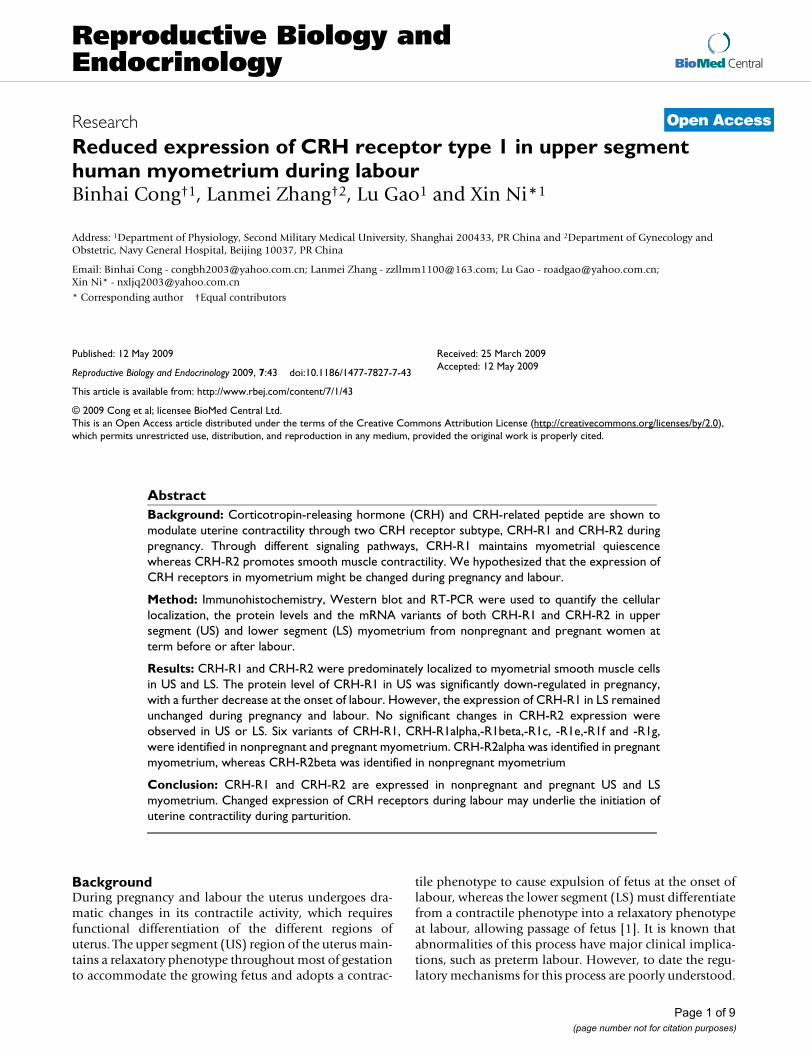
Reproductive Biology and Endocrinology
ss
Open AcceResearchReduced expression of CRH receptor type 1 in upper segment human myometrium during labourBinhai Cong†1, Lanmei Zhang†2, Lu Gao1 and Xin Ni*1Address: 1Department of Physiology, Second Military Medical University, Shanghai 200433, PR China and 2Department of Gynecology and Obstetric, Navy General Hospital, Beijing 10037, PR China
Email: Binhai Cong - [email protected]; Lanmei Zhang - [email protected]; Lu Gao - [email protected]; Xin Ni* - [email protected]
* Corresponding author †Equal contributors
AbstractBackground: Corticotropin-releasing hormone (CRH) and CRH-related peptide are shown tomodulate uterine contractility through two CRH receptor subtype, CRH-R1 and CRH-R2 duringpregnancy. Through different signaling pathways, CRH-R1 maintains myometrial quiescencewhereas CRH-R2 promotes smooth muscle contractility. We hypothesized that the expression ofCRH receptors in myometrium might be changed during pregnancy and labour.
Method: Immunohistochemistry, Western blot and RT-PCR were used to quantify the cellularlocalization, the protein levels and the mRNA variants of both CRH-R1 and CRH-R2 in uppersegment (US) and lower segment (LS) myometrium from nonpregnant and pregnant women atterm before or after labour.
Results: CRH-R1 and CRH-R2 were predominately localized to myometrial smooth muscle cellsin US and LS. The protein level of CRH-R1 in US was significantly down-regulated in pregnancy,with a further decrease at the onset of labour. However, the expression of CRH-R1 in LS remainedunchanged during pregnancy and labour. No significant changes in CRH-R2 expression wereobserved in US or LS. Six variants of CRH-R1, CRH-R1alpha,-R1beta,-R1c, -R1e,-R1f and -R1g,were identified in nonpregnant and pregnant myometrium. CRH-R2alpha was identified in pregnantmyometrium, whereas CRH-R2beta was identified in nonpregnant myometrium
Conclusion: CRH-R1 and CRH-R2 are expressed in nonpregnant and pregnant US and LSmyometrium. Changed expression of CRH receptors during labour may underlie the initiation ofuterine contractility during parturition.
BackgroundDuring pregnancy and labour the uterus undergoes dra-matic changes in its contractile activity, which requiresfunctional differentiation of the different regions ofuterus. The upper segment (US) region of the uterus main-tains a relaxatory phenotype throughout most of gestationto accommodate the growing fetus and adopts a contrac-
tile phenotype to cause expulsion of fetus at the onset oflabour, whereas the lower segment (LS) must differentiatefrom a contractile phenotype into a relaxatory phenotypeat labour, allowing passage of fetus [1]. It is known thatabnormalities of this process have major clinical implica-tions, such as preterm labour. However, to date the regu-latory mechanisms for this process are poorly understood.
Published: 12 May 2009
Reproductive Biology and Endocrinology 2009, 7:43 doi:10.1186/1477-7827-7-43
Received: 25 March 2009Accepted: 12 May 2009
This article is available from: http://www.rbej.com/content/7/1/43
© 2009 Cong et al; licensee BioMed Central Ltd. This is an Open Access article distributed under the terms of the Creative Commons Attribution License (http://creativecommons.org/licenses/by/2.0), which permits unrestricted use, distribution, and reproduction in any medium, provided the original work is properly cited.
Page 1 of 9(page number not for citation purposes)
Reproductive Biology and Endocrinology 2009, 7:43 http://www.rbej.com/content/7/1/43
An increasing body of evidence suggests that corticotro-pin-releasing hormone (CRH) is an important factor inthe regulation of human pregnancy and parturition [2-4].During human pregnancy, the placenta and fetal mem-branes produce large amounts of CRH [5,6]. Synthesis ofCRH in these tissues increases exponentially with advanc-ing gestation and, by term, it is present in high concentra-tions in the maternal and fetal blood [6-8]. Abnormalincreases or excessive levels of placental CRH are signifi-cant risk factors for an earlier onset of spontaneous birth[7,8]. Because of this, CRH has been proposed to regulatea placental clock that controls a cascade of physiologicalevents leading to parturition [7]. Although the precise bio-logical functions of CRH during pregnancy are not welldefined, it appears to exert a number of effects in intrau-terine tissues. It has been shown to stimulate prostaglan-din production in placenta and fetal membranes [9-11],and to enhance estrogen production whilst reduce proges-terone production in cultured placental trophoblasts[12,13]. A number of studies indicate that CRH isinvolved in the regulation of myometrial contractility dur-ing pregnancy [14,15].
It has been demonstrated that CRH belongs to a family ofpeptides that includes urocortin I (UCNI), urocortin II(UCNII) and urocortin III (UCNIII) as well as fish uro-tensin I and frog peptide sauvagine [16]. UCNI, UCNIIand UCNIII have also been identified in human intrauter-ine tissues including placenta, fetal membrane and myo-metrium throughout gestation [15,17,18]. All the CRHfamily peptides exert their effects through two subtypes ofCRH receptors, termed CRH-R1 and CRH-R2. Thesereceptors shared 70% homology at amino acid level[19,20]. Several splice variants of the mRNA for CRH-R1and CRH-R2 have been found, eight variants for CRH-R1and three variants for CRH-R2 [20-22]. It has been shownthat both CRH-R1 and CRH-R2 are expressed in humannonpregnant and pregnant myometrium [23-26]. Currentevidence suggests that CRH-R1 and CRH-R2 activationexert distinct actions in the regulation of myometrial con-tractility during pregnancy. CRH-R1 activation up-regu-lates the expression of constitutive form of nitric oxidesynthase, thereby promoting myometrium quiescence[27]. In contrast, CRH-R2 activation can activate ERK1/2and RhoA pathways that actively promote myometrialcontractility [28].
Our hypothesis is that differential expression of CRH-Rsmay be important for regulating the contractile activity ofuterus in the US and LS during pregnancy and labour.However, studies regarding CRH-R1 and CRH-R2 expres-sion in human pregnant myometrium have so far beenlimited to LS region [23-26]. To date there is a lack ofinformation about the expression of CRH-Rs within theUS of human myometrium during pregnancy and labour.
The purpose of this study was to determine whether therewere any changes in the expression of CRH-Rs in humanUS and LS myometrium during pregnancy and parturi-tion. We therefore determined the protein levels of CRH-R1 and CRH-R2 in the US and LS by Western blot analysisand showed the localization of CRH-Rs in US and LSmyometrium by immunohistochemistry. In addition, themRNA splicing variants of CRH-R1 and CRH-R2 were alsoidentified by RT-PCR.
MethodsTissue collectionPaired upper and lower uterine segmental myometrial tis-sues from pregnant and nonpregnant women were col-lected in Navy General Hospital, the teaching hospital ofSecond Military Medical University, Beijing, China.Approval of this study was granted by human ethic com-mittee of Navy General Hospital as well as human ethiccommittee of Second Military Medical University. Eachpatient signed approved informed consent form to partic-ipate in this study. Nonpregnant (NP) myometrium tis-sues were obtained from normal cycling patients (n = 8)undergoing hysterectomy for fibroids.
Pregnant myometrial tissues were collected at cesareansection from pregnant women at term gestation and weredivided into two groups: (1) term no labour (TNL, 37–41weeks, n = 10), (2) term labour (TL, 37–41 weeks, n = 9).Labour was defined as regular contractions (<5 min apart)plus membrane rupture and cervical dilation (>3 cm)with no augmentation. Indications for cesarean sectionincluded breech presentation, placenta previa, previouscesarean section, cephalopelvic disproportion, failure oflabour to progress, fetal distress, or maternal request.None of the women included in this study had evidenceof underlying disease (e.g. hypertension, diabetes, preec-lampsia, intrauterine growth restriction, etc.). LS uterinesamples were collected from the upper margin of the LSuterine incision, while US uterine samples were takenfrom just below fundus. The nonpregnant tissues weretaken from the normal part of uterus without fibrosis con-tamination. Collected samples were placed in phosphate-buffered saline (PBS) on ice and transported to the labo-ratory. Myometrial tissues were separated from any sero-sal or decidual components and then rinsed in PBS,frozen immediately in liquid nitrogen and stored at -80 Cfor Western blot and PCR analysis. Parts of the tissueswere also placed in 10% phosphate buffered formalin forimmunohistochemical analysis.
ImmunohistochemistryParaffin sections (5 μm) were cut, rehydrated and micro-waved in citric acid buffer to retrieve antigens. After inhi-bition of endogenous peroxidases with 3% H2O2,nonspecific antibody binding was blocked with 10% rab-
Page 2 of 9(page number not for citation purposes)
Reproductive Biology and Endocrinology 2009, 7:43 http://www.rbej.com/content/7/1/43
bit serum for 30 min. Serial sections were then incubatedwith specific antibodies against human CRH-R1 (sc-12381, Santa Cruz Biotechnology, Santa Cruz, California,USA) and CRH-R2 (sc-20550, Santa Cruz) (1:500) over-night at 4°C. The CRH-R1 antibody is directed against anepitope between amino acid positions 81 and 109 ofCRH-R1 of human origin, where no sequence homologyexists to CRH-R2. The CRH-R2 antibody was raisedagainst a peptide mapping near the C-terminus of CRH-R2 of human origin. Mouse antihuman smooth muscle α-actin-specific monoclonal antibody (Dako Inc. Carpinte-ria, California) was used to stained the muscle cells. Thebound antibodies were detected with the biotin-streptavi-din-peroxidase system (UltraSensitive-SP-kit, MaiXin Bio-technology, Fuzhou, China) and diaminobenzidine(Sigma) was used as chromogen. Counterstaining wasperformed with hemalum. Negative controls were per-formed by substituting primary antibody with a normalserum or IgG in same dilution. To confirm the specificityof primary antibody, preabsorption of the primary anti-body with a tenfold excess of the blocking peptides sc-12381P or sc-20550P (Santa Cruz) was performed.
Western blot analysisApproximately 70 mg of human myometrial tissue washomogenized in ice-cold lysis buffer consisting of 60 mMTris-HCl, 2% sodium dodecyl sulfate (SDS), 10% sucrose,2 mM phenylmethylsulfonyl fluoride (Merck, Darmstadt,Germany), 1 mM sodium orthovanadate (Sigma-Aldrich),10 μg/ml aprotinin (Bayer, Leverkusen, Germany).Lysates were then quickly sonified in ice bath, boiled 5min at 95 C, and stored at -80 C until used. Protein con-centrations were measured using a modified Bradfordassay and samples were diluted in sample buffer (250 mMTris-HCl (pH 6.8), containing 4% SDS, 10% glycerol, 2%β-mercaptoethanol, and 0.002% bromophenol blue) andboiled for a further 5 min. Samples (50 μg) were separatedon an SDS-8% polyacrylamide gel, and the proteins wereelectrophoretically transferred to a nitrocellulose mem-brane at 300 mA for 1.5 h in a transfer buffer containing20 mM Tris, 150 mM glycine, and 20% methanol. Themembrane was then blocked in TBS containing 0.1%Tween-20(TBST) and 5% dried milk powder (wt/vol) for2 h at room temperature. After three washes with TBST,the nitrocellulose membranes were incubated with pri-mary antibody for CRH-R1 or CRH-R2 (1:500) at 4°Covernight. After another three washes with TBST, themembranes were incubated with a secondary horseradishperoxidase-conjugated IgG (1:1000) for 1 h at room tem-perature and further washed for 30 min with TBST. Immu-noreactive proteins were visualized using the enhancedchemiluminescence Western blotting detection system(Santa Cruz). The light-emitting bands were detected withX-ray film. The resulting band intensities were quantitatedusing an image scanning densitometer (Furi Technology,
Shanghai, China). To control sampling errors, the ratio ofband intensities to β-actin was obtained to quantify therelative protein expression level.
Total RNA extraction and cDNA synthesisTotal RNA was extracted from the samples of myo-metrium by a method based on Chomczynski and Sacchi[29]. Briefly, frozen tissue samples were powdered underliquid nitrogen and homogenized in 1 ml of TRIzol rea-gent (Invitrogen, Grand Island, New York, USA). Subse-quent extraction of total RNA from the tissues wasconducted according to the protocol provided by themanufacturer. All RNA samples were treated with deoxyri-bonuclease I (Promega, Biotech. Co Ltd, Beijing, China).The purity and integrity of the RNA was checked spectro-scopically and by gel electrophoresis. Two microgram oftotal RNA was reverse transcribed using the SuperScript®
first-strand synthesis system (Invitrogen) and stored at -20°C.
PCR and nested PCR for CRH-R1 and -R2 variantsThe specific primers for the amplification of the CRH-R2variants and CRH-R1α/CRH-R1β were used as describedpreviously [22,23,30]. The nucleotide sequences of theprimers were as followings: (1) CRH-R2α, sense 5'-GAGCTGCTCTTGGACGGC-3', antisense 5'-GACAAG-GGCGATGCGGTA-3'; (2) CRH-R2β, sense 5'-CCCTCAC-CAACCTCTCAGGTCC-3', antisense 5'-CAGGTCATACTTCCTCTGCTTGTC-3'; (3) CRH-R2γ,sense 5'-CTCAAGCAATCTGCCTACCT-3', antisense 5'-GGCTCACACTGTGAGTAGTT-3'; (4) CRHR1 α/β, sense5'-GGCAGCTAGTGGTTCGGCC-3', antisense 5'-TCG-CAGGCACCGGATGCTC-3'. PCR reaction solution con-sisted of 2.0 μl diluted cDNA, 0.4 μmol/L of each pairedprimers, 2.5 mmol/L Mg2+, 250 μmol/L deoxynucleotidetriphosphates, 1 U Taq DNA polymerase (Qiagen, Beijing,China), and 1× PCR buffer. PCR reaction was set at 94 C(45 s), 58 C (45 s), 72 C (1 min) in a total 40–80 cycleswith a final extension step at 72 C for 10 min.
Primers for nested PCR were previously described [21-23],and were designed to distinguish the different variants ofCRH-R1. The nucleotide sequences of the first roundprimers were: exons 2–7, sense 5'-TCCGTCTCGTCAAG-GCCCTTC-3', antisense 5'-GGCTCATGGTTAGCTGGAC-CAC-3'; exons 9–14, sense 5'-CCATTGGGAAGCTGTACTACGAC-3', antisense 5'-GCTT-GATGCTGTGAAAGCTGACAC-3'. The nucleotidesequences of the second round primers were: exons 2–7,sense 5'-TGTCCCTGGCCAGCAACATCTC-3', antisense 5'-AGTGGATGATGTTTCGCAGGCAC-3';exons 9–14, sense5'-GGGTGTACACCGACTACATCTAC-3', antisense 5'-TCTTCCGGATGGCAGAACGGAC-3'. RT-products(cDNA) from tissues were used as template for the fistround of PCR. After 40 cycles of amplification, 2 μL of the
Page 3 of 9(page number not for citation purposes)
Reproductive Biology and Endocrinology 2009, 7:43 http://www.rbej.com/content/7/1/43
reaction mixture was used for additional 40-cycle amplifi-cation. The primers used in this second round of amplifi-cation were internal to the first set of primers.
Ten microliters of the reaction mixture were subsequentlyelectrophoresed on a 1.5% agarose gel and visualized byethidium bromide, using a 100 bp DNA ladder (Invitro-gen) to estimate the band sizes. As a negative control forall of the reactions, distilled water was used in place ofcDNA. The identity of the PCR products was confirmed bysequencing. Sequence data were analyzed using Blastnucleic acid database searches from the National Centrefor Biotechnology Information (NCBI).
StatisticsProtein expression levels of CRH-R1 and CRH-R2 weredetermined by densitometric analysis (Furi Technology,Shanghai, China). Peak count values were expressed asdensitometric units. The data are presented as mean ±SEM. All data were tested for homogeneity of variance byBartlett's test. The results indicated that the data were nor-mally distributed. Student's t-test was used in the analysisof these data. One-way ANOVA with Student-Newman-Keuls was used for multiple comparisons. A P value < 0.05was considered statistically significant.
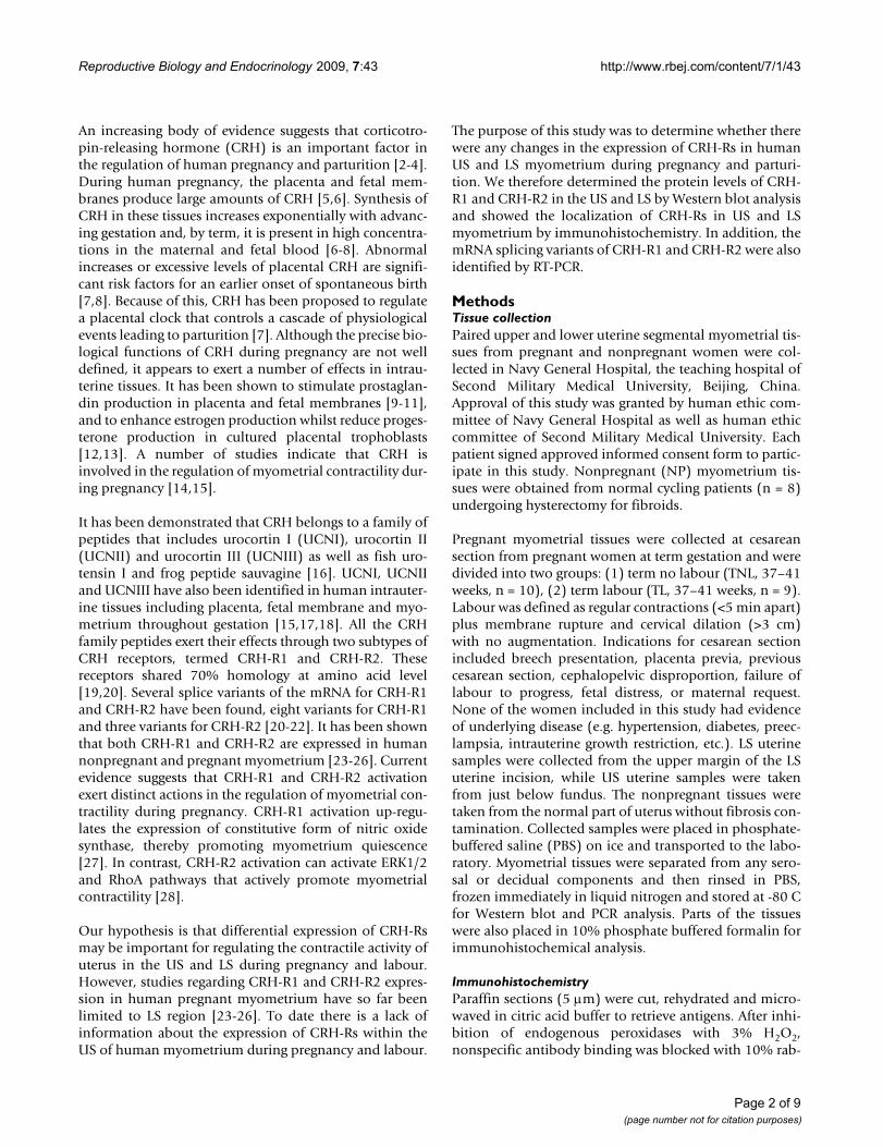
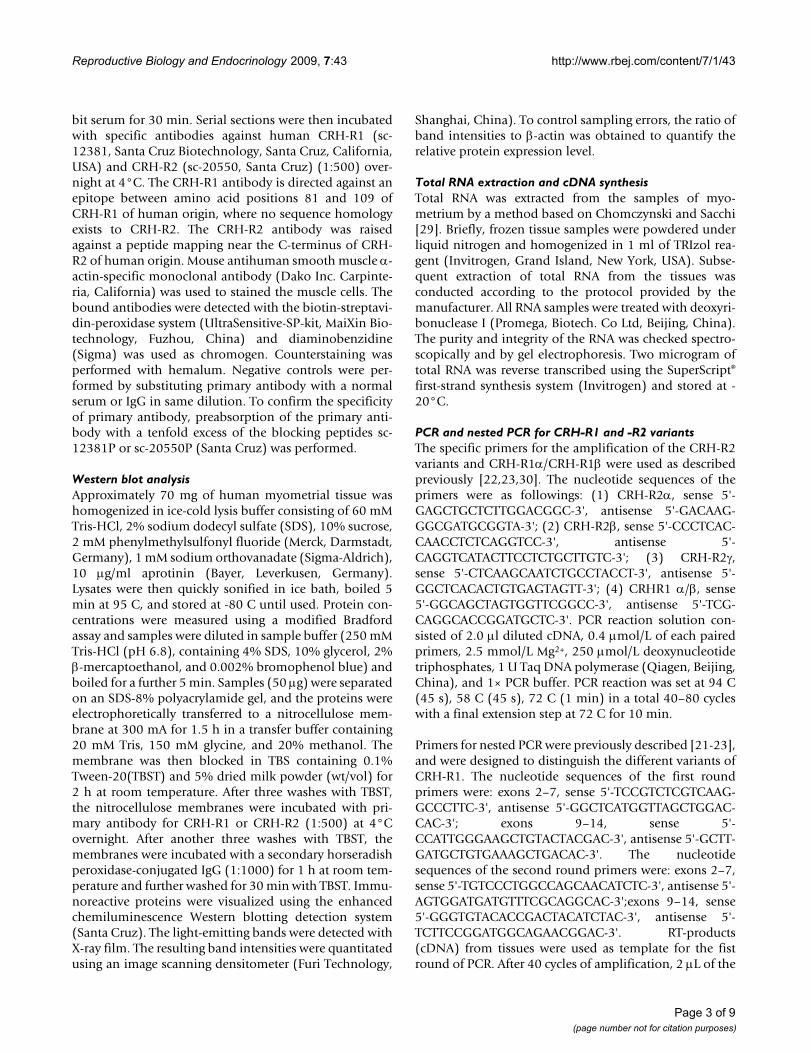
ResultsLocalization of CRH-R1 and CRH-R2 in non-pregnant and pregnant human myometriumBoth CRH-R1 and CRH-R2 were identified in nonpreg-nant (Fig. 1) and pregnant (Fig. 2) myometrium. Localiza-tion of CRH-R1 and CRH-R2 showed that these receptorswere highly expressed in the myometrial smooth musclein US and LS (Fig. 1A–C and Fig. 2A, B and 2D–F arrow-head). Positive staining of CRH-R1 and CRH-R2 was alsoseen in vascular smooth muscle cells (Fig. 1D and Fig. 2Carrowheads). Staining in the US and LS was similar ineach of groups and no dramatic changes in overall stain-ing intensity or localization were observed with preg-nancy or with labour (Fig. 2). CRH-R1 and CRH-R2staining was eliminated when antibody was pre-absorbedby synthetic peptide (Fig. 1E and 1F, respectively).
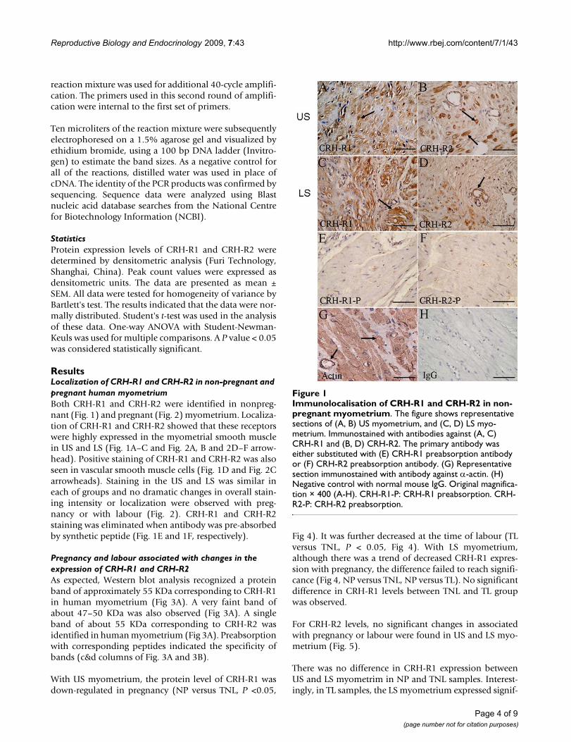
Pregnancy and labour associated with changes in the expression of CRH-R1 and CRH-R2As expected, Western blot analysis recognized a proteinband of approximately 55 KDa corresponding to CRH-R1in human myometrium (Fig 3A). A very faint band ofabout 47–50 KDa was also observed (Fig 3A). A singleband of about 55 KDa corresponding to CRH-R2 wasidentified in human myometrium (Fig 3A). Preabsorptionwith corresponding peptides indicated the specificity ofbands (c&d columns of Fig. 3A and 3B).
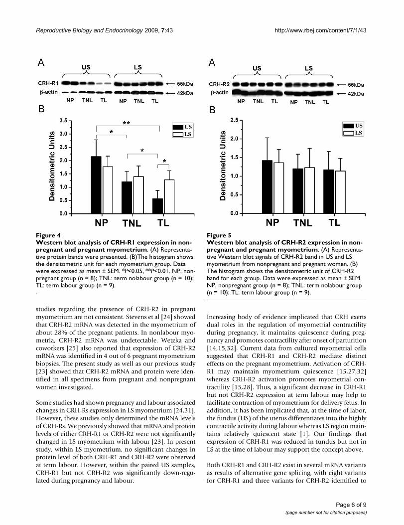
With US myometrium, the protein level of CRH-R1 wasdown-regulated in pregnancy (NP versus TNL, P <0.05,
Fig 4). It was further decreased at the time of labour (TLversus TNL, P < 0.05, Fig 4). With LS myometrium,although there was a trend of decreased CRH-R1 expres-sion with pregnancy, the difference failed to reach signifi-cance (Fig 4, NP versus TNL, NP versus TL). No significantdifference in CRH-R1 levels between TNL and TL groupwas observed.
For CRH-R2 levels, no significant changes in associatedwith pregnancy or labour were found in US and LS myo-metrium (Fig. 5).
There was no difference in CRH-R1 expression betweenUS and LS myometrim in NP and TNL samples. Interest-ingly, in TL samples, the LS myometrium expressed signif-
Immunolocalisation of CRH-R1 and CRH-R2 in nonpregnant myometriumFigure 1Immunolocalisation of CRH-R1 and CRH-R2 in non-pregnant myometrium. The figure shows representative sections of (A, B) US myometrium, and (C, D) LS myo-metrium. Immunostained with antibodies against (A, C) CRH-R1 and (B, D) CRH-R2. The primary antibody was either substituted with (E) CRH-R1 preabsorption antibody or (F) CRH-R2 preabsorption antibody. (G) Representative section immunostained with antibody against α-actin. (H) Negative control with normal mouse IgG. Original magnifica-tion × 400 (A-H). CRH-R1-P: CRH-R1 preabsorption. CRH-R2-P: CRH-R2 preabsorption.
Page 4 of 9(page number not for citation purposes)
Reproductive Biology and Endocrinology 2009, 7:43 http://www.rbej.com/content/7/1/43
icantly more CRH-R1 when compared with US, in partdue to the significant reduction in CRH-R1 expression inthe US at the labour onset (Fig 4). No significant differ-ence in CRH-R2 expression between US and LS myomet-rim was found across all groups (Fig 5).
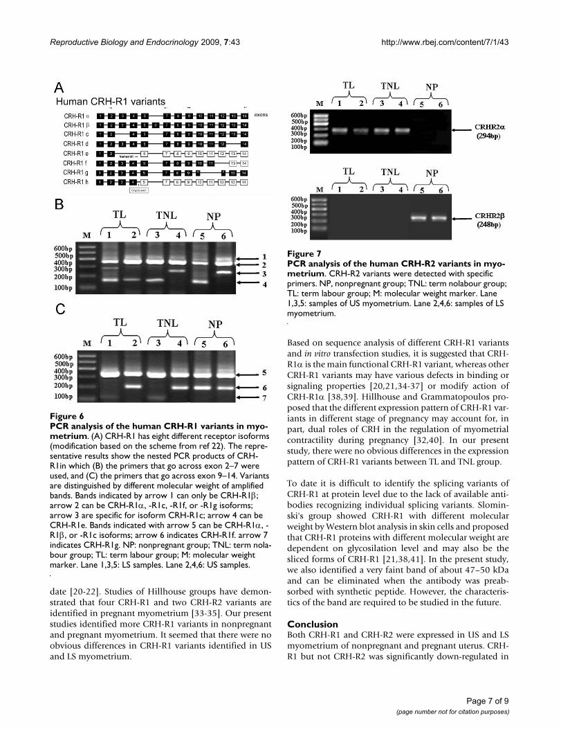
CRH-R1 and CRH-R2 variants in nonpregnant and pregnant myometriumRT-PCR and nested PCR analysis showed that CRH-R1α,-R1β,-R1c, -R1e,-R1f and -R1g were identified in nonpreg-nant as well as pregnant US and LS samples (Fig 6B and6C). Other CRH-R1 variants were undetected. CRH-R1αand -R1β were detected in all biopsies from pregnant andnonpregnant women, whereas CRH-R1c, -R1e,-R1f and -R1g were not detected in all biopsies. The detection ratesof CRH-R1c, -R1e,-R1f and -R1g was various in NP, TNL
and TL groups. For instance, within US, CRH-R1c wasidentified in 1/9TL and 3/10 TNL samples. CRH-R1e wasdetected in 2/9 TL and 3/10 TNL biopsies. CRH-R1f and -R1g were detected in all TL tissues and 4/10 TNL tissues.Within LS, 3/10 TNL and 4/9 TL tissues for CRH-R1c, 3/10TNL and 5/9 TL tissues for CRH-R1e, 4/10 TNL and 6/9 TLsamples for CRH-R1f, and 3/10 TNL and 5/9 TL tissues forCRH-R1g were identified.
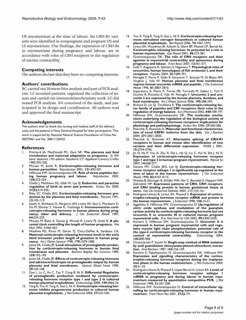
CRH-R2 has three variants, termed CRH-R2α, CRH-R2βand CRH-R2γ [20]. PCR analysis using specific primers forthese CRH-R2 variants resulted in the detection of CRH-R2α in all of the pregnant myometrial biopsies and CRH-R2β in all of nonpregnant tissues (Fig 7). We are unable todetected CRH-R2γ (data not shown).
DiscussionIn the present study, we demonstrated that, for the firsttime, CRH-R1 and CRH-R2 expression in paired US andLS samples from pregnant women who were undergoinglabour and not undergoing labour. We found that CRH-R1 is significantly reduced during labour, however, thisdecrease appeared to be restricted to the US region ofuterus.
The presence of CRH-R1 in human myometrium is inaccordance with published data [23-26,31]. However, the
Immunohistochemistry analysis of CRH receptors in myo-metrium from term labour (TL) and term nolabour (TNL) patientsFigure 2Immunohistochemistry analysis of CRH receptors in myometrium from term labour (TL) and term nola-bour (TNL) patients. The figure shows representative sec-tions of (A, C) US and (E, G) LS myometrium in TNL group, as well as (B, D) US and (F, H) LS myometrium in TL group. (A, B, E, F) Immunostained with CRH-R1 antibody. (C, D, G, H) Immunostained with CRH-R2 antibody. Original magnifi-cation × 400 (A-H).
Distribution of CRH-R1 and CRH-R2 in human myometrium by Western blot analysisFigure 3Distribution of CRH-R1 and CRH-R2 in human myo-metrium by Western blot analysis. (A) Western blot analysis of CRH-R1. a&b, immunoblotting using CRH-R1 anti-body; c&d, preabsorption blots. (B) Western blot analysis of CRH-2. a&b, immunoblotting using CRH-R2 antibody; c&d, preabsorption blots.
Page 5 of 9(page number not for citation purposes)
Reproductive Biology and Endocrinology 2009, 7:43 http://www.rbej.com/content/7/1/43
studies regarding the presence of CRH-R2 in pregnantmyometrium are not consistent. Stevens et al [24] showedthat CRH-R2 mRNA was detected in the myometrium ofabout 28% of the pregnant patients. In nonlabour myo-metria, CRH-R2 mRNA was undetectable. Wetzka andcoworkers [25] also reported that expression of CRH-R2mRNA was identified in 4 out of 6 pregnant myometriumbiopsies. The present study as well as our previous study[23] showed that CRH-R2 mRNA and protein were iden-tified in all specimens from pregnant and nonpregnantwomen investigated.
Some studies had shown pregnancy and labour associatedchanges in CRH-Rs expression in LS myometrium [24,31].However, these studies only determined the mRNA levelsof CRH-Rs. We previously showed that mRNA and proteinlevels of either CRH-R1 or CRH-R2 were not significantlychanged in LS myometrium with labour [23]. In presentstudy, within LS myometrium, no significant changes inprotein level of both CRH-R1 and CRH-R2 were observedat term labour. However, within the paired US samples,CRH-R1 but not CRH-R2 was significantly down-regu-lated during pregnancy and labour.
Increasing body of evidence implicated that CRH exertsdual roles in the regulation of myometrial contractilityduring pregnancy, it maintains quiescence during preg-nancy and promotes contractility after onset of parturition[14,15,32]. Current data from cultured myometrial cellssuggested that CRH-R1 and CRH-R2 mediate distincteffects on the pregnant myometrium. Activation of CRH-R1 may maintain myometrium quiescence [15,27,32]whereas CRH-R2 activation promotes myometrial con-tractility [15,28]. Thus, a significant decrease in CRH-R1but not CRH-R2 expression at term labour may help tofacilitate contraction of myometrium for delivery fetus. Inaddition, it has been implicated that, at the time of labor,the fundus (US) of the uterus differentiates into the highlycontractile activity during labour whereas LS region main-tains relatively quiescent state [1]. Our findings thatexpression of CRH-R1 was reduced in fundus but not inLS at the time of labour may support the concept above.
Both CRH-R1 and CRH-R2 exist in several mRNA variantsas results of alternative gene splicing, with eight variantsfor CRH-R1 and three variants for CRH-R2 identified to
Western blot analysis of CRH-R1 expression in nonpregnant and pregnant myometriumFigure 4Western blot analysis of CRH-R1 expression in non-pregnant and pregnant myometrium. (A) Representa-tive protein bands were presented. (B)The histogram shows the densitometric unit for each myometrium group. Data were expressed as mean ± SEM. *P<0.05, **P<0.01. NP, non-pregnant group (n = 8); TNL: term nolabour group (n = 10); TL: term labour group (n = 9).
Western blot analysis of CRH-R2 expression in nonpregnant and pregnant myometriumFigure 5Western blot analysis of CRH-R2 expression in non-pregnant and pregnant myometrium. (A) Representa-tive Western blot signals of CRH-R2 band in US and LS myometrium from nonpregnant and pregnant women. (B) The histogram shows the densitometric unit of CRH-R2 band for each group. Data were expressed as mean ± SEM. NP, nonpregnant group (n = 8); TNL: term nolabour group (n = 10); TL: term labour group (n = 9).
Page 6 of 9(page number not for citation purposes)
Reproductive Biology and Endocrinology 2009, 7:43 http://www.rbej.com/content/7/1/43
date [20-22]. Studies of Hillhouse groups have demon-strated that four CRH-R1 and two CRH-R2 variants areidentified in pregnant myometrium [33-35]. Our presentstudies identified more CRH-R1 variants in nonpregnantand pregnant myometrium. It seemed that there were noobvious differences in CRH-R1 variants identified in USand LS myometrium.
Based on sequence analysis of different CRH-R1 variantsand in vitro transfection studies, it is suggested that CRH-R1α is the main functional CRH-R1 variant, whereas otherCRH-R1 variants may have various defects in binding orsignaling properties [20,21,34-37] or modify action ofCRH-R1α [38,39]. Hillhouse and Grammatopoulos pro-posed that the different expression pattern of CRH-R1 var-iants in different stage of pregnancy may account for, inpart, dual roles of CRH in the regulation of myometrialcontractility during pregnancy [32,40]. In our presentstudy, there were no obvious differences in the expressionpattern of CRH-R1 variants between TL and TNL group.
To date it is difficult to identify the splicing variants ofCRH-R1 at protein level due to the lack of available anti-bodies recognizing individual splicing variants. Slomin-ski's group showed CRH-R1 with different molecularweight by Western blot analysis in skin cells and proposedthat CRH-R1 proteins with different molecular weight aredependent on glycosilation level and may also be thesliced forms of CRH-R1 [21,38,41]. In the present study,we also identified a very faint band of about 47–50 kDaand can be eliminated when the antibody was preab-sorbed with synthetic peptide. However, the characteris-tics of the band are required to be studied in the future.
ConclusionBoth CRH-R1 and CRH-R2 were expressed in US and LSmyometrium of nonpregnant and pregnant uterus. CRH-R1 but not CRH-R2 was significantly down-regulated in
PCR analysis of the human CRH-R1 variants in myometriumFigure 6PCR analysis of the human CRH-R1 variants in myo-metrium. (A) CRH-R1 has eight different receptor isoforms (modification based on the scheme from ref 22). The repre-sentative results show the nested PCR products of CRH-R1in which (B) the primers that go across exon 2–7 were used, and (C) the primers that go across exon 9–14. Variants are distinguished by different molecular weight of amplified bands. Bands indicated by arrow 1 can only be CRH-R1β; arrow 2 can be CRH-R1α, -R1c, -R1f, or -R1g isoforms; arrow 3 are specific for isoform CRH-R1c; arrow 4 can be CRH-R1e. Bands indicated with arrow 5 can be CRH-R1α, -R1β, or -R1c isoforms; arrow 6 indicates CRH-R1f. arrow 7 indicates CRH-R1g. NP: nonpregnant group; TNL: term nola-bour group; TL: term labour group; M: molecular weight marker. Lane 1,3,5: LS samples. Lane 2,4,6: US samples.
PCR analysis of the human CRH-R2 variants in myometriumFigure 7PCR analysis of the human CRH-R2 variants in myo-metrium. CRH-R2 variants were detected with specific primers. NP, nonpregnant group; TNL: term nolabour group; TL: term labour group; M: molecular weight marker. Lane 1,3,5: samples of US myometrium. Lane 2,4,6: samples of LS myometrium.
Page 7 of 9(page number not for citation purposes)
Reproductive Biology and Endocrinology 2009, 7:43 http://www.rbej.com/content/7/1/43
US myometrium at the time of labour. Six CRH-R1 vari-ants were identified in nonpregnant and pregnant US andLS myometrium. Our findings, the expression of CRH-Rsin myometrium during pregnancy and labour, are inaccordance with roles of CRH receptors in the regulationof uterine contractility.
Competing interestsThe authors declare that they have no competing interests.
Authors' contributionsBC carried out Western blot analysis and part of PCR anal-ysis. LZ recruited patients, organized the collection of tis-sues and carried out immunohistochemistry work. LG didnested PCR analysis. XN conceived of the study, and par-ticipated in its design and coordination. All authors readand approved the final manuscript.
AcknowledgementsThe authors wish to thank the nursing and medical staff of the delivery suite, and the patients in Navy General Hospital for their participation. This work is supported by National Natural Science Foundation of China No. 30575961 and No. 30811120433.
References1. Pritchard JA, MacDonald PC, Gant NF: The placenta and fetal
membranes and maternal adaptation to pregnancy. In Wil-liams' obstetrics 17th edition. Stamford, CT: Appleton Century Crofts;1985:295-320.
2. McLean M, Smith R: Corticotrophin-releasing hormone andhuman parturition. Reproduction 2001, 121:493-501.
3. Hillhouse EW, Grammatopoulos DK: Role of stress peptides dur-ing human pregnancy and labour. Reproduction 2002,124:323-329.
4. Challis J, Matthews SG, Gibb W, Lye SJ: Endocrine and paracrineregulation of birth at term and preterm. Endocr Rev 2000,21(5):514-550.
5. Riley SC, Challis JRG: Corticotorphin-releasing hormone pro-duction by the placenta and fetal membranes. Placenta 1991,12:105-119.
6. Sasaki A, Shinkawa O, Margioris AN, Liotta AS, Sato S, Murakami D,Go M, Shimizu Y, Hanew K, Yoshinaga K: Immunoreactive corti-cotropin-releasing hormone in human plasma during preg-nancy, labor and delivery. J Clin Endocrinol Metab 1987,64:224-229.
7. McLean M, Bistis A, Davies JJ, Woods R, Lowry PJ, Smith R: A pla-cental clock controlling the length of human pregnancy. NatMed 1995, 1:460-463.
8. Wadhwa PD, Porto M, Garite TJ, Chicz-DeMet A, Sandman CA:Maternal corticotropin-releasing hormone levels in the earlythird trimester predict length of gestation in human preg-nancy. Am J Obstet Gynecol 1998, 179:1079-1085.
9. Jones SA, Challis JR: Local stimulation of prostaglandin produc-tion by corticotropin-releasing hormone in human fetalmembranes and placenta. Biochem Biophys Res Commun 1989,159:192-199.
10. Jones SA, Challis JR: Effects of corticotropin-releasing hormoneand adrenocorticotropin on prostaglandin output by humanplacenta and fetal membranes. Gynecol Obstet Invest 1990,29:165-168.
11. Gao L, Lu C, Xu C, Tao Y, Cong B, Ni X: Differential Regulationof prostaglandin production mediated by corticotropin-releasing hormone receptor type 1 and type 2 in culturedhuman placental trophoblasts. Endocrinology 2008, 149:2866-76.
12. Yang R, You X, Tang X, Gao L, Ni X: Corticotropin-releasing hor-mone inhibits progesterone production in cultured humanplacental trophoblasts. J Mol Endocrinol 2006, 37:533-540.
13. You X, Yang R, Tang X, Gao L, Ni X: Corticotropin-releasing hor-mone stimulates estrogen biosynthesis in cultured humanplacental trophoblasts. Biol Reprod 2006, 74:1067-1072.
14. Linton EA, Woodman JR, Asboth G, Glynn BP, Plested CP, Bernal AL:Corticotrophin releasing hormone: its potential for a role inhuman myometrium. Exp Physiol 2001, 86:273-281.
15. Grammatopoulos DK: The role of CRH receptors and theiragonists in myometrial contractility and quiescence duringpregnancy and labour. Front Biosci 2007, 12:561-571.
16. Suda T, Kageyama K, Sakihara S, Nigawara T: Physiological roles ofurocortins, human homologues of fish urotensin I, and theirreceptors. Peptides 2004, 25:1689-701.
17. Petraglia F, Florio P, Gallo R, Simoncini T, Saviozzi M, Di Blasio AM,Vaughan J, Vale W: Human placenta and fetal membranesexpress human urocortin mRNA and peptide. J Clin EndocrinolMetab 1996, 81:3807-3810.
18. Imperatore A, Florio P, Torres PB, Torricelli M, Galleri L, Toti P,Occhini R, Picciolini E, Vale W, Petraglia F: Urocortin 2 and uro-cortin 3 are expressed by the human placenta, deciduas, andfetal membranes. Am J Obstet Gynecol 2006, 195:288-295.
19. Richard D, Lin Q, Timofeeva E: The corticotropin-releasing fac-tor family of peptides and CRF receptors: their roles in theregulation of energy balance. Eur J Pharmacol 2002, 440:189-197.
20. Hillhouse EW, Grammatopoulos DK: The molecular mecha-nisms underlying the regulation of the biological activity ofcorticotropin-releasing hormone receptors: implications forphysiology and pathophysiology. Endocr Rev 2006, 27:260-286.
21. Pisarchik A, Slominski A: Molecular and functional characteriza-tion of novel CRFR1 isoforms from the skin. Eur J Biochem2004, 271:2821-2830.
22. Pisarchik A, Slominski AT: Alternative splicing of CRH-R1receptors in human and mouse skin: identification of newvariants and their differential expression. FASEB J 2001,15:2754-2756.
23. Jin D, He P, You X, Zhu X, Dai L, He Q, Liu C, Hui N, Sha J, Ni X:Expression of corticotropin-releasing hormone receptortype 1 and type 2 in human pregnant myometrium. Reprod Sci2007, 14:568-577.
24. Stevens MY, Challis JRG, Lye SJ: Corticotropin-releasing hor-mone receptor subtype 1 is significantly up-regulated at thetime of labor in the human myometrium. J Clin EndocrinolMetab 1998, 83:4107-4115.
25. Wetzka B, Sehringer B, Schäfer WR, Hör C, Benedek E, Deppert WR,Zahradnik HP: Expression patterns of CRH, CRH receptors,and CRH binding protein in human gestational tissue atterm. Exp Clin Endocrinol Diabetes 2003, 111:154-161.
26. Rodríguez-Liñares B, Linton EA, Phaneuf S: Expression of cortico-trophin-releasing hormone receptor mRNA and protein inthe human myometrium. J Endocrinol 1998, 156:15-21.
27. Aggelidou E, Hillhouse EW, Grammatopoulos D: Up-regulation ofnitric oxide synthase and modulation of the guanylatecyclase activity by corticotrophin-releasing hormone but noturocortin II or urocortin III in cultured human pregnantmyometrial cells. Proc Natl Acad Sci USA 2002, 99:3300-3305.
28. Karteris E, Hillhouse EW, Grammatopoulos D: Urocortin II isexpressed in human pregnant myometrial cells and regu-lates myosin light chain phosphorylation: potential role ofthe type-2 corticotropin-releasing hormone receptor in thecontrol of myometrial contractility. Endocrinology 2004,145:890-900.
29. Chomczynski P, Sacchi N: Single-step method of RNA isolationby acid guanidinium thiocyanate-phenol-chloroform extrac-tion. Anal Biochem 1987, 162:156-159.
30. Karteris E, Papadopoulou N, Grammatopoulos DK, Hillhouse EW:Expression and signalling characteristics of the cortico-trophin-releasing hormone receptors during the implanta-tion phase in the human endometrium. J Mol Endocrinol 2004,32:21-32.
31. Rodríguez-Liñares B, Phaneuf S, López Bernal A, Linton EA: Levels ofcorticotrophin-releasing hormone receptor subtype 1mRNA in pregnancy and during labour in human myo-metrium measured by quantitative competitive PCR. J MolEndocrinol 1998, 21:201-208.
32. Hillhouse EW, Grammatopoulos D: Control of intracellular sig-nalling by corticotropin-releasing hormone in human myo-metrium. Front Horm Res 2001, 27:66-74.
Page 8 of 9(page number not for citation purposes)
Reproductive Biology and Endocrinology 2009, 7:43 http://www.rbej.com/content/7/1/43
Publish with BioMed Central and every scientist can read your work free of charge
"BioMed Central will be the most significant development for disseminating the results of biomedical research in our lifetime."
Sir Paul Nurse, Cancer Research UK
Your research papers will be:
available free of charge to the entire biomedical community
peer reviewed and published immediately upon acceptance
cited in PubMed and archived on PubMed Central
yours — you keep the copyright
Submit your manuscript here:http://www.biomedcentral.com/info/publishing_adv.asp
BioMedcentral
33. Grammatopoulos D, Dai Y, Chen J, Karteris E, apadopoulou N, Eas-ton AJ, Hillhouse EW: Human corticotripin-releasing hormonereceptor: Difference in subtype expression between preg-nant and nonpregnant myometria. J Clin Endocrinol Metab 1998,83:2539-2544.
34. Grammatopoulos D, Dai Y, Randeva HS, Levine MA, Karteris E, Eas-ton AJ, Hillhouse EW: A novel spliced variant of the type 1 cor-ticotropin-releasing hormone receptor with a deletion in theseventh transmembrane domain present in the human preg-nant term myometrium and fetal membranes. Mol Endocrinol1999, 13:2189-2202.
35. Markovic D, Papadopoulou N, Teli T, Randeva H, Levine MA, Hill-house EW, Grammatopoulos DK: Differential responses of corti-cotropin-releasing hormone receptor type 1 variants toprotein kinase C phosphorylation. J Pharmacol Exp Ther 2006,319:1032-1042.
36. Nabhan C, Xiong Y, Xie LY, Abou-Samra AB: The alternativelyspliced type II corticotropin-releasing factor receptor, stablyexpressed in LLCPK-1 cells, is not well coupled to the G pro-tein(s). Biochem Biophys Res Commun 1995, 212:1015-1021.
37. Wille S, Sydow S, Palchaudhuri MR, Spiess J, Dautzenberg FM: Iden-tification of amino acids in the N-terminal domain of corti-cotropin-releasing factor receptor 1 that are importantdeterminants of high-affinity ligand binding. J Neurochem 1999,72:388-395.
38. Zmijewski MA, Slominski AT: RF1 receptor splicing in epidermalkeratinocytes: potential biological role and environmentalregulations. J Cell Physiol 2009, 218:C593-602.
39. Slominski A, Zbytek B, Zmijewski M, Slominski RM, Kauser S, Worts-man J, Tobin DJ: Corticotropin releasing hormone and theskin. Front Biosci 2006, 11:2230-2248.
40. Markovic D, Vatish M, Gu M, Slater D, Newton R, Lehnert H, Gram-matopoulos DK: The onset of labor alters corticotropin-releasing hormone type 1 receptor variant expression inhuman myometrium: putative role of interleukin-1beta.Endocrinology 2007, 148:3205-3213.
41. Slominski A, Zbytek B, Pisarchik A, Slominski RM, Zmijewski MA,Wortsman J: CRH functions as a growth factor/cytokine in theskin. J Cell Physiol 2006, 206:780-791.
Page 9 of 9(page number not for citation purposes)