Embed Size (px)
Citation preview
ORIGINAL RESEARCHpublished: 31 March 2016
doi: 10.3389/fmicb.2016.00421
Frontiers in Microbiology | www.frontiersin.org 1 March 2016 | Volume 7 | Article 421
Edited by:
Thomas Dandekar,
University of Würzburg, Germany
Reviewed by:
Haider Abdul-Lateef Mousa,
University of Basrah, Iraq
Wolfgang Eisenreich,
Technische Universität München,
Germany
Ute Hentschel,
University of Kiel, Germany
*Correspondence:
Balakrishnan Prithiviraj
Specialty section:
This article was submitted to
Infectious Diseases,
a section of the journal
Frontiers in Microbiology
Received: 22 January 2016
Accepted: 16 March 2016
Published: 31 March 2016
Citation:
Kulshreshtha G, Borza T, Rathgeber B,
Stratton GS, Thomas NA, Critchley A,
Hafting J and Prithiviraj B (2016) Red
Seaweeds Sarcodiotheca
gaudichaudii and Chondrus crispus
down Regulate Virulence Factors of
Salmonella Enteritidis and Induce
Immune Responses in Caenorhabditis
elegans. Front. Microbiol. 7:421.
doi: 10.3389/fmicb.2016.00421
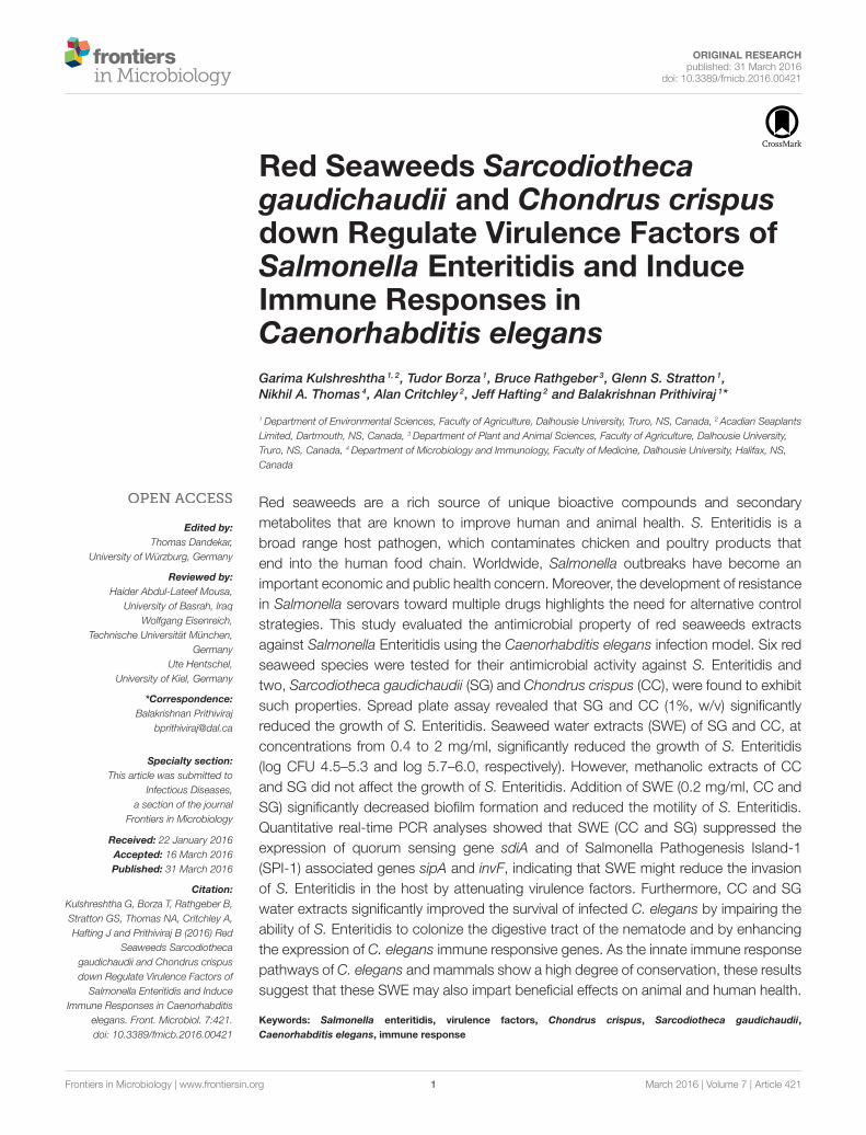
Red Seaweeds Sarcodiothecagaudichaudii and Chondrus crispusdown Regulate Virulence Factors ofSalmonella Enteritidis and InduceImmune Responses inCaenorhabditis elegans
Garima Kulshreshtha 1, 2, Tudor Borza 1, Bruce Rathgeber 3, Glenn S. Stratton 1,
Nikhil A. Thomas 4, Alan Critchley 2, Jeff Hafting 2 and Balakrishnan Prithiviraj 1*
1Department of Environmental Sciences, Faculty of Agriculture, Dalhousie University, Truro, NS, Canada, 2 Acadian Seaplants
Limited, Dartmouth, NS, Canada, 3Department of Plant and Animal Sciences, Faculty of Agriculture, Dalhousie University,
Truro, NS, Canada, 4Department of Microbiology and Immunology, Faculty of Medicine, Dalhousie University, Halifax, NS,
Canada
Red seaweeds are a rich source of unique bioactive compounds and secondary
metabolites that are known to improve human and animal health. S. Enteritidis is a
broad range host pathogen, which contaminates chicken and poultry products that
end into the human food chain. Worldwide, Salmonella outbreaks have become an
important economic and public health concern. Moreover, the development of resistance
in Salmonella serovars toward multiple drugs highlights the need for alternative control
strategies. This study evaluated the antimicrobial property of red seaweeds extracts
against Salmonella Enteritidis using the Caenorhabditis elegans infection model. Six red
seaweed species were tested for their antimicrobial activity against S. Enteritidis and
two, Sarcodiotheca gaudichaudii (SG) and Chondrus crispus (CC), were found to exhibit
such properties. Spread plate assay revealed that SG and CC (1%, w/v) significantly
reduced the growth of S. Enteritidis. Seaweed water extracts (SWE) of SG and CC, at
concentrations from 0.4 to 2 mg/ml, significantly reduced the growth of S. Enteritidis
(log CFU 4.5–5.3 and log 5.7–6.0, respectively). However, methanolic extracts of CC
and SG did not affect the growth of S. Enteritidis. Addition of SWE (0.2 mg/ml, CC and
SG) significantly decreased biofilm formation and reduced the motility of S. Enteritidis.
Quantitative real-time PCR analyses showed that SWE (CC and SG) suppressed the
expression of quorum sensing gene sdiA and of Salmonella Pathogenesis Island-1
(SPI-1) associated genes sipA and invF, indicating that SWE might reduce the invasion
of S. Enteritidis in the host by attenuating virulence factors. Furthermore, CC and SG
water extracts significantly improved the survival of infected C. elegans by impairing the
ability of S. Enteritidis to colonize the digestive tract of the nematode and by enhancing
the expression of C. elegans immune responsive genes. As the innate immune response
pathways of C. elegans and mammals show a high degree of conservation, these results
suggest that these SWE may also impart beneficial effects on animal and human health.
Keywords: Salmonella enteritidis, virulence factors, Chondrus crispus, Sarcodiotheca gaudichaudii,
Caenorhabditis elegans, immune response
Kulshreshtha et al. Red Seaweeds Regulate S.Enteritidis Virulence
INTRODUCTION
Food-borne pathogen Salmonella enterica subsp. enterica serovarEnteritidis (S. Enteritidis) is the world’s leading cause ofegg associated salmonellosis in humans (Sheela et al., 2003;Govaris et al., 2010). S. Enteritidis is a broad range hostpathogen carried by chicken and poultry products to humanfood chain. In humans, Salmonella infection causes foodpoisoning and intestinal infections associated with mucosalinflammation and diarrhea leading, in some cases, to mortality(Yim et al., 2010).Worldwide, Salmonella outbreaks have becomean important public health and economic concern (WorldHealth Organization, 2014). World Health Organization globalSalmonella survey program estimated that a number of 500–2000deaths occur each year (Betancor et al., 2010; Yim et al., 2010).In 2003, 12.7% of all Salmonella cases were due to S. Enteritidis;in 2009 the percentage increased to 32.1% (Nesbitt et al.,2012). In Canada and United States, each year approximately1.4 million people are infected with non-typhoid Salmonellaserotype (Nesbitt et al., 2012;Middleton et al., 2014). The nationalenteric disease surveillance report from 2011 indicated that inUnited States S. Enteritidis is the most dominant serotype fromclinical and non-clinical sources (Center for Disease Controland Prevention, 2012), while in Canada is one of the top threenon-typhoidal serovars (Galanis et al., 2012).
In chickens, S. Enteritidis colonizes the gastrointestinal tract,from where infection can extend to organs such as the ovariesand the oviduct to eventually localize inside the egg and embryo(Guard-Petter, 2001). The ability of S. Enteritidis to establishpersistent infection in avian tissues including egg is responsiblefor its invasion into the human food chain (Revolledo et al., 2009;Yim et al., 2010).
Upon consumption of contaminated water or food, S.Enteritidis recognizes and adhere to the host epithelium (Pontier-Bres et al., 2012). Bacteria penetrate the intestinal epithelium bysuppressing signal transduction pathways leading to cytoskeletonrearrangement into the host cell. This is followed by the deliveryof the effector proteins, which suppress the immune responseof the host, establishing a persistent infection (Groisman andMouslim, 2000; Brown et al., 2005). The survival capabilities of S.Enteritidis are enhanced by quorum sensing and by the formationof biofilms on a variety of biotic and abiotic surfaces (De Kievitand Iglewski, 2000; Parker and Guard-Petter, 2001; Prouty et al.,2002; Brossard and Campagnari, 2012).
Salmonella infection can be fatal in immunocompromisedpatients if not treated with antibiotics. Fluoroquinolones andcephalosporin are most commonly used antibiotics to treatinfections caused by Salmonella serovars (Baucheron et al., 2004).However, the development of resistance in Salmonella serovarstoward multiple drugs highlights the urgent need for alternativestrategies to control this pathogen (Acheson and Hohmann,2001). Previously, bacteriophages, antimicrobial peptides, andessential oils have been used or investigated as alternatives toantibiotics (Fratamico and Cooke, 1996; Joerger, 2003).
Salmonella also infects the nematode Caenorhabditis elegans,a widely used model organism (Aballay et al., 2000; Aballayand Ausubel, 2002; Sifri et al., 2005). Several studies have
shown that bacterial pathogens such as Pseudomonas aeruginosa,Staphylococcus aureus, Vibrio sp., Salmonella Typhimurium, E.coli 0157:H7, and Enterococcus faecalis have similar pathogenicmechanisms in nematodes and higher animals (Aballay et al.,2000; Aballay and Ausubel, 2002; Breger et al., 2007). Forexample, the pathosystem C. elegans—E. faecalis has been usedas high throughput model to screen compounds with potentialanti-infective and anti-microbial properties, applicable to cureinfections in higher animals and humans (Moy et al., 2009).Additionally, Tenor et al. (2004) have shown that C. elegans isan attractive model to study the interaction between Salmonellaeffector protein and host innate immunity because there isa significant overlap between virulence factors of Salmonellarequired for both, nematode and human pathogenesis. C.elegans react to Salmonella infection by activating the innateimmune response through the p38 mitogen-activated proteinkinase (PMK-1) and subsequently by synthesizing antimicrobialpeptides, mechanisms that are similar to immune responses inhumans (Aballay et al., 2003; Alegado and Tan, 2008).
Red seaweeds are a rich source of lipids, polysaccharides,proteins, bioactive compounds and of secondary metabolitessuch as polyphenols as well as of minerals, which impartseveral health benefits (Pujol et al., 2002; Bansemir et al., 2004;Yuan et al., 2005; Lins et al., 2009; Gómez-Ordóñez et al.,2012; Souza et al., 2012). Edible red seaweeds, Sarcodiothecagaudichaudii and Chondrus crispus, are abundant along thecoasts of the eastern Pacific Ocean and of western AtlanticOcean (Gabrielson, 1982; Guiry and Guiry, 2016) and certainstrains of red seaweeds are commercially cultivated in land(Hafting et al., 2012). Red seaweeds have been recently exploredas potential sources of products with antimicrobial properties.The main polysaccharides in these seaweeds, the carrageenans,were shown to have antiviral properties as well as antitumor,anticoagulant and immunomodulatory effects (Campo et al.,2009; de Jesus Raposo et al., 2015). It has been shown that otherred seaweed compounds such as derived brominated furanonesreduced swimming motility, flagellar biosynthesis in Salmonellaserovar Typhimurium and showed biofilm inhibiting activities(Janssens et al., 2008). Recently, components of cultivated redseaweeds have been shown to improve the immune responseof C. elegans to Pseudomonas aeruginosa (PA-14) through theinduction of PMK-1 and Daf-2/daf-16 insulin signaling pathways(Liu et al., 2013). Furthermore, enzymatic extracts of C. crispuswere identified as effective against HSV-1 virus, indicatingpotential antiviral activity of sulphated polysaccharides in theextracts (Kulshreshtha et al., 2015). In another study, feedsupplementation with S. gaudichaudii and C. crispus reducedthe prevalence of pathogenic bacteria such as Clostridiumperfringens in the chicken gut while the relative abundanceof beneficial bacteria such as Bifidobacterium longum andStreptococcus salivarius was found to be increased (Kulshreshthaet al., 2014). Here, we report the effects of water extracts fromS. gaudichaudii and C. crispus on Salmonella Enteritidis usingthe C. elegans infection model. In addition, we also examinedthe effects of water extracts on biofilm formation, motility,quorum sensing signaling and virulence factors in S. Enteritidis.Red seaweeds are a rich source of lipids, polysaccharides,
Frontiers in Microbiology | www.frontiersin.org 2 March 2016 | Volume 7 | Article 421
Kulshreshtha et al. Red Seaweeds Regulate S.Enteritidis Virulence
proteins, bioactive compounds and of secondary metabolitessuch as polyphenols as well as of minerals, which impartseveral health benefits (Pujol et al., 2002; Bansemir et al., 2004;Yuan et al., 2005; Lins et al., 2009; Gómez-Ordóñez et al.,2012; Souza et al., 2012). Edible red seaweeds, Sarcodiothecagaudichaudii and Chondrus crispus, are abundant along thecoasts of the eastern Pacific Ocean and of western AtlanticOcean (Gabrielson, 1982; Guiry and Guiry, 2016) and certainstrains of red seaweeds are commercially cultivated in land(Hafting et al., 2012). Red seaweeds have been recently exploredas potential sources of products with antimicrobial properties.The main polysaccharides in these seaweeds, the carrageenans,were shown to have antiviral properties as well as antitumor,anticoagulant and immunomodulatory effects (Campo et al.,2009; de Jesus Raposo et al., 2015). It has been shown that otherred seaweed compounds such as derived brominated furanonesreduced swimming motility, flagellar biosynthesis in Salmonellaserovar Typhimurium and showed biofilm inhibiting activities(Janssens et al., 2008). Recently, components of cultivated redseaweeds have been shown to improve the immune responseof C. elegans to Pseudomonas aeruginosa (PA-14) through theinduction of PMK-1 and Daf-2/daf-16 insulin signaling pathways(Liu et al., 2013). Furthermore, enzymatic extracts of C. crispuswere identified as effective against HSV-1 virus, indicatingpotential antiviral activity of sulphated polysaccharides in theextracts (Kulshreshtha et al., 2015). In another study, feedsupplementation with S. gaudichaudii and C. crispus reduced theprevalence of pathogenic bacteria such as Clostridium perfringensin the chicken gut while the relative abundance of beneficialbacteria such as Bifidobacterium longum and Streptococcussalivarius was found to be increased (Kulshreshtha et al., 2014).Here, we report the effects of water extracts from S. gaudichaudiiand C. crispus on Salmonella Enteritidis using the C. elegansinfection model. In addition, we also examined the effects ofwater extracts on biofilm formation, motility, quorum sensingsignaling and virulence factors in S. Enteritidis.
MATERIALS AND METHODS
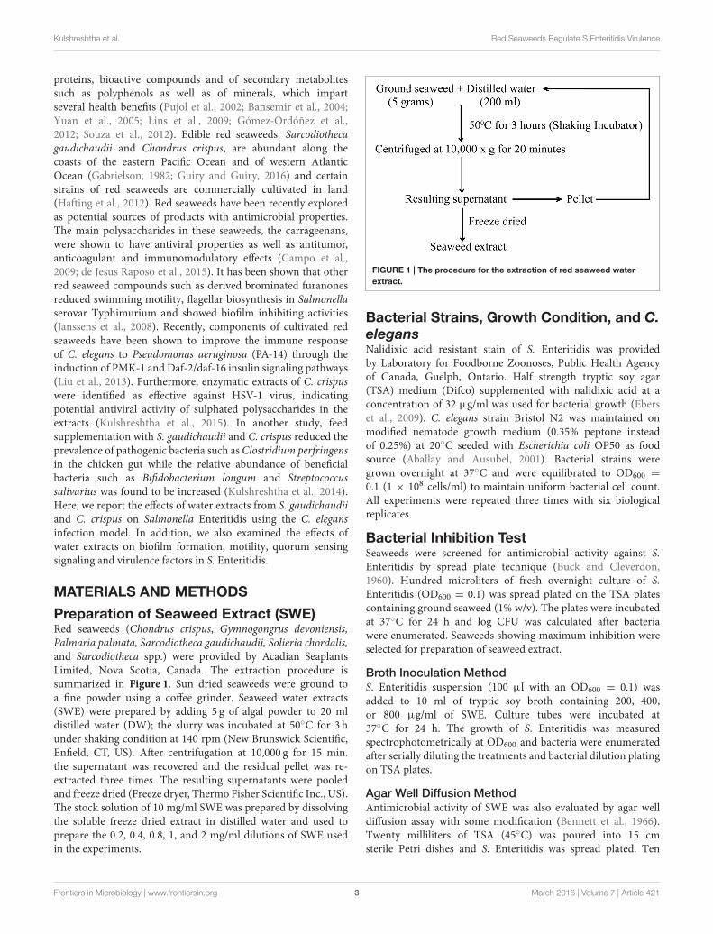
Preparation of Seaweed Extract (SWE)Red seaweeds (Chondrus crispus, Gymnogongrus devoniensis,Palmaria palmata, Sarcodiotheca gaudichaudii, Solieria chordalis,and Sarcodiotheca spp.) were provided by Acadian SeaplantsLimited, Nova Scotia, Canada. The extraction procedure issummarized in Figure 1. Sun dried seaweeds were ground toa fine powder using a coffee grinder. Seaweed water extracts(SWE) were prepared by adding 5 g of algal powder to 20 mldistilled water (DW); the slurry was incubated at 50◦C for 3 hunder shaking condition at 140 rpm (New Brunswick Scientific,Enfield, CT, US). After centrifugation at 10,000 g for 15 min.the supernatant was recovered and the residual pellet was re-extracted three times. The resulting supernatants were pooledand freeze dried (Freeze dryer, Thermo Fisher Scientific Inc., US).The stock solution of 10 mg/ml SWE was prepared by dissolvingthe soluble freeze dried extract in distilled water and used toprepare the 0.2, 0.4, 0.8, 1, and 2 mg/ml dilutions of SWE usedin the experiments.
FIGURE 1 | The procedure for the extraction of red seaweed water
extract.
Bacterial Strains, Growth Condition, and C.
elegansNalidixic acid resistant stain of S. Enteritidis was providedby Laboratory for Foodborne Zoonoses, Public Health Agencyof Canada, Guelph, Ontario. Half strength tryptic soy agar(TSA) medium (Difco) supplemented with nalidixic acid at aconcentration of 32 µg/ml was used for bacterial growth (Eberset al., 2009). C. elegans strain Bristol N2 was maintained onmodified nematode growth medium (0.35% peptone insteadof 0.25%) at 20◦C seeded with Escherichia coli OP50 as foodsource (Aballay and Ausubel, 2001). Bacterial strains weregrown overnight at 37◦C and were equilibrated to OD600 =
0.1 (1 × 10⁸ cells/ml) to maintain uniform bacterial cell count.All experiments were repeated three times with six biologicalreplicates.
Bacterial Inhibition TestSeaweeds were screened for antimicrobial activity against S.Enteritidis by spread plate technique (Buck and Cleverdon,1960). Hundred microliters of fresh overnight culture of S.Enteritidis (OD600 = 0.1) was spread plated on the TSA platescontaining ground seaweed (1% w/v). The plates were incubatedat 37◦C for 24 h and log CFU was calculated after bacteriawere enumerated. Seaweeds showing maximum inhibition wereselected for preparation of seaweed extract.
Broth Inoculation Method
S. Enteritidis suspension (100 µl with an OD600 = 0.1) wasadded to 10 ml of tryptic soy broth containing 200, 400,or 800 µg/ml of SWE. Culture tubes were incubated at37◦C for 24 h. The growth of S. Enteritidis was measuredspectrophotometrically at OD600 and bacteria were enumeratedafter serially diluting the treatments and bacterial dilution platingon TSA plates.
Agar Well Diffusion Method
Antimicrobial activity of SWE was also evaluated by agar welldiffusion assay with some modification (Bennett et al., 1966).Twenty milliliters of TSA (45◦C) was poured into 15 cmsterile Petri dishes and S. Enteritidis was spread plated. Ten
Frontiers in Microbiology | www.frontiersin.org 3 March 2016 | Volume 7 | Article 421
Kulshreshtha et al. Red Seaweeds Regulate S.Enteritidis Virulence
millimeter wells were bored using a sterile cork borer and knownconcentrations of SWE were added into the wells in the plates.The plates were incubated for 24 h at 37◦C and antimicrobialactivity was measured using vernier caliper to determinethe zone of growth inhibition. H2O was used as negativecontrol.
Effect of SWE on BacterialMotility—Swimming and SwarmingAbility of SWE to alter bacterial motility was tested as describedby Rashid and Kornberg (2000) with some modifications (Difcobacteriological agar instead of agarose). Single colony of bacteriafrom overnight grown culture was spotted using a steriletoothpick on swim plates or on swarm plates containing knownconcentration of SWE (200 µg/ml). All plates were sealed withparafilm to prevent dehydration and swim plates were incubatedat 30◦C for 14–15 h while swarm plates for 24 h.
Biofilm Formation AssayOvernight grown S. Enteritidis culture was diluted 1:100 intryptic soy broth containing known concentration of SWE (200µg/ml). Two hundred µl of the aliquot was dispensed into96 wells polyvinyl chloride microtitre plates. The plates wereincubated statically at 28◦C for 24 h. Biofilm formation wasquantified by staining the wells with 20 µl of crystal violet (CV)(0.14% (w/v) in water) at room temperature for 20 min. The wellswere washed three times in distilled water to remove excess CV.CV stained cells were eluted with 95% ethanol and optical densitywas measured at OD600.
Effect of SWE on Expression of Virulenceand Quorum Sensing Related GenesFor gene expression analysis, S. Enteritidis with an initial OD600
of 0.1 was cultured at 37◦C TSB in the presence and absence(control) of SWE with shaking at 160 rpm. Bacterial cells wereharvested by centrifugation at 12,000 g for 10 min. Total RNAwas extracted using Trizol (Invitrogen) as described by themanufacturer. The RNA was quantified by NanoDrop ND-2000spectrophotometer (NanoDrop Technologies Wilmington, DE)and the quality was assessed by agarose gel electrophoresis. RNAfrom each biological replicate was used for cDNA synthesisusing the High Capacity cDNA reverse transcription kit (AppliedBiosystems). The relative transcript levels of quorum sensing,virulence, and flagella associated genes were quantified usingStepOnePlus Real time PCR (Applied Biosystems, ON, Canada).The 10 µl reaction mix contained 2 ng of cDNA, 5 µl PromegaGoTaq SYBR green master mix (Promega North America,Madison, WI, USA) and 300 nM of gene specific primers(Supplementary Table 1). 16SrRNA and tufA genes were used asinternal control and the relative expression levels were calculatedusing the 11Ct method.
C. elegans Killing AssayModified nematode growth medium was used to establishC. elegans-S. Enteritidis pathosystem as described by Aballay et al.(2000). C. elegans killing assay was conducted by two methods asdescribed below:
Incorporating SWE into the media: Treatment plates wereprepared by supplementing SWE to nematode growth media(NGM) to a final concentration of 200, 400, and 800 µg/ml. Celegans population was synchronized by placing adult nematodeson NGM plates to lay egg for 4–6 h. Eggs were incubatedfor 2 days at 20 ± 2◦C to ensure uniform adult population.Thirty to forty synchronized L4 nematodes were used foreach assay. Heat killed S. Enteritidis and E. coli OP50 wereused as control and 70 µM fluorodeoxyuridine (FuDR) wasused to prevent the development of progeny. The plates wereincubated at 25◦C and scored for live vs. dead worms every 24h. A worm was considered dead when it failed to respond toplate tapping or a gentle touch with a platinum wire. Wormskilled as a result of being stuck to the wall of the platewere excluded from the analysis. Nematodes were subjectedto a combination of three pre-treatments with SWE to targetvirulence of bacteria and immune response of C elegans asdescribed below:
(a) Pre-treatment of bacteria with SWE: Synchronized wormswere infected with S. Enteritidis grown overnight on NGMplates containing 200, 400, 800 µg/ml of SWE to test itsefficacy in reducing bacterial virulence.
(b) Pre-treatment of nematodes with SWE: Synchronizedpopulations of worms were maintained on NMG plates fromegg stage containing 200, 400, 800µg/ml of SWE. Pre-treatedL4 nematodes were transferred to S. Enteritidis treatmentplates.
(c) Pre-treatment of bacteria and nematodes with SWE:Synchronized worms from egg stage maintained on NGMplated were infected with S. Enteritidis grown overnight onNGM plates containing 200, 400, 800 µg/ml of SWE.
Adding SWE over the media: S. Enteritidis was grown onmodifiedNGM plates and 200, 400, 800 µg/ml of SWE was addedover the media along with food source. The killing assay wasperformed with three combination of pre-treatment as describedabove.
S. Enteritidis Colonization Assay of C.elegans GutS. Enteritidis count of from C. elegans gut was determinedaccording to the modified method previously described byPrithiviraj et al. (2005). For each replicate, six adult C. eleganswere picked from the treatment plates and transferred into a 1.5ml microfuge tube containing 500µL ofM9 buffer supplementedwith 20 µg/ml gentamicin and washed three times to removebacteria from C. elegans surface. The nematodes were disruptedin a microfuge tube containing 50 µL of M9 medium with 1%Triton X-100 using a microfuge pestle. The resulting slurry wasserially diluted and plated on TSA medium and the number ofCFU was counted.
Effect of SWE on Expression of ImmuneResponse Genes in C. elegansTreatment plates were prepared by supplementing SWE to NGMto a final concentration of 400 µg/ml. C. elegans were infected
Frontiers in Microbiology | www.frontiersin.org 4 March 2016 | Volume 7 | Article 421
Kulshreshtha et al. Red Seaweeds Regulate S.Enteritidis Virulence
with S. Enteritidis and approximately 100 worms per treatmentwere harvested 5 days after exposure to S. Enteritidis. Therewere the four conditions used in the experiment: (1) C. elegansfed on SWE, (2) C. elegans fed on S. Enteritidis, (3) C. elegansfed on SWE and on S. Enteritidis, and (4) C. elegans fed onheat killed E. coli OP50. Worms were transferred into 1.5 mlmicrofuge tubes and washed three times inM9 buffer to eliminateexcess bacteria. Excess buffer was pipetted out and total RNAwas extracted using Trizol (Invitrogen) following manufacturer’sprotocol. RNA quality and quantity determination, cDNAsynthesis and quantitative real time PCR were performed aspreviously described. The immune responsive genes specificprimers used for these experiments are listed in SupplementaryTable 2. Relative expression levels were determined by 11Ctmethod and ama-1 was used as a reference gene while heat killedE. coli OP50 samples were used as control.
Statistical AnalysisA completely randomized design was followed to analyze effectsof application method, concentration, and antimicrobial assays.All experiments were performed three times with at least threebiological replicates. Data was analyzed using ANOVA one-wayanalysis of variance with a P-value of 0.05 using the statisticalsoftware Minitab 17 (Minitab Inc., PA, USA) and SAS, version9.4 for Windows (SAS Institute Inc., NC, USA). If significantmain effects were found with ANOVA, the Tukey’s procedure wasused to compare differences among the least-square means. Thestandard deviation (SD) was reported with the mean. Differenceswere considered significant when P was < 0.05.
RESULTS
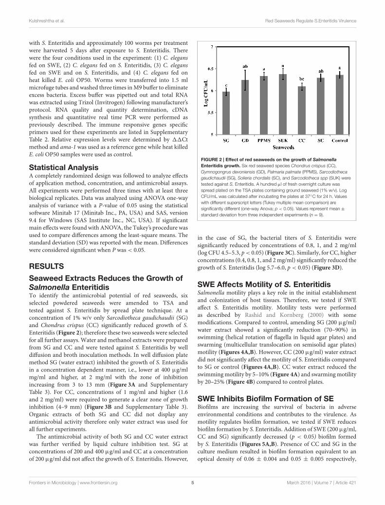
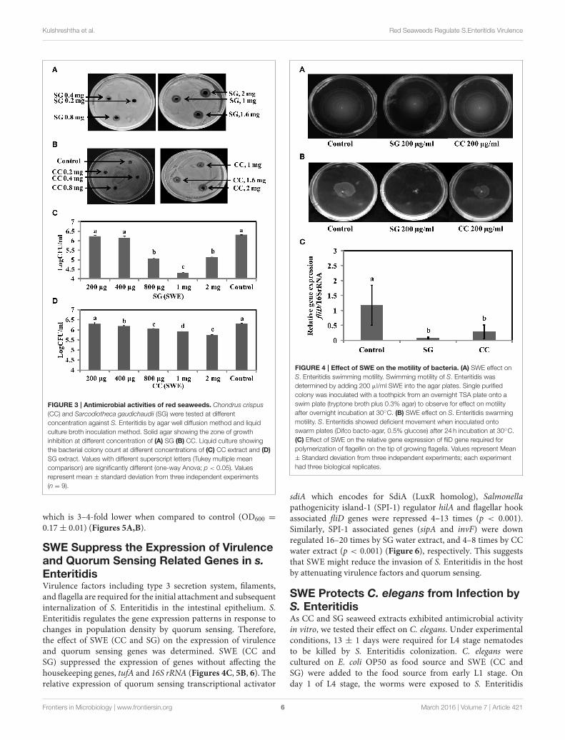
Seaweed Extracts Reduces the Growth ofSalmonella EnteritidisTo identify the antimicrobial potential of red seaweeds, sixselected powdered seaweeds were amended to TSA andtested against S. Enteritidis by spread plate technique. At aconcentration of 1% w/v only Sarcodiotheca gaudichaudii (SG)and Chondrus crispus (CC) significantly reduced growth of S.Enteritidis (Figure 2); therefore these two seaweeds were selectedfor all further assays. Water and methanol extracts were preparedfrom SG and CC and were tested against S. Enteritidis by welldiffusion and broth inoculation methods. In well diffusion platemethod SG (water extract) inhibited the growth of S. Enteritidisin a concentration dependent manner, i.e., lower at 400 µg/mlmg/ml and higher, at 2 mg/ml with the zone of inhibitionincreasing from 3 to 13 mm (Figure 3A and SupplementaryTable 3). For CC, concentrations of 1 mg/ml and higher (1.6and 2 mg/ml) were required to generate a clear zone of growthinhibition (4–9 mm) (Figure 3B and Supplementary Table 3).Organic extracts of both SG and CC did not display anyantimicrobial activity therefore only water extract was used forall further experiments.
The antimicrobial activity of both SG and CC water extractwas further verified by liquid culture inhibition test. SG atconcentrations of 200 and 400 µg/ml and CC at a concentrationof 200 µg/ml did not affect the growth of S. Enteritidis. However,
FIGURE 2 | Effect of red seaweeds on the growth of Salmonella
Enteritidis growth. Six red seaweed species Chondrus crispus (CC),
Gymnogongrus devoniensis (GD), Palmaria palmate (PPMS), Sarcodiotheca
gaudichaudii (SG), Solieria chordalis (SC), and Sarcodiotheca spp (SUK) were
tested against S. Enteritidis. A hundred µl of fresh overnight culture was
spread plated on the TSA plates containing ground seaweed (1% w/v). Log
CFU/mL was calculated after incubating the plates at 37◦C for 24 h. Values
with different superscript letters (Tukey multiple mean comparison) are
significantly different (one-way Anova; p < 0.05). Values represent mean ±
standard deviation from three independent experiments (n = 9).
in the case of SG, the bacterial titers of S. Enteritidis weresignificantly reduced by concentrations of 0.8, 1, and 2 mg/ml(log CFU 4.5–5.3, p< 0.05) (Figure 3C). Similarly, for CC, higherconcentrations (0.4, 0.8, 1, and 2mg/ml) significantly reduced thegrowth of S. Enteritidis (log 5.7–6.0, p < 0.05) (Figure 3D).
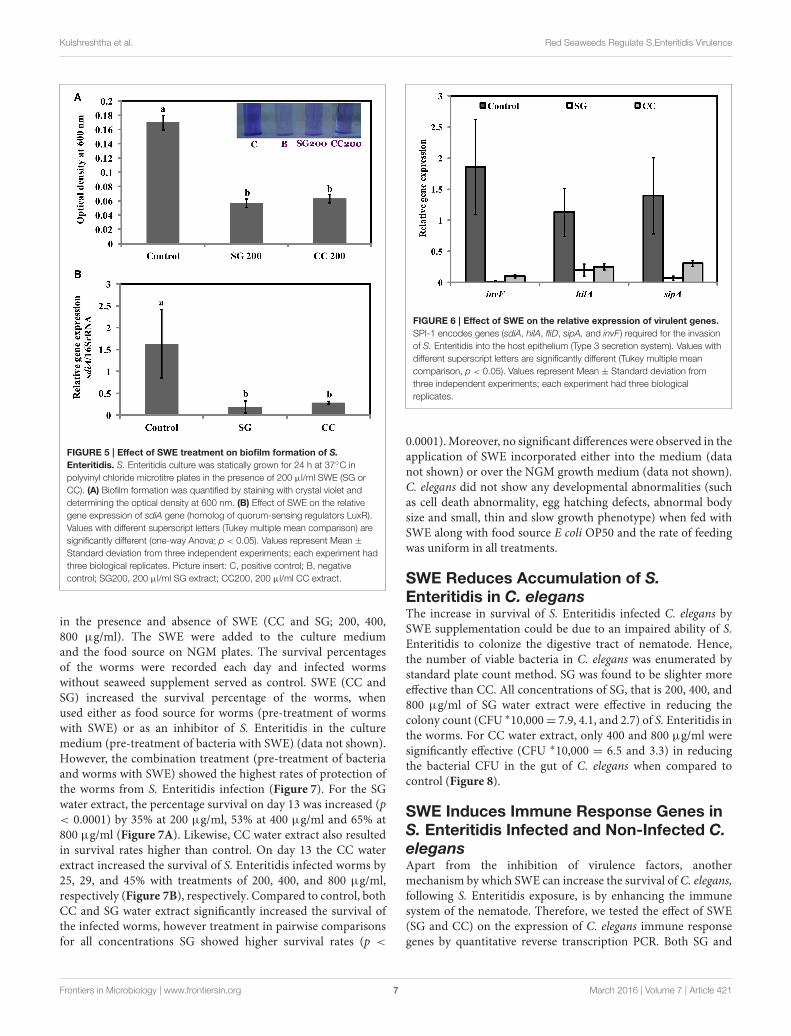
SWE Affects Motility of S. EnteritidisSalmonella motility plays a key role in the initial establishmentand colonization of host tissues. Therefore, we tested if SWEaffect S. Enteritidis motility. Motility tests were performedas described by Rashid and Kornberg (2000) with somemodifications. Compared to control, amending SG (200 µg/ml)water extract showed a significantly reduction (70–90%) inswimming (helical rotation of flagella in liquid agar plates) andswarming (multicellular translocation on semisolid agar plates)motility (Figures 4A,B). However, CC (200 µg/ml) water extractdid not significantly affect the motility of S. Enteritidis comparedto SG or control (Figures 4A,B). CC water extract reduced theswimmingmotility by 5–10% (Figure 4A) and swarmingmotilityby 20–25% (Figure 4B) compared to control plates.
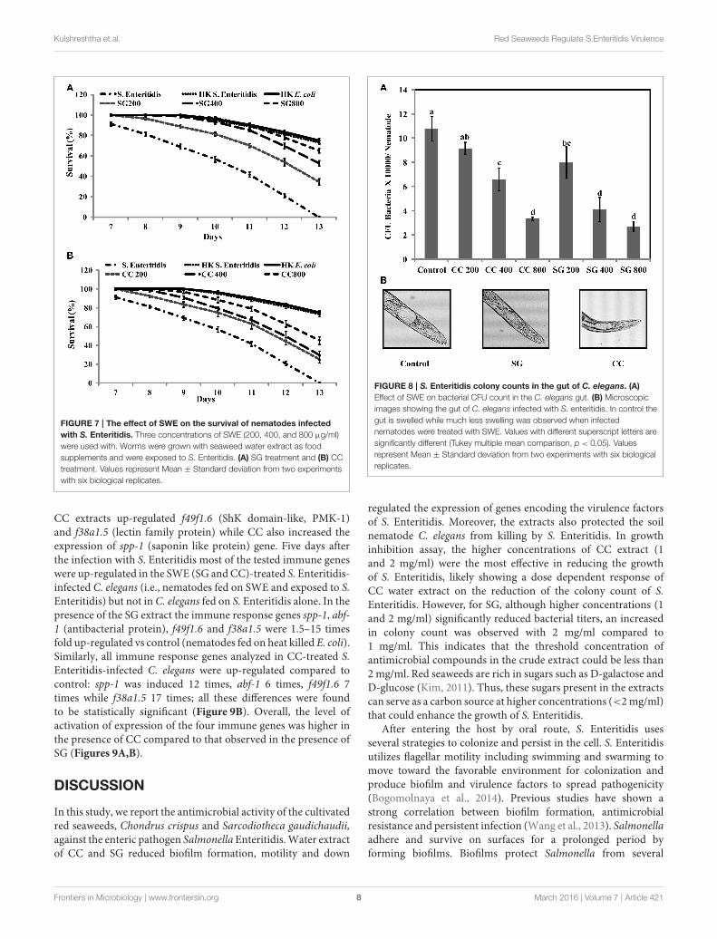
SWE Inhibits Biofilm Formation of SEBiofilms are increasing the survival of bacteria in adverseenvironmental conditions and contributes to the virulence. Asmotility regulates biofilm formation, we tested if SWE reducesbiofilm formation by S. Enteritidis. Addition of SWE (200µg/ml,CC and SG) significantly decreased (p < 0.05) biofilm formedby S. Enteritidis (Figures 5A,B). Presence of CC and SG in theculture medium resulted in biofilm formation equivalent to anoptical density of 0.06 ± 0.004 and 0.05 ± 0.005 respectively,
Frontiers in Microbiology | www.frontiersin.org 5 March 2016 | Volume 7 | Article 421
Kulshreshtha et al. Red Seaweeds Regulate S.Enteritidis Virulence
FIGURE 3 | Antimicrobial activities of red seaweeds. Chondrus crispus
(CC) and Sarcodiotheca gaudichaudii (SG) were tested at different
concentration against S. Enteritidis by agar well diffusion method and liquid
culture broth inoculation method. Solid agar showing the zone of growth
inhibition at different concentration of (A) SG (B) CC. Liquid culture showing
the bacterial colony count at different concentrations of (C) CC extract and (D)
SG extract. Values with different superscript letters (Tukey multiple mean
comparison) are significantly different (one-way Anova; p < 0.05). Values
represent mean ± standard deviation from three independent experiments
(n = 9).
which is 3–4-fold lower when compared to control (OD600 =
0.17± 0.01) (Figures 5A,B).
SWE Suppress the Expression of Virulenceand Quorum Sensing Related Genes in s.
EnteritidisVirulence factors including type 3 secretion system, filaments,and flagella are required for the initial attachment and subsequentinternalization of S. Enteritidis in the intestinal epithelium. S.Enteritidis regulates the gene expression patterns in response tochanges in population density by quorum sensing. Therefore,the effect of SWE (CC and SG) on the expression of virulenceand quorum sensing genes was determined. SWE (CC andSG) suppressed the expression of genes without affecting thehousekeeping genes, tufA and 16S rRNA (Figures 4C, 5B, 6). Therelative expression of quorum sensing transcriptional activator
FIGURE 4 | Effect of SWE on the motility of bacteria. (A) SWE effect on
S. Enteritidis swimming motility. Swimming motility of S. Enteritidis was
determined by adding 200 µl/ml SWE into the agar plates. Single purified
colony was inoculated with a toothpick from an overnight TSA plate onto a
swim plate (tryptone broth plus 0.3% agar) to observe for effect on motility
after overnight incubation at 30◦C. (B) SWE effect on S. Enteritidis swarming
motility. S. Enteritidis showed deficient movement when inoculated onto
swarm plates (Difco bacto-agar, 0.5% glucose) after 24 h incubation at 30◦C.
(C) Effect of SWE on the relative gene expression of fliD gene required for
polymerization of flagellin on the tip of growing flagella. Values represent Mean
± Standard deviation from three independent experiments; each experiment
had three biological replicates.
sdiA which encodes for SdiA (LuxR homolog), Salmonellapathogenicity island-1 (SPI-1) regulator hilA and flagellar hookassociated fliD genes were repressed 4–13 times (p < 0.001).Similarly, SPI-1 associated genes (sipA and invF) were downregulated 16–20 times by SG water extract, and 4–8 times by CCwater extract (p < 0.001) (Figure 6), respectively. This suggeststhat SWE might reduce the invasion of S. Enteritidis in the hostby attenuating virulence factors and quorum sensing.
SWE Protects C. elegans from Infection byS. EnteritidisAs CC and SG seaweed extracts exhibited antimicrobial activityin vitro, we tested their effect on C. elegans. Under experimentalconditions, 13 ± 1 days were required for L4 stage nematodesto be killed by S. Enteritidis colonization. C. elegans werecultured on E. coli OP50 as food source and SWE (CC andSG) were added to the food source from early L1 stage. Onday 1 of L4 stage, the worms were exposed to S. Enteritidis
Frontiers in Microbiology | www.frontiersin.org 6 March 2016 | Volume 7 | Article 421
Kulshreshtha et al. Red Seaweeds Regulate S.Enteritidis Virulence
FIGURE 5 | Effect of SWE treatment on biofilm formation of S.
Enteritidis. S. Enteritidis culture was statically grown for 24 h at 37◦C in
polyvinyl chloride microtitre plates in the presence of 200 µl/ml SWE (SG or
CC). (A) Biofilm formation was quantified by staining with crystal violet and
determining the optical density at 600 nm. (B) Effect of SWE on the relative
gene expression of sdiA gene (homolog of quorum-sensing regulators LuxR).
Values with different superscript letters (Tukey multiple mean comparison) are
significantly different (one-way Anova; p < 0.05). Values represent Mean ±
Standard deviation from three independent experiments; each experiment had
three biological replicates. Picture insert: C, positive control; B, negative
control; SG200, 200 µl/ml SG extract; CC200, 200 µl/ml CC extract.
in the presence and absence of SWE (CC and SG; 200, 400,800 µg/ml). The SWE were added to the culture mediumand the food source on NGM plates. The survival percentagesof the worms were recorded each day and infected wormswithout seaweed supplement served as control. SWE (CC andSG) increased the survival percentage of the worms, whenused either as food source for worms (pre-treatment of wormswith SWE) or as an inhibitor of S. Enteritidis in the culturemedium (pre-treatment of bacteria with SWE) (data not shown).However, the combination treatment (pre-treatment of bacteriaand worms with SWE) showed the highest rates of protection ofthe worms from S. Enteritidis infection (Figure 7). For the SGwater extract, the percentage survival on day 13 was increased (p< 0.0001) by 35% at 200 µg/ml, 53% at 400 µg/ml and 65% at800 µg/ml (Figure 7A). Likewise, CC water extract also resultedin survival rates higher than control. On day 13 the CC waterextract increased the survival of S. Enteritidis infected worms by25, 29, and 45% with treatments of 200, 400, and 800 µg/ml,respectively (Figure 7B), respectively. Compared to control, bothCC and SG water extract significantly increased the survival ofthe infected worms, however treatment in pairwise comparisonsfor all concentrations SG showed higher survival rates (p <
FIGURE 6 | Effect of SWE on the relative expression of virulent genes.
SPI-1 encodes genes (sdiA, hilA, fliD, sipA, and invF ) required for the invasion
of S. Enteritidis into the host epithelium (Type 3 secretion system). Values with
different superscript letters are significantly different (Tukey multiple mean
comparison, p < 0.05). Values represent Mean ± Standard deviation from
three independent experiments; each experiment had three biological
replicates.
0.0001).Moreover, no significant differences were observed in theapplication of SWE incorporated either into the medium (datanot shown) or over the NGM growth medium (data not shown).C. elegans did not show any developmental abnormalities (suchas cell death abnormality, egg hatching defects, abnormal bodysize and small, thin and slow growth phenotype) when fed withSWE along with food source E coli OP50 and the rate of feedingwas uniform in all treatments.
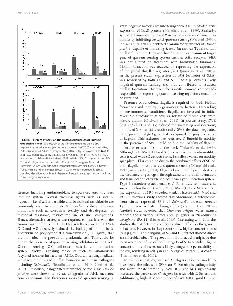
SWE Reduces Accumulation of S.Enteritidis in C. elegansThe increase in survival of S. Enteritidis infected C. elegans bySWE supplementation could be due to an impaired ability of S.Enteritidis to colonize the digestive tract of nematode. Hence,the number of viable bacteria in C. elegans was enumerated bystandard plate count method. SG was found to be slighter moreeffective than CC. All concentrations of SG, that is 200, 400, and800 µg/ml of SG water extract were effective in reducing thecolony count (CFU ∗10,000= 7.9, 4.1, and 2.7) of S. Enteritidis inthe worms. For CC water extract, only 400 and 800 µg/ml weresignificantly effective (CFU ∗10,000 = 6.5 and 3.3) in reducingthe bacterial CFU in the gut of C. elegans when compared tocontrol (Figure 8).
SWE Induces Immune Response Genes inS. Enteritidis Infected and Non-Infected C.
elegansApart from the inhibition of virulence factors, anothermechanism by which SWE can increase the survival of C. elegans,following S. Enteritidis exposure, is by enhancing the immunesystem of the nematode. Therefore, we tested the effect of SWE(SG and CC) on the expression of C. elegans immune responsegenes by quantitative reverse transcription PCR. Both SG and
Frontiers in Microbiology | www.frontiersin.org 7 March 2016 | Volume 7 | Article 421
Kulshreshtha et al. Red Seaweeds Regulate S.Enteritidis Virulence
FIGURE 7 | The effect of SWE on the survival of nematodes infected
with S. Enteritidis. Three concentrations of SWE (200, 400, and 800 µg/ml)
were used with. Worms were grown with seaweed water extract as food
supplements and were exposed to S. Enteritidis. (A) SG treatment and (B) CC
treatment. Values represent Mean ± Standard deviation from two experiments
with six biological replicates.
CC extracts up-regulated f49f1.6 (ShK domain-like, PMK-1)and f38a1.5 (lectin family protein) while CC also increased theexpression of spp-1 (saponin like protein) gene. Five days afterthe infection with S. Enteritidis most of the tested immune geneswere up-regulated in the SWE (SG andCC)-treated S. Enteritidis-infected C. elegans (i.e., nematodes fed on SWE and exposed to S.Enteritidis) but not in C. elegans fed on S. Enteritidis alone. In thepresence of the SG extract the immune response genes spp-1, abf-1 (antibacterial protein), f49f1.6 and f38a1.5 were 1.5–15 timesfold up-regulated vs control (nematodes fed on heat killed E. coli).Similarly, all immune response genes analyzed in CC-treated S.Enteritidis-infected C. elegans were up-regulated compared tocontrol: spp-1 was induced 12 times, abf-1 6 times, f49f1.6 7times while f38a1.5 17 times; all these differences were foundto be statistically significant (Figure 9B). Overall, the level ofactivation of expression of the four immune genes was higher inthe presence of CC compared to that observed in the presence ofSG (Figures 9A,B).
DISCUSSION
In this study, we report the antimicrobial activity of the cultivatedred seaweeds, Chondrus crispus and Sarcodiotheca gaudichaudii,against the enteric pathogen Salmonella Enteritidis.Water extractof CC and SG reduced biofilm formation, motility and down
FIGURE 8 | S. Enteritidis colony counts in the gut of C. elegans. (A)
Effect of SWE on bacterial CFU count in the C. elegans gut. (B) Microscopic
images showing the gut of C. elegans infected with S. enteritidis. In control the
gut is swelled while much less swelling was observed when infected
nematodes were treated with SWE. Values with different superscript letters are
significantly different (Tukey multiple mean comparison, p < 0.05). Values
represent Mean ± Standard deviation from two experiments with six biological
replicates.
regulated the expression of genes encoding the virulence factorsof S. Enteritidis. Moreover, the extracts also protected the soilnematode C. elegans from killing by S. Enteritidis. In growthinhibition assay, the higher concentrations of CC extract (1and 2 mg/ml) were the most effective in reducing the growthof S. Enteritidis, likely showing a dose dependent response ofCC water extract on the reduction of the colony count of S.Enteritidis. However, for SG, although higher concentrations (1and 2 mg/ml) significantly reduced bacterial titers, an increasedin colony count was observed with 2 mg/ml compared to1 mg/ml. This indicates that the threshold concentration ofantimicrobial compounds in the crude extract could be less than2 mg/ml. Red seaweeds are rich in sugars such as D-galactose andD-glucose (Kim, 2011). Thus, these sugars present in the extractscan serve as a carbon source at higher concentrations (<2mg/ml)that could enhance the growth of S. Enteritidis.
After entering the host by oral route, S. Enteritidis usesseveral strategies to colonize and persist in the cell. S. Enteritidisutilizes flagellar motility including swimming and swarming tomove toward the favorable environment for colonization andproduce biofilm and virulence factors to spread pathogenicity(Bogomolnaya et al., 2014). Previous studies have shown astrong correlation between biofilm formation, antimicrobialresistance and persistent infection (Wang et al., 2013). Salmonellaadhere and survive on surfaces for a prolonged period byforming biofilms. Biofilms protect Salmonella from several
Frontiers in Microbiology | www.frontiersin.org 8 March 2016 | Volume 7 | Article 421
Kulshreshtha et al. Red Seaweeds Regulate S.Enteritidis Virulence
FIGURE 9 | Effect of SWE on the relative expression of immune
responsive genes. Expression of the immune response genes spp-1
(saponin like protein), abf-1 (antibacterial protein), f49f1.6 (ShK domain-like,
PMK-1) and f38a1.5 (lectin family protein) after 5 days of exposure to (A) SG
or (B) CC was analyzed by quantitative reverse transcription PCR. SG-inf, C.
elegans fed on SG and infected with S. Enteritidis; SG, C. elegans fed on SG;
E. coli, C. elegans fed on heat killed E. coli; SE, C. elegans fed on S.
Enteritidis. Values with different superscript letters are significantly different
(Tukey multiple mean comparison, p < 0.05). Values represent Mean ±
Standard deviation from three independent experiments; each experiment had
three biological replicates.
stresses including antimicrobials, temperature and the hostimmune system. Several chemical agents such as sodiumhypochlorite, alkaline peroxide and benzalkonium chloride arecommonly used to eliminate Salmonella biofilms. However,limitations such as corrosion, toxicity and development ofmicrobial resistance, restrict the use of such compounds.Hence, alternative strategies are required to interfere with theSalmonella biofilm formation. Our results showed that SWE(CC and SG) effectively reduced the buildup of biofilm by S.Enteritidis on polystyrene at a concentration (200 µg/ml) thatdid not affect the growth of planktonic cell. This could bedue to the presence of quorum sensing inhibitors in the SWE.Quorum sensing (QS), cell-to-cell bacterial communicationsystem involves signaling molecules such as autoinducers(acylated homoserine lactones, AHL). Quorum sensing mediatesvirulence, motility and biofilm formation in human pathogensincluding Salmonella (Jesudhasan et al., 2010; Choi et al.,2012). Previously, halogenated furanones of red algae Deliseapulchra were shown to be an antagonist of AHL mediatedgene expression. The furanones inhibited quorum sensing in
gram negative bacteria by interfering with AHL mediated geneexpression of LuxR protein (Manefield et al., 1999). Similarly,synthetic furanones improved P. aeruginosa clearance from lungsin mice by inhibiting bacterial quorum sensing (Wu et al., 2004).Janssens et al. (2008) identified brominated furanones of Deliseapulchra, capable of inhibiting S. enterica serovar Typhimuriumbiofilm formation. They concluded that the expression of targetgene of quorum sensing system such as AHL receptor SdiAwas not altered on treatment with brominated furanones.Biofilm formation was reduced by repressing the expressionof the global flagellar regulator flhD (Janssens et al., 2008).In the present study, expression of sdiA (activator of SdiA)was repressed by both CC and SG. The algal extracts likelyimpaired quorum sensing and thus contributed to reducedbiofilm formation. However, the specific seaweed compoundsresponsible for repressing quorum-sensing regulators remain tobe identified.
Presence of functional flagella is required for both biofilmformations and motility in gram-negative bacteria. Dependingon environmental conditions, flagella are involved in initialreversible attachment as well as release of motile cells frommature biofilm (Chelvam et al., 2014). In present study, SWE(200 µg/ml, CC and SG) reduced the swimming and swarmingmotility of S. Enteritidis. Additionally, SWE also down-regulatedthe expression of fliD gene that is required for polymerizationof flagellin. This indicates that restricted S. Enteritidis motilityin the presence of SWE could be due the inability of flagellinmolecules to assemble onto the hook (Yokoseki et al., 1995).Although both SWE (CC and SG) reduced S. Enteritidis motility,cells treated with SG extracts formed smaller swarms on motilityagar plates. This could be due to the combined effects of SG onboth, flagellar biosynthesis and quorum sensing (Manefield et al.,1999; Janssens et al., 2008). Flagellar based motility contributes tothe virulence of pathogen through adhesion, biofilm formationand translocation of virulent protein via Type 3 secretion system.Type 3 secretion system enables S. Enteritidis to invade andsurvive within the cell (Galan, 2001). SWE (CC and SG) reducedthe expression of SP-1 encoded virulent factors hilA, invF, andsipA. A previous study showed that obacunone, a triterpenoidfrom citrus, repressed SP-1 of Salmonella enterica serovarTyphimurium mediated through hilA (Vikram et al., 2012).Another study revealed that Chondrus crispus water extractreduced the virulence factors and QS genes in Pseudomonasaeruginosa (PA-14) (Liu et al., 2013). Interestingly, in both thestudies, the extracts did not show a direct effect on the growthof bacteria. However, in the present study, higher concentrations(800 µg/ml, 1 and 2 mg/ml) of SG and CC extract showed directantimicrobial effect. The growth inhibition activity might be dueto an alteration of the cell wall integrity of S. Enteritidis. Higherconcentrations of the extracts likely changed the permeability ofthe cell, resulting in cell lysis and leakage of intracellular content(Hierholtzer et al., 2013).
In the present study, we used C. elegans infection model toinvestigate the effects of SWE on S. Enteritidis pathogenicityand worm innate immunity. SWE (CC and SG) significantlyincreased the survival of C. elegans infected with S. Enteritidis.Additionally, highest concentration of SWE (800 µg/ml CC and
Frontiers in Microbiology | www.frontiersin.org 9 March 2016 | Volume 7 | Article 421
Kulshreshtha et al. Red Seaweeds Regulate S.Enteritidis Virulence
SG), significantly reduced the accumulation of S. Enteritidis inC. elegans gut. The reduced S. Enteritidis colonization could bepartially due to the decrease in the ability of bacterial to attach tothe surface of the intestinal epithelium ofC. elegans (Aballay et al.,2000). The reduction in bacterial attachment might be due to theaffect of SWE on biosynthesis of flagellar components. Comparedto control, low concentration of SWE (200 µg/ml CC and SG),significantly improved the survival of the worms, however, itdid not affect the population of bacteria in the gut. This datasuggest that low concentrations of CC and SG SWE increasedthe survival of nematodes by repressing the expression of SP-1genes hilA and infF that are essential for virulence and killingin C. elegans (Tenor et al., 2004). C. elegans immune responsesup-regulate the expression of defense related genes required tocombat the infection caused by invading pathogens, includingSalmonella (Alegado and Tan, 2008). In the present study, inC. elegans infected with S. Enteritidis, both CC and SG waterextracts were found to induce the expression of immune relatedgenes f49f1.6 (regulated by the PMK-1), spp-1, abf-1, and lectinfamily protein f38a1.5. The level of expression of these geneswas higher in the presence of CC compared to SG water extractindicating a stronger effect of CC extract on C. elegans immunesystem. Notably, the expression of immune related genes wasalso induced without infection, indicating that SWE can augmentimmune responses in C. elegans (Liu et al., 2013).
In conclusion, SWE (CC and SG) inhibited the growth,motility and biofilm formation of S. Enteritidis. Furthermore,gene expression analysis showed that SWE inhibited the quorumsensing, virulence and motility related genes. This indicates thata possible mechanism of S. Enteritidis inhibition by SWE couldbe by interfering with flagellar biosynthesis. Another possibilityis that SWE inhibit quorum sensing through compounds that arestructurally similar to auto-inducers. Additionally, SWE reducedS. Enteritidis colonization in C. elegans and increased the survivalof infected worms. Both CC and SG increased the survival of
C. elegans; however, the mode of action and the level of activityappear to be different. SG was more effective as an antimicrobial,reducing S. Enteritidis invasion, whereas CC stimulated more theimmune responsive genes, enhancing C. elegans immunity and,thereby, increasing their survival. Further studies are requiredto provide more support to these findings and to contributeto the understanding of the inhibitory mode of action of SWEon S. Enteritidis. Moreover, additional investigations are neededto identify and isolate the compound(s) responsible for theantibacterial activity. Taken together, our data indicate that CCand SG water extracts have significant antimicrobial effects onS. Enteritidis, improving the survival of S. Enteritidis-infected C.elegans.
AUTHOR CONTRIBUTIONS
Conceived and designed the experiments: GK and BP. Performedthe experiments, contributed with reagents, biological materialsor assisted with data acquisition, analysis and interpretation: GK,TB, BR, GS, NT, AC, JH, and BP. Drafted the manuscript: GK,
TB, BR, GS, NT, AC, JH, and BP. All authors critically revisedand approved the final version.
ACKNOWLEDGMENTS
This work was supported by a Natural Sciences and EngineeringResearch Council of Canada (NSERC) doctoral scholarshipgranted to GK (grant number-445754).
SUPPLEMENTARY MATERIAL
The Supplementary Material for this article can be foundonline at: http://journal.frontiersin.org/article/10.3389/fmicb.2016.00421
REFERENCES
Aballay, A., and Ausubel, F. M. (2001). Programmed cell death mediated
by ced-3 and ced-4 protects Caenorhabditis elegans from Salmonella
Typhimurium-mediated killing. Proc. Natl. Acad. Sci. U.S.A. 98, 2735. doi:
10.1073/pnas.041613098
Aballay, A., Drenkard, E., Hilbun, L. R., and Ausubel, F. M. (2003). Caenorhabditis
elegans innate immune response triggered by Salmonella enterica requires
intact LPS and is mediated by aMAPK signaling pathway. Curr. Biol. 13, 47–52.
doi: 10.1016/S0960-9822(02)01396-9
Aballay, A., Yorgey, P., and Ausubel, F. M. (2000). Salmonella Typhimurium
proliferates and establishes a persistent infection in the intestine of
Caenorhabditis elegans. Curr. Biol. 10, 1539–1542. doi: 10.1016/S0960-
9822(00)00830-7
Aballay, A., and Ausubel, F. M. (2002). Caenorhabditis elegans as a host for
the study of host-pathogen interactions. Curr. Opin. Microbiol. 5, 97. doi:
10.1016/S1369-5274(02)00293-X
Acheson D. and Hohmann E. L. (2001). Nontyphoidal salmonellosis. Clin. Infect.
Dis. 32, 263–269. doi: 10.1086/318457
Alegado, R. A., and Tan, M. (2008). Resistance to antimicrobial peptides
contributes to persistence of Salmonella typhimurium in the C. elegans
intestine. Cell. Microbiol. 10, 1259–1273. doi: 10.1111/j.1462-5822.2008.
01124.x
Bansemir, A., Just, N., Michalik, M., Lindequist, U., and Lalk, M. (2004). Extracts
and sesquiterpene derivatives from the red alga Laurencia chondrioides with
antibacterial activity against fish and human pathogenic bacteria. Chem. Biodiv.
1, 463–467. doi: 10.1002/cbdv.200490039
Baucheron, S., Tyler, S., Boyd, D., Mulvey, M., Chaslus-Dancla, E., and Cloeckaert,
A. (2004). AcrAB-TolC directs efflux-mediated multidrug resistance in
Salmonella enterica serovar Typhimurium DT104. Antimicrob. Agents Chem.
48, 3729–3735. doi: 10.1128/AAC.48.10.3729-3735.2004
Bennett, J. V., Brodie, J. L., Benner, E. J., and Kirby, W. M. (1966). Simplified,
accurate method for antibiotic assay of clinical specimens. Appl. Microbiol. 14,
170–177.
Betancor, L., Pereira, M., Martinez, A., Giossa, G., Fookes, M., Flores, K., et al.
(2010). Prevalence of Salmonella enterica in poultry and eggs in Uruguay
during an epidemic due to Salmonella enterica serovar Enteritidis. J. Clin.
Microbiol. 48, 2413. doi: 10.1128/jcm.02137-09
Bogomolnaya, L. M., Aldrich, L., Ragoza, Y., Talamantes, M., Andrews, K. D.,
McClelland, M., et al. (2014). Identification of novel factors involved in
modulating motility of Salmonella enterica serotype Typhimurium. PLoS ONE
9:e111513. doi: 10.1371/journal.pone.0111513
Breger, J., Fuchs, B. B., Aperis, G., Moy, T. I., Ausubel, F. M., and
Mylonakis, E. (2007). Antifungal chemical compounds identified using a C.
elegans pathogenicity assay. PLoS Pathog. 3:e18. doi: 10.1371/journal.ppat.00
30018
Frontiers in Microbiology | www.frontiersin.org 10 March 2016 | Volume 7 | Article 421
Kulshreshtha et al. Red Seaweeds Regulate S.Enteritidis Virulence
Brossard, K. A., and Campagnari, A. A. (2012). The Acinetobacter baumannii
biofilm-associated protein plays a role in adherence to human epithelial cells.
Infect. Immun. 80, 228–233. doi: 10.1128/IAI.05913-11
Brown, N. F., Vallance, B. A., Coombes, B. K., Valdez, Y., Coburn, B. A., and Finlay,
B. B. (2005). Salmonella Pathogenicity Island 2 is expressed prior to penetrating
the intestine. PLoS Pathog. 1:e32. doi: 10.1371/journal.ppat.0010032
Buck, J. D., and Cleverdon, R. C. (1960). The spread plate as a method
for the enumeration of marine bacteria. Limnol. Oceanogr. 5:78. doi:
10.4319/lo.1960.5.1.0078
Campo, V. L., Kawano, D. F., Da Silva, D. B., and Carvalho, I. (2009).
Carrageenans: Biological properties, chemical modifications and
structural analysis – a review. Carbohydr. Polym. 77, 167–180. doi:
10.1016/j.carbpol.2009.01.020
Center for Disease Control and Prevention (2012). Salmonella. Atlanta, GA: CDC.
Available online at: http://www.cdc.gov/Salmonella/enteritidis/ (Accessed June
2015).
Chelvam, K. K., Chai, L. C., and Thong, K. L. (2014). Variations in motility and
biofilm formation of Salmonella enterica serovar Typhi. Gut Pathog. 6:2. doi:
10.1186/1757-4749-6-2
Choi, J., Shin, D., Kim, M., Park, J., Lim, S., and Ryu, S. (2012). LsrR-mediated
quorum sensing controls invasiveness of Salmonella Typhimurium
by regulating SPI-1 and flagella genes. PLoS ONE 7:e37059. doi:
10.1371/journal.pone.0037059
de Jesus Raposo, M. F., de Morais, A. M., and de Morais, R. M. (2015). Marine
polysaccharides from algae with potential biomedical applications.Mar. Drugs
13, 2967–3028. doi: 10.3390/md13052967
De Kievit, T., and Iglewski, B. H. (2000). Bacterial quorum sensing in pathogenic
relationships. Infect. Immun. 68:4839. doi: 10.1128/iai.68.9.4839-4849.2000
Ebers, K. L., Zhang, C. Y., Zhang, M. Z., Bailey, R. H., and Zhang, S. (2009).
Transcriptional profiling avian beta-defensins in chicken oviduct epithelial cells
before and after infection with Salmonella enterica serovar Enteritidis. BMC
Microbiol. 9:153. doi: 10.1186/1471-2180-9-153
Fratamico, P. M., and Cooke, P. H. (1996). Isolation of bdellovibrios that prey
on Escherichia coli O157:H7 and Salmonella species and application for
removal of prey from stainless steel surfaces. J. Food Safety 16, 161–173. doi:
10.1111/j.1745-4565.1996.tb00157.x
Gabrielson, P. W. (1982). Morphological studies of members of the tribe
Agardhielleae (Solieriaceae, Rhodophyta). I. Sarcodiotheca furcata (Setchell et
Gardner) Kylin and Sarcodiotheca dichotoma (Howe) Dawson. Phycologia 1,
75–85. doi: 10.2216/i0031-8884-21-1-75.1
Galan, J. E. (2001). Salmonella interactions with host cells: Type III Secretion at
Work. Annu. Rev. Cell Dev. Biol. 17:53. doi: 10.1146/annurev.cellbio.17.1.53
Galanis, E., Parmley, J., De With, N., and British Columbia Integrated Survey
(2012). Integrated surveillance of Salmonella along the food chain using
existing data and resources in British Columbia, Canada. Food Res. Int. 45,
795–801. doi: 10.1016/j.foodres.2011.04.015
Gómez-Ordóñez, E., Jiménez-Escrig, A., and Rupérez, P. (2012). Effect of
the red seaweed Mastocarpus stellatus intake on lipid metabolism and
antioxidant status in healthy Wistar rats. Food Chem. 135, 806–811. doi:
10.1016/j.foodchem.2012.04.138
Govaris, A., Solomakos, N., Pexara, A., and Chatzopoulou, P. S. (2010). The
antimicrobial effect of oregano essential oil, nisin and their combination
against Salmonella Enteritidis in minced sheep meat during refrigerated
storage. Int. J. Food Microbiol. 137, 175–180. doi: 10.1016/j.ijfoodmicro.2009.
12.017
Groisman, E. A., and Mouslim, C. (2000). Molecular mechanisms of Salmonella
pathogenesis. Curr. Opin. Infect. Dis. 13, 519. doi: 10.1097/00001432-
200010000-00014
Guard-Petter, J. (2001). The chicken, the egg and Salmonella enteritidis. Environ.
Microbiol. 3, 421–430. doi: 10.1046/j.1462-2920.2001.00213.x
Guiry M. D., and Guiry G. M. (2016).World-Wide Electronic Publication. Galway:
National University of Ireland. Available online at: http://www.algaebase.org
Hafting, J. T., Critchley, A. T., Cornish, M. L., Hubley, S. A., and Archibald, A. F.
(2012). On-land cultivation of functional seaweed products for human usage. J.
Appl. Phycol. 24, 385–392. doi: 10.1007/s10811-011-9720-1
Hierholtzer, A., Chatellard, L., Kierans, M., Akunna, J. C., and Collier, P. J. (2013).
The impact and mode of action of phenolic compounds extracted from brown
seaweed on mixed anaerobic microbial cultures. J. Appl. Microbiol. 4, 964–973.
doi: 10.1111/jam.12114
Janssens, J. C. A., Steenackers, H., Robijns, S., Gellens, E., Levin, J., Zhao, H.,
et al. (2008). Brominated furanones inhibit biofilm formation by Salmonella
enterica serovar Typhimurium. Appl. Environ. Microbiol. 74, 6639–6648. doi:
10.1128/AEM.01262-08
Jesudhasan, P. R., Cepeda,M. L.,Widmer, K., Dowd, S. E., Soni, K. A., Hume,M. E.,
et al. (2010). Transcriptome analysis of genes controlled by luxS/Autoinducer-
2 in Salmonella enterica serovar Typhimurium. Foodborne Pathog. Dis. 7,
399–410. doi: 10.1089/fpd.2009.0372
Joerger, R. D. (2003). Alternatives to antibiotics: bacteriocins, antimicrobial
peptides and bacteriophages. Poultry Sci. 82, 640–647. doi: 10.1093/ps/82.4.640
Kim, S. (2011). Handbook of Marine Macroalgae: Biotechnology and Applied
Phycology. Chichester: John Wiley and Sons.
Kulshreshtha, G., Burlot, A., Marty, C., Critchley, A., Hafting, J., Bedoux, G., et al.
(2015). Enzyme-assisted extraction of bioactive material from Chondrus crispus
and Codium fragile and its effect on Herpes Simplex Virus (HSV-1).Mar. Drugs
13, 558–580. doi: 10.3390/md13010558
Kulshreshtha, G., Rathgeber, B., Stratton, G., Thomas, N., Evans, F., Critchley,
A., et al. (2014). Feed supplementation with red seaweeds, Chondrus crispus
and Sarcodiotheca gaudichaudii, affects performance, egg quality, and gut
microbiota of layer hens. Poultry Sci. 93, 2991–3001. doi: 10.3382/ps.2014-
04200
Lins, K. O. A. L., Bezerra, D. P., Alves, A. P. N. N., Alencar, N. M. N., Lima, M. W.,
Torres, V. M., et al. Costa-Lotufo L (2009). Antitumor properties of a sulfated
polysaccharide from the red seaweed Champia feldmannii (Diaz-Pifferer). J.
Appl. Toxicol. 29, 20. doi: 10.1002/jat.1374
Liu, J., Hafting, J., Critchley, A. T., Banskota, A. H., and Prithiviraj, B. (2013).
Components of the cultivated red seaweed Chondrus crispus enhance the
immune response of Caenorhabditis elegans to Pseudomonas aeruginosa
through the pmk-1, daf-2/daf-16, and skn-1 pathways. Appl. Environ.
Microbiol. 79, 7343–7350. doi: 10.1128/AEM.01927-13
Manefield, M., de Nys, R., Kumar, N., Read, R., Givskov, M., Steinberg, P., et al.
(1999). Evidence that halogenated furanones from Delisea pulchra inhibit
Acylated homoserine lactone (AHL)-mediated gene expression by displacing
the AHL signal from its receptor protein. Microbiology 145, 283–291. doi:
10.1099/13500872-145-2-283
Middleton, D., Savage, R., Tighe, M. K., Vrbova, L., Walton, R., Whitfield, Y.,
et al. (2014). Risk factors for sporadic domestically acquired Salmonella serovar
Enteritidis infections: a case-control study in Ontario, Canada, 2011. Epidemiol.
Infect. 142, 1411–1421. doi: 10.1017/S0950268813001945
Moy, T. I., Conery, A. L., Larkins-Ford, J., Wu, G., Mazitschek, R., Casadei, G.,
et al. (2009). High-throughput screen for novel antimicrobials using a whole
animal infection model. ACS Chem. Biol. 4, 527–533. doi: 10.1021/cb900084v
Nesbitt, A., Ravel, A., Murray, R., McCormick, R., Savelli, C., Finley, R., et al.
(2012). Integrated surveillance and potential sources of Salmonella Enteritidis
in human cases in Canada from 2003 to 2009. Epidemiol. Infect. 140, 1757–1772.
doi: 10.1017/S0950268811002548
Parker, C. T., and Guard-Petter, J. (2001). Contribution of flagella and invasion
proteins to pathogenesis of Salmonella enterica serovar Enteritidis in chicks.
FEMS Microbiol. Lett. 204, 287. doi: 10.1111/j.1574-6968.2001.tb10899.x
Pontier-Bres, R., Prodon, F., Munro, P., Rampal, P., Lemichez, E., Peyron, J.
F., et al. (2012). Modification of Salmonella Typhimurium motility by the
probiotic yeast strain Saccharomyces boulardii. PLoS ONE 7:e33796. doi:
10.1371/journal.pone.0033796
Prithiviraj, B., Bais, H. P., Weir, T., Suresh, B., Najarro, E. H., Dayakar, B.
V., et al. (2005). Down regulation of virulence factors of Pseudomonas
aeruginosa by salicylic acid attenuates its virulence on Arabidopsis
thaliana and Caenorhabditis elegans. Infect. Immun. 73, 5319–5328. doi:
10.1128/IAI.73.9.5319-5328.2005
Prouty, A. M., Schwesinger, W. H., and Gunn, J. S. (2002). Biofilm formation and
interaction with the surfaces of gallstones by Salmonella spp. Infect. Immun.
70:2640. doi: 10.1128/IAI.70.5.2640-2649.2002
Pujol, C. A., Estevez, J. M., Carlucci, M. J., Ciancia, M., Cerezo, A. S., and Damonte,
E. B. (2002). Novel dl-galactan hybrids from the red seaweed Gymnogongrus
torulosus a potent inhibitors of Herpes Simplex Virus and dengue virus.Antivir.
Chem. Chemother. 13, 83. doi: 10.1177/095632020201300202
Frontiers in Microbiology | www.frontiersin.org 11 March 2016 | Volume 7 | Article 421
Kulshreshtha et al. Red Seaweeds Regulate S.Enteritidis Virulence
Rashid, M. H., and Kornberg, A. (2000). Inorganic polyphosphate is needed for
swimming, swarming, and twitching motilities of Pseudomonas aeruginosa.
Proc. Natl. Acad. Sci. U.S.A. 97, 4885. doi: 10.1073/pnas.060030097
Revolledo, L., Ferreira, C. S. A., and Ferreira, A. J. P. (2009). Prevention of
Salmonella Typhimurium colonization and organ invasion by combination
treatment in broiler chicks. Poultry Sci. 88, 734–743. doi: 10.3382/ps.2008-
00410
Sheela, R. R., Babu, U., Mu, J., Elankumaran, S., Bautista, D. A., Raybourne, R. B.,
et al. (2003). Immune Responses against Salmonella enterica Serovar Enteritidis
infection in virally immunosuppressed chickens. Clin. Diagn. Lab. Immunol.
10:670. doi: 10.1128/cdli.10.4.670-679.2003
Sifri, C. D., Begun, J., and Ausubel, F. M. (2005). The worm has turned microbial
virulence modeled in Caenorhabditis elegans. Trends Microbiol. 13, 119–127.
doi: 10.1016/j.tim.2005.01.003
Souza, B. W. S., Cerqueira, M. A., Bourbon, A. I., Pinheiro, A. C., Martins, J.
T., Teixeira, J. A., et al. (2012). Chemical characterization and antioxidant
activity of sulfated polysaccharide from the red seaweedGracilaria birdiae. Food
Hydrocoll. 27, 287–292. doi: 10.1016/j.foodhyd.2011.10.005
Tenor, J. L., McCormick, B. A., Ausubel, F. M., and Aballay, A. (2004).
Caenorhabditis elegans-based screen identifies Salmonella virulence factors
required for conserved host-pathogen interactions. Curr. Biol. 14, 1018–1024.
doi: 10.1016/j.cub.2004.05.050
Vikram, A., Jayaprakasha, G. K., Jesudhasan, P. R., Pillai, S. D., and Patil, B.
S. (2012). Obacunone represses Salmonella pathogenicity islands 1 and 2 in
an envZ-dependent fashion. Appl. Environ. Microbiol. 78, 7012–7022. doi:
10.1128/AEM.01326-12
Wang, H., Ye, K., Wei, X., Cao, J., Xu, X., and Zhou, G. (2013). Occurrence,
antimicrobial resistance and biofilm formation of Salmonella isolates from
a chicken slaughter plant in china. Food Control 33, 378–384. doi:
10.1016/j.foodcont.2013.03.030
World Health Organization (2014). Antimicrobial Resistance: Global Report on
Surveillance: World Health Organization. Geneva: WHO Press.
Wu, H., Song, Z., Hentzer, M., Andersen, J. B., Molin, S., Givskov, M., et al. (2004).
Synthetic furanones inhibit quorum-sensing and enhance bacterial clearance in
Pseudomonas aeruginosa lung infection in mice. J. Antimicrob. Chemother. 53,
1054–1061. doi: 10.1093/jac/dkh223
Yim, L., Betancor, L., Martinez, A., Giossa, G., Bryant, C., Maskell, D., et al.
(2010). Differential phenotypic diversity among epidemic spanning Salmonella
enterica Serovar Enteritidis Isolates from Humans or Animals. Appl. Environ.
Microb. 76, 6812–6820. doi: 10.1128/AEM.00497-10
Yokoseki, T., Kutsukake, K., Ohnishi, K., and Iino, T. (1995). Functional-
analysis of the flagellar genes in the flid operon of Salmonella
Typhimurium. Microbiology 141, 1715–1722. doi: 10.1099/13500872-141-
7-1715
Yuan, Y. V., Carrington, M. F., and Walsh, N. A. (2005). Extracts from dulse
(Palmaria palmata) are effective antioxidants and inhibitors of cell proliferation
in vitro. Food Chem. Toxicol. 43, 1073–1081. doi: 10.1016/j.fct.2005.
02.012
Conflict of Interest Statement: The authors declare that the research was
conducted in the absence of any commercial or financial relationships that could
be construed as a potential conflict of interest.
Copyright © 2016 Kulshreshtha, Borza, Rathgeber, Stratton, Thomas, Critchley,
Hafting and Prithiviraj. This is an open-access article distributed under the terms
of the Creative Commons Attribution License (CC BY). The use, distribution or
reproduction in other forums is permitted, provided the original author(s) or licensor
are credited and that the original publication in this journal is cited, in accordance
with accepted academic practice. No use, distribution or reproduction is permitted
which does not comply with these terms.
Frontiers in Microbiology | www.frontiersin.org 12 March 2016 | Volume 7 | Article 421


![APUNTES MARKETING pmk[1]](https://img.pdfslide.us/doc/110x75/5571f95b49795991698f62a1/apuntes-marketing-pmk1.jpg)