Embed Size (px)
Citation preview

17
Recurrent Disease in theTransplanted Kidney
Many patients receiving renal allografts become identified simplyas recipients of kidney transplantation. All subsequent eventsinvolving changes in renal function are attributed to the
process and natural history of transplantation itself: acute and chronicrejection, immunosuppressive drug nephrotoxicity, graft vasculaturethrombosis or stenosis, ischemia, infection, and lymphoproliferativedisorders. However, it is important to remember the nature of theunderlying disease that caused the initial renal failure, even if the diseaseoccurred many years previously. Recurrence of the primary diseaseoften causes pathologic changes within the allograft; clinical manifes-tations such as proteinuria and hematuria; and less commonly, renalfailure. Thus, focal segmental glomerulosclerosis (FSGS) frequentlycauses recurrent proteinuria after transplantation, which may begin asearly as minutes after the graft is vascularized [1]. All patients withdiabetes develop recurrent basement membrane and mesangial pathologywithin their allografts [2], and recurrent oxalate deposition can causerapid renal allograft failure in patients with oxalosis [3]. Identifyingpatients at particular risk of primary disease recurrence allows consideration of therapeutic maneuvers that may minimize the incidenceof recurrence.
Living-related transplantation poses additional dilemmas. For manynephritides good evidence exists for an increased incidence of recurrentprimary disease in related as opposed to cadaveric grafts. Data fromthe Eurotransplant Registry suggests a fourfold increased incidence ofrecurrence of glomerulonephritis, causing graft loss in grafts from livingrelated donors (16.7% vs 4%) [4].
Finally, the recurrence of glomerulonephritis after transplantation,in particular, can cause specific diagnostic problems. It may be causedby recurrent disease, development of de novo glomerulonephritis in thetransplanted organ, or transplanted glomerulonephritis from a donorwith unrecognized disease. Glomerulonephritis after transplantationmust be distinguished from chronic rejection causing glomerulopathyand cyclosporine-induced glomerulotoxicity. Each of the followingdiseases can present diagnostic dilemmas and cause graft failure:
Jeremy B. Levy
C H A P T E R

17.2 Transplantation as Treatment of End-Stage Renal Disease
recurrence of FSGS, mesangial immunoglobulin A disease,hemolytic uremic syndrome, mesangiocapillary glomeru-lonephritis, and anti–glomerular basement membrane disease.
Overall, three groups of diseases recur in patients with transplantations: metabolic disorders, especially primary hyper-oxaluria and diabetes; systemic diseases, including systemiclupus erythematosus, sickle cell disease, systemic sclerosis,hepatitis C virus–associated nephropathies and systemic vasculitis; and a variety of glomerulonephritides. For immune-mediated systemic diseases the standard transplantationimmunosuppressive regimens often prevent recurrence of primary
disease, which also may be true for the glomerulonephritides.Some evidence exists that in the glomerulonephritides there is areduced incidence of recurrence with the use of cyclosporine.Confirmed recurrence of all the glomerulonephritides causesgraft loss in 4% of adults and 7% of children receiving allografts[4,5]. Although few data exist on the treatment of most formsof recurrent nephritis, plasma exchange or immunoadsorptionare proving beneficial at reducing nephrotic range proteinuriain recurrent FSGS [6,7], and recurrent renal oxalate depositionoften can be abrogated after transplantation in patients withprimary hyperoxaluria [8,9].
DISEASES THAT RECUR AFTER KIDNEY TRANSPLANTATION
Metabolic
Diabetes mellitus
Oxalosis
Amyloidosis
Fabry’s disease
Systemic
Systemic lupus erythematosus
Systemic vasculitis
Sickle cell disease
Hepatitis C virus–associated nephropathy
Systemic sclerosis
Glomerulonephritis
Immunoglobulin A nephropathy
Focal segmental glomerulosclerosis
Henoch-Schonlein purpura
Membranous nephropathy
MCGN
Hemolytic uremic syndrome
Anti–glomerular basement membrane disease
DIFFERENTIAL DIAGNOSIS OFRECURRENT DISEASE AFTERKIDNEY TRANSPLANTATION
De novo glomerulonephritis
Transplanted glomerulonephritis
Chronic rejection
Acute allograft glomerulopathy
Chronic allograft glomerulopathy
Cyclosporine toxicity
Acute rejection
Allograft ischemia
Cytomegalovirus infection
FIGURE 17-1
Many diseases can recur in transplanted kidneys, although fewercause graft failure. Those disorders that can cause loss of allograftsinclude oxalosis (primary hyperoxaluria) and some glomerulonephri-tides, particularly mesangiocapillary glomerulonephritis (MCGN),focal segmental glomerulosclerosis, and sometimes hemolytic uremicsyndrome. Diabetes recurs almost universally in isolated renal graftsbut rarely causes graft failure. Histologic recurrence of diabetic vascular pathology and glomerular pathology is much more infrequentin patients receiving combined pancreas and kidney transplantations[10,11]. Hepatitis C virus is now recognized as a cause of a numberof problems after transplantation, including an increased risk ofrecurrent and de novo glomerulonephritis (MCGN and membranous)and allograft glomerulopathy [12].
FIGURE 17-2
Acute cellular rejection and cyclosporine toxicity usually can be distinguished easily fromrecurrent glomerular disease. Recurrent hemolytic uremic syndrome, however, can cause amicroangiopathy similar to cyclosporine toxicity, with erythrocyte fragments visible bothin blood films and within glomerular capillary loops. The major diagnostic difficulty lieswith chronic rejection, especially in the form of transplantation glomerulopathy, and denovo or transplanted glomerulonephritis. Chronic transplantation glomerulopathy occursin 4% of renal allografts and usually is associated with proteinuria of more than 1 g/d,beginning a few months after transplantation. Chronic glomerulopathy shares some featureswith both recurrent mesangiocapillary glomerulonephritis type I and hemolytic uremicsyndrome: glomerular capillary wall thickening, mesangial expansion, and double contourpatterns of the capillary walls with mesangial cell interposition [13]. Thus, a definitivediagnosis of recurrent nephritis may require histologic characterization of the underlyingprimary renal disease and a graft biopsy before transplantation.
17.3Recurrent Disease in the Transplanted Kidney
A
B
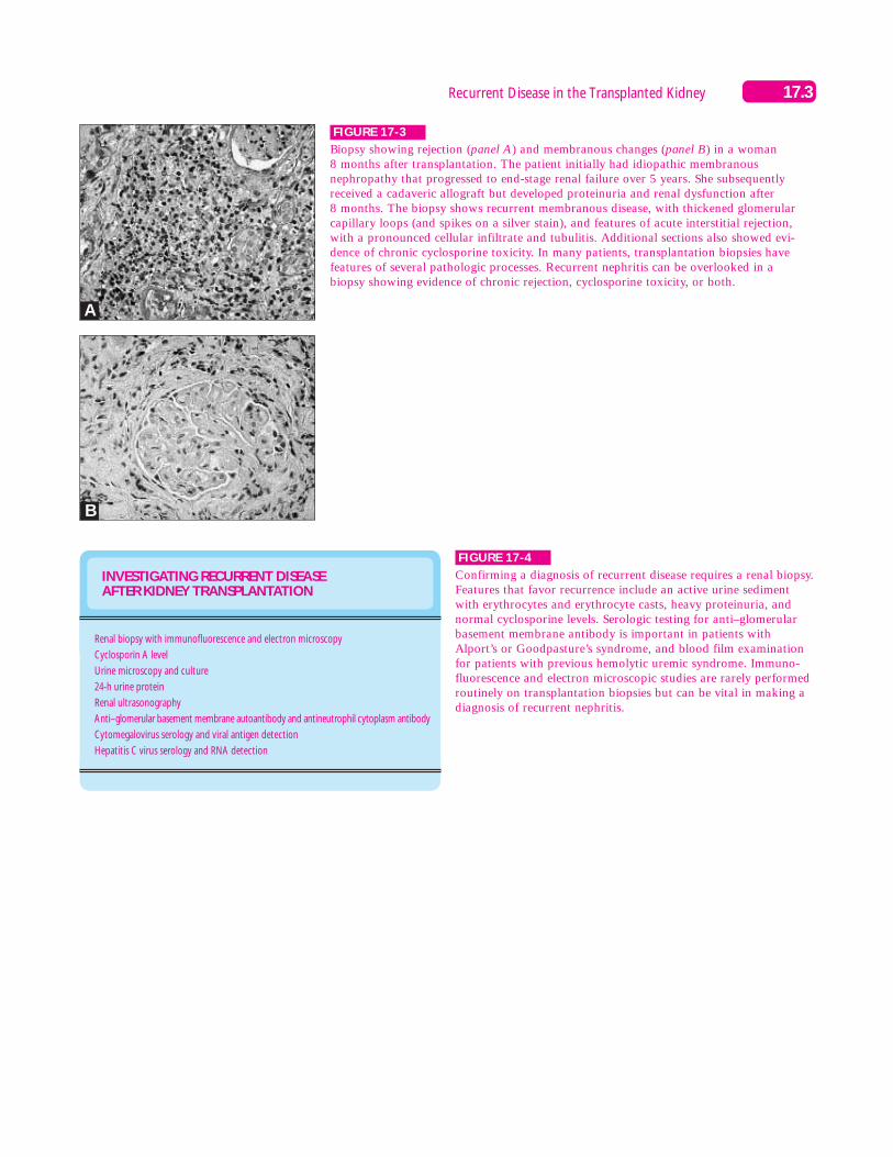
FIGURE 17-3
Biopsy showing rejection (panel A) and membranous changes (panel B) in a woman 8 months after transplantation. The patient initially had idiopathic membranousnephropathy that progressed to end-stage renal failure over 5 years. She subsequentlyreceived a cadaveric allograft but developed proteinuria and renal dysfunction after 8 months. The biopsy shows recurrent membranous disease, with thickened glomerularcapillary loops (and spikes on a silver stain), and features of acute interstitial rejection,with a pronounced cellular infiltrate and tubulitis. Additional sections also showed evi-dence of chronic cyclosporine toxicity. In many patients, transplantation biopsies have features of several pathologic processes. Recurrent nephritis can be overlooked in a biopsy showing evidence of chronic rejection, cyclosporine toxicity, or both.
INVESTIGATING RECURRENT DISEASE AFTER KIDNEY TRANSPLANTATION
Renal biopsy with immunofluorescence and electron microscopy
Cyclosporin A level
Urine microscopy and culture
24-h urine protein
Renal ultrasonography
Anti–glomerular basement membrane autoantibody and antineutrophil cytoplasm antibody
Cytomegalovirus serology and viral antigen detection
Hepatitis C virus serology and RNA detection
FIGURE 17-4
Confirming a diagnosis of recurrent disease requires a renal biopsy.Features that favor recurrence include an active urine sedimentwith erythrocytes and erythrocyte casts, heavy proteinuria, andnormal cyclosporine levels. Serologic testing for anti–glomerularbasement membrane antibody is important in patients withAlport’s or Goodpasture’s syndrome, and blood film examinationfor patients with previous hemolytic uremic syndrome. Immuno-fluorescence and electron microscopic studies are rarely performedroutinely on transplantation biopsies but can be vital in making adiagnosis of recurrent nephritis.
17.4 Transplantation as Treatment of End-Stage Renal Disease
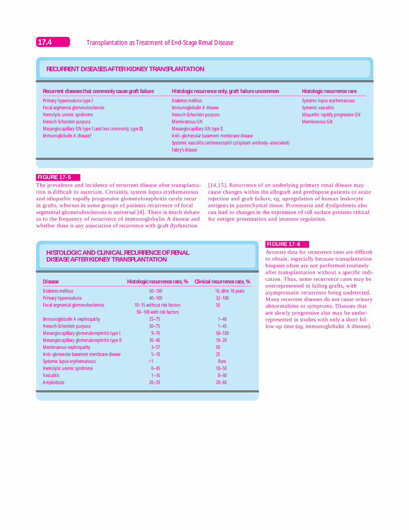
HISTOLOGIC AND CLINICAL RECURRENCE OF RENAL DISEASE AFTER KIDNEY TRANSPLANTATION
Disease
Diabetes mellitus
Primary hyperoxaluria
Focal segmental glomerulosclerosis
Immunoglobulin A nephropathy
Henoch-Schonlein purpura
Mesangiocapillary glomerulonephritis type I
Mesangiocapillary glomerulonephritis type II
Membranous nephropathy
Anti–glomerular basement membrane disease
Systemic lupus erythematosus
Hemolytic uremic syndrome
Vasculitis
Amyloidosis
Histologic recurrence rate, %
50–100
40–100
10–15 without risk factors
50–100 with risk factors
25–75
30–75
9–70
30–40
3–57
5–10
<1
0–45
1–16
20–33
Clinical recurrence rate, %
10, after 10 years
32–100
50
1–40
1–45
50–100
10–20
50
25
Rare
10–50
0–40
20–60
FIGURE 17-6
Accurate data for recurrence rates are difficultto obtain, especially because transplantationbiopsies often are not performed routinelyafter transplantation without a specific indi-cation. Thus, some recurrence rates may beoverrepresented in failing grafts, withasymptomatic recurrence being undetected.Many recurrent diseases do not cause urinaryabnormalities or symptoms. Diseases thatare slowly progressive also may be under-represented in studies with only a short fol-low-up time (eg, immunoglobulin A disease).
RECURRENT DISEASES AFTER KIDNEY TRANSPLANTATION
Recurrent diseases that commonly cause graft failure
Primary hyperoxaluria type I
Focal segmental glomerulosclerosis
Hemolytic uremic syndrome
Henoch-Schonlein purpura
Mesangiocapillary GN type I (and less commonly, type II)
Immunoglobulin A disease?
Histologic recurrence only, graft failure uncommon
Diabetes mellitus
Immunoglobulin A disease
Henoch-Schonlein purpura
Membranous GN
Mesangiocapillary GN type II
Anti–glomerular basement membrane disease
Systemic vasculitis (antineutrophil cytoplasm antibody–associated)
Fabry’s disease
Histologic recurrence rare
Systemic lupus erythematosus
Systemic vasculitis
Idiopathic rapidly progressive GN
Membranous GN
FIGURE 17-5
The prevalence and incidence of recurrent disease after transplanta-tion is difficult to ascertain. Certainly, system lupus erythematosusand idiopathic rapidly progressive glomerulonephritis rarely recurin grafts, whereas in some groups of patients recurrence of focalsegmental glomerulosclerosis is universal [4]. There is much debateas to the frequency of recurrence of immunoglobulin A disease andwhether there is any association of recurrence with graft dysfunction
[14,15]. Recurrence of an underlying primary renal disease maycause changes within the allograft and predispose patients to acuterejection and graft failure, eg, upregulation of human leukocyteantigens in parenchymal tissue. Proteinuria and dyslipidemia alsocan lead to changes in the expression of cell surface proteins criticalfor antigen presentation and immune regulation.
17.5Recurrent Disease in the Transplanted Kidney
0 5 10 15 20 250
100
80
60
40
20
Gra
ft s
urv
ival
, %
Grafted before 1983, yA
Patients with glomerulonephritisPatients without glomerulonephritis
0 5 100
100
80
60
40
20
Gra
ft s
urv
ival
, %
Grafted since 1983, yB
Patients with glomerulonephritisPatients without glomerulonephritis
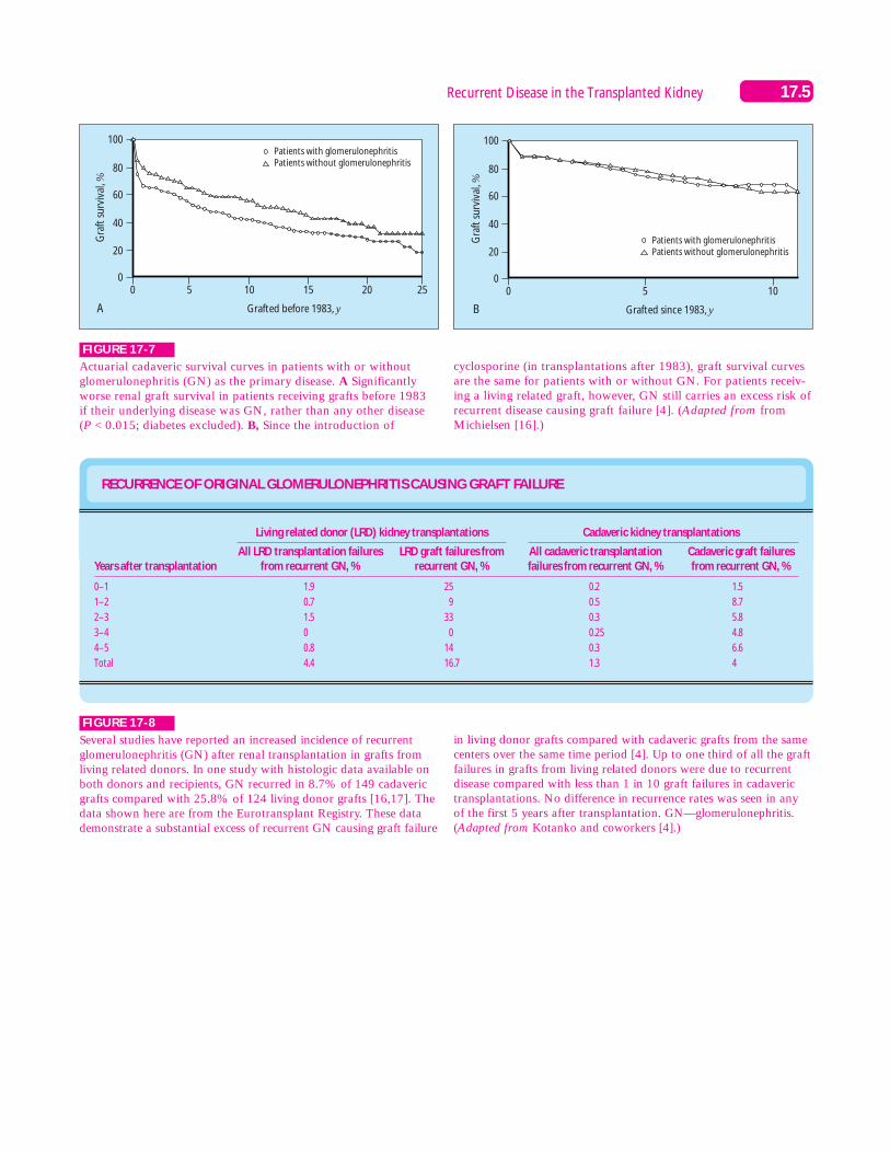
FIGURE 17-7
Actuarial cadaveric survival curves in patients with or withoutglomerulonephritis (GN) as the primary disease. A Significantlyworse renal graft survival in patients receiving grafts before 1983 if their underlying disease was GN, rather than any other disease(P < 0.015; diabetes excluded). B, Since the introduction of
cyclosporine (in transplantations after 1983), graft survival curvesare the same for patients with or without GN. For patients receiv-ing a living related graft, however, GN still carries an excess risk ofrecurrent disease causing graft failure [4]. (Adapted from fromMichielsen [16].)
RECURRENCE OF ORIGINAL GLOMERULONEPHRITIS CAUSING GRAFT FAILURE
Years after transplantation
0–1
1–2
2–3
3–4
4–5
Total
All LRD transplantation failuresfrom recurrent GN, %
1.9
0.7
1.5
0
0.8
4.4
Living related donor (LRD) kidney transplantations
LRD graft failures fromrecurrent GN, %
25
9
33
0
14
16.7
Cadaveric kidney transplantations
Cadaveric graft failuresfrom recurrent GN, %
1.5
8.7
5.8
4.8
6.6
4
All cadaveric transplantationfailures from recurrent GN, %
0.2
0.5
0.3
0.25
0.3
1.3
FIGURE 17-8
Several studies have reported an increased incidence of recurrentglomerulonephritis (GN) after renal transplantation in grafts fromliving related donors. In one study with histologic data available onboth donors and recipients, GN recurred in 8.7% of 149 cadavericgrafts compared with 25.8% of 124 living donor grafts [16,17]. Thedata shown here are from the Eurotransplant Registry. These datademonstrate a substantial excess of recurrent GN causing graft failure
in living donor grafts compared with cadaveric grafts from the samecenters over the same time period [4]. Up to one third of all the graftfailures in grafts from living related donors were due to recurrentdisease compared with less than 1 in 10 graft failures in cadaverictransplantations. No difference in recurrence rates was seen in any of the first 5 years after transplantation. GN—glomerulonephritis.(Adapted from Kotanko and coworkers [4].)
17.6 Transplantation as Treatment of End-Stage Renal Disease
CAUSE OF GRAFT LOSS IN RENAL GRAFT RECIPIENTS WITH DIABETES DURING THE FIRST AND SECOND DECADES
Cause
Deaths with functioning grafts:
Cardiovascular disease
Sepsis
Malignancy
Other
Rejection
Recurrent diabetic nephropathy
Technical
Other
First decade, %(No. of patients)
56 (104)
16
14
2
24
31 (62)
0 (0)
8 (14)
5 (9)
Second decade, %(No. of patients)
76 (19)
40
4
16
16
16(4)
8 (2)
0 (0)
0 (0)
134320
Transplant
Glomerular capillarybasement membrane
thickening
Mesangialexpansion,
microalbuminuria
Hyaline vasculopathyalmost universal
18% of patients havesevere
mesangial expansion(Kimmelsteil-Wilson
nodules)
Years
FIGURE 17-10
Recurrence of diabetes in renal allografts is a common histologicfinding but a rare cause of graft loss. The most frequent cause ofdeath in the second decade after transplantation was cardiovasculardisease, and the most common cause of graft loss was the death ofa patient with a functioning graft. Only 2 of 100 patients survivingmore than 10 years suffered graft loss from recurrent diabeticnephropathy, occurring at 12.6 and 13.6 years after transplantation[2]. The incidence of vascular complications and the need for ampu-tations, however, are substantially increased in patients with diabetesreceiving transplantations. In most centers, overall graft survival ratesare lower for recipients with diabetes than for those without diabetes.(Adapted from Najarian and coworkers [2].)
FIGURE 17-11
Diabetic changes in renal allografts transplanted into patients withdiabetes. Diabetic changes (especially glomerular capillary wallthickening and hyaline vasculopathy) probably occur in all theserecipients [2,10]. Diabetic changes occur slowly, however, and rarelyare severe enough to cause graft dysfunction. The serum creatinineat 10 years in 95 patients from Minnesota with renal allograftsfunctioning for more than 10 years was 1.5 ´0.1 mg/dL (mean ´standard error of the mean) and in 10 patients with allograft functionfor 15 or more years was 1.6 ´0.3 mg/dL [2]. Classic nodularglomerulosclerosis is much rarer. Recurrence of diabetic nephropathycan be prevented by simultaneous pancreatic and renal transplanta-tion. At 2 years, most patients receiving a combined pancreatic andkidney graft have no histologic changes on renal biopsy and normalbasement membrane thickness on electron microscopy of glomerulartissue [10,11]. Intensive insulin treatment with good glycemic controlafter transplantation also prevents the development of recurrentglomerular and arteriolar lesions.
0 5 10 150
5
10
15
20
25
30
35
40
45
20
Rec
urr
ence
, %
Time of follow-up, yA
P<0.02
Nephx
No Nephx
0 5 10 150
5
10
15
20
25
30
35
40
20
Gra
ft lo
ss fr
om
rec
urr
ence
, %
Time of follow-up, yB
P<0.01
Nephx
No Nephx
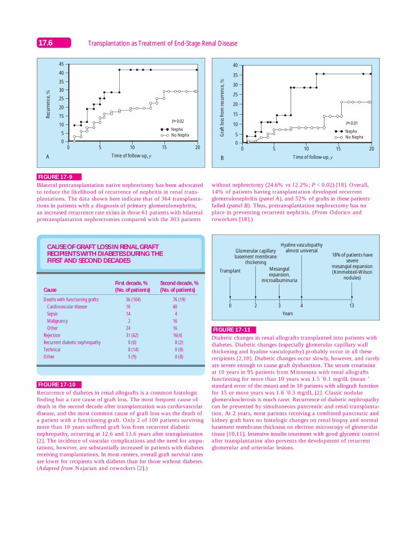
FIGURE 17-9
Bilateral pretransplantation native nephrectomy has been advocatedto reduce the likelihood of recurrence of nephritis in renal trans-plantations. The data shown here indicate that of 364 transplanta-tions in patients with a diagnosis of primary glomerulonephritis, an increased recurrence rate exists in those 61 patients with bilateralpretransplantation nephrectomies compared with the 303 patients
without nephrectomy (24.6% vs 12.2%; P < 0.02) [18]. Overall,14% of patients having transplantation developed recurrentglomerulonephritis (panel A), and 52% of grafts in these patientsfailed (panel B). Thus, pretransplantation nephrectomy has noplace in preventing recurrent nephritis. (From Odorico andcoworkers [18].)

17.7Recurrent Disease in the Transplanted Kidney
Glyoxylate
Oxalate
Alanine: glyoxylateaminotransferase
(AGT)
Cofactor: pyridoxine
Glyoxylatereductase
Glycolate
Glycine
Lactate dehydrogenaseL-α-hydroxy acid oxidase
Glycolate oxidase
FIGURE 17-12
Primary hyperoxaluria type I in renal failure. Primary hyperoxaluriatype I is an autosomal recessive inborn error of metabolism resultingfrom a deficiency (or occasionally incorrect subcellular localization)of hepatic peroxisomal alanine–glyoxylate aminotransferase [8].Patients excrete excess oxalate as a result of the increased glyoxylatepool. In many patients, renal disease is manifested by chronic renalfailure. Once the glomerular filtration rate has decreased below 25mL/min the combination of oxalate overproduction and reducedurinary excretion leads to systemic oxalosis, with calcium oxalatedeposition in many tissues. Renal transplantation alone has yieldedpoor results in the past, with 1-year graft survival rates of only26% [3]. Combined hepatorenal transplantation simultaneouslyreplaces renal function and corrects the underlying metabolic defect.The 1-year liver graft survival rate is 88%, with patient survival of80% at 5 years. Of 24 renal grafts from the European experienceof hepatorenal transplantation, 17 were still functioning at 3 monthsto 2 years after transplantation [19].
FIGURE 17-13
Histologic slide of a patient who received an isolated renal allograftfor primary hyperoxaluria type I in which oxylate crystals are seenclearly within the tubules and interstitium. The major hazards forthe renal graft after transplantation include early acute nephrocal-cinosis caused by rapid mobilization of the systemic oxalate deposits.Acute tubular obstruction by calcium oxalate crystals also canoccur. Late nephrocalcinosis leads to progressive loss of renal functionover several years. Rejection episodes are less common in patientsreceiving combined liver and kidney grafts than in those receivingkidney transplantation alone [3,19]. Acute rejection with renal dysfunction, however, causes additional episodes of acute calciumoxalate deposition in the kidney. Recurrent oxalosis can be seen asearly as 3 months after transplantation.
PATIENT MANAGEMENT IN RENAL OR HEPATORENALTRANSPLANTATIONS FOR PRIMARY HYPEROXALURIA
Aggressive preoperative dialysis (and possibly continued postoperatively)
Maintenance of high urine output
Low oxalate, low ascorbic acid, diet low in vitamin D
Phosphate supplements
Magnesium glycerophosphate
High-dose pyridoxine (500 mg/d)
Thiazide diuretics
FIGURE 17-14
Daily hemodialysis for at least 1 week before transplantationdepletes the systemic oxalate pool to some extent. Some centerscontinue aggressive hemodialysis after transplantation, regardlessof the renal function of the transplanted organ. In patients receivingcombined hepatorenal grafts, dietary measures to reduce oxalateproduction are not as important as they are in patients receivingisolated kidney grafts. In these patients, excess production ofoxalate from glyoxylate still occurs. Magnesium and phosphatesupplements are powerful inhibitors of calcium oxalate crystallizationand should be used in all recipients, whereas thiazide diuretics mayreduce urinary calcium excretion. Pyridoxine is a cofactor for alanine–glyoxylate aminotransferase and can increase the activity of the enzymein some patients. Pyridoxine has no role in combined hepatorenaltransplantation. For most patients the ideal option is probably acombined transplantation when their glomerular filtration ratedecreases below 25 mL/min [8,9].

17.8 Transplantation as Treatment of End-Stage Renal Disease
A B
FIGURE 17-16
Microradioangiography comparing the vasculature of the kidney in a patient withno disease (panel A) and a patient withhomozygous sickle cell disease (panel B)[22]. Despite the frequency of renal damagein sickle cell disease, only 4% of patientsprogress to end-stage renal disease, and littleexperience exists with renal transplantation.Three patients have been reported withrecurrent sickle cell nephropathy. In onecase, a patient developed renal dysfunction3.5 years after transplantation; a biopsyshowed glomerular sclerosis, tubular atrophy, and interstitial fibrosis, withoutfeatures of rejection. A second study reportedrecurrent sickle cell nephropathy leading tograft failure in two of eight patients receivingtransplantation [23]. Concentration defectswere observed within 12 months of grafting.Patients also suffered an increased incidenceof sickle cell crises after renal transplantation,possibly associated with the increase inhematocrit.
AMYLOIDOGENIC AND RELATED DISEASES CAUSING RENAL FAILURE
Disease
Nonhereditary
Systemic amyloidosis associated with chronic inflammatory disorders (especially rheumatoid arthritis)
Systemic amyloidosis associated with immune dyscrasia: multiple myeloma, monoclonal gammopathy,occult immune dyscrasia, lymphoma
Hereditary
Familial Mediterranean fever
Ostertag-type (autosomal dominant, early hypertension, and renal impairment)
Muckle-Wells syndrome (deafness, nephropathy, urticaria, and limb pain)
Hereditary renal amyloidosis
Familial nephropathic systemic amyloidosis
Light chain deposition disease
Fibril protein
Amyloid A
AL
Amyloid A
Not known
Not known
Fibrinogen
Apolipoprotein A
AL or immunoglobulin light chains
Precursor protein
Serum amyloid A
Monoclonal immunoglobulin light chain
Serum amyloid A
Not known
Not known
Fibrinogen
Apolipoprotein A
Immunoglobulin light chains
FIGURE 17-15
The most common cause of amyloidosis leading to renal failure isrheumatoid arthritis [20]. However, increasing numbers of patientswith myeloma and AL amyloid, or primary amyloidosis, are nowreceiving peripheral blood stem cell transplantations or bone mar-row allografts. Thus, these patients are surviving long enough toconsider renal transplantation. Over 60 patients with renal failureresulting from systemic amyloid A (AA) amyloidosis have beenreported to have received renal allografts. Graft survival in thesepatients is the same as that of a matched population. Histologic
recurrence of renal amyloid has been reported in 20% to 33% ofthese grafts within 2 years of transplantation [20,21]. Patient survivalis reduced, owing to infections and vascular complications, to 68% at1 year and 51% at 2 years. Recurrence is characterized by proteinuria11 months to 3 years after transplantation. Recurrent light chaindeposition disease is found in half of patients receiving allografts, withgraft loss in one third despite plasmapheresis and chemotherapy [4].Heavy proteinuria is seen at the onset of recurrence. AL—primaryamyloidosis.
17.9Recurrent Disease in the Transplanted Kidney
FEATURES OF RECURRENT SYSTEMIC LUPUS ERYTHEMATOSUS
Rash
Arthralgia
Proteinuria (usually nonnephrotic)
Increasing anti-DNA antibody titers
Increasing antinuclear antibody titers
Decreasing complement levels (C3 and C4)
FIGURE 17-17
Nephritis caused by systemic lupus erythe-matosus (SLE) rarely recurs in transplanta-tions. SLE accounts for approximately 1%of all patients receiving allografts, and lessthan 1% of these will develop recurrentrenal disease. Time to recurrence has beenreported as 1.5 to 9 years after transplanta-tion [24,25]. Cyclosporine therapy does notprevent recurrence. It is reasonable toensure that serologic test results for SLE areminimally abnormal before transplantationand certainly that patients have no evidenceof active extrarenal disease. Patients withlupus anticoagulant and anticardiolipinantibodies are at risk of thromboembolicevents, including renal graft vein or arterythrombosis. These patients may requireanticoagulation therapy, or platelet inhibi-tion with aspirin.
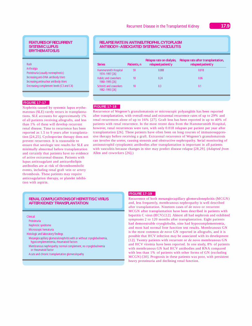
RELAPSE RATE IN ANTINEUTROPHIL CYTOPLASM ANTIBODY–ASSOCIATED SYSTEMIC VASCULITIS
Series
Hammersmith Hospital1974–1997 [26]
Habitz and coworkers1980–1995 [26]
Schmitt and coworkers1982–1993 [26]
Patients, n
59
18
18
Relapse rate on dialysis,relapses/patient/y
0.088
0.24
0.3
Relapse rate after transplantation,relapses/patient/y
0.018
0.06
0.1
FIGURE 17-18
Recurrence of Wegener’s granulomatosis or microscopic polyangiitis has been reportedafter transplantation, with overall renal and extrarenal recurrence rates of up to 29% andrenal recurrences alone of up to 16% [27]. Graft loss has been reported in up to 40% ofpatients with renal recurrence. In the most recent data from the Hammersmith Hospital,however, renal recurrences were rare, with only 0.018 relapses per patient per year aftertransplantation [26]. These patients have often been on long courses of immunosuppres-sive therapy before receiving a graft. Extrarenal recurrence of Wegener’s granulomatosiscan involve the ureter, causing stenosis and obstructive nephropathy. Serial monitoring ofantineutrophil cytoplasmic antibodies after transplantation is important in all patients with vasculitis because changes in titer may predict disease relapse [28,29]. (Adapted fromAllen and coworkers [26].)
RENAL COMPLICATIONS OF HEPATITIS C VIRUSAFTER KIDNEY TRANSPLANTATION
Clinical:
Proteinuria
Nephrotic syndrome
Microscopic hematuria
Histologic and laboratory findings
Mesangiocapillary glomerulonephritis with or without cryoglobulinemia, hypocomplementemia, rheumatoid factors
Membranous nephropathy: normal complement, no cryoglobulinemia or rheumatoid factor
Acute and chronic transplantation glomerulopathy
FIGURE 17-19
Recurrence of both mesangiocapillary glomerulonephritis (MCGN)and, less frequently, membranous nephropathy is well describedafter transplantation. Nineteen cases of de novo or recurrentMCGN after transplantation have been described in patients withhepatitis C virus (HCV) [12]. Almost all had nephrosis and exhibitedsymptoms 2 to 120 months after transplantation. Eight patientshad demonstrable cryoglobulin, nine had hypocomplementemia,and most had normal liver function test results. Membranous GNis the most common de novo GN reported in allografts, and it ispossible that HCV infection may be associated with its development[12]. Twenty patients with recurrent or de novo membranous GNand HCV viremia have been reported. In one study, 8% of patientswith membranous GN had HCV antibodies and RNA comparedwith less than 1% of patients with other forms of GN (excludingMCGN) [30]. Prognosis in these patients was poor, with persistentheavy proteinuria and declining renal function.

17.10 Transplantation as Treatment of End-Stage Renal Disease
RISK FACTORS FOR RECURRENT FOCAL SEGMENTALGLOMERULOSCLEROSIS AFTER TRANSPLANTATION
Recurrence rate, %
50
80–100
75–85
10–15
Risk factor
Age <5 y
Age < 15 y with progression to end-stage renal diseasewithin 3 y
First graft lost from focal segmental glomerulosclerosis
Adults without risk factors
Graft loss occurs in half of all patients with recurrent focal segmental glomerulosclerosisand nephrotic syndrome.
FIGURE 17-20
Focal segmental glomerulosclerosis accounts for 7% to 10% ofpatients requiring renal replacement therapy. The overall recurrencerate is approximately 20% to 30% [1,4,31]. These numbers, however,may be an underestimate because of biopsy sampling errors. Patientsat high risk for recurrence can be identified, particularly childrenwith rapid evolution of their original disease and mesangial expansionon biopsy [1,32]. Recurrence manifests with proteinuria (often10–40 g/d), developing hours to weeks after transplantation. Inchildren the mean time to recurrence is 14 days. Recurrence is notbenign and leads to graft loss in up to half of patients. Patients athighest risk for recurrence should not receive grafts from livingrelated donors.
A. RECURRENT FOCAL SEGMENTAL GLOMERULOSCLEROSIS AND ACUTE RENAL FAILURE AFTER TRANSPLANTATION
Acute renal failure (23)
No acute renal failure (50)
Patients with no recurrence, n
7
40
Patients with recurrence, n
16
10
FIGURE 17-21
Patients with recurrent focal segmentalglomerulosclerosis are at substantiallyincreased risk of developing both acuterenal failure (panel A) after transplantationand acute rejection episodes (panel B). Inone study, 23 of 26 patients with recurrencedeveloped one or more episodes of rejec-tion, compared with only 11 of 40 patientswithout recurrence [31]. Although the mecha-nism for the increased rate of acute dysfunc-tion and rejection is unclear, proteinuria anddyslipidemia may alter the expression of cellsurface immunoregulatory molecules andmajor histocompatibility complex antigens.(Adapted from Kim and coworkers [31].)
B. ACUTE REJECTION EPISODES AMONG ACUTE RENAL FAILURE CASES
>1 acute rejection episode
No rejection
Patients with recurrence
Acute renal failure
16
0
No acute renal failure
7
3
Patients with no recurrence,no acute renal failure
11
29

17.11Recurrent Disease in the Transplanted Kidney
550
10
4
3
2
1
8
6
4
2
155
Seru
m c
reat
inin
e, m
g/d
L
Uri
nar
y p
rote
in e
xcre
tio
n, g
/d
Day after transplantationA
6 3
500 5405200
10
12
10
6
8
2
4
8
6
4
2
580560
Seru
m c
reat
inin
e, m
g/d
L
Uri
nar
y p
rote
in e
xcre
tio
n, g
/d
Day after transplantationC
4
400 5000
10
5
4
3
2
1
8
6
4
2
600
Seru
m c
reat
inin
e, m
g/dL
Uri
nar
y p
rote
in e
xcre
tio
n, g
/d
Day after transplantationD
3
60 110 160 2100
10
10
8
6
4
2
8
6
4
2
260
Seru
m c
reat
inin
e, m
g/d
L
Uri
nar
y p
rote
in e
xcre
tio
n, g
/d
Day after transplantationB
55 2
FIGURE 17-22
Serum creatinine concentrations and urinary protein excretion in four patients (A–D) withrecurrent nephrotic syndrome after transplantation treated by protein adsorption. Each bar indicates one cycle of treatment and the numbers above the bars indicate the sessions of treatment in that cycle. A number of studies have demonstrated that both plasmaexchange and protein adsorption (using protein A sepharose), can decrease urinary proteinexcretion in recurrent focal segmental glomerulosclerosis [6,7,33]. Four examples areshown here. In this study, protein excretion decreased by 82% but returned to pretreatmentlevels within 2 months in seven of eight patients. More intensive treatment regimens haveled to longer remissions [7]. The nature of the circulating factor responsible for proteinleakage is unknown. There are case reports of children with recurrent focal segmentalglomerulosclerosis responding to high-dose intravenous cyclosporine with remission ofnephrotic syndrome. However, cyclosporine does not prevent recurrence when used as part of the initial immunosuppressive regimen. (Adapted from Dantal and coworkers [6].)
DIFFERENTIAL DIAGNOSIS OF SEGMENTAL GLOMERULAR SCARS ON TRANSPLANTATION BIOPSY
Diagnosis
Recurrent FSGS
Rejection
Cyclosporine-related
De novo FSGS
Other glomerulonephritides
Features
Recurrent heavy proteinuria within 3 mo
Original disease caused renal failure in <3 y
Insidious onset of proteinuria
Features of chronic rejection on biopsy, especially vascular sclerosis and glomerulopathy
Previous thrombotic microangiopathy affecting glomeruli
Original disease not FSGS
Chronic rejection excluded
Characteristic immunohistology and electron microscopy, especially in immunoglobulin
A disease
FSGS—focal segmental glomerulosclerosis.
FIGURE 17-23
Segmental glomerular scars in a functioninggraft is a common finding. The interpreta-tion of the biopsy requires knowledge of the previous histology in the native kidneys and the clinical course after transplantation.Immunohistology and electron microscopycan be particularly helpful in this setting.Recurrent focal segmental glomerulosclerosisis the most common cause of early massiveproteinuria. Both rejection and cyclosporinetherapy, however, can cause segmental scarsindistinguishable from those of focal seg-mental glomerulosclerosis. Recurrent or denovo immunoglobulin A disease in an allo-graft also can cause segmental glomerularscarring, but with mesangial hypercellularity,immunoglobulin A detectable by immuno-staining, and paramesangial deposits on electron microscopy.

17.12 Transplantation as Treatment of End-Stage Renal Disease
RECURRENT IMMUNOGLOBULIN A DISEASE
Features
Histologic recurrence, 25%–75%
Clinical recurrence, 1%–40%
Time to recurrence, 2 mo to 4 y
Clinical presentation: asymptomatic, low-grade proteinuria, microscopic hematuria
Susceptibility: human leukocyte antigen B35, DR4; immunoglobulin A rheumatoid factors
Graft loss, <10%
FIGURE 17-24
Up to 75% of patients with immunoglobulin A (IgA) disease develophistologic recurrence within their grafts, which usually presents withmicroscopic hematuria and proteinuria [4,14,15]. Many patients,however, only will have recurrence noted on a routine biopsy aftertransplantation. Most studies suggest that the risk of graft lossresulting from recurrent disease is low (<10%) [4]. However, long-term follow-up in some studies has suggested an increasing rate ofgraft loss with time, approaching 20% at 46 months [14,15].Conversely, one study has documented 100% graft survival at 2years in patients with IgA disease who had IgA anti–human leukocyteantigen (HLA) antibodies [34]. The mechanism is unclear. Theassociation of IgA disease and the HLA alleles B35 and DR4 mayexplain the increased risk of recurrence in grafts from living relateddonors because family members are more likely to share HLA genes.
FIGURE 17-25
Histologic slide of a biopsy from a patient with recurrent immuno-globulin A (IgA) nephropathy. This patient developed proteinuria 9months after receiving a cadaveric allograft. The biopsy shows featuresof recurrent IgA disease with mesangial expansion and a glomerulartuft adhesion to Bowman’s capsule. Immunohistology confirmeddeposition of IgA in the mesangium. At the earliest stages of recur-rence, mesangial IgA and complement C3 are detectable by 3months after transplantation, with electron-dense deposits in theparamesangium but normal appearance on light microscopy. Inpatients with progressive renal dysfunction, crescents often arefound in the glomerulus.
RECURRENT HENOCH-SCHONLEIN PURPURA
Features
Risk of recurrence, 30%–75%
Clinical recurrence, up to 45%
Time to recurrence, immediately to 20 mo
Clinical presentation: often asymptomatic; hematuria, proteinuria, arthralgia, purpuric rash, melena
Susceptibility: rapid development of renal failure in native kidneys, age >14 y
Graft loss: up to 20%, increased in grafts from living related donors
FIGURE 17-26
Most studies have shown that histologic recurrence of Henoch-Schonlein purpura (HSP) is common but rarely causes graft loss.Grafts from living related donors have a substantially increasedrisk of failure as a result of recurrent HSP. Patients can developboth renal and extrarenal manifestations of HSP, especially arthral-gia. Rapid evolution of the original disease and older age at presen-tation (>14 y) seem to be risk factors for clinical recurrence.Cyclosporine does not prevent recurrence. It has been arbitrarilysuggested that transplantation should be avoided for 12 monthsafter resolution of the purpura; however, individual cases of recur-rent disease have been reported despite delays of over 3 yearsbetween resolution of purpura and grafting.

17.13Recurrent Disease in the Transplanted Kidney
MESANGIOCAPILLARY GLOMERULONEPHRITIS
Type II
50%–100%
10%–20%
1 mo to 7 y (usually <1 y)
Frequently asymptomaticnonnephrotic proteinuria,microscopic hematuria
Male, rapidly progressivecourse of initial disease,nephrotic syndrome aftertransplantation
Type I
9%–70%
30%–40%
2 wk to 7 y (median, 1.5 y)
Rarely asymptomatic; proteinuria, nephrotic syndrome, microscopichematuria
Grafts from living related donor
Feature
Histologic recurrence
Clinical recurrence
Time to recurrence
Clinical presentation
Risk factors
FIGURE 17-27
Both mesangiocapillary glomerulonephritis (MCGN) type I (mesan-gial and subendothelial deposits) and type II (dense deposit disease)commonly recur after transplantation. Silent recurrence is foundmore often in type II disease, whereas recurrence of type I MCGNfrequently causes nephrotic syndrome and graft failure [35]. Anincreased risk of recurrence of type I MCGN occurs in grafts fromliving related donors. Type II disease recurs more often in malepatients who progressed rapidly to end-stage renal failure beforetransplantation. The onset of nephrotic syndrome in type II diseaseusually heralds graft failure. No established treatment for recurrentdisease exists, although anecdotally aspirin plus dipyridamole andcyclophosphamide have been used with some success in recurrenttype I MCGN. Plasma exchange has been reported to improve thehistologic changes and induce a clinical remission in one patientwith recurrence of type II MCGN [36].
Subendothelialdeposits
Basementmembrane
Mononuclearcell nucleus
Capillarylumen Interpositioned
mesangialcell
Podocytes
EndothelialcellA
Continuous band ofelectron-dense materialin basement membrane
Capillarylumen
Cell nucleus
Endothelial cell Basement membrane
Podocytefoot
processes
B
FIGURE 17-28
Electron micrographs of mesangiocapillary glomerulonephritis (MCGN) type I (A) andtype II (B). The histologic features of recurrence are the same as for the primary disease. In type II MCGN the ribbonlike band of electron-dense material within the glomerularbasement membrane has been observed as early as 3 weeks after transplantation. Initially,the recurrence is focal but subsequently progresses to involve most of the capillary walls.Failing grafts frequently have segmental glomerular necrosis and extracapillary crescents.Making the diagnosis is not difficult when electron microscopy has been performed on the transplantation biopsy. In MCGN type I, electron-dense deposits first appear in themesangium and subsequently in a subendothelial position. Mesangial cell interposition frequently is visible on electron microscopy, and on light microscopy the capillary wallsappear thickened and show a double contour. The differential diagnosis is MCGN causedby acute or chronic transplantation glomerulopathy. Global changes, immune deposits,and increased mesangial cells, however, are rare in chronic transplantation glomerulopathy.Endocapillary proliferation and macrophages within capillary loops are important featuresof acute transplantation glomerulopathy, which usually are absent in recurrent MCGN [13].

17.14 Transplantation as Treatment of End-Stage Renal Disease
FEATURES OF RECURRENT AND DE NOVO MEMBRANOUS NEPHROPATHY AFTER TRANSPLANTATION
Features
Incidence
Clinical presentation
Time of onset
Histology
Risk factors for graft failure
Incidence of graft failure
De novo membranous
2%–5%
Often asymptomatic; proteinuria, nephrotic syndrome develops slowly
4 mo to 6 y (mean 22 mo)
Identical to native membranous nephropathy, often shows features ofchronic rejection
None specific
Increased over controls; may be as high as 50% but most patients alsohave chronic rejection
Recurrent membranous
3%–57%
Proteinuria, nephrotic syndrome develops rapidly
1 wk to 2 y (mean 10 mo)
Identical to native membranous nephropathy, often shows features ofchronic rejection
Male gender, aggressive clinical course
50%–60%, but some studies have shown no increased graft failure ratecompared with other nephritides
FIGURE 17-29
Recurrence of membranous nephropathy in transplantations is variable,with studies reporting incidences from 3% to 57% [4,37]. The majordifferential diagnosis is de novo membranous nephropathy in patientswith a different underlying renal pathology. De novo allograft mem-branous glomerulonephritis reported in 2% to 5% of transplantationsis often asymptomatic and usually associated with chronic rejection
FIGURE 17-30
Histologic slide of a biopsy showing extensive spike formationalong the glomerular basement membrane. This woman had recurrentmembranous disease 8 months after transplantation. She developednephrotic range proteinuria and subsequent renal dysfunction.Both recurrent and de novo membranous glomerulonephritis areindistinguishable from idiopathic membranous nephropathy. Theinitial lesions are generally stage I or II, although the deposits subsequently become diffuse and intramembranous.
FIGURE 17-31 (see Color Plate)
Histologic slide showing deposition of anti–glomerular basementmembrane (GBM) antibody along the GBM, which is seen in overhalf of patients with Goodpasture’s syndrome who receive an allograftwhile circulating antibodies are still detectable [39]. In most of thesecases no histologic abnormalities are seen within the glomerulus, how-ever, and patients remain asymptomatic with normal renal function.Approximately 25% of patients with antibody deposition will developfeatures of crescentic and rapidly progressive glomerulonephritis andsubsequently suffer graft loss. Delaying transplantation for at least 6months after antibodies have become undetectable reduces therecurrence rate to only 5% to 15%.
[38]. In contrast, recurrent disease frequently causes nephrotic syn-drome, developing within the first 2 years after transplantation. Dataon the incidence of graft failure attributable to membranous diseaseare confusing. Cyclosporine therapy has made no difference in theincidence of the two entities, and hepatitis C virus infection may beassociated with membranous disease after transplantation.
17.15Recurrent Disease in the Transplanted Kidney
00
100
50
1512963
No treatment
Immunosuppressionalone
With plasmaexchange
+immunosuppression
An
tib
od
y ti
ter,
%
Time, mo
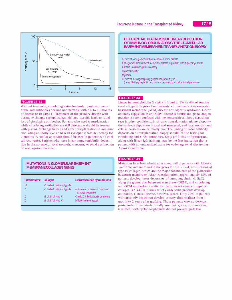
FIGURE 17-32
Without treatment, circulating anti–glomerular basement mem-brane autoantibodies become undetectable within 6 to 18 monthsof disease onset [40,41]. Treatment of the primary disease withplasma exchange, cyclophosphamide, and steroids leads to rapidloss of circulating antibodies. Patients who need transplantationwhile circulating antibodies are still detectable should be treatedwith plasma exchange before and after transplantation to minimizecirculating antibody levels and with cyclophosphamide therapy for2 months. A similar approach should be used in patients with clini-cal recurrence. Patients who have linear immunoglobulin deposi-tion in the absence of focal necrosis, crescents, or renal dysfunctiondo not require treatment.
DIFFERENTIAL DIAGNOSIS OF LINEAR DEPOSITIONOF IMMUNOGLOBULIN ALONG THE GLOMERULARBASEMENT MEMBRANE IN TRANSPLANTATION BIOPSY
Recurrent anti–glomerular basement membrane disease
Anti–glomerular basement membrane disease in patients with Alport’s syndrome
Chronic transplant glomerulopathy
Diabetes mellitus
Myeloma
Recurrent mesangiocapillary glomerulonephritis type I (rarely fibrillary nephritis, and normal cadaveric grafts after initial perfusion)
FIGURE 17-33
Linear immunoglobulin G (IgG) is found in 1% to 4% of routinerenal allograft biopsies from patients with neither anti–glomerularbasement membrane (GBM) disease nor Alport’s syndrome. Linearantibody deposition in anti-GBM disease is diffuse and global and, inpractice, is rarely confused with the nonspecific antibody depositionseen in other conditions. In chronic transplantation glomerulopathythe antibody deposition is focal and segmental, and focal necrosis andcellular crescents are extremely rare. The finding of linear antibodydeposits on a transplantation biopsy should lead to testing for circulating anti-GBM antibodies. Early graft loss or dysfunction,along with linear IgG staining, may be the first indication that apatient with an unidentified cause for end-stage renal disease hasAlport’s syndrome.
MUTATIONS IN GLOMERULAR BASEMENT MEMBRANE COLLAGEN GENES
Chromosome
13
2
X
X
Collagen
�1 and �2 chains of type IV
�3 and �4 chains of type IV
�5 chain of type IV
�6 chain of type IV
Diseases caused by mutations
Autosomal recessive or dominantAlport’s syndrome
Classic X-linked Alport’s syndrome
Diffuse leiomyomatosis
FIGURE 17-34
Mutations have been identified in about half of patients with Alport’ssyndrome and are found in the genes for the �3, �4, or �5 chains oftype IV collagen, which are the major constituents of the glomerularbasement membrane. After transplantation, approximately 15% ofpatients develop linear deposition of immunoglobulin G (IgG)along the glomerular basement membrane (GBM), and circulatinganti-GBM antibodies specific for the �3 or �5 chains of type IVcollagen [42–44]. It is unclear why only some patients developantibodies. Clinical disease, however, is rare. Only 20% of patientswith antibody deposition develop urinary abnormalities from 1month to 2 years after grafting. Those patients who do developproteinuria or hematuria usually lose their grafts. In some cases,treatment with cyclophosphamide did not prevent graft loss.
17.16 Transplantation as Treatment of End-Stage Renal Disease
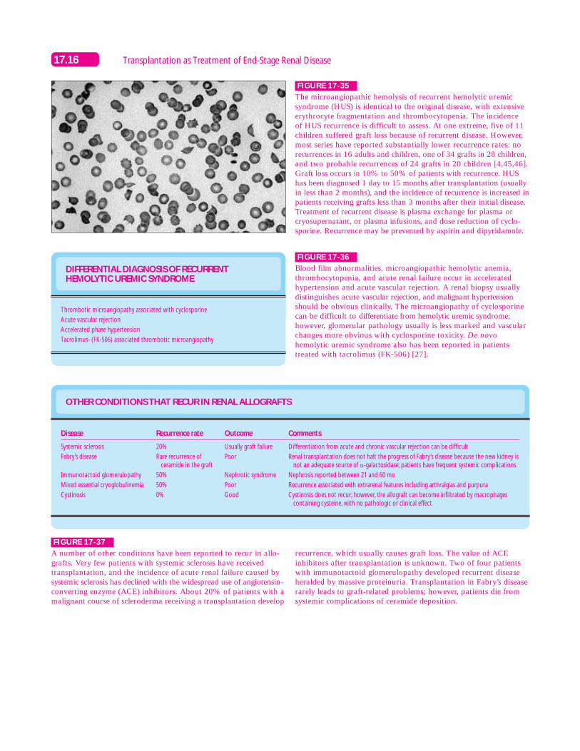
FIGURE 17-35
The microangiopathic hemolysis of recurrent hemolytic uremic syndrome (HUS) is identical to the original disease, with extensiveerythrocyte fragmentation and thrombocytopenia. The incidence of HUS recurrence is difficult to assess. At one extreme, five of 11children suffered graft loss because of recurrent disease. However,most series have reported substantially lower recurrence rates: norecurrences in 16 adults and children, one of 34 grafts in 28 children,and two probable recurrences of 24 grafts in 20 children [4,45,46].Graft loss occurs in 10% to 50% of patients with recurrence. HUShas been diagnosed 1 day to 15 months after transplantation (usuallyin less than 2 months), and the incidence of recurrence is increased inpatients receiving grafts less than 3 months after their initial disease.Treatment of recurrent disease is plasma exchange for plasma orcryosupernatant, or plasma infusions, and dose reduction of cyclo-sporine. Recurrence may be prevented by aspirin and dipyridamole.
DIFFERENTIAL DIAGNOSIS OF RECURRENT HEMOLYTIC UREMIC SYNDROME
Thrombotic microangiopathy associated with cyclosporine
Acute vascular rejection
Accelerated phase hypertension
Tacrolimus- (FK-506) associated thrombotic microangiopathy
OTHER CONDITIONS THAT RECUR IN RENAL ALLOGRAFTS
Disease
Systemic sclerosis
Fabry’s disease
Immunotactoid glomerulopathy
Mixed essential cryoglobulinemia
Cystinosis
Recurrence rate
20%
Rare recurrence ofceramide in the graft
50%
50%
0%
Outcome
Usually graft failure
Poor
Nephrotic syndrome
Poor
Good
Comments
Differentiation from acute and chronic vascular rejection can be difficult
Renal transplantation does not halt the progress of Fabry’s disease because the new kidney isnot an adequate source of �-galactosidase; patients have frequent systemic complications
Nephrosis reported between 21 and 60 mo
Recurrence associated with extrarenal features including arthralgias and purpura
Cystinosis does not recur; however, the allograft can become infiltrated by macrophages containing cysteine, with no pathologic or clinical effect
FIGURE 17-36
Blood film abnormalities, microangiopathic hemolytic anemia,thrombocytopenia, and acute renal failure occur in acceleratedhypertension and acute vascular rejection. A renal biopsy usuallydistinguishes acute vascular rejection, and malignant hypertensionshould be obvious clinically. The microangiopathy of cyclosporinecan be difficult to differentiate from hemolytic uremic syndrome;however, glomerular pathology usually is less marked and vascularchanges more obvious with cyclosporine toxicity. De novohemolytic uremic syndrome also has been reported in patientstreated with tacrolimus (FK-506) [27].
FIGURE 17-37
A number of other conditions have been reported to recur in allo-grafts. Very few patients with systemic sclerosis have receivedtransplantation, and the incidence of acute renal failure caused bysystemic sclerosis has declined with the widespread use of angiotensin-converting enzyme (ACE) inhibitors. About 20% of patients with amalignant course of scleroderma receiving a transplantation develop
recurrence, which usually causes graft loss. The value of ACEinhibitors after transplantation is unknown. Two of four patientswith immunotactoid glomerulopathy developed recurrent diseaseheralded by massive proteinuria. Transplantation in Fabry’s diseaserarely leads to graft-related problems; however, patients die fromsystemic complications of ceramide deposition.

17.17Recurrent Disease in the Transplanted Kidney
MANAGEMENT OF RECURRENT DISEASE AFTER KIDNEY TRANSPLANTATION
Treatment of recurrence
Plasma exchange, immunoadsorption, steroids,angiotensin-converting enzyme inhibitors, nonsteroidal anti-inflammatory drugs
With crescents: plasma exchange, cytotoxics
?Steroids
Aspirin, dipyridamole
?Plasma exchange
?Cytotoxics and steroids
Plasma exchange, cyclophosphamide
Plasma exchange, plasma infusion
Cyclophosphamide and steroids
Glycemic control
Aggressive perioperative dialysis, hydration, low oxalatediet, low ascorbic acid diet, phosphate supplements,magnesium glycerophosphate, pyridoxine
Disease
Focal segmental glomerulosclerosis
Immunoglobulin A nephropathy
Henoch-Schonlein purpura
Mesangiocapillary glomerulonephritis type I
Mesangiocapillary glomerulonephritis type II
Membranous nephropathy
Anti–glomerular basement membrane disease
Hemolytic uremic syndrome
Antineutrophil cytoplasm antibody–associated vasculitis
Diabetes
Oxalosis
FIGURE 17-38
No controlled data exist on the managementof recurrent disease after transplantation.For patients with primary hyperoxaluria,measures to prevent further deposition ofoxalate have proved successful in controllingrecurrent renal oxalosis [9]. In diabetesmellitus, the pathophysiology of recurrentnephropathy undoubtedly reflects the sameinsults as those causing the initial renal failure,and good evidence exists that glycemic controlcan slow the development of end-organdamage. Plasma exchange and immuno-adsorption are promising therapies forpatients with nephrosis who have recurrentfocal segmental glomerulosclerosis; however,these therapies do not provide sustainedremission [6,7]. In all these cases, establishinga diagnosis of recurrent disease is critical inidentifying a possible treatment modality.
WHEN TO AVOID USING LIVING RELATED DONORSIN KIDNEY TRANSPLANTATION
Focal segmental glomerulosclerosis with risk factors for early recurrence
Henoch-Schonlein purpura
Mesangiocapillary glomerulonephritis type I
Mesangiocapillary glomerulonephritis type II with risk factors (familial immunoglobulin A nephropathy and hemolytic uremic syndrome)
FIGURE 17-39
In these diseases, rapid recurrence leading to graft failure is frequentenough to warrant extreme caution in using living related donors.Even excluding these conditions, the overall rate of recurrence ofglomerulonephritis is substantially increased in living related donors,and patients should be made aware of this risk [4]. For familial diseases, the risk of recurrence is even higher (eg, some familieswith immunoglobulin A disease and hemolytic uremic syndrome).Finally, recurrent glomerulonephritis has been reported in up to30% of renal isografts, with disease onset between 2 weeks and 16 years after grafting.
References
1. Tejani A, Stablein DH: Recurrence of focal segmental glomerulonephritisposttransplantation: a special report of the North American PediatricRenal Transplant Cooperative Study. J Am Soc Nephrol 1992,2(suppl):258–263.
2. Najarian JS, Kaufman DB, Fryd DS, et al.: Long term survival followingkidney transplantation in 100 type I diabetic patients. Transplantation1989, 47:106–113.
3. Broyer M, Brunner FP, Brynger H, et al.: Kidney transplantation inprimary oxalosis: data from the EDTA registry. Nephrol DialTransplant 1990, 5:332–336.
4. Kotanko P, Pusey CD, Levy JB: Recurrent glomerulonephritis followingrenal transplantation. Transplantation 1997, 63:1045–1052.
5. Cameron JS: Recurrent primary disease following renal transplantation.In Advanced Renal Medicine. Edited by Raine AEG. Oxford: OxfordUniversity Press; 1992:435–448.
6. Dantal J, Bigot E, Bogers W, et al.: Effect of plasma protein adsorptionon protein excretion in kidney-transplant recipients with recurrentnephrotic syndrome. N Engl J Med 1994, 330:7–14.
7. Artero ML, Sharma R, Savin VJ, et al.: Plasmapheresis reduces protein-uria and serum capacity to injure glomeruli in patients with recurrentfocal glomerulosclerosis. Am J Kidney Dis 1994, 23:574–581.
8. Watts RWE: Primary hyperoxaluria type 1. Q J Med 1994, 87:593–599.9. Allen AR, Thompson EM, Williams G, et al.: Selective renal trans-
plantation in primary hyperoxaluria type 1. Am J Kidney Dis 1996,27:891–895.
10. Bilous RW, Mauer SM, Sutherland DE, et al.: The effects of pancreastransplantation on the glomerular structure of renal allografts in patientswith insulin-dependent diabetes. N Engl J Med 1989, 321:80–85.
11. Remuzzi G, Ruggenenti P, Mauer SM: Pancreas and kidney/pancreastransplants: experimental medicine or real improvement? Lancet1994, 343:27–31.
12. Morales JM, Campistol JM, Andres A, et al.: Glomerular diseases inpatients with hepatitis C virus infection after renal transplantation.Curr Opinion Nephrol Hypertens 1997, 6:511–515.
13. Porter KA: Renal transplantation. In Pathology of the Kidney. Editedby Heptinstall RH. Boston: Little, Brown; 1992:1799–1934.

17.18 Transplantation as Treatment of End-Stage Renal Disease
14. Odum J, Peh CA, Clarkson AR, et al.: Recurrent mesangial IgAnephritis following renal transplantation. Nephrol Dial Transplant1994, 9:309–312.
15. Ohmacht C, Kliem V, Burg M, et al.: Recurrent IgA nephropathy afterrenal transplantation: a significant contributor to graft loss.Transplantation 1997.
16. Michielsen P: Recurrence of the original disease. Does this influencerenal graft failure? Kidney Int 1995, 48(suppl 52):79–84.
17. O’Meara Y, Green A, Carmody M, et al.: Recurrent glomerulonephritisin renal transplants: fourteen years’ experience. Nephrol DialTransplant 1989, 4:730–734.
18. Odorico JS, Knechtle SJ, Rayhill SC, et al.: The influence of nativenephrectomy on the incidence of recurrent disease following renaltransplantation for primary glomerulonephritis. Transplantation1996, 61:228–234.
19. Watts RWE, Danpure CJ, De Pauw L, et al.: Combined liver-kidneyand isolated liver transplantation in primary hyperoxaluria type 1.Nephrol Dial Transplant 1991, 6:502–511.
20. Pasternack A, Ahonen J, Kuhlback B: Renal transplantation in 45patients with amyloidosis. Transplantation 1986, 42:598–601.
21. Livneh A, Zemer D, Siegal B, et al.: Colchicine prevents kidney trans-plant amyloidosis in familial Mediterranean fever. Nephron 1992,60:418–422.
22. Statius van Eps LW: Nature of concentrating defect in sickle cellnephropathy. Lancet 1970, i:450–454.
24. Montgomery R, Zibari G, Hill GS, et al.: Renal transplantation inpatients with sickle cell nephropathy. Transplantation 1994,58:618–620.
24. Goss JA, Cole BR, Jendrisak MD: Renal transplantation for systemiclupus erythematosus and recurrent lupus nephritis: a single centerexperience and review of the literature. Transplantation 1991,52:805–810.
25. Lochhead KM, Pirsch JD, D’Alessandro AM, et al.: Risk factors forrenal allograft loss in patients with systemic lupus erythematosus.Kidney Int 1996, 49:512–517.
26. Allen AR, Pusey CD, Gaskin G: ANCA associated vasculitis: outcomeand relapse on renal replacement therapy. J Am Soc Nephrol 1997,8:81A.
27. Dantal J, Giral M, Hoormant M, et al.: Glomerulonephritis recurrencesafter transplantation. Curr Opin Nephrol Hypertens 1995, 4:146–152.
28. Jayne DR, Gaskin G, Pusey CD, et al.: ANCA and predicting relapsein systemic vasculitis. Q J Med 1995, 88:127–133.
29. De’Oliviera J, Gaskin G, Pusey CD, et al.: Relationship between diseaseactivity and anti-neutrophil cytoplasmic antibody concentration inlong-term management of systemic vasculitis. Am J Kidney Dis 1995,25:380.
30. Takishita Y, Ishikawa S, Okada K: Two cases of membranousglomerulonephritis associated with hepatitis C virus. Nippon JinzoGakkai Shi 1994, 36:1203–1207.
31. Kim EM, Striegel J, Kim Y, et al.: Recurrence of steroid resistantnephrotic syndrome in kidney transplants is associated with increasedacute renal failure and acute rejection. Kidney Int 1994, 45:1440–1445.
32. Senggutuvan P, Cameron JS, Hartley RB, et al.: Recurrence of focalsegmental glomerulosclerosis in transplanted kidneys: analysis of inci-dence and risk factors in 59 allografts. Pediatr Nephrol 1990, 4:21–8.
33. Savin VJ, Sharma R, Sharma M, et al.: Circulating factor associatedwith increased glomerular permeability to albumin in recurrent focalglomerulosclerosis. N Engl J Med 1996, 334:878–883.
34. Mathew TH: Recurrence of disease following renal transplantation.Am J Kidney Dis 1988, 12:85–96.
35. Glicklich D, Matas AJ, Sablay LB, et al.: Recurrent membranoprolif-erative glomerulonephritis type I in successive renal transplants. Am JNephrol 1987, 7:143–149.
36. Oberkircher OR, Enama M, West JC, et al.: Regression of recurrentmembranoproliferative glomerulonephritis type II in a transplantedkidney after plasmapheresis. Transplant Proc 1988, 20:418–423.
37. Couchoud C, Pouteil-Noble C, Colon S, et al.: Recurrence of mem-branous nephropathy after renal transplantation. Transplantation1995, 59:1275–1279.
38. Schwarz A, Krause PH, Offermann G, et al.: Impact of de novo mem-branous glomerulonephritis on the clinical course after kidney trans-plantation. Transplantation 1994, 58:650–654.
39. Levy JB, Pusey CD: Anti-GBM antibody mediated disease. InNephrology. Edited by Wilkinson R, Jamison R. London: Chapman& Hall; 1997:599–615.
40. Peters DK, Rees AJ, Lockwood CM, et al.: Treatment and prognosisin antibasement membrane antibody mediated nephritis. TransplantProc 1982, 14:513–21.
41. Simpson IJ, Doak PB, Williams LC, et al.: Plasma exchange inGoodpasture’s syndrome. Am J Nephrol 1982, 2:301–311.
42. Turner AN, Rees AJ: Goodpasture’s disease and Alport’s syndromes.Ann Rev Med 1996, 47:377–386.
43. Kalluri R, van den Heuvel LP, Smeets HJ, et al.: A COL4A3 genemutation and post-transplant anti-� 3(IV) collagen alloantibodies inAlport syndrome. Kidney Int 1995, 47:1199–1204.
44. Ding J, Zhou J, Tryggvason K, et al.: COL4A5 deletions in threepatients with Alport syndrome and posttransplant antiglomerularbasement membrane nephritis. J Am Soc Nephrol 1994, 5:161–168.
45. Gagnadoux MF, Habib R, Broyer M: Outcome of renal transplanta-tion in 34 cases of childhood hemolytic uremic syndrome and the roleof cyclosporine. Transplant Proc 1994, 26:269–270.
46. Agarwal A, Mauer SM, Matas AJ, et al.: Recurrent hemolytic uremicsyndrome in an adult renal allograft recipient: current concepts andmanagement. J Am Soc Nephrol 1995, 6:1160–1169.

![GRAFTED TOMATO - Iserv1].pdf · GRAFTED TOMATO Grafted onto ... Grafting joins the top part of one plant (the scion) to the root ... (TPIE) - January 18-20, 2012 Spring Trials in](https://img.pdfslide.us/doc/110x75/5aa1ea047f8b9a436d8c452d/grafted-tomato-1pdfgrafted-tomato-grafted-onto-grafting-joins-the-top-part.jpg)