Embed Size (px)
Citation preview

Acta Botánica Mexicana
ISSN: 0187-7151
Instituto de Ecología, A.C.
México
Robinson, Néstor M.; Galicia-García, Citlalli; Okolodkov, Yuri B
NEW RECORDS OF GREEN (CHLOROPHYTA) AND BROWN ALGAE (PHAEOPHYCEAE) FOR
CABEZO REEF, NATIONAL PARK SISTEMA ARRECIFAL VERACRUZANO, GULF OF MEXICO
Acta Botánica Mexicana, núm. 101, octubre, 2012, pp. 11-48
Instituto de Ecología, A.C.
Pátzcuaro, México
Available in: http://www.redalyc.org/articulo.oa?id=57424816002
How to cite
Complete issue
More information about this article
Journal's homepage in redalyc.org
Scientific Information System
Network of Scientific Journals from Latin America, the Caribbean, Spain and Portugal
Non-profit academic project, developed under the open access initiative

Acta Botanica Mexicana 101: 11-48 (2012)
11
NEW RECORDS OF GREEN (CHLOROPHYTA) AND BROWN ALGAE (PHAEOPHYCEAE) FOR CABEZO REEF, NATIONAL PARK SISTEMA
ARRECIFAL VERACRUZANO, GULF OF MEXICO
Néstor M. robiNsoN1, Citlalli GaliCia-GarCía1 & Yuri b. okolodkov2,3
1Instituto Tecnológico de Boca del Río, Laboratorio de Biología, km 12 carretera Veracruz-Córdoba, 94290 Boca del Río,
Veracruz, México.2Universidad Veracruzana, Instituto de Ciencias Marinas y Pesquerías,
Calle Hidalgo núm. 617, Colonia Río Jamapa, 94290 Boca del Río, Veracruz, México.
3Author for correspondence: [email protected]
ABSTRACT
Descriptions of 13 green and 12 brown algal species from 20 genera and 15 families collected on Cabezo Reef of the National Park Sistema Arrecifal Veracruzano in the southwestern Gulf of Mexico, which are new records for the reef, are given. The species belong to the chlorophyte genera Caulerpa, Cladophoropsis, Codium, Dictyosphaeria, Ernodesmis, Halimeda, Neomeris, Parvocaulis, Percursaria and Rhipocephalus and the phaeophycean genera Canistrocarpus, Colpomenia, Cladosiphon, Dictyerpa, Dictyota, Ectocarpus, Padina, Rosenvingea, Sargassum and Sphacelaria. The family Dictyotaceae contained the largest number of species (6). The descriptions include morphometric and biological data and are accompanied by photographs and line drawings for each species. Data on the geographic distribution in the State of Veracruz, park and the Gulf of Mexico in general are also given. Percursaria percursa is a new record for the park, while Dictyerpa jamaicensis, Sargassum furcatum, Caulerpa racemosa var. occidentalis and Codium isthmocladum subsp. clavatum are new records for the State of Veracruz. Most of the specimens of Chlorophyta were found in the vegetative stage, whereas all the brown algal species except Dictyerpa jamaicensis and Sphacelaria rigidula possessed gametangia or sporangia.
Key words: anatomy, brown algae, green algae, Gulf of Mexico, new records, taxonomy.

Acta Botanica Mexicana 101: 11-48 (2012)
12
RESUMEN
Se presentan las descripciones de 13 especies de algas verdes y 12 de algas pardas de 20 géneros y 15 familias, que son nuevos registros para el arrecife Cabezo ubicado en la parte sureste del Parque Nacional Sistema Arrecifal Veracruzano en el suroeste del Golfo de México. Las especies pertenecen a los géneros de clorofitas Caulerpa, Cladophoropsis, Codium, Dictyosphaeria, Ernodesmis, Halimeda, Neomeris, Parvocaulis, Percursaria y Rhipocephalus, y los géneros de algas pardas Canistrocarpus, Colpomenia, Cladosiphon, Dictyerpa, Dictyota, Ectocarpus, Padina, Rosenvingea, Sargassum y Sphacelaria. La familia Dictyotaceae fue la mejor representada en cuanto al número de especies (6). Las descripciones incluyen datos morfométricos y biológicos y están acompañadas con fotografías y dibujos a línea para cada especie. También se presenta la información sobre la distribución de las especies en el estado de Veracruz, el parque y el Golfo de México en general. Percursaria percursa es un nuevo registro para el parque, mientras Dictyerpa jamaicensis, Sargassum furcatum, Caulerpa racemosa var. occidentalis y Codium isthmocladum subsp. clavatum lo son para el estado de Veracruz. La mayor proporción de ejemplares pertenecientes a Chlorophyta se encontraron en estado vegetativo, mientras que los especímenes de algas pardas excluyendo a las especies Dictyerpa jamaicensis y Sphacelaria rigidula, se encontraron ya sea con gametangios o esporangios.
Palabras clave: algas pardas, algas verdes, anatomía, Golfo de México, nuevos registros, taxonomía.
INTRODUCTION
In the State of Veracruz, a total of 54 brown and 100 green algal species have been found, and in the National Park Sistema Arrecifal Veracruzano (NPSAV) in particular, 39 brown and 74 green algal species (Lehman, 1993; Dreckmann, 1998; Ortega et al., 2001; Galicia-García & Morales-García, 2007, González-Gándara et al., 2007; Godínez-Ortega et al., 2009). The largest number of species was encoun-tered on Isla Enmedio, Isla Verde, Isla Sacrificios and Hornos Reef, which is propor-tional to the number of studies carried out at these sites, followed by La Blanquilla, Santiaguillo, La Gallega, Punta Gorda, Giote, Ingenieros and Blanca reefs. There is no published information on brown and green algae from Cabezo Reef. According to Lehman (2007), Santiaguillo is third in terms of registered brown and green algal species (28).

Robinson et al.: New records of green and brown algae for the Sistema Arrecifal Veracruzano
13
To contribute to our knowledge of the morphology and species of Chlorophyta and Phaeophyceae of the NPSAV occurring on Cabezo Reef and to document their records were the main purposes of the present study.
MATERIAL AND METHODS
During CEPIA boat trips within the NPSAV, specimens of green and brown algae were collected on Cabezo Reef (Fig. 1) on 11th March and 12th Novem-ber 2008 and 3rd June 2010 during snorkeling at 0.5 to 1.5 m depth, manually or with a knife, and placed into a 500-ml plastic bottle or a Ziplock plastic bag filled with seawater from the sampling site. Immediately after sampling, a stock 37% formaldehyde solution was added to the samples to a final concentration of 4%. The samples were incorporated into the collection of liquid samples and the herbarium of macroalgae of the Instituto de Ciencias Marinas y Pesquerías de la Universidad Veracruzana (ICIMAP-UV). In the laboratory, morphological fea-
Cabezo
Anegadilla
Santiaguillo
Topatillo
Anegada de Afuera
Enmedio
Rizo
Chopas
Polo
Ingeniero
Laguna de Mandinga
Sacrificios
Pájaros
El Verde
Anegada de AdentroBlanquilla
Bajo PaducahLavandera
Hornos
Bajo MerseyTerranova
Galleguilla
Gallega
Punta Gorda19º15'N
19º10'N
19º05'N
96º10'W
5 km
96º05'W 96º00'W 95º 55'W 95º 50'W
Los BajitosBlanca
Giote
Punta Coyol
National ParkSistema Arrecifal Veracruzano
Veracruz
Boca del Río
MandingaAntón Lizardo
Gulfof Mexico
RíoMoreno
RíoJamapa
Fig. 1. Sampling site (filled circle) in the National Park Sistema Arrecifal Veracruzano. Hatched areas are coral reefs.

Acta Botanica Mexicana 101: 11-48 (2012)
14
tures were observed and described using a stereoscopic low-magnification Carl Zeiss Stemi 2000C microscope. When necessary, cross-sections of algal thalli were made with a razor and photographed using an Olympus BX51 microscope equipped with phase-contrast objectives and an Olympus C7070 Wide Zoom 7.1-megapixel digital camera. A camera lucida was also used to make line draw-ings. Specimens were identified with the use of specialized literature (Taylor, 1960; Joly, 1967; Earle, 1969; Schneider & Searles, 1991; Flores-Moya & Conde, 1998; Littler & Littler, 2000; Moreira & Suárez, 2002; Solé & Foldats, 2003; Boraso de Zaixso, 2004; Dawes & Mathieson, 2008; Miranda Alves et al., 2010; Norris, 2010). In our species descriptions, anatomical and morphological terms for macroalgal structures given by Dawes & Mathieson (2008) were followed, with some exceptions.
Abbreviations used: bas. – basionym; diam. – diameter; GOM – Gulf of Mexico; ICIMAP-UV-AL – herbarium of macroalgae of the Institute for Marine Sciences and Fisheries of the University of Veracruz; LS – liquid sample; Mpio. – municipality; syn. – synonym.
RESULTS
Descriptions of 13 green and 12 brown algal species from 15 families collect-ed on Cabezo Reef are given below. They include morphometric and biological data and are accompanied by photographs and line drawings for each species. The data on the geographic distribution in the NPSAV, the State of Veracruz and the Gulf of Mexico in general are also presented. However, the primary literature sources cited in floristic lists by Dreckmann (1998), Ortega et al. (2001) and Galicia-García & Morales-García (2007) were omitted. More recent publications (Orduña-Medrano, 2004; González-Gándara et al., 2007; Lehman, 2007; Godínez-Ortega et al., 2009) were considered. The taxa are given in the order following Fredericq et al. (2009).
Division Chlorophyta Family Ulvaceae
1. Percursaria percursa (C. Agardh) Rosenvinge, 1893 (Pl. 1, Fig. 1 and 2; Pl. 8, Fig. 1)Bas.: Conferva percursa C. Agardh, 1817: 87.Syn.: Enteromorpha percursa (C. Agardh) J. Agardh, 1842: 15.

Robinson et al.: New records of green and brown algae for the Sistema Arrecifal Veracruzano
15
Thallus filamentous, erect and ramified, 7 mm long. Pale green. Principal branches (62.5)75-80(113) μm in diam., consisting of 3-4 longitudinal rows of quadrangular or rectangular cells 25-40 μm long and 20-25(37.5) μm wide. Fila-ments uniform in diam., 32-38 μm, consisting of 2 rows of cells of very regular shape, rectangular, 22.5-27.5 μm long and 17.5 μm in diam. Cells of the thicker branches are less regular in shape. Median branches 20-25 μm long and 52.5-62.5 μm in diam. Terminal branchlets consist of one row of cells 12.5-17.5 μm long and 12.5-17.5 μm in diam. Single laminar, parietal chloroplast, with 1-3 pyrenoids. Epiphyte on Bryothamnion triquetrum (S. G. Gmelin) M. A. Howe (Rhodophyta).
Examined specimens: LS-8 (1 March 2008).A new record for the NPSAV.Elsewhere in Veracruz: Mpio. San Andrés Tuxtla: Playa Escondida (Valen-
zuela, 1987).GOM: SE (Fredericq et al., 2009).References: Taylor, 1960: 54; Boraso de Zaixso, 2004: 101, pl. 7, fig. 20;
Dawes & Mathieson, 2008: 30, pl. 1, fig. 11 and 12.
Family Siphonocladaceae
2. Cladophoropsis membranacea (Hofman Bang ex C. Agardh) Børgesen, 1905 (Pl. 1, Fig. 3-5; Pl. 8, Fig. 2 and 3)Bas.: Conferva membranacea Hofman Bang ex C. Agardh, 1824: 120.Syn.: Cladophora membranacea (Hofman Bang ex C. Agardh) Kütz., 1843: 271; Acrosiphonia membranacea (Hofman Bang ex C. Agardh) J. Agardh, 1846: 104; Aegagropila membranacea (C. Agardh) Kütz., 1854: 4, pl. 14.
Thallus filamentous, gregarious. Filaments uniseriate, 175-250 μm in diam., forming dense cespitose clumps of 15 cm width. Glossy light green. Branching al-ternate inferiorly and unilateral superiorly. Lateral filaments 100-140 μm in diam., originating from the parietal cells as extensions, having an open connection with the parent cells. Chloroplasts reticulate, with numerous pyrenoids. It is attached to the substrate by a stolon with rhizoids with finger-shaped extremities (specialized hapteroid cells) oriented horizontally, pale or lightly colored.
Examined specimens: ICIMAP-UV-AL 08, 08R (12 November 2008).NPSAV: Isla de Enmedio, Isla Verde, Isla Santiaguillo, Gallega Reef (Ortega
et al., 2001; Galicia-García & Morales-García, 2007).

Acta Botanica Mexicana 101: 11-48 (2012)
16
Elsewhere in Veracruz: Mpio. Tuxpan: Isla de Lobos; Mpio. Actopan: La Mancha (Punta Mancha); Playa Paraíso (La Mancha), Villa Rica (Punta Villa Rica); Mpio. Alto Lucero: Boca Andrea, El Morro (Punta del Morro), Laguna Verde; Mpio. Catemaco: Laguna de Sontecomapan (Dreckmann, 1998; Ortega et al., 2001).
GOM: NW, SW and SE (Fredericq et al., 2009).References: Taylor, 1960: 118; Littler & Littler, 2000: 332, 333 (fig.); Dawes &
Mathieson, 2008: 55, pl. 4, fig. 5.
3. Dictyosphaeria cavernosa (Forsskäl) Børgesen, 1932 (Pl. 1, Fig. 6 and 7; Pl. 8, Fig. 4 and 5)Bas.: Ulva cavernosa Forsskål, 1775: 187.Syn.: Valonia favulosa C. Agardh, 1823 (1822-1823): 432; Dictyosphaeria favulosa (C. Agardh) Decaisne ex Endlicher, 1843: 18.
Thallus pseudoparenchymatous, globular, saclike, hollow, spherical when young and irregularly lobed and ruptured when mature, 5-6 cm in diam. Bright green, loses color rapidly after exposure to light. Membrane cells 0.8-1.2 mm in diam., arranged in one row, angular or polyhedral in surface view, beehive-like, at-tached to each other by hapteroid cells of 50-60 μm diam., forming a continuous row of primary cells alternately arranged.
Examined specimens: ICIMAP-UV-AL 08 (12 November 2008).NPSAV: Playa Mocambo, San Juan de Ulúa, Hornos Reef, Isla de Enmedio, Isla
Sacrificios, Isla Verde (Ortega et al., 2001; Galicia-García & Morales-García, 2007). Elsewhere in Veracruz: Mpio. Tuxpan: Isla de Lobos, Tuxpan Reef, marine
littoral of Tampamachoco lagoon; Mpio. Úrsulo Galván: Barra de Chachalacas (Dreckmann, 1998; Ortega et al., 2001; González-Gándara et al., 2007).
GOM: NE, SW and SE (Fredericq et al., 2009).References: Taylor, 1960: 116; Littler & Littler, 2000: 332, 333 (fig.); Dawes &
Mathieson, 2008: 56, pl. 4, fig. 6-8.
Family Valoniaceae
4. Ernodesmis verticillata (Kütz.) Børgesen, 1912 (Pl. 1, Fig. 8-10; Pl. 8, Fig. 6)Bas.: Valonia verticillata Kütz., 1847: 165.
Thallus vesicular, dense, forming spherical clumps, 5 cm long. Translucent yellowish green, loses color rapidly after exposure to light. Branching verticillate

Robinson et al.: New records of green and brown algae for the Sistema Arrecifal Veracruzano
17
1 2 3
4
5 6 7
8 9 10
Plate 1. Chlorophyta. Fig. 1-2. Percursaria percursa: 1. fragment of filament composed of 2-3 rows of cells; 2. apical fragment of vegetative filament. Fig. 3-5. Cladophoropsis membranacea: 3. fragment of filament with uniseriate branching; 4. chloroplasts forming a parietal reticulum; 5. hapteroid basal cell. Fig. 6 and 7. Dictyosphaeria cavernosa: 6. surface view of primary cells; 7. surface view of hapteroid basal cells (arrowhead). Fig. 8-10. Ernodesmis verticillata: 8. tenacular cells (arrowhead); 9. basal cell with ring constrictions; 10. thallus with verticillate branching. Scale bars: 25 μm in Fig. 1, 2, 4, 5 and 7; 100 μm in Fig. 3, 6, 8 and 9; 2 mm in Fig. 10.
Acta Botanica Mexicana 101: 11-48 (2012)
18
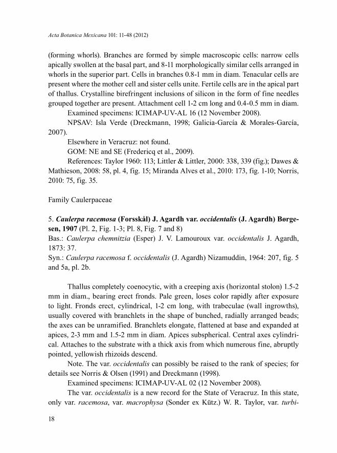
(forming whorls). Branches are formed by simple macroscopic cells: narrow cells apically swollen at the basal part, and 8-11 morphologically similar cells arranged in whorls in the superior part. Cells in branches 0.8-1 mm in diam. Tenacular cells are present where the mother cell and sister cells unite. Fertile cells are in the apical part of thallus. Crystalline birefringent inclusions of silicon in the form of fine needles grouped together are present. Attachment cell 1-2 cm long and 0.4-0.5 mm in diam.
Examined specimens: ICIMAP-UV-AL 16 (12 November 2008).NPSAV: Isla Verde (Dreckmann, 1998; Galicia-García & Morales-García,
2007).Elsewhere in Veracruz: not found.GOM: NE and SE (Fredericq et al., 2009).References: Taylor 1960: 113; Littler & Littler, 2000: 338, 339 (fig.); Dawes &
Mathieson, 2008: 58, pl. 4, fig. 15; Miranda Alves et al., 2010: 173, fig. 1-10; Norris, 2010: 75, fig. 35.
Family Caulerpaceae
5. Caulerpa racemosa (Forsskål) J. Agardh var. occidentalis (J. Agardh) Børge-sen, 1907 (Pl. 2, Fig. 1-3; Pl. 8, Fig. 7 and 8)Bas.: Caulerpa chemnitzia (Esper) J. V. Lamouroux var. occidentalis J. Agardh, 1873: 37. Syn.: Caulerpa racemosa f. occidentalis (J. Agardh) Nizamuddin, 1964: 207, fig. 5 and 5a, pl. 2b.
Thallus completely coenocytic, with a creeping axis (horizontal stolon) 1.5-2 mm in diam., bearing erect fronds. Pale green, loses color rapidly after exposure to light. Fronds erect, cylindrical, 1-2 cm long, with trabeculae (wall ingrowths), usually covered with branchlets in the shape of bunched, radially arranged beads; the axes can be unramified. Branchlets elongate, flattened at base and expanded at apices, 2-3 mm and 1.5-2 mm in diam. Apices subspherical. Central axes cylindri-cal. Attaches to the substrate with a thick axis from which numerous fine, abruptly pointed, yellowish rhizoids descend.
Note. The var. occidentalis can possibly be raised to the rank of species; for details see Norris & Olsen (1991) and Dreckmann (1998).
Examined specimens: ICIMAP-UV-AL 02 (12 November 2008).The var. occidentalis is a new record for the State of Veracruz. In this state,
only var. racemosa, var. macrophysa (Sonder ex Kütz.) W. R. Taylor, var. turbi-

Robinson et al.: New records of green and brown algae for the Sistema Arrecifal Veracruzano
19
nata (J. Agardh) Eubank, have been reported; var. racemosa f. condensata Weber-van Bosse, var. racemosa f. reducta Børgesen, var. gracilis (Zanardini) Weber-van Bosse, var. laetevirens (Montagne) Weber-van Bosse f. cylindrica (Sonder) Weber-van Bosse and var. lamourouxii (Turner) Weber-van Bosse have been reported for the Atlantic Mexican coast in general (Ortega et al., 2001; Galicia-García & Mo-rales-García, 2007; González-Gándara et al., 2007).
GOM: Caulerpa racemosa is distributed throughout the gulf (Fredericq et al., 2009).
References: Taylor, 1960: 151; Littler & Littler, 2000: 370, 371 (fig.); Sch-neider & Searles, 1991: 93; Dawes & Mathieson, 2008: 74, pl. 379, fig. 28 and 29.
6. Caulerpa sertularioides (S. G. Gmelin) M. A. Howe, 1905 (Pl. 2, Fig. 4-6; Pl. 8, Fig. 9 and 10)Bas.: Fucus sertularioides S. G. Gmelin, 1768: 151, pl. 15, fig. 4. Syn.: Fucus plumaris Forsskål, 1775: 190; Caulerpa plumaris (Forsskål) C. Agardh, 1823 (1822-1823): 436.
Thallus completely coenocytic, with a creeping axis and erect feather-like fronds, sometimes branched, up to 5 cm long and 0.5-1 cm wide. Dark green. Fronds erect, with trabeculae (wall ingrowths). Branchlets opposite, cylindrical, needle-shaped, curved upwards, 3-5 mm long and 400-500 μm in diam., with bluntly point-ed apices. Central axes cylindrical, 1-1.5 mm in diam. Horizontal stolon creeping, extended, cylindrical, 2.0-2.5 mm in diam., usually about 20 cm long, sometimes up to 2 m. Attaches to the substrate by a stolon from which fine, abruptly pointed, yel-lowish filamentous rhizoids descend.
Examined specimens: ICIMAP-UV-AL 01, 01R, 01R1 (12 November 2008).NPSAV: San Juan de Ulúa, Blanquilla, Punta Gorda, Gallega, Pájaros, Hor-
nos, Ingeniero and Giote reefs, Isla Sacrificios, Isla Verde, Isla Santiaguillo (Ortega et al., 2001; Galicia-García & Morales-García, 2007; Godínez-Ortega et al., 2009).
Elsewhere in Veracruz: Mpio. Tuxpan: Isla de Lobos, Tuxpan Reef (f. brevi-ceps (J. Agardh) Svedelius, f. farlowii (Weber-van Bosse) Børgesen and f. longi-seta (Bory) Svedelius; González-Gándara et al., 2007), marine littoral of Tam-pamachoco lagoon; Mpio. Actopan: La Mancha (Punta Mancha), Playa Paraíso (La Mancha), Villa Rica (Punta Villa Rica); Mpio. Alto Lucero: Boca Andrea, El Morro (Punta del Morro); Laguna Verde; Mpio. San Andrés Tuxtla: Montepío (Punta Morrillos) (Dreckmann, 1998; Ortega et al., 2001; González-Gándara et al., 2007).

Acta Botanica Mexicana 101: 11-48 (2012)
20
GOM: NE, SW and SE (Fredericq et al., 2009).References: Taylor 1960: 144; Littler & Littler, 2000: 374, 375 (fig.); Dawes &
Mathieson, 2008: 75, pl. 6, fig. 26- 29; Norris, 2010: 96, fig. 46.
Family Codiaceae
7. Codium isthmocladum Vickers subsp. clavatum (Collins et Hervey) P. C. Silva, 1960 (Pl. 2, Fig. 7 and 8; Pl. 8, Fig. 11 and 12)Bas.: Codium decorticatum var. clavatum Collins et Hervey, 1917: 56.
Thallus pseudoparenchymatous, erect, compact, spongy, 5-8 cm long. Light green when alive, olive green in dried specimens, loses color after exposure to light. Branching irregular in basal part and dichotomous in superior parts, branches cylin-drical, 2-4 mm in diam. Trichomes per utricle are single (usually) or few, deciduous, 300-350 µm long. Apical wall of the utricles 0.5-2 cm long and 50-55 µm wide. The revised specimens were in the vegetative stage.
Examined specimens: ICIMAP-UV-AL 51, 51R, 51R2 (12 November 2008).The subsp. clavatum is a new record for the State of Veracruz. The species C.
isthmocladum was reported for this area (Valenzuela, 1987; Dreckmann, 1998), and the var. isthmocladum was found on Isla Sacrificios and Hornos Reef of the NPSAV (Ortega et al., 2001).
GOM: Codium isthmocladum is distributed throughout the gulf (Fredericq et al., 2009).
References: Taylor, 1960: 186, 187; Schneider & Searles, 1991: 86; Littler & Littler, 2000: 352, 353 (fig.); Dawes & Mathieson, 2008: 80, pl. 5, fig. 15 and 16.
8. Codium taylorii P. C. Silva, 1960 (Pl. 2, Fig. 9-11; Pl. 9, Fig. 1 and 2)
Thallus pseudoparenchymatous, erect, forming dense clumps, 7-8 cm long. Green, loses color rapidly after exposure to light. Branching irregular, sometimes cervicorn in basal part and dichotomous at the extremities. Branches firm, spongy, smooth, 4-5 mm in diam., with rounded apices, formed by utricles that are constrict-ed at base and claviform and rounded at apices, 450-1000 μm long and 100-350 μm wide. Apices of the utricles have very thin walls (4-5 μm). Trichomes scarce, up to 1000 μm long, inserted near the apices. Gametangia elongate, 200-320 μm long and 50-75 μm in diam., usually one per utricle, inserted into its middle part. Attaches to the substrate by a crusty disc 0.8-1 cm in diam.

Robinson et al.: New records of green and brown algae for the Sistema Arrecifal Veracruzano
21
1 2 3
4
7
10
8
11
9
5 6
Plate 2. Chlorophyta. Fig. 1-3. Caulerpa racemosa var. occidentalis: 1. claviform apex of a branchlet; 2. longitudinal section of horizontal stolon; 3. transverse section of erect frond showing trabecula (arrowhead). Fig. 4-6. Caulerpa sertularioides: 4. apex of a branchlet. 5. transverse section of erect axis showing trabecula (arrowhead); 6. ramified rhizoids. Fig. 7 and 8. Codium isthmocladum subsp. clavatum: 7. utricle showing hair scars (arrowhead); 8. utricle with a surface hair (arrowhead). Fig. 9-11. Codium taylorii: 9. utricle with rounded apex; 10. medullary filaments (arrowhead) and utricles; 11. utricle with a gametangium (arrowhead). Scale bars: 25 μm in Fig. 7, 9 and 11; 50 μm in Fig. 3-5; 100 μm in Fig. 1, 2, 6, 8 and 10.

Acta Botanica Mexicana 101: 11-48 (2012)
22
Examined specimens: ICIMAP-UV-AL 07R, LS-26 (12 November 2008).NPSAV: Playa Mocambo, Hornos Reef, Isla de Enmedio, Isla Verde (Dreck-
mann, 1998; Ortega et al., 2001; Galicia-García & Morales-García, 2007).Elsewhere in Veracruz: Mpio. Tuxpan: Isla de Lobos; Mpio. Alto Lucero: El
Morro (Punta del Morro) (Dreckmann, 1998; Ortega et al., 2001).GOM: NE, NW and SE (Fredericq et al., 2009).References: Taylor, 1960: 188; Schneider & Searles, 1991: 87; Littler & Littler,
2000: 354, 355 (fig.); Dawes & Mathieson 2008: 81, pl. 5, fig. 19-21.
Family Halimedaceae
9. Halimeda opuntia (L.) J. V. Lamouroux, 1816 (Pl. 3, Fig. 1-3; Pl. 9, Fig. 3-6)Bas.: Corallina opuntia L., 1758: 805.
Thallus pseudoparenchymatous, erect, formed by articulated sequences of segments, strongly calcified, forming compact mats 10-15 cm in diam. Yellowish green, loses color rapidly after exposure to light. Densely branched in various pla-nes. Segments plane, kidney-shaped, sometimes grooved, 0.3-0.5 cm long and 0.7-12 cm wide. Medullary siphons 40-70 μm in diam., paired but not fused in nodes, forming simple groups. Superficial utricles in groups of 4-5, rounded or polygonal, 25-40 cm in diam. Subsuperficial utricles 30-50 μm long, supporting 2-4 superficial utricles. In surface view, cortex formed by hexagonal cells 15-30 μm in diam. Atta-ches to the substrate by multiple parts of thallus.
Examined specimens: ICIMAP-UV-AL 72, 72R (11 March 2008); LS-19, 22, 38 (10 April 2008), 32 (12 November 2008), 1 (3 June 2010).
NPSAV: Blanquilla, Hornos, Pájaros, Gallega, Galleguilla and Anegada de Adentro reefs, Isla de Enmedio, Isla Verde, Isla Sacrificios, Isla Santiaguillo (Dreck-mann, 1998; Ortega et al., 2001; Galicia-García & Morales-García, 2007; Godínez-Ortega et al., 2009).
Elsewhere in Veracruz: Mpio. Tuxpan: Isla de Lobos, marine littoral of Tam-pamachoco lagoon; Mpio. Actopan: Playa Paraíso (La Mancha) (Dreckmann, 1998; Ortega et al., 2001).
GOM: NE, NW and SE (Fredericq et al., 2009).References: Taylor, 1960: 176; Littler & Littler, 2000: 406, 407 (fig.); Dawes
& Mathieson 2008: 84, pl. 8, fig. 29 and 31; Bandeira-Pedrosa et al., 2004: 365, fig. 3-5, 21, 28-30.

Robinson et al.: New records of green and brown algae for the Sistema Arrecifal Veracruzano
23
10. Halimeda scabra M. A. Howe, 1905 (Pl. 3, Fig. 4-6; Pl. 9, Fig. 7-10)
Thallus pseudoparenchymatous, erect, calcareous, 8 cm long. Dark green. Branched in a single plane. Segments moderately calcified, oval or compressed, without ribs, 0.2-1 cm long and 0.4-1.4 cm wide, margin entire, rounded. Superfi-cial utricles elongate, with bulbous extremities, 90-100 µm long. Medullary siphons fused 2-3 times, principal utricles in 3-4 rows, 70-80 µm long and 30-40 µm wide, with a prominent central spine. Gametangia formed on the surface from the surface utricles. Attaches to the substrate by a fibrose segment.
Examined specimens: LS-4 (10 April 2008), 1 (3 June 2010).NPSAV: Isla Verde (Galicia-García & Morales-García, 2007).Elsewhere in Veracruz: Mpio. Actopan: La Mancha (Punta Mancha) (Dreck-
mann, 1998; Ortega et al., 2001).GOM: NE, SW and SE (Fredericq et al., 2009).References: Taylor, 1960: 180; Littler & Littler 2000: 406, 407 (fig.); Dawes &
Mathieson 2008: 83, pl. 9, fig. 1 and 2.
Family Udoteaceae
11. Rhipocephalus phoenix (Ellis et Sol.) Kütz, f. brevifolius A. Gepp et E. S. Gepp, 1911 (Pl. 3, Fig. 7-10; Pl. 10, Fig. 1-3)
Thallus pseudoparenchymatous, erect, 3-6 cm long, moderately calcified, sol-itary or in pairs, consisting of capitulum or cap (composed of narrow blades radially arranged), simple stalk and holdfast of fine siphons interlaced with sand particles. Dark green, loses color rapidly after exposure to light. Stipes cylindrical, smooth, 1-4 cm long and 2-3 mm in diam. Surface siphons compact, dichotomously branched in a single plane, with thin walls and rounded finger-like apices. Cap ovoid, 2-3 cm long; blades minute, 0.6-1 cm long. Siphons in blades are laterally fused together, 200-220 µm in proximal part and 50-100 µm in distal part, constricted at base, after the first dichotomous division. Siphons in stipes with repeatedly ramified append-ages, finger-like, grouped into packages. Attaches to the substrate by a rhizoidal mass (holdfast).
Examined specimens: LS-37 (10 April 2008).NPSAV: Isla Verde, Isla de Enmedio (Dreckmann, 1998; Ortega et al., 2001;
Galicia-García & Morales-García, 2007).

Acta Botanica Mexicana 101: 11-48 (2012)
24
Elsewhere in Veracruz: Mpio. Tuxpan: Isla de Lobos, Tuxpan Reef (Dreck-mann, 1998; Ortega et al., 2001; González-Gándara et al., 2007).
GOM: NE, SW and SE (Fredericq et al., 2009).References: Taylor, 1960: 174; Littler & Littler, 2000: 418, 419 (fig.); Dawes &
Mathieson, 2008: 98, pl. 9, fig. 27-30.
Family Dasycladaceae
12. Neomeris annulata Dickie, 1874 (Pl. 3, Fig. 11-13; Pl. 10, Fig. 4 and 5)Syn.: Neomeris kelleri Cramer, 1887: 3-9, 39, pl. 1; pl. 2: fig. 1-12; pl. 3: fig. 1 and 2.
Thallus pseudoparenchymatous, erect, 0.4-1 cm long and 1-3 mm wide. Light green, loses color rapidly after exposure to light. Solitary, cylindrical, arched down-wards, lightly calcified. Apices of the filaments fine, deciduous. Central axis 300-400 μm wide. Branches of the central axis arranged spirally, leaving scars after be-ing detached; terminated with two superficial cells encircling a gametangium, cells polyhedral or globular, 100-130 μm in diam., with bulbous apices. Rows of superfi-cial cells aligned with 50-150 μm distance between them. Gametangium elongate or oval, 120-150 μm long and 60-80 μm in diam. Attachment disc is formed by short siphons. Usually grows on fragments of corals.
Examined specimens: ICIMAP-UV-AL 09, 09R (12 November 2008); LS-24, 28 (1 March 2008).
NPSAV: Blanquilla Reef, Isla Sacrificios, Isla Verde, Isla Santiaguillo (Dreck-mann, 1998; Ortega et al., 2001; Galicia-García & Morales-García, 2007).
Elsewhere in Veracruz: Mpio. Tuxpan: Isla de Lobos, Tuxpan Reef (Dreck-mann, 1998; Ortega et al., 2001; González-Gándara et al., 2007).
GOM: NE, SW and SE (Fredericq et al., 2009).References: Taylor, 1960: 101; Littler & Littler, 2000: 438, 439 (fig.); Dawes &
Mathieson, 2008: 108, pl. 9, fig. 12 and 13.
Family Polyphysaceae
13. Parvocaulis polyphysoides (P. L. Crouan et H. M. Crouan) S. Berger, U. Fe-ttweiss, S. Gleissberg, L. B. Liddle, U. Richter, H. Sawitsky et G. C. Zuccarello, 2003 (Pl. 4, Fig. 1-3; Pl. 10, Fig. 6-8)Bas.: Acetabularia polyphysoides P. L. Crouan et H. M. Crouan in Schramm et Mazé, 1865: 42.

Robinson et al.: New records of green and brown algae for the Sistema Arrecifal Veracruzano
25
1 2 3
4
7
11
8
12
9 10
5 6
13
Plate 3. Chlorophyta. Fig. 1-3. Halimeda opuntia: 1. Ramified medullary siphons; 2. surface view of a segment; 3. surface utricles (arrowhead). Fig. 4-6. Halimeda scabra: 4. surface utricles with a prominent central spine; 5. surface view of utricles; 6. a developing group of utricles (arrowhead). Fig. 7-10. Rhipocephalus phoenix f. brevifolius: 7. general view of thallus; 8. basal siphon of a blade without apparent constriction (arrowhead); 9. constriction of a siphon after the first dichotomy; 10. lateral appendages that form stipe cortex. Fig. 11-13. Neomeris annulata: 11. Central axis showing verticillate branchlet scars (arrowhead); 12. gametangium (arrowhead) encircled by surface cells; 13. attachment structure formed by short siphons (arrowhead). Scale bars: 25 μm in Fig. 1-6 and 9-13; 100 μm in Fig. 8; 2 mm in Fig. 7.

Acta Botanica Mexicana 101: 11-48 (2012)
26
Thallus unbranched, erect, minute, umbrella-shaped, 1-4 mm long, with a solitary plane or slightly concave downwards disc (cap) 1-5 mm in diam., with 21-23 rays. Deep brown green or bright green. Margins of the rays slightly sharp-pointed by 6-9 scars left by sterile hairs, arranged elliptically at base of the rays. Cylindrical calcified stalk 4 mm long and 300-600 µm wide. Gametangia in mature thallus are rays containing cysts. Cysts spherical, numerous, 50-100 µm in diam., sometimes up to 50 cysts per ray.
Examined specimens: ICIMAP-UV-AL 13, 13R (12 November 2008); LS-24 (1 March 2008).
NPSAV: Blanquilla Reef, Isla Verde, Isla Sacrificios (Dreckmann, 1998; Ortega et al., 2001; Galicia-García & Morales-García, 2007).
Elsewhere in Veracruz: not found.GOM: SE (Fredericq et al., 2009).References: Taylor, 1960: 104; Littler & Littler, 2000: 444, 445 (fig.).
Class PhaeophyceaeFamily Ectocarpaceae
14. Ectocarpus rallsiae Vickers, 1905 (Pl. 4, Fig. 4 and 5; Pl. 10, Fig. 9 and 10).Syn.: Feldmannia rallsiae (Vickers) G. Hamel, 1939: 67; Giffordia rallsiae (Vick-ers) W. R. Taylor, 1960: 208. Hincksia rallsiae (Vickers) P. C. Silva in P. C. Silva, Meñez et Moe, 1987: 73 (for synonymy, see Wynne, 2011: 112, note 430).
Thallus filamentous, fine, forming clumps of 0.5-1 cm. Light brown. Fila-ments uniseriate, branched. Branching spacious, irregular to dichotomous. In sur-face view, cells rectangular, 50-100 µm long and 25-50 µm wide. Plurangia sessile, fusiform, 75-125 µm long and 20-30 µm in diam. Phaeoplasts parietal, ribbon-like, with numerous pyrenoids. Attaches to the substrate by creeping basal filaments. Epiphyte on the seagrass Thalassia testudinum Banks et Sol. ex König.
Examined specimens: LS-39 (12 November 2008).NPSAV: Port of Veracruz, Hornos Reef, Isla de Enmedio (Ortega et al., 2001;
Galicia-García & Morales-García, 2007).Elsewhere in Veracruz: Mpio. Actopan: Playa Paraíso (La Mancha); Mpio.
Alto Lucero: Boca Andrea, Playa el Morro (Punta del Morro) (Ortega et al., 2001).
GOM: NE, NW, SE and SW (Fredericq et al., 2009).References: Taylor, 1960: 208; Dawes & Mathieson, 2008: 147, pl. 17, fig. 4.

Robinson et al.: New records of green and brown algae for the Sistema Arrecifal Veracruzano
27
Family Sphacelariaceae
15. Sphacelaria rigidula Kütz., 1843 (Pl. 4, Fig. 6 and 7; Pl. 10, Fig. 11)Syn.: Sphacelaria furcigera Kütz., 1855: 27, pl. 90, fig. 2.
Thallus filamentous, forming small tight clumps, 5-7 mm long. Yellowish brown. Branching irregular to radial, scarce to frequent. Filaments linear, cylindri-cal, segments 50-75 μm in diam. and 1-1.5 diameters long, with 1-3 longitudinal walls. Lateral hairs abundant, 112-120 μm long and 12.5 μm in diam. Propagules with two cylindrical arms, some of them tapered; arms 310-363 μm long and 17.5-25.0 μm in diam.; their stalk below the bifurcation of the arms 245 μm long and 27.5-30 μm in diam. Attaches by a stolon. Epiphyte on the red alga Bryothamnion triquetrum.
Examined specimens: LS-8 (1 March 2008).NPSAV: Isla Verde, Isla de Enmedio, Isla Santiaguillo (Dreckmann, 1998;
Ortega et al., 2001; Galicia-García & Morales-García, 2007).Elsewhere in Veracruz: Mpio. Actopan: Playa Paraíso (La Mancha), Villa
Rica (Punta Villa Rica) (Dreckmann, 1998; Ortega et al., 2001).GOM: throughout the gulf (Fredericq et al., 2009).References: Taylor, 1960: 210; Schneider & Searles, 1991: 152; Littler & Lit-
tler, 2000: 252, 253 (fig.); Mendoza-González et al., 2000: 27, fig. 34-37; Dawes & Mathieson, 2008: 133, pl. 16, fig. 1; Norris, 2010: 112, fig. 51.
Family Dictyotaceae
16. Canistrocarpus cervicornis (Kütz.) De Paula et De Clerck in De Clerck, Leli-aert, Verbruggen, Lane, De Paula, Payo et Coppejans, 2006 (Pl. 4, Fig. 8-11; Pl. 11, Fig. 1-5)Bas: Dictyota cervicornis Kütz., 1859: 11, pl. 24: fig. 2.Syn.: Dictyota fasciola Harvey, 1852: 108, pl. 8B; Dictyota indica Sonder ex Kütz., 1859: 8, pl. 17: fig. 1; Dictyota pardalis Kütz., 1859: 16-17, pl. 39: fig. 2; Dictyota dichotoma var. curvula P. L. Crouan et H. M. Crouan, 1878: 119.
Thallus pseudoparenchymatous, erect, dense, thin, blades are easily frag-mented. Dark brown, some portions olive green. Branching regularly dichotomous, sometimes in terminal branches, one of bifurcations being shorter. Blade margins entire. Branches ribbon-like, slightly inrolled, 3 mm wide, 200 μm thick, slightly

Acta Botanica Mexicana 101: 11-48 (2012)
28
thinner with each subsequent division. Apices rounded. Proximal internodes 7-8 mm long, median 5-7 mm long and distal ones 3-5 mm long. Medullary cells ir-regular or rectangular, 100-200 μm wide, arranged in one row of 22-25 cells across the blade width. Cortical cells spherical, 25 μm in diam.; surface cells rectangular, 80-130 μm long and 50-70 μm wide. Bifurcations forming 15-45o angles. Sporangia solitary, spherical, 70-150 μm in diam., surrounded by a ring of cells (paraphyses).
Examined specimens: LS-1 (3 June 2010).NPSAV: Blanca, Blanquilla and Punta Gorda reefs, Isla Sacrificios, Isla
Verde, Isla de Enmedio (Dreckmann, 1998; Ortega et al., 2001; Galicia-García & Morales-García, 2007).
Elsewhere in Veracruz: Mpio. Tuxpan: Isla de Lobos; Mpio. San Andrés Tuxt-la: Montepío (Punta Morrillos); Mpios. Pueblo Viejo, Tampico Alto, Ozuluama, Ta-malín and Tamiahua (Laguna de Tamiahua) (Dreckmann, 1998; Ortega et al., 2001).
GOM: throughout the gulf (Fredericq et al., 2009).References: Taylor, 1960: 222; Earle, 1969: 153, fig. 60; Schneider & Searles,
1991: 157; Littler & Littler, 2000: 260, 261 (fig.); Dawes & Mathieson, 2008: 121, pl. 13, fig. 3; Solé & Foldats, 2003: 47, fig. 3 and 4.
17. Dictyerpa jamaicensis F. S. Collins, 1901 (Pl. 4, Fig. 12-14; Pl. 11, Fig. 6 and 7)Syn.: Vaughaniella rupicola Børgesen, 1950: 3-10, fig. 1-8; 1951: 11-14, fig. 4.
Thallus pseudoparenchymatous, forming small entangled mats, 2-3 cm wide, not calcified, rough. Brown, orange or golden brown. Branching opposite or densely irregular. Axes ribbon-like, slightly compressed, 0.2-0.3 mm long and 0.6-0.8 mm wide. Apices curved upwards. Medulla consisting of irregular-shaped cells, up to 6 rows of cells thick; cells 50-100 μm in diam. Surface cells pigmented, 25-35 μm in diam. Attaches to the substrate by rhizoids originating from creeping axes.
Note. Silva et al. (1996), Littler & Littler (2000) and Dawes & Mathieson (2008) conclude that Dictyerpa jamaicensis represents a growth stage in the life history of Padina but cannot be assigned with certainty to any particular species.
Examined specimens: LS-6 (10 April 2008).A new record for the State of Veracruz.GOM: in deep waters of the gulf (Dawes & Mathieson, 2008).References: Littler & Littler, 2000: 272, 273 (fig.).
18. Dictyota bartayresiana J. V. Lamouroux, 1809 (Pl. 5, Fig. 1-4; Pl. 11, Fig. 8-10)Syn.: Dictyota bartayresii J. V. Lamouroux, 1809; Zonaria bartayresiana (J. V.

Robinson et al.: New records of green and brown algae for the Sistema Arrecifal Veracruzano
29
1 2 3
4 5 6
9
12 13 14
7 8
10 11
Plate 4. Chlorophyta and Phaeophyceae. Fig. 1-3. Parvocaulis polyphysoides: 1. reproductive stage showing rays with cysts (arrowhead); 2. detail of the corona superior (arrowhead); 3. stipe. Fig. 4 and 5. Ectocarpus rallsiae: 4. filament with plurangia and intercalar meristem (arrowhead); 5. sessile plurangium (arrowhead). Fig. 6 and 7. Sphacelaria rigidula: 6. filaments with lateral branchlets (arrowhead); 7. a propagule (arrowhead). Fig. 8-11. Canistrocarpus cervicornis: 8. transverse section of sporophyte blade; 9. sporangium surrounded by a circle of cells (arrowhead); 10. surface view of vegetative blades; 11. apex of a vegetative blade showing apical cells. Fig. 12-14. Dictyerpa jamaicensis: 12. general view of thallus; 13. transverse section of a vegetative branch; 14. surface view of a vegetative branch. Scale bars: 25 μm in Fig. 2, 5 and 9-11; 50 μm in Fig. 4, 6, 7 and 13; 100 μm in Fig. 1, 3, 8 and 14; 2 mm in Fig. 12.
Acta Botanica Mexicana 101: 11-48 (2012)
30
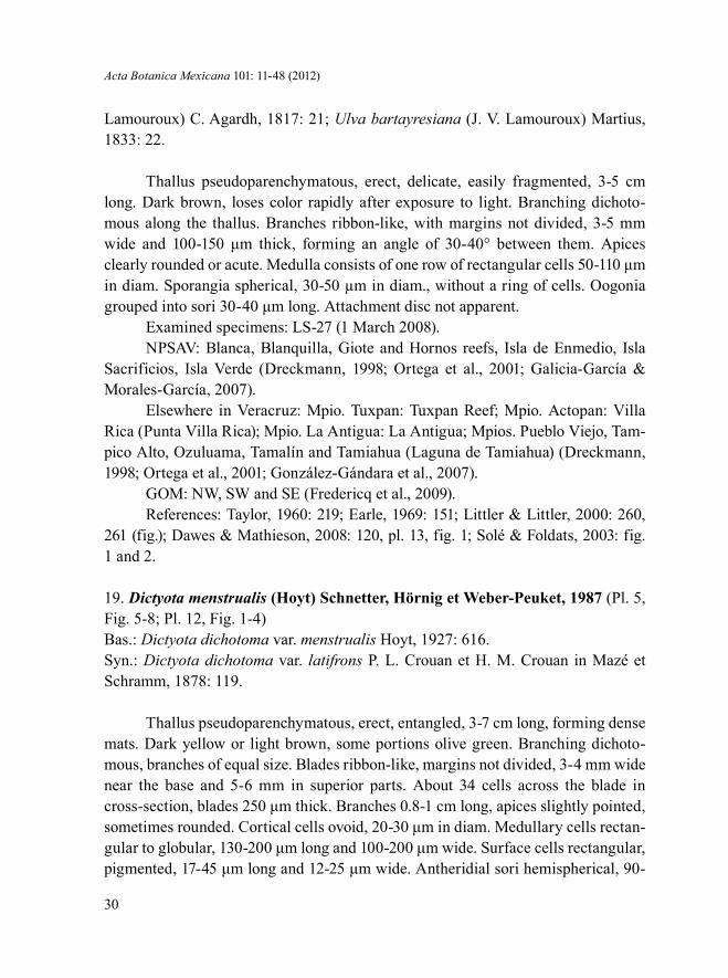
Lamouroux) C. Agardh, 1817: 21; Ulva bartayresiana (J. V. Lamouroux) Martius, 1833: 22.
Thallus pseudoparenchymatous, erect, delicate, easily fragmented, 3-5 cm long. Dark brown, loses color rapidly after exposure to light. Branching dichoto-mous along the thallus. Branches ribbon-like, with margins not divided, 3-5 mm wide and 100-150 μm thick, forming an angle of 30-40° between them. Apices clearly rounded or acute. Medulla consists of one row of rectangular cells 50-110 μm in diam. Sporangia spherical, 30-50 µm in diam., without a ring of cells. Oogonia grouped into sori 30-40 μm long. Attachment disc not apparent.
Examined specimens: LS-27 (1 March 2008).NPSAV: Blanca, Blanquilla, Giote and Hornos reefs, Isla de Enmedio, Isla
Sacrificios, Isla Verde (Dreckmann, 1998; Ortega et al., 2001; Galicia-García & Morales-García, 2007).
Elsewhere in Veracruz: Mpio. Tuxpan: Tuxpan Reef; Mpio. Actopan: Villa Rica (Punta Villa Rica); Mpio. La Antigua: La Antigua; Mpios. Pueblo Viejo, Tam-pico Alto, Ozuluama, Tamalín and Tamiahua (Laguna de Tamiahua) (Dreckmann, 1998; Ortega et al., 2001; González-Gándara et al., 2007).
GOM: NW, SW and SE (Fredericq et al., 2009).References: Taylor, 1960: 219; Earle, 1969: 151; Littler & Littler, 2000: 260,
261 (fig.); Dawes & Mathieson, 2008: 120, pl. 13, fig. 1; Solé & Foldats, 2003: fig. 1 and 2.
19. Dictyota menstrualis (Hoyt) Schnetter, Hörnig et Weber-Peuket, 1987 (Pl. 5, Fig. 5-8; Pl. 12, Fig. 1-4)Bas.: Dictyota dichotoma var. menstrualis Hoyt, 1927: 616. Syn.: Dictyota dichotoma var. latifrons P. L. Crouan et H. M. Crouan in Mazé et Schramm, 1878: 119.
Thallus pseudoparenchymatous, erect, entangled, 3-7 cm long, forming dense mats. Dark yellow or light brown, some portions olive green. Branching dichoto-mous, branches of equal size. Blades ribbon-like, margins not divided, 3-4 mm wide near the base and 5-6 mm in superior parts. About 34 cells across the blade in cross-section, blades 250 μm thick. Branches 0.8-1 cm long, apices slightly pointed, sometimes rounded. Cortical cells ovoid, 20-30 μm in diam. Medullary cells rectan-gular to globular, 130-200 μm long and 100-200 μm wide. Surface cells rectangular, pigmented, 17-45 μm long and 12-25 μm wide. Antheridial sori hemispherical, 90-

Robinson et al.: New records of green and brown algae for the Sistema Arrecifal Veracruzano
31
100 μm, located along both sides of the blade. Bifurcations forming 60-90° angles. Sporangia grouped, 60-80 μm in diam., along both sides of the blade. Attachment disc fibrous. Epiphyte on the chlorophyte Halimeda scabra.
Note. The morphology of the species is widely variable. Its diagnostic fea-tures frequently overlap with those of other species. The species distribution in the Atlantic is still confused. The encountered specimens may belong to a species com-plex, and its varieties and forms are to be described (Solé & Foldats, 2003).
Examined specimens: ICIMAP-UV-AL 80, 80R, 80R1 (12 November 2008); LS-1 (3 June 2010).
NPSAV: Playa del Balneario Villa del Mar; Playa Mocambo, Blanca, Blan-quilla, Giote and Hornos reefs, Isla de Enmedio, Isla Sacrificios (Ortega et al., 2001; Galicia-García & Morales-García, 2007).
Elsewhere in Veracruz: Mpio. Tuxpan: Tuxpan Reef; Mpio. Actopan: Playa Paraíso (La Mancha), Villa Rica (Punta Villa Rica); Mpio. Alto Lucero: Boca An-drea, Laguna Verde, Playa el Morro (Punta del Morro); Mpio. Boca del Río: Laguna de Mandinga; Mpio. San Andrés Tuxtla: Montepío (Punta Morrillos); Mpio. Tuxpan: Tuxpan; Mpios. Pueblo Viejo, Tampico Alto, Ozuluama, Tamalín and Tamiahua (Laguna de Tamiahua) (Ortega et al., 2001; González-Gándara et al., 2007).
GOM: throughout the gulf (Fredericq et al., 2009).References: Taylor, 1960: 218; Schneider & Searles, 1991: 158; Littler & Lit-
tler, 2000: 266, 267 (fig.); Solé & Foldats, 2003: 60, fig. 13 and 14; Dawes & Mathie-son, 2008: 123, pl. 13, fig. 13 and 14.
20. Dictyota pulchella Hörnig et Schnetter, 1988 (Pl. 5, Fig. 9-12; Pl. 12, Fig. 5-8; Pl. 13, Fig. 1)Syn.: Dictyota divaricata J. V. Lamouroux, 1809: 43; Zonaria divaricata (J. V. Lamouroux) C. Agardh, 1817: xxi.
Thallus pseudoparenchymatous, erect, forming dense entangled mats. Light brown. Branching widely dichotomous. Bifurcations forming 90-100° angles at base and in superior parts, 60-120°. Blades compressed, ribbon-like, margins not divided, 2-3 mm wide in basal part, the width being continuously diminished with every bi-furcation, down to 0.5-0.8 mm. Apices pointed. Surface cells rectangular, 40-50 μm long and 10-14 μm wide. Chloroplasts several. Medullary cells arranged in a single row, up to 23 cells in transverse section, rectangular to globular, 60-130 μm in diam. Cortical cells spherical, strongly pigmented, 12-18 μm in diam. Sporangia spherical, 70-120 μm in diam., with a ring of basal cells. Antheridia 50 μm long.

Acta Botanica Mexicana 101: 11-48 (2012)
32
Note. A ring of cells that encircles the sporangia is not mentioned in the re-vised literature (Earle, 1969; Schneider & Searles, 1991; Littler & Littler, 2000; Solé & Foldats, 2003; Dawes & Mathieson, 2008). Although this structure was observed in the examined specimens, other morphological features and size correspond well to Dictyota pulchella.
Examined specimens: ICIMAP-UV-AL 26, 26R (12 November 2008); LS-1 (3 June 2008).
NPSAV (as Dictyota divaricata): Giote Reef, Isla de Enmedio, Isla Sacrifi-cios, Isla Verde, Isla Santiaguillo, Isla de Enmedio (Ortega et al., 2001; Galicia-García & Morales-García, 2007).
Elsewhere in Veracruz (as Dictyota divaricata): Mpio. Tuxpan: Isla de Lobos, Tuxpan Reef (Ortega et al., 2001; González-Gándara et al., 2007).
GOM: throughout the gulf (Fredericq et al., 2009).References: Taylor, 1960: 219; Earle, 1969: 160, fig. 45, 46 and 58; Schneider
& Searles, 1991: 160; Littler & Littler, 2000: 268, 269 (fig.); Solé & Foldats, 2003: 65, fig. 19 and 20; Dawes & Mathieson, 2008: 125, pl. 14, fig. 1- 4.
21. Padina pavonica (L.) Thivy in W. R. Taylor, 1960 (Pl. 5, Fig. 13 and 14; Pl. 6, Fig. 1; Pl. 13, Fig. 2-5)Bas.: Fucus pavonicus L., 1753: 1162.Syn.: Padina pavonia J. V. Lamouroux, 1816: 304; Zonaria pavonia C. Agardh, 1820: 125; Padina mediterranea Bory de Saint-Vincent, 1827: 590.
Thallus pseudoparenchymatous, erect, fronds fan-shaped, moderately calci-fied, 10 cm long. Light brown. Fronds grouped into dense clumps. Apical margins inrolled, composed of two rows of cells 45-80 μm thick. Blades 100-115 μm thick at base, formed by 3 rows of cells; their middle parts 80-100 μm thick, formed by 2-3 rows of cells. Sterile hairs numerous, 20-25 μm in diam., located near the base of fronds. Sporangia ovoid, 60-100 μm in diam., with indusium, located along the concentric bands near the apical margin only on one side of the blades. Attaches to the substrate by a mass of rhizoids 1.5 cm in diam.
Examined specimens: ICIMAP-UV-AL 27, 27R (12 November 2008); LS-12 (1 March 2008), 2 (12 November 2008), 29 (10 April 2008).
NPSAV: Blanquilla Reef, Isla de Enmedio (Galicia-García & Morales-García, 2007).Elsewhere in Veracruz: Mpio. Actopan: Playa Paraíso (La Mancha), Villa Rica
(Punta Villa Rica); Mpio. Alto Lucero: Playa el Morro (Punta del Morro); Mpio. San Andrés Tuxtla: Montepío (Punta Morrilos); Mpio. Tuxpan: Barra de Tuxpan,

Robinson et al.: New records of green and brown algae for the Sistema Arrecifal Veracruzano
33
1 2 3
4 5
7 8 9
10
13 14
12
6
11
Plate 5. Phaeophyceae. Fig. 1-4. Dictyota bartayresiana: 1. transverse section of a blade showing sporangia (arrowhead); 2. apical portion of the blade; 3. surface view of a vegetative blade; 4. transverse section of a blade showing a developing group of hairs. Fig. 5-8. Dictyota menstrualis: 5. apical portion of a vegetative blade; 6. surface view of a row of oogonia (arrowhead); 7. transverse section of a blade; 8. antheridial sorus. Fig. 9-12. Dictyota pulchella: 9. surface view of a sporangium with a ring of paraphyses (arrowhead); 10. transverse section of a vegetative blade; 11. apical portion of the blade; 12. antheridia (arrowhead). Fig. 13 and 14. Padina pavonica: 13. general view of thallus; 14. transverse section of the middle part of a blade showing oogonia (arrowhead). Scale bars: 25 μm in Fig. 1, 3-6, 8-10 and 12; 50 μm in Fig. 7, 11 and 14; 100 μm in Fig. 2; 2 mm in Fig. 13.

Acta Botanica Mexicana 101: 11-48 (2012)
34
Blanquilla, Enmedio, Tanhuijo and Tuxpan reefs; Isla de Lobos, Isla de Enmedio (Dreckmann, 1998; Ortega et al., 2001).
GOM: SW and SE (Fredericq et al., 2009).References: Taylor, 1960: 234; Littler & Littler, 2000: 274, 275 (fig.); Dawes &
Mathieson, 2008: 128, pl. 14, fig. 15.
Family Chordariaceae
22. Cladosiphon occidentalis Kylin, 1940 (Pl. 6, Fig. 2-4; Pl. 13, Fig. 6-8)Syn.: Eudesme zosterae sensu Dawes & Mathieson (2008: 140), non (J.
Agardh) Kylin: 85, fig. 20; Cladosiphon zosterae sensu Dawes & Mathieson (2008: 140), non (J. Agardh) Kylin: 1940: 28, pl. 4, fig. 9.
Thallus pseudoparenchymatous, smooth, gelatinous, worm-like, small, 1-3 cm long, sometimes densely branched. Light brown, golden brown or yellow. Axes tubular, covered with fine colored hairs, up to 150 µm long and 8-15 µm in diam. Inward medullary cells compact, longitudinally elongate, cylindrical, 100-250 µm long and 30-75 µm in diam. Surface filaments strongly pigmented, radially arranged, 100-300 µm long. Sporangia plurilocular, elongate, 30-40 µm long, located at the extremities of the surface filaments. Attaches to the substrate by a basal disc 0.5 mm in diam. Epiphyte on the seagrass Thalassia testudinum.
Examined specimens: LS-39 (12 November 2008).NPSAV: Hornos and Blanca reefs, Isla Verde, Isla Sacrificios, Isla de Enme-
dio (Dreckmann, 1998; Galicia-García & Morales-García, 2007).Elsewhere in Veracruz: not found.GOM: throughout the gulf (Fredericq et al., 2009).References: Taylor, 1960: 247; Earle, 1969: 180, fig. 72, 75, 76 and 83; Sch-
neider & Searles, 1991: 136; Littler & Littler, 2000: 244, 245 (fig.); Dawes & Mathie-son, 2008: 140, pl. 17, fig. 1 and 2.
Family Scytosiphonaceae
23. Colpomenia sinuosa (Mertens ex Roth) Derbès et Solier, 1851 (Pl. 6, Fig. 5 and 6; Pl. 13, Fig. 9; Pl. 14, Fig. 1)Bas.: Ulva sinuosa Mertens ex Roth, 1806: 327, pl. 12; Encoelium sinuosum (Mertens ex Roth) C. Agardh, 1820: 146; Stilophora sinuosa (Mertens ex Roth) C. Agardh, 1827: 642; Asperococcus sinuosus (Mertens ex Roth) Bory de Saint-Vincent, 1832: 326; Hydroclathrus sinuosus (Mertens ex Roth) Zanardini, 1843: 39.

Robinson et al.: New records of green and brown algae for the Sistema Arrecifal Veracruzano
35
Thallus pseudoparenchymatous, rigid, sessile, sac-like, irregularly lobed, creeping, 3-8 cm in diam., formed by 4-6 rows of cells, 0.3-0.4 mm thick. Light brown or golden brown. Medullary cells hyaline, irregular-shaped, 160-200 µm in diam., continuously diminishing in size towards the surface layer (1-2 rows) of small, strongly pigmented cells 6-12 µm in diam. Sporangia plurilocular, numerous, located on the blade surface, elongate, 10-20 µm long and 5-8 µm wide. Sterile hairs up to 150 µm long and 10-13 µm in diam., located on the blade surface.
Examined specimens: LS-36 (12 November 2008).NPSAV: Playa Mocambo, Blanquilla, Hornos and Blanca reefs, Isla Verde,
Isla Sacrificios, Isla de Enmedio, Isla Santiaguillo (Ortega et al., 2001; Galicia-Gar-cía & Morales-García, 2007).
Elsewhere in Veracruz: Mpio. Actopan: Playa Paraíso (La Mancha), Villa Rica (Punta Villa Rica); Mpio. Alto Lucero: Boca Andrea, Laguna Verde, Playa el Morro (Punta el Morro); Mpio. San Andrés Tuxtla: Montepío (Punta Morrillos); Mpio. Tuxpan: Barra de Tuxpan, Isla de Lobos, Tuxpan Reef, marine littoral of Tam-pamachoco lagoon; Mpio. de Cazones: Barra de Cazones (Dreckmann, 1998; Ortega et al., 2001; González-Gándara et al., 2007).
GOM: SW and SE (Fredericq et al., 2009).References: Taylor, 1960: 260; Earle, 1969: 200, fig. 100 and 101; Schneider &
Searles, 1991: 146; Littler & Littler, 2000: 248, 249 (fig.); Dawes & Mathieson, 2008: 150, pl. 18, fig. 10 and 11; Norris, 2010: 192, fig. 92a.
24. Rosenvingea intricata (J. Agardh) Børgesen, 1914 (Pl. 6, Fig. 7-9; Pl. 14, Fig. 2-4)Bas.: Asperococcus intricatus J. Agardh, 1847: 7.
Thallus pseudoparenchymatous, erect, delicate, smooth, not gelatinous, easily fragmented, up to 10 cm long. Light brown or golden brown. Branching widely dichoto-mous, in some parts alternate. Axes slightly compressed, 1.2-1.5 mm in diam. Apices pointed, with numerous apical cells. Surface cells rectangular, 7-10 µm in diam. Branches containing 3-4 rows of interior hyaline cells 40-100 µm in diam. and one row of small ex-ternal cells, strongly pigmented, 10-12 µm in diam. Sporangia plurilocular, ovoid, 40-50 µm long, located on visible sori on the blade surface. Surface hairs grouped into clumps.
Examined specimens: LS-27 (1 March 2008).NPSAV: Gallega and Hornos reefs (Dreckmann, 1998; Ortega et al., 2001;
Galicia-García & Morales-García, 2007).Elsewhere in Veracruz: Mpio. Alto Lucero: Playa el Morro (Punta el Morro);
Mpio. de Tamiahua (Laguna de Tamiahua) (Dreckmann, 1998; Ortega et al., 2001).

Acta Botanica Mexicana 101: 11-48 (2012)
36
1
4 5
6
8 9
7
2 3
Plate 6. Phaeophyceae. Fig. 1. Padina pavonica: transverse section of the inrolled margin of two layers of cells. Fig. 2-4. Cladosiphon occidentalis: 2. transverse section of a branch showing medullary cells and lateral filaments (arrowhead); 3. general view of thalli; 4. plurilocular sporangia on tips of lateral filaments (arrowhead). Fig. 5 and 6. Colpomenia sinuosa: 5. general view of irregularly lobulated thallus; 6. transverse section of the membrane showing sterile hairs and plurilocular sporangia (arrowhead). Fig. 7-9. Rosenvingea intricata: 7. apical portion of a branch; 8. general view of thallus. 9. transverse section of a branch showing plurilocular sporangia (arrowhead). Scale bars: 25 μm in Fig. 4, 6 and 9; 50 μm in Fig. 1 and 2; 100 μm in Fig. 7; 2 mm in Fig: 3, 5 and 8.

Robinson et al.: New records of green and brown algae for the Sistema Arrecifal Veracruzano
37
GOM: NW, SW and SE (Fredericq et al., 2009).References: Taylor, 1960: 262; Earle, 1969: 207, fig. 108-112; Dawes & Ma-
thieson, 2008: 153, pl. 18, fig. 18 and 19; Norris, 2010: 198, fig. 96.
Family Sargassaceae
25. Sargassum furcatum Kütz., 1843 (Pl. 7, Fig. 1-5; Pl. 14, Fig. 5-7)Syn.: Sargassum vulgare C. Agardh (var.) furcatum (Kütz.) Kuntze, 1880: 229.
Thallus pseudoparenchymatous, erect, with numerous branches originating from the base area. Golden brown. Branchlets numerous, short, 1-1.6 cm long, all the axes muriculated. Blades are present principally in young parts of thallus, thin, linear-lanceolate, clearly serrulate (toothed), subpercurrent, with a rib, usually di-chotomously divided 1-4 times, 3-3.5 cm long and 1-1.5 cm wide. Cryptostomata small, located irregularly on the blades, 192-228 μm in diam. Air-bladders absent. Receptacles warty, cylindrical, 4-8 mm long. Male conceptacles 144 μm in diam. Female conceptacles 168 μm in diam.
Note. The species has been previously reported for the State of Quintana Roo (Caribbean Sea), and most likely it is in the process of expanding its range (Dreck-mann, 1998).
Examined specimens: ICIMAP-UV-AL 69 (11 March 2008); LS-14 (11 March 2008). A fragment of thallus was found floating in the water column.
A new record for the State of Veracruz. For this species, Ortega et al. (2001: 333) only indicate “Veracruz” without giving any details about localities.
GOM: SW and SE (Fredericq et al., 2009).References: Taylor, 1960: 277; Flores-Moya & Conde, 1998: 68, fig. 1; Moreira
& Suárez, 2002: 53, fig. 1.
DISCUSSION
On Cabezo Reef a total of 46 macroalgal species were identified, of which 21 (45.6%) belong to red algae (Galicia-García, pers. obs.), 13 (28.26%) to green algae and 12 (26.08%) to brown algae. The families Corallinaceae (6 species), Dictyo-taceae (6) and Rhodomelaceae (5) were best represented in the number of species.
Out of 25 species given in the present work, Percursaria percursa is a new record for NPSAV, while Dictyerpa jamaicensis, Sargassum furcatum, Caulerpa

Acta Botanica Mexicana 101: 11-48 (2012)
38
1 2
3
4 5
Plate 7. Phaeophyceae. Fig. 1-5. Sargassum furcatum: 1. general view of thallus; 2. transverse section of the principal axis; 3. transverse section of a blade; 4. transverse section of a blade showing cryptostoma (arrowhead) with a bunch of sterile hairs; 5. surface view of a branch. Scale bars: 25 μm in Fig. 5; 50 μm in Fig: 4; 100 μm in Fig. 2 and 3; 2 mm en Fig. 1.

Robinson et al.: New records of green and brown algae for the Sistema Arrecifal Veracruzano
39
1
23
6
1011 12
7
8
9
5
4
Plate 8. Chlorophyta. Fig. 1. Percursaria percursa: portion of a biseriate filament. Fig. 2 and 3. Cladophoropsis membranacea: 2: filament with unilateral branching, showing lateral filaments with open connection to parent cells; 3. apical portion of a filament. Fig. 4 and 5. Dictyosphaeria cavernosa: 4. surface polygonal cell; 5. row of hapteroid cells. Fig. 6. Ernodesmis verticillata: thallus with elongated bulbous cells, with verticillate branching. Fig. 7 and 8. Caulerpa racemosa var. occidentalis: 7. apical portion of a branchlet; 8. characteristic branch with branchlets radially arranged. Fig. 9 and 10. Caulerpa sertularioides: 9. transverse section of a horizontal stolon; 10. general view of thallus. Fig. 11 and 12. Codium isthmocladum subsp. clavatum: 11. utricle with a surface hair; 12. general view of dichotomously ramified branches. Scale bars: 25 μm in Fig. 5; 50 μm in Fig. 4; 100 μm in Fig. 1, 2, 3, 7, 9 and 11; 2 mm in Fig. 6, 8, 10 and 12.

Acta Botanica Mexicana 101: 11-48 (2012)
40
1 2 3
4
89
10
5
6 7
Plate 9. Chlorophyta. Fig. 1 and 2. Codium taylorii: 1. dichotomously ramified branches; 2. utricles and a medullary filament. Fig. 3-6. Halimeda opuntia: 3. surface view of a segment; 4. general view of segments; 5. surface and subsurface utricles and medullary siphons; 6. fragment of medullary siphons. Fig. 7-10. Halimeda scabra: 7. assemblage of developing utricles; 8. general view of segments; 9. surface view of a segment; 10. surface utricles with a prominent central spine. Scale bars: 25 μm in Fig. 3, 5, 7, 9 and 10; 50 μm in Fig. 6; 100 μm in Fig. 2; 2 mm in Fig. 1, 4 and 8.

Robinson et al.: New records of green and brown algae for the Sistema Arrecifal Veracruzano
41
1 2 3 4
5
8
9
11
10
67
Plate 10. Chlorophyta and Phaeophyceae. Fig. 1-3. Rhipocephalus phoenix f. brevifolius. 1. general view of thallus; 2. appendages of the siphons of a stipe; 3. first dichotomic ramification without constrictions. Fig. 4 and 5. Neomeris annulata: 4. gametangium surrounded by surface cells; 5. central axis showing branchlet scars. Fig. 6-8. Parvocaulis polyphysoides: 6. general view of thallus; 7. rays (gametangia) containing cysts; 8. part of the corona superior showing hair scars (arrowheads). Fig. 9 and 10. Ectocarpus rallsiae: 9. general view of filaments with plurangia; 10. sessile plurangium on a filament. Fig. 11. Sphacelaria rigidula: filament with a propagule (on the left above) and the conspicuous apical cell of the filament. Scale bars: 25 μm in Fig. 2, 8 and 10; 50 μm in Fig. 9 and 11; 100 μm in Fig. 3-5 and 7; 2 mm in Fig. 1 and 6.

Acta Botanica Mexicana 101: 11-48 (2012)
42
12 3
4
7
9 10
8
5
6
Plate 11. Phaeophyceae. Fig. 1-5. Canistrocarpus cervicornis: 1. general view of blades; 2. transverse section of a blade with rhizoids; 3. apical portion of a blade; 4. surface view of a vegetative blade; 5. transverse section of a blade showing a sporangium (below) surrounded by a ring of cells (paraphyses). Fig. 6 and 7. Dictyerpa jamaicensis: 6. view of oppositely ramified branches; 7. transverse section of a vegetative branch. Fig. 8-10. Dictyota bartayresiana: 8. general view of blades; 9. transverse section of a blade showing a developing group of hairs (above); 10. transverse section of a blade showing a sporangium (below). Scale bars: 25 μm in Fig. 3-5, 9 and 10; 50 μm in Fig. 7; 100 μm in Fig. 2; 2 mm in Fig. 1, 6 and 8.

Robinson et al.: New records of green and brown algae for the Sistema Arrecifal Veracruzano
43
1
34
5
67
8
2
Plate 12. Phaeophyceae. Fig. 1-4. Dictyota menstrualis: 1. surface view of a vegetative blade; 2. general view of the blades with proliferations; 3. transverse section of a blade showing a sporangium (above); 4. transverse section of a blade showing an antheridial sorus (above). Fig. 5-8. Dictyota pulchella: 5. surface view of a vegetative blade; 6. general view of a blade; 7. detail of two apices; 8. transverse section of a vegetative blade. Scale bars: 25 μm in Fig. 1, 4 and 5; 50 μm in Fig. 3 and 7; 100 μm in Fig. 8; 2 mm in Fig. 2 and 6.

Acta Botanica Mexicana 101: 11-48 (2012)
44
1
3
5
6
7
98
4
2
Plate 13. Phaeophyceae. Fig. 1. Dictyota pulchella: an antheridial sorus. Fig. 2-5. Padina pavonica: 2. general view of a fan-like blade; 3. transverse section of a blade showing oogonia (above); 4. longitudinal section of inrolled blade margin consisting of two layers of cells; 5. transverse section of a proximal part of a blade. Fig. 6-8. Cladosiphon occidentalis: 6. general aspect of thallus; 7. surface view of the medullary cells; 8. lateral view of medullary cells and radial filaments bearing plurilocular sporangia. Fig. 9. Colpomenia sinuosa: a globose lobulated thallus. Scale bars: 25 μm in Fig. 1; 50 μm in Fig. 3-5, 7 and 8; 2 mm in Fig. 2, 6 and 9.

Robinson et al.: New records of green and brown algae for the Sistema Arrecifal Veracruzano
45
1
2
3
5 6
7
4
Plate 14. Phaeophyceae. Fig. 1. Colpomenia sinuosa: transverse section of surface membrane showing sterile hairs and plurilocular sporangia (above). Fig. 2-4. Rosenvingea intricata: 2. distal portion of a branch; 3. transverse section of a branch showing sterile hairs and plurilocular sporangia (above); 4. general view of branches. Fig. 5-7. Sargassum furcatum: 5. general view of a branch with blades; 6. transverse section of a blade showing cryptostoma with numerous sterile hairs (above); 7. transverse section of a blade showing midrib cells. Scale bars: 25 μm in Fig. 1, 3 and 6; 100 μm in Fig. 2 and 7; 2 mm in Fig. 4 and 5.

Acta Botanica Mexicana 101: 11-48 (2012)
46
racemosa var. occidentalis and Codium isthmocladum subsp. clavatum are new re-cords for the State of Veracruz. Percursaria percursa is known from Labrador to New Jersey, the Gulf of Mexico and Venezuela, as well as from the northeastern and tropical Atlantic (type location: Hofmansgave, Denmark), the Mediterranean and Black seas, the Indo-Pacific region, Australia, and Antarctica (Dawes & Mathieson, 2008).
Most chlorophyte genera (at least Caulerpa J. V. Lamouroux, Cladophoropsis Børgesen, Dictyosphaeria Decaisne, Ernodesmis Børgesen, Neomeris J. V. Lamour-oux, Parvocaulis S. Berger, U. Fettweiss, S. Gleissberg, L. B. Liddle, U. Richter, H. Sawitsky, H. et G. C. Zuccarello and Rhipocephalus Kütz.) have exclusively tropical-subtropical distributions (Littler & Littler, 2000; Guiry & Guiry, 2011). Two genera, Ernodesmis and Rhipocephalus, seem to be endemic to the tropical western Atlantic (Guiry & Guiry, 2011). Codium Stackhouse, Halimeda J. V. Lamouroux and Percur-saria Bory de Saint-Vincent are distributed throughout the tropical and temperate zones. The data on the geographic distribution of the phaeophycean genera are not sufficient to form reliable conclusions, although they seem to have a wider distribu-tion in general. We conclude that at least the chlorophyte component of the macroal-gal flora of Cabezo Reef has, to a greater extent, a tropical affiliation at the generic level.
ACKNOWLEDGMENTS
Our thanks to Captain Cipriano Anaya-Cruz for logistic support, Luz Elena Mateo-Cid and A. Catalina Mendoza-González for their hospitality in the Laboratory of Phycology at Escuela Nacional de Ciencias Biológicas del Instituto Politécnico Na-cional in Mexico City, Horacio Pérez-España from Instituto de Ciencias Marinas y Pes-querías, Universidad Veracruzana (ICIMAP-UV) for both logistic and financial sup-port with the boat trip in 2010. The help of Sachico Hayasaka-Ramírez (ICIMAP-UV) in obtaining necessary literature is very much appreciated. Marcia M. Gowing from the University of California at Santa Cruz, California, USA, kindly improved the writing style. The present study was a part of the project of the Dirección General de Investiga-ciones de la Universidad Veracruzana “Algas de la zona arrecifal Veracruzana, Golfo de México, con énfasis en las algas rojas, diatomeas y dinoflagelados” (2007-2009) given to YBO. Financial support of “Programa de Mejoramiento del Profesorado” to the project “Patrones de distribución de la diversidad y biomasa de grupos funcionales clave para el Sistema Arrecifal Veracruzano” (2011-2012) is also appreciated.

Robinson et al.: New records of green and brown algae for the Sistema Arrecifal Veracruzano
47
LITERATURE CITED
Bandeira-Pedrosa, M. E., S. M. B. Pereira & E. C. Oliveira. 2004. Taxonomy and distribution of the green algal genus Halimeda (Bryopsidales, Chlorophyta) in Brazil. Rev. Brasil. Bot. 27(2): 363-377.
Boraso de Zaixso, A. 2004. Chlorophyta marinas de la Argentina. Historia Natural (Buenos Aires), Ser. 2, 3(11): 95-119.
Dawes, C. J. & A. C. Mathieson. 2008. The seaweeds of Florida. University Press of Florida, Gainesville, Florida. viii + 591 pp., 51 pl.
Dreckmann, K. M. 1998. Clasificación y nomenclatura de las macroalgas marinas bentónicas del Atlántico mexicano. Comisión Nacional para el Conocimiento y Uso de la Biodiversidad. México, D.F., Mexico. 140 pp.
Earle, S. A. 1969. Phaeophyta of the eastern Gulf of Mexico. Phycologia 7(2): 71-254.Flores-Moya, A. & F. Conde. 1998. Nuevas citas de macroalgas para las Islas Chafarinas.
Acta Bot. Malac. 23: 197-228. Fredericq, S., T. O. Cho, S. A. Earle, C. G. Frederico, D. M. Krayesky, L. E. Mateo-Cid, A.
C. Mendoza-González, J. N. Norris & A. M. Suárez. 2009. Seaweeds of the Gulf of Mexico. In: Tunnell Jr., J. W., D. L. Felder & S. A. Earl (eds.). Gulf of Mexico origin, waters and biota. Vol. 1. Biodiversity. Harte Research Institute for Gulf of Mexico Studies Series, Texas A&M University Press. Corpus Christi, USA. pp. 187-259.
Galicia-García, C. & A. Morales-García. 2007. Investigaciones sobre macroalgas realizadas en el Sistema Arrecifal Veracruzano. In: Granados-Barba, A., L. Abarca-Arenas & J. M. Vargas-Hernández (eds.). Investigaciones Científicas en el Sistema Arrecifal Veracruzano. Universidad Autónoma de Campeche. Campeche, Mexico. pp. 141-160.
Godínez-Ortega, J. L., P. Ramírez-García & K. Pedraza-Venegas. 2009. Cambios en la flora béntica de Arrecife Hornos (Veracruz, México). TIP Rev. Esp. Cienc. Quím. Biol. 12(2): 59-65.
González-Gándara, C., M. Cruz-Arellano, C. Domínguez-Barradas, A. Serrano-Solís & A. de J. Basañez-Muñoz. 2007. Macroalgas asociadas a cuatro hábitats del arrecife Tuxpan, Veracruz, México. Rev. UDO Agrícola 7(1): 252-257.
Guiry, M. D. & G. M. Guiry. 2011. AlgaeBase. World-wide electronic publication, National University of Ireland, Galway. http://www.algaebase.org; searched on 6 July 2011.
Joly, A. B. 1967. Gêneros de algas marinhas da costa atlântica latino-americana. Edit. Universidad de São Paulo. São Paulo, Brasil. 461 pp.
Lehman, R. L. 1993. Field and laboratory investigations of the macroalgae of Enmedio Coral Reef, with specific taxonomic reference to the genus Caulerpa. PhD dissertation. Texas A&M University. Texas, USA. 161 pp.
Lehman, R. L. 2007. Algas de los arrecifes. In: Tunnell Jr., J. W., E. A. Chávez & K. Withers (eds.). Arrecifes coralinos del sur del Golfo de México. Edición del Centro Interdisciplinario de Ciencias Marinas, Instituto Politécnico Nacional. La Paz, Baja California Sur, Mexico. pp. 129-140.
Littler, D. S. & M. M. Littler. 2000. Caribbean reef plants. An identification guide to the reef plants of the Caribbean, Bahamas, Florida and Gulf of Mexico. Offshore Graphics, Inc., Washington D.C., USA. 542 pp.

Acta Botanica Mexicana 101: 11-48 (2012)
48
Mendoza-González, A. C., L. E. Mateo-Cid, R. Aguilar-Rosas & L. E. Aguilar-Rosas. 2000. La familia Sphacelariaceae (Sphacelariales, Phaeophyta) en las costas de México. Polibotánica 11: 21-48.
Miranda Alves, A., L. M. de Souza Gestinari & C. W. do Nascimento Moura. 2010. La familia Valoniaceae (Chlorophyta) en el estado de Bahía, Brasil: aspectos morfológicos y de distribución. Hidrobiológica 20(2): 171-184.
Moreira, L. & A. M. Suárez. 2002. Estudio del género Sargassum C. Agardh, 1820 (Phaeophyta, Fucales, Sargassaceae) en aguas cubanas. 1. Sargassum furcatum Kützing, nuevo reporte. Rev. Invest. Mar. 23(1): 53-54.
Norris, J. N. 2010. Marine algae of the northern Gulf of California: Chlorophyta and Phaeophyceae. Smithsonian Institution Scholarly Press. Washington, D.C., USA. x + 276 pp.
Norris, J. N. & J. L. Olsen. 1991. Deep-water green algae from the Bahamas, including Cladophora vandenhoekii sp. nov. (Cladophorales). Phycologia 30(4): 315-328.
Orduña-Medrano, R. E. 2004. Distribución y abundancia de la ficoflora en la llanura arrecifal Isla Sacrificios, Veracruz, México. (verano 2002 e invierno 2003). Tesis profesional. Facultad de Biología, Campus Xalapa, Universidad Veracruzana. Xalapa, Mexico. 124 pp.
Ortega, M. M., J. L. Godínez & G. Garduño-Solórzano. 2001. Catálogo de algas bénticas de las costas del Golfo de México y mar Caribe. Universidad Nacional Autónoma de México. México, D.F., Mexico. 594 pp.
Schneider, C. W. & R. B. Searles. 1991. Seaweeds of the southeastern United States. Duke University Press, Durham & London. UK. 553 pp.
Silva, P. C., P. W. Basson & R. L. Moe. 1996. Catalogue of the benthic marine algae of the Indian Ocean. University of California Publications in Botany 79. University of California Press. Berkeley, USA. 1289 pp.
Solé, M. A. & E. Foldats. 2003. El género Dictyota (Phaeophyceae, Dictyotales) en el Caribe Venezolano. Acta Bot. Venez. 26(1): 41- 82.
Taylor, W. R. 1960. Marine algae of the eastern tropical and subtropical coasts of the Americas. University of Michigan, Ann Arbor, USA. xi + 870 pp.
Valenzuela, D. H. 1987. Contribución al conocimiento de la vegetación marina del litoral rocoso de Playa Escondida, Veracruz. Tesis de licenciatura. Instituto Politécnico Nacional, Escuela Nacional de Ciencias Biológicas. México, D.F., Mexico. 112 pp.
Wynne, M. J. 2011. A checklist of benthic marine algae of the tropical and subtropical Western Atlantic: third revision. Nova Hedw., Beih. 140: 1-166.
Recibido en agosto de 2011.
Aceptado en febrero de 2012.