Embed Size (px)
Citation preview
RECONSTRUCTIVE
Anterolateral Thigh Free Flap for ComplexComposite Central Chest Wall DefectReconstruction with ExtrathoracicMicrovascular Anastomoses
Michele Di Candia, M.D.Frank C. Wells, F.R.C.S.
Charles M. Malata,M.R.C.S., F.R.C.S.(Glasg.),
F.R.C.S.(Plast.)
Cambridge, United Kingdom
Background: Complex central chest wall resection defects present a challeng-ing management problem for both thoracic and reconstructive surgeons. Al-though most chest wall defects can be repaired using local and regional flaps,more complicated cases require increasingly sophisticated techniques such asmicrosurgical free tissue transfer. This study reviews a single plastic surgeon’sexperience over a 4-year period with complex chest wall reconstruction usingthe anterolateral thigh free flap.Methods: Five female patients who underwent the above procedure between 2004and 2007 were reviewed retrospectively. The clinicopathologic details recordedincluded histologic diagnosis, extent of resection, type of skeletal defect, flap size,receipt vessels, ischemia time, and flap/donor-site complications. Skeletal recon-struction used methylmethacrylate/polypropylene mesh sandwich prostheses.Results: The indications for surgery were metastatic breast cancer (n � 3), ad-vanced primary fibrosarcoma (n � 1), and extensive radionecrosis (n � 1). Theaverage surface area of the chest wall resection was 197 cm2 (range, 156 to 270 cm2).The four patients who underwent partial sternectomy and rib resection requiredskeletal reconstruction and subsequent ventilatory support postoperatively in theintensive care unit. The mean anterolateral thigh flap size was 188 cm2 (range, 143to252cm2);noneof thedonorsiteswasskingrafted.Therewas100percent flapsurvival,and the prostheses remained fully covered in all cases after a mean follow-up of 16months (range, 5 to 28 months). No major complications were observed.Conclusions: The anterolateral thigh free flap is a safe and reliable option forreconstructing complicated composite chest wall defects. It therefore providesa practical alternative when regional pedicled flap options are unavailable orinadequate. (Plast. Reconstr. Surg. 126: 1, 2010.)
Historically, the predominant considerationin oncologic chest wall reconstruction wasrespiratory mechanics rather than achiev-
ing total extirpation of the neoplasm.1 Since the
first known chest wall reconstruction in the eigh-teenth century by Tansini,2 the advent of antibi-otics, improvements in anesthesia and criticalcare, and developments in ablative and recon-structive surgery have allowed extensive chest wallresection to be performed with acceptable mor-bidity and mortality. Common indications forsuch surgery include locally advanced breast can-cer, radionecrosis, and primary or recurrent tu-
From the Department of Plastic and Reconstructive Surgery,Addenbrooke’s University Hospital, Cambridge UniversityHospitals NHS Foundation Trust, and the Department ofCardiothoracic Surgery, Papworth Hospital.Received for publication March 15, 2010; revised April 3,2010.Presented at the Winter Meeting of the British Association ofPlastic, Reconstructive, and Aesthetic Surgeons, in London,United Kingdom, December 1 through 3, 2009, and the 45thAnnual Congress of the European Society for Surgical Re-search, in Geneva, Switzerland, June 9 through 12, 2010.Copyright ©2010 by the American Society of Plastic Surgeons
DOI: 10.1097/PRS.0b013e3181ef679c
Disclosure: None of the authors has any financialdisclosures or commercial associations that mightpose or create a conflict of interest with informationpresented in this article. The creation of this articlewas not supported by any outside funds.
www.PRSJournal.com 1rich3/zpr-prs/zpr-prs/zpr01110/zpr3877-10z xppws S�1 8/13/10 6:19 4/Color Figure(s): F1-4 Art: PRS202498 Input-nlm
Foot
AQ: 1
mors involving multiple layers (soft tissue, ribs/sternum, and intrathoracic organs).3 Primary ormetastatic malignancies of the chest wall tend tobe aggressive and often infiltrate deeply, thus ne-cessitating extensive ablative surgery to ensure tu-mor-free margins.4 The aim of reconstruction is torestore reliable and durable soft-tissue coverage toprotect the intrathoracic structures; avoid disrup-tion of respiratory functions; and provide, if pos-sible, acceptable aesthetic outcomes.5
The first option is the use of locoregional muscleand myocutaneous flaps,6,7 as they permit immediate“single-stage” chest wall reconstruction.5–8 However,for cases in which the resections are too wide forlocoregional muscle flaps or involve the supportingskeletal elements and/or the vital intrathoracicstructures, the reconstructive problem becomesmore complex and therefore distant, reliable, well-vascularized flap coverage is mandatory.9,10
Microsurgical free tissue transfers, with theirsuperior vascularity, are particularly indicated insituations in which regional flaps will be resectedduring the ablative procedure or their vascularsupply is compromised or too small to cover thedefect.9–11 Large central composite chest wall de-fects present such a challenge, especially in asso-ciation with tumor recurrence and radiotherapy.Despite the large number of publications on freeflap chest wall reconstruction,9–12 there is to datea paucity of literature regarding the use of theanterolateral thigh free flap for reconstruction forthis specific purpose.13–15 The objective of thisstudy therefore was to analyze a single plastic sur-geon’s (C.M.M.) experience with five central com-posite chest wall reconstructions using the antero-lateral thigh free flap and to outline its indicationsand benefits.
PATIENTS AND METHODSA retrospective chart review of five patients
who underwent anterolateral thigh free flap chestwall reconstruction between 2004 and 2007 per-formed by a single plastic surgeon (C.M.M.) wasperformed. Details collected included sex, age,pathologic diagnosis, extent of chest wall resec-tion, type of skeletal defect, flap size, recipientvessels, ischemia times, flap/donor-site outcomes,and length of hospital stay.
Preoperatively, all five patients had plain chestradiography and underwent either computed to-mographic scanning or magnetic resonance im-aging to evaluate the nature, location, and extentof the abnormality. In patients with suspected dis-tant metastases, staging computed tomographicand magnetic resonance imaging scans were also
obtained. The clinical tumor, nose, metastasisstage was then assigned.
Tumor resection and flap harvest were per-formed simultaneously by the cardiothoracic andreconstructive surgical teams to reduce the totaloperating time. After ablative resection, the recip-ient vessels were prepared on the side of the neckcontralateral to the central venous line. The flapwas then divided and transferred to the chest de-fect, and microvascular anastomoses were per-formed to neck recipient vessels using interrupted9-0 monofilament nylon for the artery and 8-0monofilament nylon running suture for the vein.
In four cases needing partial rib and sternalresection, a methylmethacrylate/polypropylene(Marlex; Ethicon, Inc., Somerville, N.J.) meshsandwich was fabricated by the cardiothoracic sur-geon (F.C.W.). The size of the defect was esti-mated with a ruler and a double layer of Marlexwas cut to approximately 10 percent larger thanthis size. Next, the bone cement (methylmethac-rylate) was mixed and dispensed onto one of thesheets of Marlex, where it was spread to a 2 to4-mm thickness and contoured to the correctshape required to fill the defect. A second Marlexlayer was then laid on top of it and gently pressedso that the mesh immersed itself into the still-softbone wax (cases 1 and 2).
The whole prosthetic sandwich was thenshaped to the contour of the chest wall over thebase of the curve until the right degree of curva-ture was achieved. The sandwich was then allowedto harden. During this exothermic reaction, thewhole device became very hot, and it was allowedto cool before being sewn into place. Then, 2-0Ethibond sutures (Ethicon) were placed aroundthe edge of the wound, with any rib ends or bonebeing pierced with a Brad awl so that the sutureswere passed through them easily.
When this was complete, the sandwich wassewn into place using interrupted lengths of 2-0Ethibond sutures. This gave a very secure base onwhich to inset the free flap. Suction drains wereplaced in donor and recipient sites, and prophy-lactic antibiotics were administered until removalof the drains.
RESULTSBetween 2004 and 2007, five patients under-
went central chest wall resection with immediatereconstruction using the anterolateral thigh freeflap. All patients were women, with a mean age of61 years (range, 52 to 69 years), and the mostcommon indication for surgery was metastasisfrom breast cancer (n � 3).
Plastic and Reconstructive Surgery • November 2010
2
rich3/zpr-prs/zpr-prs/zpr01110/zpr3877-10z xppws S�1 8/13/10 6:19 4/Color Figure(s): F1-4 Art: PRS202498 Input-nlm
The mean area of soft-tissue resection was 197cm2 (range, 156 to 270 cm2), and four patients (80percent) required skeleton resection consisting ofpart of the sternum and a variable number of ribs(Table 1). This was reconstructed with a methyl-methacrylate/Marlex mesh sandwich prosthesis.Flap size ranged from 143 to 252 cm2, with anaverage of 188 cm2. Microvascular anastomoses tothe neck vessels were performed in an averageischemia time of 80 minutes (range, 68 to 103minutes). No intraoperative or postoperative re-vision of the vessels was required, and all of theflaps were successful.
The four patients needing skeletal reconstruc-tion were managed postoperatively in the inten-sive care unit for an average of 3 days (range, 2 to7 days) before transfer to the plastic surgery wardto continue flap monitoring and subsequent care.The median subsequent stay on the surgical wardwas 10 days (Table 2). None of the five patientsdied intraoperatively or during their hospital stay.No major complications were observed in the re-cipient site and, where used, the prosthesis re-mained covered in all cases.
Three patients had prolonged seromas of thedonor site that required repeated aspiration. Onepatient had persistent pain at the residual fascialata just above the knee. This required antiinflam-matory drug treatment, massage, and physicaltherapy for a few months postoperatively. Fol-low-up ranged from 6 to 28 months, and two pa-tients have died as a result of metastatic cancer at7 and 9 months after discharge from the hospital.
CASE REPORTS
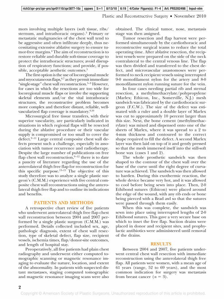
Case 1A 64-year-old woman (patient 2 in Tables 1 and 2) presented
in February of 2004 with a T2N2aM0 invasive ductal carcinomaof the left breast that was treated by total mastectomy withaxillary lymph node dissection and six cycles of adjuvant cyclo-phosphamide, methotrexate, and 5-fluorouracil chemotherapyand 5 weeks of postoperative radiotherapy.
Three years later, she presented with a mass in the middlethird of her sternum that was histologically diagnosed as aregional recurrence of the previous breast cancer. The massgrew rapidly (Fig. 1, above, left), and the computed tomographicscan showed bony destruction and invasion of the intrathoracicorgans (pleura and left lung). A radical sternectomy with sac-rifice of eight adjacent ribs was planned. During the tumorresection, both internal mammary vessels were sacrificed. Skel-etal reconstruction to provide rigid stabilization of the chestwall was undertaken using composite synthetic materials madeby the cardiothoracic surgeon (Fig. 1, above, center and above,right); the resulting wide soft-tissue defect that extended be-tween the second intercostal space down to the xiphoid processwas too large for repair with locoregional flaps. The defect wastherefore covered with a left free anterolateral thigh flap in- Ta
ble
1.
Surg
ical
Det
ails
ofF
ive
Pat
ien
tsW
ho
Un
der
wen
tC
entr
alC
hes
tW
allR
esec
tio
nan
dR
eco
nst
ruct
ion
wit
hA
nte
rola
tera
lTh
igh
Free
Flap
s
Pat
ient
Age
(yr)
*P
atho
logi
cD
iagn
osis
Che
stD
efec
tD
efec
tSi
ze(c
m)
Flap
Size
(cm
)R
ecip
ient
Ves
sels
Isch
emia
Tim
e(m
in)
Flap
Com
plic
atio
nsD
onor
-Sit
eC
ompl
icat
ions
159
Met
asta
tic
brea
stca
nce
rSt
ern
uman
d6
ribs
(par
tial
)12
�15
17�
11FA
/IJV
72N
one
Sero
ma
264
Met
asta
tic
brea
stca
nce
rSt
ern
uman
d8
ribs
(par
tial
)15
�18
21�
12FA
/IJV
77N
one
Non
e
369
Rad
ion
ecro
sis
No
skel
etal
12�
1316
�9
LA
/IJV
81N
one
Pain
462
Met
asta
tic
brea
stca
nce
rSt
ern
uman
d4
ribs
(par
tial
)14
�16
18�
12FA
/IJV
68N
one
Sero
ma
552
Prim
ary
fibr
osar
com
aSt
ern
uman
d4
ribs
(par
tial
)12
�13
13�
11FA
/IJV
103
Non
eN
one
FA,
faci
alar
tery
;IJ
V,
inte
rnal
jugu
lar
vein
;L
A,
lingu
alar
tery
.*A
gesh
own
isag
eat
the
tim
eof
ches
tw
all
surg
ery.
Volume 126, Number 5 • Composite Chest Wall Defect Repair
3
T1
T2
F1
rich3/zpr-prs/zpr-prs/zpr01110/zpr3877-10z xppws S�1 8/13/10 6:19 4/Color Figure(s): F1-4 Art: PRS202498 Input-nlm
corporating a small segment of the vastus lateralis muscle to fillin the large thoracic dead space.
The flap was easily orientated and inset (Fig. 1, below, left)such that the pedicle was superior. It was passed under a sub-cutaneous tunnel from the chest to the neck to achieve com-fortable microvascular anastomoses to the facial artery and theinternal jugular vein recipient vessels (Fig. 1, below, center). Thepostoperative course was without any complications andthe skeletal and soft-tissue reconstruction was adequate andstable (Fig. 1, below, right).
Fig. 1. Case 1 (patient 2 in Tables 1 and 2). (Above, left) Preoperative view of a 64-year-old woman with a large mass over the sternumindicating recurrence of the previous left breast cancer. (Above, center) Wide resection of the tumor. (Above, right) Skeletal stabi-lization using composite synthetic materials (methylmethacrylate/Marlex mesh prosthesis. An anterolateral thigh free flap with asmall segment of vastus lateralis muscle was used to repair the soft-tissue defect. Its easy orientation (below, left) allows comfortablemicrovascular anastomoses in the neck recipient site (below, center). (Below, right) Preoperative view of the patient at follow-up 8months after surgery.
Table 2. Recovery and Follow-Up
PatientIntensive Care
Unit Stay (days)Long Hospital
Stay (days)Follow-Up
(mo)
1 3 8 6*2 2 9 283 0 6 194 7 17 5*5 4 11 21*Patient died as a result of metastatic cancer.
Plastic and Reconstructive Surgery • November 2010
4
rich3/zpr-prs/zpr-prs/zpr01110/zpr3877-10z xppws S�1 8/13/10 6:19 4/Color Figure(s): F1-4 Art: PRS202498 Input-nlm
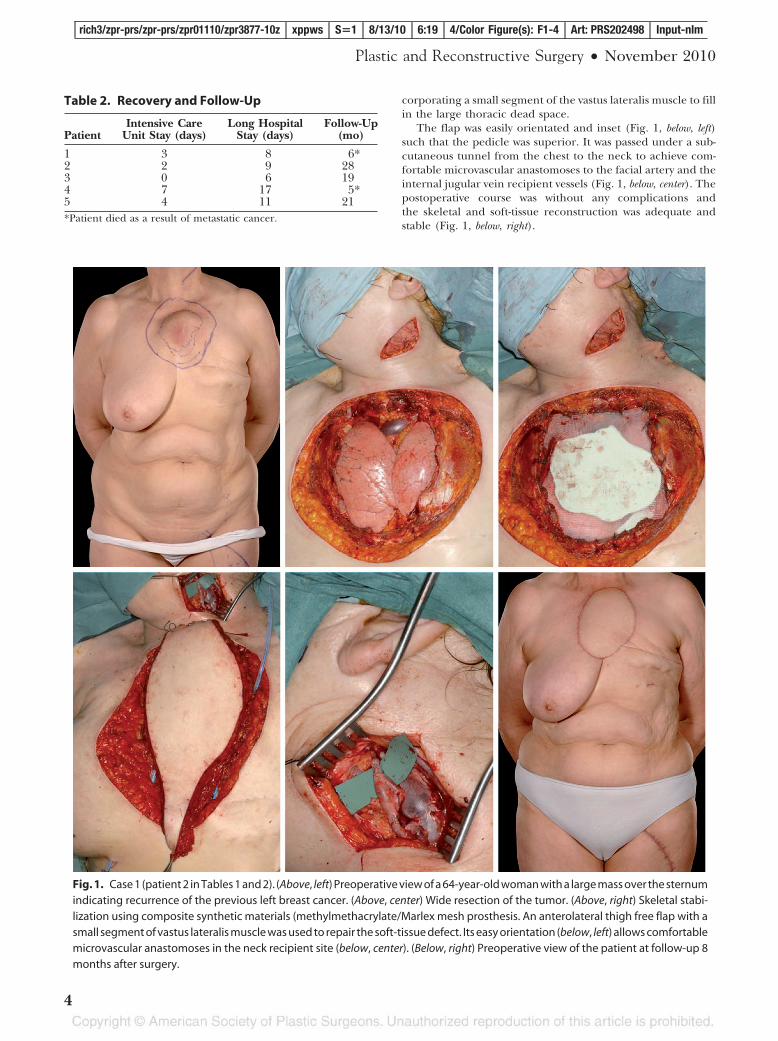
Case 2A 52-year-old woman (patient 5 in Table 1 and 2) presented
in January of 2004 with a large mass over the sternal manubriumthat extended rapidly toward the base of the neck (Fig. 2, above,left). The core biopsy revealed a primitive fibrosarcoma of thechest wall. As the computed tomographic scan showed no dis-tant metastases and chemotherapy and radiotherapy werejudged unsuitable, the patient was referred for surgery. Thesuperior two-thirds of the sternum was resected together withpartial excision of four ribs. A rigid support made of Marlexmesh and methylmethacrylate was anchored to the rib rem-nants and clavicle, thus providing protection for the intratho-racic organs (Fig. 2, below, left).
A left fasciocutaneous anterolateral thigh free flap was usedto cover the remaining soft-tissue defect. The recipient vesselsused were the left facial artery and internal jugular vein. Thehospital stay was uneventful, and the patient had a good func-tional and aesthetic outcome (Fig. 2, right).
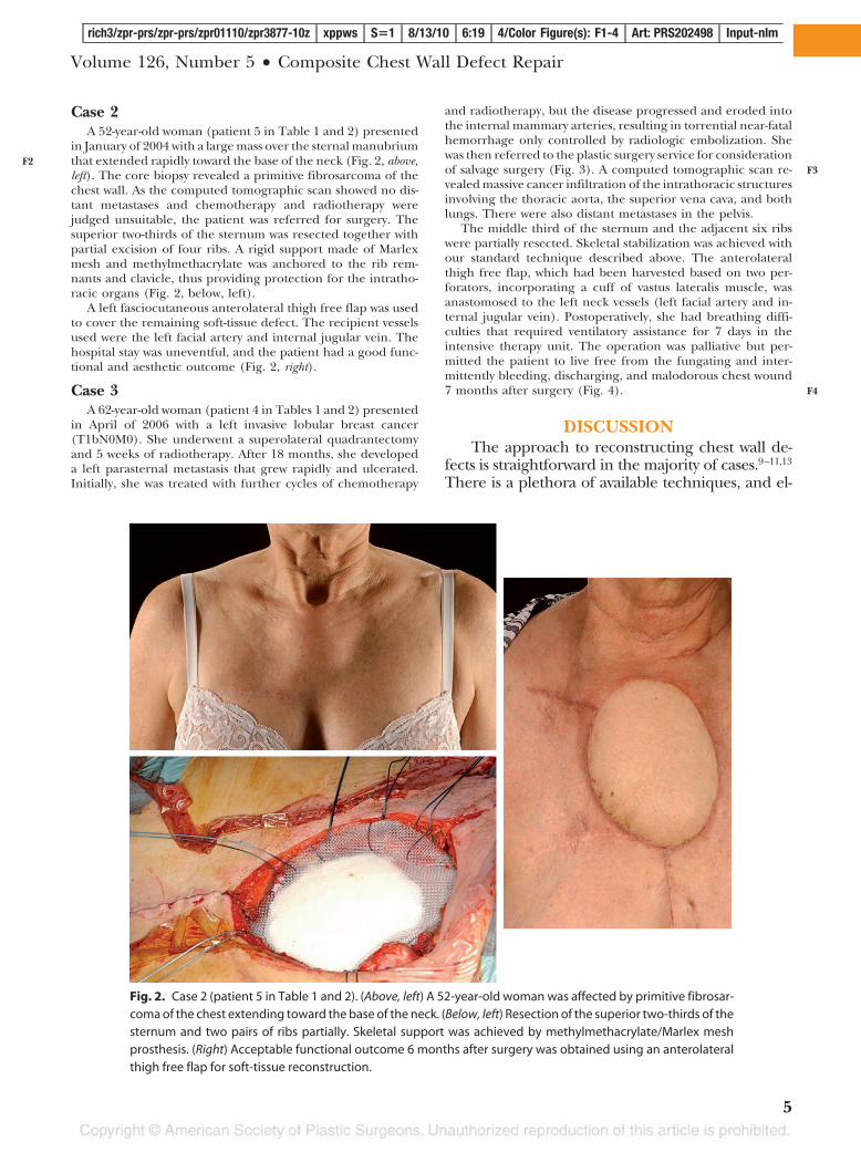
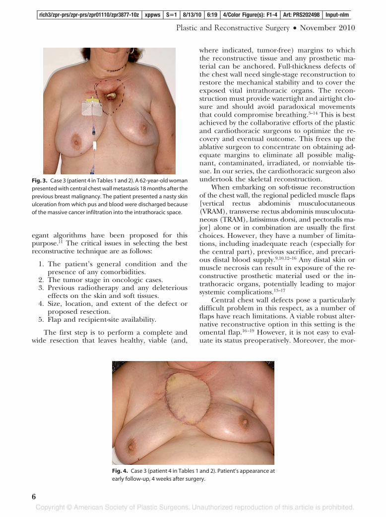
Case 3A 62-year-old woman (patient 4 in Tables 1 and 2) presented
in April of 2006 with a left invasive lobular breast cancer(T1bN0M0). She underwent a superolateral quadrantectomyand 5 weeks of radiotherapy. After 18 months, she developeda left parasternal metastasis that grew rapidly and ulcerated.Initially, she was treated with further cycles of chemotherapy
and radiotherapy, but the disease progressed and eroded intothe internal mammary arteries, resulting in torrential near-fatalhemorrhage only controlled by radiologic embolization. Shewas then referred to the plastic surgery service for considerationof salvage surgery (Fig. 3). A computed tomographic scan re-vealed massive cancer infiltration of the intrathoracic structuresinvolving the thoracic aorta, the superior vena cava, and bothlungs. There were also distant metastases in the pelvis.
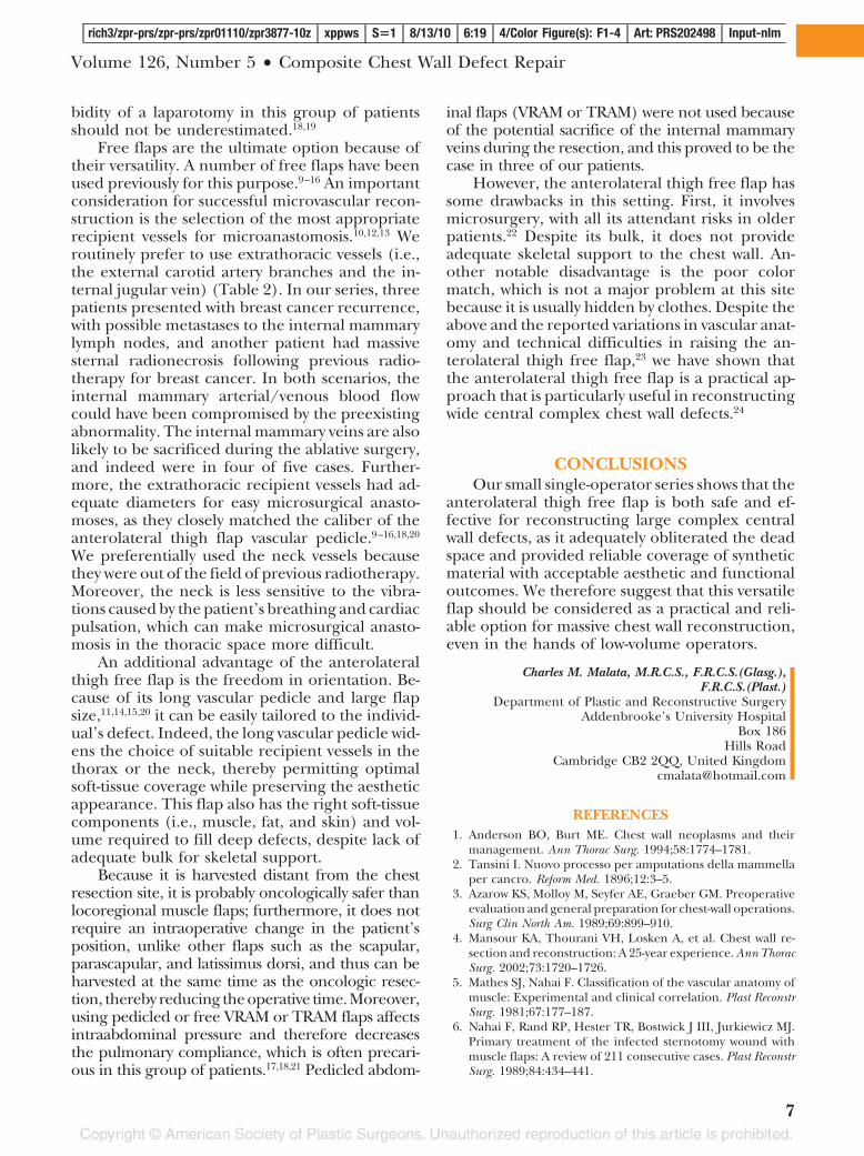
The middle third of the sternum and the adjacent six ribswere partially resected. Skeletal stabilization was achieved withour standard technique described above. The anterolateralthigh free flap, which had been harvested based on two per-forators, incorporating a cuff of vastus lateralis muscle, wasanastomosed to the left neck vessels (left facial artery and in-ternal jugular vein). Postoperatively, she had breathing diffi-culties that required ventilatory assistance for 7 days in theintensive therapy unit. The operation was palliative but per-mitted the patient to live free from the fungating and inter-mittently bleeding, discharging, and malodorous chest wound7 months after surgery (Fig. 4).
DISCUSSIONThe approach to reconstructing chest wall de-
fects is straightforward in the majority of cases.9–11,13
There is a plethora of available techniques, and el-
Fig. 2. Case 2 (patient 5 in Table 1 and 2). (Above, left) A 52-year-old woman was affected by primitive fibrosar-coma of the chest extending toward the base of the neck. (Below, left) Resection of the superior two-thirds of thesternum and two pairs of ribs partially. Skeletal support was achieved by methylmethacrylate/Marlex meshprosthesis. (Right) Acceptable functional outcome 6 months after surgery was obtained using an anterolateralthigh free flap for soft-tissue reconstruction.
Volume 126, Number 5 • Composite Chest Wall Defect Repair
5
F2F3
F4
rich3/zpr-prs/zpr-prs/zpr01110/zpr3877-10z xppws S�1 8/13/10 6:19 4/Color Figure(s): F1-4 Art: PRS202498 Input-nlm
egant algorithms have been proposed for thispurpose.11 The critical issues in selecting the bestreconstructive technique are as follows:
1. The patient’s general condition and thepresence of any comorbidities.
2. The tumor stage in oncologic cases.3. Previous radiotherapy and any deleterious
effects on the skin and soft tissues.4. Size, location, and extent of the defect or
proposed resection.5. Flap and recipient-site availability.
The first step is to perform a complete andwide resection that leaves healthy, viable (and,
where indicated, tumor-free) margins to whichthe reconstructive tissue and any prosthetic ma-terial can be anchored. Full-thickness defects ofthe chest wall need single-stage reconstruction torestore the mechanical stability and to cover theexposed vital intrathoracic organs. The recon-struction must provide watertight and airtight clo-sure and should avoid paradoxical movementsthat could compromise breathing.3–14 This is bestachieved by the collaborative efforts of the plasticand cardiothoracic surgeons to optimize the re-covery and eventual outcome. This frees up theablative surgeon to concentrate on obtaining ad-equate margins to eliminate all possible malig-nant, contaminated, irradiated, or nonviable tis-sue. In our series, the cardiothoracic surgeon alsoundertook the skeletal reconstruction.
When embarking on soft-tissue reconstructionof the chest wall, the regional pedicled muscle flaps[vertical rectus abdominis musculocutaneous(VRAM), transverse rectus abdominis musculocuta-neous (TRAM), latissimus dorsi, and pectoralis ma-jor] alone or in combination are usually the firstchoices. However, they have a number of limita-tions, including inadequate reach (especially forthe central part), previous sacrifice, and precari-ous distal blood supply.9,10,12–16 Any distal skin ormuscle necrosis can result in exposure of the re-constructive prosthetic material used or the in-trathoracic organs, potentially leading to majorsystemic complications.13–17
Central chest wall defects pose a particularlydifficult problem in this respect, as a number offlaps have reach limitations. A viable robust alter-native reconstructive option in this setting is theomental flap.16–19 However, it is not easy to eval-uate its status preoperatively. Moreover, the mor-
Fig. 4. Case 3 (patient 4 in Tables 1 and 2). Patient’s appearance atearly follow-up, 4 weeks after surgery.
Fig. 3. Case 3 (patient 4 in Tables 1 and 2). A 62-year-old womanpresented with central chest wall metastasis 18 months after theprevious breast malignancy. The patient presented a nasty skinulceration from which pus and blood were discharged becauseof the massive cancer infiltration into the intrathoracic space.
Plastic and Reconstructive Surgery • November 2010
6
rich3/zpr-prs/zpr-prs/zpr01110/zpr3877-10z xppws S�1 8/13/10 6:19 4/Color Figure(s): F1-4 Art: PRS202498 Input-nlm
bidity of a laparotomy in this group of patientsshould not be underestimated.18,19
Free flaps are the ultimate option because oftheir versatility. A number of free flaps have beenused previously for this purpose.9–16 An importantconsideration for successful microvascular recon-struction is the selection of the most appropriaterecipient vessels for microanastomosis.10,12,13 Weroutinely prefer to use extrathoracic vessels (i.e.,the external carotid artery branches and the in-ternal jugular vein) (Table 2). In our series, threepatients presented with breast cancer recurrence,with possible metastases to the internal mammarylymph nodes, and another patient had massivesternal radionecrosis following previous radio-therapy for breast cancer. In both scenarios, theinternal mammary arterial/venous blood flowcould have been compromised by the preexistingabnormality. The internal mammary veins are alsolikely to be sacrificed during the ablative surgery,and indeed were in four of five cases. Further-more, the extrathoracic recipient vessels had ad-equate diameters for easy microsurgical anasto-moses, as they closely matched the caliber of theanterolateral thigh flap vascular pedicle.9–16,18,20
We preferentially used the neck vessels becausethey were out of the field of previous radiotherapy.Moreover, the neck is less sensitive to the vibra-tions caused by the patient’s breathing and cardiacpulsation, which can make microsurgical anasto-mosis in the thoracic space more difficult.
An additional advantage of the anterolateralthigh free flap is the freedom in orientation. Be-cause of its long vascular pedicle and large flapsize,11,14,15,20 it can be easily tailored to the individ-ual’s defect. Indeed, the long vascular pedicle wid-ens the choice of suitable recipient vessels in thethorax or the neck, thereby permitting optimalsoft-tissue coverage while preserving the aestheticappearance. This flap also has the right soft-tissuecomponents (i.e., muscle, fat, and skin) and vol-ume required to fill deep defects, despite lack ofadequate bulk for skeletal support.
Because it is harvested distant from the chestresection site, it is probably oncologically safer thanlocoregional muscle flaps; furthermore, it does notrequire an intraoperative change in the patient’sposition, unlike other flaps such as the scapular,parascapular, and latissimus dorsi, and thus can beharvested at the same time as the oncologic resec-tion, thereby reducing the operative time. Moreover,using pedicled or free VRAM or TRAM flaps affectsintraabdominal pressure and therefore decreasesthe pulmonary compliance, which is often precari-ous in this group of patients.17,18,21 Pedicled abdom-
inal flaps (VRAM or TRAM) were not used becauseof the potential sacrifice of the internal mammaryveins during the resection, and this proved to be thecase in three of our patients.
However, the anterolateral thigh free flap hassome drawbacks in this setting. First, it involvesmicrosurgery, with all its attendant risks in olderpatients.22 Despite its bulk, it does not provideadequate skeletal support to the chest wall. An-other notable disadvantage is the poor colormatch, which is not a major problem at this sitebecause it is usually hidden by clothes. Despite theabove and the reported variations in vascular anat-omy and technical difficulties in raising the an-terolateral thigh free flap,23 we have shown thatthe anterolateral thigh free flap is a practical ap-proach that is particularly useful in reconstructingwide central complex chest wall defects.24
CONCLUSIONSOur small single-operator series shows that the
anterolateral thigh free flap is both safe and ef-fective for reconstructing large complex centralwall defects, as it adequately obliterated the deadspace and provided reliable coverage of syntheticmaterial with acceptable aesthetic and functionaloutcomes. We therefore suggest that this versatileflap should be considered as a practical and reli-able option for massive chest wall reconstruction,even in the hands of low-volume operators.
Charles M. Malata, M.R.C.S., F.R.C.S.(Glasg.),F.R.C.S.(Plast.)
Department of Plastic and Reconstructive SurgeryAddenbrooke’s University Hospital
Box 186Hills Road
Cambridge CB2 2QQ, United [email protected]
REFERENCES1. Anderson BO, Burt ME. Chest wall neoplasms and their
management. Ann Thorac Surg. 1994;58:1774–1781.2. Tansini I. Nuovo processo per amputations della mammella
per cancro. Reform Med. 1896;12:3–5.3. Azarow KS, Molloy M, Seyfer AE, Graeber GM. Preoperative
evaluation and general preparation for chest-wall operations.Surg Clin North Am. 1989;69:899–910.
4. Mansour KA, Thourani VH, Losken A, et al. Chest wall re-section and reconstruction: A 25-year experience. Ann ThoracSurg. 2002;73:1720–1726.
5. Mathes SJ, Nahai F. Classification of the vascular anatomy ofmuscle: Experimental and clinical correlation. Plast ReconstrSurg. 1981;67:177–187.
6. Nahai F, Rand RP, Hester TR, Bostwick J III, Jurkiewicz MJ.Primary treatment of the infected sternotomy wound withmuscle flaps: A review of 211 consecutive cases. Plast ReconstrSurg. 1989;84:434–441.
Volume 126, Number 5 • Composite Chest Wall Defect Repair
7
rich3/zpr-prs/zpr-prs/zpr01110/zpr3877-10z xppws S�1 8/13/10 6:19 4/Color Figure(s): F1-4 Art: PRS202498 Input-nlm
7. Jurkiewicz MJ, Bostwick J III, Hester TR, Bishop JB, Craver J.Infected median sternotomy wound: Successful treatment bymuscle flaps. Ann Surg. 1980;191:738–744.
8. Brown RG, Fleming WH, Jurkiewicz MJ. An island flap of thepectoralis major muscle. Br J Plast Surg. 1977;30:161–165.
9. Tukiainen E, Popov P, Asko-Seljavaara S. Microvascular re-constructions of full thickness oncological chest wall defects.Ann Surg. 2003;238:794–802.
10. Cordeiro PG, Santamaria E, Hidalgo D. The role of micro-surgery in reconstruction of oncologic chest wall defects.Plast Reconstr Surg. 2001;108:1924–1930.
11. Gore SM, Akhavani MA, Kang N, Chana JS. Chest wall re-construction using a turbocharged chimaeric anterolateralthigh flap. J Plast Reconstr Aesthet Surg. 2008;61:438–441.
12. Losken A, Thourani VH, Carlson GW, et al. A reconstructivealgorithm for plastic surgery following extensive chest wallresection. Br J Plast Surg. 2004;57:295–302.
13. Tukiainen E, Popov P, Asko-Seljavaara S. Microvascular re-constructions of full-thickness oncological chest wall defects.Ann Surg. 2003;238:794–801; discussion 801–802.
14. Wei FC, Jain V, Celik N, Chen HC, Chuang DC, Lin CH. Havewe found an ideal soft-tissue flap? An experience with 672anterolateral thigh flaps. Plast Reconstr Surg. 2002;109:2219–2226; discussion 2227–2230.
15. Kuo YR, Jeng SF, Kuo MH, Liu YT, Lai PW. Versatility of thefree anterolateral thigh flap for reconstruction of soft-tissuedefects: Review of 140 cases. Ann Plast Surg. 2002;48:161–166.
16. Arnold PG, Pairolero PC. Chest wall reconstruction: An ac-count of 500 consecutive patients. Plast Reconstr Surg. 1996;98:804–810.
17. Cohen M, Ramasastry SS. Reconstruction of complex chestwall defects. Am J Surg. 1996;172:35–40.
18. Chang RR, Mehrara BJ, Hu QY, Disa JJ, Cordeiro PG. Re-construction of complex oncologic chest wall defects: A 10year experience. Ann Plast Surg. 2004;52:471–479.
19. Jurkiewicz MJ, Arnold PG. The omentum: An account of itsuse in the reconstruction of the chest wall. Ann Surg. 1977;185:548–554.
20. Malata CM, Tehrani H, Kumiponjera D, Hardy DG, MoffatDA. Use of anterolateral thigh and lateral arm fasciocuta-neous free flaps in lateral skull base reconstruction. Ann PlastSurg. 2006;57:169–175; discussion 176.
21. Yamamoto Y, Nohira K, Shintomi Y, Sugihara T, Ohura T.Turbo charging the vertical rectus abdominis myocutaneous(turbo-VRAM) flap for reconstruction of extensive chest walldefects. Br J Plast Surg. 1994;47:103–107.
22. Malata CM, Cooter RD, Batchelor AG, Simpson KH, Brown-ing FS, Kay SP. Microvascular free-tissue transfers in elderlypatients: The leeds experience. Plast Reconstr Surg. 1996;98:1234–1241.
23. Cormack GC, Lamberty BG. The Arterial Anatomy of SkinFlaps. New York: Churchill Livingstone; 1994:85–103, 132–148.
24. Di Candia M, Lie KH, Kumiponjera D, Malata CM. Versatility ofthe anterolateral thigh free flap for soft tissue reconstruction: TheCambridge experience. Br J Surg. 2008;95(Suppl 6):1–104.
Plastic and Reconstructive Surgery • November 2010
8
rich3/zpr-prs/zpr-prs/zpr01110/zpr3877-10z xppws S�1 8/13/10 6:19 4/Color Figure(s): F1-4 Art: PRS202498 Input-nlm
JOBNAME: AUTHOR QUERIES PAGE: 1 SESS: 3 OUTPUT: Fri Aug 13 06:20:23 2010/rich3/zpr�prs/zpr�prs/zpr01110/zpr3877�10z
AQ1: AUTHOR—Author list: The Journal allows the use of academic degrees (e.g., M.D., M.A.,M.S.) or fellowship affiliations (e.g., F.R.C.S., M.R.C.S.) but not both. For authors with bothdegrees and affiliations, the fellowship affiliations were used. If the academic degrees arepreferred, please indicate such on the page proof. Please also note that the Journal allows amaximum of three degrees/affiliations (preferably the three highest earned).
AUTHOR QUERIES
AUTHOR PLEASE ANSWER ALL QUERIES 1