Embed Size (px)
Citation preview

Fisheries Research 96 (2009) 148–159
Contents lists available at ScienceDirect
Fisheries Research
journa l homepage: www.e lsev ier .com/ locate / f i shres
Reconstructing individual shape histories of fish otoliths: A newimage-based tool for otolith growth analysis and modeling
Ronan Fableta,d,∗, Anatole Chesselb, Sebastien Carbinib,Abdesslam Benzinouc, Hélène de Pontualb
a Institut Telecom/Telecom Bretagne/UMR 3192 LabSTICC, CS 83818, Technopôle Brest-Iroise, 29238 Brest Cedex 3, Franceb Ifremer/STH, BP 70, 29280 Plouzané, Francec ENIB/RESO, Technopôle Brest-Iroise 29280 Plouzane, Franced Université européenne de Bretagne, France
a r t i c l e i n f o
Article history:Received 31 July 2008Received in revised form 10 October 2008Accepted 17 October 2008
Keywords:Otolith imaging
a b s t r a c t
In this paper is presented a novel image processing tool for the extraction of geometric information inotolith images. It relies on the reconstruction of individual otolith shape histories from otolith images.Based on the proposed non-parametric level-set representation of otolith shape history, applications tothe extraction of growth axes and ring structures in otolith images are first considered. A second categoryof applications concern the analysis of 2D otolith growth. The potential of the proposed framework isillustrated on real otolith images for various species (e.g., cod, pollock) and discussed with a particular
Shape dynamicsGrowth ring extractionG2
emphasis on the genericity of the approach and on applications such as otolith shape analysis, multi-proxyotolith analysis, otolith modeling.
© 2008 Elsevier B.V. All rights reserved.
1
cecfidptem(vasoy
2
2poge1slasliia
0d
rowth axis extractionD otolith growth
. Introduction and problem statement
As they grow according to an accretionary process, fish otolithsan be viewed as a succession of three-dimensional concentric lay-rs. The composition of these layers, in terms of physico-chemicalharacteristics, varies according to endogenous and exogenousactors (Panfili et al., 2002). The accretionary process generallynvolves a periodic rhythmicity, typically daily and/or seasonal,eposit, such that these biological structures depict concentric ringatterns, also called growth marks, in an observation plane goinghrough the initial core . These characteristics provide the basis forxploiting these structures as biological archives to define environ-ental proxies (e.g., for instance to reconstruct temperature series)
Hoie et al., 2004), or to reconstruct individual life traits (e.g., indi-idual age and growth information or migration paths) (Fablet et
l., 2007). To further stress the key importance of these biologicaltructures in marine ecology, it can be pointed out that well overne million fish (Campana and Thorrold, 2001) are analyzed eachear to estimate age structures for fish stock assessment.∗ Corresponding author at: Telecom Bretagne CS 83818, Technopôle Brest-Iroise,9238 Brest Cedex 3, France.
E-mail address: [email protected] (R. Fablet).URL: http://perso.enst-bretagne.fr/ronanfablet (R. Fablet).
op(oistacn
165-7836/$ – see front matter © 2008 Elsevier B.V. All rights reserved.oi:10.1016/j.fishres.2008.10.011
Following ongoing developments (Alvarez et al., 2008; Fablet,006; Fablet et al., 2007) aimed at information extraction and inter-retation in fish otolith images, this paper addresses the extractionf geometric otolith characteristics and their application to otolithrowth modelling and analysis. Though extensively studied andxploited (Campana and Casselman, 1993; de Pontual and Prouzet,988), the analysis of the shape of fish otoliths and other calcifiedtructures has usually been restricted to the analysis of the out-ine of the otolith in a given observation plane, especially for stocknd species discrimination. However, the presence of internal ringtructures potentially provides the mean for back-tracking the evo-ution of the shape of the otolith from the core to the edge. Suchnformation is of great interest for analyzing, modelling and extract-ng the main features of otolith growth. Recently, we developed
new computational tool aimed at reconstructing the sequencef the successive shapes associated with an accretionary growthrocess in a given observation plane containing the otolith coreFablet et al., 2008b). We benefit from this representation of thetolith growth to develop new solutions for information extractionn otolith images. Experiments on real otolith images for various
pecies for various species are reported, and, we investigate a quan-itative analysis of the 2D otolith growth. The genericity of thepproach is further discussed as well as its broad interest for appli-ations to otolith shape analysis, multi-proxy otolith analysis andumerical otolith modelling.
Resea
2
2
iigvcuh(etct2o
t1
2f
soroptf
�
ToavBeal
Fla
•
•
•
•
gmactaFitttiTveprpac
E
R. Fablet et al. / Fisheries
. Materials and methods
.1. Otolith material
In this study, the considered biological material is a set ofmages of whole otoliths or otolith sections associated with annterpretation of the internal growth structures in terms of age androwth pattern. Otolith sections have been prepared in the trans-erse plane. We focus on seasonal growth and thick sections areonsidered. The proposed methodological developments are eval-ated for several species (namely, examples of cod (Gadus morhua),ake (Merluccius merluccius), plaice (Pleuronectes platessa), pollockPollachius virens), and whiting (M erlangus merlangus) are consid-red). These species are chosen for the panel of complexity levelshey convey in terms of image contrast and ring structures. Thishoice is also aimed at demonstrating the improvements comparedo previous work (Fablet, 2006; Guillaud et al., 2002; Palmer et al.,005; Traodec et al., 2000) which was mainly limited to the analysisf whole plaice otoliths.
Otolith images have been acquired under a binocular usingransmitted or reflected light depending on the species with a000 × 1000 digital camera.
.2. Reconstruction of individual histories of 2D otolith shapesrom images
The core of the proposed computational framework is the recon-truction of the evolution of the 2D otolith shape in a givenbservation plane from an image. With a view to modelling andepresenting the 2D otolith growth, we adopt a level-set settingf the accretionary growth process. It relies on the definition of aotential function U such that the 2D shape �t(U) of the otolith atime t is given by a level line of U, that is to say the set of points por which the associated potential value U(p) equals t:
t(U) = {p ∈R2 such that U(p) = t} (1)
his level-set representation of the accretionary growth of fishtoliths is illustrated (Fig. 1). The potential function U is displayeds a 3D surface, and the successive level-lines of U, for potentialalues uniformly sampled, are visualized in the horizontal plane.
y definition, potential function U is convex: as it can be consid-red that fish otoliths never resorb (Panfili et al., 2002), the shapet time t is included in the shape at any time t′ posterior to t. Thisevel-set setting is of great interest for several reasons:ig. 1. Level-set representation of the accretionary growth of fish otoliths: eachevel-line of the increasing potential function represents the shape of the otolith atgiven age.
wt〈tegioeioarica
t
t
s
rch 96 (2009) 148–159 149
It intrinsically conforms to the requirements that the accretionarygrowth is normal to the shape and that the successive shapes areconcentric.1
It is a compact representation of a series of successive shapes, thewhole series of shapes being represented by a single mathemat-ical function U.It is generic as it accounts for elliptic-like shapes, such as wholeplaice otoliths, as well as more complex non-convex examplessuch as hake or cod otolith sections.It is non-parametric. Contrary to the parametric approach pro-posed in (Alvarez et al., 2008), no assumption is made on theevolution of the shape, so that subsequent analysis is not biasedby some parametric a priori which may not be fulfilled in practice.
Our goal is to fit the level-set model U to an otolith image in aiven observation plane, such that the successive level-lines of Uatch the internal rings of the otolith. We further assume that we
re provided with additional constraints, referred to as boundaryonditions, at least the position of the nucleus of the otolith andhe edge of the otolith which can be extracted automatically (Caond Fablet, 2006). Additional internal rings may also be provided.itting model U is then viewed as its interpolation to the wholemage domain given known boundary constraints. This interpola-ion is stated as the minimization of an energy criterion involvingwo different terms. The first term is a regularisation energy set-ing that the successive shapes �t(U) should be smooth. This terms computed as the sum of the perimeters of all the shapes {�t(U)}.he second term relies on image-based features. Exploiting pre-ious work on the estimation of local image orientations (Chesselt al., 2006), this term states that the normal to shape {�t(U)} atoint p should be orthogonal to the local tangent to ring structures,eferred to as the local orientation and denoted by w(p). An exam-le of an estimated map of local image orientations is reported forpollock otolith section (Fig. 3). Formally, the considered energy
riterion is given by:
(U) = (1 − �)
∫t ∈ [0,T]
∫p ∈ �t (U)
1
+ �
∫t ∈ [0,T]
∫p ∈ �t (U)
·∣∣∣⟨ ∇U(p)
|∇U(p)| , ω(p)⟩∣∣∣ (2)
here � is a weight setting the relative influence of the twoerms, ∇U(p)/|∇U(p)| the orientation of shape �t(U) at point p and∇U(p)/|∇U(p)|, ω(p)〉 the scalar product evaluating whether thewo orientations are orthogonal. We let the reader refer to (Fablett al., 2008a, b) for details on the numerical implementation of theradient-based minimization of criterion E. Cross-validation exper-ments carried out on synthetic examples have shown that valuesf � in the range [0.4, 0.8] are optimal in terms of mean-squarerror (Fablet et al., 2008a). Though no theoretical evidence can val-date this experimental statement, our experiments on a variety oftolith images show that setting � to 0.6 is appropriate in practicend that results are stable if � is set in the range [0.4,0.8]. This is cor-oborated by the numerous applications of variational techniquesn computer vision (Sethian, 1999). Concerning the computational
ost, the proposed scheme is implemented as a C code2 under Linuxnd runs in about 1 min for a 1000 × 1000 image.If the otolith growth pattern along a given growth axis is known,he estimated potential function U provides at any pixel p an age
1 The term “concentric” should not be understood in a strict sense. We mean herehat the shape at time t is included in all the shapes posterior to time t.
2 All source codes, Matlab and C codes currently running under Linux, used in thistudy are available on request to [email protected].
1 Resea
e2tmdfi
2
tWiglo
gttbeaog
cfGoHg(vWtaohtmd
(atrhilnttappl
2
etqb
tfiuoocfsat
•
•
•
•
•
g
a
••
tion, especially geometric shape features, growth measures andopacity descriptors?
• Are these relations between the 2D features of the accretion con-stant in space and time?
50 R. Fablet et al. / Fisheries
stimate. If not, potential function U only provides the successiveD shapes of the otolith from the core to the edge. In that case,he values of U refer to the actual age up to a contrast change (i.e., a
onotonic increasing function). This second situation occurs whenealing with automated otolith imaging for instance for automatedsh ageing (Fablet, 2006).
.3. Extraction of geometric structures in otolith images
From a fitted potential function U, we consider applications tohe automated extraction of geometric structures in otolith images.
e here illustrate this great potential for three different types ofnformation, namely growth shapes, internal ring structures androwth axis. We only briefly review the proposed approaches andet the reader refer to (Fablet et al., 2008a) for detailed presentationsn the associated algorithms.
A first straightforward by-product is the sequence of the 2Drowth shapes of the otolith issued from the level-lines of poten-ial function U. If function U is time-calibrated, the 2D shape ofhe otolith can be extracted at any age, i.e. at any precision (yearly,iannual, etc.) as a level-line of U. Such information is of key inter-st regarding otolith shape analysis and classification (Campanand Casselman, 1993): either as a potentially discriminant featurer for normalization purposes when samples from different ageroups have to be dealt with.
A second application is the extraction of the opaque and translu-ent ring curves in otolith images. It serves for instance as a basisor age and growth estimation (Fablet, 2006; Traodec et al., 2000).ood performances have been reported for images of whole plaicetoliths (Traodec et al., 2000; Fablet, 2006; Palmer et al., 2005).owever, these otoliths depict very clear ring structures and theirrowth can be viewed as mainly radial. For more complex imagese.g., hake or cod otolith images), the methods proposed in pre-ious work do not succeed in correctly extracting ring structures.e can address these issues using the proposed otolith shape his-
ory model. Growth ring structures correspond to image valleysnd ridges (together known as creases), which are the relief curvesf the landscape obtained when the image intensity is seen as aeight map. We then propose to extract ring curves as portions ofhe level-lines of U depicting high values of a local contrast-based
easure. Formally, our method is implemented within a contrarioetection framework (Desolneux et al., 2001, 2003).
When focusing on temporal signals archived by fish otolithse.g., growth patterns, migrations, environmental records, etc.), thenalysis is mainly one-dimensional from the core to the edge ofhe otoliths. Hence, the extraction and the standardization of theeference growth axis is a crucial step. To our knowledge, no toolas been developed to this end yet. As the accretionary growth
s normal to the surface, growth axes can be defined as pathsinking the growth centre to the edge such that these paths areormal to the ring structures. A straightforward solution wouldhen be to extract growth axes as integral lines of the orienta-ion field ∇U/|∇U|. This solution is however numerically unstablend a more robust variational setting is proposed. Using a minimalath scheme (Cohen, 2005), growth axes are retrieved as smoothaths from the core to the edge, locally, as normal as possible to the
evel-lines of U.
.4. Quantitative analysis of the 2D otolith growth
Whereas a huge amount of work has been dedicated to thextraction and the analysis of one-dimensional otolith growth pat-erns (Campana and Thorrold, 2001; Panfili et al., 2002), the actualuantitative analysis of the 2D growth has, to our knowledge, onlyeen occasionally considered. Such an analysis is of great interest
carc
rch 96 (2009) 148–159
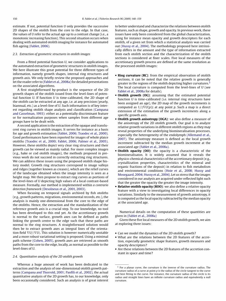
o better understand and characterize the relations between otolitheatures, such as shape, growth and opacity. In previous work, thesessues have only been considered from the global characterization,sing for instance mean opacity and growth descriptors for eachtolith, of a given set from which a statistical analysis was carriedut (Hussy et al., 2004). The methodology proposed here intrinsi-ally differs in the amount and the type of information extractedrom each otolith section and the characterization of the otolithections is considered at finer scales. Five local measures of theccretionary growth process are defined at the same resolution ashe processed otolith images:
Ring curvature (RC): from the empirical observation of otolithsections, it can be noted that the relative growth is generallygreater in the regions of the otolith depicting higher curvatures.3
The local curvature is computed from the level-lines of U (seeFablet et al., 2008a for details).Otolith growth (OG): assuming that the estimated potentialfunction U is time-calibrated (i.e. internal ring structures havebeen assigned an age), the 2D map of the growth increments iscomputed as 1/‖∇U(p)‖ at any point p. Such a map is a directextension of the estimation of the growth increments along aspecific growth axis.Otolith growth anisotropy (OGA): we also define a measure ofthe anisotropy of the 2D otolith growth. Our goal is to analyzerelative growth variations in different otolith regions as they mayreveal properties of the underlying biomineralisation processes,especially the heterogeneity of the endolymph (Allemand et al.,2007). The anisotropy measure is defined as the local growthincrement subtracted by the median growth increment at theassociated age (Fablet et al., 2008a).Otolith opacity (OO): the opacity is a characteristic of thebiomineralisaton. It is widely assumed that it reveals thephysico-chemical characteristics of the accretionary deposit (e.g.,crystallization properties, characteristics of the mineral andorganic fractions of the deposit) in relation to fish metabolismand environmental conditions (Hoie et al., 2008; Hussy andMosegaard, 2004; Hussy et al., 2004). Let us stress that the imagesconsidered in our analysis are acquired under reflected light suchthat the greater the opacity the greater the image intensity.Relative otolith opacity (ROO): we also define a relative opacityfeature with a view to investigating local differences in opacityvariations. Similarly to the measurement of growth anisotropy, itis computed as the local opacity subtracted by the median opacityat the associated age.
Numerical details on the computation of these quantities areiven in (Fablet et al., 2008a)
Given these five local measures of the 2D otolith growth, we aimt exploring three issues:
Can we model the dynamics of the 2D otolith growth?What are the relations between the 2D features of the accre-
3 For a planar curve, the curvature is the inverse of the curvature radius. Theurvature radius of a curve at point p is the radius of the circle tangent to the curvend best fitting to the curve. For instance, the curvature radius of the circle is itsadius and straight lines have an infinite curvature radius and equivalently a nullurvature.

R. Fablet et al. / Fisheries Resea
Fig. 2. Sketch of the polar-like representation used for the quantitative analysis ofotolith growth features: for any point, the associated radius-like value � is defined asthe age distance from the nucleus along the growth axis, that is to say the value of thepppa
palsicnwSft
sft
3
3
todplldovct
paprmis
3
(
Fo(f
otential function U, and the angle-like value � refers to the growth axis which theoint belongs to and is indexed by the corresponding point on the outline. Outlineoints are indexed and sorted using the point on the horizontal line from the nucleuss the origin of the contour.
To proceed to this quantitative analysis, a key feature of the pro-osed representation of the accretionary growth is that it providesstandardized frame. More precisely, it naturally defines a polar-
ike representation (Fig. 2). Formally, polar maps are interpolateduch that point (�, �) in the polar image refers to the point in themage along the growth axis � at a time distance � from the otolithentre. This polar analysis can be exploited to spatially discrimi-ate specific otolith zones, such as the ventral and dorsal regionshich correspond to different angular sectors in the polar images.
imilarly, it permits studying the distribution of the otolith growtheatures in these zones. Moreover, the analysis can also be restricted
o specific age intervals.The reported statistical analysis is carried out using standardtatistical tools, such as the factor analysis and correlation statistics,rom age 0.5 to age 4 for four different zones in the transverse plane:he dorsal, ventral, distal and proximal zones.
iscas
ig. 3. Reconstruction of the series of growth shapes for a pollock (Pollachius virens) otrientation field using the AMLE (right); second row, estimated potential function represeright). The constraints (here, the otolith centre) are superimposed to the otolith imageunction U as white curves.
rch 96 (2009) 148–159 151
. Results
.1. Reconstruction of individual histories of 2D otolith shape
The illustration of the different steps of the reconstruction ofhe otolith shape history is exemplified by an image of a pollocktolith section (Fig. 3): in addition to the otolith image (top left) areepicted the estimated orientation field ω (top right), the estimatedotential function U (bottom left), with uniformly sampled level-
ines projected onto the horizontal plane, and uniformly sampledevel-lines superimposed to the otolith image (bottom right). Theepicted level-lines are not expected to correspond to image ridgesr valleys, as they only result from uniformly sampled potentialalues of the estimated function U. Note that the proposed approachan also exploit closed or partial internal rings to further constrainhe reconstruction of potential function U (Fablet et al., 2008a).
Results for three other fish species, namely plaice (P leuronecteslatessa), cod (Gadhus morua) and hake (M erluccius merluccius)re presented (Fig. 4). The best results are obtained for the wholelaice otolith, as it involves the clearer structures. The resultseported for the whiting (Gadhus morua) and cod (Merlangiuserlangus) otoliths demonstrate that we are also capable of approx-
mately recovering the complex and non-isomorphic evolution ofuch otolith shapes from images depicting lower contrasts.
.2. Geometric information extraction in otolith images
Besides the illustration of the extraction of the 2D otolith shapesFig. 4), the application to the automated extraction of ring structure
s reported (Fig. 5) for three otolith images: an image of a transverseection of a pollock otolith, an image of a transverse section of aod otolith and an image of a transverse section of a hake otolith. Inll cases, the proposed approach detects meaningful ring parts. Ithould be stressed that the reported results do not involve any post-olith image depicted above: first row, original otolith image (left) and extractednted as a 3D surface (left) and estimated shape history superimposed to the imageas black curves, and the equally sampled level sets of the reconstructed potential

152 R. Fablet et al. / Fisheries Research 96 (2009) 148–159
Fig. 4. Reconstruction of the individual shape histories for three fish otoliths: for a plaice (Pleuronectes platessa) otolith (first row), a cod (Gadhus morua) otolith (second row),and a whiting (Merlangius merlangus) otolith (third row. For each row, the otolith image and the series of shape superimposed to the otolith image are reported. Only theotolith centre and the outline are used as constraints.
Fig. 5. Examples of the automated extraction of ring structures in three otolith images: an image of pollock otolith section (first row), an image of a cod otolith section (secondrow), an image of a hake otolith section (third row). The otolith image with the curves detected using the proposed a contrario approach is reported (left), as well as the setof detected curves alone (centre). A comparison to the results obtained from the template-based method (right) described in (Fablet, 2006) is also displayed.

R. Fablet et al. / Fisheries Research 96 (2009) 148–159 153
F th axa
paisic
stetowitotaiTh
3
v
wcJy4aiF
gitmatctdrcv
ig. 6. Growth axis extraction: original otolith images (left column), extracted growre the one reported in Figs. 3 and 4.
rocessing steps, for instance, removing the shorter curves. Suchn additional interpretation step is application-dependent as fornstance for automated fish ageing in (Fablet, 2006). The compari-on to previous work (Fablet, 2006) demonstrates the significantmprovement brought by the proposed approach, especially foromplex samples such as the hake otolith image.
The automated extraction of the growth axis is carried out foreveral images (Fig. 6). For each otolith image, the growth axes fromhe otolith centre to points sampled along the edge of the otolith arextracted. As expected, the extraction of the growth axes stresseshat the growth is mainly radial for the considered whole plaicetolith, whereas for the three transverse sections, namely pollock,hiting and cod otoliths, the growth can be regarded as radial only
n the distal zone of the otolith. The growth axes reconstructed inhe ventral and dorsal zones are especially curved. The extractionf the growth axes tends to enhance the main growth axis alonghe ventral–dorsal axis. Overall, the extraction of the growth seemsppropriate. Locally, some holes appear. They correspond to areasn which a local shift in the curvature of the growth axis occurs.hough being expected, the importance of this shift is likely to beere locally overestimated in some areas.
.3. Quantitative analysis of 2D otolith growth
The quantitative analysis of the 2D otolith growth in the trans-erse plane is carried out for pollock otolith sections. This species is
lc
of
is superimposed to the otolith image (right column). The processed otolith images
ell-suited for such an analysis, as clear yearly opaque and translu-ent rings are visible on transverse otolith sections (Hoberman andensen, 1962). The reported experiments are based on the anal-sis of 10 otolith sections of individuals belonging to age groupsand 5. These individuals were caught in the Northeast Atlantic
nd sampled in the auction room of Boulogne/Mer. Our analysiss also exemplified with the pollock otolith sections depicted inig. 3.
We first depict the five different local features of the 2D otolithrowth, otolith growth anisotropy, ring curvature, otolith opac-ty and relative otolith opacity (Fig. 7). It should be stressed thathis two-dimensional growth information is inferred from the
odel and that no secondary validation of these estimations isvailable. The estimated growth maps however visually conformo expectations. This visual analysis stresses that the greater theurvature, the greater the growth. The 2D growth map also illus-rates that the fast growth zones, especially in the ventral andorsal areas, are associated with greater opacity values undereflected light. On the contrary, slow growth periods are asso-iated with lower opacity values. From the inspection of theariations of the map of the relative opacities with respect to
ocal shape curvatures, not all otolith zones undergo the same pro-ess.The comparison of the evolution of the growth increments andf the associated opacities, as a function of the age, along four dif-erent growth axis chosen in the dorsal, ventral, distal and proximal

154 R. Fablet et al. / Fisheries Research 96 (2009) 148–159
F bottomg
zoattfooafafwoestFef
i(mC
bgci
emai1atIwnfttlf
ig. 7. Quantitative analysis of the 2D otolith growth: from left to right, and top torowth, ring curvature, otolith growth anisotropy, relative otolith opacity.
one, further illustrate these points (Fig. 8). Whereas the variationsf the opacity are synchronous for the four growth axes, this is nots clear for the growth increments. For instance, the growth alonghe ventral axis does not follow the evolution of the growths alonghe dorsal and distal axis between age 2 and 3. Opacity and growthollow similar decreasing trends when the fish gets older in the fourtolith zones. With a view to evaluating how similar or different thetolith growth is in the distal, proximal, dorsal and ventral zones,factor analysis has been carried using the five local features. This
actor analysis (FA) (Fig. 9) shows that the different otolith zonesre clearly separated in the FA feature space (t-test, p < 0.001). Theactor space is mainly structured by curvature and growth features,hereas the contribution of opacity characteristics is weaker. Not
nly the mean characteristics appear different between the differ-nt otolith zones, but also the relations between these features. Aimilar analysis has been carried out for the nine other otolith sec-ions which confirm this result (t-tests, p < 0.001 in all cases). Thisactor analysis also indicates that positive correlations are observedspecially, between ring curvature, growth increment and opacityeatures.
A correlation analysis is also carried out (Fig. 10). Regard-ng otolith growth features, positive and meaningful correlationsp < 0.001) are found between ring curvature and growth incre-
ent as well as between ring curvature and growth anisotropy.oncerning opacity features, significant correlations are observed
oraft
, opacity acquired under reflected light of a 5-year pollock otolith section, otolith
etween growth increment and opacity, as well as betweenrowth anisotropy and relative opacity. Note that no significantorrelation is retrieved between ring curvature and opac-ty.
These correlations are evaluated over the whole section. How-ver, as indicated by the factor analysis, the different otolith zonesay not exhibit the same type of relations. A similar correlation
nalysis is then carried out for the dorsal, distal, ventral and prox-mal otolith zones. Results are reported for the considered set of0 transverse pollock otolith sections (Fig. 11). Growth incrementnd ring curvature are significantly correlated to ring curvature inhe ventral and dorsal zones as well as globally for all sections.n the distal and proximal zones, growth and ring curvature areeakly correlated, and growth anisotropy and ring curvature areegatively correlated. These negative correlations are significant
or only 4 sections out of 10 in the distal zones. It should be stressedhat the distal and proximal zones are the ones in which nega-ive curvature values are found, i.e. areas in which the shape is notocally convex with respect to the otolith core. Regarding opacityeatures, the global correlations are significant for only 3 sections
ut of 10 between opacity features and ring curvature. Greater cor-elations are observed between respectively growth and opacity,nd, growth anisotropy and relative opacity. They are significantor 9 sections out of 10. Focusing on the different otolith zones,he dorsal zone depict similar characteristics. In the ventral zone,
R. Fablet et al. / Fisheries Research 96 (2009) 148–159 155
Fig. 8. Comparison of the features of the otolith growth along the main growth axis of the dorsal, distal, ventral and proximal zones: otolith image with superimposed growthaxis (top), associated growth increment series (bottom left), associated opacity series (bottom right).
Fig. 9. Projection of the otolith growth features in the frame of the two first factors of a factor analysis (FA): otolith image with superimposed growth axis correspondingto the four otolith zones considered for the factor analysis (i.e., the dorsal, distal, ventral and proximal zones of the otolith) (top), position of the different otolith growthfeatures (growth increment, curvature, opacity, growth anisotropy, relative opacity) (black lines), data set associated with the dorsal, distal, ventral and proximal otolith zones(respectively, star, square, circle and cross markers) (bottom).

156 R. Fablet et al. / Fisheries Research 96 (2009) 148–159
F h anisd ng curg ures fc
ocnfnpgmggoc
4
4
uo
ig. 10. Correlation analysis applied to the otolith growth features (growth, growtepicted in Fig. 7: from left to right and top to bottom, ring curvature vs. growth, rirowth vs. opacity and growth anisotropy vs. relative opacity. Otolith growth featoefficient and the associated p-statistic.
pacity is significantly correlated to growth for all sections, but theorrelation between relative opacity and growth anisotropy is sig-ificant for only six sections. The distal and proximal zones do not
ollow this pattern. In the distal zone, growth and opacity are sig-ificantly correlated (9 out of 10 sections), but both negative andositive correlations are observed between relative opacity androwth anisotropy, only very few being significant. In the proxi-
al zone, opposite observations can be made: relative opacity androwth anisotropy are positively and significantly correlated, butrowth and opacity are positively and significantly correlated fornly six sections, one section even depicting a significant negativeorrelation.
waei2
otropy, ring curvature, opacity and relative opacity) for the pollock otolith sectionvature vs. growth anisotropy, ring curvature vs. opacity, ring curvature vs. opacity,rom age 0.5 to 4 are considered. For each plot are given the Pearson correlation
. Discussion
.1. Model genericity and contributions w.r.t. previous work
A new tool has been presented for the reconstruction of individ-al shape histories of otolith sections. Relying on the representationf the accretionary growth of fish otoliths by a potential function
hose level-lines are the successive 2D shapes of the otolith ingiven observation plane, the proposed variational formulationxploits orientation-related cues to fit this model to a given otolithmage. Compared to the previous work presented in (Alvarez et al.,008), new contributions are brought:

R. Fablet et al. / Fisheries Research 96 (2009) 148–159 157
Fig. 11. Correlation analysis applied to the otolith growth features (growth, growth anisotropy, ring curvature, opacity and relative opacity) for a set of 10 pollock otoliths ture vv rrelativ are cv
•
•
•
•
eewactPso(oilrlefiIsg
ections: from left to right and top to bottom, ring curvature vs. growth, ring curvas. opacity and growth anisotropy vs. relative opacity. For each plot the Pearson coentral, proximal) and the whole section. Otolith growth features from age 0.5 to 4alue is moreover marked by a square.
Contrary to (Alvarez et al., 2008), we do not only rely on shapeinterpolation between boundary constraints (in (Alvarez et al.,2008), the first ring and the outline are considered). The imagecontent, more precisely a dense field of local image orientations, isexploited to constrain the estimation of the shape history model.This flexibility is exploited to perform an automated reconstruc-tion of the otolith shape history using only the position of thenucleus as an internal constraint. It also authorizes for consid-ering a non-parametric shape history model. The process can beimproved using additional internal ring constraints, set either asclosed ring or open curves.In (Alvarez et al., 2008) a parametric model is proposed. Theunderlying assumption is that model parameters are constantalong radials from the core to the edge. More precisely, the localgrowth magnitude is modelled as a linear function of the ringcurvature. The analysis of the 2D otolith growth carried out inthis study for transverse sections of pollock otoliths shows thatthis empirical assumption is not satisfied. In contrast, a non-parametric and generic model is proposed. This non-parametricsetting is proven robust and flexible to account both for elliptic-like samples, such as plaice otoliths, and for more complex shapes,such as those of pollock and cod otoliths. Being non-parametric,our approach also provides the mean for carrying out the analysisof the 2D otolith growth with no particular assumption that mayconstrain and limit this analysis.Our model distinguishes the geometric component of the otolithshape history and the associated growth pattern. While theimage-based variational minimization solves for the first task,additional information such as the positions of the annual rings
along a given interpretation axis permits calibrating the level-setshape model using the fish age as an actual time scale.Appropriate minimization methods have been developed so thatthe computational time required for model fitting is typically ofa minute for a 1000 × 1000 image of an otolith section. This pro-iaaar
s. growth anisotropy, ring curvature vs. opacity, ring curvature vs. opacity, growthon coefficients are reported as black dots for the four otolith zones (dorsal, distal,onsidered. When meaningful (p-statistic below 0.001), the associated correlation
cessing time includes both the computation of the orientationfield and the estimation of the potential U. Compared to the highcomputational load required by the scheme proposed in (Alvarezet al., 2008)(several hours for a 1000 × 1000 image), it providesthe mean for exploiting the estimated level-set representation forvarious tasks and applications.
The proposed representation of otolith shape history has beenxploited to extract ring structures and growth axis. To our knowl-dge, the latter application has not been reported in previousork. In both cases, the shape history model being non-parametric,
pplications for complex samples, such as hake otoliths, can beonsidered. Reported results stress the interest of our contribu-ion compared to previous work (Traodec et al., 2000; Fablet, 2006;almer et al., 2005). More can be gained, especially for complexpecies such as hake, from a combination between the extractionf ring structures and the reconstruction of otolith shape historyChessel et al., 2008). In fact, the quality of the reconstruction of thetolith shape history relies on the quality of the estimation of localmage orientations. In most cases, the balance between the regu-arization and orientation-driven term in Eq. (2) leads to a relevanteconstruction of the otolith shape history. If image orientations areocally too noisy or incoherent, the regularization term may how-ver not suffice to reach a correct reconstruction. This may happenor complex samples such as hake otoliths. A first solution may be tontroduce additional partial or closed rings as boundary constraints.f an automated procedure is sought, techniques iterating the recon-truction of the otolith shape history and the extraction of partialrowth rings have been shown to lead to significant improvements
n the reconstruction of shape history and the extraction of growthxes (Chessel et al., 2008). These improvements mainly come frombetter determination of local image orientations through thedditional constraints brought by the detection of partial growthings.
1 Resea
tfibdcfwi
4
qgaap
o2gfqlgtaTtratlacBtzicz
wtgoP(
4
otcPaboagatd
prtitciismaofticmfImc2nedoTmciıatsonmfoth
tatt(mycFeaaarchanges, etc.) will be easier at an individual level given inter-individual otolith variabilities. A second important contribution isthe extension of one-dimensional otolith models as proposed in
58 R. Fablet et al. / Fisheries
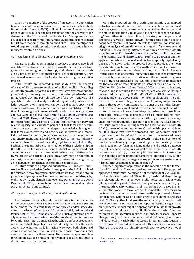
Given the genericity of the proposed framework, the applicationo other examples of accretionary growth processes, such as shell-sh or corals (Ubutaka, 2003), will be of interest. Another issue toe considered would be the reconstruction and the analysis of theynamics of the 3D shape of the otolith. Such 3D representationsould be deduced from multiple parallel 2D otolith sections as usedor 3D brain mapping from 2D scanned slices. Such investigationsould require specific technical developments to acquire images
n successive otolith planes.
.2. New local otolith signatures and otolith growth analysis
Regarding otolith growth analysis, we have proposed new localuantitative features of 2D otolith growth, i.e. ring curvature,rowth increment, growth anisotropy and relative opacity, whichre by-products of the estimation level-set representation. Theyre viewed as new means for locally characterizing the accretionrocess.
Initial results are reported in this study from the analysisf a set of 10 transverse sections of pollock otoliths. RegardingD otolith growth, reported results stress how asynchronous therowth along different growth axes can be and emphasize the needor advanced 2D (and possibly 3D) tools for growth analysis. Theuantitative statistical analysis exhibits significant positive corre-ation between otolith opacity and growth, and, relative opacity androwth anisotropy. This can be regarded as a quantitative evalua-ion of the strength of these relationships which are broadly knownnd evaluated at a global level (Panfili et al., 2002; Campana andhorrold, 2001; Hussy and Mosegaard, 2004). Focusing on the lat-er relationship, the absence of a significant correlation betweenelative opacity and curvature points out that local opacity vari-tions are not constrained by shape characteristics. It suggestshat local otolith growth and opacity can be viewed as a modu-ation of two factors: a global factor related to fish metabolismnd environment and a local factor related to the local physico-hemical characteristics of the endolymph (Allemand et al., 2007).esides, the quantitative characterization of these relationships inhe different otolith zones (i.e., ventral, dorsal, proximal and dorsalones) indicates that for some relationships (e.g., relative opac-ty vs. growth anisotropy) a global mean law may be relevant. Inontrast, for other relationships (e.g., curvature vs. local growth),one-dependent relationships seem more appropriate.
In future work the proposed quantitative 2D analysis frame-ork will be exploited to further investigate at the individual level
he relations between physico-chemical otolith features and otolithrowth and opacity, as well as the relations between otolith opacity,tolith growth, endolymph heterogeneity (Allemand et al., 2007;ayan et al., 1999), fish metabolism and environmental variablese.g., temperature and salinity).
.3. A generic tool for otolith analysis and applications
The proposed approach performs the extraction of the seriesf the successive otolith shapes. Otolith shape has been proveno be among the relevant features for species and/or stock dis-rimination issues (Campana and Casselman, 1993; de Pontual androuzet, 1987; Parisi-Barabad et al., 2005). Such application gener-lly relies on the characterization of the otolith outline, for instancey Fourier descriptors. Considering the whole and/or subsequencesf the individual shape histories considerably enriches the avail-
ble characterization, as it intrinsically conveys both shape androwth information. Curvature and growth anisotropy maps maylso be of interest for these issues. These novel shape-based fea-ures should lead to significant improvements of stock and speciesiscrimination from fish otoliths.(
m
rch 96 (2009) 148–159
From the proposed otolith growth representation, an adaptedolar-like coordinate system, where the angular information �efers to a growth axis (indexed w.r.t. a point along the outline) andhe radius information � to an age, has been proposed for analyz-ng 2D otolith sections. Exemplified in our study for the spatial andemporal analysis of otolith growth features, this otolith-specificoordinate system is of broad interest: for instance, for standard-zing the analysis of one-dimensional transects for one or severalndividuals or evaluating differences or similarities w.r.t. otolithampling zones. Fish length backcalculation from otolith measure-ents (Campana and Thorrold, 2001; Panfili et al., 2002) is another
pplication. Whereas backcalculation laws typically exploit onlyne specific growth axis, the proposed setting provides the meanor extending such laws to any growth axis as well as ensuringhe standardization of the reference growth axis (Fig. 8). Regard-ng the extraction of chemical signatures, the proposed frameworkan contribute to the standardization and the automatic program-ing of transect characteristics (e.g., spots locations), for instance
or the analysis of trace elements or isotopes by using WDS, LA-CPMS or SIMS (de Pontual and Geffen, 2003). In some applications,
icrodrilling is required for the subsequent analysis of isotopiconcentrations by using IRMS, MC-ICPMS or TIMS (Hoie et al.,004; Klaue et al., 2002; Barnett-Johnson et al., 2005). The defi-ition of the micro-drilling trajectories is of primary importance tonsure that growth-consistent otolith zones are sampled. Micro-rilling trajectories are defined by both a prior manual recordingf otolith reference lines and an interpolation between those lines.his quite tedious process presents a risk of mismatching inter-ediate trajectories and internal otolith rings, resulting in noisy
hemical measurements. It has been recognized that such analyt-cal issues might be a limiting factor for a joint analysis of otolith18 O (a proxy of water temperature) and otolith opacity (Hoie etl., 2004). In contrast, from the proposed framework, micro-drillingrajectories could be defined from portions of the estimated level-et representation of the otolith growth. In the same context oftolith microchemistry, the proposed representation also providesew means for performing a joint analysis and a fusion betweenultiple chemical signatures, as well as with image-based otolith
eatures (e.g., opacity), issues being far from trivial. An illustrationf the potential of the proposed framework is reported (Fig. 12) forhe fusion of the opacity image and oxygen isotope signatures of aake otolith (Desenfant et al unpublished).4
Another important application is the modelling of the forma-ion of fish otoliths. The contributions are two-fold. The proposedpproach first permits investigating, at the individual level, a quan-itative characterization of 2D otolith growth and determininghe relevant relationship between otolith features. Previous workHussy and Mosegaard, 2004) relied on global characteristics (e.g.,
ean otolith opacity vs. mean otolith growth). Such a global anal-sis is rather coarse to formulate and test modelling hypothesis. Inontrast, such issues can be addressed with the proposed scheme.or instance, hypotheses on otolith growth considered in (Alvarezt al., 2008)(e.g., that local growth can be radially parameterized)re shown not to be satisfied and reported results suggest thatn exponential model might be appropriate to relate local growthnisotropy and relative opacity. Similarly, the analysis of tempo-al shifts in the accretion regimes (e.g., checks, seasonal opacity
Hussy et al., 2004) to a joint 2D growth-opacity predictive model
4 An animated version can be visualized at public.enst-bretagne.eu/∼rfablet/ottolith.htm.

R. Fablet et al. / Fisheries Resea
Fig. 12. Illustration of the image-based fusion of oxygen isotope signatures andoıii
ois
A
itwmSrpA
R
A
A
B
C
a
C
C
C
C
C
d
d
d
D
D
F
F
F
F
G
H
H
H
H
H
K
P
P
P
P
S
tolith opacity for a hake otolith: acquired transect of the oxygen isotope ratios18 O superimposed to the otolith image acquired under reflected light (top), fusedmage comprising in the upper part opacity information and the lower part thenterpolated ı18 O values.
f the accretion of the otolith.5 Such a model would be of greatnterest to better understand the conditions of the formation of theuccessive opaque and translucent layers of the biomineral.
cknowledgements
We are grateful to Andre Ogor for the acquisition of the otolithmages. We also thank F. Cao for fruitful discussion on the minimiza-ion of variational issue based on level-set representations. Thisork has been carried out with the financial support from the Com-ission of the European Communities, specific program “Specific
upport to Policies”, SSP8-044132 “AFISA”. It does not necessarilyeflect its views and in no way anticipates the Commission’s futureolicy in this area. This work has also been supported by nationalNR grant “OTOCAL”.
eferences
llemand, D., Mayer-Gostan, N., de Pontual, H., Boeuf, G., Payan, P., 2007. Handbookof biomineralization. In: Bauerlein, E. (Ed.), Fish Otolith Calcification in Relationto Endolymph Chemistry. Wiley, pp. 291–308 (Chapter).
lvarez, A., Palmer, M., Tomas, J., Morales-Nin, B., 2008. A two-dimensionnal otolithgrowth inverse model. J. Fish Biol. 3 (72), 512–522.
arnett-Johnson, R., Grimes, F.R.C., MacFarlane, R., 2005. Validation of sr isotopes inotoliths by laser ablation multicollector inductively coupled plasma mass spec-trometry (la-mc-icpms): opening avenues in fisheries science applications. Can.J. Fish. Aquat. Sci. 62 (11), 2425–2430.
ampana, S., Casselman, J., 1993. Stock discrimination using otolith shape analysis.Can. J. Fish. Aquat. Sci. 50 (5), 1062–1083.
5 An example of a numerical simulation of an otolith growth model can be visu-lized at public.enst-bretagne.eu/∼rfablet/mottolith.htm.
T
U
rch 96 (2009) 148–159 159
ampana, S., Thorrold, S., 2001. Otoliths, increments, and elements: keys to a com-prehensive understanding of fish populations? Can. J. Fish. Aquat. Sci. 58 (1),30–38.
ao, F., Fablet, R., 2006. Automatic morphological detection of otolith nucleus. Pat-tern Recogn. Lett. 27 (6), 658–666.
hessel, A., Cao, F., Fablet, R., 2006. Interpolation of orientation: an axiomaticapproach. In: Proc. Eur. Conf. on Comp. Vis., ECCV06, LNCS 3954, Graz, pp.241–254.
hessel, A., Fablet, R., Kervrann, C., Cao, F., 2008. Otolith image by computer vision:extraction growth rings and recovering shape evolution of accretionary struc-tures. In: Proc. Int. Conf. on Bio-Insp. Sig. Proc. Sys., Biosignal 2008, Madeira, pp.490–497.
ohen, L.D., 2005. Minimal paths and fast marching methods for image analysis.In: Paragios, N., Chen, Y., Faugeras, O. (Eds.), Mathematical Models in ComputerVision: The Handbook. Springer, pp. 97–112.
e Pontual, H., Geffen, A., 2003. Otolith microchemistry. In: Panfili, J., de Pontual,H.T., Wright, P. (Eds.), Manual of Fish Sclerochronology. Editions Ifremer, pp.243–304.
e Pontual, H., Prouzet, P., 1987. Atlantic salmon, Salmo salar l., stock discriminationby scale-shape analysis. Aquat. Fish. Man. 18, 277–289.
e Pontual, H., Prouzet, P., 1988. Numerical analysis of scale morphology to discrim-inate between atlantic salmon stocks. Aquat. Liv. Res. 1 (1), 17–27.
esolneux, A., Moisan, L., Morel, J., 2001. Edge detection by helmoltz principle. J.Math. Imag. Vis. 14 (3), 271–284.
esolneux, A., Moisan, L., Morel, J.-M., 2003. A grouping principle and four applica-tions. IEEE Trans. Pattern Anal. Mach. Intell. 25 (4), 508–513.
ablet, R., 2006. Semi-local extraction of ring structures in images of biological hardtissues: application to the bayesian interpretation of fish otoliths for age andgrowth estimation. Can. J. Fish. Aquat. Sci. 43 (6), 1414–1428.
ablet, R., Chessel, A., Carbini, S., Benzinou, A., de Pontual, H., 2008a. Mottolith: anew tool for otolith imaging, online report. Ifremer/LASAA. http://perso.enst-bretagne.fr/ronanfablet.
ablet, R., Chessel, S.P.A., Benzinou, A., Cao, F., 2008b. 2D image-based reconstruc-tion of shape deformation of biological structures using a variational level-setapproach. Comp. Vis. Imag. Understand. 111 (3), 295–306.
ablet, R., Daverat, F., de Pontual, H., 2007. Unsupervised bayesian reconstructionof individual life histories chronologies from otolith signatures: case study ofSr:Ca transects of eel (anguilla anguilla) otoliths. Can. J. Fish. Aquat. Sci. 64, 152–165.
uillaud, A., Benzinou, A., Troadec, H., Rodin, V., Bihan, J.L., 2002. Autonomous agentsfor edge detection and continuity perception on otolith images. Imag. Vis. Comp.20 (13–14), 955–968.
oberman, J., Jensen, A., 1962. The growth rate of new england pollock. Trans. Am.Fish. Soc. 91, 227–228.
oie, H., Folkvord, A., Mosegaard, H., Li, L., Clausen, L., Norberg, B., Geffen, A., 2008.Restricted fish feeding reduces cod otolith opacity. J. Appl. Ichthyol. 24 (2),138–143.
oie, H., Otterlei, E., Folkvord, A., 2004. Temperature-dependent fractionation ofstable oxygen isotopes in otoliths of juvenile cod (gadus morhua l). ICES J. Mar.Sci. 61 (2), 243–251.
ussy, K., Mosegaard, H., 2004. Atlantic cod (gadus morhua) growth and otolithaccretion characteristics modelled in a bioenergetics context. Can. J. Fish. Aquat.Sci. 61 (6), 1021–1031.
ussy, K., Mosegaard, H., Jessen, F., 2004. Effect of age and temperature on aminoacid composition and the content of different protein types of juvenile atlanticcod (gadus morhua) otoliths. Can. J. Fish. Aquat. Sci. 61 (6).
laue, A., Blum, J., Folt, C., Nislow, K., Kennedy, B., 2002. Robust and efficient detectionof salient convex groups. Can. J. Fish. Aqat. Sci. 59 (6), 925–929.
almer, M., Alvarez, A., Tomas, J., Morales-Nin, B., 2005. A new method for robust fea-ture extraction of otolith growth marks using fingerprint recognition methods.Mar. Fresh. Res. 56, 791–794.
anfili, J., Pontual, H.D., Troadec, H., Wright, P., 2002. Manual of Fish Sclerochronol-ogy. Editions Ifremer.
arisi-Barabad, V., Lombarte, A., Garcia-Ladona, E., Cabestany, J., Piera, J., Chico,O., 2005. Otolith shape contour analysis using affine transformation invariantwavelet transforms and curvature scale space representation. Mar. Fresh. Res.56 (5), 795–804.
ayan, P., Edeyer, A., Pontual, H.D., Borelli, G., Boeuf, G., Mayer-Gostan, N., 1999.Chemical composiion of saccular endolymph and otolith in fish inner ear:lack of spatial uniformity. Am. J. Phys.: Reg. Int. Comp. Phys. 277 (1), 123–131.
ethian, J., 1999. Level Set Methods and Fast Marching Methods: Evolving Interfacesin Computational Geometry, Fluid Mechanics, Computer Vision and Materials
Science. Cambridge University Press.raodec, H., Benzinou, A., Rodin, V., Bihan, J.L., 2000. Use of deformable templatesfor otolith 2D growth ring detection by digital image processing. Fish. Res. 46(1–3), 155–163.
butaka, T., 2003. Pattern of growth rate around aperture and shell form in Bivalvia:a theoretical morphological study. Paleobiology 29 (4), 480–491.