Embed Size (px)
Citation preview
Systematic & Applied Acarology 21(5): 583–595 (2016)http://doi.org/10.11158/saa.21.5.3
583© Systematic & Applied Acarology Society
ISSN 1362-1971 (print)ISSN 2056-6069 (online)
Reconditioning of the Nalepa collection of eriophyoid mites (Acari-formes, Eriophyoidea)
PHILIPP E. CHETVERIKOV1,2,6, CHRISTOPH HÖRWEG3, MAXIM I. KOZLOV4 & JAMES W.AMRINE, JR.5
1 Department of Invertebrate Zoology, Saint-Petersburg State University, Universitetskaya nab., 7/9, 199034, St. Petersburg, Russia 2 Zoological Institute, Russian Academy of Sciences, Universitetskaya Embankment 1, 199034 St. Petersburg, Russia3 Natural History Museum Vienna, 3. Zoology (Invertebrates), Burgring 7, 1010 Vienna, Austria 4 Department of Chemistry, Lomonosov Moscow State University, Leninskie gory, 1/3, 119991, Moscow, Russia5 West Virginia University, Division of Plant and Soil Sciences, P.O. Box 6108, Morgantown, West Virginia, 26505-6108 USA6 Corresponding author. E-mail: [email protected]
Abstract
Alfred Nalepa (19.XII.1856–11.XII.1929), an Austrian acarologist, described about 460 eriophyoid species. Hereported new taxa in short communications usually published in “Anzeiger der Kaiserlichen Akademie derWissenschaften in Wien” and later prepared detailed descriptions for separate publication. For most Nalepanspecies the date of the first listing in Anzeiger is the valid date of the taxon name. His archive, library andcollection are kept in the Natural History Museum of Vienna, Austria (NHMW). The collection consists of 24boxes with 1073 vials containing plant material with extracted mites collected during 1887‒1929. All boxes arelabeled according to the first letters of the host-plant names and are sorted alphabetically; the vials are numberedand labeled. A jotter, presumably representing the hand-written catalog of the vial collection, and work diaries,containing indications of numbers of the vials, were found in the Nalepa archives. Nalepa used picric acid,hydrochloric acid, ethanol, formalin and creosote for preservation of mites. In all vials the preservative totallyevaporated so that only dry sediment remains at the bottom of the vials. A solution containing ethanol, ether andacetic acid was found to be appropriate for dissolving the sediment. A simple, fast protocol for recovering mitesfrom vials and making good slides was developed. It includes four steps: 1) opening the vial; 2) dissolving thesediment; 3) treating mites in lactic acid; 4) slide mounting. All digital data obtained from the Nalepa archives(database of the vials, copies of the jotter, reprints and drawings of mites) will be available for scientists at theweb site of NHMW http://www.nhm-wien.ac.at/en/nalepa.
Key words: new protocol, recovery, taxonomical revision, phytoparasitic arthropods
Introduction
Doctor August Nemesius Alfred Nalepa (19.XII.1856–11.XII.1929) was an Austrian acarologistfamous for his pioneer studies of tiny phytoparasitic mites of the superfamily Eriophyoidea.Bibliographic data on this scientist can be found in two obituaries (Masee 1930, Rechinger 1930)and in a very sincere paper by Shevtshenko (1967) dedicated to the 110th anniversary of the birth ofA. Nalepa. Systematization of the knowledge on eriophyoids obtained by European acarologists in19th and early 20th centuries and faunistic studies are essential contributions by A. Nalepa toacarology, which contributed greatly to the development of eriophyoidology in the middle of 20th
century. According to Keifer (1975) and Newkirk (1984) A. Nalepa published 113 papers anddescribed about 330 new species. Elevation of many of his trinomials and tetranomials has resulted
Article
584 SYSTEMATIC & APPLIED ACAROLOGY VOL. 21
in about 462 named mites. Fundamental observations on anatomy, ecology and taxonomy oferiophyoids by A. Nalepa (for details, see Shevtshenko 1967 p. 470 and Keifer 1975 pp. 329–332)influenced many scientists and inspired them to study eriophyoids in different countries andcontinents after his death. At present it is hard to find a paper on eriphyoid mites which does not citeat least one of Nalepa’s papers.
Along with his papers, the private archives and scientific collections are the most valuableheritage left by Dr. A. Nalepa to his followers. This important collection was shrouded in mysteryfor decades and was thought to be lost (Shevtshenko 1967) until it was discovered being kept in threesmall cupboards in the Natural History Museum in Vienna, Austria, under curation by Dr. J. Gruber(Amrine and Manson 1996, p. 386). Since then Dr. J. Gruber has retired and in 2007 a new curator,C. Hörweg MSc, replaced him. The collection includes reprints of Nalepa papers, his personal library(mainly books and papers on eriophyoids by Nalepa’s contemporaries) and numerous glass vials.Only dried sediment is present on the bottom of the vials, whereas the preservative medium hadtotally evaporated (Amrine et al. 2003). All of these conditions greatly challenge the identificationand search for eriophyoid mites in the Nalepa collection. Several researchers attempted to recovermites from the vials and mount them to get neotypes (de Lillo et al. 2010, p. 288; Xue et al. 2015, p.77; Dr. D. Knihinicki and Dr. C. Craemer personal communications, 2014). Only once it wassuccessful and resulted in slides of Phytoptochetus tristichus Nalepa 1917 which were good enoughto assign a neotype and draw the mite (Amrine et al. 2003, pp. 2, 50, fig. 50). However the protocoldeveloped for recovering and mounting P. tristichus was quite complicated and clearing took aperiod of two months (de Lillo et al. 2010, p. 288) implying that development of an easier and shorterprotocol is very important.
In 2012 a group of eriophyidologists attending the VIIth EURAAC symposium in Vienna (Drs.C. Flechtmann, R. Petanović, D. Navia, B. Vidović, C. Craemer and P. Chetverikov) visited theNalepa collection and concluded that it should be carefully reconditioned and an optimal procedurefor recovering mites and making neotypes should be developed. In 2014 a two year project“Reconditioning of the Nalepa collection at NHMW” was initiated. This project was primarily aimedto recondition the Nalepa collection in order to make it available for a broad range of scientists. Themain goals of this study were: 1) to evaluate, describe and improve the current condition of thecollection; 2) to develop the best technique for recovering mites from vials and making slides; 3) tomake a digital database of vials from the Nalepa collection and 4) to find the best way for preservingthe collection for the future. In this paper we present the most significant results obtained during theproject which will be important for eriophyoid studies.
Material and Methods
Reconditioning of the collection and databases. All the boxes containing vials were labeledaccording to the first letters of the host-plant names (A,B,C etc.) and sorted alphabetically. Insideevery box the vials containing material from one plant genus were put together. Those vialscontaining damaged plant organs (but not sediment) were additionally marked with a red dot. Thedeformed old cork stoppers not properly fitting to the vials were removed, stored in a zip-plastic bag;they were replaced by new ones (VWR® Cat. No. 217-1017 & 217-1016) and new carton labels withnumbers were glued (with UHU® hart, Art. Nr. 45510). All data from labels which was possible torecognize were entered into an Excel file and a database of all the vials was created. Using reprintsfrom the Nalepa archive and data from Rechinger (1930), Shevtshenko (1967) and Newkirk (1984)we compiled the full list of papers by A. Nalepa following the system of Nalepa’s papers proposedby Newkirk (1984). Digital copies (pdf files) of most of these papers were obtained from our
5852016 CHETVERIKOV et al.: RECONDITIONING OF THE NALEPA COLLECTION
personal collections and completed with several rare papers which were found in the Nalepa archiveand scanned at NHMW. Additionally all the images from Nalepa’s papers and some of his originaldrawings of mites were scanned with high resolution (300 dpi, half-tone) and saved separately as .pdffiles.
Testing methods for recovering mites from sediment and making slides. For recoveringmites from the vials we searched for mites under a stereomicroscope Leica MZ6 in the solutionsobtained by dissolving the sediment from the vials in various cold and hot media (distilled water,70% ethanol, 5% acetic acid, diethyl ether (C2H5)2O and mixtures of all these chemicals in differentcombinations). For making permanent slides we applied modified Berlese medium (Amrine andManson 1996) and tested pretreatment in 20% KOH or technical DL-lactic acid. For heating thesolutions with sediment and slide mounts we used a Heraeus EW-BAL Thermostat Oven; differenttemperature (from 70°C to 115°C) and time (from 30 minutes to 72 hours) were tested. The final andbest protocol for recovering mites is described below under the section “Protocol developed in thisstudy”. Photographs were obtained using a Nikon Coolpix R310, and microphotographs of therecovered mites and crystals inside vials were obtained using a stereomicroscope Nikon SMZ25equipped with a microscope camera Nikon DS-Ri2. Quality of the slides was checked using aconventional light microscope Leitz Diaplan.
Results
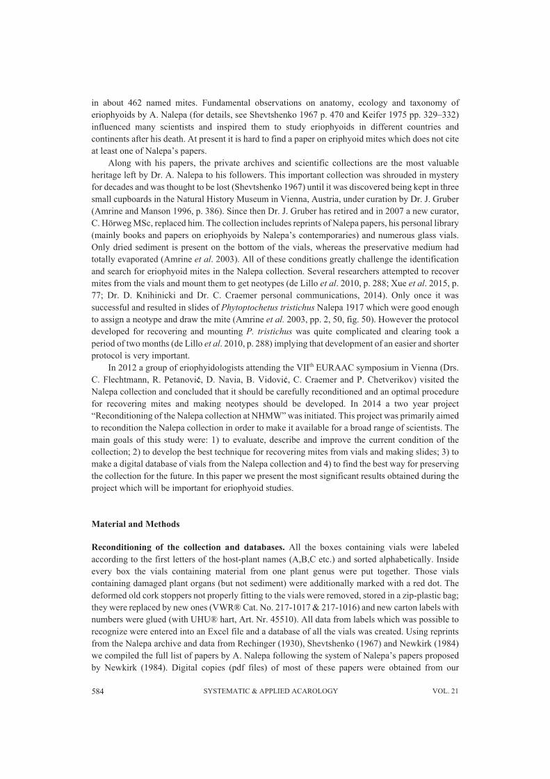
General characteristics of the collection (Fig. 1, Tab. 1). All of Nalepa’s materials are located inthree cupboards (#90, #91, & #92). All the papers from the Nalepa’s library written by his precursorsand contemporaries (Thomas, Trotter, Schlechtendal etc.) are in the upper part of cupboard #90. Theboxes with vials (mainly material from Austria), glass bottles and envelops (with material fromSamoa, Java, New Zealand, Australia and Barbados) are in the cupboard #91; a small plastic boxcontaining the type specimen of Phytoptochetus tristichus is also in this cupboard. In total, there are24 boxes (23 square, wooden boxes about 20x20x20 cm and one white carton box of larger size)containing 1073 vials collected from 1887 (the oldest record) till 1929 (the last record). All the boxesare labeled according to the first letters of the host-plant names (A,B,C etc.) and sortedalphabetically. Inside every box, the vials containing material from one plant genus are kept together(Table 1). In the cupboard #92 the original pencil drawings, reprints of Nalepa’s papers and hiscorrespondence and diaries are kept.
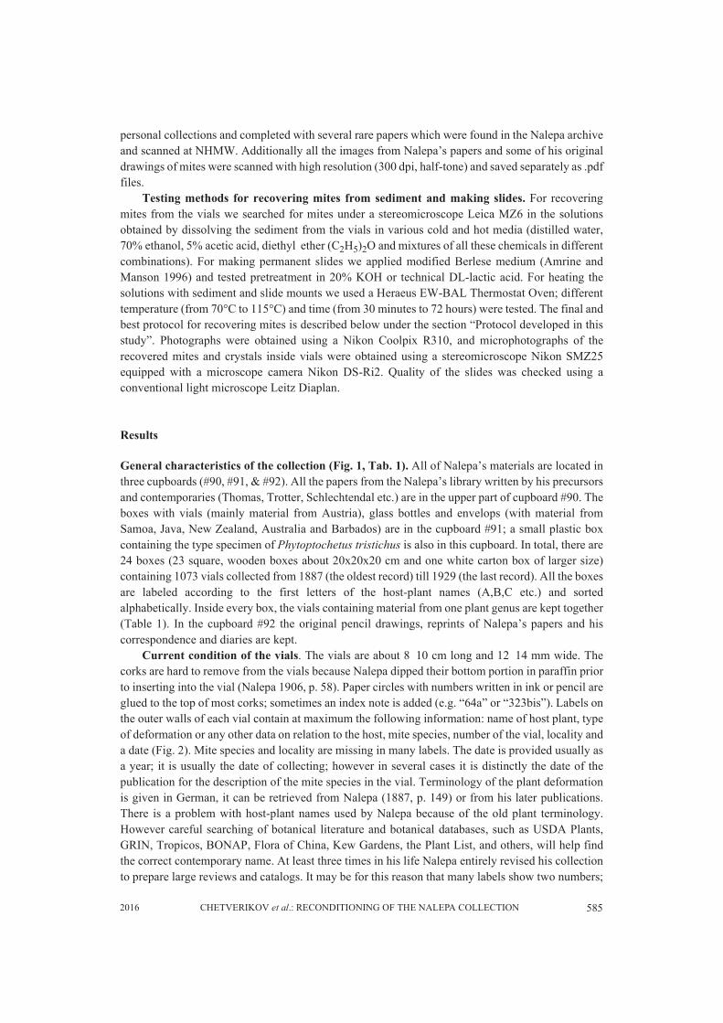
Current condition of the vials. The vials are about 8‒10 cm long and 12‒14 mm wide. Thecorks are hard to remove from the vials because Nalepa dipped their bottom portion in paraffin priorto inserting into the vial (Nalepa 1906, p. 58). Paper circles with numbers written in ink or pencil areglued to the top of most corks; sometimes an index note is added (e.g. “64a” or “323bis”). Labels onthe outer walls of each vial contain at maximum the following information: name of host plant, typeof deformation or any other data on relation to the host, mite species, number of the vial, locality anda date (Fig. 2). Mite species and locality are missing in many labels. The date is provided usually asa year; it is usually the date of collecting; however in several cases it is distinctly the date of thepublication for the description of the mite species in the vial. Terminology of the plant deformationis given in German, it can be retrieved from Nalepa (1887, p. 149) or from his later publications.There is a problem with host-plant names used by Nalepa because of the old plant terminology.However careful searching of botanical literature and botanical databases, such as USDA Plants,GRIN, Tropicos, BONAP, Flora of China, Kew Gardens, the Plant List, and others, will help findthe correct contemporary name. At least three times in his life Nalepa entirely revised his collectionto prepare large reviews and catalogs. It may be for this reason that many labels show two numbers;
586 SYSTEMATIC & APPLIED ACAROLOGY VOL. 21
one of them preceeded by the letter “K”, suggesting possible reference to a lost catalog (“Katalog”in German) or his 1929 published Katalog. Several vials contain an additional small label insideindicating the reference for the description of the mite species and/or detailed collecting data.
FIGURE1. Photographs of the cupboards (on the right) and boxes with vials inside cupboard #91 (on the left).
FIGURE 2. Microphotographs showing a vial, cork, label and the reconstructed text from the label (from leftto right).
The content of the vials. Nalepa (1906, p. 56) reported that he added three different media tothe vials: #1 a mixture of picric acid, distilled water and concentrated hydrochloric acid, 1:100:2(“Pikrinsalzsäure”); #2 a mixture of 94% ethanol and concentrated hydrochloric acid, 100:2(“Säurealkohol”) and #3 80% ethanol. The media #1 and #2 were used both for extracting mites (byshaking plant material with medium in a tube) and preservation; prior to preservation in the vial,mites in media #1 and #2 were diluted in 1:4‒5 with water and heated to 50‒60°C. Medium
5872016 CHETVERIKOV et al.: RECONDITIONING OF THE NALEPA COLLECTION
TABLE 1. Characteristics of the boxes with vials from Nalepa’s collection. Note: plant genera are given exactlythe same as indicated in the labels, using the spelling style of the time (e.g. Evonymus, Gossipium).
* the box Java-II has four compartments: II/1, II/2, II/3 and II/4.
Box Number of vials in a box
Genera of plants mentioned on the vial labels (number of the vials from exact plant genus is mentioned in brackets)
A1 59 Abies (3), Acacia (1), Acer (55)A2 45 Acer (45)A3 37 Aesculus (4), Ajuga (2), Alnus (31)A4 42 Alyssum (2), Amelanchier (2), Anchusa (2), Andromeda (1), Aposeris (3), Arabis (3),
Arctostaphylos (2), Artemisia (16), Asperula (5), Atragene (1), Atriplex (2), Avena (1),Azalea (2)
B 45 Bartsia (1), Bellidiastrum (1), Berberis (2), Bertoroa (1), Betonica (1), Betula (21),Brachypodium (1), Bromus (2), Bucida (2), Buxus (13)
C1 54 Camellina (2), Campanula (8), Carlina (1), Carpinus (11), Carum (2), Cedrus (2),Centaurea (7), Cerastium (1), Chondrilla (4), Cinnamomum (1), Cirsium (1), Cistus (1),Clematis (5), Convolvus (3), Cornus (2), Coronilla (2), Cydonia (1)
C2 48 Colutea (2), Corylus (22), Cotoneaster (6), Crataegus (14), Crepis (1), Cytisus (3)DE 26 Dactylis (1), Doryconium (1), Echinospermum (2), Echium (2), Empetrum (1), Erigeron
(1), Erodium (3), Ervum (1), Erysimum (1), Eugenia (2), Eupatorium (2), Euphorbia (1),Euphrasia (2), Evonymus (6)
F 39 Fraxinus (13), Fragaria (4), Fagus (22)G 27 Galium (27)
GHIJ 47 Genista (1), Gentiana (3), Geranium (5), Geum (5), Gossipium (2), Helianthemum (3),Hibiscus (4), Hieracium (4), Hippophae (1), Hutschinsia (1), Hypocharis (1), Ipomoea (1), Juglans (7), Juniperus (6), Jurinea (2), unknown (1)
L 31 Lactuca (1), Larix (3), Laurus (2), Lepidium (2), Linosyris (1), Lonicera (7), Lotus (4),Lycium (2), Lycopsis (1), Lysimachia (8)
MNO 26 Malva (2), Mangifera (2), Medicago (2), Mentha (2), Moehringia (2), Molinia (1), Ononis (2), Origanum (8), Orlaya (1), Ornithopus (2), Oxalis (1), unknown (1)
P1 54 Paederota (1), Passerina (1), Pedicularis (1), Peucedanum (1), Phlomis (3), Pimpinella (3), Pinus (6), Pistacia (3), Plantago (1), Potentilla (3), Pyrus (31)
P2 56 Polygala (2), Populus (19), Poterium (3), Prunus (30), Pteris (2)QR 47 Quercus (18), Robinia (3), Ranunculus (4), Rhamnus (3), Rhodiola (3), Rhododendron
(4), Rosa (2), Rubia (2), Rubus (2), Ribes (6)S1 61 Salicornia (1), Salix (60)S2 44 Sambucus (8), Sarothamnus (2), Saxifraga (3), Scabiosa (2), Serratula (1), Seseli (2),
Sisymbrium (2), Solanum (3), Sonchus (1), Sorbus (20)S3 33 Salvia (7), Sedum (8), Spiraea (1), Spiraeanthemum (1), Staphylea (1), Stellaria (3),
Suaeda (2), Symphyandra (1), Syringa (9)T 59 Tilia (59)
TU 48 Tamarix (1), Tanacetum (3), Taraxacum (3), Taxus (1), Teucrium (1), Thalictrum (1),Thesium (2), Thymus (4), Torilis (2), Trifolium (1), Trinia (1), Triticum (1), Ulmus (27)
V 37 Veronica (6), Viburnum (11), Vicia (2), Viola (3), Vitex (1), Vitis (14)Java-I 48 Acacia (2), Acalypha (1), Acronychia (1), Alangium (1), Allophyllus (1), Aporosa (1),
Bauhinia (2), Beilschmiedia (1), Buettneria (1), Callicarpa (1), Canarium (1),Cinnamomum (1), Clerodendron (2), Conocephalus (1), Cordia (1), Crotalaria (2),Cryptocarya (2), Dianthera (1), Ehretia (1), Elaeocarpus (2), Evodia (2), Ficus (7),Flacourtia (1), Fluggea (1), Glochidium (5), Gonania (1), Haasia (=Dehaasia, 1),Hibiscus (4)
Java-II/1* 12 Indigofera (3), Ipomoea (2), Laportea (2), Lepistemon (1), Litsea (3), unknown (1)Java-II/2 13 Macaranga (1), Macropanax (1), Mallotus (2), Melastoma (1), Melochia (1), Merremia
(1), Mikania (1), Morinda (1), Nephrolepis (3), Oldenlandia (1)Java-II/3 17 Paederia (1), Pavetta (1), Peristrophe (1), Pluchea (1), Pometia (1), Premna (2),
Pterospermum (1), Quisqualis (1), Rubus (1), Ruellia (1), Sandoricum (2), Sesbania (1), Solanum (1), Streblus (1), Strobilanthes (1)
Java-II/4 18 Tetracera (1), Toddalia (1), Triumphaeta (3), Unona (1), Vangueria (1), Villebrunnea (3), Viburnum (1), Vitex (2), Weinmannia (4), Wendlandia (1)
588 SYSTEMATIC & APPLIED ACAROLOGY VOL. 21
#3 was used for preservation of galls and other damaged tissues caused by mites. Nalepa also usedto add a small amount of creosote in the vials to avoid microbial contamination. Besides thesechemicals, “formalin” is mentioned in several labels suggesting that it was also used as apreservative. However in most labels indication of the medium is absent so that it is not possible toknow which medium was actually applied.
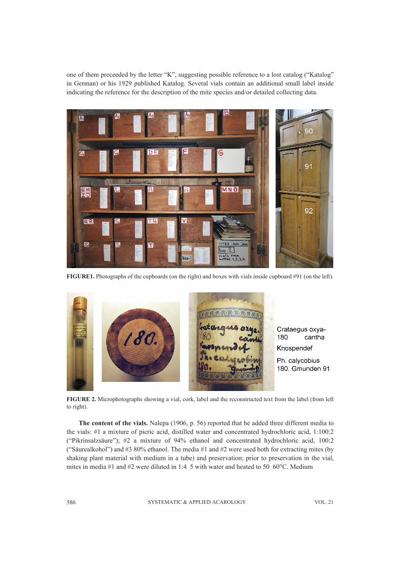
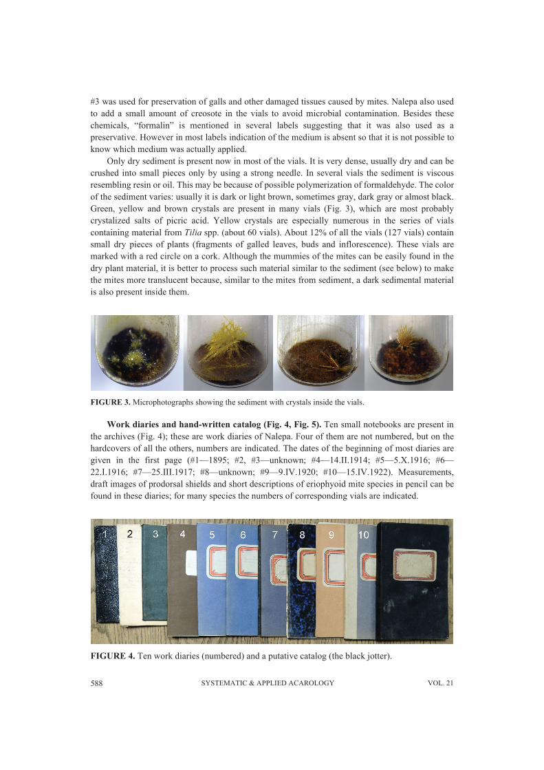
Only dry sediment is present now in most of the vials. It is very dense, usually dry and can becrushed into small pieces only by using a strong needle. In several vials the sediment is viscousresembling resin or oil. This may be because of possible polymerization of formaldehyde. The colorof the sediment varies: usually it is dark or light brown, sometimes gray, dark gray or almost black.Green, yellow and brown crystals are present in many vials (Fig. 3), which are most probablycrystalized salts of picric acid. Yellow crystals are especially numerous in the series of vialscontaining material from Tilia spp. (about 60 vials). About 12% of all the vials (127 vials) containsmall dry pieces of plants (fragments of galled leaves, buds and inflorescence). These vials aremarked with a red circle on a cork. Although the mummies of the mites can be easily found in thedry plant material, it is better to process such material similar to the sediment (see below) to makethe mites more translucent because, similar to the mites from sediment, a dark sedimental materialis also present inside them.
FIGURE 3. Microphotographs showing the sediment with crystals inside the vials.
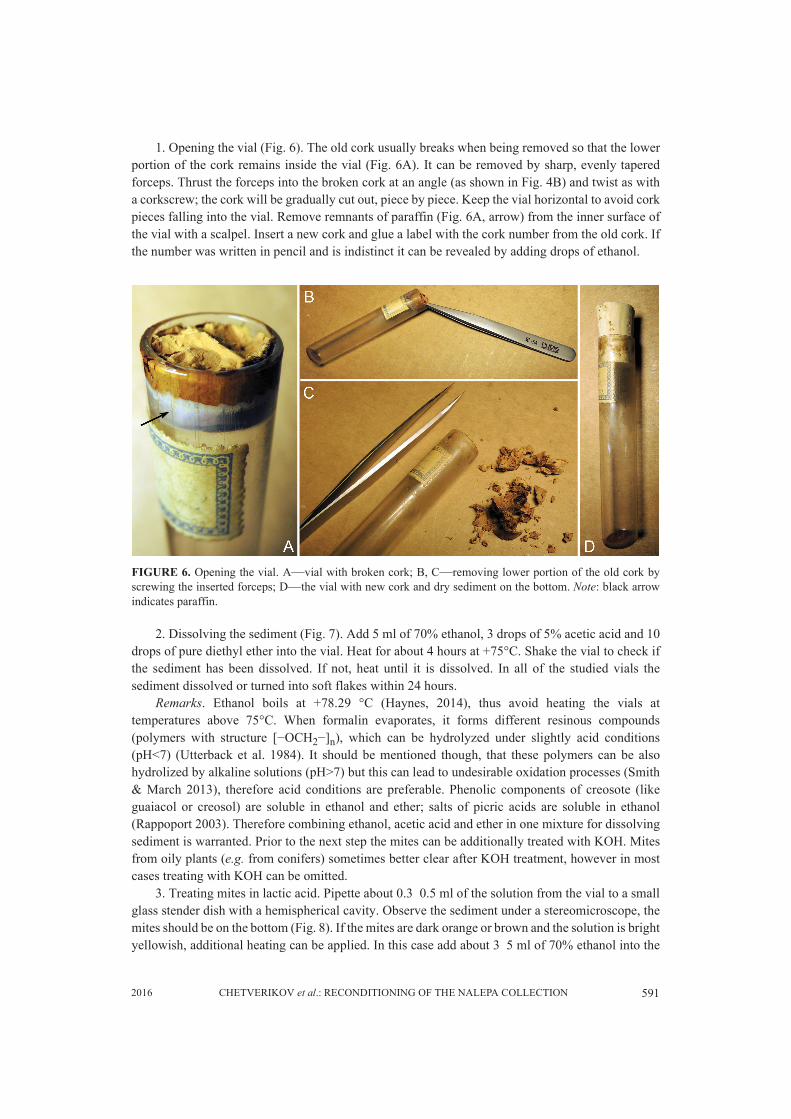
Work diaries and hand-written catalog (Fig. 4, Fig. 5). Ten small notebooks are present inthe archives (Fig. 4); these are work diaries of Nalepa. Four of them are not numbered, but on thehardcovers of all the others, numbers are indicated. The dates of the beginning of most diaries aregiven in the first page (#1—1895; #2, #3—unknown; #4—14.II.1914; #5—5.X.1916; #6—22.I.1916; #7—25.III.1917; #8—unknown; #9—9.IV.1920; #10—15.IV.1922). Measurements,draft images of prodorsal shields and short descriptions of eriophyoid mite species in pencil can befound in these diaries; for many species the numbers of corresponding vials are indicated.
FIGURE 4. Ten work diaries (numbered) and a putative catalog (the black jotter).

5892016 CHETVERIKOV et al.: RECONDITIONING OF THE NALEPA COLLECTION
FIGURE 5. A photograph showing six examples of the putative hand-written catalog with page (black circle)and vial numbers (arrow) indicated. The reference to a herbarium sample is indicated by a black rectangle.
Along with the work diaries another important document was found in the Nalepa archives (Fig.5). It is a small jotter of 220 pages (each page is enumerated by hand) with black hardcover. Althoughthe date “1911/12” is indicated in the hardcover, this document contains information about materialfrom Java suggesting much later date of use. We assume that this jotter is possibly the very hand-written catalog mentioned by previous authors (Amrine and Manson 1996; Amrine et al. 2003; deLillo et al. 2010) and which was thought to be lost. The texts in the jotter are written in pencil in anold-fashioned mode using “cursive writing” (stenography) with a lot of abbreviations, making it hardto understand. However, comparison with the two catalogs published by Nalepa (1911, 1929)

590 SYSTEMATIC & APPLIED ACAROLOGY VOL. 21
revealed that the jotter might have been a draft for these publications as most descriptions from thejotter precisely fit to the texts from the two published catalogs. Many species descriptions (but farfrom all of them) include indication of the numbers of vials with corresponding material (Fig. 5,arrows). On page 157 a reference to a herbarium specimen of Strobilanthes sp. is given in thedescription of Eriophyes strobilanthis (Fig. 5, rectangle); the herbarium specimen and the vials withthis mite species have not been found in the collection.
The jotter consists of four parts: pages 1‒58; 60‒137; 138‒196 and the last unnumbered 24pages. In the first 58 pages the descriptions of new mite species from Java collected in 1914 aregiven, the host index is provided on pages 55‒58. Pages 60‒134 contain descriptions and records ofEuropean species of eriophyoids, host index for these species is given on pages 135‒137. On pages138‒196 descriptions of new species from Java collected in 1921 as well as data on European speciesare given in a jumble; another host index for mites from Java is given on pages 179‒180. The lastpart of the jotter contains some unfinished tables and notes on the mites from Acer spp. with pencildrawings showing different forms of the modified trichomes of the erinea caused by eriophyoids onAcer pseudoplatanus and A. campestre; several pages are blank. Careful, future study of this jotterand the work diaries will be necessary to better understand the content of each vial from thecollection.
Database of the vials and other digital data obtained. The following digital data wereprepared: 1) pdf copies of 108 Nalepa papers and 2) pdf copies of the jotter and ten work diaries; 3)a list of the eriophyoid mite species and genera described by A. Nalepa; 4) high resolution copies ofthe figures from all Nalepa papers and his original pencil drawings of the mites; 5) pdf copies of thepapers about A. Nalepa and his work; 6) photographs of the collection and 7) database of the vials.The database organization is shown in Table 2. All the digital data are available for scientists throughthe Internet at the NHMW site http://www.nhm-wien.ac.at/en/nalepa.
TABLE 2 A portion of the Excel file showing the organization of the database
Technique for recovering mites from vials and making slides. Analysis of the literaturerevealed that previous authors scrabbled dry sediment on the bottom of a vial, removed it and lookedfor the mites under a stereomicroscope. According to our experiments this is the reason why most ofthe previous attempts to recover mites were unsuccessful because 1) the mites are usually tightlyembedded into the sediment and thus are hard to be visualized and 2) scrabbling dry sediment oftenresults in crumbling the mites (not only breaking legs and setae but also totally destroying theexoskeleton). The protocol developed in this study implies that the sediment should be liquefied and/or dissolved and then the mites are collected. Following this protocol about 80% of the resulting slidemounts are of good quality and appropriate for studying morphology of mites, and is quite similar tomounting live mites, although more time consuming. The protocol includes four steps: 1) opening avial; 2) dissolving the sediment; 3) treating mites in lactic acid; 4) slide mounting. Description andremarks on these stages are as follows:
Box # Vial # Plant genus Plant species Damage Mite(s)Species
Coll. date Locality Content
C2 180 Crataegus oxyacantha Knospendef. P. calycobius 1891? Gmunden dark brown sediment
DE 190 Evonymus europeus Blattrandroll. Cecidophyes convolvens
1889? ? dark sediment
GHIJ 130a Galium aparine ? ? VI.1926 ? brown sediment
GHIJ 639 Geranium palustre Erineum def. Blt ? 1908 ? leaf with erineum
5912016 CHETVERIKOV et al.: RECONDITIONING OF THE NALEPA COLLECTION
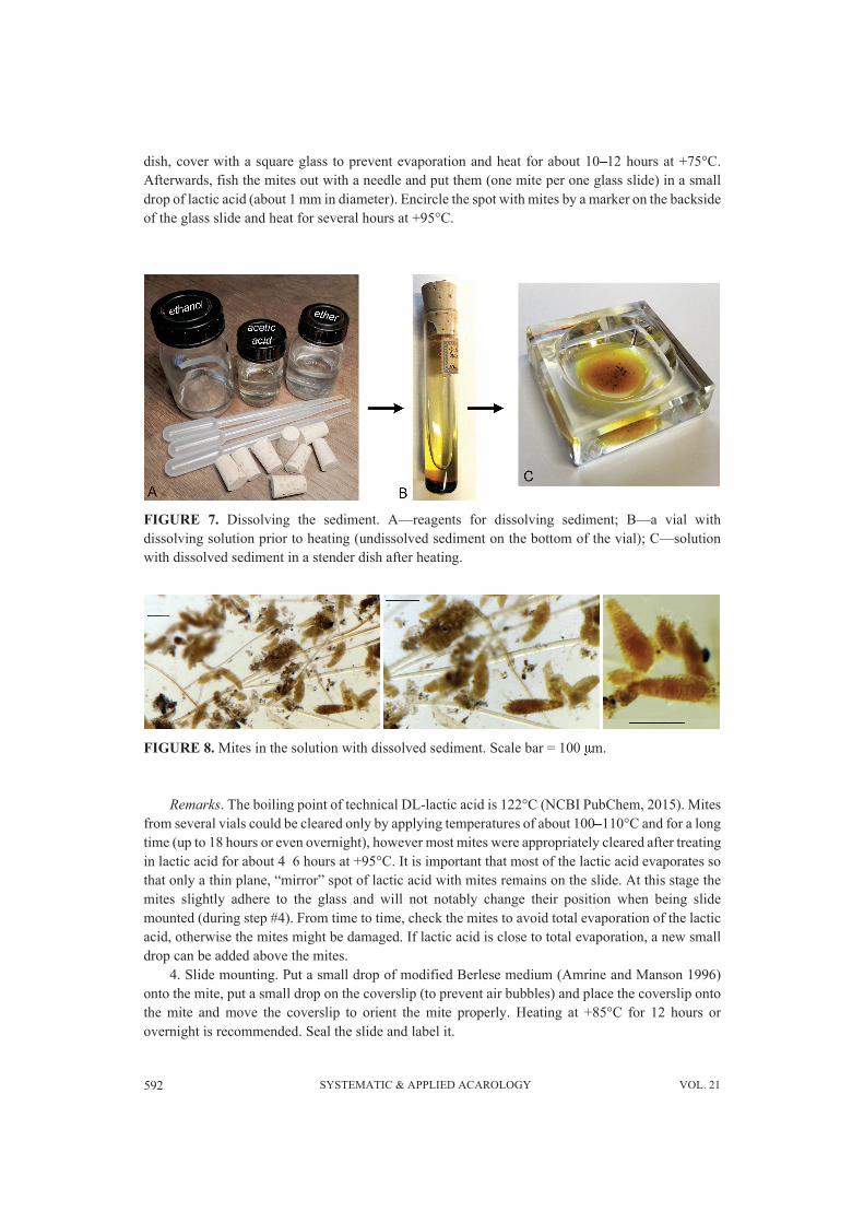
1. Opening the vial (Fig. 6). The old cork usually breaks when being removed so that the lowerportion of the cork remains inside the vial (Fig. 6A). It can be removed by sharp, evenly taperedforceps. Thrust the forceps into the broken cork at an angle (as shown in Fig. 4B) and twist as witha corkscrew; the cork will be gradually cut out, piece by piece. Keep the vial horizontal to avoid corkpieces falling into the vial. Remove remnants of paraffin (Fig. 6A, arrow) from the inner surface ofthe vial with a scalpel. Insert a new cork and glue a label with the cork number from the old cork. Ifthe number was written in pencil and is indistinct it can be revealed by adding drops of ethanol.
FIGURE 6. Opening the vial. A—vial with broken cork; B, C—removing lower portion of the old cork byscrewing the inserted forceps; D—the vial with new cork and dry sediment on the bottom. Note: black arrowindicates paraffin.
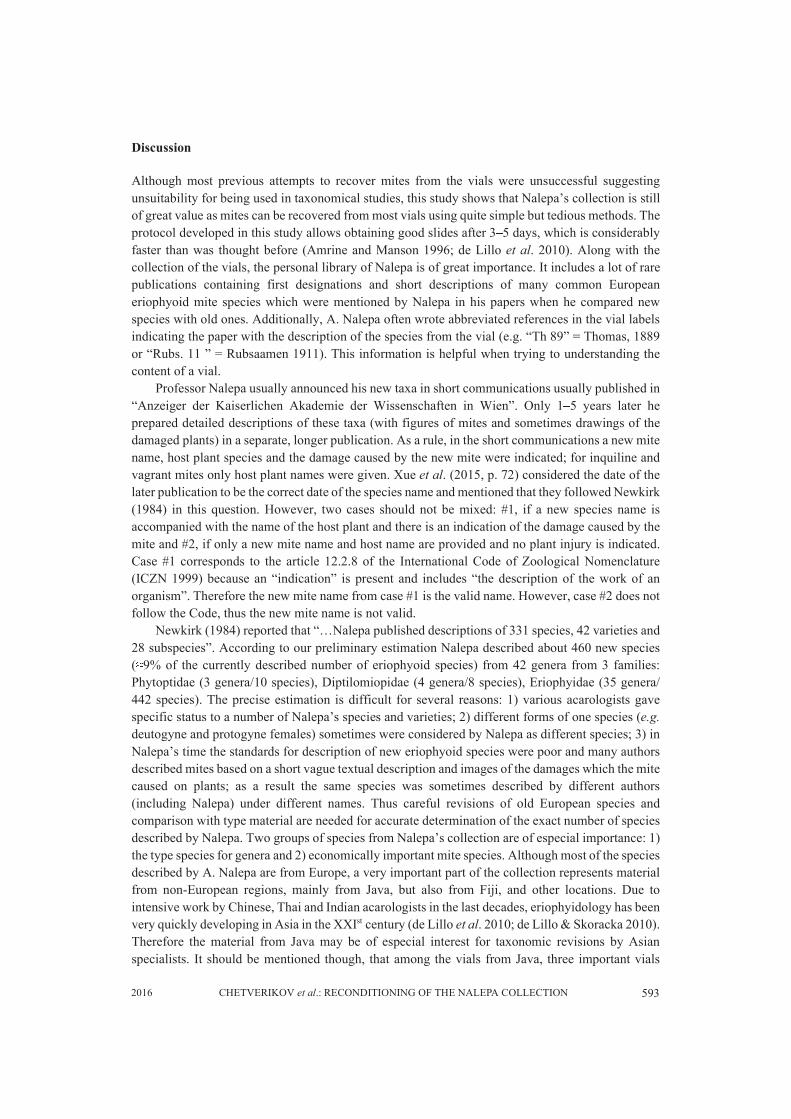
2. Dissolving the sediment (Fig. 7). Add 5 ml of 70% ethanol, 3 drops of 5% acetic acid and 10drops of pure diethyl ether into the vial. Heat for about 4 hours at +75°C. Shake the vial to check ifthe sediment has been dissolved. If not, heat until it is dissolved. In all of the studied vials thesediment dissolved or turned into soft flakes within 24 hours.
Remarks. Ethanol boils at +78.29 °C (Haynes, 2014), thus avoid heating the vials attemperatures above 75°C. When formalin evaporates, it forms different resinous compounds(polymers with structure [−OCH2−]n), which can be hydrolyzed under slightly acid conditions(pH<7) (Utterback et al. 1984). It should be mentioned though, that these polymers can be alsohydrolized by alkaline solutions (pH>7) but this can lead to undesirable oxidation processes (Smith& March 2013), therefore acid conditions are preferable. Phenolic components of creosote (likeguaiacol or creosol) are soluble in ethanol and ether; salts of picric acids are soluble in ethanol(Rappoport 2003). Therefore combining ethanol, acetic acid and ether in one mixture for dissolvingsediment is warranted. Prior to the next step the mites can be additionally treated with KOH. Mitesfrom oily plants (e.g. from conifers) sometimes better clear after KOH treatment, however in mostcases treating with KOH can be omitted.
3. Treating mites in lactic acid. Pipette about 0.3‒0.5 ml of the solution from the vial to a smallglass stender dish with a hemispherical cavity. Observe the sediment under a stereomicroscope, themites should be on the bottom (Fig. 8). If the mites are dark orange or brown and the solution is brightyellowish, additional heating can be applied. In this case add about 3‒5 ml of 70% ethanol into the
592 SYSTEMATIC & APPLIED ACAROLOGY VOL. 21
dish, cover with a square glass to prevent evaporation and heat for about 10‒12 hours at +75°C.Afterwards, fish the mites out with a needle and put them (one mite per one glass slide) in a smalldrop of lactic acid (about 1 mm in diameter). Encircle the spot with mites by a marker on the backsideof the glass slide and heat for several hours at +95°C.
FIGURE 7. Dissolving the sediment. A—reagents for dissolving sediment; B—a vial withdissolving solution prior to heating (undissolved sediment on the bottom of the vial); C—solutionwith dissolved sediment in a stender dish after heating.
FIGURE 8. Mites in the solution with dissolved sediment. Scale bar = 100 μm.
Remarks. The boiling point of technical DL-lactic acid is 122°C (NCBI PubChem, 2015). Mitesfrom several vials could be cleared only by applying temperatures of about 100‒110°C and for a longtime (up to 18 hours or even overnight), however most mites were appropriately cleared after treatingin lactic acid for about 4‒6 hours at +95°C. It is important that most of the lactic acid evaporates sothat only a thin plane, “mirror” spot of lactic acid with mites remains on the slide. At this stage themites slightly adhere to the glass and will not notably change their position when being slidemounted (during step #4). From time to time, check the mites to avoid total evaporation of the lacticacid, otherwise the mites might be damaged. If lactic acid is close to total evaporation, a new smalldrop can be added above the mites.
4. Slide mounting. Put a small drop of modified Berlese medium (Amrine and Manson 1996)onto the mite, put a small drop on the coverslip (to prevent air bubbles) and place the coverslip ontothe mite and move the coverslip to orient the mite properly. Heating at +85°C for 12 hours orovernight is recommended. Seal the slide and label it.
5932016 CHETVERIKOV et al.: RECONDITIONING OF THE NALEPA COLLECTION
Discussion
Although most previous attempts to recover mites from the vials were unsuccessful suggestingunsuitability for being used in taxonomical studies, this study shows that Nalepa’s collection is stillof great value as mites can be recovered from most vials using quite simple but tedious methods. Theprotocol developed in this study allows obtaining good slides after 3‒5 days, which is considerablyfaster than was thought before (Amrine and Manson 1996; de Lillo et al. 2010). Along with thecollection of the vials, the personal library of Nalepa is of great importance. It includes a lot of rarepublications containing first designations and short descriptions of many common Europeaneriophyoid mite species which were mentioned by Nalepa in his papers when he compared newspecies with old ones. Additionally, A. Nalepa often wrote abbreviated references in the vial labelsindicating the paper with the description of the species from the vial (e.g. “Th 89” = Thomas, 1889or “Rubs. 11 ” = Rubsaamen 1911). This information is helpful when trying to understanding thecontent of a vial.
Professor Nalepa usually announced his new taxa in short communications usually published in“Anzeiger der Kaiserlichen Akademie der Wissenschaften in Wien”. Only 1‒5 years later heprepared detailed descriptions of these taxa (with figures of mites and sometimes drawings of thedamaged plants) in a separate, longer publication. As a rule, in the short communications a new mitename, host plant species and the damage caused by the new mite were indicated; for inquiline andvagrant mites only host plant names were given. Xue et al. (2015, p. 72) considered the date of thelater publication to be the correct date of the species name and mentioned that they followed Newkirk(1984) in this question. However, two cases should not be mixed: #1, if a new species name isaccompanied with the name of the host plant and there is an indication of the damage caused by themite and #2, if only a new mite name and host name are provided and no plant injury is indicated.Case #1 corresponds to the article 12.2.8 of the International Code of Zoological Nomenclature(ICZN 1999) because an “indication” is present and includes “the description of the work of anorganism”. Therefore the new mite name from case #1 is the valid name. However, case #2 does notfollow the Code, thus the new mite name is not valid.
Newkirk (1984) reported that “…Nalepa published descriptions of 331 species, 42 varieties and28 subspecies”. According to our preliminary estimation Nalepa described about 460 new species(≈9% of the currently described number of eriophyoid species) from 42 genera from 3 families:Phytoptidae (3 genera/10 species), Diptilomiopidae (4 genera/8 species), Eriophyidae (35 genera/442 species). The precise estimation is difficult for several reasons: 1) various acarologists gavespecific status to a number of Nalepa’s species and varieties; 2) different forms of one species (e.g.deutogyne and protogyne females) sometimes were considered by Nalepa as different species; 3) inNalepa’s time the standards for description of new eriophyoid species were poor and many authorsdescribed mites based on a short vague textual description and images of the damages which the mitecaused on plants; as a result the same species was sometimes described by different authors(including Nalepa) under different names. Thus careful revisions of old European species andcomparison with type material are needed for accurate determination of the exact number of speciesdescribed by Nalepa. Two groups of species from Nalepa’s collection are of especial importance: 1)the type species for genera and 2) economically important mite species. Although most of the speciesdescribed by A. Nalepa are from Europe, a very important part of the collection represents materialfrom non-European regions, mainly from Java, but also from Fiji, and other locations. Due tointensive work by Chinese, Thai and Indian acarologists in the last decades, eriophyidology has beenvery quickly developing in Asia in the XXIst century (de Lillo et al. 2010; de Lillo & Skoracka 2010).Therefore the material from Java may be of especial interest for taxonomic revisions by Asianspecialists. It should be mentioned though, that among the vials from Java, three important vials
594 SYSTEMATIC & APPLIED ACAROLOGY VOL. 21
containing type material of Cecidodectes euzonus Nalepa 1917 (type species of genus CecidotectesNalepa 1917), Diptilomiopus javanicus Nalepa 1917 (type species for genus Diptilomiopus Nalepa1916) and Eriophyes strobilanthis Nalepa 1921, were not found; this material is probably lost.Collection of these species in type localities and designations of the neotypes will be necessary.
The scientists who are interested in recovering mites from Nalepa’s vials or depositing typematerial at NHMW are welcome to contact the curator of the collection via e-mail([email protected]). It is preferable if the recovering process is performed atNHMW; in this case the scientists might consider a 4‒5 day working visit to NHMW. All thematerials after recovery should be saved and all the slides should be appropriately labeled,catalogued and kept in the collection. The most promising approach to work with the collection isrevising groups of vials from the same host plant. In this case a limited number of possible specieswill be recovered. They can be easily identified based on the original descriptions especially thosespecies which had been described before 1910. The reason for this is, that after 1910, Nalepa did notprovide figures of the mites in his descriptions. However, in general, his later descriptions seem tobe more detailed which slightly compensates for the absence of figures. Brief analysis of the newdatabase for the vials suggests that usually, the higher the number indicated on the vial cork the laterit was collected. This fact simplifies searching the relevant papers containing descriptions of thepossible species.
In the beginning of this study it was hoped that the current condition of the collection might beimproved by adding an appropriate solution to all the vials. After developing the protocol forrecovering mites, we concluded that it is better to keep the vials as they are. Besides Nalepa’scollection, there are several more important old collections of eriophyoids (e.g. slides and envelopeswith plant material of H. H. Keifer in USA, “Cecidotheca Italica” in Italy, “Cecidotheca Rossica” inRussia and others). Accurate revisions and making digital profiles of these collections is one of theimportant goals for the future. Every year dozens of new species of eriophyoids are describedworldwide (de Lillo & Skoracka 2010), not all of the descriptions are of appropriate quality. To avoidfuture chaos in eriophyoid taxonomy it is important to decrease the speed of creating inadequatelydescribed new taxa, to intensify faunistic surveys for revising long forgotten information by previousauthors (e.g. a study by Hellrigl (2003) for old Trotter species), in order to update the nomenclatureand to involve older material in taxonomical studies.
Acknowledgements
We are grateful to Ms. Slavica Marinković (University of Belgrade, Serbia) and Ms. Mercia E.Duarte (Alagoas Federal University, Brazil) for their assistance in making slides and preparing thedigital copy of the hand-written catalog. We are also thankful to Dr. Eugenia A. Desnitskaya (Saint-Petersburg State University, Russia) for her help in translating Nalepa’s papers. This study wassupported by Pro Acarologia Basiliensis (PAB).
References
Amrine, J.W.Jr. & Manson, D.C.M. (1996) Preparation, mounting and descriptive study of Eriophyoid mites.In: Lindquist, E.E., Sabelis, M.W., Bruin, J. (eds.) Eriophyoid mites: their biology, natural enemies andcontrol. World Crop Pests 6. Amsterdam, Elsevier. pp. 383–396.
Amrine, J.W.Jr., Stasny, T.A., Flechtmann, C.H.W. (2003) Revised keys to the world genera of the Eriophyoi-dea (Acari: Prostigmata). Michigan, Indira Publishing House. 244 pp.
de Lillo, E. & Skoracka, A. (2010) What’s “cool” on eriophyoid mites? In: Ueckermann, E.A. (ed.) Eriophyoid
5952016 CHETVERIKOV et al.: RECONDITIONING OF THE NALEPA COLLECTION
Mites: Progress and Prognoses. Amsterdam, Springer. pp. 3–30. http://dx.doi.org/10.1007/978-90-481-9562-6_2.
de Lillo, E., Craemer, C., Amrine, J.W.Jr. & Nuzzaci, G. (2010) Recommended procedures and techniques formorphological studies of Eriophyoidea (Acari: Prostigmata). In: Ueckermann, E.A. (ed.) EriophyoidMites: Progress and Prognoses. Amsterdam, Springer. pp. 283–307. http://dx.doi.org/10.1007/978-90-481-9562-6_15.
Haynes, W.M. (Ed) (2014) CRC handbook of chemistry and physics. Boca Raton, Florida, CRC press. 2704pp.
Hellrigl, K. (2003) Faunistik der Gallmilben Südtirols (Acari: Eriophyoidea). Gredleriana, 3, 77–142.ICZN. International Commission on Zoological Nomenclature (1999) International Code of Zoological
Nomenclature, 4th edition. London, International Trust for Zoological Nomenclature, xxix + 306 pp.Keifer, H.H. (1975) Eriophyoidea Nalepa. Injurious eriophyoid mites. In: Jeppson, L.R., Keifer, H.H., Baker,
E.W. (eds.) Mites injurious to economic plants. Berkeley, University of California Press. pp. 327–533.Masee, A.M. (1930) Obituary of Dr. A. N. A. Nalepa. Nature, 3142(125), 96.Nalepa, A. (1887) Die Anatomie der Phytopten. Sitzungsbericht der Kaiserlichen Akademie der Wissen-
schaften in Wien, Mathematisch-naturwissenschaftliche Classe 96(4), 115-165.Nalepa, A. (1906) Über das Praeparieren und Konservieren der Gallmilben. Marcellia, 5(2), 49–61.Nalepa, A. (1911) Eriophyiden. Gallenmilben. In: Rübsaamen, E.H. (ed.) Die Zoocecidien durch Tiere
erzeugte Pflanzengallen Deutschlands und ihre Bewohner. Stuttgart, Zoologica. pp. 166‒293.Nalepa, A. (1917) Neue Gallmilben. Anzeiger der Kaiserlichen Akademie der Wissenschaften in Wien, 54(5),
52‒53.Nalepa, A. (1921) Eriophyiden aus Java III. Treubia, 2(1), 146–153.Nalepa, A. (1929) Neuer Katalog der bisher beschriebenen Gallmilben, ihrer Gallen und Wirtspflanzen. Mar-
cellia, 25(1‒4), 67‒183.NCBI PubChem, National Center for Biotechnology Information. PubChem Compound Database. CID=612.
https://pubchem.ncbi.nlm.nih.gov/compound/612 (accessed Oct. 19, 2015)Newkirk, R.A. (1984) Eriophyid mites of Alfred Nalepa. Entomological Society of America, Thomas Say
Foundation Publications, 137 pp.Rappoport, Z. (2003) The Chemistry of phenols. Chichester, John Wiley & Sons Ltd, 1667 pp.Rechinger, K. (1930) Prof. Dr. Alfred Nalepa. Verhandlungen der Zoologisch – Botanischen Gesellschaft in
Wien, 80 (1‒2), 69–75.Shevtchenko, V.G. (1967) To the 110th anniversary of Docter Alferd Nalepa. Acarologia, 9(3), 467–474.Smith, M.B. & March, J. (2013) Advanced organic chemistry. Reactions, Mechanisms and Structure. New
York, John Wiley & Sons Ltd, 2080 pp.Utterback, D.F., Millington, D.S., Gold, A. (1984) Characterization and determination of formaldehyde oligo-
mers by capillary column gas chromatography. Journal of Analytical Chemistry, 56(3), 470–473. http://dx.doi.org/10.1021/ac00267a040.
Xue, X.F., Han, X. & Zhang, Z.Q. (2015) Correct identification and biosecurity decision-making: Two speciesinstead of one in Aceria genistae complex (Acari: Eriophyidae) in New Zealand. Systematic and AppliedAcarology, 30(1), 71–86.http://dx.doi.org/10.11158/saa.20.1.8.
Submitted: 5 Nov. 2015; accepted by: Zhi-Qiang Zhang 14 Jan. 2016; published: 2 May 2016