Embed Size (px)
Citation preview
REVIEW ARTICLE
Reconciling the bizarre inheritance of microtubulesin complex (euglenid) microeukaryotes
Naoji Yubuki & Brian S. Leander
Received: 3 October 2011 /Accepted: 10 October 2011 /Published online: 4 November 2011# Springer-Verlag 2011
Abstract We introduce a hypothetical model that explainshow surface microtubules in euglenids are generated,integrated and inherited with the flagellar apparatus fromgeneration to generation. The Euglenida is a very diversegroup of single-celled eukaryotes unified by a complex cellsurface called the “pellicle”, consisting of proteinaceousstrips that run along the longitudinal axis of the cell andarticulate with one another along their lateral margins. Thestrips are positioned beneath the plasma membrane and arereinforced with subtending microtubules. Euglenids repro-duce asexually, and the two daughter cells inherit pelliclestrips and associate microtubules from the parent cell in asemi-conservative pattern. In preparation for cell division,nascent pellicle strips develop from the anterior end of thecell and elongate toward the posterior end between twoparent (mature) strips, so that the total number of pelliclestrips and underlying microtubules is doubled in thepredivisional cell. Each daughter cell inherits an alternatingpattern of strips consisting of half of the nascent strips andhalf of the parent (mature) strips. This observationcombined with the fact that the microtubules underlyingthe strips are linked to the flagellar apparatus created acytoskeletal riddle: how do microtubules associated with analternating pattern of nascent strips and mature stripsmaintain their physical relationship to the flagellar apparatus
when the parent cell divides? The model of microtubularinheritance articulated here incorporates known patterns ofcytoskeletal semi-conservatism and two new inferences: (1) amultigenerational “pellicle microtubule organizing center”(pMTOC) extends from the dorsal root of the flagellarapparatus, encircles the flagellar pocket, and underpins themicrotubules of the pellicle; and (2) prior to cytokinesis,nascent pellicle microtubules fall within one of two “left/right” constellations that are linked to one of the two newdorsal basal bodies.
Keywords Basal body . Cytokinesis . Cytoskeleton .
Euglenida . Euglenozoa . Eukaryotes . Flagellar apparatus .
Microtubules . Pellicle . Ultrastructure
AbbreviationsDB Dorsal basal bodyDR Dorsal rootIR Intermediate rootMTOC Microtubular organizing centerpMTOC Pellicle microtubule organizing centerVB Ventral basal bodyVR Ventral root
Introduction
Diversity in the organization of the microtubular cytoskel-eton reflects fundamental differences between the majorlineages, or “supergroups”, of eukaryotes. The microtubulesin the vast majority of microbial eukaryotes originate fromone or more microtubular roots that stem from the basalbodies of the flagellar apparatus (Moestrup 1982, 2000;Sleigh 1988; Beech et al. 1991; Brugerolle 1991; Triemer
Handling Editor: David Robinson
Electronic supplementary material The online version of this article(doi:10.1007/s00709-011-0340-z) contains supplementary material,which is available to authorized users.
N. Yubuki (*) :B. S. LeanderCanadian Institute for Advanced Research, Program in IntegratedMicrobial Biodiversity, Departments of Botany and Zoology,University of British Columbia,Vancouver, BC, Canadae-mail: [email protected]
Protoplasma (2012) 249:859–869DOI 10.1007/s00709-011-0340-z
and Farmer 1991; Roberts and Roberts 1991; Andersen1991). These microtubules can radiate in complex spatialpatterns in order to support the cell surface, a feedingapparatus (if present), systems for locomotion, and a mitoticspindle. The inheritance of microtubules from a parent cellto its daughter cells often requires sophisticated cellularcoordination and transformation, especially in groups ofeukaryotes with complex cell surface features.
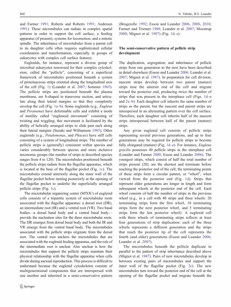
Euglenids, for instance, represent a diverse group ofmicrobial eukaryotes renowned for their complex cytoskel-eton, called the “pellicle”, consisting of a superficialframework of microtubules positioned beneath a systemof proteinaceous strips oriented along the longitudinal axisof the cell (Fig. 1) (Leander et al. 2007; Sommer 1965).The pellicle strips are positioned beneath the plasmamembrane, are S-shaped in transverse section, and articu-late along their lateral margins so that they completelyenvelop the cell (Fig. 1a–b). Some euglenids (e.g., Euglenaand Peranema) have deformable cells and exhibit a modeof motility called “euglenoid movement” consisting oftwisting and wiggling; this movement is facilitated by theability of helically arranged strips to slide past each alongtheir lateral margins (Suzaki and Williamson 1985). Othereuglenids (e.g., Petalomonas, and Phacus) have stiff cellsconsisting of a system of longitudinal strips. The number ofpellicle strips is (generally) consistent within species andvaries considerably between species and more inclusivetaxonomic groups (the number of strips in described speciesranges from 4 to 120). The microtubules positioned beneaththe pellicle strips radiate from the flagellar apparatus, whichis located at the base of the flagellar pocket (Fig. 1c). Themicrotubules extend anteriorly along the inner wall of theflagellar pocket before turning posteriorly at the opening ofthe flagellar pocket to underlie the superficially arrangedpellicle strips (Fig. 1c).
The microtubular organizing center (MTOC) of euglenidcells consists of a tripartite system of microtubular rootsassociated with the flagellar apparatus: a dorsal root (DR),an intermediate root (IR) and a ventral root (VR). Two basalbodies—a dorsal basal body and a ventral basal body—provide the nucleation sites for the three microtubular roots.The DR emerges from dorsal basal body and both the IR andVR emerge from the ventral basal body. The microtubulesassociated with the pellicle strips originate from the dorsalroot. The ventral root anchors the microtubules that areassociated with the euglenid feeding apparatus, and the role ofthe intermediate root is unclear. Also unclear is how themicrotubules that support the pellicle strips maintain theirphysical relationship with the flagellar apparatus when cellsdivide during asexual reproduction. This process is difficult tounderstand because the euglenid cytoskeleton consists ofmultigenerational components that are interspersed withone another and inherited in a semi-conservative pattern
(Brugerolle 1992; Esson and Leander 2006, 2008, 2010;Farmer and Triemer 1988; Leander et al. 2007; Moestrup2000; Mignot et al. 1987) (Fig. 1d–e).
The semi-conservative pattern of pellicle stripdevelopment
The duplication, segregation, and inheritance of pelliclestrips from one generation to the next have been describedin detail elsewhere (Esson and Leander 2006; Leander et al.2007; Mignot et al. 1987). In preparation for cell division,nascent strips develop between two parent (mature)strips near the anterior end of the cell and migratetoward the posterior end, producing twice the number ofstrips that was present in the interphase cell (Figs. 1d–eand 2a–b). Each daughter cell inherits the same number ofstrips as the parent, but the nascent and parent strips areinterspersed in an alternating pattern over the cell surface.Therefore, each daughter cell inherits half of the nascentstrips interspersed between half of the parent (mature)strips.
Any given euglenid cell consists of pellicle stripsrepresenting several previous generations, and up to fourgenerations may be required for pellicle strips to becomefully elongated (mature) (Fig. 1d–e). For instance, Euglenagracilis possesses 40 pellicle strips in the interphase cell(Leander and Farmer 2000; Esson and Leander 2006). Theyoungest strips, which consist of half the total number ofstrips present (20), are the shortest and terminate beforereaching the posterior end of the cell; the terminating pointsof these strips form a circular pattern, or “whorl”, whenviewed from the posterior end (Fig. 1d). Strips thatrepresent older generations are longer in length and formsubsequent whorls at the posterior end of the cell. Eachwhorl consists of half the number of strips in the previouswhorl (e.g., in a cell with 40 strips and three whorls: 20terminating strips form the first whorl, 10 terminatingstrips form the next posterior whorl, and 5 terminatingstrips form the last posterior whorl). A euglenid cellwith three whorls of terminating strips reflects at leastfour generations of strip duplication: each of the threewhorls represents a different generation and the stripsthat reach the posterior tip of the cell represents thefourth (and older) generations (Esson and Leander 2006;Leander et al. 2007).
The microtubules beneath the pellicle duplicate inparallel to the pattern of strip inheritance described above(Mignot et al. 1987). Pairs of new microtubules develop inbetween existing pairs of microtubules and support theinner wall of the flagellar pocket (Fig. 2c). The newmicrotubules turn toward the posterior end of the cell at theopening of the flagellar pocket and migrate beneath the
860 N. Yubuki, B.S. Leander
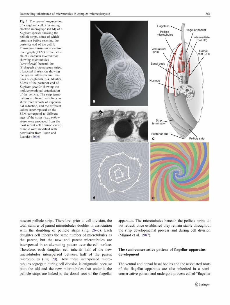
nascent pellicle strips. Therefore, prior to cell division, thetotal number of paired microtubules doubles in associationwith the doubling of pellicle strips (Fig. 2b–c). Eachdaughter cell inherits the same number of microtubules asthe parent, but the new and parent microtubules areinterspersed in an alternating pattern over the cell surface.Therefore, each daughter cell inherits half of the newmicrotubules interspersed between half of the parentmicrotubules (Fig. 2d). How these interspersed micro-tubules segregate during cell division is enigmatic, becauseboth the old and the new microtubules that underlie thepellicle strips are linked to the dorsal root of the flagellar
apparatus. The microtubules beneath the pellicle strips donot retract; once established they remain stable throughoutthe strip developmental process and during cell division(Mignot et al. 1987).
The semi-conservative pattern of flagellar apparatusdevelopment
The ventral and dorsal basal bodies and the associated rootsof the flagellar apparatus are also inherited in a semi-conservative pattern and undergo a process called “flagellar
b c
a
ed
FlagellumFlagellar pocket
Nucleus
Strip termination
Pellicle strip
Basal body
Pellicle microtubules
VB DB
Posterior end
Dorsal root (DR)
Intermediate root (IR)
Ventral root (VR)
Fig. 1 The general organizationof a euglenid cell. a Scanningelectron micrograph (SEM) of aEuglena species showing thepellicle strips, some of whichterminate before reaching theposterior end of the cell. bTransverse transmission electronmicrograph (TEM) of the pelli-cle of Colacium mucronatumshowing microtubules(arrowheads) beneath the(S-shaped) proteinaceous strips.c Labeled illustration showingthe general ultrastructural fea-tures of euglenids. d–e. IdenticalSEMs of the posterior end ofEuglena gracilis showing themultigenerational organizationof the pellicle. The strip termi-nations are linked with lines toshow three whorls of exponen-tial reduction, and the differentcolors superimposed on theSEM correspond to differentages of the strips (e.g., yellowstrips were produced from themost recent cell division event).d and e were modified withpermission from Esson andLeander (2006)
Reconciling inheritance of microtubules in complex microeukaryote 861
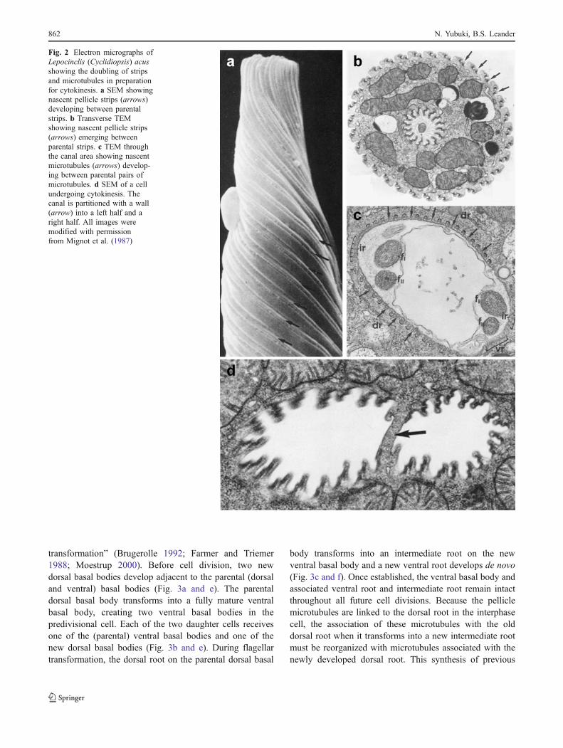
transformation” (Brugerolle 1992; Farmer and Triemer1988; Moestrup 2000). Before cell division, two newdorsal basal bodies develop adjacent to the parental (dorsaland ventral) basal bodies (Fig. 3a and e). The parentaldorsal basal body transforms into a fully mature ventralbasal body, creating two ventral basal bodies in thepredivisional cell. Each of the two daughter cells receivesone of the (parental) ventral basal bodies and one of thenew dorsal basal bodies (Fig. 3b and e). During flagellartransformation, the dorsal root on the parental dorsal basal
body transforms into an intermediate root on the newventral basal body and a new ventral root develops de novo(Fig. 3c and f). Once established, the ventral basal body andassociated ventral root and intermediate root remain intactthroughout all future cell divisions. Because the pelliclemicrotubules are linked to the dorsal root in the interphasecell, the association of these microtubules with the olddorsal root when it transforms into a new intermediate rootmust be reorganized with microtubules associated with thenewly developed dorsal root. This synthesis of previous
Fig. 2 Electron micrographs ofLepocinclis (Cyclidiopsis) acusshowing the doubling of stripsand microtubules in preparationfor cytokinesis. a SEM showingnascent pellicle strips (arrows)developing between parentalstrips. b Transverse TEMshowing nascent pellicle strips(arrows) emerging betweenparental strips. c TEM throughthe canal area showing nascentmicrotubules (arrows) develop-ing between parental pairs ofmicrotubules. d SEM of a cellundergoing cytokinesis. Thecanal is partitioned with a wall(arrow) into a left half and aright half. All images weremodified with permissionfrom Mignot et al. (1987)
862 N. Yubuki, B.S. Leander
studies made it extremely difficult to comprehend how thedevelopment and inheritance of the microtubules associatedwith the flagellar apparatus were coordinated with thedevelopment and inheritance of the microtubules associatedwith the pellicle strips. Our goal here was to establish atestable model that explains the process of microtubularreorganization during the interconnected processes of stripduplication and flagellar transformation.
Reconciling the inheritance patterns of pellicleand flagellar microtubules
Figures 4, 5 and 6 illustrate our model for the organizationand inheritance of the euglenid cytoskeleton. For illustrativepurposes, we consider a euglenid cell with 16 pellicle strips,16 pairs of subtendingmicrotubules, and two generations (i.e.,posterior whorls) of strip reduction (Fig. 4a.i–ii). Our model
of microtubular integration and inheritance is based on twokey inferences.
Inference 1
The pellicle microtubules do not extend directly from thedorsal root, which stands in contrast to previous interpre-tations that have been illustrated in the literature (Shin et al.2001; Solomon et al. 1987; Willey and Wibel 1987;Supplementary Fig. 1). Instead, we propose that the pelliclemicrotubules extend from an intermediary “pellicle micro-tubule organizing center” (pMTOC) that branches from thedorsal root of the dorsal basal body (Fig. 4a.iii). ThepMTOC encircles the posterior end of the flagellar pocketand supports all of the microtubules that underlie thepellicle strips. The distal end of the pMTOC terminates nearthe intermediate root, which branches from the ventral basalbody (Fig. 4a.iii).
DBVB
VR IR DR
VBVB
VRIR IR
DR
VR
IR
DR
IRVR
DB
VB
DB
VB
d e f
a b c
VB VR
DB
IR
DR
VB VR
IR DB
DR
VB
IR
DB
DR
VR
VB
DBDR
IR
VR
VRVB IR
DB DR
Fig. 3 Flagellar transformation in euglenids. a–c Transmissionelectron micrographs (TEM) of Entosiphon sulcatum. d–f.Corresponding schematic drawings of euglenid flagellar transforma-tion. The parental components of the flagellar apparatus are show ingreen and the de novo components are shown in red d. The interphasecell contains two basal bodies (DB and VB) and three microtubularroots (DR, IR and VR). a, e Just before cytokinesis, two new basal
bodies (arrowheads) form de novo near the parental basal bodies. bNew dorsal microtubular roots form on the new dorsal basal bodies;the parent dorsal basal body transforms into a new ventral basal bodyand develops a ventral root de novo. c, f Separation of two pairs ofbasal bodies, each consisting of one new dorsal basal body and oneventral basal body. a, b and c were modified with permission fromBrugerolle (1992)
Reconciling inheritance of microtubules in complex microeukaryote 863
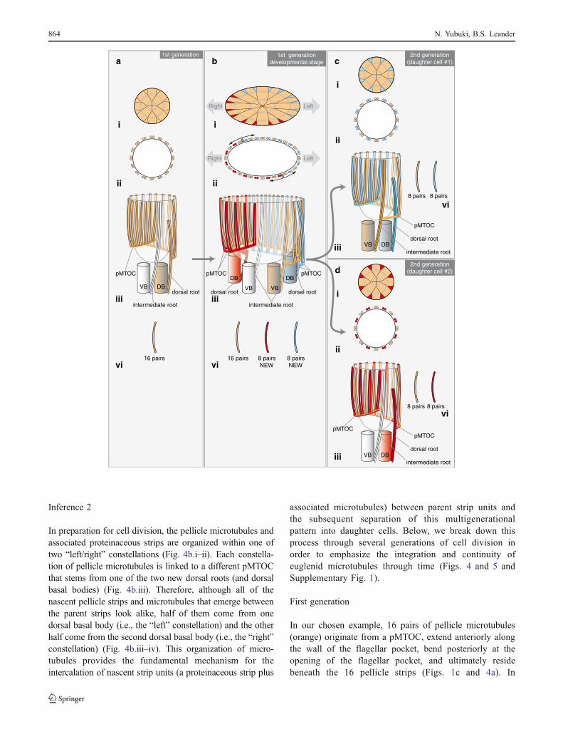
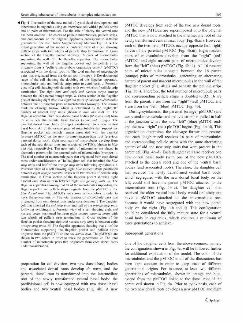
Inference 2
In preparation for cell division, the pellicle microtubules andassociated proteinaceous strips are organized within one oftwo “left/right” constellations (Fig. 4b.i–ii). Each constella-tion of pellicle microtubules is linked to a different pMTOCthat stems from one of the two new dorsal roots (and dorsalbasal bodies) (Fig. 4b.iii). Therefore, although all of thenascent pellicle strips and microtubules that emerge betweenthe parent strips look alike, half of them come from onedorsal basal body (i.e., the “left” constellation) and the otherhalf come from the second dorsal basal body (i.e., the “right”constellation) (Fig. 4b.iii–iv). This organization of micro-tubules provides the fundamental mechanism for theintercalation of nascent strip units (a proteinaceous strip plus
associated microtubules) between parent strip units andthe subsequent separation of this multigenerationalpattern into daughter cells. Below, we break down thisprocess through several generations of cell division inorder to emphasize the integration and continuity ofeuglenid microtubules through time (Figs. 4 and 5 andSupplementary Fig. 1).
First generation
In our chosen example, 16 pairs of pellicle microtubules(orange) originate from a pMTOC, extend anteriorly alongthe wall of the flagellar pocket, bend posteriorly at theopening of the flagellar pocket, and ultimately residebeneath the 16 pellicle strips (Figs. 1c and 4a). In
16 pairs
VB DB
1st generation
dorsal root
intermediate root
i
ii
iii
vi
pMTOC
VB VB
DB DB
dorsal root
intermediate root
dorsal root
1st generationdevelopmental stage
16 pairs 8 pairs NEW
8 pairs NEW
i
ii
iii
vi
pMTOC pMTOC
LeftRight
LeftRight
8 pairs 8 pairs
VB DBdorsal root
intermediate root
i
ii
iii
vi
pMTOC
2nd generation(daughter cell #1)
2nd generation(daughter cell #2)
8 pairs 8 pairs
VB DB
i
ii
iii
vi
pMTOC
dorsal root
intermediate root
pMTOC
a b c
d
864 N. Yubuki, B.S. Leander
preparation for cell division, two new dorsal basal bodiesand associated dorsal roots develop de novo, and theparental dorsal root is transformed into the intermediateroot of the newly transformed ventral basal body; thepredivisional cell is now equipped with two dorsal basalbodies and two ventral basal bodies (Fig. 4b). A new
pMTOC develops from each of the two new dorsal roots,and the new pMTOCs are superimposed onto the parentalpMTOC that is now attached to the intermediate root of thenewly transformed ventral basal body (Fig. 4b.iii). However,each of the two new pMTOCs occupy opposite (left–right)halves of the parental pMTOC (Fig. 4b.iii). Eight nascentpairs of microtubules develop from the “right” (red)pMTOC, and eight nascent pairs of microtubules developfrom the “left” (blue) pMTOC (Fig. 4b.iii). All 16 nascentpairs of microtubules elongate between the 16 parent(orange) pairs of microtubules, generating an alternatingpattern of parent and nascent microtubules in the wall of theflagellar pocket (Fig. 4b.ii) and beneath the pellicle strips(Fig. 5b.i). Therefore, the total number of microtubule pairsand corresponding pellicle strips is doubled to 32: 16 arefrom the parent, 8 are from the “right” (red) pMTOC, and8 are from the “left” (blue) pMTOC (Fig. 4b).
During cytokinesis, the parental (orange) pMTOC (andassociated microtubules and pellicle strips) is pulled in halfat the junction where the new “left” (blue) pMTOC endsand the new “right” (red) pMTOC begins (Fig. 4b–d). Thisorganization determines the cleavage furrow and ensuresthat each daughter cell receives 16 pairs of microtubulesand corresponding pellicle strips with the same alternatingpattern of old and new strip units that were present in theparent cell (Fig. 4c–d). Each daughter cell also receives onenew dorsal basal body (with one of the new pMTOCsattached to the dorsal root) and one of the ventral basalbodies (and associated roots). Therefore, the daughter cellthat received the newly transformed ventral basal body,which segregated with the new dorsal basal body on theleft, could still have the parent pMTOC attached to theintermediate root (Fig. 4b–c). The daughter cell thatreceived the older ventral basal body would definitely nothave a pMTOC attached to the intermediate rootbecause it would have segregated with the new dorsalbody on the right (Fig. 4b and d). This configurationcould be considered the fully mature state for a ventralbasal body in euglenids, which requires a minimum ofthree generations to achieve.
Subsequent generations
One of the daughter cells from the above scenario, namelythe configuration shown in Fig. 4c, will be followed furtherfor additional explanation of the model. The color of themicrotubules and the pMTOC in all of the illustrations hasbeen kept constant in order to keep track of differentgenerational origins. For instance, at least two differentgenerations of microtubules, shown in orange and blue,extend from the pMTOC linked to the dorsal root of theparent cell shown in Fig. 5a. Prior to cytokinesis, each ofthe two new dorsal roots develops a new pMTOC and eight
Fig. 4 Illustration of the new model of cytoskeletal development andinheritance in euglenids using an interphase cell with16 pellicle stripsand 16 pairs of microtubules. For the sake of clarity, the ventral roothas been omitted. The colors of pellicle microtubules, pellicle strips,and components of the flagellar apparatus correspond to differentgenerations. See also Online Supplementary Material Fig. S1. a Theinitial generation of the model. i. Posterior view of a cell showingpellicle strips with two whorls of pellicle strip termination. ii. Crosssection of the flagellar pocket showing 16 pairs of microtubulessupporting the wall. iii. The flagellar apparatus. The microtubulessupporting the wall of the flagellar pocket and the pellicle stripsoriginate from a “pellicle microtubule organizing center” (pMTOC)associated with the dorsal root. iv. The total number of microtubulepairs that originated from the dorsal root (orange). b Developmentalstage of the cell showing the doubling of the flagellar apparatus,microtubular pairs and pellicle strips prior to cytokinesis. i. Posteriorview of a cell showing pellicle strips with two whorls of pellicle striptermination. The eight blue and eight red nascent strips emergebetween the 16 parental orange strips. ii. Cross section of the flagellarpocket showing eight blue and eight red pairs of nascent microtubulesbetween the 16 parental pairs of microtubules (orange). The arrowsmark the cleavage furrow, which is determined by the “right/left”clusters of nascent strip units (shown in blue and red). iii. Theflagellar apparatus. Two new dorsal basal bodies (blue and red) formde novo near the parental basal bodies (white and orange). Theparental dorsal basal body (orange) transforms into a new ventralbasal body. All of the orange pairs of microtubules that support theflagellar pocket and pellicle remain associated with the parental(orange) pMTOC on the new (orange) intermediate root (previousparental dorsal root). Eight new pairs of microtubules develop fromeach of the new dorsal roots and associated pMTOCs (shown in blueand red, respectively). The new pairs of microtubles are placed inalternative pattern with the parental pairs of microtubules (orange). iv.The total number of microtubule pairs that originated from each dorsalroots under consideration. c The daughter cell that inherited the bluestrip units and half of the orange strip units following cytokinesis. i.Posterior view of a cell showing eight blue nascent strips positionedbetween eight orange parental strips with two whorls of pellicle striptermination. ii. Cross section of the flagellar pocket showing eightnascent blue strip units in between eight orange strip units. iii. Theflagellar apparatus showing that all of the microtubules supporting theflagellar pocket and pellicle strips originate from the pMTOC on theblue dorsal root. The pMTOCs are shown in two colors in order totrack the generations. iv. The total number of microtubule pairs thatoriginated from each dorsal roots under consideration. d The daughtercell that inherited the red strip units and half of the orange strip unitsfollowing cytokinesis. i. Posterior view of a cell showing eight rednascent strips positioned between eight orange parental strips withtwo whorls of pellicle strip termination. ii. Cross section of theflagellar pocket showing eight red nascent strip units in between eightorange strip units. iii. The flagellar apparatus showing that all of themicrotubules supporting the flagellar pocket and pellicle stripsoriginate from the pMTOC on the red dorsal root. The pMTOCs areshown in two colors in order to track the generations. iv. The totalnumber of microtubule pairs that originated from each dorsal rootsunder consideration
�
Reconciling inheritance of microtubules in complex microeukaryote 865
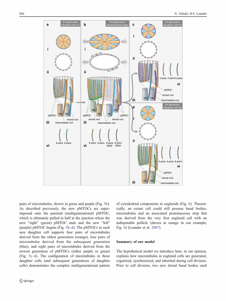
pairs of microtubules, shown in green and purple (Fig. 5b).As described previously, the new pMTOCs are super-imposed onto the parental (multigenerational) pMTOC,which is ultimately pulled in half at the junction where thenew “right” (green) pMTOC ends and the new “left”(purple) pMTOC begins (Fig. 5b–d). The pMTOCs in eachnew daughter cell supports four pairs of microtubulesderived from the oldest generation (orange), four pairs ofmicrotubules derived from the subsequent generation(blue), and eight pairs of microtubules derived from thenewest generation of pMTOCs (either purple or green)(Fig. 5c–d). The configuration of microtubules in thesedaughter cells (and subsequent generations of daughtercells) demonstrates the complex multigenerational pattern
of cytoskeletal components in euglenids (Fig. 6). Theoret-ically, an extant cell could still possess basal bodies,microtubules and an associated proteinaceous strip thatwas derived from the very first euglenid cell with anindisputable pellicle (shown in orange in our example;Fig. 6) (Leander et al. 2007).
Summary of our model
The hypothetical model we introduce here, in our opinion,explains how microtubules in euglenid cells are generated,organized, synchronized, and inherited during cell division.Prior to cell division, two new dorsal basal bodies each
a b c
d
8 pairs 8 pairs
VB DB
dorsal rootintermediate root
i
ii
iii
vi
pMTOC
2nd generation(daughter cell #1)
VB VB
DB DB
dorsal root
intermediate root
dorsal root
8 pairs 8 pairs 8 pairs NEW
8 pairs NEW
2nd generationdevelopmental stage
i
ii
iii
vi
pMTOC pMTOC
LeftRight
LeftRight
4 pairs 4 pairs 8 pairs
VB DB
3rd generation(daughter cell #1)
i
ii
iii
vi
dorsal root
intermediate root
pMTOC
4 pairs 4 pairs 8 pairs
VB DB
3rd generation(daughter cell #2)
dorsal root
intermediate root
i
ii
iii
vi
pMTOC
866 N. Yubuki, B.S. Leander
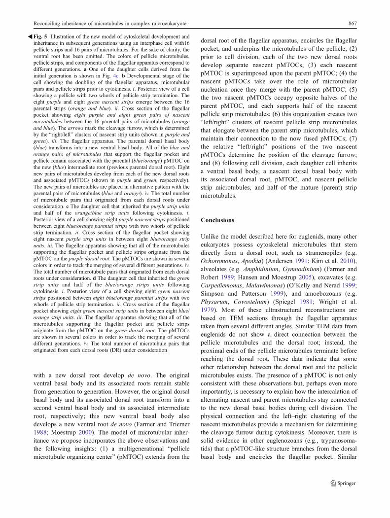
with a new dorsal root develop de novo. The originalventral basal body and its associated roots remain stablefrom generation to generation. However, the original dorsalbasal body and its associated dorsal root transform into asecond ventral basal body and its associated intermediateroot, respectively; this new ventral basal body alsodevelops a new ventral root de novo (Farmer and Triemer1988; Moestrup 2000). The model of microtubular inher-itance we propose incorporates the above observations andthe following insights: (1) a multigenerational “pelliclemicrotubule organizing center” (pMTOC) extends from the
dorsal root of the flagellar apparatus, encircles the flagellarpocket, and underpins the microtubules of the pellicle; (2)prior to cell division, each of the two new dorsal rootsdevelop separate nascent pMTOCs; (3) each nascentpMTOC is superimposed upon the parent pMTOC; (4) thenascent pMTOCs take over the role of microtubularnucleation once they merge with the parent pMTOC; (5)the two nascent pMTOCs occupy opposite halves of theparent pMTOC, and each supports half of the nascentpellicle strip microtubules; (6) this organization creates two“left/right” clusters of nascent pellicle strip microtubulesthat elongate between the parent strip microtubules, whichmaintain their connection to the now fused pMTOCs; (7)the relative “left/right” positions of the two nascentpMTOCs determine the position of the cleavage furrow;and (8) following cell division, each daughter cell inheritsa ventral basal body, a nascent dorsal basal body withits associated dorsal root, pMTOC, and nascent pelliclestrip microtubules, and half of the mature (parent) stripmicrotubules.
Conclusions
Unlike the model described here for euglenids, many othereukaryotes possess cytoskeletal microtubules that stemdirectly from a dorsal root, such as stramenopiles (e.g.Ochoromonas, Apoikia) (Andersen 1991; Kim et al. 2010),alveolates (e.g. Amphidinium, Gymnodinium) (Farmer andRobert 1989; Hansen and Moestrup 2005), excavates (e.g.Carpediemonas, Malawimonas) (O’Kelly and Nerad 1999;Simpson and Patterson 1999), and amoebozoans (e.g.Physarum, Covostelium) (Spiegel 1981; Wright et al.1979). Most of these ultrastructural reconstructions arebased on TEM sections through the flagellar apparatustaken from several different angles. Similar TEM data fromeuglenids do not show a direct connection between thepellicle microtubules and the dorsal root; instead, theproximal ends of the pellicle microtubules terminate beforereaching the dorsal root. These data indicate that someother relationship between the dorsal root and the pelliclemicrotubules exists. The presence of a pMTOC is not onlyconsistent with these observations but, perhaps even moreimportantly, is necessary to explain how the intercalation ofalternating nascent and parent microtubules stay connectedto the new dorsal basal bodies during cell division. Thephysical connection and the left–right clustering of thenascent microtubules provide a mechanism for determiningthe cleavage furrow during cytokinesis. Moreover, there issolid evidence in other euglenozoans (e.g., trypanosoma-tids) that a pMTOC-like structure branches from the dorsalbasal body and encircles the flagellar pocket. Similar
Fig. 5 Illustration of the new model of cytoskeletal development andinheritance in subsequent generations using an interphase cell with16pellicle strips and 16 pairs of microtubules. For the sake of clarity, theventral root has been omitted. The colors of pellicle microtubules,pellicle strips, and components of the flagellar apparatus correspond todifferent generations. a One of the daughter cells derived from theinitial generation is shown in Fig. 4c. b Developmental stage of thecell showing the doubling of the flagellar apparatus, microtubularpairs and pellicle strips prior to cytokinesis. i. Posterior view of a cellshowing a pellicle with two whorls of pellicle strip termination. Theeight purple and eight green nascent strips emerge between the 16parental strips (orange and blue). ii. Cross section of the flagellarpocket showing eight purple and eight green pairs of nascentmicrotubules between the 16 parental pairs of microtubules (orangeand blue). The arrows mark the cleavage furrow, which is determinedby the “right/left” clusters of nascent strip units (shown in purple andgreen). iii. The flagellar apparatus. The parental dorsal basal body(blue) transforms into a new ventral basal body. All of the blue andorange pairs of microtubules that support the flagellar pocket andpellicle remain associated with the parental (blue/orange) pMTOC onthe new (blue) intermediate root (previous parental dorsal root). Eightnew pairs of microtubules develop from each of the new dorsal rootsand associated pMTOCs (shown in purple and green, respectively).The new pairs of microtubles are placed in alternative pattern with theparental pairs of microtubules (blue and orange). iv. The total numberof microtubule pairs that originated from each dorsal roots underconsideration. c The daughter cell that inherited the purple strip unitsand half of the orange/blue strip units following cytokinesis. i.Posterior view of a cell showing eight purple nascent strips positionedbetween eight blue/orange parental strips with two whorls of pelliclestrip termination. ii. Cross section of the flagellar pocket showingeight nascent purple strip units in between eight blue/orange stripunits. iii. The flagellar apparatus showing that all of the microtubulessupporting the flagellar pocket and pellicle strips originate from thepMTOC on the purple dorsal root. The pMTOCs are shown in severalcolors in order to track the merging of several different generations. iv.The total number of microtubule pairs that originated from each dorsalroots under consideration. d The daughter cell that inherited the greenstrip units and half of the blue/orange strips units followingcytokinesis. i. Posterior view of a cell showing eight green nascentstrips positioned between eight blue/orange parental strips with twowhorls of pellicle strip termination. ii. Cross section of the flagellarpocket showing eight green nascent strip units in between eight blue/orange strip units. iii. The flagellar apparatus showing that all of themicrotubules supporting the flagellar pocket and pellicle stripsoriginate from the pMTOC on the green dorsal root. The pMTOCsare shown in several colors in order to track the merging of severaldifferent generations. iv. The total number of microtubule pairs thatoriginated from each dorsal roots (DR) under consideration
�
Reconciling inheritance of microtubules in complex microeukaryote 867
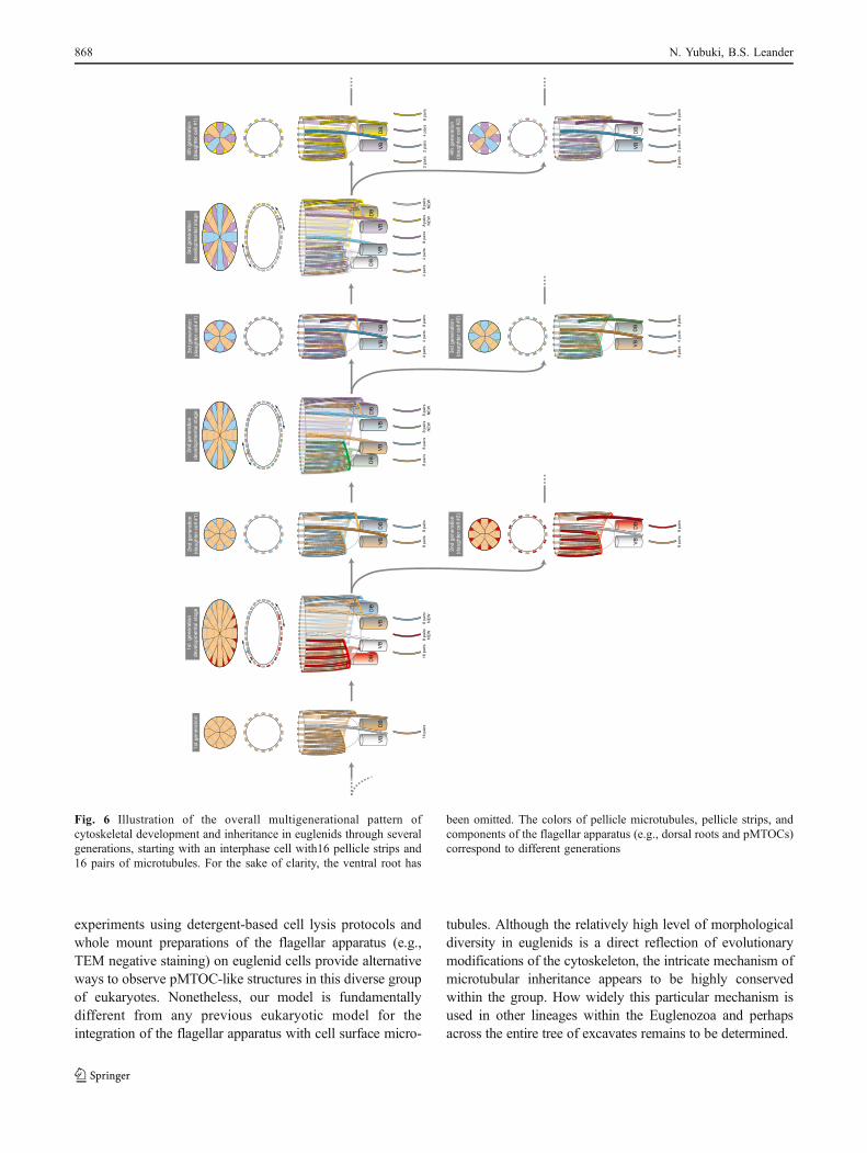
experiments using detergent-based cell lysis protocols andwhole mount preparations of the flagellar apparatus (e.g.,TEM negative staining) on euglenid cells provide alternativeways to observe pMTOC-like structures in this diverse groupof eukaryotes. Nonetheless, our model is fundamentallydifferent from any previous eukaryotic model for theintegration of the flagellar apparatus with cell surface micro-
tubules. Although the relatively high level of morphologicaldiversity in euglenids is a direct reflection of evolutionarymodifications of the cytoskeleton, the intricate mechanism ofmicrotubular inheritance appears to be highly conservedwithin the group. How widely this particular mechanism isused in other lineages within the Euglenozoa and perhapsacross the entire tree of excavates remains to be determined.
16 p
airs
VB
DB
1st g
ener
atio
n
VB
VB
DB
DB
1st
gene
ratio
nde
velo
pmen
tal s
tage
16 p
airs
8 pa
irs N
EW
8 pa
irs N
EW
8 pa
irs8
pairs
VB
DB
2nd
gene
ratio
n(d
augh
ter
cell
#1)
2nd
gene
ratio
n(d
augh
ter
cell
#2)
8 pa
irs8
pairs
VB
DB
VB
VB
DB
DB
4 pa
irs4
pairs
8 pa
irs8
pairs
NE
W8
pairs
NE
W
3rd
gene
ratio
nde
velo
pmen
tal s
tage
4 pa
irs4
pairs
8 pa
irs
VB
DB
3rd
gene
ratio
n(d
augh
ter
cell
#1)
4 pa
irs4
pairs
8 pa
irs
VB
DB
3rd
gene
ratio
n(d
augh
ter
cell
#2)
VB
VB
DB
DB
8 pa
irs8
pairs
8 pa
irs N
EW
8 pa
irs N
EW
2nd
gene
ratio
nde
velo
pmen
tal s
tage
2 pa
irs2
pairs
4 pa
irs8
pairs
VB
DB
4th
gene
ratio
n(d
augh
ter
cell
#1)
VB
DB
4th
gene
ratio
n(d
augh
ter
cell
#2)
2 pa
irs2
pairs
4 pa
irs8
pairs
Fig. 6 Illustration of the overall multigenerational pattern ofcytoskeletal development and inheritance in euglenids through severalgenerations, starting with an interphase cell with16 pellicle strips and16 pairs of microtubules. For the sake of clarity, the ventral root has
been omitted. The colors of pellicle microtubules, pellicle strips, andcomponents of the flagellar apparatus (e.g., dorsal roots and pMTOCs)correspond to different generations
868 N. Yubuki, B.S. Leander
Acknowledgements This work was supported by grants from theTula Foundation (Centre for Microbial Diversity and Evolution at theUniversity of British Columbia) and the Canadian Institute forAdvanced Research, Program in Integrated Microbial Biodiversity.
References
Andersen RA (1991) The cytoskeleton of chromophyte algae.Protoplasma 164:143–159
Beech PL, Heimann K, Melkonian M (1991) Development of theflagellar apparatus during the cell cycle in unicellular algae.Protoplasma 164:23–37
Brugerolle G (1991) Flagellar and cytoskeletal systems in amitochon-drial flagellates: Archamoeba, Metamonada and Parabasala.Protoplasma 164:70–90
Brugerolle G (1992) Flagellar apparatus duplication and partition,flagellar transformation during division in Entosiphon sulcatum.BioSystems 28(1–3):203–209
Esson HJ, Leander BS (2006) A model for the morphogenesis of stripreduction patterns in phototrophic euglenids: evidence forheterochrony in pellicle evolution. Evol Dev 8(4):378–388.doi:10.1111/j.1525-142X.2006.00110.x
Esson HJ, Leander BS (2008) Novel pellicle surface patterns onEuglena obtusa (Euglenophyta) from the marine benthic envi-ronment: implications for pellicle development and evolution. JPhycol 44(1):132–141
Esson HJ, Leander BS (2010) Evolution of distorted pelliclepatterns in rigid photosynthetic euglenids (Phacus Dujardin).J Eukaryot Microbiol 57(1):19–32. doi:10.1111/j.1550-7408.2009.00447.x
Farmer MA, Robert KR (1989) Comperative analyses of thedinoflagellate flagellar apparatus. III. Freeze substitution ofAmphydinium rhyncocephalum. J Phycol 25:280–292
Farmer MA, Triemer RE (1988) Flagellar systems in the euglenoidflagellates. BioSystems 21(3–4):283–291
Hansen G, Moestrup Ø (2005) Flagellar apparatus and nuclearchambers of the green dinoflagellate Gymnodinium chlorophorum.Phycol Res 53(2):169–181
Kim E, Yubuki N, Leander BS, Graham LE (2010) Ultrastructure and18S rDNA Phylogeny of Apoikia lindahlii comb. nov. (Chrys-ophyceae) and its epibiontic protists, Filos agilis gen. et sp. nov.(Bicosoecida) and Nanos amicus gen. et sp. nov. (Bicosoecida).Protist 161(2):177–196. doi:10.1016/j.protis.2009.09.003
Leander BS, Farmer MA (2000) Comparative morphology of theeuglenid oellicle. I. Patterns of strips and pores. J EukaryotMicrobiol 47(5):469–479
Leander BS, Esson HJ, Breglia SA (2007) Macroevolution of complexcytoskeletal systems in euglenids. BioEssays 29(10):987–1000.doi:10.1002/bies.20645
Mignot JP, Brugerolle G, Bricheux G (1987) Intercalary stripdevelopment and dividing cell morphogenesis in the euglenidCyclidiopsis acus. Protoplasma 139(1):51–65
Moestrup Ø (1982) Flagellar structure in algae: a review, with newobservations particularly on the Chrysophyceae, Phaeophyceae(Fucophyceae), Euglenophyceae and Reckertia. Phycologia 21(4):427–528
Moestrup Ø (2000) The flagellate cytoskeleton. Introduction of ageneral terminology for microtubular flagellar roots in protists.In: Leadbeater BSC, Green JC (eds) The flagellate. Unity,diversity and evolution. The systematics association specialvolume series 59. Taylor & Francis Limited, London, pp 69–94
O’Kelly CJ, Nerad TA (1999) Malawimonas jakobiformis n. gen., n.sp. (Malawimonadidae n. fam.): a Jakoba-like heterotrophicnanoflagellate with discoidal mitochondrial cristae. J Euk Micro-biol 46(5):522–531. doi:10.1111/j.1550-7408.1999.tb06070.x
Roberts KR, Roberts JE (1991) The flagellar apparatus and cytoskel-eton of the dinoflagellates. A comparative overview. Protoplasma164:105–122
Shin W, Boo SM, Triemer RE (2001) Ultrastructure of the basal bodycomplex and putative vestigial feeding apparatus in Phacuspleuronectes (Euglenophyceae). J Phycol 37(5):913–921
Simpson AGB, Patterson DJ (1999) The ultrastructure of Carpedie-monas membranifera (Eukaryota) with reference to the “excavatehypothesis”. Eur J Protistol 35:353–370
Sleigh MA (1988) Flagellar root maps allow speculative comparisonsof root patterns and of their ontogeny. BioSystems 21(3–4):277–282
Solomon JA, Walne PL, Kivic PA (1987) Entosiphon sulcatum(Euglenophyceae): flagellar roots of the basal body complexand reservoir region. J Phycol 23(1):85–98
Sommer JR (1965) The Ultrastructure of the pellicle complex ofEuglena gracilis. J Cell Biol 24:253–257
Spiegel FW (1981) Phylogenetic significance of the flagellar apparatus inprotostelids (Eumycetozoa). BioSystems 14(3–4):491–499
Suzaki T, Williamson RE (1985) Euglenoid movement in Euglenafusca: evidence for sliding between pellicular strips. Protoplasma124(1–2):137–146
Triemer RE, Farmer MA (1991) An ultrastructural comparison of themitotic apparatus, feeding apparatus, flagellar apparatus and cyto-skeleton in euglenoids and kinetoplastids. Protoplasma 164:91–104
Willey RL, Wibel RG (1987) Flagellar roots and the reservoircytoskeleton of Colacium libellae (Euglenophyceae). J Phycol23(s2):283–288
Wright M, Moisand A, Mir L (1979) The structure of the flagellarapparatus of the swarm cells of Physarum polycephalum.Protoplasma 100(3):231–250. doi:10.1007/bf01279314
Reconciling inheritance of microtubules in complex microeukaryote 869