Embed Size (px)
Citation preview
443
L A B O R A T O R Y 9
Recombination of Antibiotic ResistanceGenes
LABORATORY 9 BEGINS AN EXPERIMENTAL STREAM designed to construct and analyzea recombinant DNA molecule. The starting reagents are the relaxed plasmidspAMP and pKAN, each of which carries a single antibiotic resistance gene: ampi-cillin in pAMP and kanamycin in pKAN. The goal is to construct a recombinantplasmid that contains both ampicillin and kanamycin resistance genes. This lab-oratory is divided into two parts: Restriction Digest of Plasmids pAMP and pKANand Ligation of pAMP and pKAN Restriction Fragments.
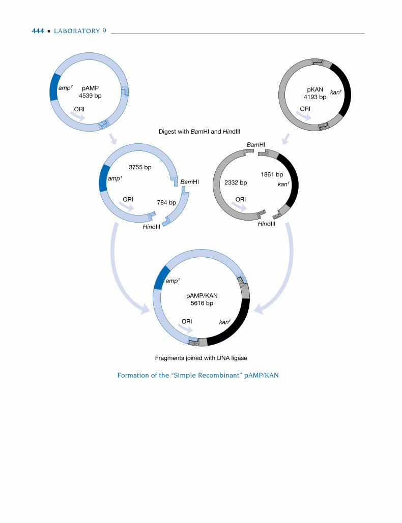
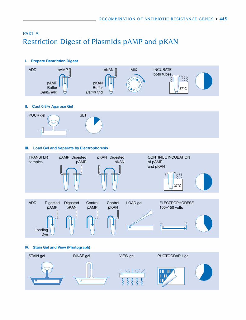
• Part A provides a procedure whereby samples of both plasmids are digestedin separate restriction reactions with BamHI and HindIII. Following incuba-tion at 37ºC, samples of digested pAMP and pKAN are analyzed by agarosegel electrophoresis to confirm proper cutting. Each plasmid contains a singlerecognition site for each enzyme, yielding only two restriction fragments.Cleavage of pAMP yields fragments of 784 bp and 3755 bp, and cleavage ofpKAN yields fragments of 1861 bp and 2332 bp.
• Part B provides a technique for ligation of pAMP and pKAN restriction frag-ments. The restriction digests of pAMP and pKAN are heated to destroyBamHI and HindIII activity. A sample from each reaction is mixed with DNAligase plus ATP and incubated at room temperature. Complementary BamHIand HindIII “sticky ends” hydrogen-bond to align restriction fragments. Ligasecatalyzes the formation of phosphodiester bonds that covalently link the DNAfragments to form stable recombinant DNA molecules.
Equipment and materials for this laboratory are available from the CarolinaBiological Supply Company (see Appendix 1).
443-456 DNA Sci Lab 09 11/9/05 1:54 PM Page 443
444 ■ LABORATORY 9
Digest with BamHI and HindIII
HindIII HindIII
BamHI
BamHI
pAMP4539 bp
pAMP/KAN5616 bp
ORI
amp r
amp r
amp r
kan r
kan r
kan r
ORI
3755 bp
2332 bp1861 bp
784 bp ORI
pKAN4193 bp
ORI
ORI
Fragments joined with DNA ligase
Formation of the “Simple Recombinant” pAMP/KAN
443-456 DNA Sci Lab 09 11/9/05 1:54 PM Page 444
PART A
Restriction Digest of Plasmids pAMP and pKAN
RECOMBINATION OF ANTIBIOTIC RESISTANCE GENES ■ 445
ADD MIXpAMP pKAN
I. Prepare Restriction Digest
II. Cast 0.8% Agarose Gel
III. Load Gel and Separate by Electrophoresis
IV. Stain Gel and View (Photograph)
INCUBATEboth tubes
POUR gel SET
TRANSFERsamples
pAMP DigestedpAMP
STAIN gel RINSE gel VIEW gel PHOTOGRAPH gel
37°C
37°C
pAMPBuffer
Bam/Hind
pKANBuffer
Bam/Hind
pKAN CONTINUE INCUBATIONof pAMPand pKAN
DigestedpKAN
ADD
LoadingDye
DigestedpAMP
DigestedpKAN
ControlpAMP
ControlpKAN
LOAD gel ELECTROPHORESE100–150 volts
– +
443-456 DNA Sci Lab 09 11/9/05 1:54 PM Page 445
PRELAB NOTES
Review Prelab Notes in Laboratory 3, DNA Restriction Analysis.
Plasmid Substitution
The process of constructing and analyzing recombinant molecules is not trivial.However, good results can be expected if the directions are followed carefully.These protocols have been optimized for the teaching plasmids pAMP andpKAN, and the extensive analysis of results is based entirely on recombinant mol-ecules derived from these parent molecules.
The Prudent Control
In Section III, samples of the restriction digests are analyzed by agarose gel elec-trophoresis, prior to ligation, to confirm complete cutting by the endonucleases.This prudent control is standard experimental procedure. If pressed for time,omit electrophoresis and ligate DNA directly following the restriction digest.However, be sure to pretest the activity of BamHI and HindIII to determine theincubation time needed for complete digestion.
It is fairly impractical to use methylene blue staining for this step, whichdemands a rapid and sensitive assay to check for complete digestion of the plas-mid DNAs. Methylene blue destaining requires at least 30 minutes, and it couldfail to detect a small but possibly significant amount of uncut DNA. However, ifusing methylene blue staining for this lab, refer to the staining procedure in StepIVB of Laboratory 8 (Part B).
Saving DNA
Restriction reactions and controls in this experiment use a relatively largeamount of plasmid DNA, which is the most expensive reagent used in thecourse. To minimize expense, the protocol directs that the lab be prepared by setting upaliquots of exactly the required volumes of pAMP and pKAN into 1.5-ml tubes. Then thereagents for restriction digestion are added directly to these aliquots of DNA.
PRELAB PREPARATION
Before performing this Prelab Preparation, please refer to the cautions indicatedon the Laboratory Materials list.
1. Mix in 1:1 proportion: BamHI + HindIII (6 µl per experiment).
2. Prepare aliquots for each experiment:
5.5 µl of 0.20 µg/µl pAMP (store on ice)5.5 µl of 0.20 µg/µl pKAN (store on ice)5 µl of 0.10 µg/µl pAMP (store on ice)5 µl of 0.10 µg/µl pKAN (store on ice)20 µl of 2x restriction buffer (store on ice)6.0 µl of BamHI/HindIII500 µl of distilled water500 µl of loading dye
446 ■ LABORATORY 9
443-456 DNA Sci Lab 09 11/9/05 1:54 PM Page 446
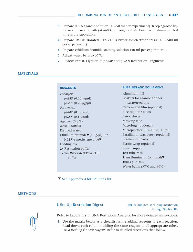
3. Prepare 0.8% agarose solution (40–50 ml per experiment). Keep agarose liq-uid in a hot-water bath (at ~60ºC) throughout lab. Cover with aluminum foilto retard evaporation.
4. Prepare 1x Tris/Borate/EDTA (TBE) buffer for electrophoresis (400–500 mlper experiment).
5. Prepare ethidium bromide staining solution (50 ml per experiment).
6. Adjust water bath to 37ºC.
7. Review Part B, Ligation of pAMP and pKAN Restriction Fragments.
MATERIALS
METHODS
I. Set Up Restriction Digest (40–60 minutes, including incubation
through Section III)
Refer to Laboratory 3, DNA Restriction Analysis, for more detailed instructions.
1. Use the matrix below as a checklist while adding reagents to each reaction.Read down each column, adding the same reagent to all appropriate tubes.Use a fresh tip for each reagent. Refer to detailed directions that follow.
RECOMBINATION OF ANTIBIOTIC RESISTANCE GENES ■ 447
REAGENTS
For digest:
pAMP (0.20 µg/µl)
pKAN (0.20 µg/µl)
For control:
pAMP (0.1 µg/µl)
pKAN (0.1 µg/µl)
Agarose (0.8%)
BamHI/HindIII
Distilled water
Ethidium bromide (1 µg/µl) (or
0.025% methylene blue )
Loading dye
2x Restriction buffer
1x Tris /Borate/EDTA (TBE)
buffer
SUPPLIES AND EQUIPMENT
Aluminum foil
Beakers for agarose and for
waste/used tips
Camera and film (optional)
Electrophoresis box
Latex gloves
Masking tape
Microfuge (optional)
Micropipettor (0.5–10 µl) + tips
Parafilm or wax paper (optional)
Permanent marker
Plastic wrap (optional)
Power supply
Test tube rack
Transilluminator (optional)
Tubes (1.5-ml)
Water baths (37ºC and 60ºC)
See Appendix 4 for Cautions list.
443-456 DNA Sci Lab 09 11/9/05 1:54 PM Page 447
pAMP pKAN BamHI/Tube 0.2 μg/μl 0.2 μg/μl 2x Buffer HindIII
Digested pAMP 5.5 μl — 7.5 μl 2 μlDigested pKAN — 5.5 μl 7.5 μl 2 μl
2. Collect 2x buffer and BamHI/HindIII (on ice), and the tubes containing pAMPand pKAN plasmid DNA, and place in a test tube rack on the lab bench.
3. Add 7.5 μl of 2x restriction buffer to the pAMP and pKAN tubes.
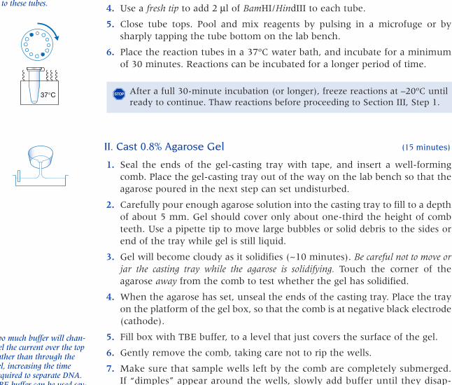
4. Use a fresh tip to add 2 μl of BamHI/HindIII to each tube.
5. Close tube tops. Pool and mix reagents by pulsing in a microfuge or bysharply tapping the tube bottom on the lab bench.
6. Place the reaction tubes in a 37ºC water bath, and incubate for a minimumof 30 minutes. Reactions can be incubated for a longer period of time.
II. Cast 0.8% Agarose Gel (15 minutes)
1. Seal the ends of the gel-casting tray with tape, and insert a well-formingcomb. Place the gel-casting tray out of the way on the lab bench so that theagarose poured in the next step can set undisturbed.
2. Carefully pour enough agarose solution into the casting tray to fill to a depthof about 5 mm. Gel should cover only about one-third the height of combteeth. Use a pipette tip to move large bubbles or solid debris to the sides orend of the tray while gel is still liquid.
3. Gel will become cloudy as it solidifies (~10 minutes). Be careful not to move orjar the casting tray while the agarose is solidifying. Touch the corner of theagarose away from the comb to test whether the gel has solidified.
4. When the agarose has set, unseal the ends of the casting tray. Place the trayon the platform of the gel box, so that the comb is at negative black electrode(cathode).
5. Fill box with TBE buffer, to a level that just covers the surface of the gel.
6. Gently remove the comb, taking care not to rip the wells.
7. Make sure that sample wells left by the comb are completely submerged.If “dimples” appear around the wells, slowly add buffer until they disap-pear.
448 ■ LABORATORY 9
To avoid confusion, keep0.1 μg/μl control pAMPand pKAN aside untilneeded in Section III.
To minimize waste andexpense, aliquots of plas-mid DNA can be set up intubes labeled pAMP andpKAN. Add reagents direct-ly to these tubes.
After a full 30-minute incubation (or longer), freeze reactions at –20ºC untilready to continue. Thaw reactions before proceeding to Section III, Step 1.
�
Too much buffer will chan-nel the current over the toprather than through thegel, increasing the timerequired to separate DNA.TBE buffer can be used sev-eral times; do not discard.If using buffer remainingin electrophoresis box froma previous experiment, rockchamber back and forth toremix ions that have accu-mulated at either end.
Cover the electrophoresis tank and save the gel until ready to continue. Gelwill remain in good condition for at least several days if it is completely sub-merged in buffer.
�
37°C
443-456 DNA Sci Lab 09.qxd 7/12/07 8:54 PM Page 448
III. Load Gel and Separate by Electrophoresis (20–30 minutes)
Only a fraction of the BamHI/HindIII digests of pAMP and pKAN are separatedby electrophoresis to check whether plasmids are completely cut. These restric-tion samples are separated by electrophoresis along with uncut pAMP and pKANas controls.
1. Use a permanent marker to label two clean 1.5-ml tubes:
Digested pAMPDigested pKAN
2. Remove original tubes labeled Digested pAMP and Digested pKAN from the37ºC water bath.
Transfer a 5-µl sample of plasmid from the original Digested pAMP tube intothe clean Digested pAMP tube.
Transfer a 5-µl sample of plasmid from the original Digested pKAN tube intothe clean Digested pKAN tube.
3. Immediately return the original Digested pAMP and Digested pKAN tubes tothe water bath, and continue incubating at 37ºC during electrophoresis.
4. Collect 1.5-ml tubes containing 5 µl each of purified plasmid at 0.1 µg/µl;label tubes:
Control pAMPControl pKAN
5. Add 1 µl of loading dye to each tube of Digested and Control pAMP andpKAN. Close tube tops, and mix by tapping the tube bottom on the labbench, pipetting in and out, or pulsing in microfuge.
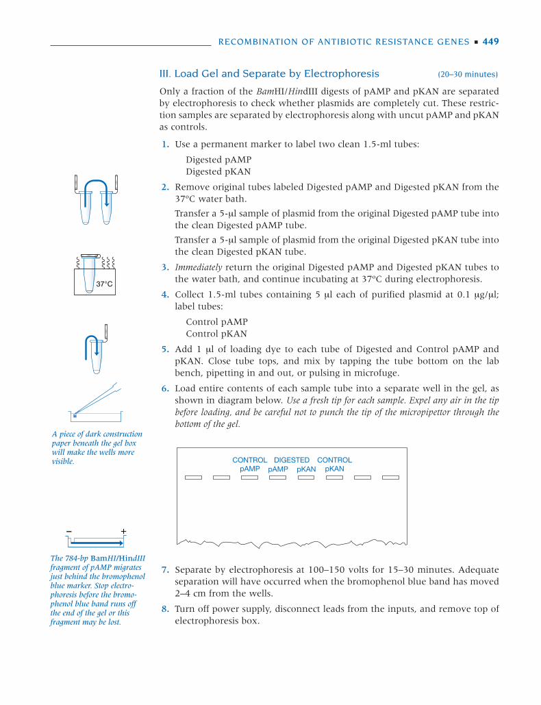
6. Load entire contents of each sample tube into a separate well in the gel, asshown in diagram below. Use a fresh tip for each sample. Expel any air in the tipbefore loading, and be careful not to punch the tip of the micropipettor through thebottom of the gel.
7. Separate by electrophoresis at 100–150 volts for 15–30 minutes. Adequateseparation will have occurred when the bromophenol blue band has moved2–4 cm from the wells.
8. Turn off power supply, disconnect leads from the inputs, and remove top ofelectrophoresis box.
RECOMBINATION OF ANTIBIOTIC RESISTANCE GENES ■ 449
CONTROLpAMP
CONTROLpKAN
DIGESTEDpAMP pKAN
The 784-bp BamHI/HindIIIfragment of pAMP migratesjust behind the bromophenolblue marker. Stop electro-phoresis before the bromo-phenol blue band runs offthe end of the gel or thisfragment may be lost.
37°C
– +
A piece of dark constructionpaper beneath the gel boxwill make the wells morevisible.
443-456 DNA Sci Lab 09 11/9/05 1:54 PM Page 449
9. Carefully remove the casting tray from the electrophoresis chamber, andslide the gel into a disposable weigh boat or other shallow tray. Label thestaining tray with your name.
10. Stain and view gel as described in Section IV.
IV. Stain Gel with Ethidium Bromide and View (Photograph) (10–15 minutes)
1. Flood the gel with ethidium bromide solution (1 μg/ml), and allow to stainfor 5–10 minutes.
2. Following staining, use a funnel to decant as much ethidium bromide solu-tion as possible from the staining tray back into storage container.
3. Rinse the gel and tray under running tap water.
4. If desired, the gel can be destained in tap water or distilled water for 5 min-utes or more to remove background ethidium bromide.
5. View under UV transilluminator or other UV source.
6. Photograph with a Polaroid or digital camera.
7. If both digests look complete, or nearly so (see Results and Discussion), con-tinue on to Part B, Ligation of pAMP and pKAN Restriction Fragments. Thereaction will have gone to completion with the additional incubation duringelectrophoresis.
8. If either or both digests look very incomplete, add another 1 μl of BamHI/HindIII solution and incubate for an additional 20 minutes. Then continue onto Part B, Ligation of pAMP and pKAN Restriction Fragments.
9. Take time for responsible cleanup.
a. Wipe down camera, transilluminator, and staining area.
b. Wash hands before leaving lab.
450 ■ LABORATORY 9
Cover electrophoresis tank and save gel until ready to continue. Gel can bestored in a zip-lock plastic bag and refrigerated overnight for viewing/pho-tographing the next day. However, over longer periods of time, the DNA will dif-fuse through the gel and the bands will become indistinct or disappear entirely.
�
Staining may be performedby an instructor in a con-trolled area when studentsare not present.
CAUTIONReview Responsible Handling of Ethidium Bromide in Laboratory 3. Wear latexgloves when staining, viewing, and photographing gels and during cleanup. Confineall staining to a restricted sink area. For further information, see Appendix 4.
Freeze BamHI/HindIII reactions at –20ºC until ready to continue. Thaw reac-tions before proceeding to Part B, Ligation of pAMP and pKAN RestrictionReactions.
�
Ethidium bromide solutionmay be reused to stain 15 ormore gels. Dispose of spentstaining solution as explainedin Laboratory 3.
CAUTIONUltraviolet light can damage eyes. Never look at unshielded UV light source withnaked eyes. View only through a filter or safety glasses that absorb harmful wave-lengths. For further information, see Appendix 4.
443-456 DNA Sci Lab 09.qxd 7/12/07 8:54 PM Page 450
RESULTS AND DISCUSSION
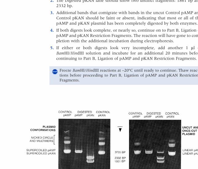
Examine the photograph of your stained gel (or view on a light box). Compareyour gel with the ideal gel shown below, and check whether both plasmids havebeen completely digested by BamHI and HindIII.
1. The Digested pAMP lane should show two distinct fragments: 784 bp and3755 bp.
2. The Digested pKAN lane should show two distinct fragments: 1861 bp and2332 bp.
3. Additional bands that comigrate with bands in the uncut Control pAMP andControl pKAN should be faint or absent, indicating that most or all of thepAMP and pKAN plasmid has been completely digested by both enzymes.
4. If both digests look complete, or nearly so, continue on to Part B, Ligation ofpAMP and pKAN Restriction Fragments. The reaction will have gone to com-pletion with the additional incubation during electrophoresis.
5. If either or both digests look very incomplete, add another 1 μl ofBamHI/HindIII solution and incubate for an additional 20 minutes beforecontinuing to Part B, Ligation of pAMP and pKAN Restriction Fragments.
RECOMBINATION OF ANTIBIOTIC RESISTANCE GENES ■ 451
Freeze BamHI/HindIII reactions at –20ºC until ready to continue. Thaw reac-tions before proceeding to Part B, Ligation of pAMP and pKAN RestrictionFragments.
�
1861
Ideal Gel Partial Digest
443-456 DNA Sci Lab 09.qxd 7/12/07 8:54 PM Page 451
452 ■ LABORATORY 9
PART B
Ligation of pAMP and pKAN Restriction Fragments
DigestedpAMP
ADD Ligation
INCUBATEat roomtemperature2–24 hours
MIX
DigestedpKAN
Digested pAMPDigested pKAN
Lig Buf/ATPH2O
Ligase
HEATboth tubes
65°C
Ligation
443-456 DNA Sci Lab 09 11/9/05 1:54 PM Page 452
PRELAB NOTES
DNA Ligase
Use only T4 DNA ligase. E. coli DNA ligase requires different reaction conditionsand cannot be substituted in this experiment. Cohesive-end units are used to cali-brate ligase activity: One unit of enzyme ligates 50% of HindIII fragments of λDNA (6 µg in 20 µl) in 30 minutes at 16ºC. This unit is used by New EnglandBiolabs (NEB) and Carolina Biological Supply Company (CBS).
Researchers typically incubate ligation reactions overnight at room temper-ature. For brief ligations, down to a minimum of 1 hour, it is essential to choose a high-concentration T4 DNA ligase with at least 100–500 cohesive-end units/µl.
For Further Information
The protocol presented here is based on the following published method:
Cohen S.N., Chang A.C.Y., Boyer H.W., and Helling R.B. 1973. Construction of biologi-cally functional bacteria plasmids in vitro. Proc. Natl. Acad. Sci. 70: 3240–3244.
PRELAB PREPARATION
1. Obtain fresh 2x ligation buffer/ATP solution. ATP is somewhat unstable insolution, so do not use very old buffer/ATP and take care to keep frozenwhen not in use.
2. T4 DNA ligase is critical to the experiment and rather expensive. Make onealiquot of ligase sufficient for all experiments, and hold on ice during the lab-oratory. We suggest that the instructor dispense ligase directly into eachexperimenter’s reaction tube.
3. Prewarm water bath to 65ºC.
4. Dispose of 2x restriction buffer from Part A, Restriction Digest of PlasmidspAMP and pKAN, to avoid mistaking it for 2x ligation buffer/ATP.
MATERIALS
RECOMBINATION OF ANTIBIOTIC RESISTANCE GENES ■ 453
REAGENTS
Digested pAMP (from part A)
Digested pKAN (from part A)
Distilled water
2x Ligation buffer/ATP
T4 DNA ligase
SUPPLIES AND EQUIPMENT
Beaker for waste/used tips
Microfuge (optional)
Micropipettor (0.5–10-µl) + tips
Test tube rack
Tube (1.5-ml)
Water bath (65ºC)
443-456 DNA Sci Lab 09 11/9/05 1:54 PM Page 453
METHODS
Ligate pAMP and pKAN (30 minutes, including incubation)
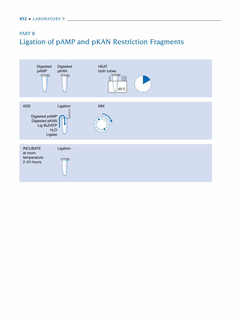
1. Incubate the tubes labeled Digested pAMP and Digested pKAN (from Part A)in a 65ºC water bath for 10 minutes.
2. Label a clean 1.5-ml tube LIG (for ligation).
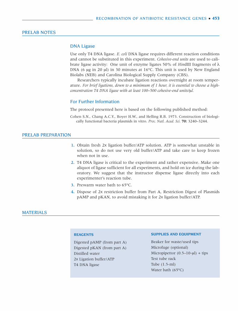
3. Use the matrix below as a checklist while adding reagents to the LIG tube.Use a fresh tip for each reagent. Refer to detailed directions that follow.
Digested Digested 2x LigationTube pAMP pKAN Buffer/ATP Water Ligase
LIG 3 μl 3 μl 10 μl 3 μl 1 μl
4. Collect reagents (except ligase), and place them in test tube rack on lab bench.
5. Add 3 μl of Digested pAMP.
6. Use a fresh tip to add 3 μl of Digested pKAN.
7. Use a fresh tip to add 10 μl of 2x ligation buffer/ATP.
8. Use a fresh tip to add 3 μl of distilled water.
9. Use a fresh tip to add 1 μl of DNA ligase. Carefully check that the droplet ofligase is on the inside wall of the tube.
10. Close tube top. Pool and mix reagents by pulsing in a microfuge or by sharplytapping the tube bottom on the lab bench.
11. Incubate the reaction for 2–24 hours at room temperature.
12. If time permits, ligation may be confirmed by electrophoresing 5 μl of the lig-ation reaction, along with BamHI/HindIII digests of pAMP and pKAN. Noneof the parent BamHI/HindIII fragments should be observed in the lane of lig-ated DNA, which should show multiple bands of high-molecular-weightDNA high up on the gel.
RESULTS AND DISCUSSION
Ligation of the four BamHI/HindIII restriction fragments of pAMP and pKAN(refer to diagram on page 444) produces many types of hybrid molecules,including plasmids composed of more than two fragments. However, only thoseconstructs possessing an origin of replication will be maintained and expressed.Three different replicating plasmids, with selectable antibiotic resistance, are cre-ated by ligating combinations of two BamHI/HindIII fragments:
• Ligation of the 784-bp fragment to the 3755-bp fragment regenerates pAMP.
• Ligation of the 1861-bp fragment to the 2332-bp fragment regenerates pKAN.
• Ligation of the 1861-bp fragment to the 3755-bp fragment produces the “sim-ple recombinant” plasmid, pAMP/KAN, in which the kanamycin resistancegene has been fused into the pAMP backbone.
The ligation products will be tested in Laboratory 10.
454 ■ LABORATORY 9
Step 1 is critical. Heat dena-tures protein, thus inactivat-ing the restriction enzymes.
Ligase may be dispensed byyour instructor.
For brief ligations of 2–4hours, it is essential to use ahigh-concentration T4 DNAligase with at least 100–500cohesive-end units/μl.
Freeze the reaction at –20ºC until ready to continue. Thaw the reactionbefore proceeding to Laboratory 10.
�
65°C
443-456 DNA Sci Lab 09.qxd 7/12/07 8:54 PM Page 454
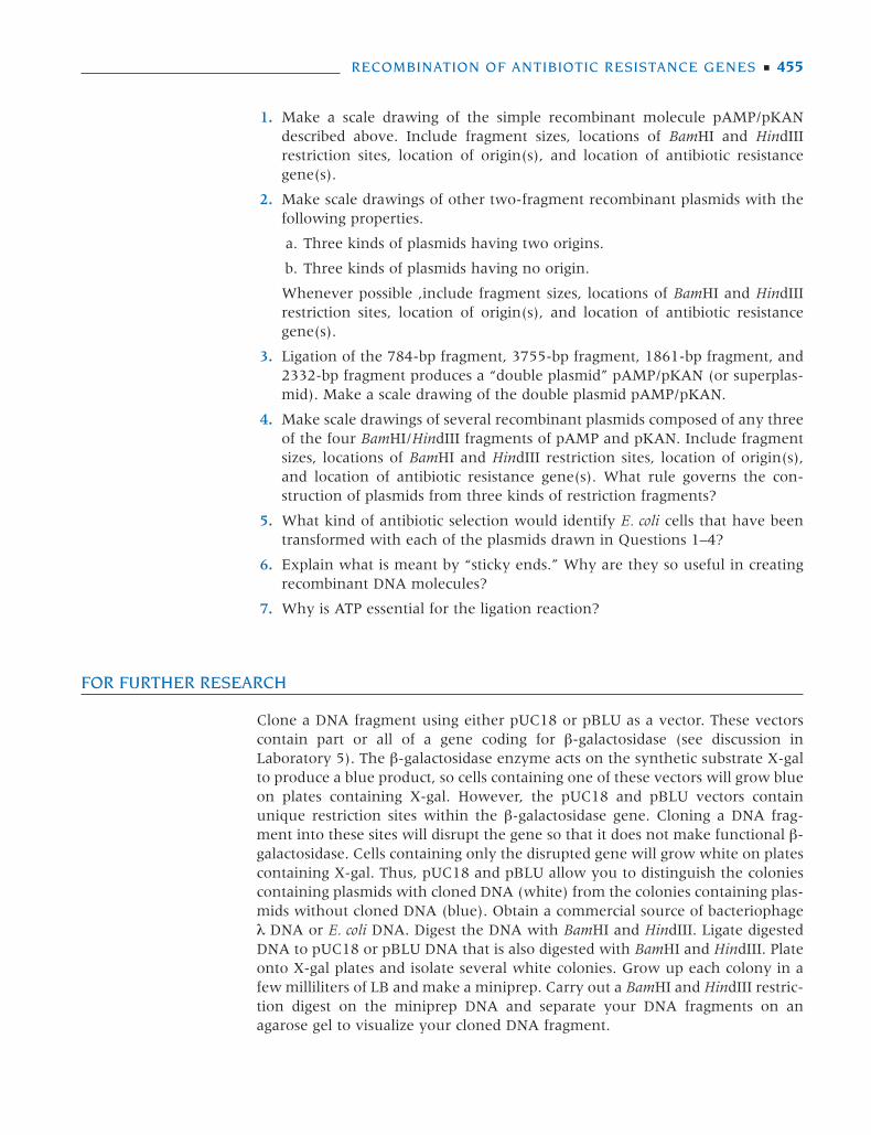
1. Make a scale drawing of the simple recombinant molecule pAMP/pKANdescribed above. Include fragment sizes, locations of BamHI and HindIIIrestriction sites, location of origin(s), and location of antibiotic resistancegene(s).
2. Make scale drawings of other two-fragment recombinant plasmids with thefollowing properties.
a. Three kinds of plasmids having two origins.
b. Three kinds of plasmids having no origin.
Whenever possible ,include fragment sizes, locations of BamHI and HindIIIrestriction sites, location of origin(s), and location of antibiotic resistancegene(s).
3. Ligation of the 784-bp fragment, 3755-bp fragment, 1861-bp fragment, and2332-bp fragment produces a “double plasmid” pAMP/pKAN (or superplas-mid). Make a scale drawing of the double plasmid pAMP/pKAN.
4. Make scale drawings of several recombinant plasmids composed of any threeof the four BamHI/HindIII fragments of pAMP and pKAN. Include fragmentsizes, locations of BamHI and HindIII restriction sites, location of origin(s),and location of antibiotic resistance gene(s). What rule governs the con-struction of plasmids from three kinds of restriction fragments?
5. What kind of antibiotic selection would identify E. coli cells that have beentransformed with each of the plasmids drawn in Questions 1–4?
6. Explain what is meant by “sticky ends.” Why are they so useful in creatingrecombinant DNA molecules?
7. Why is ATP essential for the ligation reaction?
FOR FURTHER RESEARCH
Clone a DNA fragment using either pUC18 or pBLU as a vector. These vectorscontain part or all of a gene coding for β-galactosidase (see discussion inLaboratory 5). The β-galactosidase enzyme acts on the synthetic substrate X-galto produce a blue product, so cells containing one of these vectors will grow blueon plates containing X-gal. However, the pUC18 and pBLU vectors containunique restriction sites within the β-galactosidase gene. Cloning a DNA frag-ment into these sites will disrupt the gene so that it does not make functional β-galactosidase. Cells containing only the disrupted gene will grow white on platescontaining X-gal. Thus, pUC18 and pBLU allow you to distinguish the coloniescontaining plasmids with cloned DNA (white) from the colonies containing plas-mids without cloned DNA (blue). Obtain a commercial source of bacteriophageλ DNA or E. coli DNA. Digest the DNA with BamHI and HindIII. Ligate digestedDNA to pUC18 or pBLU DNA that is also digested with BamHI and HindIII. Plateonto X-gal plates and isolate several white colonies. Grow up each colony in afew milliliters of LB and make a miniprep. Carry out a BamHI and HindIII restric-tion digest on the miniprep DNA and separate your DNA fragments on anagarose gel to visualize your cloned DNA fragment.
RECOMBINATION OF ANTIBIOTIC RESISTANCE GENES ■ 455
443-456 DNA Sci Lab 09 11/9/05 1:54 PM Page 455

443-456 DNA Sci Lab 09 11/9/05 1:54 PM Page 456
457
L A B O R A T O R Y 10
Transformation of E. coli withRecombinant DNA
IN PART A, CLASSIC PROCEDURE FOR PREPARING COMPETENT CELLS, E. coli cells arerendered competent to uptake plasmid DNA using a method essentiallyunchanged since its publication in 1970 by Morton Mandel and Akiko Higa. Theprocedure begins with vigorous E. coli cells grown in suspension culture. Cellsare harvested in mid-log phase by centrifugation and incubated at 0ºC with twosuccessive changes of calcium chloride solution.
This procedure is more involved than the rapid colony protocol introducedin Laboratory 5. However, the classical procedure typically achieves transforma-tion efficiencies ranging from 5 x 10⁴ to 5 x 10⁶ colonies per microgram of plas-mid—a 2–200-fold increase over the colony procedure. The enhanced efficien-cy is important when transforming ligated DNA (composed primarily of relaxedcircular plasmids and linear DNA), which produces 5–100 times fewer transfor-mants than plasmid DNA purified from E. coli (containing a high proportion ofthe supercoiled form).
In Part B, Transformation of E. coli with Recombinant DNA, the competentE. coli cells are transformed with the ligation products from Laboratory 9,Recombination of Antibiotic Resistance Genes. Ligated plasmid DNA is added toone sample of competent cells, and purified pAMP and pKAN plasmids areadded as controls to two other samples. The cell suspensions are incubated withthe plasmid DNAs for 20 minutes at 0ºC. Following a brief heat shock at 42ºC,the cells recover in LB broth for 40–60 minutes at 37ºC. Unlike ampicillin selec-tion in Laboratory 5, Rapid Colony Transformation of E. coli with Plasmid DNA,the recovery step is essential for the kanamycin selection in this lab. Samples oftransformed cells are plated onto three types of LB agar: with ampicillin(LB/amp), with kanamycin (LB/kan), and with both ampicillin and kanamycin(LB/amp+kan).
The ligation reaction produces many kinds of recombinant molecules com-posed of BamHI/HindIII fragments, including the religated parental plasmidspAMP and pKAN. The object is to select for transformed cells with dual antibiot-ic resistance, which must contain a 3755-bp fragment from pAMP containing theampicillin resistance gene (plus the origin of replication) and a 1861-bp fragmentfrom pKAN containing the kanamycin resistance gene. Bacteria transformedwith a single plasmid containing these sequences, or those doubly transformedwith both pAMP and pKAN plasmids, form colonies on the LB/amp+kan plate.
Equipment and materials for this laboratory are available from the CarolinaBiological Supply Company (see Appendix 1).
457-472 DNA Sci Lab 10 11/9/05 1:56 PM Page 457
458 ■ LABORATORY 10
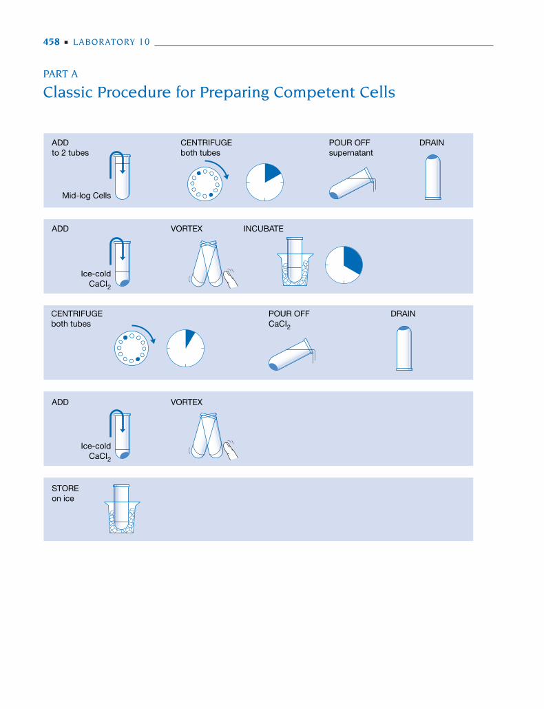
PART A
Classic Procedure for Preparing Competent Cells
ADDto 2 tubes
CENTRIFUGEboth tubes
POUR OFFsupernatant
DRAIN
Mid-log Cells
ADD
ADD
STOREon ice
VORTEX INCUBATE
DRAINCENTRIFUGEboth tubes
VORTEX
POUR OFFCaCI2
Ice-coldCaCI2
Ice-coldCaCI2
457-472 DNA Sci Lab 10 11/9/05 1:56 PM Page 458
PRELAB NOTES
Review Prelab Notes in Laboratories 1, 2, and 5 regarding sterile techniques, E.coli culture, and transformation.
Seasoning Cells for Transformation
If possible, schedule experiments so that competent cells (Part A) are preparedone day prior to transformation with recombinant DNA (Part B). “Seasoning”cells for 12–24 hours at 0ºC (an ice bath inside the refrigerator) generallyincreases transformation efficiency five- to tenfold. This enhanced efficiency willhelp ensure successful cloning of the recombinant molecules produced inLaboratory 9.
For Further Information
The protocol presented here is based on the following published methods:
Cohen S.N., Chang A.C., and Hsu L. 1972. Nonchromosomal antibiotic resistance in bac-teria: Genetic transformation of Escherichia coli by R-factor DNA. Proc. Natl. Acad. Sci.69: 2110–2114.
Dagert M. and Ehrlich S.D. 1979. Prolonged incubation in calcium chloride improves thecompetence of Escherichia coli cells. Gene 6: 23.
Mandel M. and Higa A. 1970. Calcium-dependent bacteriophage DNA infection. J. Mol.Biol. 53: 159–162.
PRELAB PREPARATION
Before performing this Prelab Preparation, please refer to the cautions indicatedon the Laboratory Materials list.
1. On the day before this lab, begin an E. coli culture from a streaked plate ofMM294, according to the protocol in Laboratory 2B, Overnight SuspensionCulture.
2. PLAN AHEAD. Make sure that you have a streaked plate of viable E. coli cellsfrom which to inoculate overnight. Also, streak your E. coli strain on LB/ampand LB/kan plates to ensure that a resistant strain has not been used by mis-take.
3. Approximately 2–4 hours before the lab, begin an E. coli culture according tothe protocol in Laboratory 2C, Mid-log Suspension Culture. Cells are optimalfor transformation when the culture reaches an OD
550of 0.3–0.40. More sim-
ply, cells inoculated into room temperature LB will be ready to transformafter 2 hours, 15 minutes. Hold cells in mid-log phase by storing the cultureon ice for up to 2 hours prior to beginning calcium chloride treatment. Eachexperiment requires 20 ml of mid-log suspension culture.
4. Sterilize 50 mM calcium chloride (CaCl2) solution and LB broth by autoclav-
ing or filtering through a 0.45-µm or 0.22-µm filter (Nalgene or Corning).Filtered CaCl
2can be stored in a filter collection container or transferred to
sterile 50-ml conical tubes.
TRANSFORMATION OF E. COLI WITH RECOMBINANT DNA ■ 459
457-472 DNA Sci Lab 10 11/9/05 1:56 PM Page 459
5. Prepare aliquots for each experiment:
15 ml of 50 mM CaCl2
in a sterile 50-ml tube (store on ice)two 10-ml cultures of mid-log E. coli cells in sterile 15-ml culture tubes
(store on ice)12 µl of 0.005 µg/µl pAMP in a 1.5-ml tube (store on ice)12 µl of 0.005 µg/µl pKAN in a 1.5-ml tube (store on ice)
6. Review Part B, Transformation of E. coli with Recombinant DNA.
MATERIALS
METHODS
Prepare Competent Cells (40–50 minutes)
This entire experiment must be performed under sterile conditions. Review steriletechniques in Laboratory 1, Measurements, Micropipetting, and SterileTechniques.
1. Place sterile tube of CaCl2
solution on ice.
2. Obtain two 15-ml tubes each with 10 ml of mid-log cells, and label with yourname.
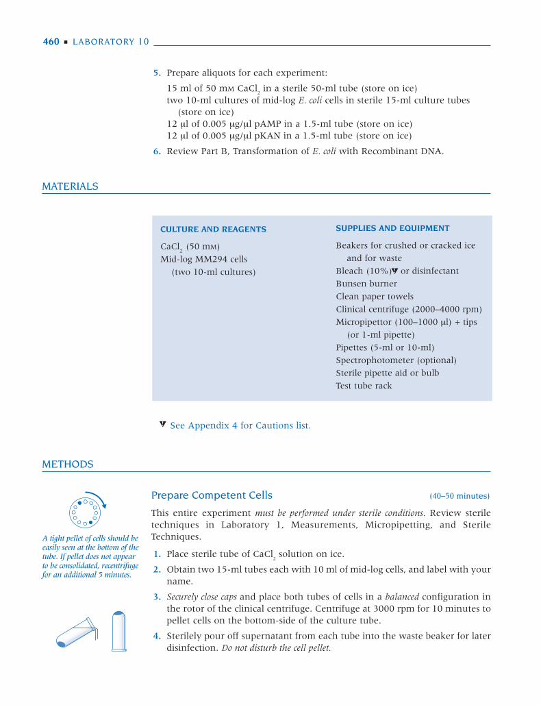
3. Securely close caps and place both tubes of cells in a balanced configuration inthe rotor of the clinical centrifuge. Centrifuge at 3000 rpm for 10 minutes topellet cells on the bottom-side of the culture tube.
4. Sterilely pour off supernatant from each tube into the waste beaker for laterdisinfection. Do not disturb the cell pellet.
460 ■ LABORATORY 10
CULTURE AND REAGENTS
CaCl2
(50 mM)
Mid-log MM294 cells
(two 10-ml cultures)
SUPPLIES AND EQUIPMENT
Beakers for crushed or cracked ice
and for waste
Bleach (10%) or disinfectant
Bunsen burner
Clean paper towels
Clinical centrifuge (2000–4000 rpm)
Micropipettor (100–1000 µl) + tips
(or 1-ml pipette)
Pipettes (5-ml or 10-ml)
Spectrophotometer (optional)
Sterile pipette aid or bulb
Test tube rack
See Appendix 4 for Cautions list.
A tight pellet of cells should beeasily seen at the bottom of thetube. If pellet does not appearto be consolidated, recentrifugefor an additional 5 minutes.
457-472 DNA Sci Lab 10 11/9/05 1:56 PM Page 460
a. Remove cap from the culture tube, and briefly flame mouth. Do not placecap on lab bench.
b. Carefully pour off supernatant. Invert culture tube, and tap gently on thesurface of a clean paper towel to drain thoroughly.
c. Reflame mouth of culture tube, and replace cap.
5. Use a 5- or 10-ml pipette to sterilely add 5 ml of ice-cold CaCl2
solution toeach culture tube:
a. Remove cap from CaCl2
tube. Do not place cap on lab bench.
b. Withdraw 5 ml of CaCl2
and replace cap.
c. Remove cap of the culture tube. Do not place cap on lab bench.
d. Expel CaCl2
into culture tube and replace cap.
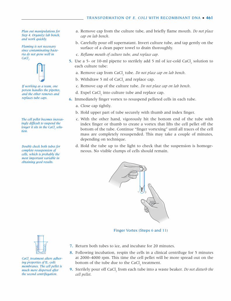
6. Immediately finger vortex to resuspend pelleted cells in each tube.
a. Close cap tightly.
b. Hold upper part of tube securely with thumb and index finger.
c. With the other hand, vigorously hit the bottom end of the tube withindex finger or thumb to create a vortex that lifts the cell pellet off thebottom of the tube. Continue “finger vortexing” until all traces of the cellmass are completely resuspended. This may take a couple of minutes,depending on technique.
d. Hold the tube up to the light to check that the suspension is homoge-neous. No visible clumps of cells should remain.
TRANSFORMATION OF E. COLI WITH RECOMBINANT DNA ■ 461
Plan out manipulations forStep 4. Organize lab bench,and work quickly.
Flaming is not necessarysince contaminating bacte-ria do not grow well inCaCl
2.
If working as a team, oneperson handles the pipettes,and the other removes andreplaces tube caps.
The cell pellet becomes increas-ingly difficult to suspend thelonger it sits in the CaCl
2solu-
tion.
Double check both tubes forcomplete resuspension ofcells, which is probably themost important variable inobtaining good results.
Finger Vortex (Steps 6 and 11)
7. Return both tubes to ice, and incubate for 20 minutes.
8. Following incubation, respin the cells in a clinical centrifuge for 5 minutesat 2000–4000 rpm. This time the cell pellet will be more spread out on thebottom of the tube due to the CaCl
2treatment.
9. Sterilely pour off CaCl2from each tube into a waste beaker. Do not disturb the
cell pellet.
CaCl2
treatment alters adher-ing properties of E. colimembranes. The cell pellet ismuch more dispersed afterthe second centrifugation.
457-472 DNA Sci Lab 10 11/9/05 1:56 PM Page 461
a. Remove the cap from the culture tube. Do not place the cap on the labbench.
b. Carefully pour off supernatant. Invert the culture tube, and tap gently onthe surface of a clean paper towel to drain thoroughly.
c. Replace cap.
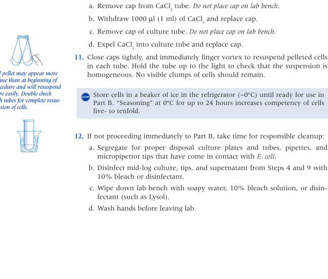
10. Use a 100–1000-μl micropipettor (or 1-ml pipette) to sterilely add 1000 μl(1 ml) of fresh, ice-cold CaCl
2to each tube.
a. Remove cap from CaCl2
tube. Do not place cap on lab bench.
b. Withdraw 1000 μl (1 ml) of CaCl2
and replace cap.
c. Remove cap of culture tube. Do not place cap on lab bench.
d. Expel CaCl2
into culture tube and replace cap.
11. Close caps tightly, and immediately finger vortex to resuspend pelleted cellsin each tube. Hold the tube up to the light to check that the suspension ishomogeneous. No visible clumps of cells should remain.
12. If not proceeding immediately to Part B, take time for responsible cleanup:
a. Segregate for proper disposal culture plates and tubes, pipettes, andmicropipettor tips that have come in contact with E. coli.
b. Disinfect mid-log culture, tips, and supernatant from Steps 4 and 9 with10% bleach or disinfectant.
c. Wipe down lab bench with soapy water, 10% bleach solution, or disin-fectant (such as Lysol).
d. Wash hands before leaving lab.
462 ■ LABORATORY 10
Cell pellet may appear morediffuse than at beginning ofprocedure and will resuspendmore easily. Double checkboth tubes for complete resus-pension of cells.
Store cells in a beaker of ice in the refrigerator (~0ºC) until ready for use inPart B. “Seasoning” at 0ºC for up to 24 hours increases competency of cellsfive- to tenfold.
�
457-472 DNA Sci Lab 10.qxd 7/12/07 8:57 PM Page 462

TRANSFORMATION OF E. COLI WITH RECOMBINANT DNA ■ 463
PART B
Transformation of E. coli with Recombinant DNA
ADD PLACE ON ICEall tubes
Competentcells
+ pLIG
ADD andMIX
HEAT SHOCKall tubes90 seconds
INCUBATEall tubes
ADDto all tubes
SPREAD9 plates
INCUBATEplates15–20 hours
Competentcells
+ pAMP
Competentcells
+ pKAN
LigatedpAMP/pKAN
+ pLIG
LB
pAMP
+ pAMP
pKAN
+ pKAN INCUBATEall tubes
INCUBATEall tubes
42°C
37°C
37°C
457-472 DNA Sci Lab 10 11/9/05 1:56 PM Page 463
PRELAB NOTES
Review Prelab Notes in Laboratory 5, Rapid Colony Transformation of E. coliwith Plasmid DNA.
Equipment Substitutions
A standard 1-ml pipette or transfer pipette can be substituted for a 100–1000-µlmicropipettor.
Recovery Period
A 40–60-minute postincubation recovery at 37ºC, with shaking, is essential priorto plating transformed cells on kanamycin, which acts quickly to kill any cellthat is not actively expressing the resistance protein.
For Further Information
The protocol presented here is based on the following published method:
Cohen S.N., Chang A.C.Y., Boyer H.W., and Helling R.B. 1973. Construction of biologi-cally functional bacteria plasmids in vitro. Proc. Natl. Acad. Sci. 70: 3240–3244.
PRELAB PREPARATION
Before performing this Prelab Preparation, please refer to the cautions indicatedon the Laboratory Materials list.
1. Prepare for each experiment:
three LB+ampicillin plates (labeled LB/amp)three LB+kanamycin plates (labeled LB/kan)three LB+ampicillin+kanamycin plates (labeled LB/amp+kan)
If only one control transformation, with either pAMP or pKAN, is done, thenone less plate of each type is required.
2. Adjust water baths to 42ºC and 37ºC.
3. Prewarm incubator to 37ºC.
4. To retard evaporation, keep beaker of ethanol covered with Parafilm, plasticwrap, or, if using a small beaker, the lid from a Petri dish. Retrieve and reuseethanol exclusively for flaming.
5. If using spreading beads, carefully place five to seven beads into a sterile 1.5-ml tube. Tube can be used as scooper. Prepare nine tubes per experiment.
464 ■ LABORATORY 10
457-472 DNA Sci Lab 10 11/9/05 1:56 PM Page 464
MATERIALS
METHODS
Perform E. coli Transformation (70–90 minutes)
This entire experiment must be performed under sterile conditions. Review ster-ile techniques in Laboratory 1, Measurements, Micropipetting, and SterileTechniques.
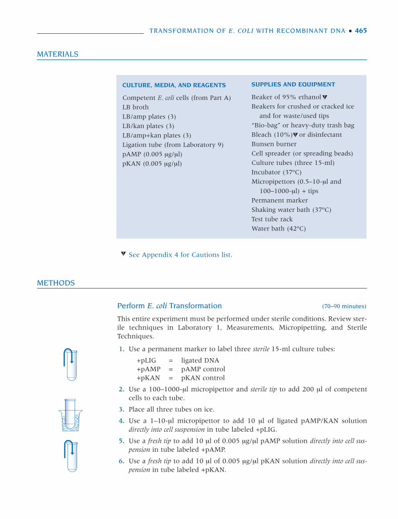
1. Use a permanent marker to label three sterile 15-ml culture tubes:
+pLIG = ligated DNA+pAMP = pAMP control+pKAN = pKAN control
2. Use a 100–1000-µl micropipettor and sterile tip to add 200 µl of competentcells to each tube.
3. Place all three tubes on ice.
4. Use a 1–10-µl micropipettor to add 10 µl of ligated pAMP/KAN solutiondirectly into cell suspension in tube labeled +pLIG.
5. Use a fresh tip to add 10 µl of 0.005 µg/µl pAMP solution directly into cell sus-pension in tube labeled +pAMP.
6. Use a fresh tip to add 10 µl of 0.005 µg/µl pKAN solution directly into cell sus-pension in tube labeled +pKAN.
TRANSFORMATION OF E. COLI WITH RECOMBINANT DNA ■ 465
CULTURE, MEDIA, AND REAGENTS
Competent E. coli cells (from Part A)
LB broth
LB/amp plates (3)
LB/kan plates (3)
LB/amp+kan plates (3)
Ligation tube (from Laboratory 9)
pAMP (0.005 µg/µl)
pKAN (0.005 µg/µl)
SUPPLIES AND EQUIPMENT
Beaker of 95% ethanol
Beakers for crushed or cracked ice
and for waste/used tips
“Bio-bag” or heavy-duty trash bag
Bleach (10%) or disinfectant
Bunsen burner
Cell spreader (or spreading beads)
Culture tubes (three 15-ml)
Incubator (37ºC)
Micropipettors (0.5–10-µl and
100–1000-µl) + tips
Permanent marker
Shaking water bath (37ºC)
Test tube rack
Water bath (42ºC)
See Appendix 4 for Cautions list.
457-472 DNA Sci Lab 10 11/9/05 1:56 PM Page 465
7. Close caps, and tap tubes with finger to mix. Avoid making bubbles in sus-pension or splashing suspension up the sides of the tubes.
8. Return all three tubes to ice for 20 minutes.
9. While cells are incubating on ice, use a permanent marker to label all nineLB agar plates with your name and the date. Divide plates into three sets ofthree plates each and mark as follows:
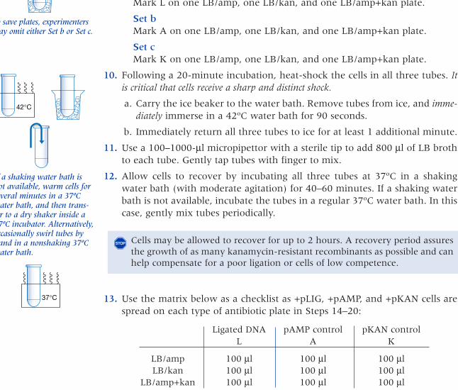
Set aMark L on one LB/amp, one LB/kan, and one LB/amp+kan plate.
Set bMark A on one LB/amp, one LB/kan, and one LB/amp+kan plate.
Set cMark K on one LB/amp, one LB/kan, and one LB/amp+kan plate.
10. Following a 20-minute incubation, heat-shock the cells in all three tubes. Itis critical that cells receive a sharp and distinct shock.
a. Carry the ice beaker to the water bath. Remove tubes from ice, and imme-diately immerse in a 42ºC water bath for 90 seconds.
b. Immediately return all three tubes to ice for at least 1 additional minute.
11. Use a 100–1000-μl micropipettor with a sterile tip to add 800 μl of LB brothto each tube. Gently tap tubes with finger to mix.
12. Allow cells to recover by incubating all three tubes at 37ºC in a shakingwater bath (with moderate agitation) for 40–60 minutes. If a shaking waterbath is not available, incubate the tubes in a regular 37ºC water bath. In thiscase, gently mix tubes periodically.
13. Use the matrix below as a checklist as +pLIG, +pAMP, and +pKAN cells arespread on each type of antibiotic plate in Steps 14–20:
Ligated DNA pAMP control pKAN controlL A K
LB/amp 100 μl 100 μl 100 μlLB/kan 100 μl 100 μl 100 μl
LB/amp+kan 100 μl 100 μl 100 μl
14. Use a micropipettor with a sterile tip to add 100 μl of cell suspension fromthe tube labeled pLIG onto three plates marked L. Do not allow suspensions tosit on plates too long before proceeding to Step 15 or 16. Spread cells using one ofthe methods described in Steps 15 and 16.
15. Sterilize the cell spreader, and spread cells over the surface of each L plate insuccession.
466 ■ LABORATORY 10
Store remainder of ligatedDNA at 4ºC. Separate by elec-trophoresis with cut pAMPand pKAN controls in Labor-atory 12 to observe the prod-ucts of the ligation reaction.
To save plates, experimentersmay omit either Set b or Set c.
If a shaking water bath isnot available, warm cells forseveral minutes in a 37ºCwater bath, and then trans-fer to a dry shaker inside a37ºC incubator. Alternatively,occasionally swirl tubes byhand in a nonshaking 37ºCwater bath.
Cells may be allowed to recover for up to 2 hours. A recovery period assuresthe growth of as many kanamycin-resistant recombinants as possible and canhelp compensate for a poor ligation or cells of low competence.
�
If too much liquid isabsorbed by agar, cells willnot be evenly distributed.
42°C
37°C
457-472 DNA Sci Lab 10.qxd 7/12/07 8:57 PM Page 466
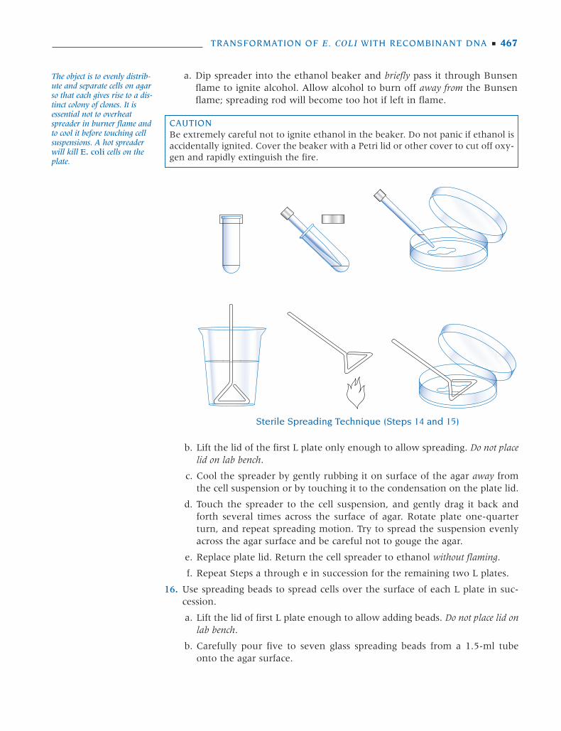
a. Dip spreader into the ethanol beaker and briefly pass it through Bunsenflame to ignite alcohol. Allow alcohol to burn off away from the Bunsenflame; spreading rod will become too hot if left in flame.
TRANSFORMATION OF E. COLI WITH RECOMBINANT DNA ■ 467
The object is to evenly distrib-ute and separate cells on agarso that each gives rise to a dis-tinct colony of clones. It isessential not to overheatspreader in burner flame andto cool it before touching cellsuspensions. A hot spreaderwill kill E. coli cells on theplate.
CAUTIONBe extremely careful not to ignite ethanol in the beaker. Do not panic if ethanol isaccidentally ignited. Cover the beaker with a Petri lid or other cover to cut off oxy-gen and rapidly extinguish the fire.
Sterile Spreading Technique (Steps 14 and 15)
b. Lift the lid of the first L plate only enough to allow spreading. Do not placelid on lab bench.
c. Cool the spreader by gently rubbing it on surface of the agar away fromthe cell suspension or by touching it to the condensation on the plate lid.
d. Touch the spreader to the cell suspension, and gently drag it back andforth several times across the surface of agar. Rotate plate one-quarterturn, and repeat spreading motion. Try to spread the suspension evenlyacross the agar surface and be careful not to gouge the agar.
e. Replace plate lid. Return the cell spreader to ethanol without flaming.
f. Repeat Steps a through e in succession for the remaining two L plates.
16. Use spreading beads to spread cells over the surface of each L plate in suc-cession.
a. Lift the lid of first L plate enough to allow adding beads. Do not place lid onlab bench.
b. Carefully pour five to seven glass spreading beads from a 1.5-ml tubeonto the agar surface.
457-472 DNA Sci Lab 10 11/9/05 1:56 PM Page 467
c. Close plate lid and use a swirling motion to move glass beads around theentire surface of the plate. This evenly spreads the cell suspension on theagar suface. Continue swirling until the cell suspension is absorbed intothe agar.
d. Repeat Steps a through c in succession for the remaining two L plates.
17. Use a fresh sterile tip to add 100 µl of cell suspension from tube labeled +pAMPonto three plates marked A.
18. Repeat Step 15a–f or Step 16a–d to spread cells over the surface of each Aplate in succession.
19. Use a fresh sterile tip to add 100 µl of cell suspension from tube labeled +pKANonto three plates marked K.
20. Repeat Step 15a–f or Step 16a–d to sterilize cell spreader and spread cellsover the surface of each K plate in succession.
21. If Step 15 was used, reflame spreader one last time before placing it on labbench.
22. Let plates set for several minutes to allow suspension to become absorbedinto agar. If Step 16 was used, invert plates and gently tap plate bottoms, sothat the spreading beads fall into plate lids. Carefully pour beads from eachlid into a storage container for reuse.
23. Stack plates and tape them in a bundle to keep the experiment together.Place plates upside down in a 37ºC incubator, and incubate for 15–20 hours.
24. After initial incubation, store plates at 4ºC to arrest E. coli growth and to slowthe growth of any contaminating microbes.
25. Take time for responsible cleanup:
a. Segregate for proper disposal culture plates and tubes, pipettes, andmicropipettor tips that have come in contact with E. coli.
b. Disinfect overnight cell suspensions, tubes, and tips with 10% bleach ordisinfectant.
c. Wipe down lab bench with soapy water, 10% bleach solution, or disin-fectant (such as Lysol).
d. Wash hands before leaving lab.
RESULTS AND DISCUSSION
Observe colonies through the bottom of the culture plate, using a permanentmarker to mark each colony as it is counted. If the experiment worked well,5–50 colonies should be observed on the L LB/amp+kan experimental plate,500–5000 colonies on the A LB/amp control plate, and 200–2000 colonies onthe K LB/kan control plate. (If plates are very crowded, draw lines on the bot-tom of the plate to divide it into equal-sized sections. Count one sector estimat-ed as being representative of the whole plate. After counting, multiply by thenumber of sectors.) Approximately ten times fewer colonies should be observedon the corresponding L LB/amp plate and L LB/kan plate. An extended recov-ery period would inflate these numbers. (Question 3 explains how to compute
468 ■ LABORATORY 10
Save L LB/amp and L LB/kan plates if planning to doLaboratory 11. Save L LB/amp+kan as a source ofcolonies to begin overnightsuspension cultures if plan-ning to do Laboratory 12.
37°C
457-472 DNA Sci Lab 10 11/9/05 1:56 PM Page 468
transformation efficiency.) If plates have been overincubated or left at roomtemperature for several days, “satellite” colonies may be observed on the LB/amp plates. Satellite colonies are never observed on the LB/kan or LB/amp+kanplates.
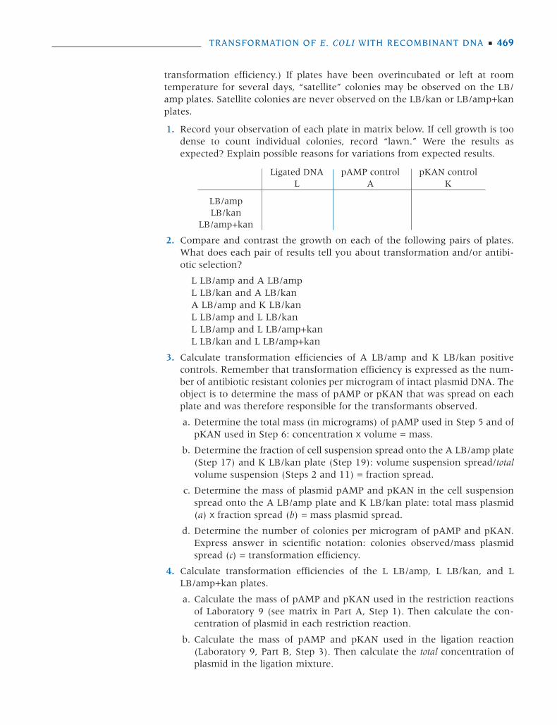
1. Record your observation of each plate in matrix below. If cell growth is toodense to count individual colonies, record “lawn.” Were the results asexpected? Explain possible reasons for variations from expected results.
Ligated DNA pAMP control pKAN controlL A K
LB/ampLB/kan
LB/amp+kan
2. Compare and contrast the growth on each of the following pairs of plates.What does each pair of results tell you about transformation and/or antibi-otic selection?
L LB/amp and A LB/ampL LB/kan and A LB/kanA LB/amp and K LB/kanL LB/amp and L LB/kanL LB/amp and L LB/amp+kanL LB/kan and L LB/amp+kan
3. Calculate transformation efficiencies of A LB/amp and K LB/kan positivecontrols. Remember that transformation efficiency is expressed as the num-ber of antibiotic resistant colonies per microgram of intact plasmid DNA. Theobject is to determine the mass of pAMP or pKAN that was spread on eachplate and was therefore responsible for the transformants observed.
a. Determine the total mass (in micrograms) of pAMP used in Step 5 and ofpKAN used in Step 6: concentration x volume = mass.
b. Determine the fraction of cell suspension spread onto the A LB/amp plate(Step 17) and K LB/kan plate (Step 19): volume suspension spread/totalvolume suspension (Steps 2 and 11) = fraction spread.
c. Determine the mass of plasmid pAMP and pKAN in the cell suspensionspread onto the A LB/amp plate and K LB/kan plate: total mass plasmid(a) x fraction spread (b) = mass plasmid spread.
d. Determine the number of colonies per microgram of pAMP and pKAN.Express answer in scientific notation: colonies observed/mass plasmidspread (c) = transformation efficiency.
4. Calculate transformation efficiencies of the L LB/amp, L LB/kan, and LLB/amp+kan plates.
a. Calculate the mass of pAMP and pKAN used in the restriction reactionsof Laboratory 9 (see matrix in Part A, Step 1). Then calculate the con-centration of plasmid in each restriction reaction.
b. Calculate the mass of pAMP and pKAN used in the ligation reaction(Laboratory 9, Part B, Step 3). Then calculate the total concentration ofplasmid in the ligation mixture.
TRANSFORMATION OF E. COLI WITH RECOMBINANT DNA ■ 469
457-472 DNA Sci Lab 10 11/9/05 1:56 PM Page 469
c. Use this concentration in calculations following Steps a–d of Question 3above.
5. Compare the transformation efficiencies that you calculated for the ALB/amp plate in this laboratory and the +pAMP plate in Laboratory 5. Bywhat factor is the classical procedure more or less efficient than colony trans-formation? What differences in the protocols contribute to the increase inefficiency?
6. Compare the transformation efficiencies that you calculated for controlpAMP and pKAN versus the ligated pAMP and pKAN. How can you accountfor the differences in efficiency? Take into account the formal definition oftransformation efficiency.
FOR FURTHER RESEARCH
Interpretable experimental results can only be achieved when the classic trans-formation protocol can be repeated with reproducible results. Only attempt theexperiments below when you are able to routinely achieve 500–2000 colonieson the A LB/amp plate.
1. Design and execute an experiment to compare the transformation efficien-cies of linear versus circular plasmid DNAs. Keep molecular weight constant.
2. Design and execute a series of experiments to test the relative importance ofeach of the four major steps of most transformation protocols: (1) preincu-bation, (2) incubation, (3) heat shock, and (4) recovery. Which steps areabsolutely necessary?
3. Design and execute a series of experiments to compare the transformingeffectiveness of CaCl
2versus salts of other monovalent (+), divalent (++), and
trivalent (+++) cations.
a. Make up 50 mM solutions of each salt.
b. Check the pH of each solution, and buffer to pH 7 when necessary.
c. Is CaCl2
unique in its ability to facilitate transformation?
d. Is there any consistent difference in the transforming ability of monova-lent versus divalent versus trivalent cations?
4. Test the effect of pH differences on transformation. First, prepare a series of50 mM CaCl
2/20 mM Tris transformation solutions at pH 5.0, 5.5, 6.0, 6.5,
7.0, 7.5, 8.0, 8.5, and 9.0. Next grow a 100-ml culture of E. coli to mid-logphase (OD
5500.4–0.6). Remove nine 10-ml aliquots and prepare competent
cells with the above series of transformation solutions. Then, transform eachset of competent cells using a plasmid DNA, such as pAMP. For consistency,select from only one dilution tube of plasmid DNA for all of your transfor-mations. Follow identical protocols for each transformation so that pH is theonly variable. Plate transformations on selective media and incubateovernight. The next day, count and determine which pH transformationsolution has the highest efficiency.
5. Carry out a similar experiment to determine the effect of adding dithiothre-itol (DTT) to the transformation at various concentrations. Use 50 mM
470 ■ LABORATORY 10
457-472 DNA Sci Lab 10 11/9/05 1:56 PM Page 470
CaCl2/20 mM Tris transformation solution at pH 7 (or whichever pH you
have determined works best). Try DTT at 0 mM, 0.5 mM, 1 mM, 2 mM, 5 mM,and 10 mM.
6. Design a series of experiments to determine saturating conditions for trans-formation reactions.
a. Transform E. coli using the following DNA concentrations:
0.00001 µg/µl0.00005 µg/µl0.0001 µg/µl0.0005 µg/µl0.001 µg/µl0.005 µg/µl0.01 µg/µl0.05 µg/µl0.1 µg/µl
b. Plot a graph of DNA mass versus colonies per plate.
c. Plot a graph of DNA mass versus transformation efficiency.
d. At what mass does the reaction appear to become saturated?
e. Repeat the experiment with concentrations clustered on either side of thepresumed saturation point to produce a fine saturation curve.
7. Repeat Experiment 6 above, but transform with a 1:1 mixture of pAMP andpKAN at each concentration. Plate transformants on LB/amp, LB/kan, andLB/amp+kan plates. Be sure to include a 40–60-minute recovery, with shaking.
a. Calculate the percentage of double transformations at each mass.
colonies amp+kan plate
colonies amp plate + colonies kan plate
b. Plot a graph of DNA mass versus colonies per plate.
c. Plot a graph of DNA mass versus percentage of double transformations.Under saturating conditions, what percentage of bacteria are doublytransformed?
8. Plot a recovery curve for E. coli transformed with pKAN. Allow cells to recov-er for 0–120 minutes at 20-minute intervals.
a. Plot a graph of recovery time versus colonies per plate.
b. At what time point is antibiotic expression maximized?
c. Can you discern a point at which the cells began to replicate?
9. Attempt to isolate pAMP/KAN recombinants using the colony transforma-tion protocol in Laboratory 5. What trick would increase the likelihood ofretrieving ampicillin/kanamycin-resistant colonies?
TRANSFORMATION OF E. COLI WITH RECOMBINANT DNA ■ 471
457-472 DNA Sci Lab 10 11/9/05 1:56 PM Page 471

457-472 DNA Sci Lab 10 11/9/05 1:56 PM Page 472


![White Paper Antibiotic Use and Resistance: Moving Forward ... - Symp... · White Paper: Antibiotic Use & Resistance [3] BACKGROUND The symposium Antibiotic Use and Resistance: Moving](https://img.pdfslide.us/doc/110x75/5f0aa9c27e708231d42cb9b0/white-paper-antibiotic-use-and-resistance-moving-forward-symp-white.jpg)