Embed Size (px)
Citation preview
Reciprocal Regulation of Endocytosisand Metabolism
Costin N. Antonescu1, Timothy E. McGraw2, and Amira Klip3
1Department of Chemistry and Biology, Ryerson University, Toronto, Ontario M5B 2K3, Canada2Department of Biochemistry, Weill Medical College of Cornell University, New York, New York 100653Program in Cell Biology, The Hospital for Sick Children, Toronto, Ontario M5G 1X8, Canada
Correspondence: [email protected]; [email protected]
The cellular uptake of many nutrients and micronutrients governs both their cellular avail-ability and their systemic homeostasis. The cellular rate of nutrient or ion uptake (e.g.,glucose, Fe3þ, Kþ) or efflux (e.g., Naþ) is governed by a complement of membrane transport-ers and receptors that show dynamic localization at both the plasma membrane and definedintracellular membrane compartments. Regulation of the rate and mechanism of endocytosiscontrols the amounts of these proteins on the cell surface, which in many cases determinesnutrient uptake or secretion. Moreover, the metabolic action of diverse hormones is initiatedupon binding to surface receptors that then undergo regulated endocytosis and show distinctsignaling patterns once internalized. Here, we examine how the endocytosis of nutrienttransporters and carriers as well as signaling receptors governs cellular metabolism andthereby systemic (whole-body) metabolite homeostasis.
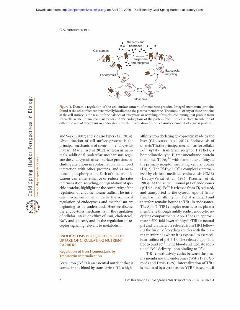
Interactions between the cell and its environ-ment obligatorily involve events at the plasma
membrane. Cell-surface proteins mediate nutri-ent uptake, product release, and the sensing ofenvironmental changes, including signals fromother cells. Appropriate sensing and response toextracellular cues is essential for the individualcell’s survival and for the coordinated cellularbehavior in multicellular organisms. Accord-ingly, maintenance and dynamics of membraneproteins are fundamental mechanisms of cellu-lar homeostasis and survival.
Most plasma membrane proteins are in de-fined equilibria with intracellular endosomalcompartments, such that the amount of a givenprotein at the plasma membrane is determined
by the balance of its endocytosis and its recy-cling back to the cell surface from endosomesand other intracellular compartments (Fig. 1).Changes in the kinetics of membrane proteintraffic acutely affect the levels of individual pro-teins at the cell surface and thereby impact howcells intake nutrients, sense the environment,and respond to external cues.
Selective molecular mechanisms triggertraffic of plasma membrane proteins throughendomembranes. Among them, ubiquitinationand phosphorylation stand out as they can di-rectly target the cargo proteins. Ubiquitinationis the covalent attachment of the 76-amino acidpolypeptide ubiquitin to the 1-amino groupof specific lysine residues (reviewed by Miranda
Editors: Sandra L. Schmid, Alexander Sorkin, and Marino Zerial
Additional Perspectives on Endocytosis available at www.cshperspectives.org
Copyright # 2014 Cold Spring Harbor Laboratory Press; all rights reserved; doi: 10.1101/cshperspect.a016964
Cite this article as Cold Spring Harb Perspect Biol 2014;6:a016964
1
on April 22, 2020 - Published by Cold Spring Harbor Laboratory Press http://cshperspectives.cshlp.org/Downloaded from
and Sorkin 2007; and see also Piper et al. 2014).Ubiquitination of cell-surface proteins is theprincipal mechanism of control of endocytosisin yeast (MacGurn et al. 2012), whereas in mam-mals, additional molecular mechanisms regu-late the endocytosis of cell-surface proteins, in-cluding alterations in conformation that impactinteraction with other proteins, and as men-tioned, phosphorylation. Each of these modifi-cations can either enhance or reduce the ratesinternalization, recycling, or degradation of spe-cific proteins, highlighting the complexity of theregulation of endomembrane traffic. The intri-cate mechanisms that underlie the reciprocalregulation of endocytosis and metabolism arebeginning to be understood. Here we discussthe endocytosis mechanisms in the regulationof cellular intake or efflux of iron, cholesterol,Naþ, and glucose, and in the regulation of re-ceptor signaling relevant to metabolism.
ENDOCYTOSIS IS REQUIRED FOR THEUPTAKE OF CIRCULATING NUTRIENTCARRIERS
Regulation of Iron Homeostasis byTransferrin Internalization
Ferric iron (Fe3þ) is an essential nutrient that iscarried in the blood by transferrin (Tf ), a high-
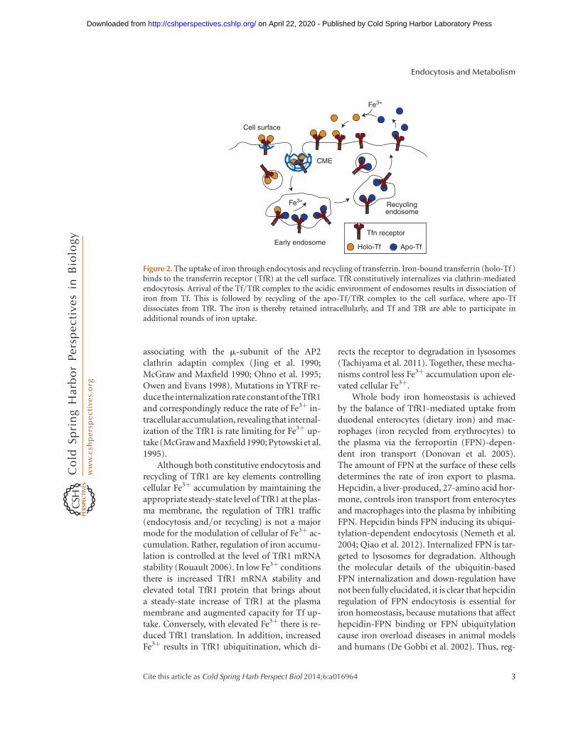
affinity iron chelating glycoprotein made by theliver (Gkouvatsos et al. 2012). Endocytosis ofdiferric Tf isthe principal mechanismforcellularFe3þ uptake. Transferrin receptor 1 (TfR1), ahomodimeric type II transmembrane proteinthat binds Tf-Fe2
3þ with nanomolar affinity, isthe primary receptor mediating cellular uptake(Fig. 2). The Tf-Fe2
3þ:TfR1 complex is internal-ized by clathrin-mediated endocytosis (CME)(Dautry-Varsat et al. 1983; Klausner et al.1983). At the acidic luminal pH of endosomes(pH 5.5–6.0), Fe3þ is released from Tf, reduced,and transported to the cytosol. Apo-Tf (iron-free) has high affinity for TfR1 at acidic pH andtherefore remains bound to TfR1 in endosomes.The Apo-Tf:TfR1 complex returns to the plasmamembrane through mildly acidic, endocytic re-cycling compartments. Apo-Tf has an approxi-mate�500-fold loweraffinity for TfR1atneutralpH and it is therefore released from TfR1 follow-ing the fusion of recycling vesicles with the plas-ma membrane (where it is exposed to extracel-lular milieu of pH 7.4). The released apo-Tf isfree to bind Fe3þ in the blood and mediate addi-tional Fe3þ delivery upon binding to TfR1.
TfR1 constitutively cycles between the plas-ma membrane and endosomes (Watts 1985; Gi-rones and Davis 1989). Internalization of TfR1is mediated by a cytoplasmic YTRF-based motif
Cell surface
Endocytosis
Exocytosis/recycling
Endosomes
Nutrients andhormones
Transporters and receptors
Figure 1. Dynamic regulation of the cell-surface content of membrane proteins. Integral membrane proteinsfound at the cell surface are dynamically localized to the plasma membrane. The amount of any of these proteinsat the cell surface is the result of the balance of exocytosis or recycling of vesicles containing that protein fromintracellular membrane compartments and the endocytosis of the protein from the cell surface. Regulation ofeither the rate of exocytosis or endocytosis results in alteration of the cell-surface content of a given protein.
C.N. Antonescu et al.
2 Cite this article as Cold Spring Harb Perspect Biol 2014;6:a016964
on April 22, 2020 - Published by Cold Spring Harbor Laboratory Press http://cshperspectives.cshlp.org/Downloaded from
associating with the m-subunit of the AP2clathrin adaptin complex (Jing et al. 1990;McGraw and Maxfield 1990; Ohno et al. 1995;Owen and Evans 1998). Mutations in YTRF re-ducethe internalization rateconstantof theTfR1and correspondingly reduce the rate of Fe3þ in-tracellularaccumulation, revealing that internal-ization of the TfR1 is rate limiting for Fe3þ up-take (McGrawand Maxfield 1990; Pytowski et al.1995).
Although both constitutive endocytosis andrecycling of TfR1 are key elements controllingcellular Fe3þ accumulation by maintaining theappropriate steady-state level of TfR1 at the plas-ma membrane, the regulation of TfR1 traffic(endocytosis and/or recycling) is not a majormode for the modulation of cellular of Fe3þ ac-cumulation. Rather, regulation of iron accumu-lation is controlled at the level of TfR1 mRNAstability (Rouault 2006). In low Fe3þ conditionsthere is increased TfR1 mRNA stability andelevated total TfR1 protein that brings abouta steady-state increase of TfR1 at the plasmamembrane and augmented capacity for Tf up-take. Conversely, with elevated Fe3þ there is re-duced TfR1 translation. In addition, increasedFe3þ results in TfR1 ubiquitination, which di-
rects the receptor to degradation in lysosomes(Tachiyama et al. 2011). Together, these mecha-nisms control less Fe3þ accumulation upon ele-vated cellular Fe3þ.
Whole body iron homeostasis is achievedby the balance of TfR1-mediated uptake fromduodenal enterocytes (dietary iron) and mac-rophages (iron recycled from erythrocytes) tothe plasma via the ferroportin (FPN)-depen-dent iron transport (Donovan et al. 2005).The amount of FPN at the surface of these cellsdetermines the rate of iron export to plasma.Hepcidin, a liver-produced, 27-amino acid hor-mone, controls iron transport from enterocytesand macrophages into the plasma by inhibitingFPN. Hepcidin binds FPN inducing its ubiqui-tylation-dependent endocytosis (Nemeth et al.2004; Qiao et al. 2012). Internalized FPN is tar-geted to lysosomes for degradation. Althoughthe molecular details of the ubiquitin-basedFPN internalization and down-regulation havenot been fully elucidated, it is clear that hepcidinregulation of FPN endocytosis is essential foriron homeostasis, because mutations that affecthepcidin-FPN binding or FPN ubiquitylationcause iron overload diseases in animal modelsand humans (De Gobbi et al. 2002). Thus, reg-
CME
Fe3+
Fe3+
Cell surface
Early endosome
Recyclingendosome
Tfn receptor
Apo-TfHolo-Tf
Figure 2. The uptake of iron through endocytosis and recycling of transferrin. Iron-bound transferrin (holo-Tf )binds to the transferrin receptor (TfR) at the cell surface. TfR constitutively internalizes via clathrin-mediatedendocytosis. Arrival of the Tf/TfR complex to the acidic environment of endosomes results in dissociation ofiron from Tf. This is followed by recycling of the apo-Tf/TfR complex to the cell surface, where apo-Tfdissociates from TfR. The iron is thereby retained intracellularly, and Tf and TfR are able to participate inadditional rounds of iron uptake.
Endocytosis and Metabolism
Cite this article as Cold Spring Harb Perspect Biol 2014;6:a016964 3
on April 22, 2020 - Published by Cold Spring Harbor Laboratory Press http://cshperspectives.cshlp.org/Downloaded from
ulation of FPN endocytosis is a physiologicallyimportant example of the acute regulation of anutrient carrier by endocytosis.
Low-Density Lipoprotein Receptor EndocytosisFacilitates Cellular Cholesterol Uptake
Cellular cholesterol uptake occurs throughbinding of low-density lipoprotein (LDL) par-ticles by the LDL receptor (LDLR), followed byreceptor endocytosis. LDLR binds apo-B100,which is present in a single copy within eachLDL particle (Jeon and Blacklow 2005; Gold-stein and Brown 2009). Upon internalization,the lower endosomal pH causes dissociationof LDL from its receptor. LDLR recycles backto the cell surface (Brown et al. 1983), whereasLDL continues to lysosomes, where cholesterolis released and rerouted. Cholesterol uptake bythis mechanism also suppresses endogenouscholesterol biosynthesis.
LDLR internalization occurs via CME (Jeonand Blacklow 2005; Goldstein and Brown 2009).Early studies identified aromatic-based endo-cytic sorting motifs on LDLR (Davis et al.1987) and the 802FDNPVY807 sequence was latershown to act as a recognition motif for the CMEadaptor protein ARH (Traub 2003). Interesting-ly, LDLR endocytosis by CME requires the en-docytic adaptor proteins ARH or Dab2, whichare dispensable for internalization of otherCME cargo such as TfR1 (He et al. 2002; Maurerand Cooper 2006).
LDL particles are �22 nm in diameter, mak-ing each LDL-LDLR complex significantly larg-er than most CME cargo. Expression of a chi-meric protein harboring the cytoplasmic regionof LDLR results in ARH- and Dab2-dependentchanges in the size of clathrin-coated pits, evenin the absence of LDL (Mettlen et al. 2010). Thisindicates that incorporation of LDLR-ARH/Dab2 complexes into endocytic structures may“customize” CME to accommodate these largercargoes.
LDLR also contributes to the internalizationof other lipoproteins, including very-low-den-sity lipoproteins (VLDL). This raises the ques-tion of how the internalization of a single recep-tor may regulate the unique homeostasis of
distinct lipoproteins. Interestingly, LDL uptakebut not that of VLDL by LDLR involves ARHS-nitrosylation at C199 and C286 (Zhao et al.2013), required for ARH interaction with AP2.This suggests a mechanism by which LDLR-de-pendent cellular internalization of LDL can beseparately regulated from that of VLDL, there-by defining a mechanism by which the systemicmetabolism of these different lipoproteins canbe distinctly regulated.
Strikingly, of the nearly 1000 mutations inLDLR linked to genetic hypercholesterolemia(Leigh et al. 2008), several impair LDLR inter-nalization but not LDL binding (Hobbs et al.1990). In particular, one of the first monoge-netic mutations identified in LDLR leading tohypercholesterolemia was Y807C; thus mutatedLDLR shows markedly lower recruitment ofLDLR to clathrin-coated pits and virtually ab-lated LDLR endocytosis (Davis et al. 1986).Moreover, mutations in ARH that impairLDLR internalization also lead to autosomal-recessive hypercholesterolemia (Soutar et al.2003). Collectively, these studies reveal a criticalcontribution of LDLR endocytosis to systemiccholesterol homeostasis (Bonfleur et al. 2010).
ENDOCYTOSIS LIMITS GLUCOSE UPTAKEAND Naþ EFFLUX THROUGH MEMBRANETRANSPORTERS
In contrast to the cellular internalization of ironand cholesterol carriers described above, othernutrients and micronutrients are taken up di-rectly through membrane-bound transportersstraight into the cytosol. Efflux also occurs di-rectly from the cytosol to the extracellular mi-lieu across membrane transporters. The rate ofnutrient or micronutrient uptake/efflux islargely determined by the permanence of theircognate transporters at the cell surface, which isfundamentally controlled by the balance of theirendocytosis and recycling.
GLUT Endocytosis Limits the Rate of CellularGlucose Uptake
A family of facilitative glucose transporters,GLUTs, is responsible for the transport of glu-
C.N. Antonescu et al.
4 Cite this article as Cold Spring Harb Perspect Biol 2014;6:a016964
on April 22, 2020 - Published by Cold Spring Harbor Laboratory Press http://cshperspectives.cshlp.org/Downloaded from
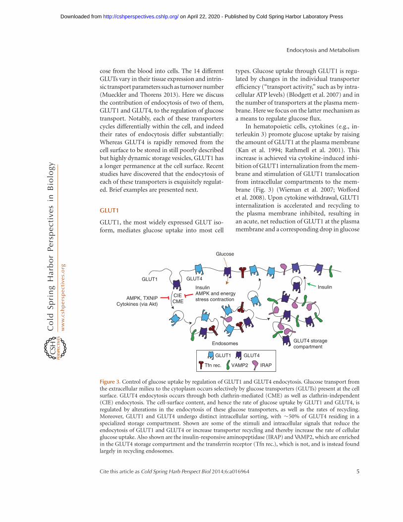
cose from the blood into cells. The 14 differentGLUTs vary in their tissue expression and intrin-sic transport parameters such asturnover number(Mueckler and Thorens 2013). Here we discussthe contribution of endocytosis of two of them,GLUT1 and GLUT4, to the regulation of glucosetransport. Notably, each of these transporterscycles differentially within the cell, and indeedtheir rates of endocytosis differ substantially:Whereas GLUT4 is rapidly removed from thecell surface to be stored in still poorly describedbut highly dynamic storage vesicles, GLUT1 hasa longer permanence at the cell surface. Recentstudies have discovered that the endocytosis ofeach of these transporters is exquisitely regulat-ed. Brief examples are presented next.
GLUT1
GLUT1, the most widely expressed GLUT iso-form, mediates glucose uptake into most cell
types. Glucose uptake through GLUT1 is regu-lated by changes in the individual transporterefficiency (“transport activity,” such as by intra-cellular ATP levels) (Blodgett et al. 2007) and inthe number of transporters at the plasma mem-brane. Here we focus on the latter mechanism asa means to regulate glucose flux.
In hematopoietic cells, cytokines (e.g., in-terleukin 3) promote glucose uptake by raisingthe amount of GLUT1 at the plasma membrane(Kan et al. 1994; Rathmell et al. 2001). Thisincrease is achieved via cytokine-induced inhi-bition of GLUT1 internalization from the mem-brane and stimulation of GLUT1 translocationfrom intracellular compartments to the mem-brane (Fig. 3) (Wieman et al. 2007; Woffordet al. 2008). Upon cytokine withdrawal, GLUT1internalization is accelerated and recycling tothe plasma membrane inhibited, resulting inan acute, net reduction of GLUT1 at the plasmamembrane and a corresponding drop in glucose
Glucose
GLUT4
InsulinAMPK and energystress contraction
CIECME
GLUT1
AMPK, TXNIPCytokines (via Akt)
Endosomes GLUT4 storagecompartment
Insulin
GLUT4GLUT1
Tfn rec. VAMP2 IRAP
Figure 3. Control of glucose uptake by regulation of GLUT1 and GLUT4 endocytosis. Glucose transport fromthe extracellular milieu to the cytoplasm occurs selectively by glucose transporters (GLUTs) present at the cellsurface. GLUT4 endocytosis occurs through both clathrin-mediated (CME) as well as clathrin-independent(CIE) endocytosis. The cell-surface content, and hence the rate of glucose uptake by GLUT1 and GLUT4, isregulated by alterations in the endocytosis of these glucose transporters, as well as the rates of recycling.Moreover, GLUT1 and GLUT4 undergo distinct intracellular sorting, with �50% of GLUT4 residing in aspecialized storage compartment. Shown are some of the stimuli and intracellular signals that reduce theendocytosis of GLUT1 and GLUT4 or increase transporter recycling and thereby increase the rate of cellularglucose uptake. Also shown are the insulin-responsive aminopeptidase (IRAP) and VAMP2, which are enrichedin the GLUT4 storage compartment and the transferrin receptor (Tfn rec.), which is not, and is instead foundlargely in recycling endosomes.
Endocytosis and Metabolism
Cite this article as Cold Spring Harb Perspect Biol 2014;6:a016964 5
on April 22, 2020 - Published by Cold Spring Harbor Laboratory Press http://cshperspectives.cshlp.org/Downloaded from
transport. The molecular mechanism for cyto-kine-induced changes in GLUT1 traffic has notbeen fully described. It is known that it occursdownstream from the Akt kinase, because en-forced expression of constitutively active Aktprevents enhanced GLUT1 internalization andreduced recycling upon growth factor with-drawal (Wieman et al. 2007).
In a different physiological context, cellularenergy deprivation enacts an emergency re-sponse that increases glucose uptake. This is inpart mediated by an increase in surface GLUT1,which is in the short run mediated by inhibitionof its constitutive endocytosis, and in the longrun by enhanced GLUT1 synthesis. The ele-ments participating in the acute response wererecently found to involve the arrestin-domain-containing protein (ARRDC), thioredoxin-in-teracting protein (TXNIP). TXNIP expressioncorrelates with glucose transport and its knock-down in adipose and muscle cells lowered glu-cose uptake (Parikh et al. 2007). TXNIP affectsGLUT1 traffic (Wu et al. 2013), by forming acomplex that promotes its internalized throughclathrin-coated pits. Although ARRDCs areinvolved in the ubiquitination of membraneproteins (MacGurn et al. 2012), ubiquitinationof GLUT1 has not been reported, and it isnot known if TXNIP controls GLUT1 endocy-tosis by ubiquitination of another protein. TheTXNIP-induced drop in surface GLUT1 reducesglucose transport.
TXNIP transcription is under the regulationof a number of different glucose-sensing tran-scription complexes, CHREBP-Mlx and Mon-doA-Mlx, such that TXNIP mRNA levels in-crease with increasing glucose uptake. BecauseTXNIP has an inhibitory effect on glucose up-take, the glucose-stimulated increased TXNIPmRNA provides a negative-feedback loop forGLUT1-mediated glucose uptake. In addition,TXNIP phosphorylation by AMP-dependentprotein kinase (AMPK) promotes its degrada-tion (Wu et al. 2013). AMPK is activated whenthe AMP/ATP ratio increases, therefore AMPK-induced degradation of TXNIP will result inelevated plasma membrane GLUT1 as a conse-quence of reduced internalization, and a corre-sponding increase of glucose uptake and ATP
generation ensue. Further, TXNIP also reducesGLUT1 transcription, contributing to the long-term reduction in glucose uptake.
GLUT4
GLUT4 expression is mostly limited to muscleand adipose cells (Mueckler and Thorens 2013)and is responsible for most of the glucose re-trieval from blood following a meal. In the fast-ed state (between meals) glucose transport intoadipose and muscle cells is low because 90%–95% of GLUT4 is sequestered intracellularly(Li et al. 2001; Huang and Czech 2007). Insulin,which increases in the blood upon feeding,stimulates a rapid (within minutes) redistribu-tion of GLUT4 from intracellular compart-ments to the plasma membrane, causing a con-comitant surge in glucose transport into thesetissues (Foley et al. 2011; Rowland et al. 2011).The elevated rate of glucose transport thusachieved is behind the return of blood glucoselevels back to prefeeding levels within 1–2 h(Ferrannini et al. 1985). The redistribution ofGLUT4 to the plasma membrane is reversible.When blood insulin levels wane, GLUT4 redis-tributes back from the plasma membrane to in-tracellular compartments. Unlike intracellularGLUT1, which transits only through recyclingendosomes, GLUT4 is unique in its sorting outof recycling endosomes into the TGN and a spe-cific “GLUT4 storage” compartment. The na-ture of this compartment is highly debated,but it is apparent that it excludes certain pro-teins such as GLUT1 and most of the TfR1,whereas it is enriched in proteins such asVAMP2 and IRAP (Fig. 3) (Jedrychowski et al.2010; Foley et al. 2011; Stockli et al. 2011; Letoand Saltiel 2012). Most studies agree that theGLUT4 storage compartment is distinct fromthe TGN although it contains proteins such asSyntaxin-6 and -16, and these may contribute toits sorting or functionality. Sorting to this selec-tive compartment, unique to fat and musclecells, may prevent GLUT4 from degradationand may account for its strikingly long half-life (nearly 48 h). The localization of this uniquecompartment is also poorly defined, in part ow-ing to spatial constraints in adipose and mature
C.N. Antonescu et al.
6 Cite this article as Cold Spring Harb Perspect Biol 2014;6:a016964
on April 22, 2020 - Published by Cold Spring Harbor Laboratory Press http://cshperspectives.cshlp.org/Downloaded from
muscle cells, and to its dynamic nature. Howev-er, there is general agreement that GLUT4 se-questration is essential for its appropriate re-sponse to insulin and hence defining the fullcomplement of signals dictating the dynamicretention of GLUT4 in intracellular stores is ofutmost physiological importance.
GLUT4 Internalization in the Basal State
GLUT4 is a very long-lived protein; hence, eachmolecule can undergo numerous cycles of in-ternalization and recycling. The pronouncedintracellular GLUT4 sequestration in unstimu-lated fat and muscle cells is owing to its rapidinternalization and slow recycling. GLUT4 con-tains sequences that determine its endocyto-sis and intracellular sorting. When ectopicallyexpressed in fibroblast-like cells, both the phe-nylalanine-based motif FQQI on the GLUT4amino-terminal cytoplasmic domain and a di-leucine motif on its carboxy-terminal cytoplas-mic domain function as internalization motifs(Corvera et al. 1994; Garippa et al. 1994, 1996;Verhey et al. 1995). These motifs suffice to pro-mote internalization when transferred to otherproteins, and belong to the two major familiesof membrane protein sorting signals: aromatic-based motifs and dileucine-based motifs (Sea-man 2008; see also Traub and Bonifacino 2013).However, the aromatic-based motif on GLUT4has a phenylalanine instead of the more com-mon tyrosine. Substitution of the endogenousphenylalanine for tyrosine in the GLUT4 mo-tif promotes faster (2�) internalization of thetransporter (Garippa et al. 1996; Blot andMcGraw 2006). Similarly phenylalanine-basedmotifs on other proteins promote a slower in-ternalization compared with tyrosine-basedmotifs (e.g., McGraw and Maxfield 1990). Thebehavior of GLUT4 in fibroblast-like cells isconsistent with its internalization by a cla-thrin/AP2-dependent pathway, although themechanism of internalization of ectopically ex-pressed GLUT4 endocytosis in these cells has notbeen rigorously investigated. In contrast, morerecent studies in 3T3-L1 adipocytes reveal that,in the basal state, GLUT4 is internalized by acholesterol-dependent, nystatin-sensitive path-
way that is independent of AP2 or any of theknown GLUT4 membrane protein traffic mo-tifs (Blot and McGraw 2006). In unstimulatedL6 muscle cells, GLUT4 internalizes via botha clathrin-dependent and a cholesterol anddynamin-dependent but caveolin-independentmechanism (Antonescu et al. 2008a, 2009). Ki-netically, GLUT4 endocytosis differs from thatof the TfR1 or the receptor for a2-macroglobu-lin (Habtemichael et al. 2011), markers of CMEand non-CME, noncaveolar internalization.
GLUT4 Internalization in Responseto Insulin
In muscle and fat cells the predominant effect ofinsulin is enhanced recycling (i.e., re-exocyto-sis) of GLUT4 to the plasma membrane (Li et al.2001; Karylowski et al. 2004; Martin et al. 2006;Blodgett et al. 2007; Antonescu et al. 2008b,2008a; Ishikura et al. 2010), and this is also thecase in skeletal muscle (Karlsson et al. 2009). Inadipocytes, insulin also slows down GLUT4 in-ternalization (Fig. 3) (Czech and Buxton 1993;Blot and McGraw 2006), but this does not occurin either cardiomyocytes (Yang and Holman2005) or L6 muscle cells (Li et al. 2001; An-tonescu et al. 2008a, 2009) (although a reportto the contrary exists [Fazakerley et al. 2010]).Once internalized, insulin enhances the rate ofGLUT4 transit through recycling endosomes(Foster et al. 2001). Although the magnitudeof the effect of insulin on recycling is greaterthan on internalization, both changes contrib-ute significantly to the net redistribution ofGLUT4 to the plasma membrane. This may beone reason why the insulin-dependent net gainin surface GLUT4—and its consequent glucoseuptake—is larger in adipocytes than in muscleor heart cells. The molecular basis of insulin-induced reduction in GLUT4 internalizationin adipocytes is beginning to be understood.Under this stimulus, the FQQI motif functionsas the main GLUT4 internalization motif, pro-moting uptake via an AP2-dependent mecha-nism (Blot and McGraw 2006). Thus, the datasuggest that the GLUT4 internalization routevaries between insulin-stimulated and -unstim-ulated adipocytes (respectively, AP2-depen-
Endocytosis and Metabolism
Cite this article as Cold Spring Harb Perspect Biol 2014;6:a016964 7
on April 22, 2020 - Published by Cold Spring Harbor Laboratory Press http://cshperspectives.cshlp.org/Downloaded from
dent and -independent). How this change inroute is achieved is unknown. By comparison,GLUT4 internalizes through both clathrin-de-pendent and clathrin-independent routes inboth basal and insulin-stimulated muscle cells,and insulin does not appear to change the pro-portion or rate of internalization of either route(Antonescu et al. 2008a, 2009). GLUT4 inter-nalization appears to require the F-BAR-do-main-containing protein CIP4, perhaps tocause membrane curvature for formation of en-docytic vesicles (Hartig et al. 2009; Feng et al.2010).
What is the significance of the differentinternalization routes? Although this is still anopen question, one can make certain testablepredictions: If the cholesterol-dependent routeis faster than the FQQI-dependent one, the in-sulin-dependent shift to clathrin/AP2-mediat-ed internalization through the FQQI motif inadipocytes might allow for longer permanenceat the plasma membrane of each transporter—to enact more rounds of glucose transport—before being internalized. This would be facili-tated by the slower internalization conferredto GLUT4 by the FAAI motif (compared withthe canonical YQQI motif ). The slower inter-nalization would be compounded by the gain intransporters at the membrane resulting from in-creased recycling. In muscle, the clathrin-de-pendent pathway operating in the basal statemight confer a slower internalization that couldbe advantageous for its physiological needs.
GLUT4 Internalization in Responseto Energy Demand
Processes that increase energy demand requirean adaptive increase in carbon source to sustaincellular viability (Cartee et al. 1991). This adap-tive response in other cells is ascribed to GLUT1(see above), but muscle largely depends onGLUT4 for influx of glucose. In contrast to in-sulin stimulation, energy status signaling leadsto a marked slowdown of GLUT4 endocytosis inmuscle cells. Metabolic stress resulting fromtreatment of cardiomyocytes with metforminor oligomycin (Yang and Holman 2005) or ofL6 muscle cells with 2,4-dinitrophenol (DNP)
(Antonescu et al. 2008a) slows the rate ofGLUT4 endocytosis (Fig. 3). DNP causes aninitial and abrupt drop in cellular ATP withconcomitant activation of AMPK; within min-utes, intracellular ATP levels bounce back, coin-cident with the AMPK-dependent increase incell surface GLUT4 elicited by DNP (Klip et al.2009). Thus, AMPK reduces GLUT4 endocyto-sis to enhance GLUT4 permanence at the mem-brane. Indeed, activation of AMPK by treatmentof L6 muscle cells with the AMPK-activatorAICAR alone (Fazakerley et al. 2010) or AICARin combination with exogenous activation ofprotein kinase C (Antonescu et al. 2008a) low-ered the rate of GLUT4 endocytosis. Other ex-amples of reduced GLUT4 endocytosis uponexposure of energy stressors include the re-sponse of adipocytes to dimethyl sulfoxide (Be-renguer et al. 2011), rosiglitazone (Velebit et al.2011), metformin (Yang and Holman 2006),and chronic insulin (Pryor et al. 2000).
In physiological conditions, muscle contrac-tion vigorously uses ATP that must be replen-ished by glucose influx. Contraction increasessurface GLUT4 levels in skeletal muscle (Douenet al. 1990; Goodyear et al. 1991) and this occursthrough both AMPK- and Ca2þ-dependent sig-nals. Interestingly, in noncontracting musclecells, a depolarization-induced influx of Ca2þ
suffices to reduce GLUT4 internalization, andthis response is independent of AMPK avail-ability (Wijesekara et al. 2006). Moreover, a cal-cium ionophore, ionomycin, can slow downthe rate of GLUT4 endocytosis and promoteGLUT4 exocytosis, but only the latter effectis AMPK-dependent (Q Li, P Bilan, W Niu,et al., unpubl.). The exocytic increase in surfaceGLUT4 in exercised muscle is also AMPK de-pendent (Lauritzen et al. 2010). Intriguingly,the AICAR had only a minor effect on GLUT4exocytosis in skeletal muscle (Karlsson et al.2009), suggesting that AMPK may instead acton the endocytic step and/or that it may act inconcert with other physiological signals to in-crease surface GLUT4 during muscle contrac-tion. Hence, AMPK regulates GLUT4 endocy-tosis in conditions of energy demand (e.g., DNPtreatment) and GLUT4 exocytosis in responseto Ca2þ-dependent signals. Collectively, these
C.N. Antonescu et al.
8 Cite this article as Cold Spring Harb Perspect Biol 2014;6:a016964
on April 22, 2020 - Published by Cold Spring Harbor Laboratory Press http://cshperspectives.cshlp.org/Downloaded from
studies show that signals elicited by alterationsin energy status and during skeletal muscle con-traction converge to regulate the rates of GLUT4endocytosis and exocytosis, thereby enhancingintake of energy-providing glucose.
Na/K-ATPase Endocytosis ControlsMultiple Metabolic Functions
In all mammalian cells, an electrochemical gra-dient—creating a voltage difference that is typ-ically negative with regards to the external mi-lieu—ensures proper movement of ions andregulation of cell volume. At the core of themaintenance of this electrochemical Naþ gradi-ent lays the Naþ/Kþ-ATPase or Naþ/Kþ pump.By expending one ATP molecule per cycle ofefflux of three intracellular Naþ ions and influxof two extracellular Kþ ions, the pump ensuresrapid riddance of any Naþ ions that increase inthe cytosol as a result of numerous Naþ-depen-dent influx processes. Such preceding influxprocesses are key for lowering intracellularCa2þ (Naþ/Ca2þ exchange), Naþ-coupled ami-no acid transport, Naþ-coupled glucose uptakein epithelial cells, and for a diversity of Naþ, Kþ
and Cl2 transport processes. In fact, the Naþ/Kþ pump is the main, if not only, mechanism ofNaþ efflux from most cells. In parallel, thepump returns Kþ to cells that release or leakthis ion in the course of their electrical activity,such as during action potentials in neurons,skeletal, smooth and heart muscles, and neuro-endocrine cells. Another fundamental functionof the pump is the translocation of Naþ fromone side of epithelia to the other, that along withobligatory anion movement generates the os-motic gradient that drives water absorption orresorption.
With this tall order, it is not surprising thatthe activity of the Naþ/Kþ pump is rapidly andexquisitely regulated. This is achieved by chang-es in both the activity and the availability of thepump at the surface of the cell in question.
Structural Properties of the Naþ/Kþ Pump
The pump consists of a heterodimer of onecatalytic a subunit and one stabilizing, highly
glycosylated b subunit. The 110-kDa a sub-unit has 10 transmembrane segments, whereasthe 31.5 kDa b subunit crosses the membraneonce. The ab heterodimer is the stable form, aschanges in copy number of the a subunit re-quire concomitant changes in the b subunitto create functional units. Depending on thecell type, about 800,000 to 30 million pumpcopies are present at the cell surface at any giventime. Abnormal changes in the copy number ofpump units occur in several pathologies suchas heart failure and hypertension associatedwith excessive renal Naþ resorption. The func-tional pump involves one of four isoforms of thecatalytic a subunit and one of any of the threeisoforms of the stabilizing b subunit. The genetranscription and translation control of each ofthese subunits is tissue specific and differentiallyregulated by diverse physiological conditions, asis the catalytic activity of the a subunit. On topof that, the regulation of pump availability atthe cell membrane through exocytosis and en-docytosis is a fundamental physiological controlmechanism.
Pump Regulation by Exocytosis
The increase in pump surface exposure is bestexemplified by the increase in surfacea2 and b1subunits that occurs in skeletal muscle in re-sponse to insulin (Hundal et al. 1992; Ewartand Klip 1995; Benziane and Chibalin 2008).This rapid up-regulation of surface pump num-ber occurs independently of biosynthetic chang-es and although typically ascribed to translo-cation to the membrane, the endocytic andexocytic arms of pump traffic in response toinsulin have not been directly measured. A rapidincrease in surface pump units is vital to managethe rapid uptake of dietary Kþ (along with theNaþ, Kþ, Cl2 cotransporter) and importantlyhandles the elimination of cytosolic Naþ excessthat results from Naþ-coupled amino acid up-take and Naþ/Hþ exchange promoted by thehormone. A similar up-regulation occurs inthe contracting skeletal muscle fiber. In the cen-tral nervous system, the pump is up-regulatedon demand to mediate Kþ reuptake and ridsneurons of Naþ gained during electrical activity.
Endocytosis and Metabolism
Cite this article as Cold Spring Harb Perspect Biol 2014;6:a016964 9
on April 22, 2020 - Published by Cold Spring Harbor Laboratory Press http://cshperspectives.cshlp.org/Downloaded from
Naþ/Kþ-Pump Endocytosis Triggeredby CO2 and Hypoxia in the Lung
Changes in CO2 and O2 tension occur contin-uously in the lung, and when these changes areextreme, fluid movement needs to be regulated.This is enacted primarily through changes inthe availability of the Naþ/Kþ pump at the al-veolar cell surface, as the pump is the drivingforce for ion-accompanying fluid movement(Bertorello and Sznajder 2005). Elevations inCO2 (hypercapnia) and reductions in O2 (hy-poxia) demand an adaptive reduction in Naþ
flux through the cells, and this is achieved byreducing the number of Naþ/Kþ pumps at thecell surface, through their rapid endocytosis. Infact, the endocytosis of the pump in alveolarepithelial cells controls whole body CO2 clear-ance and alveolar fluid resorption. The reduc-tion in pump units at the cell surface also curbsATP utilization, given the high energetic de-mand imposed by pump activity (up to 30%of the alveolar cell ATP). This reduction is ben-eficial in conditions of hypoxia. However, en-docytosis of the pump can result in decreasedalveolar fluid clearance.
The lung preferentially expresses the a1 andb1 subunits. The endocytosis of pump subunitsat the basolateral surface of primary and im-mortalized alveolar cells (e.g., human A549,rat ATII0) has been recorded using cell-surfacebiotinylation of proteins followed by pulldownwith streptavidin beads. Pump endocytosisis promoted by CO2 and by hypoxia, but alsoby thrombin (Vadasz et al. 2005). The signalsregulated this endocytic movement are onlybeginning to be identified, but hypercapniaactivates ERK (extracellular signal-regulatingkinase) and inhibiting ERK reduces pump en-docytosis (Welch et al. 2010). On the otherhand, hypoxia increases CaMKK-b (calcium/calmodulin-dependent protein kinase kinase2) and ROS (reactive oxygen species), whichactivate AMPK (Vadasz et al. 2008). Indeed, in-hibiting either one of these inputs reduces hy-poxia-induced pump endocytosis. It has beenproposed that AMPK phosphorylates the atyp-ical protein kinase C-z on Thr-410, activatingit, and that this signaling pathway leads to phos-
phorylation of Ser-18 on the a1 subunit of theNaþ/Kþ pump (Gusarova et al. 2009). This isthought to lead to ubiquitylation of a lysineresidue in the vicinity of Ser-18 as preamble tothe pump endocytosis (Dada et al. 2007). Thismechanism is partly reenacted in the kidney inresponse to elevated Naþ in the basolateral mi-lieu (see below).
Finally, mitochondrial ROS are generated inthe course of hypoxia and these have been impli-cated in the endocytic regulation of the pump(Vadasz et al. 2005), apparently through phos-phorylation of the AP2 adaptin m2 subunit.The responsible kinase remains to be identified,although AAK1 (adaptor-associated proteinkinase 1) has been suggested (Chen et al. 2006).
Naþ/Kþ-Pump Endocytosis Triggeredby Naþ, Dopamine, and CardiotonicSteroids in the Kidney
In the proximal tubule of the kidney, the Naþ/Kþ pump is the driving force for Naþ and fluidrecovery from lumen to blood (Naþ resorp-tion). The kidney expresses the a1- andb1-sub-unit isoforms that are abundant at the baso-lateral membrane, from where the dimer isendocytosed in response to several stimuli (Ber-torello and Sznajder 2005).
The presence of elevated Naþ in the bloodand therefore at the peritubular interstitium ofthe proximal tubule demands a reduction inNaþ resorption from the lumen. High levels ofNaþ cause release of dopamine, a natriuretichormone that increases Naþ excretion by dimin-ishing its reabsorption, primarily in the proxi-mal tubule. Dopamine is synthesized within thekidney from L-dopa, and both local and circu-lating dopamine binds and activates DA1 andDA2 receptors that generate cyclic AMP. Dopa-mine triggers Naþ/Kþ-pump endocytosis atthe basolateral membrane of kidney epithelialcells through three coincident events, docu-mented in OK (opossum kidney) cells (Chibalinet al. 1999) and renal proximal tubule cells (Pe-demonte et al. 2005):
1. Phosphorylation of Ser-18 on thea1 subunitof the Naþ/Kþ pump (Pedemonte et al.
C.N. Antonescu et al.
10 Cite this article as Cold Spring Harb Perspect Biol 2014;6:a016964
on April 22, 2020 - Published by Cold Spring Harbor Laboratory Press http://cshperspectives.cshlp.org/Downloaded from
2005), mediated by protein kinase C-z. Thispromotes association of dynamin-2 with thea subunit (Efendiev et al. 2002). Presumably,phosphorylation of dynamin-2 at positionS848 limits basal Naþ/Kþ-pump endocy-tosis and dopamine-regulated PP2 dephos-phorylation of dynamin-2 S848 promotesNaþ/Kþ-pump internalization (Efendiev etal. 2002).
2. Simultaneously, the a1 subunit is also phos-phorylated on Y537, promoting binding ofthe p85 subunit of phosphatidylinositol-3-kinase (PI3K). Both phosphorylation eventsof a1 on Ser-18 and Y537 are required for itsendocytosis (Done et al. 2002). Binding ofPI3K to the pump occurs via interaction ofthe SH3 domain of p85 with a proline-richdomain of the a1 subunit, a region of thepump distinct from that bound by AP2 (Yu-dowski et al. 2000). In fact, PI3K activityenhances association of AP2 with the a1subunit (Yudowski et al. 2000). This resultsin formation of a complex of the Naþ/Kþ
pump, AP2, clathrin, and dynamin-2 (Pede-monte et al. 2005).
3. Finally, the adaptin m2 subunit is phosphor-ylated on T156 in response to dopamine andROS (Chen et al. 2006), and m2 binds di-rectly to the YLEL motif on the main cyto-plasmic loop of the Naþ/Kþ pump (Doneet al. 2002; Chen et al. 2006). The pump thenapparently internalizes all the way to lateendosomes (based on subcellular fraction-ation experiments) from whence it can recy-cle to the membrane (Chibalin et al. 1999).
In addition to this interesting model, otherelements contribute to pump endocytosis inepithelial cells following activation of G-pro-tein-coupled receptors (GPCRs) by dopamine,vasopressin, or adrenergic hormones. In COScells (a monkey kidney cell line), Naþ/Kþ-pump endocytosis involves its direct phosphor-ylation through GPCR-associated kinases 2and 3 and binding of arrestin 2 and 3 (Kimuraet al. 2007). In MDCK (Madin-Darby caninekidney) epithelial cells, inhibition of AMPKcontributes to Naþ/Kþ-pump endocytosis,
through sparing phosphorylation of the Rab-GAP AS160 (thereby maintaining it in itsGAP-active form) (Alves et al. 2010). This sug-gests that AS160, known to retain GLUT4 inintracellular stores, also retains the pump intra-cellularly, effectively preventing it from recy-cling to the cell surface.
Finally, additional to the Naþ-dependentrelease of dopamine that regulates the pumpthrough GPCRs, basolateral Naþ appears tocause release of cardiotonic steroids (endoge-nous, digitalis-like factors), which act on theproximal tubule cells, sending signals that pro-mote Naþ/Kþ-pump endocytosis (Bagrov et al.2009). In cell cultures, these endogenous car-diotonic steroids are emulated by using oua-bain (irrespective of its inhibitory action onpump activity). The ouabain-induced Naþ/Kþ-pump endocytosis requires clathrin (Liuet al. 2004) although the same group also re-ported endocytosis through a cholesterol- andcaveolin-1-dependent route (Liu et al. 2005).Clearly, more work is required to elucidate theendocytic route involved in the response to oua-bain and, if more than one, it is paramount toidentify which one is regulated, and how. Addi-tional regulatory factors act on the pump, suchas prostaglandins, reduce Naþ/Kþ-pump ac-tivity in the collecting renal tubular cells, andit will be important to find if this effect is alsomediated by pump endocytosis.
Collectively, the above examples illustratethe variety of stimuli that induce Naþ/Kþ-pump endocytosis and the diverse intracellu-lar mechanism that participate in this phenom-enon.
RECIPROCAL REGULATIONOF METABOLISM, ENDOCYTOSIS,AND RECEPTOR SIGNALING
Hormones and growth factors that are recog-nized by surface receptors have evolved mecha-nisms to signal differentially from the plasmamembrane and endocytic locales (see Di Fioreand von Zastrow 2014). This applies particular-ly to receptor tyrosine kinases (RTKs) andGPCRs that impact on protein, lipid, and car-bohydrate metabolism. Endocytosis is also a
Endocytosis and Metabolism
Cite this article as Cold Spring Harb Perspect Biol 2014;6:a016964 11
on April 22, 2020 - Published by Cold Spring Harbor Laboratory Press http://cshperspectives.cshlp.org/Downloaded from
mechanism to end defined signal transmission.Internalized receptors can then either recycleto the membrane or continue toward degrada-tion. Receptor removal from the membrane,whether for recycling or degradation, can leadto ligand desensitization, whereas recycling af-fords resensitization. The internalization andpostendocytic fate of the receptor-ligand com-plex governs the magnitude, duration, and spe-cificity of receptor signaling (Sorkin and vonZastrow 2009; Kholodenko et al. 2010), and istailored to conform to the physiological func-tion of each hormone. Here we illustrate theendocytic regulation of metabolic signaling bythe insulin, epidermal growth factor (EGF), b2-adrenergic, and D2 dopamine receptors.
Regulation of Insulin Action by InsulinReceptor Endocytosis
Insulin, secreted by pancreatic b cells in re-sponse to ingested glucose (Rorsman and Ren-strom 2003), binds to insulin receptors (IR)present on its main metabolic target tissues (liv-er, fat, muscle) as well as on epithelial, endothe-lial, and neuronal cells. Insulin binding to IRactivates mitogenic signaling (e.g., Shc to Erk),gene expression (e.g., Foxo), and is a criticalregulator of systemic carbohydrate and lipidmetabolism (through Akt) (Saltiel and Kahn2001). Insulin-dependent clearance of dietaryglucose from blood involves the rapid mobiliza-tion of GLUT4 vesicles described above, whichis regulated by the phosphatidylinositol-3-ki-nase to Akt axis that activates small G proteinsdirecting vesicle traffic (Zaid et al. 2008; Foleyet al. 2011).
Upon binding insulin, the IR rapidly inter-nalizes via CME in rat liver cells (Pilch et al.1983), 3T3-L1 adipocytes (Fan et al. 1982),mouse embryonic fibroblasts (Morcavallo et al.2012), and CHO cells (Paccaud et al. 1992; Car-pentier et al. 1993), as well as through non-clathrin mechanism(s) in rat adipocytes (Gus-tavsson et al. 1999; Fagerholm et al. 2009),endothelial cells (Wang et al. 2011), and mouseembryonic fibroblasts (Morcavallo et al. 2012).IR endocytosis is required for (1) transendo-thelial insulin transfer, (2) mitogenic and meta-
bolic signaling, and (3) insulin degradation toterminate its action.
IR Endocytosis in TransendothelialInsulin Transport
Transendothelial insulin transport may repre-sent the rate-limiting step of insulin action invivo (Miles et al. 1995; Barrett et al. 2009), andin aorta endothelial cells occurs through a sat-urable mechanism involving caveolar internali-zation of insulin-IR complexes (Barrett et al.2011). IR internalization and effective insulintranscytosis in endothelial cells determines in-sulin availability to muscle and fat cells. Surpris-ingly, the molecular underpinnings of thistransendothelial transfer remain obscure, pos-sibly due to suitable microvascular endothelialcell systems.
IR Endocytosis Controls Mitogenic but NotMetabolic Signaling
IR endocytosis is slow in skeletal muscle and fatcells, but fast in the liver. Upon internalizationand dissociation from insulin in endosomes, theIR retains tyrosine kinase activity (Rosen et al.1983; Klein et al. 1987) and activated insulinsignaling intermediates are associated with en-dosomes in Fao hepatoma and rat liver cells(Backer et al. 1989; Balbis et al. 2000), adipo-cytes (Kublaoui et al. 1995), and skeletal muscle(Dombrowski et al. 2000). Interestingly, block-ing IR internalization impairs insulin-stimulat-ed Shc and Erk (but not Akt) phosphorylationin hepatoma and CHO cells (Ceresa et al. 1998;Hamer et al. 2002) but not in 3T3-L1 adipocytes(Kao et al. 1998). These findings suggest that IRendocytosis is required for activation of MAPKin a cell-selective manner, but is not required forAkt activation. In fact, inhibiting IR endocytosisdoes not prevent the Akt-dependent GLUT4translocation, stimulation of glucose transport,or glycogen synthesis in adipocytes (Ceresa et al.1998; Kao et al. 1998). Consistent with thesedifferential requirements for IR endocytosis ofthe mitogenic versus metabolic facets insulinsignaling, the Erk-dependent transcription ofc-fos but not the Akt-dependent regulation of
C.N. Antonescu et al.
12 Cite this article as Cold Spring Harb Perspect Biol 2014;6:a016964
on April 22, 2020 - Published by Cold Spring Harbor Laboratory Press http://cshperspectives.cshlp.org/Downloaded from
the glucokinase gene by insulin was blocked byinhibition of IR endocytosis in INS1 cells (Uhleset al. 2007). Hence, the Akt-dependent meta-bolic facets of IR signaling do not require recep-tor endocytosis; however, IR endocytosis mayprovide negative-feedback regulation (Brann-mark et al. 2010).
IR Internalization Leading to InsulinDegradation
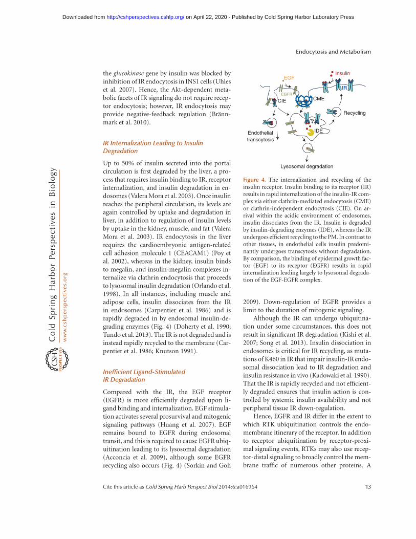
Up to 50% of insulin secreted into the portalcirculation is first degraded by the liver, a pro-cess that requires insulin binding to IR, receptorinternalization, and insulin degradation in en-dosomes (Valera Mora et al. 2003). Once insulinreaches the peripheral circulation, its levels areagain controlled by uptake and degradation inliver, in addition to regulation of insulin levelsby uptake in the kidney, muscle, and fat (ValeraMora et al. 2003). IR endocytosis in the liverrequires the cardioembryonic antigen-relatedcell adhesion molecule 1 (CEACAM1) (Poy etal. 2002), whereas in the kidney, insulin bindsto megalin, and insulin-megalin complexes in-ternalize via clathrin endocytosis that proceedsto lysosomal insulin degradation (Orlando et al.1998). In all instances, including muscle andadipose cells, insulin dissociates from the IRin endosomes (Carpentier et al. 1986) and israpidly degraded in by endosomal insulin-de-grading enzymes (Fig. 4) (Doherty et al. 1990;Tundo et al. 2013). The IR is not degraded and isinstead rapidly recycled to the membrane (Car-pentier et al. 1986; Knutson 1991).
Inefficient Ligand-StimulatedIR Degradation
Compared with the IR, the EGF receptor(EGFR) is more efficiently degraded upon li-gand binding and internalization. EGF stimula-tion activates several prosurvival and mitogenicsignaling pathways (Huang et al. 2007). EGFremains bound to EGFR during endosomaltransit, and this is required to cause EGFR ubiq-uitination leading to its lysosomal degradation(Acconcia et al. 2009), although some EGFRrecycling also occurs (Fig. 4) (Sorkin and Goh
2009). Down-regulation of EGFR provides alimit to the duration of mitogenic signaling.
Although the IR can undergo ubiquitina-tion under some circumstances, this does notresult in significant IR degradation (Kishi et al.2007; Song et al. 2013). Insulin dissociation inendosomes is critical for IR recycling, as muta-tions of K460 in IR that impair insulin-IR endo-somal dissociation lead to IR degradation andinsulin resistance in vivo (Kadowaki et al. 1990).That the IR is rapidly recycled and not efficient-ly degraded ensures that insulin action is con-trolled by systemic insulin availability and notperipheral tissue IR down-regulation.
Hence, EGFR and IR differ in the extent towhich RTK ubiquitination controls the endo-membrane itinerary of the receptor. In additionto receptor ubiquitination by receptor-proxi-mal signaling events, RTKs may also use recep-tor-distal signaling to broadly control the mem-brane traffic of numerous other proteins. A
Lysosomal degradation
Recycling
Insulin
CMECIE
IREGFR
EGF
Endothelialtranscytosis
IDE
Figure 4. The internalization and recycling of theinsulin receptor. Insulin binding to its receptor (IR)results in rapid internalization of the insulin-IR com-plex via either clathrin-mediated endocytosis (CME)or clathrin-independent endocytosis (CIE). On ar-rival within the acidic environment of endosomes,insulin dissociates from the IR. Insulin is degradedby insulin-degrading enzymes (IDE), whereas the IRundergoes efficient recycling to the PM. In contrast toother tissues, in endothelial cells insulin predomi-nantly undergoes transcytosis without degradation.By comparison, the binding of epidermal growth fac-tor (EGF) to its receptor (EGFR) results in rapidinternalization leading largely to lysosomal degrada-tion of the EGF-EGFR complex.
Endocytosis and Metabolism
Cite this article as Cold Spring Harb Perspect Biol 2014;6:a016964 13
on April 22, 2020 - Published by Cold Spring Harbor Laboratory Press http://cshperspectives.cshlp.org/Downloaded from
recent study in yeast found that activation ofmTOR resulted in control of ubiquitin ligase tar-geting by arrestin-related proteins (ARTs, ho-mologous to mammalian ARRDCs), which inturn led to ubiquitination and internalizationof a diverse group of cell-surface proteins (Mac-Gurn et al. 2011). This work raises the possibilitythat activation of mTOR by RTKs in mammalsmay lead to robustcontrolof the endomembranetraffic and function of a large number of cell-surface transporters and receptors.
Regulation of GPCR Signaling by Endocytosis
GPCRs regulate multiple aspects of cellular andsystemic metabolism. GPCR agonists stimulateexchange of GDP for GTP on specific Ga sub-units of heterotrimeric G proteins, activatingsignaling intermediates including G-protein-coupled receptor kinases (GRKs). GRKs enablebinding of b-arrestins to GPCRs, which in turndown-regulate G-protein signaling and pro-mote CME of the receptors (Shimokawa et al.2010). Although GPCR activation of the mito-genic MAPK pathway begins at the cell surface,signaling continues within endosomes (Sorkinand von Zastrow 2009). Interestingly, adrener-gic stimulation of Erk is reduced in cells withimpaired b2-adrenergic receptor (b2-AR) en-docytosis (Daaka et al. 1998). Hence, GPCRmembrane traffic and signaling are intimatelyrelated, with specific signals emanating fromdistinct cellular locale(s). GPCRs show indi-vidual signatures of GRK-dependent GPCRphosphorylation, b-arrestin binding, and ubiq-uitylation. Here, we examine the control ofmetabolism by endocytosis of b2-AR and D2-dopamine receptor (D2DR), which contributeto the peripheral and central regulation of me-tabolism, respectively.
Adrenergic Control of Lipolysis Dependson b2-AR Endocytosis
Agonist stimulation of b2-AR in adipocytescauses release of fatty acids from triglyceridestores. Adrenergic stimulation elicits rapid b2-AR CME.b2-AR then undergoes Rab11-depen-dent recycling to the cell surface (Seachrist et al.
2000; Parent et al. 2009) with minimal receptordegradation (Tsao and von Zastrow 2000), al-though prolonged adrenergic stimulation caus-es b2-AR degradation (Moore et al. 1999). Im-portantly, impairment of b2-AR endocytosisby perturbation of Arf6 or dynamin selectivelyblunts adrenergic stimulation of lipolysis (Liuet al. 2010). Consistent with a physiological con-tribution of b2-AR endocytosis to systemic me-tabolism, polymorphisms of b2-AR (R16G andQ27E) with defective adrenergic desensitization(Dishy et al. 2001) are linked to differences infat metabolism and obesity (Ukkola et al. 2000;Corbalan et al. 2002). Hence, b2-AR endocy-tosis is a critical controller of lipolysis, and sug-gests that either multiple cycles of b2-AR inter-nalization and recycling or endosomal-specificb2-AR signaling are required for stimulation oflipolysis.
D2DR Endocytosis Controls Feedingand Energy Expenditure
D2DR is highly expressed in the striatum andnucleus accumbens regions of the brain, whichregulate food intake (Stice et al. 2008; Beaulieuand Gainetdinov 2011). D2DR controls behav-ior associated with food intake and energyexpenditure (Meguid et al. 2000). D2DR under-goes rapid internalization leading to desensiti-zation following stimulation with dopamine orother ligands (Gainetdinov et al. 2004), and thisoccurs via clathrin-dependent (Paspalas et al.2006) or -independent mechanisms (Vickeryand von Zastrow 1999). Agonist-stimulated in-ternalization of D2DR is GRK- and b-arrestin-independent, and hence involves a noncanon-ical endocytic mechanism for receptor desen-sitization, although b-arrestins do contributeto D2DR signaling (Namkung et al. 2009).The adaptor protein CIN85 (Shimokawa et al.2010) as well as the BLOC-1 complex (Iizukaet al. 2007) are required for agonist-stimulatedD2DR internalization, and subsequent recycl-ing leading to D2DR resensitization occursvia a slow Rab-11-dependent pathway (Li et al.2012). Importantly, mice lacking neuronalCIN85 that showed defective D2DR endocy-tosis in the striatum were lean despite having
C.N. Antonescu et al.
14 Cite this article as Cold Spring Harb Perspect Biol 2014;6:a016964
on April 22, 2020 - Published by Cold Spring Harbor Laboratory Press http://cshperspectives.cshlp.org/Downloaded from
elevated food intake (Shimokawa et al. 2010),suggesting that D2DR endocytosis may impacton energy expenditure (or may reflect action ofother CIN85-dependent receptors). These re-sults exemplify how endocytosis in neurons crit-ically impacts on systemic metabolism.
CONCLUDING REMARKS AND FUTUREDIRECTIONS
We have examined how the regulated endocyto-sis of nutrient carriers (e.g., TfR1, LDLR),transporters (GLUTs and Na/K-ATPase), andsignaling receptors (IR, b2-AR, D2DR) facili-tate or limit the function of these importantregulators of systemic and cellular metabolism.For many of these examples, the regulation ofendocytosis is well documented, and a criticalphysiological role for endocytosis in the ho-meostasis of various nutrients is emerging.Nonetheless, there is still much to learn aboutthe molecular mechanisms regulating carrier,transporter, and receptor endocytosis withinthe context of metabolic homeostasis.
Furthermore, given the diversity of mem-brane proteins at the cell surface involved inthe cellular uptake and systemic regulation ofmetabolites, it is surprising that relatively littleis known about how the endocytosis of mosttransporters and receptors controls their func-tion. Importantly, approximately half of the 590human kinases (protein, lipid, and carbo-hydrate) are required for the normal endo-membrane traffic of vesicular stomatitis virus(VSV) and/or Simian virus 40 (SV40), markersof clathrin-dependent and clathrin-indepen-dent endocytosis, respectively (Pelkmans et al.2005). Indeed some of the kinases found to reg-ulate endomembrane traffic are metabolic en-zymes, such as FN3KRP, which controls proteinmodification by fructosamine (Pelkmans et al.2005). This suggests that endomembrane trafficis tightly coupled to or controlled by a cell’smetabolic state, and that we have only begunto understand this regulatory process. Further-more, that mTOR was found to exert broad ef-fects on the cell-surface membrane traffic of nu-merous transporters and receptors in yeast(MacGurn et al. 2011), and the central role of
mTOR as a branched chain amino acid sensor(Tokunaga et al. 2004), suggests the existence ofelaborate coordination of nutrient uptake, au-tophagy, and hormone signaling that we areonly beginning to understand.
Future work should examine how the met-abolic functions of the many cellular carriers,transporters, and receptors (e.g., lipid, nucleo-side or amino acid transporters, other signalingreceptors) are impacted by their endocytosis.Such information, along with a better under-standing of the molecular underpinnings, willprovide important insight into human metab-olism and may generate novel corrective thera-pies for metabolic disease.
ACKNOWLEDGMENTS
C.N.A. is supported by operating grant No.125854 from the Canadian Institutes of HealthResearch. A.K. is supported by Operating grantNo. 3707 from the Canadian Institutes ofHealth Research. T.E.M. is supported by NIHRO1 DK52852.
REFERENCES�Reference is also in this collection.
Acconcia F, Sigismund S, Polo S. 2009. Ubiquitin in traffick-ing: The network at work. Exp Cell Res 315: 1610–1618.
Alves DS, Farr GA, Seo-Mayer P, Caplan MJ. 2010. AS160associates with the Naþ,Kþ-ATPase and mediates theadenosine monophosphate-stimulated protein kinase-dependent regulation of sodium pump surface expres-sion. Mol Biol Cell 21: 4400–4408.
Antonescu CN, Diaz M, Femia G, Planas JV, Klip A. 2008a.Clathrin-dependent and independent endocytosis ofglucose transporter 4 (GLUT4) in myoblasts: Regulationby mitochondrial uncoupling. Traffic 9: 1173–1190.
Antonescu CN, Randhawa VK, Klip A. 2008b. DissectingGLUT4 traffic components in L6 myocytes by fluores-cence-based, single-cell assays. Meth Mol Biol 457: 367–378.
Antonescu CN, Foti M, Sauvonnet N, Klip A. 2009. Ready,set, internalize: Mechanisms and regulation of GLUT4endocytosis. Biosci Rep 29: 1–11.
Backer JM, Kahn CR, White MF. 1989. Tyrosine phosphor-ylation of the insulin receptor during insulin-stimulatedinternalization in rat hepatoma cells. J Biol Chem 264:1694–1701.
Bagrov AY, Shapiro JI, Fedorova OV. 2009. Endogenous car-diotonic steroids: Physiology, pharmacology, and noveltherapeutic targets. Pharmacol Rev 61: 9–38.
Endocytosis and Metabolism
Cite this article as Cold Spring Harb Perspect Biol 2014;6:a016964 15
on April 22, 2020 - Published by Cold Spring Harbor Laboratory Press http://cshperspectives.cshlp.org/Downloaded from
Balbis A, Baquiran G, Bergeron JJ, Posner BI. 2000. Com-partmentalization and insulin-induced translocations ofinsulin receptor substrates, phosphatidylinositol 3-ki-nase, and protein kinase B in rat liver. Endocrinology141: 4041–4049.
Barrett EJ, Eggleston EM, Inyard AC, Wang H, Li G, Chai W,Liu Z. 2009. The vascular actions of insulin control itsdelivery to muscle and regulate the rate-limiting step inskeletal muscle insulin action. Diabetologia 52: 752–764.
Barrett EJ, Wang H, Upchurch CT, Liu Z. 2011. Insulinregulates its own delivery to skeletal muscle by feed-for-ward actions on the vasculature. Am J Phys Endo Met 301:E252–E263.
Beaulieu J-M, Gainetdinov RR. 2011. The physiology, sig-naling, and pharmacology of dopamine receptors. Phar-macol Rev 63: 182–217.
Benziane B, Chibalin AV. 2008. Frontiers: Skeletal musclesodium pump regulation: A translocation paradigm.Am J Phys Endo Met 295: E553–E558.
Berenguer M, Zhang J, Bruce MC, Martinez L, Gonzalez T,Gurtovenko AA, Xu T, Marchand-Brustel Y, Govers R.2011. Dimethyl sulfoxide enhances GLUT4 translocationthrough a reduction in GLUT4 endocytosis in insulin-stimulated 3T3-L1 adipocytes. Biochimie 93: 697–709.
Bertorello AM, Sznajder JI. 2005. The dopamine paradox inlung and kidney epithelia: Sharing the same target butoperating different signaling networks. Am J Respir CellMol Biol 33: 432–437.
Blodgett DM, De Zutter JK, Levine KB, Karim P, CarruthersA. 2007. Structural basis of GLUT1 inhibition by cyto-plasmic ATP. J Gen Physiol 130: 157–168.
Blot V, McGraw TE. 2006. GLUT4 is internalized by a cho-lesterol-dependent nystatin-sensitive mechanism inhib-ited by insulin. EMBO J 25: 5648–5658.
Bonfleur ML, Vanzela EC, Ribeiro RA, de Gabriel Dori-ghello G, de Franca Carvalho CP, Collares-Buzato CB,Carneiro EM, Boschero AC, de Oliveira HCF. 2010. Pri-mary hypercholesterolaemia impairs glucose homeosta-sis and insulin secretion in low-density lipoprotein re-ceptor knockout mice independently of high-fat diet andobesity. Biochim Biophys Acta 1801: 183–190.
Brannmark C, Palmer R, Glad ST, Cedersund G, Stralfors P.2010. Mass and information feedbacks through receptorendocytosis govern insulin signaling as revealed using aparameter-free modeling framework. J Biol Chem 285:20171–20179.
Brown MS, Anderson RGW, Goldstein JL. 1983. Recyclingreceptors: The round-trip itinerary of migrant membraneproteins. Cell 32: 663–667.
Carpentier JL, Gazzano H, Van Obberghen E, Fehlmann M,Freychet P, Orci L. 1986. Intracellular pathway followedby the insulin receptor covalently coupled to 125I-photo-reactive insulin during internalization and recycling.J Cell Biol 102: 989–996.
Carpentier JL, Paccaud JP, Backer J, Gilbert A, Orci L, KahnCR, Backer J. 1993. Two steps of insulin receptor inter-nalization depend on different domains of theb-subunit.J Cell Biol 122: 1243–1252.
Cartee GD, Douen AG, Ramlal T, Klip A, Holloszy JO. 1991.Stimulation of glucose transport in skeletal muscle byhypoxia. J Appl Physiol 70: 1593–1600.
Ceresa BP, Kao AW, Santeler SR, Pessin JE. 1998. Inhibitionof clathrin-mediated endocytosis selectively attenuatesspecific insulin receptor signal transduction pathways.Mol Cell Biol 18: 3862–3870.
Chen Z, Krmar RT, Dada L, Efendiev R, Leibiger IB, Pede-monte CH, Katz AI, Sznajder JI, Bertorello AM. 2006.Phosphorylation of adaptor protein–2 m2 is essentialfor Naþ,Kþ-ATPase endocytosis in response to either Gprotein–coupled receptor or reactive oxygen species. AmJ Respir Cell Mol Biol 35: 127–132.
Chibalin AV, Ogimoto G, Pedemonte CH, Pressley TA, KatzAI, Feraille E, Berggren PO, Bertorello AM. 1999. Do-pamine-induced endocytosis of Naþ,Kþ-ATPase is initi-ated by phosphorylation of Ser-18 in the rat a subunitand is responsible for the decreased activity in epithelialcells. J Biol Chem 274: 1920–1927.
Corbalan MS, Marti A, Forga L, Martinez-Gonzalez MA,Martinez JA. 2002. The 27Glu polymorphism of theb2-adrenergic receptor gene interacts with physical ac-tivity influencing obesity risk among female subjects.Clin Genet 61: 305–307.
Corvera S, Chawla A, Chakrabarti R, Joly M, Buxton J,Czech MP. 1994. A double leucine within the GLUT4glucose transporter COOH-terminal domain functionsas an endocytosis signal. J Cell Biol 126: 979–989.
Czech MP, Buxton JM. 1993. Insulin action on the internal-ization of the GLUT4 glucose transporter in isolated ratadipocytes. J Biol Chem 268: 9187–9190.
Daaka Y, Luttrell LM, Ahn S, Rocca GJD, Ferguson SSG,Caron MG, Lefkowitz RJ. 1998. Essential role for Gprotein-coupled receptor endocytosis in the activationof mitogen-activated protein kinase. J Biol Chem 273:685–688.
Dada LA, Welch LC, Zhou G, Ben-Saadon R, Ciechanover A,Sznajder JI. 2007. Phosphorylation and ubiquitinationare necessary for Na,K-ATPase endocytosis during hy-poxia. Cell Signal 19: 1893–1898.
Dautry-Varsat A, Ciechanover A, Lodish HF. 1983. pH andthe recycling of transferrin during receptor-mediated en-docytosis. Proc Natl Acad Sci 80: 2258–2262.
Davis CG, van Driel IR, Russell DW, Brown MS, GoldsteinJL. 1987. The low density lipoprotein receptor. Identifi-cation of amino acids in cytoplasmic domain required forrapid endocytosis. J Biol Chem 262: 4075–4082.
Davis CG, Lehrman MA, Russell DW, Anderson RGW,Brown MS, Goldstein JL. 1986. The J. D. mutation infamilial hypercholesterolemia: Amino acid substitutionin cytoplasmic domain impedes internalization of LDLreceptors. Cell 45: 15–24.
De Gobbi M, Roetto A, Piperno A, Mariani R, Alberti F,Papanikolaou G, Politou M, Lockitch G, Girelli D, Far-gion S, et al. 2002. Natural history of juvenile haemochro-matosis. Brit J Haematol 117: 973–979.
� Di Fiore PP, von Zastrow M. 2014. Endocytosis, signaling,and beyond. Cold Spring Harb Perspect Biol doi: 10.1101/cshperspect.a016865.
Dishy V, Sofowora GG, Xie H-G, Kim RB, Byrne DW, SteinCM, Wood AJJ. 2001. The effect of common polymor-phisms of the b2-adrenergic receptor on agonist-medi-ated vascular desensitization. New Engl J Med 345: 1030–1035.
C.N. Antonescu et al.
16 Cite this article as Cold Spring Harb Perspect Biol 2014;6:a016964
on April 22, 2020 - Published by Cold Spring Harbor Laboratory Press http://cshperspectives.cshlp.org/Downloaded from
Doherty JJ II, Kay DG, Lai WH, Posner BI, Bergeron JJ. 1990.Selective degradation of insulin within rat liver endo-somes. J Cell Biol 110: 35–42.
Dombrowski L, Faure R, Marette A. 2000. Sustained activa-tion of insulin receptors internalized in GLUT4 vesiclesof insulin-stimulated skeletal muscle. Diabetes 49: 1772–1782.
Done SC, Leibiger IB, Efendiev R, Katz AI, Leibiger B,Berggren P-O, Pedemonte CH, Bertorello AM. 2002. Ty-rosine 537 within the Naþ,Kþ-ATPase a-subunit is es-sential for AP-2 binding and clathrin-dependent endo-cytosis. J Biol Chem 277: 17108–17111.
Donovan A, Lima CA, Pinkus JL, Pinkus GS, Zon LI, RobineS, Andrews NC. 2005. The iron exporter ferroportin/Slc40a1 is essential for iron homeostasis. Cell Met 1:191–200.
Douen AG, Ramlal T, Rastogi S, Bilan PJ, Cartee GD, VranicM, Holloszy JO, Klip A. 1990. Exercise induces recruit-ment of the “insulin-responsive glucose transporter.” Ev-idence for distinct intracellular insulin- and exercise-re-cruitable transporter pools in skeletal muscle. J Biol Chem265: 13427–13430.
Efendiev R, Yudowski GA, Zwiller J, Leibiger B, Katz AI,Berggren P-O, Pedemonte CH, Leibiger IB, BertorelloAM. 2002. Relevance of dopamine signals anchoring dy-namin-2 to the plasma membrane during Naþ,Kþ-AT-Pase endocytosis. J Biol Chem 277: 44108–44114.
Ewart HS, Klip A. 1995. Hormonal regulation of the Naþ-Kþ-ATPase: Mechanisms underlying rapid and sustainedchanges in pump activity. Am J Physiol 269: C295–C311.
Fagerholm S, Ortegren U, Karlsson M, Ruishalme I, Stral-fors P. 2009. Rapid insulin-dependent endocytosis of theinsulin receptor by caveolae in primary adipocytes. PLoSONE 4: e5985.
Fan JY, Carpentier JL, Gorden P, Obberghen EV, BlackettNM, Grunfeld C, Orci L. 1982. Receptor-mediated endo-cytosis of insulin: Role of microvilli, coated pits, andcoated vesicles. Proc Natl Acad Sci 79: 7788–7791.
Fazakerley DJ, Holman GD, Marley A, James DE, Stockli J,Coster ACF. 2010. Kinetic evidence for unique regulationof GLUT4 trafficking by insulin and AMP-activated pro-tein kinase activators in L6 myotubes. J Biol Chem 285:1653–1660.
Feng Y, Hartig SM, Bechill JE, Blanchard EG, Caudell E,Corey SJ. 2010. The Cdc42-interacting protein-4 (CIP4)gene knock-out mouse reveals delayed and decreased en-docytosis. J Biol Chem 285: 4348–4354.
Ferrannini E, Bjorkman O, Reichard GA Jr, Pilo A, OlssonM, Wahren J, DeFronzo RA. 1985. The disposal of an oralglucose load in healthy subjects. A quantitative study.Diabetes 34: 580–588.
Foley K, Boguslavsky S, Klip A. 2011. Endocytosis, recycling,and regulated exocytosis of glucose transporter 4. Bio-chemistry 50: 3048–3061.
Foster LJ, Li D, Randhawa VK, Klip A. 2001. Insulin accel-erates inter-endosomal GLUT4 traffic via phosphatidyli-nositol 3-kinase and protein kinase B. J Biol Chem 276:44212–44221.
Gainetdinov RR, Premont RT, Bohn LM, Lefkowitz RJ,Caron MG. 2004. Desensitization of G protein-coupledreceptors and neuronal functions. Annu Rev Neurosci 27:107–144.
Garippa RJ, Judge TW, James DE, McGraw TE. 1994. Theamino terminus of GLUT4 functions as an internaliza-tion motif but not an intracellular retention signal whensubstituted for the transferrin receptor cytoplasmic do-main. J Cell Biol 124: 705–715.
Garippa RJ, Johnson A, Park J, Petrush RL, McGraw TE.1996. The carboxyl terminus of GLUT4 contains a ser-ine-leucine-leucine sequence that functions as a potentinternalization motif in Chinese hamster ovary cells. JBiol Chem 271: 20660–20668.
Girones N, Davis RJ. 1989. Comparison of the kineticsof cycling of the transferrin receptor in the presenceor absence of bound diferric transferrin. Biochem J 264:35–46.
Gkouvatsos K, Papanikolaou G, Pantopoulos K. 2012. Reg-ulation of iron transport and the role of transferrin. Bio-chim Biophys Acta 1820: 188–202.
Goldstein JL, Brown MS. 2009. The LDL receptor. ArteriosclThrom Vas 29: 431–438.
Goodyear LJ, Hirshman MF, Horton ES. 1991. Exercise-in-duced translocation of skeletal muscle glucose transport-ers. Am J Physiol 261: E795–E799.
Gusarova GA, Dada LA, Kelly AM, Brodie C, Witters LA,Chandel NS, Sznajder JI. 2009. a1-AMP-activatedprotein kinase regulates hypoxia-induced Na,K-ATPaseendocytosis via direct phosphorylation of protein kinaseCz. Mol Cell Biol 29: 3455–3464.
Gustavsson J, Parpal S, Karlsson M, Ramsing C, Thorn H,Borg M, Lindroth M, Peterson KH, Magnusson K-E,Stralfors P. 1999. Localization of the insulin receptor incaveolae of adipocyte plasma membrane. FASEB J 13:1961–1971.
Habtemichael EN, Brewer PD, Romenskaia I, Mastick CC.2011. Kinetic evidence that Glut4 follows different endo-cytic pathways than the receptors for transferrin and a2-macroglobulin. J Biol Chem 286: 10115–10125.
Hamer I, Foti M, Emkey R, Cordier-Bussat M, Philippe J, DeMeyts P, Maeder C, Kahn CR, Carpentier J-L. 2002. Anarginine to cysteine252 mutation in insulin receptors froma patient with severe insulin resistance inhibits receptorinternalisation but preserves signalling events. Diabetolo-gia 45: 657–667.
Hartig SM, Ishikura S, Hicklen RS, Feng Y, Blanchard EG,Voelker KA, Pichot CS, Grange RW, Raphael RM, Klip A,et al. 2009. The F-BAR protein CIP4 promotes GLUT4endocytosis through bidirectional interactions with N-WASp and Dynamin-2. J Cell Sci 122: 2283–2291.
He G, Gupta S, Yi M, Michaely P, Hobbs HH, Cohen JC.2002. ARH is a modular adaptor protein that interactswith the LDL receptor, Clathrin, and AP-2. J Biol Chem277: 44044–44049.
Hobbs HH, Russell DW, Brown MS, Goldstein JL. 1990. TheLDL receptor locus in familial hypercholesterolemia:Mutational analysis of a membrane protein. Annu RevGenet 24: 133–170.
Huang S, Czech MP. 2007. The GLUT4 glucose transporter.Cell Met 5: 237–252.
Huang F, Goh LK, Sorkin A. 2007. EGF receptor ubiquiti-nation is not necessary for its internalization. Proc NatlAcad Sci 104: 16904–16909.
Endocytosis and Metabolism
Cite this article as Cold Spring Harb Perspect Biol 2014;6:a016964 17
on April 22, 2020 - Published by Cold Spring Harbor Laboratory Press http://cshperspectives.cshlp.org/Downloaded from
Hundal HS, Marette A, Mitsumoto Y, Ramlal T, Blostein R,Klip A. 1992. Insulin induces translocation of the a2 andb1 subunits of the Naþ/Kþ-ATPase from intracellularcompartments to the plasma membrane in mammalianskeletal muscle. J Biol Chem 267: 5040–5043.
Iizuka Y, Sei Y, Weinberger DR, Straub RE. 2007. Evidencethat the BLOC-1 protein dysbindin modulates dopamineD2 receptor internalization and signaling but not D1internalization. J Neurosci 27: 12390–12395.
Ishikura S, Antonescu CN, Klip A. 2010. Document-ing GLUT4 exocytosis and endocytosis in musclecell monolayers. Curr Prot Cell Biol doi: 10.1002/0471143030.cb1515s46.
Jedrychowski MP, Gartner CA, Gygi SP, Zhou L, Herz J,Kandror KV, Pilch PF. 2010. Proteomic analysis ofGLUT4 storage vesicles reveals LRP1 to be an importantvesicle component and target of insulin signaling. J BiolChem 285: 104–114.
Jeon H, Blacklow SC. 2005. Structure and physiologic func-tion of the low-density lipoprotein receptor. Annu RevBiochem 74: 535–562.
Jing SQ, Spencer T, Miller K, Hopkins C, Trowbridge IS.1990. Role of the human transferrin receptor cytoplasmicdomain in endocytosis: Localization of a specific signalsequence for internalization. J Cell Biol 110: 283–294.
Kadowaki H, Kadowaki T, Cama A, Marcus-Samuels B, Ro-vira A, Bevins CL, Taylor SI. 1990. Mutagenesis of lysine460 in the human insulin receptor. Effects upon receptorrecycling and cooperative interactions among bindingsites. J Biol Chem 265: 21285–21296.
Kan O, Baldwin SA, Whetton AD. 1994. Apoptosis is regu-lated by the rate of glucose transport in an interleukin 3dependent cell line. J Exp Med 180: 917–923.
Kao AW, Ceresa BP, Santeler SR, Pessin JE. 1998. Expressionof a dominant interfering dynamin mutant in 3T3L1adipocytes inhibits GLUT4 endocytosis without affectinginsulin signaling. J Biol Chem 273: 25450–25457.
Karlsson HKR, Chibalin AV, Koistinen HA, Yang J, Kouma-nov F, Wallberg-Henriksson H, Zierath JR, Holman GD.2009. Kinetics of GLUT4 trafficking in rat and humanskeletal muscle. Diabetes 58: 847–854.
Karylowski O, Zeigerer A, Cohen A, McGraw TE. 2004.GLUT4 is retained by an intracellular cycle of vesicle for-mation and fusion with endosomes. Mol Biol Cel 15:870–882.
Kholodenko BN, Hancock JF, Kolch W. 2010. Signallingballet in space and time. Nat Rev Mol Cell Biol 11: 414–426.
Kimura T, Allen PB, Nairn AC, Caplan MJ. 2007. Arrestinsand spinophilin competitively regulate Naþ,Kþ-ATPasetrafficking through association with a large cytoplasmicloop of the Naþ,Kþ-ATPase. Mol Biol Cell 18: 4508–4518.
Kishi K, Mawatari K, Sakai-Wakamatsu K, Yuasa T, Wang M,Ogura-Sawa M, Nakaya Y, Hatakeyama S, Ebina Y. 2007.APS-mediated ubiquitination of the insulin receptor en-hances its internalization, but does not induce its degra-dation. Endocr J 54: 77–88.
Klausner RD, Ashwell G, van Renswoude J, Harford JB,Bridges KR. 1983. Binding of apotransferrin to K562cells: Explanation of the transferrin cycle. Proc NatlAcad Sci 80: 2263–2266.
Klein HH, Freidenberg GR, Matthaei S, Olefsky JM. 1987.Insulin receptor kinase following internalization in iso-lated rat adipocytes. J Biol Chem 262: 10557–10564.
Klip A, Schertzer JD, Bilan PJ, Thong F, Antonescu C. 2009.Regulation of glucose transporter 4 traffic by energy dep-rivation from mitochondrial compromise. Acta Physiol196: 27–35.
Knutson VP. 1991. Cellular trafficking and processing of theinsulin receptor. FASEB J 5: 2130–2138.
Kublaoui B, Lee J, Pilch PF. 1995. Dynamics of signalingduring insulin-stimulated endocytosis of its receptor inadipocytes. J Biol Chem 270: 59–65.
Lauritzen HPMM, Galbo H, Toyoda T, Goodyear LJ. 2010.Kinetics of contraction-induced GLUT4 translocation inskeletal muscle fibers from living mice. Diabetes 59:2134–2144.
Leigh SEA, Foster AH, Whittall RA, Hubbart CS, Humph-ries SE. 2008. Update and analysis of the UniversityCollege London low density lipoprotein receptor famil-ial hypercholesterolemia database. Ann Hum Genet 72:485–498.
Leto D, Saltiel AR. 2012. Regulation of glucose transport byinsulin: Traffic control of GLUT4. Nat Rev Mol Cell Biol13: 383–396.
Li D, Randhawa VK, Patel N, Hayashi M, Klip A. 2001.Hyperosmolarity reduces GLUT4 endocytosis and in-creases its exocytosis from a VAMP2-independent poolin l6 muscle cells. J Biol Chem 276: 22883–22891.
Li Y, Roy BD, Wang W, Zhang L, Zhang L, Sampson SB, YangY, Lin D-T. 2012. Identification of two functionally dis-tinct endosomal recycling pathways for dopamine D2
receptor. J Neurosci 32: 7178–7190.
Liu J, Kesiry R, Periyasamy SM, Malhotra D, Xie Z, ShapiroJI. 2004. Ouabain induces endocytosis of plasmalemmalNa/K-ATPase in LLC-PK1 cells by a clathrin-dependentmechanism. Kidney Int 66: 227–241.
Liu J, Liang M, Liu L, Malhotra D, Xie Z, Shapiro JI. 2005.Ouabain-induced endocytosis of the plasmalemmal Na/K-ATPase in LLC-PK1 cells requires caveolin-1. KidneyInt 67: 1844–1854.
Liu Y, Zhou D, Abumrad NA, Su X. 2010. ADP-ribosylationfactor 6 modulates adrenergic stimulated lipolysis in ad-ipocytes. Am J Physiol Cell Physiol 298: C921–C928.
MacGurn JA, Hsu P-C, Smolka MB, Emr SD. 2011. TORC1regulates endocytosis via Npr1-mediated phosphoinhi-bition of a ubiquitin ligase adaptor. Cell 147: 1104–1117.
MacGurn JA, Hsu P-C, Emr SD. 2012. Ubiquitin and mem-brane protein turnover: From cradle to grave. Annu RevBiochem 81: 231–259.
Martin OJ, Lee A, McGraw TE. 2006. GLUT4 distributionbetween the plasma membrane and the intracellularcompartments is maintained by an insulin-modulatedbipartite dynamic mechanism. J Biol Chem 281: 484–490.
Maurer ME, Cooper JA. 2006. The adaptor protein Dab2sorts LDL receptors into coated pits independently ofAP-2 and ARH. J Cell Sci 119: 4235–4246.
McGraw TE, Maxfield FR. 1990. Human transferrin recep-tor internalization is partially dependent upon an aro-matic amino acid on the cytoplasmic domain. Cell Regul1: 369–377.
C.N. Antonescu et al.
18 Cite this article as Cold Spring Harb Perspect Biol 2014;6:a016964
on April 22, 2020 - Published by Cold Spring Harbor Laboratory Press http://cshperspectives.cshlp.org/Downloaded from
Meguid MM, Fetissov SO, Varma M, Sato T, Zhang L, La-viano A, Rossi-Fanelli F. 2000. Hypothalamic dopamineand serotonin in the regulation of food intake. Nutrition16: 843–857.
Mettlen M, Loerke D, Yarar D, Danuser G, Schmid SL. 2010.Cargo- and adaptor-specific mechanisms regulate clath-rin-mediated endocytosis. J Cell Biol 188: 919–933.
Miles PDG, Levisetti M, Reichart D, Khoursheed M, MoossaAR, Olefsky JM. 1995. Kinetics of insulin action in vivo:Identification of rate-limiting steps. Diabetes 44: 947–953.
Miranda M, Sorkin A. 2007. Regulation of receptors andtransporters by ubiquitination: New insights into sur-prisingly similar mechanisms. Mol Interv 7: 157–167.
Moore RH, Tuffaha A, Millman EE, Dai W, Hall HS, DickeyBF, Knoll BJ. 1999. Agonist-induced sorting of humanb2-adrenergic receptors to lysosomes during downregu-lation. J Cell Sci 112: 329–338.
Morcavallo A, Genua M, Palummo A, Kletvikova E, JiracekJ, Brzozowski AM, Iozzo RV, Belfiore A, Morrione A.2012. Insulin and insulin-like growth factor II differen-tially regulate endocytic sorting and stability of insulinreceptor isoform A. J Biol Chem 287: 11422–11436.
Mueckler M, Thorens B. 2013. The SLC2 (GLUT) family ofmembrane transporters. Mol Aspects Med 34: 121–138.
Namkung Y, Dipace C, Javitch JA, Sibley DR. 2009. G pro-tein-coupled receptor kinase-mediated phosphorylationregulates post-endocytic trafficking of the D2 dopaminereceptor. J Biol Chem 284: 15038–15051.
Nemeth E, Tuttle MS, Powelson J, Vaughn MB, Donovan A,Ward DM, Ganz T, Kaplan J. 2004. Hepcidin regulatescellular iron efflux by binding to ferroportin and induc-ing its internalization. Science 306: 2090–2093.
Ohno H, Stewart J, Fournier MC, Bosshart H, Rhee I, Miya-take S, Saito T, Gallusser A, Kirchhausen T, Bonifacino JS.1995. Interaction of tyrosine-based sorting signals withclathrin-associated proteins. Science 269: 1872–1875.
Orlando RA, Rader K, Authier F, Yamazaki H, Posner BI,Bergeron JJ, Farquhar MG. 1998. Megalin is an endocyticreceptor for insulin. J Am Soc Nephrol 9: 1759–1766.
Owen DJ, Evans PR. 1998. A structural explanation for therecognition of tyrosine-based endocytotic signals. Sci-ence 282: 1327–1332.
Paccaud JP, Siddle K, Carpentier JL. 1992. Internalization ofthe human insulin receptor. The insulin-independentpathway. J Biol Chem 267: 13101–13106.
Parent A, Hamelin E, Germain P, Parent J-L. 2009. Rab11regulates the recycling of the b2-adrenergic receptorthrough a direct interaction. Biochem J 418: 163–172.
Parikh H, Carlsson E, Chutkow WA, Johansson LE, Stor-gaard H, Poulsen P, Saxena R, Ladd C, Schulze PC, Maz-zini MJ, et al. 2007. TXNIP regulates peripheral glucosemetabolism in humans. PLoS Med 4: e158.
Paspalas CD, Rakic P, Goldman-Rakic PS. 2006. Internali-zation of D2 dopamine receptors is clathrin-dependentand select to dendro-axonic appositions in primate pre-frontal cortex. Eur J Neurosci 24: 1395–1403.
Pedemonte CH, Efendiev R, Bertorello AM. 2005. Inhibi-tion of Na,K-ATPase by dopamine in proximal tubuleepithelial cells. Sem Nephrol 25: 322–327.
Pelkmans L, Fava E, Grabner H, Hannus M, Habermann B,Krausz E, Zerial M. 2005. Genome-wide analysis of hu-man kinases in clathrin- and caveolae/raft-mediated en-docytosis. Nature 436: 78–86.
Pilch PF, Shia MA, Benson RJ, Fine RE. 1983. Coated vesiclesparticipate in the receptor-mediated endocytosis of in-sulin. J Cell Biol 96: 133–138.
� Piper RC, Dikic I, Lukacs GL. 2014. Ubiquitin-dependentsorting in endocytosis. Cold Spring Harb Perspect Biol 6:a016808.
Poy MN, Yang Y, Rezaei K, Fernstrom MA, Lee AD, Kido Y,Erickson SK, Najjar SM. 2002. CEACAM1 regulates in-sulin clearance in liver. Nature Genet 30: 270–276.
Pryor PR, Liu SC, Clark AE, Yang J, Holman GD, Tosh D.2000. Chronic insulin effects on insulin signalling andGLUT4 endocytosis are reversed by metformin. BiochemJ 348: 83–91.
Pytowski B, Judge TW, McGraw TE. 1995. An internaliza-tion motif is created in the cytoplasmic domain of thetransferrin receptor by substitution of a tyrosine at thefirst position of a predicted tight turn. J Biol Chem 270:9067–9073.
Qiao B, Sugianto P, Fung E, Del-Castillo-Rueda A, Moran-Jimenez M-J, Ganz T, Nemeth E. 2012. Hepcidin-inducedendocytosis of ferroportin is dependent on ferroportinubiquitination. Cell Met 15: 918–924.
Rathmell JC, Farkash EA, Gao W, Thompson CB. 2001. IL-7enhances the survival and maintains the size of naiveT cells. J Immunol 167: 6869–6876.
Rorsman DP, Renstrom E. 2003. Insulin granule dynamicsin pancreatic b cells. Diabetologia 46: 1029–1045.
Rosen OM, Herrera R, Olowe Y, Petruzzelli LM, Cobb MH.1983. Phosphorylation activates the insulin receptor ty-rosine protein kinase. Proc Natl Acad Sci 80: 3237–3240.
Rouault TA. 2006. The role of iron regulatory proteins inmammalian iron homeostasis and disease. Nat Chem Biol2: 406–414.
Rowland AF, Fazakerley DJ, James DE. 2011. Mapping in-sulin/GLUT4 circuitry. Traffic 12: 672–681.
Saltiel AR, Kahn CR. 2001. Insulin signalling and the regu-lation of glucose and lipid metabolism. Nature 414: 799–806.
Seachrist JL, Anborgh PH, Ferguson SSG. 2000. b2-adrenergic receptor internalization, endosomal sorting,and plasma membrane recycling are regulated by RabGTPases. J Biol Chem 275: 27221–27228.
Seaman MNJ. 2008. Endosome protein sorting: Motifs andmachinery. Cell Mol Life Sci 65: 2842–2858.
Shimokawa N, Haglund K, Holter SM, Grabbe C, Kirkin V,Koibuchi N, Schultz C, Rozman J, Hoeller D, Qiu C-H, etal. 2010. CIN85 regulates dopamine receptor endocytosisand governs behaviour in mice. EMBO J 29: 2421–2432.
Song R, Peng W, Zhang Y, Lv F, Wu H-K, Guo J, Cao Y, Pi Y,Zhang X, Jin L, et al. 2013. Central role of E3 ubiquitinligase MG53 in insulin resistance and metabolic disor-ders. Nature 494: 375–379.
Sorkin A, Goh LK. 2009. Endocytosis and intracellular traf-ficking of ErbBs. Exp Cell Res 315: 683–696.
Sorkin A, von Zastrow M. 2009. Endocytosis and signalling:Intertwining molecular networks. Nat Rev Mol Cell Biol10: 609–622.
Endocytosis and Metabolism
Cite this article as Cold Spring Harb Perspect Biol 2014;6:a016964 19
on April 22, 2020 - Published by Cold Spring Harbor Laboratory Press http://cshperspectives.cshlp.org/Downloaded from
Soutar AK, Naoumova RP, Traub LM. 2003. Genetics, clin-ical phenotype, and molecular cell biology of autosomalrecessive hypercholesterolemia. Arterioscl Throm Vas 23:1963–1970.
Stice E, Spoor S, Bohon C, Small DM. 2008. Relation be-tween obesity and blunted striatal response to food ismoderated by TaqIA A1 allele. Science 322: 449–452.
Stockli J, Fazakerley DJ, James DE. 2011. GLUT4 exocytosis.J Cell Sci 124: 4147–4159.
Tachiyama R, Ishikawa D, Matsumoto M, Nakayama KI,Yoshimori T, Yokota S, Himeno M, Tanaka Y, Fujita H.2011. Proteome of ubiquitin/MVB pathway: Possibleinvolvement of iron-induced ubiquitylation of transfer-rin receptor in lysosomal degradation. Genes Cells 16:448–66.
Tokunaga C, Yoshino K, Yonezawa K. 2004. mTOR inte-grates amino acid- and energy-sensing pathways. Bio-chem Biophys Res Commun 313: 443–446.
Traub LM. 2003. Sorting it out: AP-2 and alternate clathrinadaptors in endocytic cargo selection. J Cell Biol 163:203–208.
� Traub LM, Bonifacino JS. 2013. Cargo recognition in clath-rin-mediated endocytosis. Cold Spring Harb Perspect Biol5: a016790.
Tsao PI, von Zastrow M. 2000. Type-specific sorting of Gprotein-coupled receptors after endocytosis. J Biol Chem275: 11130–11140.
Tundo GR, Sbardella D, Ciaccio C, Bianculli A, Orlandi A,Desimio MG, Arcuri G, Coletta M, Marini S. 2013. Insu-lin-degrading enzyme (IDE) a novel heat shock-like pro-tein. J Biol Chem 288: 2281–2289.
Uhles S, Moede T, Leibiger B, Berggren P-O, Leibiger IB.2007. Selective gene activation by spatial segregation ofinsulin receptor B signaling. FASEB J 21: 1609–1621.
Ukkola O, Rankinen T, Weisnagel SJ, Sun G, Perusse L,Chagnon YC, Despres J-P, Bouchard C. 2000. Interac-tions among the a2-, b2-, and b3-adrenergic receptorgenes and obesity-related phenotypes in the QuebecFamily Study. Metabolism 49: 1063–1070.
Vadasz I, Morty RE, Olschewski A, Konigshoff M, KohstallMG, Ghofrani HA, Grimminger F, Seeger W. 2005.Thrombin impairs alveolar fluid clearance by promotingendocytosis of Naþ,Kþ-ATPase. Am J Resp Cell Mol 33:343–354.
Vadasz I, Dada LA, Briva A, Trejo HE, Welch LC, Chen J,Toth PT, Lecuona E, Witters LA, Schumacker PT, et al.2008. AMP-activated protein kinase regulates CO2-in-duced alveolar epithelial dysfunction in rats and humancells by promoting Na,K-ATPase endocytosis. J Clin In-vest 118: 752–762.
Valera Mora ME, Scarfone A, Calvani M, Greco AV, Min-grone G. 2003. Insulin clearance in obesity. J Am CollNutr 22: 487–493.
Velebit J, Chowdhury HH, Kreft M, Zorec R. 2011. Rosigli-tazone balances insulin-induced exo- and endocytosisin single 3T3-L1 adipocytes. Mol Cell Endocrinol 333:70–77.
Verhey KJ, Yeh JI, Birnbaum MJ. 1995. Distinct signals in theGLUT4 glucose transporter for internalization and fortargeting to an insulin-responsive compartment. J CellBiol 130: 1071–1079.
Vickery RG, von Zastrow M. 1999. Distinct dynamin-de-pendent and -independent mechanisms target structur-ally homologous dopamine receptors to different endo-cytic membranes. J Cell Biol 144: 31–43.
Wang H, Wang AX, Barrett EJ. 2011. Caveolin-1 is requiredfor vascular endothelial insulin uptake. Am J Physiol En-docrinol Metab 300: E134–E144.
Watts C. 1985. Rapid endocytosis of the transferrin receptorin the absence of bound transferrin. J Cell Biol 100:633–637.
Welch LC, Lecuona E, Briva A, Trejo HE, Dada LA, SznajderJI. 2010. Extracellular signal-regulated kinase (ERK)participates in the hypercapnia-induced Na,K-ATPasedownregulation. FEBS Lett 584: 3985–3989.
Wieman HL, Wofford JA, Rathmell JC. 2007. Cytokine stim-ulation promotes glucose uptake via phosphatidylinosi-tol-3 kinase/Akt regulation of Glut1 activity and traffick-ing. Mol Biol Cell 18: 1437–1446.
Wijesekara N, Tung A, Thong F, Klip A. 2006. Muscle celldepolarization induces a gain in surface GLUT4 via re-duced endocytosis independently of AMPK. Am J PhysiolEndocrinol Metab 290: E1276–E1286.
Wofford JA, Wieman HL, Jacobs SR, Zhao Y, Rathmell JC.2008. IL-7 promotes Glut1 trafficking and glucose uptakevia STAT5-mediated activation of Akt to support T-cellsurvival. Blood 111: 2101–2111.
Wu N, Zheng B, Shaywitz A, Dagon Y, Tower C, Bellinger G,Shen C-H, Wen J, Asara J, McGraw TE, et al. 2013.AMPK-dependent degradation of TXNIP upon energystress leads to enhanced glucose uptake via GLUT1.Mol Cell 49: 1167–1175.
Yang J, Holman GD. 2005. Insulin and contraction stimulateexocytosis, but increased AMP-activated protein kinaseactivity resulting from oxidative metabolism stress slowsendocytosis of GLUT4 in cardiomyocytes. J Biol Chem280: 4070–4078.
Yang J, Holman GD. 2006. Long-term metformin treatmentstimulates cardiomyocyte glucose transport through anAMP-activated protein kinase-dependent reduction inGLUT4 endocytosis. Endocrinology 147: 2728–2736.
Yudowski GA, Efendiev R, Pedemonte CH, Katz AI, Berg-gren PO, Bertorello AM. 2000. Phosphoinositide-3 ki-nase binds to a proline-rich motif in the Naþ,Kþ-ATPasea subunit and regulates its trafficking. Proc Natl Acad Sci97: 6556–6561.
Zaid H, Antonescu CN, Randhawa VK, Klip A. 2008. Insu-lin action on glucose transporters through molecularswitches, tracks and tethers. Biochem J 413: 201–215.
Zhao Z, Pompey S, Dong H, Weng J, Garuti R, Michaely P.2013. S-nitrosylation of ARH is required for LDL uptakeby the LDL receptor. J Lipid Res 54: 1550–1559.
C.N. Antonescu et al.
20 Cite this article as Cold Spring Harb Perspect Biol 2014;6:a016964
on April 22, 2020 - Published by Cold Spring Harbor Laboratory Press http://cshperspectives.cshlp.org/Downloaded from
2014; doi: 10.1101/cshperspect.a016964Cold Spring Harb Perspect Biol Costin N. Antonescu, Timothy E. McGraw and Amira Klip Reciprocal Regulation of Endocytosis and Metabolism
Subject Collection Endocytosis
Endocytosis: Past, Present, and Future
ZerialSandra L. Schmid, Alexander Sorkin and Marino Clathrin-Mediated Endocytosis
Imaging and Modeling the Dynamics of
Marcel Mettlen and Gaudenz Danuser
Endosomal SystemRab Proteins and the Compartmentalization of the
Angela Wandinger-Ness and Marino ZerialClathrin-Mediated EndocytosisEndocytic Accessory Factors and Regulation of
Christien J. Merrifield and Marko Kaksonen
Regulator of Cell Polarity and Tissue DynamicsCargo Sorting in the Endocytic Pathway: A Key
Suzanne Eaton and Fernando Martin-BelmonteSystemThe Complex Ultrastructure of the Endolysosomal
Judith Klumperman and Graça Raposo
Links to Human DiseaseCytoskeleton, Cell Cycle, Nucleus, and Beyond:and Other Endocytic Regulators in the Unconventional Functions for Clathrin, ESCRTs,
et al.Frances M. Brodsky, R. Thomas Sosa, Joel A. Ybe,
Lysosome-Related OrganellesThe Biogenesis of Lysosomes and
Dieckmann, et al.J. Paul Luzio, Yvonne Hackmann, Nele M.G.
Endocytosis of Viruses and BacteriaPascale Cossart and Ari Helenius
Endocytosis, Signaling, and BeyondPier Paolo Di Fiore and Mark von Zastrow
Responds to External CuesLysosomal Adaptation: How the Lysosome
Carmine Settembre and Andrea Ballabio
Clathrin-Independent Pathways of Endocytosis
DonaldsonSatyajit Mayor, Robert G. Parton and Julie G.
MetabolismReciprocal Regulation of Endocytosis and
Amira KlipCostin N. Antonescu, Timothy E. McGraw and
SignalingThe Role of Endocytosis during Morphogenetic
Marcos Gonzalez-Gaitan and Frank Jülicher
Cooperation?Endocytosis and Autophagy: Exploitation or
Sharon A. Tooze, Adi Abada and Zvulun ElazarDiseaseRole of Endosomes and Lysosomes in Human
Frederick R. Maxfield
http://cshperspectives.cshlp.org/cgi/collection/ For additional articles in this collection, see
Copyright © 2014 Cold Spring Harbor Laboratory Press; all rights reserved
on April 22, 2020 - Published by Cold Spring Harbor Laboratory Press http://cshperspectives.cshlp.org/Downloaded from