Embed Size (px)
Citation preview

Receptors
Receptors are part of the co-ordination systemthat detect the changes of the environment bymeans of stimuli. Two types ...• Interoceptors – receptors detecting internal stimuli, such as hunger, thirst, pain, need to urinate, and blood pressure changes, etc.• Exteroceptors – receptors detecting external stimuli, such as light, sound, movement (balance), temperature, pressure, pain, etc.

Sense organs – What to learn:
• Label diagrams of human eye and ear• List functions of parts of eye and ear• Using diagrams describe in writing formation
of an image, accommodation, binocular vision, pupillary mechanism, balance and hearing
• Long and short sightedness, astigmatism, cataracts

Internal structure of the eye
ciliary body retina choroid
yellow spot
blind spot
sclera
vitreous humour
lens
suspensory ligament
iris
aqueous humour
pupil
optic nerve
conjunctiva
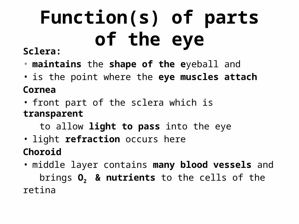
Function(s) of parts of the eyeSclera: • maintains the shape of the eyeball and • is the point where the eye muscles attachCornea• front part of the sclera which is transparent to allow light to pass into the eye• light refraction occurs hereChoroid• middle layer contains many blood vessels and brings O2 & nutrients to the cells of the retina

Function(s) of parts of the eye (cont.)
Conjunctiva • Thin membrane covers the front of the eye has
sensory nerve endings that detect the presence of foreign objects (protect eye)
Suspensory ligaments• Holds the lens in placeLens• responsible for the refraction of light to create
a sharp focussed image

Function(s) of parts of the eye (cont.)
Retina• light-sensitive membrane lines the inside of the eye ball and is made up of photoreceptorsPhotoreceptors (photo=light+receptors convert stimulus into nerve impulse). Two types.. Rods• active in dim light, allow us to see black & whiteCones • active in bright light, allow us to see colour

Function(s) of parts of the eye (cont.)
Yellow spot (fovea)• small area on retina which contains only cones• the area of sharp visionBlind spot• area where the neurons from the photo- receptors leave the eye ball via the optical nerve• There are no cones or rods in this area• Blood vessels also leaves and enter the eye ball at the blind spot

AccommodationAccommodation is the adjustment of the shapeof the lens to see objects clearly whether they are far away or close by.
Near vision
Far vision
Use the two diagrams to memorise the summary of these two processes on the next slide
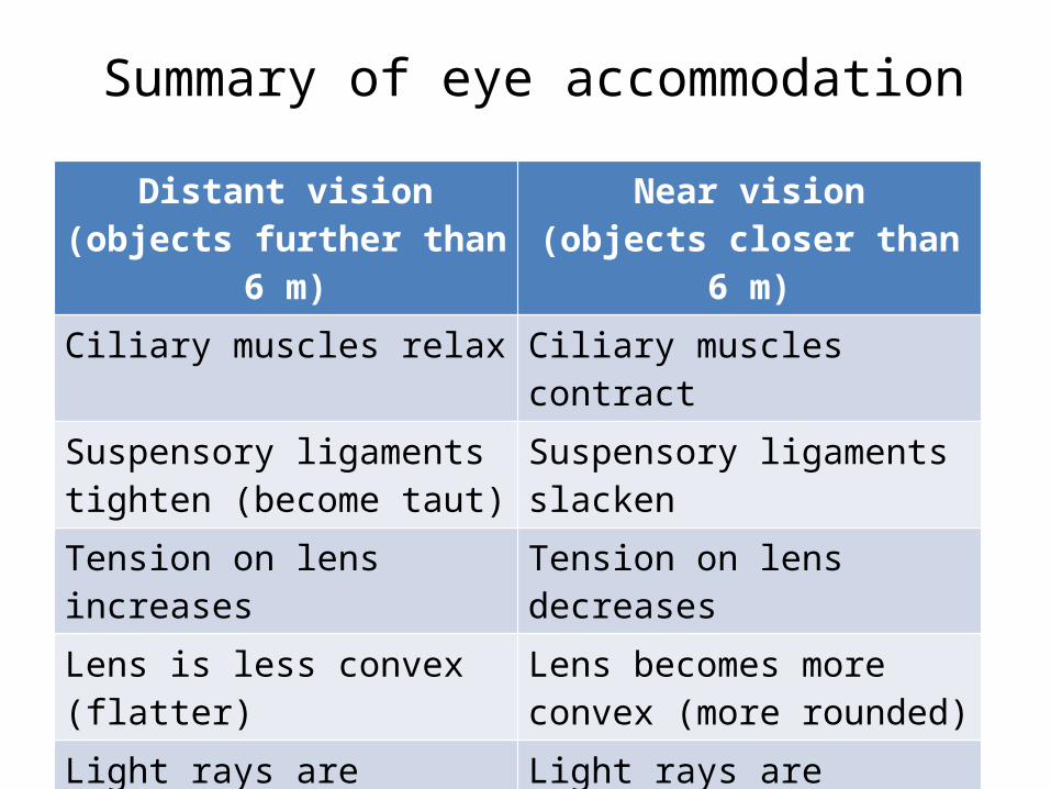
Summary of eye accommodation
Distant vision(objects further than 6 m)
Near vision(objects closer than 6 m)
Ciliary muscles relax Ciliary muscles contractSuspensory ligaments tighten (become taut)
Suspensory ligaments slacken
Tension on lens increases Tension on lens decreasesLens is less convex (flatter) Lens becomes more convex
(more rounded)Light rays are refracted (bend) less
Light rays are refracted (bend) more
Light rays are focused onto the retina
Light rays are focused onto the retina

Pupillary mechanismPupillary mechanism is the contraction of thepupil in response to light entering the eye.
Bright light
Dim light
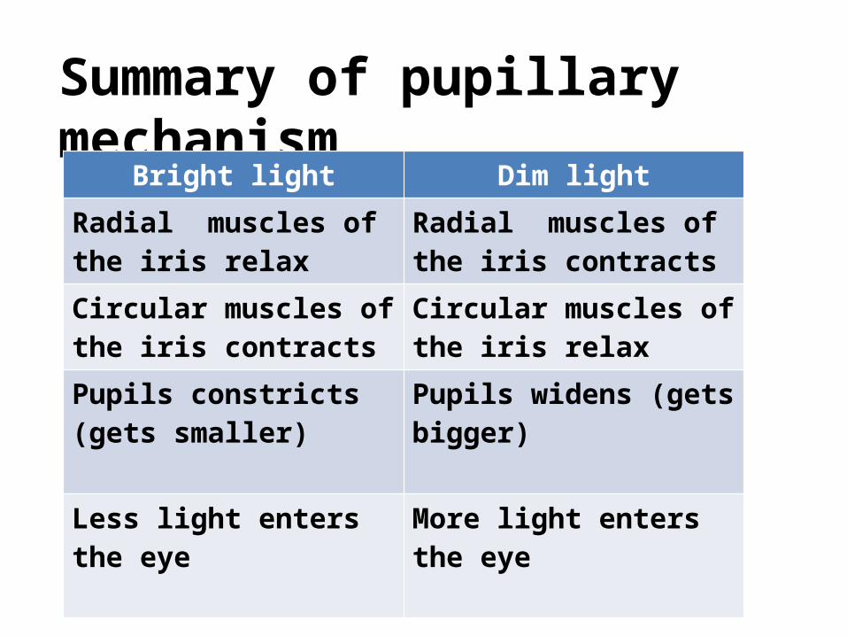
Summary of pupillary mechanismBright light Dim light
Radial muscles of the iris relax
Radial muscles of the iris contracts
Circular muscles of the iris contracts
Circular muscles of the iris relax
Pupils constricts (gets smaller)
Pupils widens (gets bigger)
Less light enters the eye More light enters the eye

Structure of the human ear
The human ear consists of the following three parts

Labelled diagram of the ear
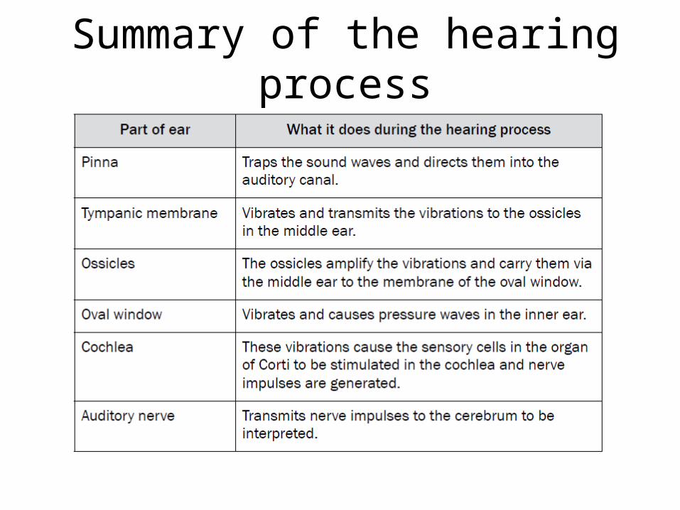
Summary of the hearing process