Embed Size (px)
Citation preview

Vet Clin Small Anim
35 (2005) 147–170
Recent Concepts in Feline LowerUrinary Tract Disease
Roger A. Hostutler, DVM, MS*,Dennis J. Chew, DVM, Stephen P. DiBartola, DVM
Department of Clinical Sciences, The Ohio State University, College of Veterinary Medicine,
Columbus, OH 43210, USA
A clinical description of lower urinary tract disease (LUTD) in cats in1925 [1] accurately described the clinical signs and the disease, and reportedit to be commonplace. The terms feline urologic syndrome (FUS) and felinelower urinary tract disease (FLUTD) have since been used to describe theconstellation of clinical signs related to irritative voiding but do not identifythe underlying etiology. Most cats with LUTD have feline idiopathic orinterstitial cystitis (FIC), but urolithiasis, bacterial urinary tract infection(UTI), anatomic malformations, neoplasia, behavioral disorders, andneurologic problems (eg, reflex dysnergia) may also occur, although moreuncommonly than FIC. Regardless of the underlying etiology, the resultantclinical signs are similar and include dysuria, stranguria, hematuria(macroscopic and microscopic), pollakiuria, and periuria (a word used torefer to urination in inappropriate places).
Obstructive and nonobstructive uropathy are broader concepts that mayalso be used to classify LUTD by the presence or absence of urethralobstruction, respectively. Obstructive uropathy is rare in female cats and isprimarily seen in male cats. The diameter of the urethra and frequency ofobstructive uropathy do not differ between castrated and intact male cats, buturethral obstruction occurs with higher frequency in castrated male cats [2].
Diseases of the lower urinary tract are becoming more apparent clinicallyas indoor cats and multicat households are becoming more common.Inappropriate elimination results in the relinquishment of approximately4 million cats annually to animal shelters because of behavior that isunacceptable to owners [3]. The estimated prevalence of LUTD in primarycare practice in the United States has been reported to be approximately
* Corresponding author.
E-mail address: [email protected] (R.A. Hostutler).
0195-5616/05/$ - see front matter � 2005 Elsevier Inc. All rights reserved.
doi:10.1016/j.cvsm.2004.08.006 vetsmall.theclinics.com
148 HOSTUTLER et al
1.5% [4]. Based on referral institution studies of cats presented withnonobstructive urinary tract disease, the two most common causes havebeen found to be FIC (55%–69%) and urolithiasis (13%–28%) [5,6]. Adiagnosis of FIC is made after routine diagnostic tests, including urinalysis,urine culture and sensitivity, radiography, ultrasonography, and contrastradiography, fail to identify an etiology. If uroendoscopy is performed andsubmucosal petechial hemorrhages (ie, glomerulations) are seen, a diagnosisof FIC is made. This nomenclature is used based on similarities of clinicalsigns seen in human beings with interstitial cystitis.
Recently, many studies have evaluated the effects of dietary andenvironmental factors on development of LUTD in cats. The influence ofbehavioral disturbances and interactions with other cats in development oflower urinary tract signs cannot be overemphasized. Most cats with lowerurinary tract signs are presented between 2 and 6 years of age, and thedisorder is uncommon in cats less than 1 year of age or greater than 10 yearsof age. When looking solely at nonobstructive disease, LUTD occurs withequal frequency in male and female cats. The risk is higher for castrated orspayed cats when adjusted for age, but the age of neutering does not seem tobe clinically relevant [2].
Body weight and diet have been reported to be risk factors whencompared with nonaffected cats. Cats that are obese and sedentary havebeen shown to have a higher incidence of LUTD, as do cats that are fedsolely dry food or fed intermittently throughout the day [2].
Another study found an association with indoor elimination, confine-ment, and sleeping; recent moves; and decreased water intake with thedevelopment of LUTD [7]. Environmental factors, such as interactions withowners, multicat households, and changes in routine, have been associatedwith LUTD and are discussed in detail later. Regardless of the ultimatemanifestations of LUTD, changes in environment, husbandry, and feedingmay decrease the recurrence rate. Recurrence rates have been reported to beas high as 45% within 6 months in male cats with obstructive uropathy [8]and 39% within 1 year in cats with nonobstructive uropathy [9].
Diagnostic workup
No clinical sign or combination of clinical signs is diagnostic ofa particular LUTD in cats. Making a diagnosis involves integrating findingsfrom the signalment, history, physical examination, clinical signs, timecourse of the disease, urinalysis with sediment evaluation, urine culture andsensitivity testing, and urinary tract imaging. The modality of imagingchosen may include a combination of plain abdominal radiography,ultrasonography of the urogenital system (which affords minimal urethralevaluation), contrast radiography, and uroendoscopy (including urethro-scopy and cystoscopy).
149FELINE LOWER URINARY TRACT DISEASE
Consideration of the signalment may be helpful in developing a list ofdifferential diagnoses. It would be uncommon (\5%) for a cat older than 10years of age to develop idiopathic cystitis. Bacterial UTIs are diagnosed inmore than 50% of cats older than 10 years of age presented with lowerurinary tract signs [10]. Likewise, it would be unusual for a young cat todevelop a bacterial UTI. Laboratory evaluation of blood work generally isunremarkable unless other diseases, such as chronic renal failure, arepresent. If concomitant disease is suspected, a complete blood cell count andserum biochemistry profile should be evaluated.
Periuria is a word that has been coined to describe the tendency of catswith irritative voiding to urinate in places other than the litter box(inappropriate urination). Periuria is the most common clinical signreported by owners of cats with LUTD, and these cats often are suspectedby veterinarians to have a behavioral disorder. Approximately half of catswith inappropriate urination as the only client-reported clinical sign havebeen reported to have interstitial cystitis diagnosed by uroendoscopy [6].The time course of the clinical signs also may be helpful in arriving ata diagnosis. Initial bouts of interstitial cystitis generally resolve within 7days with or without treatment. Other diseases, such as urolithiasis andbacterial UTI, often result in clinical signs that are present for longerperiods and may be progressive in severity unless adequate therapy isinstituted.
Urinalysis with sediment evaluation should be performed if there isrecurrence of clinical signs, evidence of underlying chronic renal failure, orprevious urinary catheterization or if a perineal urethrostomy is present.Urine dipstick pads that detect white blood cell (WBC) esterase often arepositive in the absence of pyuria in cats (ie, they frequently yield false-positive results). When evaluating feline urine sediment, care must be takennot to overinterpret the presence of bacteria. Cellular debris may exhibitBrownian motion and be misinterpreted as bacteria. Dilute urine in the faceof pyuria or significant pyuria (>5 WBCs per high-power field [hpf])regardless of urine specific gravity (USG) warrants urine culture andsensitivity testing on urine collected by cystocentesis. The presence ofcrystals in urine sediment may have no clinical importance in cats withouta stone or urethral plug, because crystals do not damage healthy urothelium.Urine that has been refrigerated or stored for hours often contains crystalsin the urinary sediment, and this phenomenon is exaggerated in urine that ishighly concentrated [11].
Plain abdominal radiographs that include the pelvic and penile urethracan be helpful in identifying radiopaque calculi (eg, struvite, oxalate) morethan 3 mm in diameter. Contrast radiography, including cystography,urethrography, and urethrocystography, is indicated in cats with recurrentor lingering clinical signs. Contrast cystography often is normal in FIC, butthe technique may be helpful in detecting small calculi, radiolucent calculi,urachal diverticula and neoplasia as well as in determining bladder wall

150 HOSTUTLER et al
thickness. Occasionally, contrast material may be seen permeating throughthe bladder wall in severe cystitis. Contrast evaluation of the urethragenerally is normal but may be helpful in diagnosing urethral strictures inmale cats and stones in the urethra [12,13]. Generally, these procedures areperformed simultaneously as a contrast urethrocystogram in male cats, thusmaximizing the information the clinician obtains from one procedure.
Abdominal ultrasonography is useful to evaluate the bladder but isunrewarding for evaluation of the entire length of the urethra. Abdominalultrasonography may detect small calculi, radiolucent calculi, and bladdermasses like polyps and neoplasia and may aid in assessing bladder wallthickness if the bladder is sufficiently distended.
Uroendoscopy is a valuable tool in evaluation of cats that have recurrentor persistent clinical signs associated with the lower urinary tract.Uroendoscopy allows visualization of the urethral and bladder mucosa,detection of small calculi not seen on abdominal ultrasonography,evaluation for urachal remnants, and direct visualization of masses thatmay be present. Uroendoscopy of female cats is performed using a rigidpediatric cystoscope, which affords much greater detail and manipulationthan can be obtained with the flexible fiberoptic ureteroscope that is used inmale cats. The rigid cystoscope may be used in male cats that have hada perineal urethrostomy performed and allows the clinician to obtainbiopsies. The 1.1-mm flexible urethroscope used in male cats is not asoptimal for evaluation of the bladder as the 3.0-mm rigid cystoscope, but itis adequate for evaluation of the urethra for the presence of strictures, plugs,spasms, and stones.
Most cases of LUTD in the cat can be managed successfully in primarycare practice. Generally, clinical signs resolve within 7 days. If clinical signspersist or recur repeatedly, further diagnostics and referral to an internist orbehaviorist may be indicated. If a cat has two or three episodes of lowerurinary tract signs within a short period, further imaging and diagnostics,such as uroendoscopy, may be indicated. Veterinary behaviorists can playan important role in evaluation and management of behavioral andenvironmental factors. Strategies to enrich the environment of the cat andlessen stresses associated with multicat households and indoor living shouldbecome a routine part of husbandry of cats (discussed in detail later insection on treatment of interstitial cystitis).
Feline interstitial/idiopathic cystitis
Pathophysiology
Multiple abnormalities of the bladder, central nervous system, andhypothalamic-pituitary-adrenal axis may lead to the clinical manifestationsof FIC. The pelvic and hypogastric nerves and their central connections inthe dorsal horn of the sacral and lumbar spinal cord provide sensory
151FELINE LOWER URINARY TRACT DISEASE
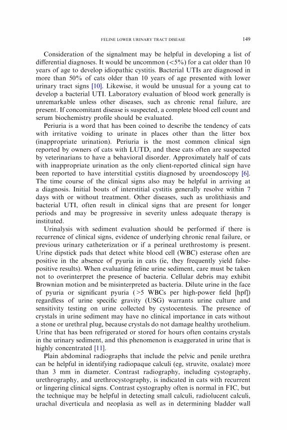
innervation to the bladder [14]. Normal bladder urothelium is lined bya specific glycosaminoglycan (GAG) called GP-51 that is believed to inhibitbacterial adherence and protect the urothelium from noxious urineconstituents. People and animals with interstitial cystitis excrete decreasedamounts of urine GAG [15] and GP-51 [16]. If the GAG layer or urotheliumis compromised, constituents of the urine can contact the sensory nerves andresult in neurogenic bladder inflammation. The sensory neurons are locatedin the submucosa and are composed primarily of unmyelinated pain fibers(C-fibers). Once these sensory fibers are stimulated, action potentials aretransmitted to the spinal cord and are perceived as pelvic pain. In additionto transmitting the sensation of pain to the brain, local axon reflexes thatlead to the release of substance P (SP), a neurotransmitter that results inlocal potentiation of the inflammation, are released. Local SP release resultsin increased vascular permeability by means of direct action on vessel wallsand through SP-mediated release of inflammatory mediators, such ashistamine from mast cells. Receptors for SP also are present on smoothmuscle and cause contraction when stimulated. Figs. 1 and 2 illustrate thenormal bladder and the described changes that lead to the clinical signs seenwith FIC.
Histologic findings in the bladders of FIC-affected cats are typical but notpathognomonic. Changes include edema, hemorrhage, and dilatation ofblood vessels in the submucosa. Increased mast cell density has been reportedin some cats with FIC when toluidine blue stain is applied [17]. Routinehematoxylin and eosin staining of specimens may reveal an intact or partiallydenuded urothelium. Electron microscopy, however, has shown areas thatlack urothelium and distortion of gap junctions [18]. These findings supportthe role of local neurogenic inflammation potentially mediated by SP and thefindings of other studies on increased bladder permeability in cats with FIC[19].
Urine - BladderLumen
Sensory Nerve
GAGLayer
Uroepithelium
MuscularLayer
Fig. 1. Normal bladder with intact urothelium and glycosaminoglycan (GAG) layer. The
urothelium and GAG act as a natural barrier to protect the underlying layers and sensory
nerves from the noxious urine.

152 HOSTUTLER et al
The overall clinical manifestations and high recurrence rates in cats withFIC also seem to involve intimate changes in the neurochemistry of thebrain. The locus coeruleus (LC) [20] and paraventricular nucleus [21]recently have been reported to be involved in the pathogenesis of FIC. TheLC is responsible for excitatory stimulation to the bladder and is activatedon bladder distention [22]. These areas in cats with FIC recently have beenreported to possess increased tyrosine hydroxylase immunoreactivity,suggesting increased catecholamine synthesis [20,21]. Affected animals alsohave increased concentrations of circulating catecholamines [23] at rest andduring stressful situations. a2-Adrenoceptors also seem to play a role in thedevelopment of FIC in cats. Centrally, a2-adrenoceptors are found in theLC and spinal cord, where they inhibit catecholamine release and pain inputto the brain, respectively [24,25]. Peripherally, a2-adrenoceptors are found inthe bladder mucosa, where they are believed to regulate blood flow.Desensitization of the central a2-receptors as a result of chronic stimulationand enhanced catecholamine release from the bladder of cats with FIC hasbeen reported [26] and may result in potentiation of the inflammatoryresponse. Finally, cats with FIC have been shown to have a suboptimalresponse to exogenous corticotropin stimulation when compared withcontrol cats [27], decreased adrenal volume when evaluated by CT [28], anda histologically greater adrenal medulla area than normal cats. Thesefindings suggest that FIC results in overactivation of the sympatheticnervous system with suboptimal activation of the hypothalamic-pituitary-adrenal axis.
Diagnosis
The terms idiopathic cystitis and interstitial cystitis often are usedinterchangeably. Idiopathic cystitis is the most common diagnosis in catswith lower urinary tract signs. The term idiopathic cystitis is used if all
Urine
Mast cells Sensory Nerves
GAG Layer
Uroepithelium
MuscularLayer
Fig. 2. Schematic of bladder of cat affected with feline interstitial cystitis (FIC). This
demonstrates loss of integrity of the glycosaminoglycan layer and urothelium. Loss of integrity
of these layers leads to recruitment of sensory nerves, recruitment and activation of mast cells,
and changes in the central nervous system that lead to the clinical signs seen with FIC.

153FELINE LOWER URINARY TRACT DISEASE
diagnostics fail to confirm the presence of another disease, such asurolithiasis or a bacterial UTI. Idiopathic cystitis generally is seen inmiddle-aged cats and is rarely diagnosed in cats older than 10 years of age,and no gender predisposition has been reported in cats with nonobstructiveFIC [7]. No clinical signs are specific for FIC, but owners of affected catsmost commonly report periuria. Cats may exhibit solely periuria, or theymay also show signs of pollakiuria, stranguria, and hematuria.
Results of radiography and urinalysis often are nonspecific in cats withFIC. Abdominal radiography may be performed to aid in the elimination ofurolithiasis as a differential diagnosis in cats with multiple recurrences ofclinical signs. Double-contrast cystography and positive-contrast urethro-graphy are recommended for cats with recurrent lower urinary tract signs inwhich no cause has been found on urinalysis, urine culture, and plainabdominal radiographs. Results from cats with FIC are normal inapproximately 85% of the cases. Focal or diffuse thickening of the bladderwall is seen in some, and contrast agent may be observed dissecting throughthe bladder wall in a few cases [13]. Ultrasonography is less invasive thanurethrocystography but is less sensitive in the detection of small lesions andprovides little information about the urethra.
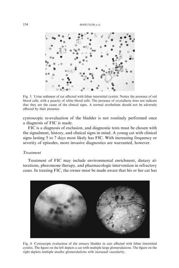
Urinalysis may identify hematuria and proteinuria, the severity of whichcan vary substantially throughout the day or over the course of several days.The absence of hematuria does not exclude a diagnosis of FIC. A paucity ofWBCs is found in the urine sediment. Crystalluria is variable and of nopathologic significance in cats with FIC. Often, crystalluria is an artifact ofrefrigeration and time of storage. Many times, bacteria are reported fromthe laboratory when, in fact, they are not present. This problem is commonin the cat and is caused by particulate material (eg, small crystals, cellulardebris, lipid droplets) that exhibit Brownian motion and may bemisidentified as bacteria. The results of urine cultures in cats with FIC arenegative (ie, no growth or \1000 colony-forming units/mL on urinecollected by cystocentesis). USG should be greater than 1.025 in cats eatingcanned foods and greater than 1.035 in cats eating dry foods. Urinalysisfindings, however, are not specific for any one LUTD. Fig. 3 shows typicalurine sediment found in cats affected with FIC.
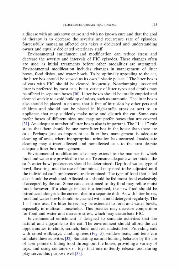
If clinical signs are continuous or frequently recur or if the episodesbecome more severe, direct visualization of the lower urinary tract usinguroendoscopy may be indicated to eliminate other differential diagnoses andto confirm the diagnosis of FIC. If uroendoscopy is performed andsubmucosal petechial hemorrhages (ie, glomerulations) are seen, the terminterstitial cystitis is appropriate (Fig. 4). Glomerulations are not seen in allcats with FIC and may be seen in some sensitive asymptomatic cats thathave undergone recent stress [29]. Other findings on cystoscopy in cats withFIC include edema, debris in the lumen of the bladder, and increasedvascularity. The severity of cystoscopic lesions does not seem to correlatewith the severity of clinical signs observed by the owner; therefore,
154 HOSTUTLER et al
cystoscopic re-evaluation of the bladder is not routinely performed oncea diagnosis of FIC is made.
FIC is a diagnosis of exclusion, and diagnostic tests must be chosen withthe signalment, history, and clinical signs in mind. A young cat with clinicalsigns lasting 5 to 7 days most likely has FIC. With increasing frequency orseverity of episodes, more invasive diagnostics are warranted, however.
Treatment
Treatment of FIC may include environmental enrichment, dietary al-terations, pheromone therapy, and pharmacologic intervention in refractorycases. In treating FIC, the owner must be made aware that his or her cat has
Fig. 3. Urine sediment of cat affected with feline interstitial cystitis. Notice the presence of red
blood cells, with a paucity of white blood cells. The presence of crystalluria does not indicate
that they are the cause of the clinical signs. A normal urothelium should not be adversely
affected by their presence.
Fig. 4. Cystoscopic evaluation of the urinary bladder in cats affected with feline interstitial
cystitis. The figure on the left depicts a cat with multiple large glomerulations. The figure on the
right depicts multiple smaller glomerulations with increased vascularity.
155FELINE LOWER URINARY TRACT DISEASE
a disease with an unknown cause and with no known cure and that the goalof therapy is to decrease the severity and recurrence rate of episodes.Successfully managing affected cats takes a dedicated and understandingowner and equally dedicated veterinary staff.
Environmental enrichment and modification can reduce stress anddecrease the severity and intervals of FIC episodes. These changes oftenare used as initial treatments before other modalities are attempted.Environmental modification includes changes in management of litterboxes, food dishes, and water bowls. To be optimally appealing to the cat,the litter box should be viewed as its own ‘‘plastic palace.’’ The litter boxesof cats with FIC should be cleaned frequently. Nonclumping unscentedlitter is preferred by most cats, but a variety of litter types and depths maybe offered in separate boxes [30]. Litter boxes should be totally emptied andcleaned weekly to avoid buildup of odors, such as ammonia. The litter boxesalso should be placed in an area that is free of intrusion by other pets andchildren and should not be placed in high-traffic areas or next to anappliance that may suddenly make noise and disturb the cat. Some catsprefer boxes of different sizes and may not prefer boxes that are covered[31]. An adequate number of litter boxes also is important. The ‘‘1 þ 1’’ rulestates that there should be one more litter box in the house than there arecats. Perhaps just as important as litter box management is adequatecleaning of areas where inappropriate urination has occurred. Inadequatecleaning may attract affected and nonaffected cats to the area despiteadequate litter box management.
Environmental modification also may extend to the manner in whichfood and water are provided to the cat. To ensure adequate water intake, thecat’s water bowl preferences should be determined. Depth of water, type ofbowl, flavoring, and the use of fountains all may need to be adjusted untilthe individual cat’s preferences are determined. The type of food that is fedalso should be evaluated. Affected cats should be fed moist food exclusivelyif accepted by the cat. Some cats accustomed to dry food may refuse moistfood, however. If a change in diet is attempted, the new food should beintroduced alongside the current diet in a separate dish. As with litter boxes,food and water bowls should be cleaned with a mild detergent regularly. The1 þ 1 rule used for litter boxes may be extended to food and water bowls,especially in multicat households. This practice may decrease competitionfor food and water and decrease stress, which may exacerbate FIC.
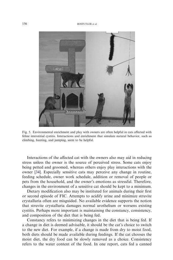
Environmental enrichment is designed to simulate activities that arenatural and enjoyable to the cat. The environment should afford the catopportunities to climb, scratch, hide, and rest undisturbed. Providing catswith raised walkways, climbing trees (Fig. 5), window seats, and tents cansimulate these activities [32]. Simulating natural hunting behavior by the useof laser pointers, hiding food throughout the house, providing a variety oftoys, and using containers or toys that intermittently release food duringplay serves this purpose well [33].
156 HOSTUTLER et al
Interactions of the affected cat with the owners also may aid in reducingstress unless the owner is the source of perceived stress. Some cats enjoybeing petted and groomed, whereas others enjoy play interactions with theowner [34]. Especially sensitive cats may perceive any change in routine,feeding schedule, owner work schedule, addition or removal of people orpets from the household, and the owner’s emotions as stressful. Therefore,changes in the environment of a sensitive cat should be kept to a minimum.
Dietary modification also may be instituted for animals during their firstor second episode of FIC. Attempts to acidify urine and minimize struvitecrystalluria often are misguided. No available evidence supports the notionthat struvite crystalluria damages normal urothelium or worsens existingcystitis. Perhaps more important is maintaining the constancy, consistency,and composition of the diet that is being fed.
Constancy refers to minimizing changes in the diet that is being fed. Ifa change in diet is deemed advisable, it should be the cat’s choice to switchto the new diet. For example, if a change is made from dry to moist food,both diets should be made available during feedings. If the cat chooses themoist diet, the dry food can be slowly removed as a choice. Consistencyrefers to the water content of the food. In one report, cats fed a canned
Fig. 5. Environmental enrichment and play with owners are often helpful in cats affected with
feline interstitial cystitis. Interactions and enrichment that simulate natural behavior, such as
climbing, hunting, and jumping, seem to be helpful.

157FELINE LOWER URINARY TRACT DISEASE
formulation of a food had only an 11% recurrence of signs over a 1-yearperiod, whereas cats fed the dry formulation of the same food had a 39%recurrence rate over the same period [35]. Feeding a canned formulationincreases the amount of water the animal is consuming and decreases USG.As a result, the concentration of potentially noxious substances in urine isreduced. Composition refers to the nutrient content of the diet being fed.Feeding of certain diets may result in excretion of noxious substances in theurine. Highly acidic urine may activate sensory nerve fibers in theurothelium. The optimal diet for cats with FIC has yet to be determined,and no commercially available cat foods are specifically designed for thetreatment of FIC.
Recently, a synthetic formulation of feline facial pheromone (Feliway;Abbott Laboratories, Abbott Park, IL) has been developed to decreaseanxiety-related behavior in cats, including urine marking and destructivescratching. This product also may have salutary benefits for cats with FIC,but such effects have not been reported. One report suggested differentbehavior in hospitalized cats exposed to Feliway compared with placebo-treated cats [36]. Other reports show reduced urine marking during Feliwaytreatment, which may be a consequence of reduced vigilance of the cats,because perception of their environment has been favorably altered [37,38].Although not specifically studied, reduced vigilance likely is related toreduction in activation of the sympathetic nervous system. The use of Feliwaymay be justified in cats with FIC to reduce the impact of an activatedsympathetic nervous system on the disease process. Feline facial pheromoneoften is used in combinationwith environmental enrichment to decrease stressin cats with FIC. Feliway is available as a spray form and, more recently, asa room diffuser. The diffuser form is reported to cover approximately 650 sq ftand lasts for approximately 30 days. The spray form is formulated in anethanol vehicle and may be sprayed in carriers approximately 15 minutesbefore transport, sprayed in cages in a veterinary hospital, or sprayed on areasof inappropriate elimination in the house.
Drug therapy may be indicated if environmental enrichment andmodification in combination with dietary modification, enhanced waterturnover, and feline facial pheromone use do not control clinical signs.Long-term drug use is reserved for the most severely affected cats that havepersistent clinical signs or those that have multiple episodes of FIC. Catssuffering from a current bout of FIC usually are treated with systemicanalgesics. Nonsteroidal anti-inflammatory drugs, such as carprofen andketoprofen, and potent analgesics, such as opioids, including butorphanol,buprenorphine, and fentanyl, seem to be beneficial in short-term pain relief.Scientific evidence to support their routine use in cats with FIC is lacking,however.
Amitriptyline is a tricyclic antidepressant that has been reported to havebenefit in the outcome of cats with FIC that is chronic and has failed othermore routine treatments [29]. Unfortunately, this study was not blinded, and

158 HOSTUTLER et al
no placebo group was included Amitriptyline has many effects. It providesanalgesia by decreasing C-fiber sensory nerve fiber transmission in thebladder, inhibits norepinephrine reuptake in the LC with subsequentdownregulation of norepinephrine outflow, potentially inhibits nociceptiveneurons in the spinal trigeminal nucleus, inhibits serotonin reuptake,stabilizes mast cells, blocks glutamate receptors and sodium channels [39],and may have anticholinergic effects. Amitriptyline was not effective inshort-term treatment of acute FIC in two recent studies [40,41]. In onestudy, clinical signs of FIC were worse in cats treated with amitriptyline,possibly as a result of abrupt withdrawal of treatment after 7 days [40].Extending the duration of therapy may have beneficial effects, however. Theseverity of clinical signs in severely affected cats treated with amitriptyline ata dose of 10 mg daily was dramatically reduced in 60% of affected cats1 year after starting therapy. Because of the potential for hepatotoxicity inpeople, serum biochemistry should be evaluated before and 1 month, 2months, and 6 months after starting amitriptyline. Other adverse effectsinclude urine retention as a result of anticholinergic effects. In the authors’experience, low doses of amitriptyline often are used with promising results.The dosage range is 2.5 to 12.5 mg given orally once daily. A typical startingdose is 5 mg daily, which is effective in many cats. The dosage may be slowlyincreased until a calming effect is seen in addition to resolution of clinicalsigns. If no favorable results are seen after approximately 4 months, thedrug should be gradually tapered and discontinued. Other medications thathave been used for FIC in cats include clomipramine, fluoxetine, andbuspirone. Oral diazepam is not recommended because of its potential tocause hepatic necrosis after oral administration in cats [42]. Glucocorticoidshave not been shown to lessen clinical signs or hasten recovery in cats withFIC.
Oral GAG replacement has been used in people with interstitial cystitiswith minimally favorable results. Theoretically, orally administered GAG isexcreted in the urine and attaches to the defective urothelium, leading todecreased bladder permeability and less neurogenic inflammation. Elmiron(pentosan polysulfate, 100-mg capsules) has been used in human patientswith IC. No evidence is available in veterinary medicine to indicate that suchreplacement decreases the severity or recurrence rate of FIC. GAGreplacements, however, can be considered for treatment of cats with severecystitis in conjunction with other treatments. Adverse effects have not beenobserved with pentosan polysulfate when given to cats at a dose of 50 mgtwice a day. Overdosage theoretically could result in coagulationabnormalities because of the anticoagulant effects of glycosaminoglycans.Adequan (polysulfated glycosaminoglycan) and Cosequin (chondroitinsulfate) are used by some practitioners for treatment of FIC in cats, butsuch use is off label and no reports document the effectiveness of thesetreatments. Fig. 6 depicts an approach to diagnosing and treating cats thathave recurrent episodes of LUTD.

159FELINE LOWER URINARY TRACT DISEASE
Urinary tract infections
Bacterial UTIs are relatively rare in cats. In younger cats, bacterial UTIsaffect less than 2% of cats presented for evaluation of LUTD [6]. In catsolder than 10 years of age presented for evaluation of lower urinary tractsigns, the probability of bacterial UTIs increases to greater than 50%. Catsthat have recurrent UTIs were initially suspected to have reinfection basedon results of susceptibility antibiotograms. More recently, it seems that cats
SignsFrequently Recuror Persist ?
Step 4
+ Amitriptyline (?) + GAG (?) + More Stress / Behavioral Modification + Pain ReliefCystoscopy (?)Repeat Urinalysis
+ Facial pheromones
+ More Stress Reduction + More Water Intake Dynamics
SignsRecur ?
Step 3
Extended Data Base: Urine Culture Contrast RadiographyUltrasonography
Minimum Data Base : Urinalysis & Abdominal X-Ray
Step 2 + Consistent Diet (Canned = BEST) + Increase Water Intake + Stress Reduction
“Watchful” Waiting – Spontaneous Resolution
Client Education – “From the Cat’s Point of View”
Litterbox Management Unscented litter/Texture/Depth Preferences/Aversions Location/Ventilation/Access to Box Increased Cleaning Frequency
Clean Up and Eliminate “Accident” Odors
Urinary History(Vertical vs Horizontal Periuria? Irritative Voidings?)
LowerUrinaryTract
Signs
Fig. 6. Stepwise approach to treatment of cats with idiopathic lower urinary tract signs. More
diagnostics should be performed when cats fail to clear their initial lower urinary tract signs
spontaneously and when signs recur to ensure that the diagnosis is really idiopathic lower
urinary tract disease. Properly controlled clinical trials may provide better approaches to
treatment in the future, but this is what we do in the interim.
160 HOSTUTLER et al
with recurrent UTIs and chronic renal failure suspected of havingreinfections actually have relapsing infection based on genetic analysis ofthe bacteria [43]. Cats that have a concurrent disease, such as diabetesmellitus or chronic renal failure [10,44], are at increased risk for developinga bacterial UTI. Cats that have recently had a urinary catheter placed forobstructive uropathy or other reasons and cats that have had a perinealurethrostomy performed also are at increased risk of developing a UTI. Insuch cases, culture and sensitivity of urine obtained by cystocentesis isa more important diagnostic consideration at first presentation.
Urinalysis with sediment evaluation may be helpful but should not beused alone to definitively diagnose UTI. As mentioned previously,identification of ‘‘bacteria’’ in feline urine sediment is problematic, becausecellular debris commonly found in the urine sediment exhibits Brownianmotion and can easily misinterpreted as bacteria. The presence of diluteurine (USG \1.030) increases the index of suspicion that a UTI may bepresent. Also, the presence of WBCs is not diagnostic of a UTI, but itincreases the index of suspicion. Pyuria may be present with many LUTDs,including FIC, without a concomitant bacterial UTI. Some cats witha confirmed UTI do not have obvious pyuria, especially when the UTIoccurs in association with dilute urine.
If a cat is definitively diagnosed with a UTI based on quantitative urineculture and sensitivity testing, antibacterial therapy should be based onsensitivity results. Treatment with the appropriate antibacterials generally isperformed for 2 to 3 weeks or for 4 to 6 weeks if pyelonephritis is suspectedbased on the physical examination, complete blood cell count, biochemistryprofile, and abdominal ultrasonography. Routine monitoring of catspredisposed to UTIs by use of urine culture is recommended. As many as33% of cats with chronic renal failure can be expected to have or developa UTI during the next 6 months to 1 year.
Special caution is warranted with the use of fluoroquinolones intreatment of cats with a UTI associated with chronic renal failure (CRF).Initial reports of sudden blindness in cats treated with enrofloxacin showedwhat seemed to be a dose-related effect in some cats treated with higherdosages [45]. Some cats treated with lower dosages also developed blindness,but the affected cats were found to have reduced renal function. Afterdosage recommendations were reduced to 5 mg/kg every 24 hours, reportsof blindness decreased dramatically. Cats with renal dysfunction develophigher plasma concentrations of fluoroquinolones and their metabolites.Retinal toxicity of fluoroquinolones may be related to peak concentrationsof drug, which favor enhanced tissue distribution. All fluoroquinolonesdemonstrate dose-dependent retinal toxicity at higher dosages. In cats withCRF and a UTI, a dosage of 3 mg/kg every 24 hours or 2.5 mg/kg every 12hours is recommended to reduce the potential for retinal toxicity. In normalcats, the concentration of fluoroquinolones in urine is high and well abovethe minimum inhibitory concentration for most pathogens. In CRF, the

161FELINE LOWER URINARY TRACT DISEASE
concentration of fluoroquinolones in urine is decreased, but the concentra-tion achieved is still above the minimum inhibitory concentration for mosturopathogens. Whether the reduced dosage regimen achieves tissueconcentrations above the minimum inhibitory concentration for offendingorganisms in the kidney of cats with upper UTI is not known.
Urolithiasis
The formation of uroliths depends on supersaturation of the urine withcalculogenic minerals. If supersaturation is sufficient and sustained, a nidusmay form on which subsequent calculus may develop. The type of calculusformed is dependent on many factors, including renal excretion of minerals,pH of the urine, presence of promoters, absence of inhibitors, concomitantbacterial infections, and possibly underlying inflammation. Clinical signsassociated with urolithiasis generally are similar to those of other LUTDs,but obstruction may occur if the stone becomes lodged in the urethra. Thiscomplication may occur in male and female cats but is much more commonin male cats.
The diagnosis of urolithiasis includes a combination of abdominalpalpation and urinary tract imaging. Routine abdominal radiography ishelpful if the uroliths are large enough (>3 mm) and radiodense. Abdominalultrasonography and double-contrast cystography are beneficial for thedetection of stones that are small (\3mm) or radiolucent. Care must be takennot to assume that urolithiasis is present based on occurrence of crystals in theurine sediment. Likewise, crystals in the urine typically are not the cause oflower urinary tract signs, and one should not equate the type of crystals seenwith the type of urolith that may be present. Crystals may be present withoutdisease, calculi may be present without crystals, and crystals of a different typemay be present in cats with calculi of a specific type. Quantitative stoneanalysis is the only way to ascertain definitively the type of urolith present. Ifuroliths are present, however, the index of suspicion for a particular type isgreatly increased when taking into account the urine pH, presence or absenceof UTI, and crystal type. Definitive long-term treatment of urolithiasisdepends on the type of calculus present. Medical dissolution may beattempted for urate and struvite calculi, but no protocol is available todissolve calcium oxalate calculi. For large calculi or those that do not respondto dissolution protocols, surgical intervention often is required. Voidingurohydropulsion may be attempted for stones up to 5 mm in female cats and1 to 2 mm in male cats. Using this technique in male cats may result inobstruction if the size of the uroliths is underestimated; thus, it should only beperformed by clinicians familiar with the technique.
All calculi that are removed from a cat should be analyzed by a diagnosticlaboratory using quantitative analysis to determine the specific type of urolithpresent. Quantitative analysis is especially useful if amixed urolith (more than
162 HOSTUTLER et al
onemineral) is present. Qualitative analysis should not be performed, becausefrequent false-positive and false-negative results occur and the relativecontribution of the different crystalloids present is not determined.
Urate urolithiasis
Urate urolithiasis accounted for approximately 6% of 20,343 calculievaluated by the University of Minnesota [46]. Portosystemic vascularanomalies can contribute to urate urolithiasis in cats, but the exact patho-genesis in most affected cats remains unknown [47]. Several risk factors, suchas UTI leading to increased urine ammonia, excessive dietary protein, andmetabolic acidosis, have been noted. It is not possible to predict which catsultimately will develop urate urolithiasis, however. The bladder is the mostcommon site for urate calculi, but they also are found in the urethra andkidneys.
Urate calculi generally are radiolucent and are not detected on surveyradiographs unless other mineral constituents are present. Double-contrastcystography and ultrasonography may be used to facilitate detection ofthese calculi. Prevention of urolith formation and dissolution of calculi maybe attempted by combining diets that are low in nucleoproteins (containingpurines) and by the addition of allopurinol. Allopurinol acts by inhibitingthe enzyme xanthine oxidase, which is required for uric acid production. Useof allopurinol may increase the risk of xanthine urolithiasis in the cat,however. The recommended dosage for allopurinol in cats is 9 mg/kg/d [48].If medical dissolution is unsuccessful, as is generally the case in urateurolithiasis secondary to portosystemic shunts, surgical removal orurohydropulsion may be necessary. Correction of the portosystemic shunt,if present, should prevent recurrence.
Struvite urolithiasis
Struvite calculi analyzed at two major laboratories performing quantita-tive analysis far outnumbered oxalate uroliths before the late 1980s. Sincethat time, possibly as a result of a shift by the pet food industry tomagnesium-restricted acidifying diets, struvite calculi have declined to approximately42%, whereas oxalates have increased to approximately 46% of the calculianalyzed [47]. The urine is sterile in approximately 95% of cases of struviteurolithiasis in cats, which is in sharp contrast to the situation in dogs, in whichstruvite urolithiasis is almost always associated with a bacterial UTI.Consequently, struvite urolithiasis in the cat is thought to be metabolic inorigin. Struvite urolithiasis associated with a UTI generally is caused by thepresence of urease-producing bacteria. Urease production results in anincrease in urine pH that favors struvite crystallization in supersaturatedurine. Struvite urolithiasis not associated with a bacterial UTI often isassociated with concentrated urine and possibly with excess consumption andexcretion of calculogenic minerals (especially magnesium) and alkaline urine.

163FELINE LOWER URINARY TRACT DISEASE
A diagnosis of struvite urolithiasis is definitively made by quantitativestone analysis. Urinalysis and urine culture and sensitivity testing areindicated in cats with suspected struvite urolithiasis to determine theunderlying etiology. Struvite uroliths usually are identified on plainabdominal radiographs because they are radiopaque and generally easilyseen. If the calculi are extremely small, ultrasonography and double-contrastcystography may be required to identify them.
Treatment of struvite urolithiasis can include surgical removal of calculi,voiding urohydropulsion, or medical calculolysis depending on the in-dividual situation. Increasing water intake is imperative in medicalmanagement of urolithiasis to promote formation of urine that is notsupersaturated with calculogenic minerals. One commercially availablecalculolytic diet (Hills s/d, Science Diet, Topeka, KS) specifically designedfor cats is acidifying, magnesium restricted, and supplemented with salt andhas been reported to be effective in cats fed the canned preparation. Unlikethe similar calculolytic diet for dogs, the formulation devised for cats is notrestricted in protein. While feeding a calculolytic diet, it is important toemphasize to the owner that no other foods, including treats, should begiven. The goal is to achieve a urine pH less than 6.3 and USG less than1.030. During therapy, abdominal radiographs should be re-evaluated at3-week intervals to ensure that therapy is working. In cats in whichconcomitant bacterial UTI is present, appropriate antibiotics should begiven during dissolution and for 2 weeks after uroliths are no longerapparent radiographically. The average time for dissolution of struvitecalculi in cats without infection was 36 days (range: 14–141 days), and inthose with UTI, it was 44 days (range: 14–92 days) [49]. If uroliths persist orincrease in size despite adequate dissolution therapy, the initial diagnosismust be questioned or the possibility of a mixed urolith should beconsidered. Occasionally, medical dissolution can be used to decrease thesize of calculi so that voiding urohydropulsion can be employed.
After clinical signs have abated and dissolution is complete, routinemonitoring by urinalysis and abdominal radiography may be indicated. Incats predisposed to bacterial UTIs (ie, those with chronic renal failure,diabetes mellitus, or perineal urethrostomy), periodic urine cultures arewarranted. In all cats that repeatedly form uroliths, regardless of type,decreasing USG by feeding canned cat foods is indicated if the cat can besuccessfully transitioned to a moist diet. Many commercial diets have beendesigned to prevent formation of new struvite stones, but no reports confirmthe effectiveness of any of these diets.
Oxalate urolithiasis
Calcium oxalate uroliths have become the most frequent type of urolithin cats based on calculi submitted to laboratories for quantitative analyses.The percentage of uroliths from cats analyzed at the University of

164 HOSTUTLER et al
Minnesota Urolith Center that were oxalates increased from approximately2% to more than 40% over an 11-year period [50]. This shift may have beenassociated with a change in diet formulation by the pet food industry in anattempt to decrease the formation of sterile struvite uroliths by decreasingthe magnesium and increasing the acid content of the diets. This strategycould have uncovered a group of cats predisposed to calcium oxalate stoneformation not previously identified because they had not been exposed toa provocative environment. Calcium oxalate urolithiasis generally occurs inolder cats (7–10 years of age) [51], frequently recurs, and generally is notassociated with a bacterial UTI. Breeds that have been reported to be at anincreased risk for calcium oxalate uroliths include the Ragdoll, BritishShorthair, Foreign Shorthair, Himalayan, Havana Brown, Scottish Fold,Persian, and Exotic Shorthair. Birman, mixed-breed, Abyssinian, andSiamese cats have been reported to have a lower risk for developing calciumoxalate uroliths [51]. Indoor housing also has been reported as a risk factorfor calcium oxalate urolithiasis [52]. This risk factor may be a consequenceof decreased voiding and water intake.
Other than the previously mentioned dietary factors, the etiology ofcalcium oxalate urolith formation generally is unknown. Systemic metabolicderangements, such as acidosis and hypercalcemia, seem to increase the risk,however. Systemic acidosis results in release of calcium carbonate from bone(a normal buffering response) and secondary calciuresis. Acidosis also maydecrease the urinary excretion of citrate, an inhibitor of calcium oxalateurolith formation. All cats that are presented with calcium oxalateurolithiasis should have their serum calcium concentration evaluated.Systemic hypercalcemia results in increased calciuresis and may increase therisk of urolith formation. As many as 35% of calcium oxalate stone-formingcats evaluated at the University of Minnesota Urolith Center have beennoted to have hypercalcemia [50]; many of these cats likely had idiopathichypercalcemia. If the hypercalcemia is not corrected, it is likely that calciumoxalate urolithiasis will recur.
Currently, no medical dissolution protocol is available for calciumoxalate calculi. If the uroliths are not voided and clinical signs are present,voiding urohydropulsion or surgical intervention is indicated. After surgicalremoval, a nonacidifying diet that is low in calcium and oxalate should befed. Phosphorus should not be restricted because of the potential forincreased gut absorption of calcium and secondary calciuresis arising asa result of low serum phosphorus concentration, and magnesium should notbe restricted because of its inhibitory effect on oxalate urolith formation.Excessive supplementation with sodium to stimulate water consumption isnot indicated because of potential augmentation of calciuresis. Potassiumcitrate (100–150 mg/kg/d) may be helpful in decreasing recurrence becauseof the inhibitory effects of citrate on calcium oxalate stone formation and itsalkalinizing effect. This effect assumes that some of the administered citratewill be excreted unmetabolized into the urine. Reports documenting the
165FELINE LOWER URINARY TRACT DISEASE
effectiveness of this treatment are lacking, however. Increasing waterconsumption by feeding canned food if possible is paramount in themanagement of urolithiasis.
Several commercially available diets have been developed that aredesigned to prevent recurrence of calcium oxalate calculi. No evidence-based outcome studies have been reported showing the effectiveness of anyof these diets to prevent recurrent urolith formation. These diets have beendeveloped based on the assumption that less urinary acidification isbeneficial. Some companies have data indicating that dietary changes alterthe relative supersaturation or activity product ratio of urine from normalcats fed these diets. Relative supersaturation and activity product ratio dataprovide surrogate information about the possibility of decreasing recurrenceof urolithiasis in clinically affected cats.
Urethral obstruction
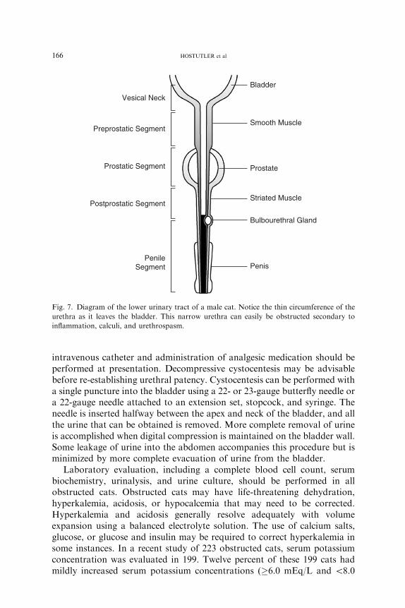
The most common cause of urethral obstruction in male cats was urethralplugs in one study [5] and idiopathic disease in a more recent report [53].When evaluated with fiberoptic urethroscopy, plugs were identified inapproximately 30% of obstructed cats in a preliminary study at The OhioState University (K.L. Cannizzo, DVM, MS; D.J. Chew, DVM, un-published observations). Other potential causes include urolithiasis with orwithout a bacterial UTI, urethral spasm, and, rarely, stricture or neoplasia.Male cats are greatly predisposed to urethral obstruction compared withfemale cats because of their extremely narrow penile urethra (Fig. 7). Large(>5 mm) uroliths cause obstruction of the female urethra, however.
The exact pathogenesis of urethral plugs has not been definitively proven.One theory is that the occurrence of UTI or inflammation with crystallurialeads to the aggregation of protein, crystals, WBCs, and red blood cells,which, in turn, are surrounded by amorphous material, leading to plugformation. Another theory suggests that chronic bladder inflammation leadsto a decrease in vascular integrity. Loss of vascular integrity then leads to anincrease in urine protein concentration, increased urine pH, crystalluria,and, ultimately, plug formation. Urethritis without plug formation is severein some cats with urethral obstruction examined by urethroscopy. It is notknown what role, if any, calicivirus-like particles seen by electronmicroscopy of urethral plugs play in the pathogenesis of urethral plugdevelopment. Any plug that is obtained after re-establishment of patencyshould be evaluated for composition by quantitative analysis. Urethralplugs generally are composed of struvite crystals. This observationcontinues to be true, despite the increased frequency of calcium oxalatecalculi and, presumably, calcium oxalate crystalluria.
At presentation, an obstructed cat should be treated on an emergencybasis. Cats that have been obstructed for more than 48 hours are most likelyto be severely ill and require uremic crisis management. Placement of an
166 HOSTUTLER et al
intravenous catheter and administration of analgesic medication should beperformed at presentation. Decompressive cystocentesis may be advisablebefore re-establishing urethral patency. Cystocentesis can be performed witha single puncture into the bladder using a 22- or 23-gauge butterfly needle ora 22-gauge needle attached to an extension set, stopcock, and syringe. Theneedle is inserted halfway between the apex and neck of the bladder, and allthe urine that can be obtained is removed. More complete removal of urineis accomplished when digital compression is maintained on the bladder wall.Some leakage of urine into the abdomen accompanies this procedure but isminimized by more complete evacuation of urine from the bladder.
Laboratory evaluation, including a complete blood cell count, serumbiochemistry, urinalysis, and urine culture, should be performed in allobstructed cats. Obstructed cats may have life-threatening dehydration,hyperkalemia, acidosis, or hypocalcemia that may need to be corrected.Hyperkalemia and acidosis generally resolve adequately with volumeexpansion using a balanced electrolyte solution. The use of calcium salts,glucose, or glucose and insulin may be required to correct hyperkalemia insome instances. In a recent study of 223 obstructed cats, serum potassiumconcentration was evaluated in 199. Twelve percent of these 199 cats hadmildly increased serum potassium concentrations (�6.0 mEq/L and \8.0
Vesical Neck
Preprostatic Segment
Prostatic Segment
Postprostatic Segment
PenileSegment
Bladder
Smooth Muscle
Prostate
Striated Muscle
Bulbourethral Gland
Penis
Fig. 7. Diagram of the lower urinary tract of a male cat. Notice the thin circumference of the
urethra as it leaves the bladder. This narrow urethra can easily be obstructed secondary to
inflammation, calculi, and urethrospasm.
167FELINE LOWER URINARY TRACT DISEASE
mEq/L), 11.6% had potassium concentrations greater than or equal to8.0 mEq/L and less than 10.0 mEq/L, and 0.5% had serum potassiumconcentrations greater than 10.0 mEq/L [54]. One study reported a 75%frequency of ionized hypocalcemia in cats with urethral obstruction [55].The presence of severe metabolic acidosis as determined by blood gasanalysis (pH\7.1) may necessitate sodium bicarbonate administration.
Establishment of urethral patency is obtained after the patient is stabilizedand properly sedated or anesthetized based on its clinical condition andoverall stability. During establishment of patency, the penis should behandled gently to avoid aggravating inflammation. After sedation and gentlepenile manipulation or massage, a urethral plug or extremely small calculicontributing to the obstruction may be expelled. All cats that are presentedwith urethral obstruction may not need placement of an indwelling urinarycatheter depending on the quality of the urethral stream and presence orabsence of systemic illness. If the animal is moribund, has a severely largebladder, or has severe azotemia or other metabolic derangements, catheterplacement is essential for adequate patient management.
Marked postobstructive diuresis may occur in cats that were obstructedfor several days or are severely azotemic. The degree of postobstructivediuresis is often proportional to the degree of azotemia. A balancedelectrolyte solution, such as lactated Ringer’s solution or Plasmalyte, oftenis adequate for rehydration and stabilization. Urine output should bemonitored to ensure that dehydration does not occur because of themagnitude of diuresis. Management of postobstructive diuresis bymonitoring the patient’s input and output (‘‘ins and outs’’) may be needed.This procedure is accomplished by providing sensible and insensible fluidneeds. Insensible losses cannot be measured and are generally considered tobe 10 mL/lb/d. Sensible losses are losses, such as urine, that can be easilymeasured. The insensible loss replacement is kept constant, and the sensiblelosses generally are measured for a given period (eg, 2–4 hours) and thenreplaced over the following time interval. Fluids may be gradually taperedafter azotemia resolves. After the cat is stabilized and while the catheter is stillin place, acepromazine (0.02–0.05mg/kg every 4–6 hours) and buprenorphine(5–20 lg/kg) or butorphanol (0.2–0.4 mg/kg every 6–8 hours) can beadministered in cats with urethral obstruction. These medications aid inrelaxing the urethral sphincter and provide pain relief. a1-Antagonists, such asphenoxybenzamine (2.5–7.5 mg every 12–24 hours) and prazosin (0.5 mgevery 8 hours), may be added to decrease urethral tone as an alternative toacepromazine. In animals that have bladder atony secondary to severeprolonged distention of the bladder, parasympathomimetic drugs, such asbethanecol (1.25–5.0 mg every 12 hours) may be added once urethral patencyhas been established. After successfulmedicalmanagement, owner counselingabout long-term medical treatment of FIC is necessary if it is suspected as theunderlying cause. Fig. 8 depicts an approach to treatment of a severelyobstructed cat on initial presentation.

168 HOSTUTLER et al
If medical management fails despite exhaustive treatment or in recurrentsevere episodes of urethral obstruction, perineal urethrostomy may beindicated. This surgery is used only in severely affected animals and onlyafter extensive owner education about the potential complications, such asurinary incontinence and stricture formation (both of which are unlikely)and increased risk of ascending UTIs [56].
References
[1] Kirk H. Urinary deposits and retention. In: The diseases of the cat and it general
management. London: Bailliere, Tindall and Cox; 1925. p. 261–7.
[2] Willeberg P. Epidemiology of naturally-occurring feline urologic syndrome. Vet Clin N Am
Small Anim Prac 1984;14:455–69.
[3] Patronek GJ, Glickman LT, Beck AM, et al. Risk factors for relinquishment of cats to an
animal shelter. J Am Vet Med Assoc 1996;209:582–8.
[4] Lund EM, Armstrong PJ, Kirk CA, et al. Health status and population characteristics of
dogs and cats examined at private veterinary practices in the United States. J Am Vet Med
Assoc 1999;214:1336–41.
[5] Kruger JM, Osborne CA, Goyal SM, et al. Clinical evaluation of cats with lower urinary
tract disease. J Am Vet Med Assoc 1991;199:211–6.
[6] Buffington CA, ChewDJ, Kendall MS, et al. Clinical evaluation of cats with nonobstructive
lower urinary tract diseases. J Am Vet Med Assoc 1997;210:46–50.
[7] Jones B, Sanson RL, Morris RS. Elucidating the risk factors of feline urologic syndrome.
NZ Vet J 1997;45:100–8.
[8] Bovee KC, Reif JS, Maguire TG, et al. Recurrence of feline urethral obstruction. J Am Vet
Med Assoc 1979;174:93–6.
[9] Barker J, Povey RC. The feline urolithiasis syndrome: a review and an inquiry into the
alleged role of dry cat food in its aetiology. J Small Anim Pract 1973;14:445–57.
MoribundCat
IV Line TherapeuticCystocentesis
ECG Stat Bloods
DehydrationVolume
Expansion
Pass Urethral Catheter
Collect/MonitorUrine Output
HyperkalemiaRescue?
HypocalcemiaRescue?
RadiographsAbdominal/Perineal
Fig. 8. Algorithm approach to an obstructed cat. Decompressive cystocentesis, fluid therapy,
and baseline blood work (including blood gas analysis with electrolytes) should be performed at
presentation. Obtaining patency of the urethra should be performed after other life-saving
measures and diagnostics are completed.
169FELINE LOWER URINARY TRACT DISEASE
[10] Bartges JW. Lower urinary tract disease in geriatric cats. In: Proceedings of the 15th
American College of Veterinary Internal Medicine Forum; 1997. p. 322–4.
[11] Sturgess CP, Hesford A, Owen H, et al. An investigation into the effects of storage on the
diagnosis of crystalluria in cats. J Feline Med Surg 2001;3:81–5.
[12] Scrivani PV, Chew DJ, Buffington CA, et al. Results of double-contrast cystography in cats
with idiopathic cystitis: 45 cases (1993–1995). J Am Vet Med Assoc 1998;212:1907–9.
[13] Scrivani PV, Chew DJ, Buffington CA, et al. Results of retrograde urethrography in cats
with idiopathic, nonobstructive lower urinary tract disease and their association with
pathogenesis. J Am Vet Med Assoc 1997;211:741–8.
[14] de Groat WC, Yoshimura N. Pharmacology of the lower urinary tract. Annu Rev
Pharmacol Toxicol 2001;41:691–721.
[15] BuffingtonCA, Blaisdell JL, Binns SP Jr, et al. Decreased urine glycosaminoglycan excretion
in cats with interstitial cystitis. J Urol 1996;155:1801–4.
[16] Byrne DS, Sedor JF, Estojak J, et al. The urinary glycoprotein GP51 as a clinical marker for
interstitial cystitis. J Urol 1999;161:1786–90.
[17] Buffington CA, Chew DJ, Woodworth BE. Animal model of human disease—feline
interstitial cystitis. Comp Pathol Bull 1997;29:3, 6.
[18] Lavelle JP, Meyers SA, Ruiz WG, et al. Urothelial pathophysiological changes in feline
interstitial cystitis: a human model. Am J Physiol Renal Physiol 2000;278(Suppl):F540–53.
[19] Gao X, Buffington CA, Au JL. Effect of interstitial cystitis on drug absorption from the
urinary bladder. J Pharmacol Exp Ther 1994;271:818–23.
[20] Reche AJ, Buffington CA. Increased tyrosine hydroxylase immunoreactivity in the locus
coeruleus of cats with interstitial cystitis. J Urol 1998;159:1045–8.
[21] Welk K, Buffington CA. Effects of interstitial cystitis on central neuropeptide and receptor
immunoreactivity in cats. Columbus: The Ohio State University; 2003. p. 31.
[22] de Groat WC, Booth AM, Yoshimura N. Neurophysiology of micturition and
its modification in animal models of human disease. In: Maggi CA, Hill CE, editors.
Nervous control of the urogenital system. The autonomic nervous system. Chur: Harwood;
1993. p. 227–90.
[23] Buffington CA, Pacak K. Increased plasma norepinephrine concentration in cats with
interstitial cystitis. J Urol 2001;165:2051–4.
[24] Stevens CW, Brenner GM. Spinal administration of adrenergic agents produces analgesia in
amphibians. Eur J Pharmacol 1996;316:205–10.
[25] Sabbe MB, Penning JP, Ozaki GT, et al. Spinal and systemic action of the alpha 2 receptor
agonist dexmedetomidine in dogs. Antinociception and carbon dioxide response.
Anesthesiology 1994;80:1057–72.
[26] Petrovaara A, Kauppila T, Jyvasjarvi E, et al. Involvement of supraspinal and spinal
segmental alpha-2-adrenergic mechanisms in the medetomidine-induced antinociception.
Neuroscience 1991;44:705–14.
[27] Westropp JL, Buffington CA. Evaluation of the hypothalamic-pituitary-adrenal axis in cats
with FIC. Presented at 15th Annual American College of Veterinary Internal Medicine
Forum. American College of Veterinary Internal Medicine; 2003.
[28] Westropp JL, Welk K, Buffington CA. Small adrenal glands in cats with feline interstitial
cystitis. J Urol 2002;170:2492–7.
[29] Chew DJ, Buffington CA, Kendall MS, et al. Amitriptyline treatment for severe recurrent
idiopathic cystitis in cats. J Am Vet Med Assoc 1998;213:1282–6.
[30] Neilson JC. Feline house soiling: elimination and marking behaviors. Vet Clin N Am Small
Anim Pract 2003;33:287–301.
[31] Overall KL. Feline elimination disorders. In: Overall KL, editor. Clinical behavioral
medicine for small animals. St. Louis: Mosby; 1997. p. 160–94.
[32] Delzio S, Ribarich C. Felinestein. New York: Harper Perennial; 1999.
[33] McCune S. Environmental enrichment for cats—a review. Second International Conference
on Environmental Enrichment, 1997.
170 HOSTUTLER et al
[34] Turner DC. The human-cat relationship. In: Bateson P, editor. The domestic cat—the biol-
ogy of its behavior. 2nd edition. Cambridge: Cambridge University Press; 2000. p. 194–206.
[35] Markwell PJ, Buffington CA, Chew DJ, et al. Clinical evaluation of commercially available
urinary acidification diets in the management of idiopathic cystitis in cats. J Am Vet Med
Assoc 1999;214:361–5.
[36] Griffith CA, Steigerwald ES, Buffington CA. Effects of a synthetic facial pheromone on
behavior of cats. J Am Vet Med Assoc 2000;217:1154–6.
[37] Mills DS, White JC. Long-term follow up of the effect of a pheromone therapy on feline
spraying behaviour. Vet Rec 2000;147:746–7.
[38] Hunthausen W. Evaluating a feline facial pheromone analogue to control urine spraying.
Vet Med 1998;143:151–6.
[39] Pena F, Neaga E, Amuzescu B, et al. Amitriptyline has a dual effect on the conductive
properties of the epithelial Na channel. J Pharm Pharmacol 2002;54:1393–8.
[40] Kruger JM, Conway TS, Kaneene JB, et al. Randomized controlled trial of the efficacy of
short-term amitriptyline administration for treatment of acute, nonobstructive, idiopathic
lower urinary tract disease in cats. J Am Vet Med Assoc 2003;222:749–58.
[41] Kraijer M, Fink-Grimmels J, Nickel RF. The short-term clinical efficacy of amitriptyline in
the management of idiopathic feline lower urinary tract disease: a controlled clinical study.
J Feline Med Surg 2003;5(3):191–6.
[42] Center SA, Elston TH, Rowland PH, et al. Fulminant hepatic failure associated with oral
administration of diazepam in 11 cats. J Am Vet Med Assoc 1996;209:618–25.
[43] Freitag T, Squires RA, Schmid J, et al. Antibiotic sensitivity profiles underestimate the
proportion of relapsing infections in cats with chronic renal failure and urinary tract
infection [abstract 10]. Presented at the American College of Veterinary Medicine Forum.
Minneapolis, June 9–12, 2004.
[44] Lulich JP, Osborne CA, O’Brien TD, et al. Feline renal failure: questions, answers,
questions. Compend Contin Educ Pract Vet 1992;14:127–53.
[45] Gelatt KN, van der Woerdt A, Ketring KL, et al. Enrofloxacin-associated retinal
degeneration in cats. Vet Ophthalmol 2001;4(2):99–106.
[46] Osborne CA, Kruger JM, Lulich J, et al. Feline lower urinary tract diseases. In: Ettinger SJ,
Feldman E, editors. Textbook of veterinary internal medicine. Philadelphia: WB Saunders;
2000. p. 1710–47.
[47] Duval D, Barsanti JA, Cornelius LM, et al. Ammonium acid urate urolithiasis in a cat.
Feline Pract 1995;23:18–22.
[48] PlumbDC.Allopurinol. In: Plumb veterinary drug handbook. 4th edition.Ames: Iowa State
University Press; 2002. p. 20.
[49] OsborneCA,Lulich JP,Kruger JM, et al.Medical dissolution of feline struvite urocystoliths.
J Am Vet Med Assoc 1990;196:1053–63.
[50] Osborne CA, Lulich JP, Thumachi R, et al. Feline urolithiasis. Etiology and pathophys-
iology. Vet Clin N Am Small Anim Pract 1996;26:217–32.
[51] Lekcharoensuk C, Lulich JP, Osborne CA, et al. Association between patient-related factors
and risk of calcium oxalate and magnesium ammonium phosphate urolithiasis in cats. J Am
Vet Med Assoc 2000;217:520–5.
[52] Kirk CA, Ling GV, Franti CE, et al. Evaluation of factors associated with development of
calcium oxalate urolithiasis in cats. J Am Vet Med Assoc 1995;207:1429–34.
[53] Gerber B. Short-term followup of cats with obstructive lower urinary tract disease. In:
Proceedings of the 13th EuropeanCongress of Veterinary InternalMedicine. Uppsala; 2003.
[54] Lee JA, Drobatz KJ. Characterization of the clinical characteristics, electrolytes, acid-base,
and renal parameters in male cats with urethral obstruction. J Vet Emerg Crit Care 2003;
13:227–33.
[55] Drobatz KJ, Hughes D. Concentration of ionized calcium in plasma from cats with urethral
obstruction. J Am Vet Med Assoc 1997;211:1392–5.
[56] Smith CW. Perineal urethrostomy. Vet Clin N Am Small Anim Pract 2002;32(4):917–25.