Embed Size (px)
Citation preview

Recent Advances in Small Animal SPECT Recent Advances in Small Animal SPECT instrumentations and techniquesinstrumentations and techniques
F. Cusanno1, M. Ballerini1, E. Cisbani1, S. Colilli1, R. Fratoni1, F. Garibaldi1, F. Giuliani1, M. Gricia1, M. Lucentini1, M. L. Magliozzi1,2, S. Majewski3, G. P. K. Mok4,
F. Santavenere1, S. Torrioli1,2, B. M. W. Tsui4, P. Veneroni1, Y. Wang4
Frontiers in Imaging Science: High Performance Nuclear Medicine Imagers for Vascular Disease Imaging , Rome, 13 – 14 November 2006
1. Istituto Superiore di Sanità, Rome, ITALY2. Università La Sapienza, Rome, ITALY 3. Jefferson Lab, Newport News VA, USA4. Johns Hopkins University, Baltimora MD, USA
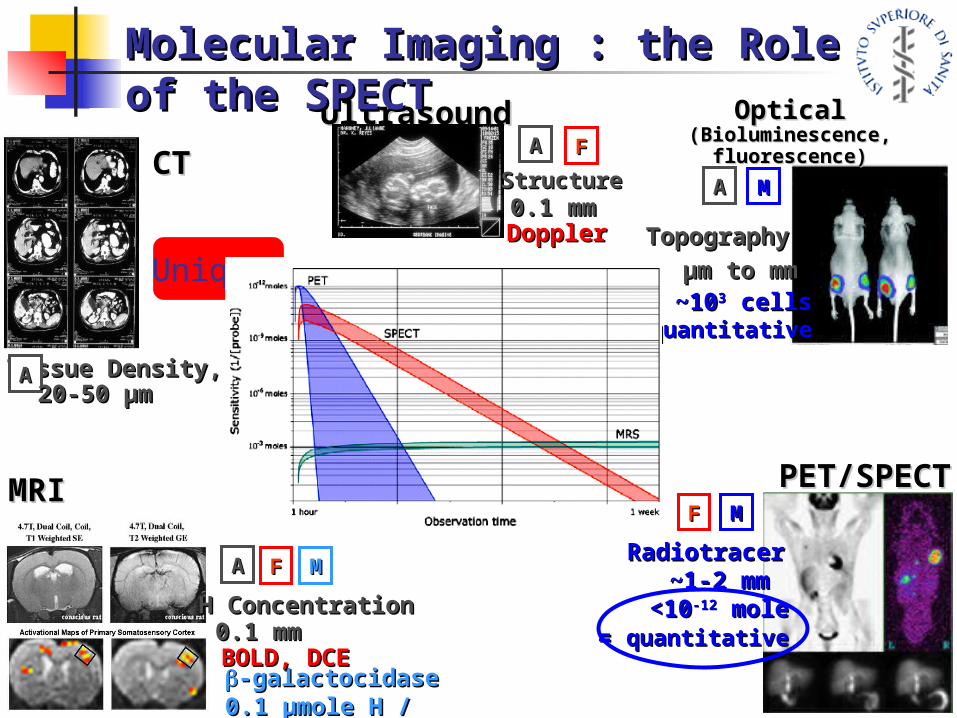
CTCT
Tissue Density, ZTissue Density, ZAA20-50 µm20-50 µm
-galactocidase-galactocidase0.1 µmole H / µmole 0.1 µmole H / µmole 3131PP
MRIMRI
AA
H ConcentrationH Concentration
MMFF
BOLD, DCEBOLD, DCE0.1 mm0.1 mm
UltrasoundUltrasound
StructureStructure
AA FF
DopplerDoppler0.1 mm0.1 mm
OpticalOptical(Bioluminescence, (Bioluminescence,
fluorescence)fluorescence)
AA
TopographyTopography
MM
~10~1033 cells cells quantitativequantitative
µm to mmµm to mm
PET/SPECTPET/SPECT
RadiotracerRadiotracer
MM
~1-2 mm~1-2 mm<10<10-12-12 mole mole
= = quantitativequantitative
FF
Molecular Imaging : the Role Molecular Imaging : the Role of the SPECTof the SPECT
Unique!
Monte Carlo to Design Advanced Monte Carlo to Design Advanced DetectorsDetectors
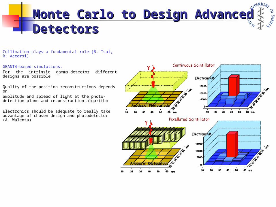
Collimation plays a fundamental role (B. Tsui, R. Accorsi)
GEANT4-based simulations:For the intrinsic gamma-detector different designs are possible
Quality of the position reconstructions depends onamplitude and spread of light at the photo-detection plane and reconstruction algorithm
Electronics should be adequate to really take advantage of chosen design and photodetector (A. Walenta)
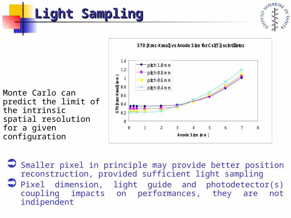
STD[Xrec-Xreal] vs Anode Size for CsI(Tl) scintillator
0
0.2
0.4
0.6
0.8
1
1.2
1.4
0 1 2 3 4 5 6 7 8
Anode Size (mm)
STD
[Xre
c-X
real
] (m
m)
pitch 1.0 mm
pitch 0.8 mm
pitch 0.6 mm
pitch 0.4 mm
Smaller pixel in principle may provide better position reconstruction, provided sufficient light sampling
Pixel dimension, light guide and photodetector(s) coupling impacts on performances, they are not indipendent
Monte Carlo can predict the limit of the intrinsic spatial resolution for a given configuration
Light SamplingLight Sampling

Array Image and Map Array Image and Map Reconstruction (1)Reconstruction (1)
The Present

Possible Future
Array Image and Map Array Image and Map Reconstruction (2)Reconstruction (2)
Application: Atherosclerotic Application: Atherosclerotic Plaques in MicePlaques in Mice
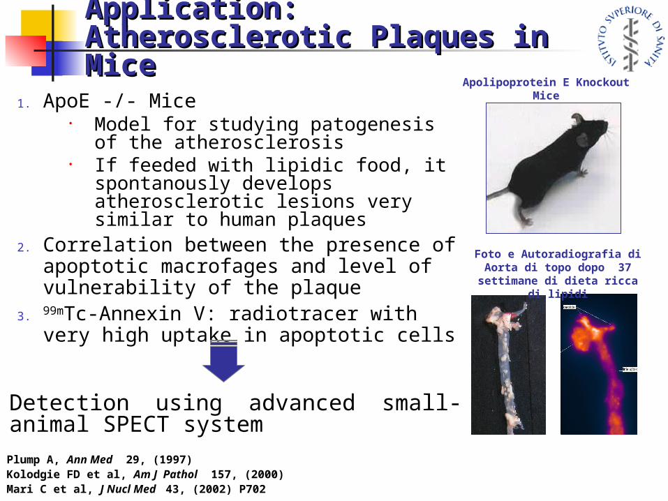
1. ApoE -/- Mice• Model for studying patogenesis of
the atherosclerosis• If feeded with lipidic food, it
spontanously develops atherosclerotic lesions very similar to human plaques
2. Correlation between the presence of apoptotic macrofages and level of vulnerability of the plaque
3. 99mTc-Annexin V: radiotracer with very high uptake in apoptotic cells
Foto e Autoradiografia di Aorta di topo dopo 37 settimane di
dieta ricca di lipidi
Apolipoprotein E Knockout Mice
Detection using advanced small-animal SPECT systemPlump A, Ann Med 29, (1997)Kolodgie FD et al, Am J Pathol 157, (2000)Mari C et al, J Nucl Med 43, (2002) P702
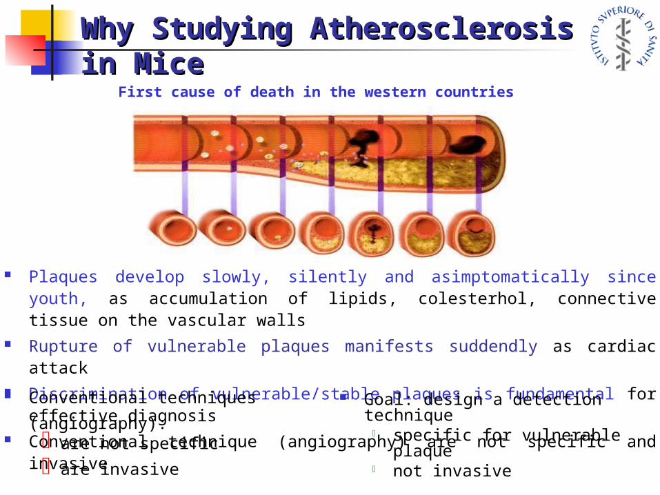
Why Studying Atherosclerosis Why Studying Atherosclerosis in Micein Mice
First cause of death in the western countries
Plaques develop slowly, silently and asimptomatically since youth, as accumulation of lipids, colesterhol, connective tissue on the vascular walls
Rupture of vulnerable plaques manifests suddendly as cardiac attack Discrimination of vulnerable/stable plaques is fundamental for effective
diagnosis Conventional technique (angiography) are not specific and invasive Conventional techniques
(angiography):are not specific
are invasive
Goal: design a detection technique specific for vulnerable plaque not invasive

Parameters for SimulationParameters for Simulation
Blankenberg FG et al, PNAS 95, (1998)
Collimator: Pinhole, 0.3˚mm aperture, magnification factor 3
Scintillator: CsI(Tl) pitch 0.5/0.8˚mm, 3˚mm thickness
CsI(Na) pitch 0.8˚mm, 3˚mm thicknessLaBr3(Ce) continuous, 3 − 5 mm thickness
PS-PMT (“offline”): Flat Panel H9500, anode size 3.0˚mmBurle Planacon 85021, anode side 1.5 mm
Assumed radiotracer distribution:Liver 12%, Kidneys 56 %, Spleen 2%, 5% ejected25 % is distributed in the tissue
H3.0 mCi di Tc99m-Annexin-V,
HPhantom size 8×3×2 cm3
Surrounding Tissue Uptake ~ 600 Bq/mm3
Plaque Uptake ~ 10÷20*600 Bq/mm3
H Plaque size ~ 0.5 × 1 × 4 mm3
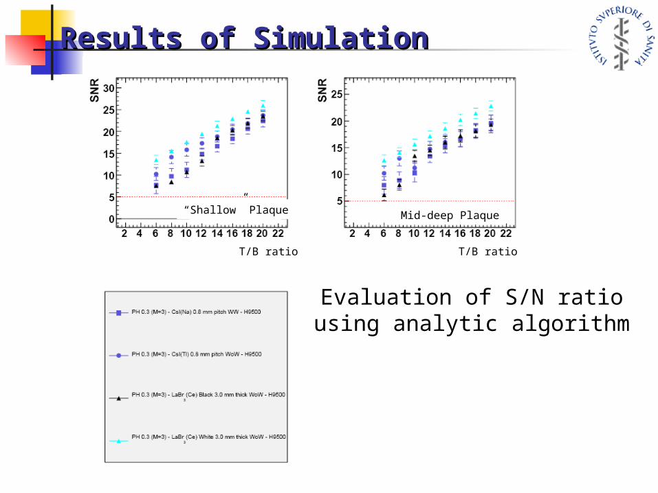
Results of SimulationResults of Simulation
T/B ratio T/B ratio
“Shallow” Plaque Mid-deep Plaque
Evaluation of S/N ratio using analytic algorithm

Preliminary Studies @ JHU (1)Preliminary Studies @ JHU (1)
Spatial Resolution Spatial Resolution 0.6 mm 0.6 mm
Detector setup:
tungsten PinHole 0.3 mm aperture, magnification
factor 3
Scintillator: CsI(Tl) 1.0 mm pitch
Photodetector: 2x2 H9500 array
Electronics: IDEAS F.E.C. 5053 and data
acquisition
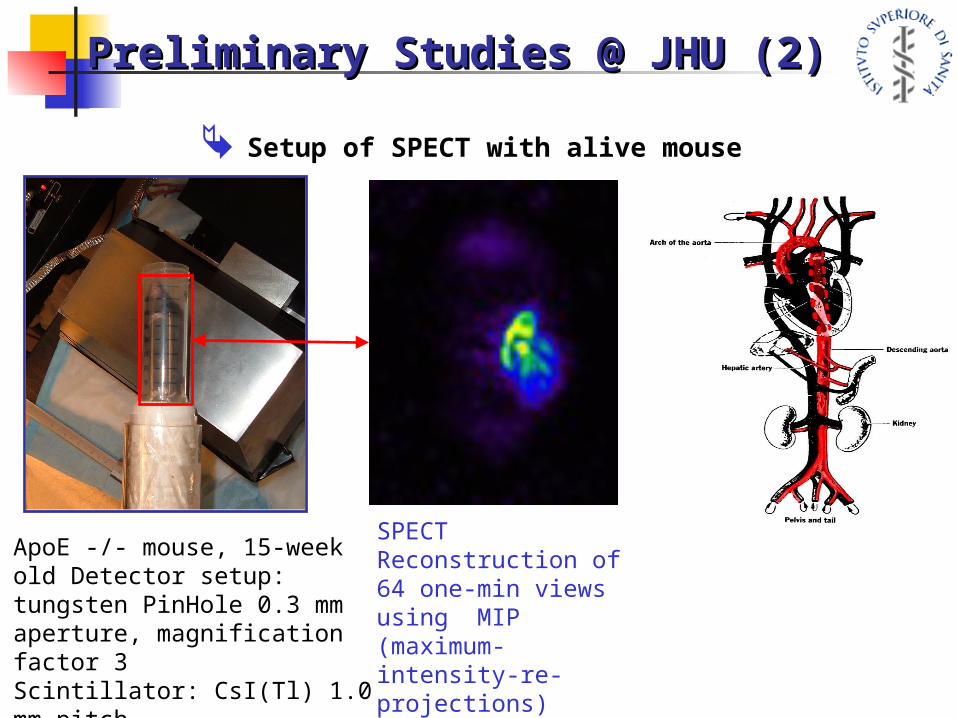
ApoE -/- mouse, 15-week old Detector setup:tungsten PinHole 0.3 mm aperture, magnification factor 3Scintillator: CsI(Tl) 1.0 mm pitchPhotodetector: 2x2 H9500 array
SPECT Reconstruction of 64 one-min viewsusing MIP (maximum-intensity-re-projections) alghoritm
Preliminary Studies @ JHU (2)Preliminary Studies @ JHU (2)
Setup of SPECT with alive mouse

Stem Cell StudiesStem Cell Studies
ConclusionsConclusions
Molecular imaging with radionuclides is a very powerful technique allowing studies of “in-vivo” biological process;
Single-Photon modality generally allows longer studies than PET and it could use different tracers, it
Spatial resolution does not have theoretical limit, better detector technology is continuosly improving performances
Advanced SPECT systems are required (for ex.) in the study of atherosclerotic plaques with mice
Others interesting studies (for ex. stem cells) could benefit the use of advanced small-animal SPECT devices
THANK YOU FOR YOUR ATTENTION!

![1 1 1 1 1 1 1 ¢ 1 , ¢ 1 1 1 , 1 1 1 1 ¡ 1 1 1 1 · 1 1 1 1 1 ] ð 1 1 w ï 1 x v w ^ 1 1 x w [ ^ \ w _ [ 1. 1 1 1 1 1 1 1 1 1 1 1 1 1 1 1 1 1 1 1 1 1 1 1 1 1 1 1 ð 1 ] û w ü](https://img.pdfslide.us/doc/110x75/5f40ff1754b8c6159c151d05/1-1-1-1-1-1-1-1-1-1-1-1-1-1-1-1-1-1-1-1-1-1-1-1-1-1-w-1-x-v.jpg)

![089 ' # '6& *#0 & 7 · 2018. 4. 1. · 1 1 ¢ 1 1 1 ï1 1 1 1 ¢ ¢ð1 1 ¢ 1 1 1 1 1 1 1ýzð1]þð1 1 1 1 1w ï 1 1 1w ð1 1w1 1 1 1 1 1 1 1 1 1 ¢1 1 1 1û](https://img.pdfslide.us/doc/110x75/60a360fa754ba45f27452969/089-6-0-7-2018-4-1-1-1-1-1-1-1-1-1-1-1-1-1.jpg)


![1 1 1 1 1 1 1 ¢ 1 1 1 - pdfs.semanticscholar.org€¦ · 1 1 1 [ v . ] v 1 1 ¢ 1 1 1 1 ý y þ ï 1 1 1 ð 1 1 1 1 1 x](https://img.pdfslide.us/doc/110x75/5f7bc722cb31ab243d422a20/1-1-1-1-1-1-1-1-1-1-pdfs-1-1-1-v-v-1-1-1-1-1-1-y-1-1-1-.jpg)
![$1RYHO2SWLRQ &KDSWHU $ORN6KDUPD +HPDQJL6DQH … · 1 1 1 1 1 1 1 ¢1 1 1 1 1 ¢ 1 1 1 1 1 1 1w1¼1wv]1 1 1 1 1 1 1 1 1 1 1 1 1 ï1 ð1 1 1 1 1 3](https://img.pdfslide.us/doc/110x75/5f3ff1245bf7aa711f5af641/1ryho2swlrq-kdswhu-orn6kdupd-hpdqjl6dqh-1-1-1-1-1-1-1-1-1-1-1-1-1-1.jpg)
![[XLS]Permit Statistical Report - New York Cityhome.nyc.gov/html/dob///downloads/excel/per121209.xls · Web viewCSD INDUSTRIES INC EG WEST 37TH LLC C/O GEMSTONE PR ALBERT FRATONI ONE](https://img.pdfslide.us/doc/110x75/5b01aea67f8b9ad85d8e84e7/xlspermit-statistical-report-new-york-viewcsd-industries-inc-eg-west-37th-llc.jpg)


![[XLS] · Web view1 1 1 2 3 1 1 2 2 1 1 1 1 1 1 2 1 1 1 1 1 1 2 1 1 1 1 2 2 3 5 1 1 1 1 34 1 1 1 1 1 1 1 1 1 1 240 2 1 1 1 1 1 2 1 3 1 1 2 1 2 5 1 1 1 1 8 1 1 2 1 1 1 1 2 2 1 1 1 1](https://img.pdfslide.us/doc/110x75/5ad1d2817f8b9a05208bfb6d/xls-view1-1-1-2-3-1-1-2-2-1-1-1-1-1-1-2-1-1-1-1-1-1-2-1-1-1-1-2-2-3-5-1-1-1-1.jpg)