Embed Size (px)
Citation preview
Pain in chronic pancreatitis: therole of neuropathic painmechanismsA M Drewes,1,2 A L Krarup,1 S Detlefsen,1,3 M-L Malmstrøm,1 G Dimcevski,4
P Funch-Jensen5
1 Mech-Sense, Department ofGastroenterology, AalborgUniversity Hospital, Aalborg,Denmark; 2 Center for Sensory-Motor Interactions, Departmentof Health Science andTechnology, Aalborg University,Denmark; 3 Department ofPathology, Aalborg UniversityHospital, Aalborg, Denmark;4 Department of MedicalGastroenterology, HaukelandUniversity Hospital, Bergen,Norway; 5 Department ofSurgical Gastroenterology L,Aarhus University Hospital,Aarhus, Denmark
Correspondence to:Professor A M Drewes, Mech-Sense, Department ofGastroenterology, AalborgUniversity Hospital, DK-9000Aalborg, Denmark; [email protected]
Published Online First19 June 2008
ABSTRACTPain mechanisms in patients with chronic pancreatitis areincompletely understood and probably multifactorial.Recently, evidence from experimental human painresearch has indicated that in many of these patients painprocessing in the central nervous system is abnormal andmimics that seen in neuropathic pain disorders. Thecurrent review focuses on several lines of evidencesupporting this hypothesis. Hence, the spontaneous andpostprandial pain in chronic pancreatitis may reflect thecharacteristic pain features seen in patients withneuropathic pain. Biochemical and histopathologicalfindings in tissues from patients with chronic pancreatitisare similar to those observed in patients with other nervefibre lesions. Experimental studies have shown thatpatients with chronic pancreatitis show signs of spinalhyper-excitability counter-balanced by segmental anddescending inhibition. Changes in the brain with corticalreorganisation to gut stimulation and increased activity inspecific electroencephalographic features characteristicfor neuropathic pain are also seen in patients with chronicpancreatitis. Finally, principles involved in the treatment ofpancreatic pain have many similarities with thoserecommended in neuropathic pain disorders. In conclu-sion, a mechanism-based understanding of pain in chronicpancreatitis may have important implications for thetreatment.
One of the most important symptoms in chronicpancreatitis (CP) is constant or recurrent abdom-inal pain that is present in 80–90% of patientsduring the course of the disease.1 Pancreatic painpresents characteristically with severe dull epigas-tric pain, often radiating directly to the back. Thepain is often recurrent, intense and long-lastingand may be associated with malnutrition, narcoticaddiction, and major socio-economic problems.Pain mechanisms are incompletely understoodand probably multifactorial. In some cases thereason for the pain is obvious, such as extrapan-creatic (eg, peptic ulcer or bile duct and duodenalstenosis due to extensive pancreatic fibrosis andinflammation) or intrapancreatic (eg, pseudocysts)complications. However, in most patients thesource of the pain remains unknown. In thesecases the following pathophysiological mechan-isms have been suggested: (1) increased intrapan-creatic pressure either within the pancreatic ductor in the parenchyma causing tissue ischaemia;
(2) inflammation in the pancreas; and (3) altera-tions in pancreatic nerves, including an increase innerve fibre diameter and evidence for neurogenicinflammation.2–4 Genetic factors probably also playa role in a patient’s pain experience.4 Because thepain mechanisms are poorly understood, treatmentis often empirical and insufficient. Animal modelshave contributed to the understanding of painpathogenesis in chronic pancreatitis.5–7 Althoughhighly relevant to our understanding of painmechanisms in general, the data should be inter-preted cautiously. Hence there are major differ-ences between pain studies in different species andstrains. Furthermore, mechanisms related to therelatively short-lasting evoked inflammation inmost animal studies on the one hand and thelong-lasting pain in humans with CP on theother are probably very different. Recently, evi-dence from experimental human pain research hasindicated that pain processing in the centralnervous system (CNS) is abnormal and inmany cases may mimic that seen in neuropathicpain disorders.8–10 Neuropathic pain is defined as‘‘pain after a lesion or disease of the afferents inthe peripheral or central nervous system thatnormally signals pain’’.11 Neuropathic pain isprevalent in all diseases in which lesions of thenerves are present, and after surgery neuropathic-like pain can be seen in 20–40% of patients.12
Although other pain mechanisms may also be ofimportance, the following lines of evidence pointtowards neuropathic pain mechanisms in patientswith CP:
c Clinical features of the pain
c Biochemical and histological findings
c Spinal changes, with neuronal hyper-excitabil-ity and amplification of the incoming afferentactivity
c Changes in the brain–gut axis
c Clinical and experimental profile of the drugsused to treat the pain
Although the characteristics listed above are notspecific for neuropathic pain mechanisms, takentogether they do support this theory. By focussingon recent neurophysiological experiments inhumans, these topics will be reviewed in thecurrent paper with the aim of improving ourunderstanding and treatment of pain in CP.
Recent advances in basic science
1616 Gut 2008;57:1616–1627. doi:10.1136/gut.2007.146621
on June 4, 2020 by guest. Protected by copyright.
http://gut.bmj.com
/G
ut: first published as 10.1136/gut.2007.146621 on 19 June 2008. Dow
nloaded from
NEUROPATHIC PAIN MECHANISMS AND CLINICALFEATURESMost nerve afferents mediating pain from thepancreas belong to the splanchnic nerves that passthrough the coeliac ganglion and enter thoracicdorsal root ganglia. The vagal nerve with cellbodies in nodose ganglia normally does notparticipate in pain signalling but can indirectlymodify spinal pain processing, as shown by in vivoelectrical stimulation.13 14 Cholinergic modulationof pancreatic inflammation has also been demon-strated (for a review, see Fregni et al15). Hence thenervous system plays a major role in the inflam-matory response as well as in pain pathogenesisand any abnormalities in the sensory system willinvariably affect the disease process.
Neuropathic pain typically presents as a stimu-lus-independent shooting or burning pain togetherwith stimulus-dependent increased sensations.16
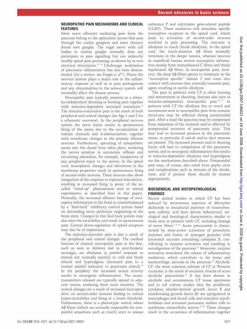
The stimulus-independent pain is the result of bothperipheral and central changes (see figs 1 and 2 fora schematic overview). In the peripheral nervoussystem the nerve lesion results in spontaneousfiring of the axons due to the accumulation ofsodium channels and a-adrenoceptors, togetherwith membrane changes in the primary afferentneurons. Furthermore, sprouting of sympatheticaxons into the dorsal horn takes place, renderingthe nerves sensitive to autonomic reflexes andcirculating adrenaline, for example, irrespective ofany peripheral input to the nerves. In the spinalcord, neuroplastic changes and alterations in themembrane properties result in spontaneous firingof second-order neurons. These neurons also showintegration of the response to repeated stimulation,resulting in increased firing (a proxy of the so-called ‘‘wind-up’’ phenomenon seen in animalexperiments, as described later in this paper).Normally, the increased afferent barrage of noci-ceptive information to the brain is counterbalancedby a ‘‘feed-back’’ inhibitory control system basedon descending nerve pathways originating in thebrain stem. Changes in this feed-back system mayalso alter the excitability and result in spontaneouspain. Central down-regulation of opioid receptorsmay also be of importance.
The stimulus-dependent pain is also a result ofthe peripheral and central changes. The cardinalfeatures of classical neuropathic pain in the skin,such as seen in diabetes and in post-herpeticneuralgia, are allodynia (a painful response tostimuli not normally painful) to cold and brushstimuli and hyperalgesia (increased pain to anormal painful stimulus) to punctuate stimuli.16
In the periphery the increased neural activityresults in neurogenic inflammation. The neuro-transmitters released are typically spread to adja-cent nerves, rendering them more sensitive. Thecentral changes are a result of increased nociceptordrive on second-order neurons leading to centralhyper-excitability and firing at a lower threshold.Furthermore, there is a phenotypic switch whereAb fibres (which are normally responsible for non-painful sensations such as touch) start to release
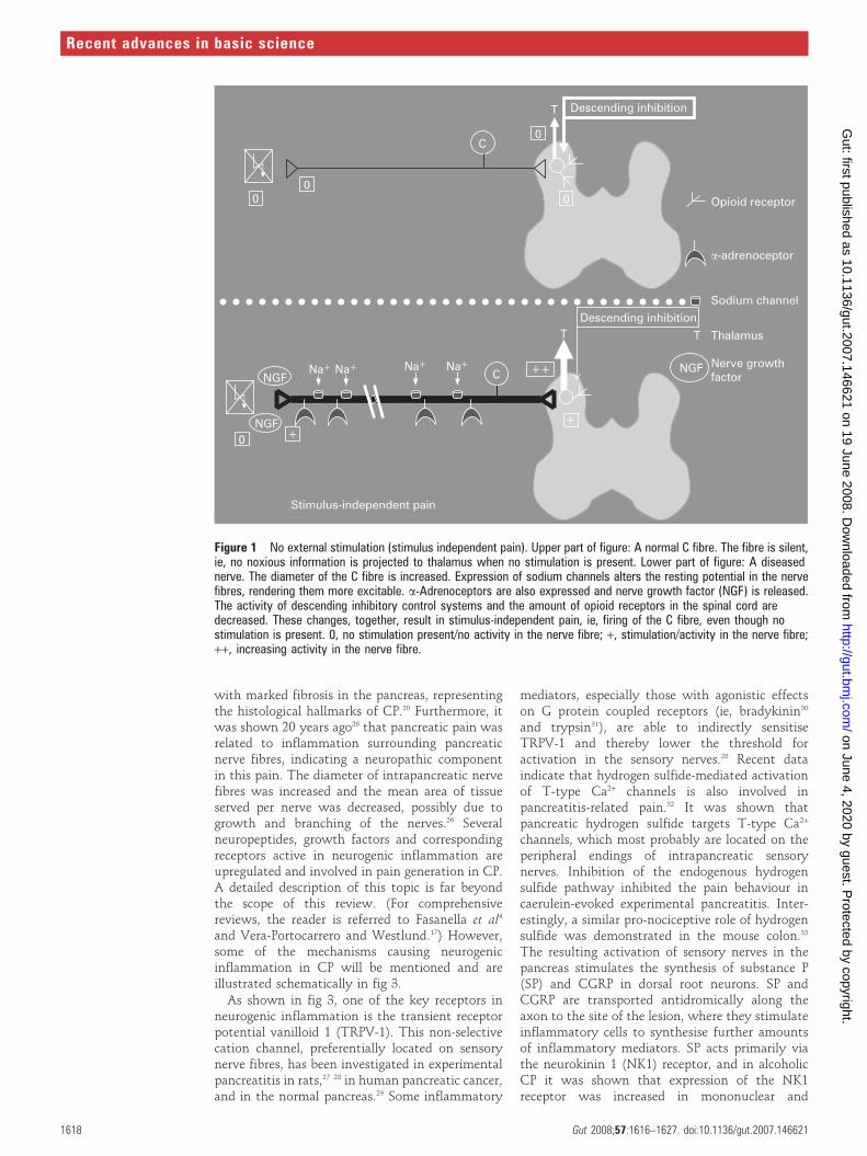
substance P and calcitonine gene-related peptide(CGRP). These mediators will stimulate specificnociceptive receptors in the spinal cord, whichleads to activation of second-order neuronsinvolved in pain processing. The outcome isallodynia to touch (brush allodynia). In the spinalcord, the touch-sensitive Ab fibres normallyterminate in the deeper lamina, whereas neuronsin superficial lamina receive nociceptive informa-tion mainly from unmyelinated C fibres and thinlymyelinated Ab fibres. In neuropathic pain, how-ever, the deep Ab fibres sprout to terminate in the‘‘nociceptive specific’’ lamina 2 and come intocontact with neurons that normally transmit pain,again resulting in tactile allodynia.
The pain in patients with CP is often burningand intermittent or shooting, features also seen instimulus-independent neuropathic pain.16 17 Inpatients with CP the allodynia due to touch andmovements seen in neuropathic pain from somaticstructures may be reflected during postprandialpain. After a meal the pancreas may be compressedfrom expansion of the stomach and/or affected bypostprandial secretion of pancreatic juice. Thismay lead to increased pressure in the pancreatictissue, in particular if calculi and/or duct stenosisare present. The increased pressure and/or shearingforces will lead to compression of the pancreaticnerves, and in neurogenic inflammation this resultsin stimulus-dependent allodynia and hyperalgesiavia the mechanisms described above. Postprandialpain may, of course, also result from peptic ulcerand complications such as stenosis of the duode-num, and if present these should be treatedappropriately.
BIOCHEMICAL AND HISTOPATHOLOGICALFINDINGSRecent animal studies in which CP has beeninduced by intravenous injection of dibutyltindichloride or intraductal injection of trinitroben-zene sulfonic acid have shown behavioural, ser-ological and histological characteristics similar tothose seen in patients with CP, including damageof nerve fibres.5 6 18 Acute pancreatitis is charac-terised by intra-acinar activation of proteolyticenzymes and fusion of zymogen granules withlysosomal vacuoles containing cathepsin B, con-tributing to enzyme activation and resulting inautodigestion of the pancreas.19 Moreover, enzymeactivation stimulates the release of inflammatorymediators, which contribute to fat tissue andhaemorrhagic necrosis in the pancreas.20 AlcoholicCP, the most common subtype of CP in Westerncountries, is the result of recurrent attacks of acutealcoholic pancreatitis.21 It has been shown inalcoholic and autoimmune CP tissue specimensand in cell culture studies that the profibroticcytokines platelet-derived growth factor B andtransforming growth factor b1 are upregulated inmacrophages and ductal cells and stimulate myofi-broblasts and activated pancreatic stellate cells tosynthesise extracellular matrix.22–25 These changesresult in the occurrence of inflammation together
Recent advances in basic science
Gut 2008;57:1616–1627. doi:10.1136/gut.2007.146621 1617
on June 4, 2020 by guest. Protected by copyright.
http://gut.bmj.com
/G
ut: first published as 10.1136/gut.2007.146621 on 19 June 2008. Dow
nloaded from
with marked fibrosis in the pancreas, representingthe histological hallmarks of CP.20 Furthermore, itwas shown 20 years ago26 that pancreatic pain wasrelated to inflammation surrounding pancreaticnerve fibres, indicating a neuropathic componentin this pain. The diameter of intrapancreatic nervefibres was increased and the mean area of tissueserved per nerve was decreased, possibly due togrowth and branching of the nerves.26 Severalneuropeptides, growth factors and correspondingreceptors active in neurogenic inflammation areupregulated and involved in pain generation in CP.A detailed description of this topic is far beyondthe scope of this review. (For comprehensivereviews, the reader is referred to Fasanella et al4
and Vera-Portocarrero and Westlund.17) However,some of the mechanisms causing neurogenicinflammation in CP will be mentioned and areillustrated schematically in fig 3.
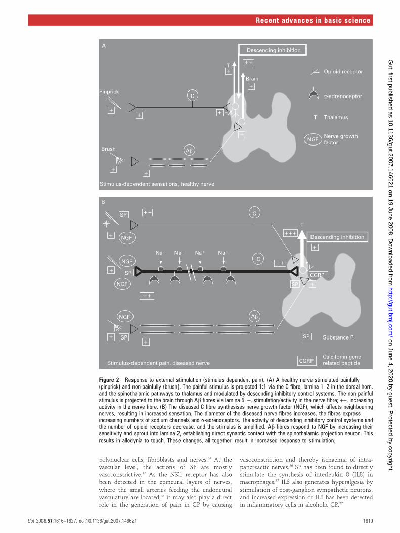
As shown in fig 3, one of the key receptors inneurogenic inflammation is the transient receptorpotential vanilloid 1 (TRPV-1). This non-selectivecation channel, preferentially located on sensorynerve fibres, has been investigated in experimentalpancreatitis in rats,27 28 in human pancreatic cancer,and in the normal pancreas.29 Some inflammatory
mediators, especially those with agonistic effectson G protein coupled receptors (ie, bradykinin30
and trypsin31), are able to indirectly sensitiseTRPV-1 and thereby lower the threshold foractivation in the sensory nerves.28 Recent dataindicate that hydrogen sulfide-mediated activationof T-type Ca2+ channels is also involved inpancreatitis-related pain.32 It was shown thatpancreatic hydrogen sulfide targets T-type Ca2+
channels, which most probably are located on theperipheral endings of intrapancreatic sensorynerves. Inhibition of the endogenous hydrogensulfide pathway inhibited the pain behaviour incaerulein-evoked experimental pancreatitis. Inter-estingly, a similar pro-nociceptive role of hydrogensulfide was demonstrated in the mouse colon.33
The resulting activation of sensory nerves in thepancreas stimulates the synthesis of substance P(SP) and CGRP in dorsal root neurons. SP andCGRP are transported antidromically along theaxon to the site of the lesion, where they stimulateinflammatory cells to synthesise further amountsof inflammatory mediators. SP acts primarily viathe neurokinin 1 (NK1) receptor, and in alcoholicCP it was shown that expression of the NK1receptor was increased in mononuclear and
Figure 1 No external stimulation (stimulus independent pain). Upper part of figure: A normal C fibre. The fibre is silent,ie, no noxious information is projected to thalamus when no stimulation is present. Lower part of figure: A diseasednerve. The diameter of the C fibre is increased. Expression of sodium channels alters the resting potential in the nervefibres, rendering them more excitable. a-Adrenoceptors are also expressed and nerve growth factor (NGF) is released.The activity of descending inhibitory control systems and the amount of opioid receptors in the spinal cord aredecreased. These changes, together, result in stimulus-independent pain, ie, firing of the C fibre, even though nostimulation is present. 0, no stimulation present/no activity in the nerve fibre; +, stimulation/activity in the nerve fibre;++, increasing activity in the nerve fibre.
Recent advances in basic science
1618 Gut 2008;57:1616–1627. doi:10.1136/gut.2007.146621
on June 4, 2020 by guest. Protected by copyright.
http://gut.bmj.com
/G
ut: first published as 10.1136/gut.2007.146621 on 19 June 2008. Dow
nloaded from
polynuclear cells, fibroblasts and nerves.34 At thevascular level, the actions of SP are mostlyvasoconstrictive.17 As the NK1 receptor has alsobeen detected in the epineural layers of nerves,where the small arteries feeding the endoneuralvasculature are located,35 it may also play a directrole in the generation of pain in CP by causing
vasoconstriction and thereby ischaemia of intra-pancreactic nerves.36 SP has been found to directlystimulate the synthesis of interleukin 8 (IL8) inmacrophages.37 IL8 also generates hyperalgesia bystimulation of post-ganglion sympathetic neurons,and increased expression of IL8 has been detectedin inflammatory cells in alcoholic CP.37
Figure 2 Response to external stimulation (stimulus dependent pain). (A) A healthy nerve stimulated painfully(pinprick) and non-painfully (brush). The painful stimulus is projected 1:1 via the C fibre, lamina 1–2 in the dorsal horn,and the spinothalamic pathways to thalamus and modulated by descending inhibitory control systems. The non-painfulstimulus is projected to the brain through Ab fibres via lamina 5. +, stimulation/activity in the nerve fibre; ++, increasingactivity in the nerve fibre. (B) The diseased C fibre synthesises nerve growth factor (NGF), which affects neighbouringnerves, resulting in increased sensation. The diameter of the diseased nerve fibres increases, the fibres expressincreasing numbers of sodium channels and a-adrenoceptors. The activity of descending inhibitory control systems andthe number of opioid receptors decrease, and the stimulus is amplified. Ab fibres respond to NGF by increasing theirsensitivity and sprout into lamina 2, establishing direct synaptic contact with the spinothalamic projection neuron. Thisresults in allodynia to touch. These changes, all together, result in increased response to stimulation.
Recent advances in basic science
Gut 2008;57:1616–1627. doi:10.1136/gut.2007.146621 1619
on June 4, 2020 by guest. Protected by copyright.
http://gut.bmj.com
/G
ut: first published as 10.1136/gut.2007.146621 on 19 June 2008. Dow
nloaded from
Evidence for a neuropathic component of paingeneration in CP was further provided by thefinding that growth associated protein 43 isincreased in pancreatic nerve fibres in alcoholicCP.38 This protein was shown to be re-expressedafter neuronal lesions,39 introducing it as a markerof neuronal growth and plasticity. Moreover, theneurotrophin nerve growth factor and its highaffinity receptor tyrosin kinase receptor A areupregulated in alcoholic CP.40 Nerve growth factoris possibly a regulator of the synthesis of SP andCGRP41 and modulates nociception in peripheralnerve fibres.42 Another neurotrophin with impacton neuropathic pain in CP is brain-derived neuro-trophic factor, which is increased in ductularcomplexes, degenerating acinar cells, and enlargednerve fibres as well as in intrapancreatic ganglia inCP.43 During peripheral inflammation, brain-derived neurotrophic factor is upregulated in dorsalroot ganglia in rats.44 Recent studies have alsoidentified artemin45 and the cannabinoid system46
as possible mediators of neural changes and painperception in CP. Artemin belongs to the family ofglial-derived neurotrophic factors and enhancessurvival, proliferation and regeneration of neurons.Both artemin and its receptor are increased in CPtissue.45 Moreover, the expression of the cannabi-noid receptor 1 is upregulated in human acutepancreatitis, a finding that is accompanied by anincrease in the endocannabinoid anandamide.46 Inanother study, cannabinoid receptors located onperipheral nociceptive endings have been shown tonegatively modulate nociceptors and inhibit releaseof neuropeptides.47
These findings point towards a complex inter-play of inflammatory cells, nerve fibres, neuropep-tides, and corresponding receptors, resulting in thephenomenon of peripheral (and subsequent cen-tral) sensitisation of the nervous system.
SPINAL CHANGES WITH HYPER-EXCITABILITYAND AMPLIFICATION OF THE INCOMING NEURALACTIVITYIn most diseases characterised by chronic pain,sensitisation of the nervous system is a cardinalfeature. This is also the case in neuropathic paindisorders. This sensitisation results in plasticchanges in the neurons at spinal (and supraspinal)sites. It has been proposed that these plastic
changes in the dorsal horn of the spinal cordcorrespond to the mechanism in ‘‘long-termpotentiation’’.48 Through a cascade of molecularchanges at the cell synapses, long-term potentia-tion results in an increase in synaptic transmissionefficacy, thus leading to an increase in synapticstrengthening which further increases electricalactivity and sensory information transmission.This sensitisation may ultimately result in anautonomous state in which the central nervoussystem reports pain even in the absence ofperipheral noxious input.49 Sensitisation includesboth peripheral and central components. Peripheralnociceptor sensitisation underlies the hyperalgesiathat develops immediately at the site of injury.However, in CP, acute inflammation of the glandhas normally ceased, and central rather thanperipheral sensitisation is thought to account formost of the symptoms experienced by the patients.In animal studies, central sensitisation is charac-terised by increased spontaneous activity,decreased firing threshold, and expansion of thereceptive fields of dorsal horn neurons.50 51 Theincrease in excitability of spinal cord nociceptiveneurons amplifies the signal coming from theperiphery, resulting in allodynia and hyperalgesia.The alterations in functional structure may resultin central plasticity and ‘‘pain memory,’’ whichafter some time may be consolidated and indepen-dent of the original peripheral input.50 51 Humanexperimental models have been used to sensitisethe gut and mimic the findings in animal studies.52
Studies using electrical and mechanical stimulihave indicated that acid perfusion of the oesopha-gus results in peripheral as well as centralsensitisation of the nervous system.53 54 In condi-tions with chronic pain there is substantialevidence for persistent central changes that outlivethe initial disease. Hence, when the peripheralstimulation (such as inflammation due to CP) hassubsided, sensitised second-order neurons continueto fire, and sub-threshold regulatory stimuli arestill perceived as painful.
Importantly, CP is often complicated by dia-betes, and it is estimated that 60–70% of patientswith diabetes suffer from mild to severe forms ofnervous system damage including autonomicneuropathy.55 Using a multimodal experimentalapproach, Frøkjær et al56 recently demonstratedthat gastrointestinal sensory nerves in patientswith diabetes are affected throughout all layers ofthe gut. The neuronal damage will invariably resultin changes in the neuronal pain matrix, includinginteractions between peripheral and central painmechanisms, and thereby the sensory processing inCP. Moreover, activation of pain related structuresin the brain might trigger behavioural responsesresulting in negative changes in eating, dailyactivity and sleep patterns that could worsen thepain.15 57
Although simplified, referred pain is a result ofvisceral and somatic fibres converging on the samesecond-order neuron (for details see Arendt-Nielsenet al58). Hence spinal hyper-excitability will be
Summary box 1
c Constant and/or recurrent abdominal pain is present in 80–90% of patientswith chronic pancreatitis
c The spontaneous and postprandial pain in chronic pancreatitis may reflect thecharacteristic stimulus independent and stimulus dependent pain featuresseen in patients with neuropathic pain
c Biochemical an histopathological features in tissues from patients withchronic pancreatitis mimic those observed in tissues from patients with othernerve fibre lesions
c The principles involved in the treatment of pancreatic pain have manysimilarities with those used in neuropathic pain disorders
Recent advances in basic science
1620 Gut 2008;57:1616–1627. doi:10.1136/gut.2007.146621
on June 4, 2020 by guest. Protected by copyright.
http://gut.bmj.com
/G
ut: first published as 10.1136/gut.2007.146621 on 19 June 2008. Dow
nloaded from
manifested by enlargement of and changes inlocalisation and sensitivity of the referred painarea.59 In patients with CP, Buscher et al60
investigated the sensitivity in the referred painareas of the abdomen. Although findings were notcompletely consistent between genders, theyshowed increased sensation to pressure (predomi-nantly a deep muscle stimulus). On the otherhand, there were higher thresholds to electricalskin stimulation in dermatome T10, the ‘‘viscer-otome’’ that shares innervation with the pancrea-tic nerves, whereas the sensation in otherdermatomes (C5, T4, L1 and L4) was normal.The authors suggested that this local hyposensi-tivity was related to segmental inhibition causedby a local feedback system in the spinal cord. Aswith the deep stimulus, changed sensation in thesomatic referred pain area (sharing central termi-nation with activated visceral nerves) has also beenshown experimentally in healthy subjects as wellas in patients with diseases such as appendicitisand choledocholithiasis.61 62 Therefore, such datagive evidence for central hyper-excitability in CP.
In experimental studies the pancreas is difficultto access and there are potentially harmfulcomplications during endoscopic procedures.Therefore it has not yet been possible in humansto investigate directly whether allodynia or othercharacteristic features of neuropathic pain arepresent during manipulation of the pancreas.However, visceral pain is typically diffuse anddifficult to localise, which to a large extent can beexplained by the widespread termination of visc-eral afferents into second-order neurons in multiplesegments of the spinal cord.63 Due to this overlap incentral termination of visceral afferents, diseasemechanisms affecting the pancreas are typicallyalso activated during pain stimulation of nearbyorgans like the duodenum and oesophagus.64
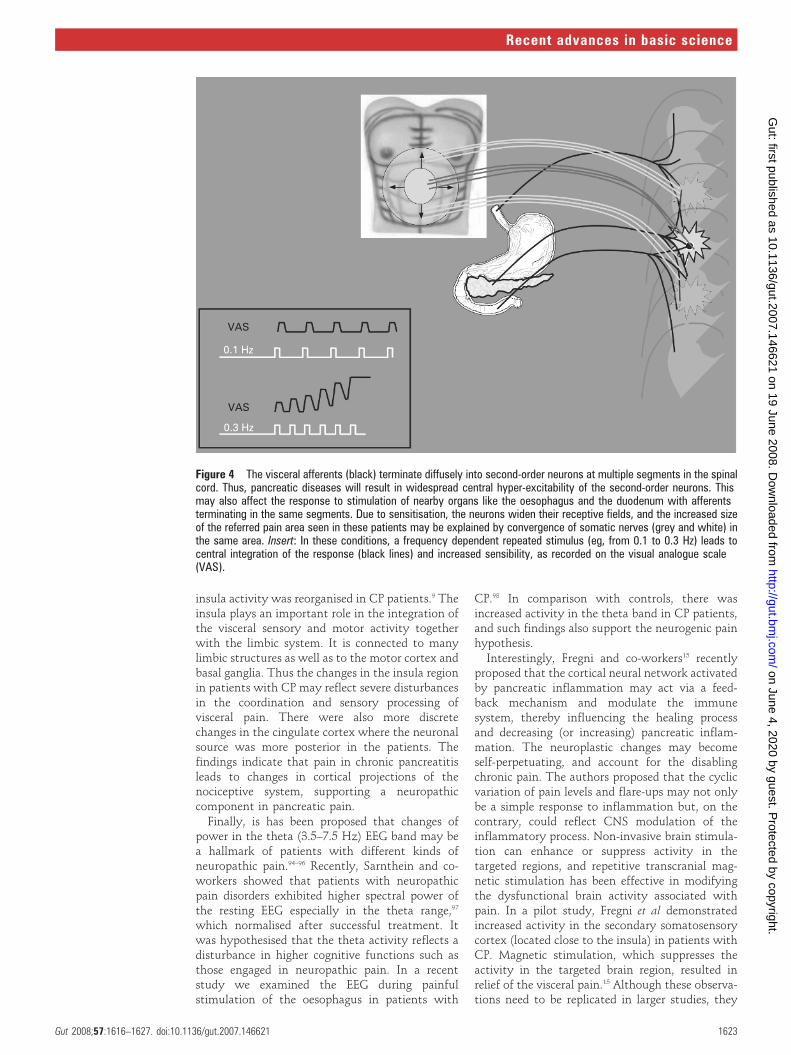
Dimcevski et al9 10 showed that the referred painarea to experimental electrical stimulation of theoesophagus in CP was increased compared withthat in healthy controls (fig 4). Because increases inthe size of the referred pain area after experimentalstimulation have also been shown in other patientgroups with organic diseases (eg, erosive and non-erosive oesophagitis, peptic ulcer disease) thisstrengthens the validity of the findings.65 66
Changes in the referred pain area, however, arenot specific for any specific pain disorder, butmerely reflect the importance of central changessuch as seen those in neuropathic pain.
Another feature that is predominant in centralsensitisation is wind-up (in humans this is termedtemporal summation or central integration).Temporal summation is a frequency dependent,gradual response of ‘‘wide-dynamic’’ spinal neu-rons to repetitive stimuli, which may lead toincreased excitability if the stimulus is sufficientlystrong.67 68 Hence, afferent fibres that are repeat-edly stimulated give a progressive increase insecond-order neuronal responsiveness. Dimcevskiet al found an increase of temporal summation inthe skin,64 muscle and oesophagus10 in patients
with CP (fig 4). Although not specific for any paindisorder, facilitated temporal summation is pre-valent in neuropathic pain patients,69–73 and thusthese findings support the important role ofneuropathic pain mechanisms in CP.
The dissociation between local sensory distur-bances in the area of nerve damage and sponta-neous pain per se suggests that the pain isneuropathic in nature. However, other mechan-isms are also of importance. The second-orderneurons in the spinal cord are subject to descendingcontrol from higher brain centres.74–77 One of thefirst systems described was diffuse noxious inhibi-tory control (DNIC), defined as ‘‘painful hetero-topic stimuli that can cause suppression ofnociceptive activity in dorsal horn neurons’’.DNIC was shown to involve a spinal–suprasp-inal–spinal feedback loop producing a descendinginhibitory control.78 Apart from being inhibitory,descending pain control from the brainstem canalso be facilitating, and the balance between theinhibiting and activating systems may determinethe overall level of excitability (and hence pain) ofthe neuron network in the dorsal horn.79 80 In ananimal model of persistent pancreatic pain Vera-Portocarrero et al81 reported that descending facil-itation from the rostral ventromedial medulla playsa critical role in the maintenance of pancreaticpain. When they selectively destroyed cells in thisbrain centre, it attenuated pancreatic-inducedhypersensitivity. Several cortical centres are con-nected to the periaquaductal grey and the raphemagnus nucleus in the medulla, which again exertcontrol over the spinal cord. The balance betweenthe excitatory system and the inhibitory modula-tion is therefore a major determinant for the finalinterpretation of the pain, and descending influ-ence from brainstem structures seems to be themajor way by which the brain controls painperception.75 76 82–84 Recent evidence supports thenotion that in patients with functional paindisorders, such as irritable bowel syndrome, theinhibitory control system functions insufficiently.This may result in an increased barrage of noxiousstimulation reaching pain centres in the brain.85 Onthe other hand, in patients with organic disordersthe descending control system is probably fullyactivated to prevent hyperalgesia evoked by theafferent activity arising in the diseased tissue.Hence, hypoalgesia to experimental visceral stimu-lation was seen in patients with diseases such asoesophagitis, Crohn’s disease and pepticulcers.65 86 87 Therefore in patients with CP therewill probably be an activation of the descendinginhibitory system, whereby the pain system triesto counterbalance the incoming nociceptive infor-mation from the pancreas. However, activation ofdescending control systems probably affects thespinal cord diffusely, resulting in widespreadhypoalgesia to stimulation from other bodyregions. Correspondingly, in an experimental studyin which the skin, oesophagus and duodenum werestimulated, generalised hypoalgesia was found in
Recent advances in basic science
Gut 2008;57:1616–1627. doi:10.1136/gut.2007.146621 1621
on June 4, 2020 by guest. Protected by copyright.
http://gut.bmj.com
/G
ut: first published as 10.1136/gut.2007.146621 on 19 June 2008. Dow
nloaded from
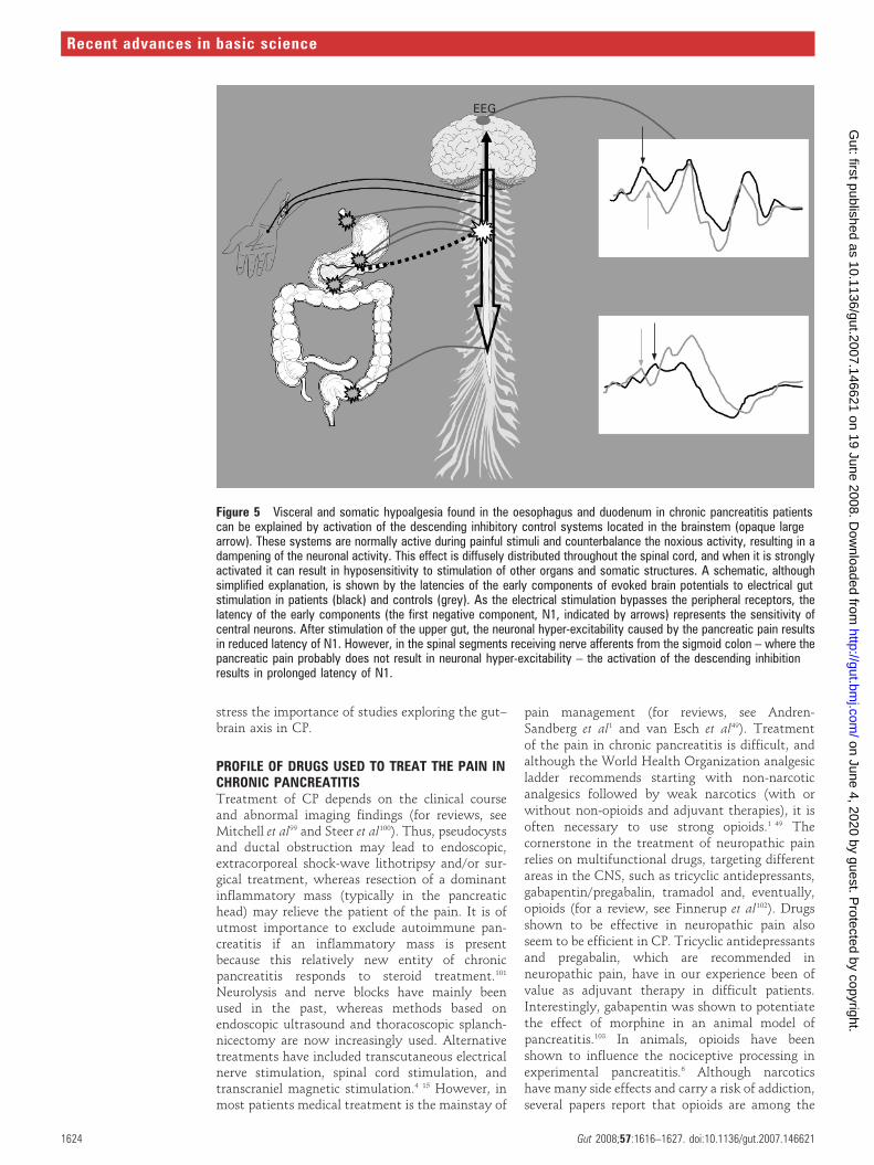
patients with CP in comparison with healthycontrols (see fig 5).
Evidence indicating increased descending inhibi-tion in CP was also seen in a study based on evokedbrain potentials resulting from painful stimulationof the oesophagus. Evoked brain potentials are thesummated outcome of a series of time-lockedelectroencephalographic (EEG) responses to exter-nal stimuli.88 89 Reduction in latency of the evokedbrain potentials may be related to hyper-excit-ability within central pain pathways evoked by thechronic pain attacks in the patients.90 This is mostprobably due to central hyper-excitability andopening of faster conducting latent connections.Correspondingly, in patients with CP the latencyto upper gut stimulations was decreased.9
However, the latency to sigmoid stimulationswas decreased (Drewes et al, unpublished data)(fig 5). The reason for these findings is probablythat, although the pain in CP evokes hyper-excitability in a widespread area of the spinal cord,this is predominant in the thoracic region. Hencethe excitability in remote lumbar segments receiv-ing afferents from the sigmoid colon could be
dampened by the descending control systemsexerting diffuse inhibition of the incoming activityat all segmental levels of the spinal cord. However,electrical stimulation may activate both vagal andsplanchnic pathways.91 Because activation of thesplanchnic/spinal pathways probably activates anendogenous pain inhibitory pathway, whereasvagal stimulation may result in facilitation, thiscan make interpretation of the data difficult.13
The most recent neurophysiological evidence forneuropathic pain mechanisms in CP is based onstudies of the brain. Several experimental andclinical studies indicate that deafferentation,chronic pain, and hyperalgesia are associated withfunctional reorganisation of the cortex. In parti-cular, there is evidence for neuroplastic changesand reorganisation of the brain in patients withamputations and neuropathic pain.92 Importantly,the degree of cortical reorganisation correlates withthe subjective pain rating, and the cortical changescan be reversed by analgesic interventions (for areview, see Wiech et al93). In recent experimentscarried out by our group, analysis of the brainsources to upper gut stimulation showed that the
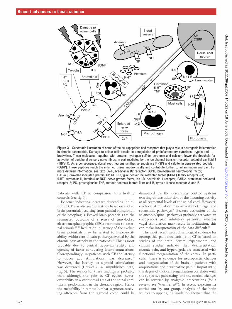
Figure 3 Schematic illustration of some of the neuropeptides and receptors that play a role in neurogenic inflammationin chronic pancreatitis. Damage to acinar cells results in upregulation of proinflammatory cytokines, trypsin andbradykinin. These molecules, together with protons, hydrogen sulfide, serotonin and calcium, lower the threshold foractivation of peripheral sensory nerve fibres, in part mediated by the ion channel transient receptor potential vanilloid 1(TRPV-1). As a consequence, dorsal root neurons synthesise substance P (SP) and calcitonin gene-related peptide(CGRP). These peptides reach the inflamed tissue antidromically and contribute further to inflammation and pain. Formore detailed information, see text. B2-R, bradykinin B2 receptor; BDNF, brain-derived neurotrophic factor;GAP-43, growth-associated protein 43; GFR-a3, glial derived neurotrophic factor (GDNF) family receptor a3;5-HT, serotonin; IL, interleukin; NGF, nerve growth factor; NK1-R, neurokinin 1 receptor; PAR-2, proteinase activatedreceptor 2; PG, prostaglandin; TNF, tumour necrosis factor; TrkA and B, tyrosin kinase receptor A and B.
Recent advances in basic science
1622 Gut 2008;57:1616–1627. doi:10.1136/gut.2007.146621
on June 4, 2020 by guest. Protected by copyright.
http://gut.bmj.com
/G
ut: first published as 10.1136/gut.2007.146621 on 19 June 2008. Dow
nloaded from
insula activity was reorganised in CP patients.9 Theinsula plays an important role in the integration ofthe visceral sensory and motor activity togetherwith the limbic system. It is connected to manylimbic structures as well as to the motor cortex andbasal ganglia. Thus the changes in the insula regionin patients with CP may reflect severe disturbancesin the coordination and sensory processing ofvisceral pain. There were also more discretechanges in the cingulate cortex where the neuronalsource was more posterior in the patients. Thefindings indicate that pain in chronic pancreatitisleads to changes in cortical projections of thenociceptive system, supporting a neuropathiccomponent in pancreatic pain.
Finally, is has been proposed that changes ofpower in the theta (3.5–7.5 Hz) EEG band may bea hallmark of patients with different kinds ofneuropathic pain.94–96 Recently, Sarnthein and co-workers showed that patients with neuropathicpain disorders exhibited higher spectral power ofthe resting EEG especially in the theta range,97
which normalised after successful treatment. Itwas hypothesised that the theta activity reflects adisturbance in higher cognitive functions such asthose engaged in neuropathic pain. In a recentstudy we examined the EEG during painfulstimulation of the oesophagus in patients with
CP.98 In comparison with controls, there wasincreased activity in the theta band in CP patients,and such findings also support the neurogenic painhypothesis.
Interestingly, Fregni and co-workers15 recentlyproposed that the cortical neural network activatedby pancreatic inflammation may act via a feed-back mechanism and modulate the immunesystem, thereby influencing the healing processand decreasing (or increasing) pancreatic inflam-mation. The neuroplastic changes may becomeself-perpetuating, and account for the disablingchronic pain. The authors proposed that the cyclicvariation of pain levels and flare-ups may not onlybe a simple response to inflammation but, on thecontrary, could reflect CNS modulation of theinflammatory process. Non-invasive brain stimula-tion can enhance or suppress activity in thetargeted regions, and repetitive transcranial mag-netic stimulation has been effective in modifyingthe dysfunctional brain activity associated withpain. In a pilot study, Fregni et al demonstratedincreased activity in the secondary somatosensorycortex (located close to the insula) in patients withCP. Magnetic stimulation, which suppresses theactivity in the targeted brain region, resulted inrelief of the visceral pain.15 Although these observa-tions need to be replicated in larger studies, they
Figure 4 The visceral afferents (black) terminate diffusely into second-order neurons at multiple segments in the spinalcord. Thus, pancreatic diseases will result in widespread central hyper-excitability of the second-order neurons. Thismay also affect the response to stimulation of nearby organs like the oesophagus and the duodenum with afferentsterminating in the same segments. Due to sensitisation, the neurons widen their receptive fields, and the increased sizeof the referred pain area seen in these patients may be explained by convergence of somatic nerves (grey and white) inthe same area. Insert: In these conditions, a frequency dependent repeated stimulus (eg, from 0.1 to 0.3 Hz) leads tocentral integration of the response (black lines) and increased sensibility, as recorded on the visual analogue scale(VAS).
Recent advances in basic science
Gut 2008;57:1616–1627. doi:10.1136/gut.2007.146621 1623
on June 4, 2020 by guest. Protected by copyright.
http://gut.bmj.com
/G
ut: first published as 10.1136/gut.2007.146621 on 19 June 2008. Dow
nloaded from
stress the importance of studies exploring the gut–brain axis in CP.
PROFILE OF DRUGS USED TO TREAT THE PAIN INCHRONIC PANCREATITISTreatment of CP depends on the clinical courseand abnormal imaging findings (for reviews, seeMitchell et al99 and Steer et al100). Thus, pseudocystsand ductal obstruction may lead to endoscopic,extracorporeal shock-wave lithotripsy and/or sur-gical treatment, whereas resection of a dominantinflammatory mass (typically in the pancreatichead) may relieve the patient of the pain. It is ofutmost importance to exclude autoimmune pan-creatitis if an inflammatory mass is presentbecause this relatively new entity of chronicpancreatitis responds to steroid treatment.101
Neurolysis and nerve blocks have mainly beenused in the past, whereas methods based onendoscopic ultrasound and thoracoscopic splanch-nicectomy are now increasingly used. Alternativetreatments have included transcutaneous electricalnerve stimulation, spinal cord stimulation, andtranscraniel magnetic stimulation.4 15 However, inmost patients medical treatment is the mainstay of
pain management (for reviews, see Andren-Sandberg et al1 and van Esch et al49). Treatmentof the pain in chronic pancreatitis is difficult, andalthough the World Health Organization analgesicladder recommends starting with non-narcoticanalgesics followed by weak narcotics (with orwithout non-opioids and adjuvant therapies), it isoften necessary to use strong opioids.1 49 Thecornerstone in the treatment of neuropathic painrelies on multifunctional drugs, targeting differentareas in the CNS, such as tricyclic antidepressants,gabapentin/pregabalin, tramadol and, eventually,opioids (for a review, see Finnerup et al102). Drugsshown to be effective in neuropathic pain alsoseem to be efficient in CP. Tricyclic antidepressantsand pregabalin, which are recommended inneuropathic pain, have in our experience been ofvalue as adjuvant therapy in difficult patients.Interestingly, gabapentin was shown to potentiatethe effect of morphine in an animal model ofpancreatitis.103 In animals, opioids have beenshown to influence the nociceptive processing inexperimental pancreatitis.6 Although narcoticshave many side effects and carry a risk of addiction,several papers report that opioids are among the
Figure 5 Visceral and somatic hypoalgesia found in the oesophagus and duodenum in chronic pancreatitis patientscan be explained by activation of the descending inhibitory control systems located in the brainstem (opaque largearrow). These systems are normally active during painful stimuli and counterbalance the noxious activity, resulting in adampening of the neuronal activity. This effect is diffusely distributed throughout the spinal cord, and when it is stronglyactivated it can result in hyposensitivity to stimulation of other organs and somatic structures. A schematic, althoughsimplified explanation, is shown by the latencies of the early components of evoked brain potentials to electrical gutstimulation in patients (black) and controls (grey). As the electrical stimulation bypasses the peripheral receptors, thelatency of the early components (the first negative component, N1, indicated by arrows) represents the sensitivity ofcentral neurons. After stimulation of the upper gut, the neuronal hyper-excitability caused by the pancreatic pain resultsin reduced latency of N1. However, in the spinal segments receiving nerve afferents from the sigmoid colon – where thepancreatic pain probably does not result in neuronal hyper-excitability – the activation of the descending inhibitionresults in prolonged latency of N1.
Recent advances in basic science
1624 Gut 2008;57:1616–1627. doi:10.1136/gut.2007.146621
on June 4, 2020 by guest. Protected by copyright.
http://gut.bmj.com
/G
ut: first published as 10.1136/gut.2007.146621 on 19 June 2008. Dow
nloaded from
best analgesics in the treatment of neuropathicpain (for a review, see Cruccu104). Wilder-Smith etal105 demonstrated that tramadol, a weak opioiddrug with several non-opioid effects in the centralnervous system, in high doses was superior totraditional opioids in the treatment of pain inCP. Among the strong opioids, oxycodone hasshown to be effective in neuropathic pain.106
Correspondingly, in an experimental study,Staahl and co-workers8 administered oxycodoneand morphine in equipotent dosages to patientswith pain due to CP. They showed that oxycodonewas superior to morphine in attenuating painevoked from somatic structures (skin and muscle)as well as from the oesophagus. The superior effectof oxycodone on pain from somatic structures wasnot seen in a similar study in healthy subjects,107
and probably the disease process in CP wasresponsible for this difference. Hence oxycodone,although a strong m-agonist, also has some effectson the k-opioid receptor. Animal experiments haveshown both a peripheral and a spinal upregulationof the k-opioid receptors in the presence ofinflammation.108 This upregulation will affect thepain system in several tissues, as has been shown inthe CP patients.109 The potential effect of areceptor differentiated effect was supported in aclinical study involving a peripherally restricted kagonist (ADL 01-0101). This agonist showedanalgesic effects in patients with pain due toCP.110 Hence in contrast to a pure m-agonist such asmorphine, opioids with more complex effects suchas tramadol and oxycodone may be of value in CP.Experimental studies should, however, be sup-ported by large-scale clinical studies.
CONCLUSIONThe pathogenesis of pain in CP is poorly under-stood. In a subset of patients, extrapancreaticdiseases such as peptic ulcer and intrapancreaticcomplications such as duct obstruction or pseudo-cysts can explain the pain and should be treatedappropriately. Endoscopic or surgical treatmentsare still the mainstay in selected cases if aninflammatory mass is present or there is substantialevidence for increased pressure in the pancreatic ductor gland.111–113 When an inflammatory mass ispresent, autoimmune pancreatitis and inflammatory
pseudotumor have to be considered.114 However, inthe majority of patients the cause of the pain is notobvious. Although the different lines of ‘‘evidence’’presented in the current paper are not specific forneuropathic pain, they point towards this mechan-ism as a major explanation for the pain seen in manypatients. If neuropathic pain dominates the clinicalpicture, surgical procedures may increase the neuro-nal lesions and worsen the pain. Furthermore,pharmacological treatment of neuropathic paindiffers from that of other pain disorders, with focuson multifunctional drugs having different effects onthe CNS, such as tricyclic antidepressants, tramadoland ion-channel inhibitors. Future studies shouldtherefore aim at developing methods to characteriseneuropathy in individual patients before they aresubjected to treatment, and at addressing theprevalence of neuropathic pain in a large series ofCP patients. Furthermore, the effect of drugseffective against neuropathy should also be evalu-ated systematically in well defined patient cohorts.
Funding: The study was supported from ‘‘SparNord Fonden’’, ‘‘DetObelske Familiefond’’ and The Danish Health Research Council.
Competing interests: None.
REFERENCES1. Andren-Sandberg A, Hoem D, Gislason H. Pain management in
chronic pancreatitis. Eur J Gastroenterol Hepatol 2002;14:957–70.2. Di Sebastiano P, di Mola FF, Bockman DE, et al. Chronic
pancreatitis: the perspective of pain generation by neuroimmuneinteraction. Gut 2003;52:907–11.
3. Witt H, Apte MV, Keim V, et al. Chronic pancreatitis: challengesand advances in pathogenesis, genetics, diagnosis, and therapy.Gastroenterology 2007;132:1557–73.
4. Fasanella KE, Davis B, Lyons J, et al. Pain in chronic pancreatitisand pancreatic cancer. Gastroenterol Clin North Am2007;36:335–64.
5. Winston JH, He ZJ, Shenoy M, et al. Molecular and behavioralchanges in nociception in a novel rat model of chronic pancreatitisfor the study of pain. Pain 2005;117:214–22.
6. Vera-Portocarrero LP, Lu Y, Westlund KN. Nociception inpersistent pancreatitis in rats: effects of morphine andneuropeptide alterations. Anesthesiology 2003;98:474–84.
7. Perides G, Tao X, West N, et al. A mouse model of ethanoldependent pancreatic fibrosis. Gut 2005;54:1461–7.
8. Staahl C, Dimcevski G, Andersen SD, et al. Differential effect ofopioids in patients with chronic pancreatitis: An experimental painstudy. Scand J Gastroenterol 2007;42:383–90.
9. Dimcevski G, Sami SA, Funch-Jensen P, et al. Pain in chronicpancreatitis: the role of reorganization in the central nervoussystem. Gastroenterology 2007;132:1546–56.
10. Dimcevski G, Staahl C, Andersen SD, et al. Assessment ofexperimental pain from skin, muscle, and esophagus in patientswith chronic pancreatitis. Pancreas 2007;35:22–9.
11. Baron R. Mechanisms of disease: neuropathic pain – a clinicalperspective. Nat Clin Pract Neurol 2006;2:95–106.
12. Kehlet H, Jensen TS, Woolf CJ. Persistent postsurgical pain: riskfactors and prevention. Lancet 2006;367:1618–25.
13. Gebhart GF. It’s chickens and eggs all over again: is centralreorganization the result or cause of persistent visceral pain?Gastroenterology 2007;132:1618–20.
14. Randich A, Gebhart GF. Vagal afferent modulation ofnociception. Brain Res Brain Res Rev 1992;17:77–99.
15. Fregni F, Pascual-Leone A, Freedman SD. Pain in chronicpancreatitis: a salutogenic mechanism or a maladaptive brainresponse? Pancreatology 2007;7:411–22.
16. Woolf CJ, Mannion RJ. Neuropathic pain: aetiology, symptoms,mechanisms, and management. Lancet 1999;353:1959–64.
17. Vera-Portocarrero L, Westlund KN. Role of neurogenicinflammation in pancreatitis and pancreatic pain. Neurosignals2005;14:158–65.
18. Xu GY, Winston JH, Shenoy M, et al. Enhanced excitability andsuppression of A-type K+ current of pancreas-specific afferent
Summary box 2
c Spinal changes due to hyper-excitability of dorsal root neurons accompanyneuropathic pain and are reflected in integration of the response to repeatedstimuli and enlargement of the referred pain areas in response to stimulationof the upper gut
c The spinal changes are counterbalanced by segmental and descendinginhibition, which may change sensibility in the gut and in somatic‘‘viscerotomes’’
c Changes in the brain with cortical reorganisation of the response to gutstimulation and increased activity in specific electroencephalographic featuresare characteristics of neuropathic pain and are also seen in chronicpancreatitis
Recent advances in basic science
Gut 2008;57:1616–1627. doi:10.1136/gut.2007.146621 1625
on June 4, 2020 by guest. Protected by copyright.
http://gut.bmj.com
/G
ut: first published as 10.1136/gut.2007.146621 on 19 June 2008. Dow
nloaded from
neurons in a rat model of chronic pancreatitis. Am J PhysiolGastrointest Liver Physiol 2006;291:G424–31.
19. Liddle RA, Nathan JD. Neurogenic inflammation andpancreatitis. Pancreatology 2004;4:551–9.
20. Kloppel G, Detlefsen S, Feyerabend B. Fibrosis of the pancreas:the initial tissue damage and the resulting pattern. Virchows Arch2004;445:1–8.
21. Kloppel G, Maillet B. The morphological basis for the evolution ofacute pancreatitis into chronic pancreatitis. Virchows Arch A PatholAnat Histopathol 1992;420:1–4.
22. Detlefsen S, Sipos B, Feyerabend B, et al. Fibrogenesis inalcoholic chronic pancreatitis: the role of tissue necrosis,macrophages, myofibroblasts and cytokines. Mod Pathol2006;19:1019–26.
23. Detlefsen S, Sipos B, Zhao J, et al. Autoimmune pancreatitis:expression and cellular source of profibrotic cytokines and theirreceptors. Am J Surg Pathol 2008;32:986–95.
24. Bachem MG, Schneider E, Gross H, et al. Identification, culture,and characterization of pancreatic stellate cells in rats andhumans. Gastroenterology 1998;115:421–32.
25. Apte MV, Haber PS, Darby SJ, et al. Pancreatic stellate cells areactivated by proinflammatory cytokines: implications forpancreatic fibrogenesis. Gut 1999;44:534–41.
26. Bockman DE, Buchler M, Malfertheiner P, et al. Analysis of nervesin chronic pancreatitis. Gastroenterology 1988;94:1459–69.
27. Xu GY, Winston JH, Shenoy M, et al. Transient receptor potentialvanilloid 1 mediates hyperalgesia and is up-regulated in rats withchronic pancreatitis. Gastroenterology 2007;133:1282–92.
28. Wick EC, Hoge SG, Grahn SW, et al. Transient receptor potentialvanilloid 1, calcitonin gene-related peptide, and substance Pmediate nociception in acute pancreatitis. Am J PhysiolGastrointest Liver Physiol 2006;290:G959–69.
29. Hartel M, di Mola FF, Selvaggi F, et al. Vanilloids in pancreaticcancer: potential for chemotherapy and pain management. Gut2006;55:519–28.
30. Burgess GM, Mullaney I, McNeill M, et al. Second messengersinvolved in the mechanism of action of bradykinin in sensoryneurons in culture. J Neurosci 1989;9:3314–25.
31. Vergnolle N. Review article: proteinase-activated receptors –novel signals for gastrointestinal pathophysiology. AlimentPharmacol Ther 2000;14:257–66.
32. Nishimura S, Fukushima O, Ishikura H, et al. Hydrogen sulfide asa novel mediator for pancreatic pain in rodents. Gut; in press.
33. Matsunami M, Tarui T, Mitani K, et al. Luminal hydrogen sulfideplays a pro-nociceptive role in mouse colon. Gut 2008; in press.
34. Shrikhande SV, Friess H, di Mola FF, et al. NK-1 receptor geneexpression is related to pain in chronic pancreatitis. Pain2001;91:209–17.
35. Kummer W, Shigemoto R, Haberberger R. Smooth muscle cellsare the site of neurokinin-1 receptor localization in the arterialsupply of the rat sciatic nerve. Neurosci Lett 1999;259:119–22.
36. Vera-Portocarrero LP, Westlund KN. Attenuation ofnociception in a model of acute pancreatitis by an NK-1antagonist. Pharmacol Biochem Behav 2004;77:631–40.
37. Di Sebastiano P, di Mola FF, Di Febbo C, et al. Expression ofinterleukin 8 (IL-8) and substance P in human chronic pancreatitis.Gut 2000;47:423–8.
38. Di Sebastiano P, Fink T, Weihe E, et al. Immune cell infiltrationand growth-associated protein 43 expression correlate with painin chronic pancreatitis. Gastroenterology 1997;112:1648–55.
39. Chong MS, Fitzgerald M, Winter J, et al. GAP-43 mRNA in ratspinal cord and dorsal root ganglia neurons: developmentalchanges and re-expression following peripheral nerve injury.Eur J Neurosci 1992;4:883–95.
40. Friess H, Zhu ZW, di Mola FF, et al. Nerve growth factor and itshigh-affinity receptor in chronic pancreatitis. Ann Surg1999;230:615–24.
41. Donnerer J, Schuligoi R, Stein C. Increased content andtransport of substance P and calcitonin gene-related peptide insensory nerves innervating inflamed tissue: evidence for aregulatory function of nerve growth factor in vivo. Neuroscience1992;49:693–8.
42. Levi-Montalcini R, Skaper SD, Dal Toso R, et al. Nerve growthfactor: from neurotrophin to neurokine. Trends Neurosci1996;19:514–20.
43. Zhu ZW, Friess H, Wang L, et al. Brain-derived neurotrophicfactor (BDNF) is upregulated and associated with pain in chronicpancreatitis. Dig Dis Sci 2001;46:1633–9.
44. Cho HJ, Kim SY, Park MJ, et al. Expression of mRNA for brain-derived neurotrophic factor in the dorsal root ganglion followingperipheral inflammation. Brain Res 1997;749:358–62.
45. Ceyhan GO, Bergmann F, Kadihasanoglu M, et al. Theneurotrophic factor artemin influences the extent of neuraldamage and growth in chronic pancreatitis. Gut 2007;56:534–44.
46. Michalski CW, Laukert T, Sauliunaite D, et al. Cannabinoidsameliorate pain and reduce disease pathology in cerulein-inducedacute pancreatitis. Gastroenterology 2007;132:1968–78.
47. Richardson JD, Kilo S, Hargreaves KM. Cannabinoids reducehyperalgesia and inflammation via interaction with peripheral CB1receptors. Pain 1998;75:111–9.
48. Liu XG, Sandkuhler J. Activation of spinal N-methyl-D-aspartateor neurokinin receptors induces long-term potentiation of spinal C-fibre-evoked potentials. Neuroscience 1998;86:1209–16.
49. van Esch AA, Wilder-Smith OH, Jansen JB, et al.Pharmacological management of pain in chronic pancreatitis. DigLiver Dis 2006;38:518–26.
50. Coderre TJ, Katz J, Vaccarino AL, et al. Contribution of centralneuroplasticity to pathological pain: review of clinical andexperimental evidence. Pain 1993;52:259–85.
51. Gebhart GF. J.J. Bonica Lecture – 2000: Physiology,pathophysiology, and pharmacology of visceral pain. Reg AnesthPain Med 2000;25:632–8.
52. Drewes AM, Gregersen H, Arendt-Nielsen L. Experimental painin gastroenterology: a reappraisal of human studies.Scand J Gastroenterol 2003;38:1115–30.
53. Drewes AM, Schipper KP, Dimcevski G, et al. Multi-modalinduction and assessment of allodynia and hyperalgesia in thehuman oesophagus. Eur J Pain 2003;7:539–49.
54. Sarkar S, Aziz Q, Woolf CJ, et al. Contribution of centralsensitisation to the development of non-cardiac chest pain.Lancet 2000;356:1154–9.
55. Duby JJ, Campbell RK, Setter SM, et al. Diabetic neuropathy: anintensive review. Am J Health Syst Pharm 2004;61:160–73.
56. Frøkjær JB, Andersen SD, Ejskaer N, et al. Gut sensations indiabetic autonomic neuropathy. Pain 2007;131:320–9.
57. Drewes AM. Pain and sleep disturbances with special referenceto fibromyalgia and rheumatoid arthritis. Rheumatology (Oxford)1999;38:1035–8.
58. Arendt-Nielsen L, Laursen RJ, Drewes AM. Referred pain as anindicator for neural plasticity. 7. Prog Brain Res 2000;129:343–56.
59. Drewes AM, Pedersen J, Liu W, et al. Controlled mechanicaldistension of the human oesophagus: sensory and biomechanicalfindings. Scand J Gastroenterol 2003;38:27–35.
60. Buscher HC, Wilder-Smith OH, van Goor H. Chronic pancreatitispatients show hyperalgesia of central origin: a pilot study.Eur J Pain 2006;10:363–70.
61. Stawowy M, Rossel P, Bluhme C, et al. Somatosensory changesin the referred pain area following acute inflammation of theappendix. Eur J Gastroenterol Hepatol 2002;14:1079–84.
62. Stawowy M, Bluhme C, Arendt-Nielsen L, et al. Somatosensorychanges in the referred pain area in patients with acutecholecystitis before and after treatment with laparoscopic or opencholecystectomy. Scand J Gastroenterol 2004;39:988–93.
63. Bielefeldt K, Christianson JA, Davis BM. Basic and clinicalaspects of visceral sensation: transmission in the CNS.Neurogastroenterol Motil 2005;17:488–99.
64. Dimcevski G, Schipper KP, Tage-Jensen U, et al. Hypoalgesia toexperimental visceral and somatic stimulation in painful chronicpancreatitis. Eur J Gastroenterol Hepatol 2006;18:755–64.
65. Mertz H, Fullerton S, Naliboff B, et al. Symptoms and visceralperception in severe functional and organic dyspepsia. Gut1998;42:814–22.
66. Reddy H, Staahl C, Arendt-Nielsen L, et al. Sensory andbiomechanical properties of the esophagus in non-erosive refluxdisease. Scand J Gastroenterol 2007;42:432–40.
67. Gebhart GF. Visceral pain-peripheral sensitisation. Gut2000;47(Suppl 4):iv54–5.
68. Mendel LM, Wall PD. Responses of single dorsal cord cells toperipheral cutaneous unmyelinated fibres. Nature 1965;206:97–9.
69. Eide PK, Rabben T. Trigeminal neuropathic pain:pathophysiological mechanisms examined by quantitativeassessment of abnormal pain and sensory perception. Neurosurg1998;43:1103–10.
70. Essick GK. Psychophysical assessment of patients withposttraumatic neuropathic trigeminal pain. J Orofac Pain2004;18:345–54.
71. Gottrup H, Nielsen J, Arendt-Nielsen L, et al. The relationshipbetween sensory thresholds and mechanical hyperalgesia innerve injury. Pain 1998;75:321–9.
72. Gottrup H, Andersen J, Arendt-Nielsen L, et al. Psychophysicalexamination in patients with post-mastectomy pain. Pain2000;87:275–84.
73. Gottrup H, Kristensen AD, Bach FW, et al. Aftersensations inexperimental and clinical hypersensitivity. Pain 2003;103:57–64.
Recent advances in basic science
1626 Gut 2008;57:1616–1627. doi:10.1136/gut.2007.146621
on June 4, 2020 by guest. Protected by copyright.
http://gut.bmj.com
/G
ut: first published as 10.1136/gut.2007.146621 on 19 June 2008. Dow
nloaded from
74. Ness TJ, Gebhart GF. Characterization of neurons responsive tonoxious colorectal distension in the T13-L2 spinal cord of the rat.J Neurophysiol 1988;60:1419–38.
75. Ness TJ, Gebhart GF. Interactions between visceral andcutaneous nociception in the rat. II. Noxious visceral stimuli inhibitcutaneous nociceptive neurons and reflexes. J Neurophysiol1991;66:29–39.
76. Ness TJ, Gebhart GF. Interactions between visceral andcutaneous nociception in the rat. I. Noxious cutaneous stimuliinhibit visceral nociceptive neurons and reflexes. J Neurophysiol1991;66:20–8.
77. Tattersall JE, Cervero F, Lumb BM. Effects of reversiblespinalization on the visceral input to viscerosomatic neurons inthe lower thoracic spinal cord of the cat. J Neurophysiol1986;56:785–96.
78. Le Bars D, Dickenson AH, Besson JM. Diffuse noxious inhibitorycontrols (DNIC). II. Lack of effect on non-convergent neurones,supraspinal involvement and theoretical implications. Pain1979;6:305–27.
79. Calvino B, Grilo RM. Central pain control. Joint Bone Spine2006;73:10–6.
80. Suzuki R, Morcuende S, Webber M, et al. What the brain tellsthe spinal cord: Lamina I/III NK1-expressing neurons control spinalactivity via descending pathways. Pain 2003;24:337–44.
81. Vera-Portocarrero LP, Xie JY, Kowal J, et al. Descendingfacilitation from the rostral ventromedial medulla maintainsvisceral pain in rats with experimental pancreatitis.Gastroenterology 2006;130:2155–64.
82. Cervero F. Visceral pain. In: Dubner R, Gebhart GF, Bond MR,eds. Pain Research and Clinical Management, Proceedings of the5th World Congress on Pain. Amsterdam: Elsevier, 1988:216–26.
83. Giamberardino MA. Recent and forgotten aspects of visceralpain. Eur J Pain 1999;3:77–92.
84. Mayer EA, Gebhart GF. Basic and clinical aspects of visceralhyperalgesia. Gastroenterology 1994;107:271–93.
85. Mayer EA, Berman S, Suyenobu B, et al. Differences in brainresponses to visceral pain between patients with irritable bowelsyndrome and ulcerative colitis. Pain 2005;115:398–409.
86. Bernstein CN, Niazi N, Robert M, et al. Rectal afferent functionin patients with inflammatory and functional intestinal disorders.Pain 1996;66:151–61.
87. Drewes AM, Reddy H, Pedersen J, et al. Multimodal painstimulations in patients with grade B oesophagitis. Gut2006;55:926–32.
88. Dawson GD. Cerebral responses to nerve stimulation in man. BrMed Bull 1950;6:326–9.
89. Hollerbach S, Tougas G, Frieling T, et al. Cerebral evokedresponses to gastrointestinal stimulation in humans. Crit RevBiomed Eng 1997;25:203–42.
90. Hobson AR, Furlong PL, Sarkar S, et al. Neurophysiologicassessment of esophageal sensory processing in noncardiacchest pain. Gastroenterol 2006;130:80–8.
91. Tougas G, Hudoba P, Fitzpatrick D, et al. Cerebral-evoked potentialresponses following direct vagal and esophageal electricalstimulation in humans. Am J Physiol 1993;264:G486–91.
92. Flor H, Elbert T, Knecht S, et al. Phantom-limb pain as aperceptual correlate of cortical reorganization following armamputation. Nature 1995;375:482–4.
93. Wiech K, Preissl H, Birbaumer N. Neuroimaging of chronic pain:phantom limb and musculoskeletal pain. Scand J RheumatolSuppl 2000;113:13–8.
94. Gucer G, Niedermeyer E, Long DM. Thalamic EEG recordings inpatients with chronic pain. J Neurol 1978;219:47–61.
95. Llinas RR, Ribary U, Jeanmonod D, et al. Thalamocorticaldysrhythmia: A neurological and neuropsychiatric syndromecharacterized by magnetoencephalography. Proc Natl AcadSci U S A 1999;96:15222–7.
96. Sarnthein J, Morel A, von Stein A, et al. Thalamocortical thetacoherence in neurological patients at rest and during a workingmemory task. Int J Psychophysiol 2005;57:87–96.
97. Sarnthein J, Stern J, Aufenberg C, et al. Increased EEG powerand slowed dominant frequency in patients with neurogenic pain.Brain 2006;129:55–64.
98. Drewes AM, Gratkowski M, Sami SA, et al. Is the pain inchronic pancreatitis of neuropathic origin? Support from EEGstudies during experimental pain. World J Gastroenterol2008;14:4020–7.
99. Mitchell RM, Byrne MF, Baillie J. Pancreatitis. Lancet2003;361:1447–55.
100. Steer ML, Waxman I, Freedman S. Chronic pancreatitis.N Engl J Med 1995;332:1482–90.
101. Kloppel G. Chronic pancreatitis, pseudotumors and other tumor-like lesions. Mod Pathol 2007;20(Suppl 1):S113–31.
102. Finnerup NB, Otto M, McQuay HJ, et al. Algorithm forneuropathic pain treatment: an evidence based proposal. Pain2005;118:289–305.
103. Smiley MM, Lu Y, Vera-Portocarrero LP, et al. Intrathecalgabapentin enhances the analgesic effects of subtherapeuticdose morphine in a rat experimental pancreatitis model.Anesthesiology 2004;101:759–65.
104. Cruccu G. Treatment of painful neuropathy. Curr Opin Neurol2007;20:531–5.
105. Wilder-Smith CH, Hill L, Osler W, et al. Effect of tramadol andmorphine on pain and gastrointestinal motor function in patientswith chronic pancreatitis. Dig Dis Sci 1999;44:1107–16.
106. Watson CP, Moulin D, Watt-Watson J, et al. Controlled-releaseoxycodone relieves neuropathic pain: a randomized controlled trialin painful diabetic neuropathy. Pain 2003;105:71–8.
107. Staahl C, Christrup LL, Andersen SD, et al. A comparative studyof oxycodone and morphine in a multi-modal, tissue-differentiatedexperimental pain model. Pain 2006;123:28–36.
108. Burton MB, Gebhart GF. Effects of kappa-opioid receptoragonists on responses to colorectal distension in rats with andwithout acute colonic inflammation. J Pharmacol Exp Ther1998;285:707–15.
109. Witt H, Apte MV, Keim V, et al. Chronic pancreatitis: challengesand advances in pathogenesis, genetics, diagnosis, and therapy.Gastroenterol 2007;132:1557–73.
110. Eisenach JC, Carpenter R, Curry R. Analgesia from a peripherallyactive kappa-opioid receptor agonist in patients with chronicpancreatitis. Pain 2003;101:89–95.
111. Okazaki K, Yamamoto Y, Kagiyama S, et al. Pressure ofpapillary sphincter zone and pancreatic main duct in patientswith alcoholic and idiopathic chronic pancreatitis. Int J Pancreatol1988;3:457–68.
112. Vestergaard H, Kruse A, Rokkjaer M, et al. Endoscopicmanometry of the sphincter of Oddi and the pancreatic and biliaryducts in patients with chronic pancreatitis. Scand J Gastroenterol1994;29:188–92.
113. Ebbehoj N, Borly L, Bulow J, et al. Pancreatic tissue fluidpressure in chronic pancreatitis. Relation to pain, morphology, andfunction. Scand J Gastroenterol 1990;25:1046–51.
114. Kloppel G. Chronic pancreatitis, pseudotumors and other tumor-like lesions. Mod Pathol 2007;20(Suppl 1):S113–31.
Recent advances in basic science
Gut 2008;57:1616–1627. doi:10.1136/gut.2007.146621 1627
on June 4, 2020 by guest. Protected by copyright.
http://gut.bmj.com
/G
ut: first published as 10.1136/gut.2007.146621 on 19 June 2008. Dow
nloaded from


![Consequences of the ablation of nonpeptidergic afferents in …...A modified version of the chronic constriction injury (CCI) lesion [9], as described previously [19], was used to](https://img.pdfslide.us/doc/110x75/60cde812aef8864b8578eccd/consequences-of-the-ablation-of-nonpeptidergic-afferents-in-a-modiied-version.jpg)