Embed Size (px)
Citation preview
BiomaterialsScience
REVIEW
Cite this: Biomater. Sci., 2018, 6, 726
Received 9th November 2017,Accepted 14th December 2017
DOI: 10.1039/c7bm01020f
rsc.li/biomaterials-science
Recent advancements in biocompatible inorganicnanoparticles towards biomedical applications
Mingxia Jiao,a,b Peisen Zhang,a,c Junli Meng,a,c Yingying Li,a,c Chunyan Liu,*a
Xiliang Luo b and Mingyuan Gao *a
Due to their intrinsic physical properties potentially useful for imaging and therapy as well as their highly
engineerable surface, biocompatible inorganic nanoparticles offer novel platforms to develop advanced
diagnostic and therapeutic agents for improved detection and more efficacious treatment of major dis-
eases. The in vivo application of inorganic nanoparticles was demonstrated more than two decades ago,
however it turns out to be very complicated as nanomaterials exhibit much more sophisticated
pharmacokinetic properties than conventional drugs. In this review, we first discuss the in vivo behavior of
inorganic nanoparticles after systematic administration, including the basic requirements for nanoparticles
to be used in vivo, the impact of the particles’ physicochemical properties on their pharmacokinetics, and
the effects of the protein corona formed across the nano–bio interface. Next, we summarize the state-
of-the-art of the preparation of biocompatible inorganic nanoparticles and bioconjugation strategies for
obtaining target-specific nanoprobes. Then, the advancements in sensitive tumor imaging towards diag-
nosis and visualization of the abnormal signatures in the tumor microenvironment, together with recent
studies on atherosclerosis imaging are highlighted. Finally, the future challenges and the potential for in-
organic nanoparticles to be translated into clinical applications are discussed.
1. Introduction
The past decade has witnessed rapid advancements in nano-technology and wide applications of nanoparticles (NPs, gener-ally defined as particles ≤100 nm in diameter) in a variety ofareas including materials science, energy, and medicine.1–7 Inparticular, inorganic NPs including iron oxide NPs, gold NPs,quantum dots (QDs) and rare earth NPs, which possess intrin-sic magnetic, optical and electrical properties, are opening the
Mingxia Jiao
Dr Mingxia Jiao is a post-doc-toral fellow in chemistry at theCollege of Chemistry andMolecular Engineering, QingdaoUniversity of Science andTechnology. She received herPh.D. in physical chemistry (2016)from the Institute of Chemistry,Chinese Academy of Sciences(ICCAS) under the supervision ofProf. Mingyuan Gao. Her majorresearch focuses on the synthesisand biomedical applications ofmultifunctional inorganic NPs.
Chunyan Liu
Dr Chunyan Liu is an associateprofessor in ICCAS. She receivedher B.Sc. in Chemistry fromXiamen University (2007), andreceived her Ph.D. (2013) inPhysical Chemistry under thesupervision of Prof. MingyuanGao in ICCAS. Her majorresearch focuses on the synthesisand biomedical applications ofnanomaterials.
aKey Laboratory of Colloid, Interface and Chemical Thermodynamics, Institute of
Chemistry, Chinese Academy of Sciences, Bei Yi Jie 2, Zhong Guan Cun,
Beijing 100190, China. E-mail: [email protected], [email protected] Laboratory of Sensor Analysis of Tumor Marker, Ministry of Education, College
of Chemistry and Molecular Engineering, Qingdao University of Science and
Technology, Qingdao 266042, ChinacSchool of Chemistry and Chemical Engineering, University of Chinese Academy of
Sciences, Beijing 100049, P. R. China
726 | Biomater. Sci., 2018, 6, 726–745 This journal is © The Royal Society of Chemistry 2018
Publ
ishe
d on
14
Dec
embe
r 20
17. D
ownl
oade
d by
Ins
titut
e of
Che
mis
try,
CA
S on
27/
03/2
018
10:1
2:37
.
View Article OnlineView Journal | View Issue
way for developing advanced technology within the biomedicalfield.8–11
One of the two major attractions for applying inorganic NPsin biomedicine is that their intrinsic physical properties, e.g.,superparamagnetism of magnetic NPs,4,12–14 surface plasmonresonance of metal NPs,15–17 luminescence of quantumdots18–20 and upconversion luminescence (UCL) NPs,8,21,22 canbe tuned by engineering the size, shape, composition, andstructure of the inorganic core for creating sensitive imagingor effective therapy. The other conspicuous attraction arisesfrom the large surface-to-volume ratio of NPs, which not onlyallows for suitable decoration of the NP surface to regulatetheir in vivo behavior, but also offers multiple surface bindingsites enabling NPs combining active targeting imaging andtherapy.2,23–25 On account of these features, NPs can serve asan excellent platform for developing multifunctional therano-stic nanovehicles and eventually realizing “precision medicine”and “personalized medicine”.
In fact, there already exist some inorganic NP-based agentswhich have been clinically approved, such as iron oxide NP-based ferumoxytol (Feraheme®), used to treat iron deficiencyanemia in people with chronic kidney disease, and ferucarbo-tran (Resovist®), a magnetic resonance imaging (MRI) contrastagent for the detection and characterization of especially smallfocal liver lesions.26 The promising pre-clinical results havealso seen many inorganic NPs move rapidly to clinical trialsrecently, e.g., Aurimmune CYT-6091 (phase II), an agent com-prised of colloidal gold NPs bound with an immune-avoidingcomponent (poly(ethylene glycol), PEG) and tumor necrosisfactor (TNF) alpha for solid tumors.25 However, the clinicaltranslation of NP agents falls behind the fast development ofthe biological applications of NPs. The toxicity of NPs is an un-avoidable aspect of concern for in vivo applications, but themain reason is that the in vivo behavior of NPs is rather com-plicated and affected by many parameters.10,27–29
Previous investigations have revealed that the in vivo behav-ior of NPs is largely determined by the size, shape, compo-sition, and the surface properties of NPs. Particularly, thesurface properties including surface structure and charge,
hydrophilicity/hydrophobicity, and reactive moieties play animportant role in nanomaterial cellular uptake, transport, andclearance.10,28,30–33 Once NPs are administered into blood,they have to first cross biological barriers and then accumulatein the target site.28,34 However, NPs are highly prone to interactacross the nano–bio interface with proteins, lipids, and otherbiomolecules in the blood stream, leading to the formation ofa dynamic “biomolecule corona” that influences the in vivo be-havior of NPs.35,36 Therefore, a deep understanding of thenano–bio interface interplay between NPs and biological mole-cules will undeniably benefit the study of in vivo applicationsof NPs and further push their clinical translation.
In order to address these problems, various biocompatiblesurface modification strategies of inorganic NPs have beendeveloped. Biocompatible polymers, such as PEG, poly(N-vinyl-2-pyrrolidone) (PVP), polysaccharides, polyacrylamide, andpoly(vinyl alcohol), are most commonly used to decorate NPsfor providing stealth characteristics to reduce or avoid non-specific interactions with opsonin proteins and uptake by thereticulo endothelial system (RES), thus resulting in longerblood half-life to help NPs accumulate in the site ofinterest.37–39 Other strategies have also emerged such aszwitterionic coating, and bio-inspired coating to obtain highlybiocompatible NPs for in vivo imaging and therapy.25,40,41
Meanwhile, on the basis of this surface engineering, targetednanoprobes, and intelligent and stimuli-responsive probes arealso being developed to improve the efficacy of diagnosis andtreatment.30,41–43
To date, numerous attempts have been focused on control-lable synthesis, surface modification of inorganic NPs, andbiomedical imaging and therapy based on biocompatible NPs.Opportunities and challenges exist side by side with the nano-technology and nanomedicine advancing rapidly. In thecurrent review, we will provide an in-depth insight into howthe factors influence the in vivo behavior of NPs, followed bythe classical and novel surface coating strategies throughin situ synthesis or post modification of inorganic NPs rep-resented by iron oxide, gold, semiconductor, and UCL NPs,and then briefly summarize the bioconjugating strategies andstate-of-the-art in in vivo imaging applications of NPs includ-ing active targeted tumor imaging and tumor microenvi-ronment responsive imaging as well as atherosclerosisimaging in recent years.
2. In vivo behaviors of inorganic NPs
The increasing biomedical applications for both diagnosticand therapeutic purposes inspire the comprehensive under-standing of the in vivo behavior of inorganic NPs. Even thoughthe toxicity of inorganic NPs is a matter of concern amongresearchers for in vivo applications all the time, it’s still farfrom conclusive and consensus. Yet the superiority of NPs inbiomedical fields promotes the fast development of nano-medicine. Previous reviews have summarized NP toxicity,which help to understand their biological effects.28,44–50 When
Mingyuan Gao
Prof. Mingyuan Gao is a FullProfessor in ICCAS. He receivedhis BSc (1989) and PhD (1995)in Polymer Chemistry andPhysics at Jilin University. Heworked as a research assistantand associate in Germany from1996 to 2002 and was anA. v. Humboldt fellow between1996 and 1998. His researchfocuses on the synthesis as wellas biological and biomedicalapplications of functional nano-materials.
Biomaterials Science Review
This journal is © The Royal Society of Chemistry 2018 Biomater. Sci., 2018, 6, 726–745 | 727
Publ
ishe
d on
14
Dec
embe
r 20
17. D
ownl
oade
d by
Ins
titut
e of
Che
mis
try,
CA
S on
27/
03/2
018
10:1
2:37
. View Article Online
NPs enter the physiological environment, the physicochemicalproperties or the integrity of NPs could be changed dramati-cally following protein binding, internalization by cells anddegradation, which would inevitably influence the in vivo be-havior of NPs. Herein, we’ll focus on the in vivo behavior andinteractions between NPs and biological factors.
2.1 Basic requirements for biomedical applications
For in vivo theranostic applications, inorganic NPs are gener-ally required to possess basic properties including water solu-bility and colloidal stability under physiological conditions,apart from bearing the desired physical properties. Generally,hydrophobic NPs are poorly dispersed in biological fluids, andtend to form aggregates due to the hydrophobic interaction,thus altering their physicochemical properties. Therefore, thewater solubility and colloidal stability under physiological con-ditions are the first requirement for in vivo applications.Meanwhile, the physical properties of NPs are expected to besteady in the circulating system after the particles are adminis-tered into the body. For example, the fluorescent stability ofQDs needs to maintain within the complex physiologicalenvironments to provide accurate physiological or pathologicalprocesses related to optical signals.
Biocompatibility is the most important prerequisite for in-organic NPs being used in vivo. The biocompatibility of amaterial refers to its ability to perform its desired function in amedical therapy without eliciting any undesirable local or sys-temic effects in the recipient of that therapy, but generatingthe most appropriate beneficial cellular or tissue response inthat specific situation, and optimizing the clinically relevantperformance of that therapy.1 Since the administered NP-based agents are desired to be eliminated from the body afterexerting their functions, the toxicities of both the inorganiccore and coating materials, local or systemic effects, circulatingbehavior and elimination pathways are the major concernstowards biomedical applications. Biocompatible surface modi-fication of NPs, on the one hand endows the NPs with watersolubility, stability, and biocompatibility, on the other handmodulates the pharmacokinetics and medical functions ofNPs aiming at different tissue or organ lesions. For instance,Bawendi et al. designed InAs-based QDs coating with threedifferent ligands. Phospholipid micelle coated QDs allowed along blood circulation time and thus enabled angiography andrelated applications such as vital sign monitoring. QDs incor-porated into lipoproteins enabled imaging of the energymetabolism of activated tissues and organs in real time. LargeQDs composite particles coating with both phospholipid andlipoprotein were bright enough for single particle tracking togenerate large-scale three-dimensional blood flow maps for aquantitative description of local tissue microenvironments.51
In addition, in view of specific diagnosis and therapy, suitablesurface functionalization is also an important aspect for estab-lishing biological targeted nanoprobes.
Besides the above requirements, reproducible and large-scale production of high quality NPs is indispensable for theirclinical translation, which raises great challenges in batch syn-
thesis. Fortunately, flow chemistry brings solutions to the poorbatch-to-batch reproducibility, and provides the possibility forlarge scale production of first-class nanomaterials. Still, thereis a long way to go for the NP-based agents to eventually liveup to clinical use.
2.2 Pharmacokinetic behaviors of NPs
A comprehensive understanding of NPs’ pharmacokineticcharacteristics is of fundamental significance for their design,synthesis, and optimization for suitable risk assessment andsafe and efficacious applications in the field of nanomedicine.Pharmacokinetic data can quantitatively reflect the exposure oforgans and tissues to the NPs, which affects not only toxicitybut also the targeting efficiency. The pharmacokinetic beha-viors including biodistribution, blood half-life, and the clear-ance pathway of NPs are often a matter of concern in thein vivo applications. Taking tumor imaging as an example,since extravasation of NPs from tumor vasculature to extra-cellular tumor microenvironment could be considered as anaccumulative process, the long half-life and high concen-tration of NPs in blood are favorable for enhancing the uptakeof NPs into the tumor site, which improves tumor targetingefficiency. In general, the pharmacokinetic behaviors of in-organic NPs are largely dominated by their hydrodynamic sizeand surface properties including surface structure and charge,and conjugated moieties.
2.2.1 Effect of particle size. The particle hydrodynamic(HD) size, which is determined by both the inorganic core sizeand the surface coating, has a tremendous impact on theuptake and clearance routes of NPs.52,53 Generally, particulatematter with a HD size larger than approximately 100 nmwould be rapidly sequestered by the RES also known as themononuclear phagocytic system (MPS), functionalizingthrough monocytes circulating in the blood, dendritic cellsand macrophages present in the liver, spleen, lung, and bonemarrow.25 In contrast, particles with a HD size of around5.5 nm or less are able to pass through the pores of the glo-merulus in the kidney, i.e., rapidly cleared via renal filtration.For instance, it was found that more than 50% of neutrallycharged 2 nm glutathione (GSH)-coated Au NPs were excretedthrough urine within 24 h after intravenous injection,54 and5.5 nm PVP-coated CuS nanodots could pass through thebarrier for efficient renal clearance and displayed substantiallyless liver and spleen uptake than that of 32 nm PEG-coatedCuS NPs.52 For the particles with a HD size between 5.5 and100 nm, the blood half-life and biodistribution also vary alongwith the particle size. For example, 45 nm PEG-coated Au NPswere demonstrated to have prolonged blood circulation timeand reduced uptake by the liver and spleen compared with90 nm PEGylated Au NPs, most of which were rapidly taken upby the RES.55 Similar results were obtained by the biodistribu-tion studies on the 6 nm and 13 nm GSH-coated Au NPs whichshow that over 40% of the 13 nm particles vs. only 4% ofthe 6 nm particles were observed in the liver.54
Although rapid clearance of NPs from the body may reducethe concern of influence of NPs on the organs, sufficiently
Review Biomaterials Science
728 | Biomater. Sci., 2018, 6, 726–745 This journal is © The Royal Society of Chemistry 2018
Publ
ishe
d on
14
Dec
embe
r 20
17. D
ownl
oade
d by
Ins
titut
e of
Che
mis
try,
CA
S on
27/
03/2
018
10:1
2:37
. View Article Online
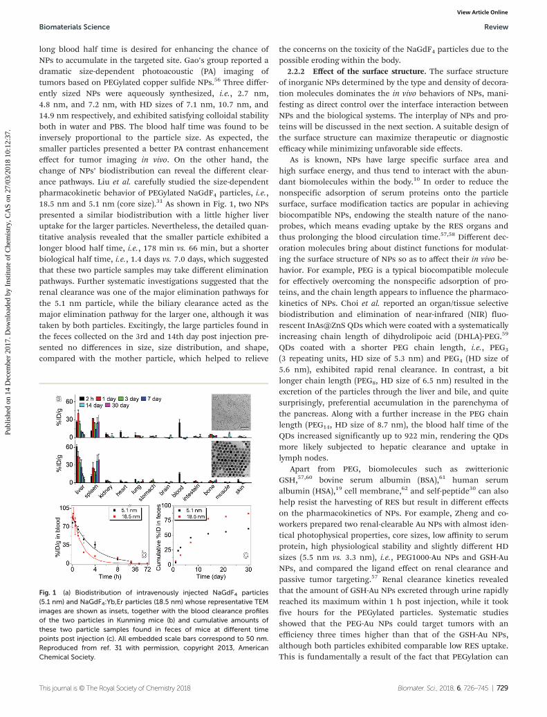
long blood half time is desired for enhancing the chance ofNPs to accumulate in the targeted site. Gao’s group reported adramatic size-dependent photoacoustic (PA) imaging oftumors based on PEGylated copper sulfide NPs.56 Three differ-ently sized NPs were aqueously synthesized, i.e., 2.7 nm,4.8 nm, and 7.2 nm, with HD sizes of 7.1 nm, 10.7 nm, and14.9 nm respectively, and exhibited satisfying colloidal stabilityboth in water and PBS. The blood half time was found to beinversely proportional to the particle size. As expected, thesmaller particles presented a better PA contrast enhancementeffect for tumor imaging in vivo. On the other hand, thechange of NPs’ biodistribution can reveal the different clear-ance pathways. Liu et al. carefully studied the size-dependentpharmacokinetic behavior of PEGylated NaGdF4 particles, i.e.,18.5 nm and 5.1 nm (core size).31 As shown in Fig. 1, two NPspresented a similar biodistribution with a little higher liveruptake for the larger particles. Nevertheless, the detailed quan-titative analysis revealed that the smaller particle exhibited alonger blood half time, i.e., 178 min vs. 66 min, but a shorterbiological half time, i.e., 1.4 days vs. 7.0 days, which suggestedthat these two particle samples may take different eliminationpathways. Further systematic investigations suggested that therenal clearance was one of the major elimination pathways forthe 5.1 nm particle, while the biliary clearance acted as themajor elimination pathway for the larger one, although it wastaken by both particles. Excitingly, the large particles found inthe feces collected on the 3rd and 14th day post injection pre-sented no differences in size, size distribution, and shape,compared with the mother particle, which helped to relieve
the concerns on the toxicity of the NaGdF4 particles due to thepossible eroding within the body.
2.2.2 Effect of the surface structure. The surface structureof inorganic NPs determined by the type and density of decora-tion molecules dominates the in vivo behaviors of NPs, mani-festing as direct control over the interface interaction betweenNPs and the biological systems. The interplay of NPs and pro-teins will be discussed in the next section. A suitable design ofthe surface structure can maximize therapeutic or diagnosticefficacy while minimizing unfavorable side effects.
As is known, NPs have large specific surface area andhigh surface energy, and thus tend to interact with the abun-dant biomolecules within the body.10 In order to reduce thenonspecific adsorption of serum proteins onto the particlesurface, surface modification tactics are popular in achievingbiocompatible NPs, endowing the stealth nature of the nano-probes, which means evading uptake by the RES organs andthus prolonging the blood circulation time.57,58 Different dec-oration molecules bring about distinct functions for modulat-ing the surface structure of NPs so as to affect their in vivo be-havior. For example, PEG is a typical biocompatible moleculefor effectively overcoming the nonspecific adsorption of pro-teins, and the chain length appears to influence the pharmaco-kinetics of NPs. Choi et al. reported an organ/tissue selectivebiodistribution and elimination of near-infrared (NIR) fluo-rescent InAs@ZnS QDs which were coated with a systematicallyincreasing chain length of dihydrolipoic acid (DHLA)-PEG.59
QDs coated with a shorter PEG chain length, i.e., PEG3
(3 repeating units, HD size of 5.3 nm) and PEG4 (HD size of5.6 nm), exhibited rapid renal clearance. In contrast, a bitlonger chain length (PEG8, HD size of 6.5 nm) resulted in theexcretion of the particles through the liver and bile, and quitesurprisingly, preferential accumulation in the parenchyma ofthe pancreas. Along with a further increase in the PEG chainlength (PEG14, HD size of 8.7 nm), the blood half time of theQDs increased significantly up to 922 min, rendering the QDsmore likely subjected to hepatic clearance and uptake inlymph nodes.
Apart from PEG, biomolecules such as zwitterionicGSH,57,60 bovine serum albumin (BSA),61 human serumalbumin (HSA),19 cell membrane,62 and self-peptide30 can alsohelp resist the harvesting of RES but result in different effectson the pharmacokinetics of NPs. For example, Zheng and co-workers prepared two renal-clearable Au NPs with almost iden-tical photophysical properties, core sizes, low affinity to serumprotein, high physiological stability and slightly different HDsizes (5.5 nm vs. 3.3 nm), i.e., PEG1000-Au NPs and GSH-AuNPs, and compared the ligand effect on renal clearance andpassive tumor targeting.57 Renal clearance kinetics revealedthat the amount of GSH-Au NPs excreted through urine rapidlyreached its maximum within 1 h post injection, while it tookfive hours for the PEGylated particles. Systematic studiesshowed that the PEG-Au NPs could target tumors with anefficiency three times higher than that of the GSH-Au NPs,although both particles exhibited comparable low RES uptake.This is fundamentally a result of the fact that PEGylation can
Fig. 1 (a) Biodistribution of intravenously injected NaGdF4 particles(5.1 nm) and NaGdF4:Yb,Er particles (18.5 nm) whose representative TEMimages are shown as insets, together with the blood clearance profilesof the two particles in Kunming mice (b) and cumulative amounts ofthese two particle samples found in feces of mice at different timepoints post injection (c). All embedded scale bars correspond to 50 nm.Reproduced from ref. 31 with permission, copyright 2013, AmericanChemical Society.
Biomaterials Science Review
This journal is © The Royal Society of Chemistry 2018 Biomater. Sci., 2018, 6, 726–745 | 729
Publ
ishe
d on
14
Dec
embe
r 20
17. D
ownl
oade
d by
Ins
titut
e of
Che
mis
try,
CA
S on
27/
03/2
018
10:1
2:37
. View Article Online
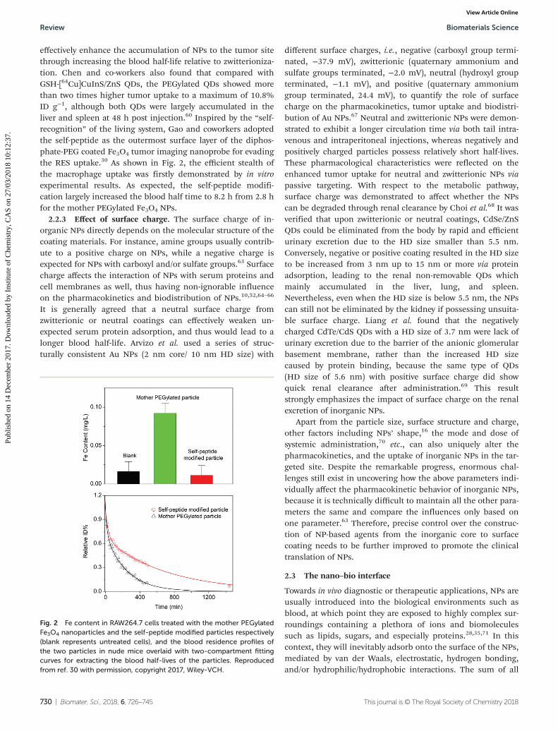
effectively enhance the accumulation of NPs to the tumor sitethrough increasing the blood half-life relative to zwitterioniza-tion. Chen and co-workers also found that compared withGSH-[64Cu]CuInS/ZnS QDs, the PEGylated QDs showed morethan two times higher tumor uptake to a maximum of 10.8%ID g−1, although both QDs were largely accumulated in theliver and spleen at 48 h post injection.60 Inspired by the “self-recognition” of the living system, Gao and coworkers adoptedthe self-peptide as the outermost surface layer of the diphos-phate-PEG coated Fe3O4 tumor imaging nanoprobe for evadingthe RES uptake.30 As shown in Fig. 2, the efficient stealth ofthe macrophage uptake was firstly demonstrated by in vitroexperimental results. As expected, the self-peptide modifi-cation largely increased the blood half time to 8.2 h from 2.8 hfor the mother PEGylated Fe3O4 NPs.
2.2.3 Effect of surface charge. The surface charge of in-organic NPs directly depends on the molecular structure of thecoating materials. For instance, amine groups usually contrib-ute to a positive charge on NPs, while a negative charge isexpected for NPs with carboxyl and/or sulfate groups.63 Surfacecharge affects the interaction of NPs with serum proteins andcell membranes as well, thus having non-ignorable influenceon the pharmacokinetics and biodistribution of NPs.10,52,64–66
It is generally agreed that a neutral surface charge fromzwitterionic or neutral coatings can effectively weaken un-expected serum protein adsorption, and thus would lead to alonger blood half-life. Arvizo et al. used a series of struc-turally consistent Au NPs (2 nm core/ 10 nm HD size) with
different surface charges, i.e., negative (carboxyl group termi-nated, −37.9 mV), zwitterionic (quaternary ammonium andsulfate groups terminated, −2.0 mV), neutral (hydroxyl groupterminated, −1.1 mV), and positive (quaternary ammoniumgroup terminated, 24.4 mV), to quantify the role of surfacecharge on the pharmacokinetics, tumor uptake and biodistri-bution of Au NPs.67 Neutral and zwitterionic NPs were demon-strated to exhibit a longer circulation time via both tail intra-venous and intraperitoneal injections, whereas negatively andpositively charged particles possess relatively short half-lives.These pharmacological characteristics were reflected on theenhanced tumor uptake for neutral and zwitterionic NPs viapassive targeting. With respect to the metabolic pathway,surface charge was demonstrated to affect whether the NPscan be degraded through renal clearance by Choi et al.68 It wasverified that upon zwitterionic or neutral coatings, CdSe/ZnSQDs could be eliminated from the body by rapid and efficienturinary excretion due to the HD size smaller than 5.5 nm.Conversely, negative or positive coating resulted in the HD sizeto be increased from 3 nm up to 15 nm or more via proteinadsorption, leading to the renal non-removable QDs whichmainly accumulated in the liver, lung, and spleen.Nevertheless, even when the HD size is below 5.5 nm, the NPscan still not be eliminated by the kidney if possessing unsuita-ble surface charge. Liang et al. found that the negativelycharged CdTe/CdS QDs with a HD size of 3.7 nm were lack ofurinary excretion due to the barrier of the anionic glomerularbasement membrane, rather than the increased HD sizecaused by protein binding, because the same type of QDs(HD size of 5.6 nm) with positive surface charge did showquick renal clearance after administration.69 This resultstrongly emphasizes the impact of surface charge on the renalexcretion of inorganic NPs.
Apart from the particle size, surface structure and charge,other factors including NPs’ shape,16 the mode and dose ofsystemic administration,70 etc., can also uniquely alter thepharmacokinetics, and the uptake of inorganic NPs in the tar-geted site. Despite the remarkable progress, enormous chal-lenges still exist in uncovering how the above parameters indi-vidually affect the pharmacokinetic behavior of inorganic NPs,because it is technically difficult to maintain all the other para-meters the same and compare the influences only based onone parameter.63 Therefore, precise control over the construc-tion of NP-based agents from the inorganic core to surfacecoating needs to be further improved to promote the clinicaltranslation of NPs.
2.3 The nano–bio interface
Towards in vivo diagnostic or therapeutic applications, NPs areusually introduced into the biological environments such asblood, at which point they are exposed to highly complex sur-roundings containing a plethora of ions and biomoleculessuch as lipids, sugars, and especially proteins.28,35,71 In thiscontext, they will inevitably adsorb onto the surface of the NPs,mediated by van der Waals, electrostatic, hydrogen bonding,and/or hydrophilic/hydrophobic interactions. The sum of all
Fig. 2 Fe content in RAW264.7 cells treated with the mother PEGylatedFe3O4 nanoparticles and the self-peptide modified particles respectively(blank represents untreated cells), and the blood residence profiles ofthe two particles in nude mice overlaid with two-compartment fittingcurves for extracting the blood half-lives of the particles. Reproducedfrom ref. 30 with permission, copyright 2017, Wiley-VCH.
Review Biomaterials Science
730 | Biomater. Sci., 2018, 6, 726–745 This journal is © The Royal Society of Chemistry 2018
Publ
ishe
d on
14
Dec
embe
r 20
17. D
ownl
oade
d by
Ins
titut
e of
Che
mis
try,
CA
S on
27/
03/2
018
10:1
2:37
. View Article Online
adsorption processes across the nano–bio interface will resultin the unintended formation of the so-called “biomoleculecorona”, of which the “protein corona” has been studied themost so far.28 It’s of great significance to consider the furtherbiointeraction and biomodification of NPs in biologicalenvironments, which may adversely impact their final utility.
The protein corona formed in the body is far more complexthan in vitro trials due to the complex environment (nearly2000 different proteins in widely varying concentrations) anddynamic processes of corona formation.35 Proteins involved inboth physiological and pathophysiological relevant processeshave been identified in the coronas of various NPs.4 Theprotein corona not only influences the adhesion to the cellmembrane and subsequent internalization of NPs, but alsohas a severe effect on the physicochemical properties of NPsand further on their pharmacokinetic behavior.10 The formedprotein corona may trigger the transformation of NPs by alter-ing their colloidal stability, either exhibiting a stabilizing effectby inducing steric stabilization or destabilizing impact causedby protein mediated bridging, charge compensation or theintroduction of charge inhomogeneity onto the NP surface.72
For example, Gao et al. found that both negatively and posi-tively charged iron oxide NPs lose their colloidal stabilitieswhen exposed to plasma proteins including serum albuminand immunoglobulin G (IgG).25 Moreover, the HD size of NPswith the corona was dramatically increased, which wouldaccelerate the macrophage uptake of the NPs into the liver,spleen, and bone marrow in the RES system. In addition,binding of opsonins such as IgG and complementary factorscould promote the clearance.73 Such an accumulation of theopsonized NPs into the RES organs is considered to be favor-able when these organs are the intended target sites. However,for delivering the inorganic NPs to tissues other than the RESorgans, the accumulation of NPs would lead to tissue toxicityas well as low theranostic efficiency by losing the targetingability, and therefore in this context minimizing the opsoniza-tion of protein to NPs becomes essential.
Currently, the protein corona is far from being understoodand still remains unpredictable, therefore, attempts to partiallyor even completely prevent protein adsorption are persistentlyinvestigated, although some proteins in the corona present apositive effect on the theranostic represented by apolipo-protein which can promote the movement of nanoprobesacross the blood brain barrier.4 The appropriate surfaceengineering of NPs, which dominates their interaction withplasma proteins, should be pursued for preparing stealth NPsor “corona-free” NPs.
3. Synthetic strategies ofbiocompatible NPs
Wet-chemical synthesis techniques provide a reliable way toprepare diversified inorganic NPs with high quality, on thebasis of which biocompatible NPs for biomedical applications
can be obtained via in situ “one-pot” synthesis of hydrophilicparticles or post surface modification of hydrophobic NPs.
3.1 In situ coatings via “one-pot” synthesis
Aqueous synthesis is in principle the simplest method toprepare water dispersible NPs, which can commonly beobtained in the presence of hydrophilic ligand molecules.3,24
Small molecules bearing chelating groups like carboxylic,thiol, and amine groups, such as citric acid, tartaric acid,mercaptoacetic acid, dimercaptosuccinic acid, phosphorylcholine, and GSH, can bind onto the particle surface as wellas the precursor or monomer surface to stabilize and regu-late the formation of NPs.1 For instance, aqueous synthesishas long been used to grow QDs including CdTe, CdSe, andCdS, involving mixing of cadmium precursors in the pres-ence of thioalkyl acids or amines in aqueous solutions fol-lowed by the injection of tellurium, selenium, or sulfur pre-cursors.3,74 In addition, biopolymers such as carbohydrates(dextran, chitosan, alginate, and arabinogalactan), proteinssuch as lipoproteins, as well as synthetic polymers such asPEG, poly(acrylic acid) (PAA), poly(methacrylic acid) (PMAA),PVP, and polyethylenimine (PEI), are often used as biocom-patible ligands in the aqueous synthesis.1,24,39,56 One repre-sentative example is an MRI contrast agent Feridex, i.e.,superparamagnetic iron oxide NPs coated with dextran,which shows good biocompatibility for FDA-approved clini-cal use.26
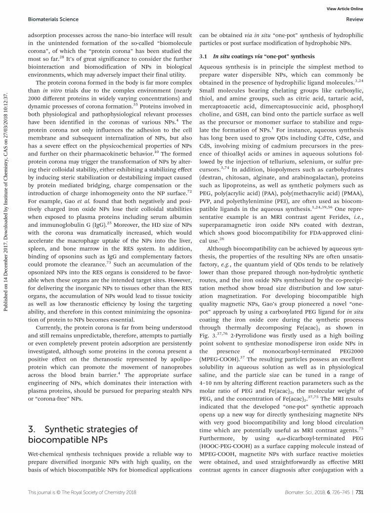
Although biocompatibility can be achieved by aqueous syn-thesis, the properties of the resulting NPs are often unsatis-factory, e.g., the quantum yield of QDs tends to be relativelylower than those prepared through non-hydrolytic syntheticroutes, and the iron oxide NPs synthesized by the co-precipi-tation method show broad size distribution and low satur-ation magnetization. For developing biocompatible highquality magnetic NPs, Gao’s group pioneered a novel “one-pot” approach by using a carboxylated PEG ligand for in situcoating the iron oxide core during the synthetic processthrough thermally decomposing Fe(acac)3 as shown inFig. 3.37,76 2-Pyrrolidone was firstly used as a high boilingpoint solvent to synthesize monodisperse iron oxide NPs inthe presence of monocarboxyl-terminated PEG2000(MPEG-COOH).37 The resulting particles possess an excellentsolubility in aqueous solution as well as in physiologicalsaline, and the particle size can be tuned in a range of4–10 nm by altering different reaction parameters such as themolar ratio of PEG and Fe(acac)3, the molecular weight ofPEG, and the concentration of Fe(acac)3.
37,75 The MRI resultsindicated that the developed “one-pot” synthetic approachopens up a new way for directly synthesizing magnetite NPswith very good biocompatibility and long blood circulationtime which are potentially useful as MRI contrast agents.75
Furthermore, by using α,ω-dicarboxyl-terminated PEG(HOOC-PEG-COOH) as a surface capping molecule instead ofMPEG-COOH, magnetite NPs with surface reactive moietieswere obtained, and used straightforwardly as effective MRIcontrast agents in cancer diagnosis after conjugation with a
Biomaterials Science Review
This journal is © The Royal Society of Chemistry 2018 Biomater. Sci., 2018, 6, 726–745 | 731
Publ
ishe
d on
14
Dec
embe
r 20
17. D
ownl
oade
d by
Ins
titut
e of
Che
mis
try,
CA
S on
27/
03/2
018
10:1
2:37
. View Article Online
specific cancer-targeting antibody.76 In addition, the particlesize was successfully tuned from 12 nm to 27 nm by increas-ing the precursor concentration while maintaining themonodispersity.
Since the iron oxide NPs costabilized by carboxylated PEGand 2-pyrrolidone were demonstrated by the same group toshow obvious nonspecific adsorption of plasma proteins dueto the positive surface potentials, diphenyl oxide, a non-coor-dinating high boiling point solvent, was used instead of2-pyrrolidone to synthesize biocompatible Fe3O4 NPs in thepresence of oleylamine and HOOC-PEG-COOH.14,77,78 Theresulting NPs exhibited terrific colloidal stability and astrongly enhanced MR contrast effect compared with the pre-vious results. Quite interestingly, the particle size of the bio-compatible Fe3O4 NPs could be effectively tuned through aunique gelification effect which is sketched in the lowerpanel of Fig. 3.14 Molecular networks could spontaneouslyform between Fe(acac)3 and HOOC-PEG-COOH with the helpof oleylamine, and be accelerated by temperature and time,which reduced the thermal decomposition rate constants ofthe Fe precursor, consequently altering the particle size. Infact, the strong coordination between the ligands and metalions is ubiquitous for synthesizing colloidal particles, whichalways opens up new ways for tuning the particle size ormorphology.
3.2 Post surface modification with biocompatible coatings
Non-aqueous synthesis represented by a thermal decompo-sition method, with the absence of complicated surfacebinding situations involving water and hydroxyl ions, can yieldhigh quality inorganic NPs with perfect monodispersity, highcrystallinity degree, and satisfying size tunability.1,4 Adequatesurface modifications are essentially required to enable thewater soluble, biocompatible, and surface functionalizableNPs for biomedical applications.
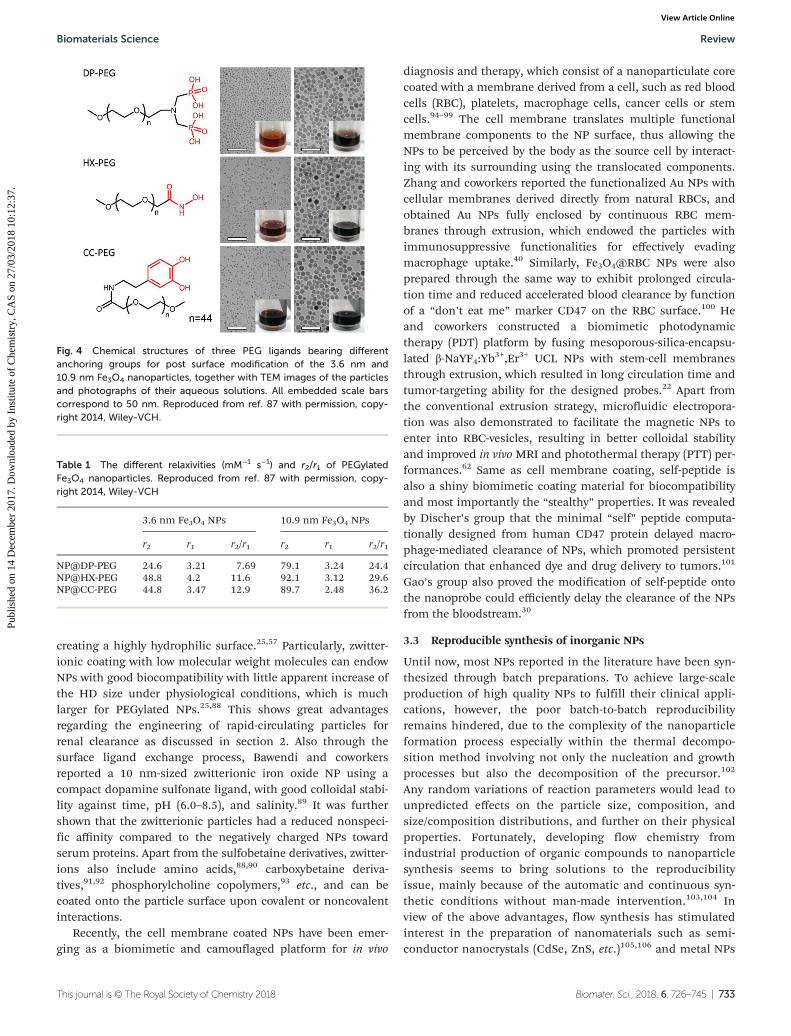
Generally, polymers, zwitterionic molecules, and bio-molecules are all available as biocompatible coatingmaterials.39,79–81 Among all the polymers used for improvingthe solubility and biocompatibility of NPs, PEG and PEG-copo-lymers are currently most popular and found to be mosteffective, and with respect to in vivo applications, the improvedstealth properties of NPs upon PEGylation are attractive due totheir high hydrophilicity, nearly neutral charge, and steric hin-drance.2,82,83 PEG-based modification of hydrophobic NPs postsynthesis can be categorized into two approaches: encapsula-tion typically by amphiphilic molecules,39 and ligand exchangeby coordinating with the anchoring groups.18,21,84 Amphiphilicmolecule encapsulation is principally based on hydrophobic–hydrophobic interactions between the hydrophobic surfaceligand of NPs and the hydrophobic segments of amphiphilicmolecules such as PEG-phospholipid, which is quite effectivefor transferring particles to aqueous phase solution but leavesa hydrophobic layer behind heavily shielding the inorganiccore.2,4 In contrast, surface ligand exchange determined by thebinding affinity of the anchoring group of the biocompatibilityligand to metal ions of NPs is proved to be a reliable approachupon suitable selection of ligands. For example, the PEGligand carrying two phosphate groups at one end and a male-imide group at the other end, denoted as dp-PEG-mal, was suc-cessfully designed by Gao and coworkers, and demonstrated toeffectively replace the oleate ligand of NaGdF4 or NaGdF4:Yb,Er NPs.31,85,86 Because of the improved binding affinity to theparticle surface, the dp-PEG-mal-coated NPs exhibited long-term colloidal stability in both water and PBS, which endowedthe particles with excellent blood circulation behavior in thefollowing in vivo applications. Notably, the anchoring groupsof surface ligands not only determine the affinity to metalions, but also tune the physical properties of NPs. Forexample, iron oxide NPs coated with PEG ligands withdifferent anchoring moieties, i.e., PEG2000 molecule bearingdiphosphate (DP), hydroxamate (HX), and catechol (CC)groups, respectively, were found to show different relaxationperformances as MRI contrast agents (shown in Fig. 4 andTable 1).87
For in vivo applications, anti-biofouling properties of thesurface coating are indispensable for efficiently directing theNPs to the region of interest. Apart from PEG molecules as out-standing anti-biofouling materials, zwitterions, containingboth positively and negatively charged groups but with overallneutral surface charge, are also found to be superior anti-bio-fouling materials through strong ionic structuring of water and
Fig. 3 The upper panel: the representative TEM images ofMPEG-COOH and HOOC-PEG-COOH modified Fe3O4 NPs, respectively.The lower panel: a sketch of the gelification process for producingdifferently sized biocompatible Fe3O4 nanoparticles, together with thephotographs of precursor solutions with different gelification degreesand representative TEM images. All embedded scale bars correspond to50 nm. Reproduced from ref. 14, 37, 75 with permission, copyright 2005and 2006, Wiley-VCH, and Copyright 2011, American Chemical Society,respectively.
Review Biomaterials Science
732 | Biomater. Sci., 2018, 6, 726–745 This journal is © The Royal Society of Chemistry 2018
Publ
ishe
d on
14
Dec
embe
r 20
17. D
ownl
oade
d by
Ins
titut
e of
Che
mis
try,
CA
S on
27/
03/2
018
10:1
2:37
. View Article Online
creating a highly hydrophilic surface.25,57 Particularly, zwitter-ionic coating with low molecular weight molecules can endowNPs with good biocompatibility with little apparent increase ofthe HD size under physiological conditions, which is muchlarger for PEGylated NPs.25,88 This shows great advantagesregarding the engineering of rapid-circulating particles forrenal clearance as discussed in section 2. Also through thesurface ligand exchange process, Bawendi and coworkersreported a 10 nm-sized zwitterionic iron oxide NP using acompact dopamine sulfonate ligand, with good colloidal stabi-lity against time, pH (6.0–8.5), and salinity.89 It was furthershown that the zwitterionic particles had a reduced nonspeci-fic affinity compared to the negatively charged NPs towardserum proteins. Apart from the sulfobetaine derivatives, zwitter-ions also include amino acids,88,90 carboxybetaine deriva-tives,91,92 phosphorylcholine copolymers,93 etc., and can becoated onto the particle surface upon covalent or noncovalentinteractions.
Recently, the cell membrane coated NPs have been emer-ging as a biomimetic and camouflaged platform for in vivo
diagnosis and therapy, which consist of a nanoparticulate corecoated with a membrane derived from a cell, such as red bloodcells (RBC), platelets, macrophage cells, cancer cells or stemcells.94–99 The cell membrane translates multiple functionalmembrane components to the NP surface, thus allowing theNPs to be perceived by the body as the source cell by interact-ing with its surrounding using the translocated components.Zhang and coworkers reported the functionalized Au NPs withcellular membranes derived directly from natural RBCs, andobtained Au NPs fully enclosed by continuous RBC mem-branes through extrusion, which endowed the particles withimmunosuppressive functionalities for effectively evadingmacrophage uptake.40 Similarly, Fe3O4@RBC NPs were alsoprepared through the same way to exhibit prolonged circula-tion time and reduced accelerated blood clearance by functionof a “don’t eat me” marker CD47 on the RBC surface.100 Heand coworkers constructed a biomimetic photodynamictherapy (PDT) platform by fusing mesoporous-silica-encapsu-lated β-NaYF4:Yb3+,Er3+ UCL NPs with stem-cell membranesthrough extrusion, which resulted in long circulation time andtumor-targeting ability for the designed probes.22 Apart fromthe conventional extrusion strategy, microfluidic electropora-tion was also demonstrated to facilitate the magnetic NPs toenter into RBC-vesicles, resulting in better colloidal stabilityand improved in vivo MRI and photothermal therapy (PTT) per-formances.62 Same as cell membrane coating, self-peptide isalso a shiny biomimetic coating material for biocompatibilityand most importantly the “stealthy” properties. It was revealedby Discher’s group that the minimal “self” peptide computa-tionally designed from human CD47 protein delayed macro-phage-mediated clearance of NPs, which promoted persistentcirculation that enhanced dye and drug delivery to tumors.101
Gao’s group also proved the modification of self-peptide ontothe nanoprobe could efficiently delay the clearance of the NPsfrom the bloodstream.30
3.3 Reproducible synthesis of inorganic NPs
Until now, most NPs reported in the literature have been syn-thesized through batch preparations. To achieve large-scaleproduction of high quality NPs to fulfill their clinical appli-cations, however, the poor batch-to-batch reproducibilityremains hindered, due to the complexity of the nanoparticleformation process especially within the thermal decompo-sition method involving not only the nucleation and growthprocesses but also the decomposition of the precursor.102
Any random variations of reaction parameters would lead tounpredicted effects on the particle size, composition, andsize/composition distributions, and further on their physicalproperties. Fortunately, developing flow chemistry fromindustrial production of organic compounds to nanoparticlesynthesis seems to bring solutions to the reproducibilityissue, mainly because of the automatic and continuous syn-thetic conditions without man-made intervention.103,104 Inview of the above advantages, flow synthesis has stimulatedinterest in the preparation of nanomaterials such as semi-conductor nanocrystals (CdSe, ZnS, etc.)105,106 and metal NPs
Fig. 4 Chemical structures of three PEG ligands bearing differentanchoring groups for post surface modification of the 3.6 nm and10.9 nm Fe3O4 nanoparticles, together with TEM images of the particlesand photographs of their aqueous solutions. All embedded scale barscorrespond to 50 nm. Reproduced from ref. 87 with permission, copy-right 2014, Wiley-VCH.
Table 1 The different relaxivities (mM−1 s−1) and r2/r1 of PEGylatedFe3O4 nanoparticles. Reproduced from ref. 87 with permission, copy-right 2014, Wiley-VCH
3.6 nm Fe3O4 NPs 10.9 nm Fe3O4 NPs
r2 r1 r2/r1 r2 r1 r2/r1
NP@DP-PEG 24.6 3.21 7.69 79.1 3.24 24.4NP@HX-PEG 48.8 4.2 11.6 92.1 3.12 29.6NP@CC-PEG 44.8 3.47 12.9 89.7 2.48 36.2
Biomaterials Science Review
This journal is © The Royal Society of Chemistry 2018 Biomater. Sci., 2018, 6, 726–745 | 733
Publ
ishe
d on
14
Dec
embe
r 20
17. D
ownl
oade
d by
Ins
titut
e of
Che
mis
try,
CA
S on
27/
03/2
018
10:1
2:37
. View Article Online
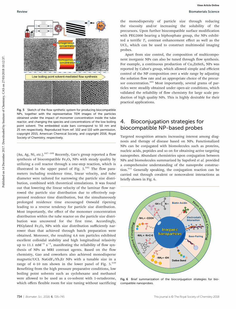
(Au, Ag, Ni, etc.).107–109 Recently, Gao’s group reported a flowsynthesis of biocompatible Fe3O4 NPs with steady quality byutilizing a coil reactor through a one-step reaction, which isillustrated in the upper panel of Fig. 5.102 The flow para-meters including residence time, linear velocity, and tubediameter were tailored for narrowing the particle size distri-bution, combined with theoretical simulations. It was foundout that lowering the linear velocity of the laminar flow nar-rowed the particle size distribution due to effectively sup-pressed residence time distribution, but the simultaneouslyprolonged residence time encouraged Ostwald ripeningleading to a reverse tendency for particle size distribution.Most importantly, the effect of the monomer concentrationdistribution within the tube reactor on the particle size distri-bution was uncovered for the first time. Accordingly,PEGylated Fe3O4 NPs with size distribution sufficiently nar-rower than that achieved through batch preparation wereobtained. Moreover, the resulting 4.6 nm particles exhibitedexcellent colloidal stability and high longitudinal relaxivityup to 11.1 mM−1 s−1, manifesting the reliability of flow syn-thesis of NPs as MRI contrast agents. Based on the flowchemistry, Gao and coworkers also achieved monodispersemagnetic/UCL NaGdF4:Yb,Er NPs with a tunable size in arange of 4–10 nm shown in the lower panel of Fig. 5.110
Benefiting from the high pressure preparative conditions, lowboiling point solvents such as cyclohexane and methanolwere allowed to be used as a co-solvent with 1-octadecene,which offers flexible room for size tuning without sacrificing
the monodispersity of particle size through reducingthe viscosity and/or increasing the solubility of theprecursors. Upon further biocompatible surface modificationwith PEG2000 bearing a biphosphate group, the NPs exhibi-ted a terrific T1 contrast enhancement effect as well as theUCL, which can be used to construct multimodal imagingprobes.
Apart from size control, the composition of multicompo-nent inorganic NPs can also be tuned through flow synthesis.For example, a continuous production of Cu2ZnSnS4 NPs wasreported by Cabot’s group, which allowed simple and efficientcontrol of the NP composition over a wide range by adjustingthe solution flow rate and an appropriate choice of the precur-sor concentration.105 Most importantly, several grams of par-ticles were steadily obtained under open-air conditions, whichvalidated the reliability of flow chemistry for large scale pro-duction of high quality NPs. This is highly desirable for theirpractical applications.
4. Bioconjugation strategies forbiocompatible NP-based probes
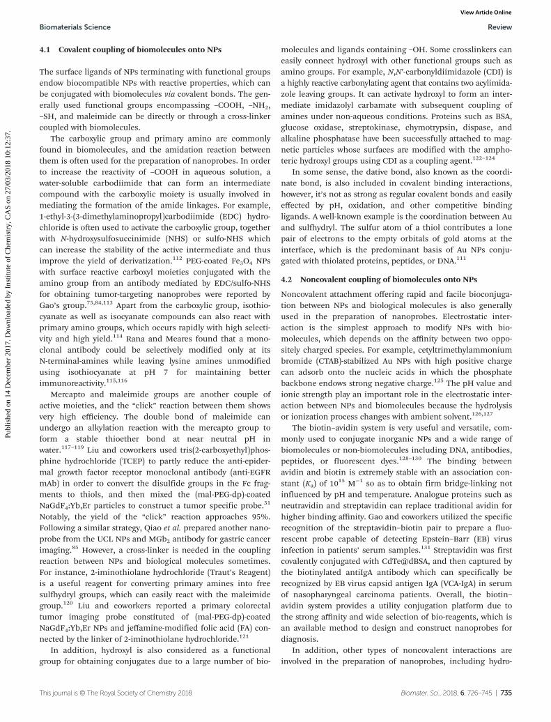
Targeted recognition attracts increasing interest among diag-nosis and therapy of disease based on NPs. FunctionalizedNPs can be conjugated with biomolecules such as proteins,nucleic acids, peptides and so on for obtaining active targetingnanoprobes. Abundant chemistries upon conjugation betweenNPs and biomolecules summarized by Sapsford et al. provideda comprehensive understanding of the nanoprobe construc-tion.111 Generally speaking, the conjugation reaction can becarried out through covalent or noncovalent interactions asbriefly shown in Fig. 6.
Fig. 6 Brief summarization of the bioconjugation strategies for bio-compatible nanoprobes.
Fig. 5 Sketch of the flow synthetic system for producing biocompatibleNPs, together with the representative TEM images of the particlesobtained under the impact of monomer concentration inside the tubereactor, and changing the species and concentrations of the low boilingpoint solvent. The embedded scale bars correspond to 50 nm and25 nm respectively. Reproduced from ref. 102 and 110 with permission,copyright 2015, American Chemical Society, and copyright 2016, RoyalSociety of Chemistry, respectively.
Review Biomaterials Science
734 | Biomater. Sci., 2018, 6, 726–745 This journal is © The Royal Society of Chemistry 2018
Publ
ishe
d on
14
Dec
embe
r 20
17. D
ownl
oade
d by
Ins
titut
e of
Che
mis
try,
CA
S on
27/
03/2
018
10:1
2:37
. View Article Online
4.1 Covalent coupling of biomolecules onto NPs
The surface ligands of NPs terminating with functional groupsendow biocompatible NPs with reactive properties, which canbe conjugated with biomolecules via covalent bonds. The gen-erally used functional groups encompassing –COOH, –NH2,–SH, and maleimide can be directly or through a cross-linkercoupled with biomolecules.
The carboxylic group and primary amino are commonlyfound in biomolecules, and the amidation reaction betweenthem is often used for the preparation of nanoprobes. In orderto increase the reactivity of –COOH in aqueous solution, awater-soluble carbodiimide that can form an intermediatecompound with the carboxylic moiety is usually involved inmediating the formation of the amide linkages. For example,1-ethyl-3-(3-dimethylaminopropyl)carbodiimide (EDC) hydro-chloride is often used to activate the carboxylic group, togetherwith N-hydroxysulfosuccinimide (NHS) or sulfo-NHS whichcan increase the stability of the active intermediate and thusimprove the yield of derivatization.112 PEG-coated Fe3O4 NPswith surface reactive carboxyl moieties conjugated with theamino group from an antibody mediated by EDC/sulfo-NHSfor obtaining tumor-targeting nanoprobes were reported byGao’s group.75,84,113 Apart from the carboxylic group, isothio-cyanate as well as isocyanate compounds can also react withprimary amino groups, which occurs rapidly with high selecti-vity and high yield.114 Rana and Meares found that a mono-clonal antibody could be selectively modified only at itsN-terminal-amines while leaving lysine amines unmodifiedusing isothiocyanate at pH 7 for maintaining betterimmunoreactivity.115,116
Mercapto and maleimide groups are another couple ofactive moieties, and the “click” reaction between them showsvery high efficiency. The double bond of maleimide canundergo an alkylation reaction with the mercapto group toform a stable thioether bond at near neutral pH inwater.117–119 Liu and coworkers used tris(2-carboxyethyl)phos-phine hydrochloride (TCEP) to partly reduce the anti-epider-mal growth factor receptor monoclonal antibody (anti-EGFRmAb) in order to convert the disulfide groups in the Fc frag-ments to thiols, and then mixed the (mal-PEG-dp)-coatedNaGdF4:Yb,Er particles to construct a tumor specific probe.31
Notably, the yield of the “click” reaction approaches 95%.Following a similar strategy, Qiao et al. prepared another nano-probe from the UCL NPs and MGb2 antibody for gastric cancerimaging.85 However, a cross-linker is needed in the couplingreaction between NPs and biological molecules sometimes.For instance, 2-iminothiolane hydrochloride (Traut’s Reagent)is a useful reagent for converting primary amines into freesulfhydryl groups, which can easily react with the maleimidegroup.120 Liu and coworkers reported a primary colorectaltumor imaging probe constituted of (mal-PEG-dp)-coatedNaGdF4:Yb,Er NPs and jeffamine-modified folic acid (FA) con-nected by the linker of 2-iminothiolane hydrochloride.121
In addition, hydroxyl is also considered as a functionalgroup for obtaining conjugates due to a large number of bio-
molecules and ligands containing –OH. Some crosslinkers caneasily connect hydroxyl with other functional groups such asamino groups. For example, N,N′-carbonyldiimidazole (CDI) isa highly reactive carbonylating agent that contains two acylimida-zole leaving groups. It can activate hydroxyl to form an inter-mediate imidazolyl carbamate with subsequent coupling ofamines under non-aqueous conditions. Proteins such as BSA,glucose oxidase, streptokinase, chymotrypsin, dispase, andalkaline phosphatase have been successfully attached to mag-netic particles whose surfaces are modified with the ampho-teric hydroxyl groups using CDI as a coupling agent.122–124
In some sense, the dative bond, also known as the coordi-nate bond, is also included in covalent binding interactions,however, it’s not as strong as regular covalent bonds and easilyeffected by pH, oxidation, and other competitive bindingligands. A well-known example is the coordination between Auand sulfhydryl. The sulfur atom of a thiol contributes a lonepair of electrons to the empty orbitals of gold atoms at theinterface, which is the predominant basis of Au NPs conju-gated with thiolated proteins, peptides, or DNA.111
4.2 Noncovalent coupling of biomolecules onto NPs
Noncovalent attachment offering rapid and facile bioconjuga-tion between NPs and biological molecules is also generallyused in the preparation of nanoprobes. Electrostatic inter-action is the simplest approach to modify NPs with bio-molecules, which depends on the affinity between two oppo-sitely charged species. For example, cetyltrimethylammoniumbromide (CTAB)-stabilized Au NPs with high positive chargecan adsorb onto the nucleic acids in which the phosphatebackbone endows strong negative charge.125 The pH value andionic strength play an important role in the electrostatic inter-action between NPs and biomolecules because the hydrolysisor ionization process changes with ambient solvent.126,127
The biotin–avidin system is very useful and versatile, com-monly used to conjugate inorganic NPs and a wide range ofbiomolecules or non-biomolecules including DNA, antibodies,peptides, or fluorescent dyes.128–130 The binding betweenavidin and biotin is extremely stable with an association con-stant (Ka) of 10
15 M−1 so as to obtain firm bridge-linking notinfluenced by pH and temperature. Analogue proteins such asneutravidin and streptavidin can replace traditional avidin forhigher binding affinity. Gao and coworkers utilized the specificrecognition of the streptavidin–biotin pair to prepare a fluo-rescent probe capable of detecting Epstein–Barr (EB) virusinfection in patients’ serum samples.131 Streptavidin was firstcovalently conjugated with CdTe@dBSA, and then captured bythe biotinylated antiIgA antibody which can specifically berecognized by EB virus capsid antigen IgA (VCA-IgA) in serumof nasopharyngeal carcinoma patients. Overall, the biotin–avidin system provides a utility conjugation platform due tothe strong affinity and wide selection of bio-reagents, which isan available method to design and construct nanoprobes fordiagnosis.
In addition, other types of noncovalent interactions areinvolved in the preparation of nanoprobes, including hydro-
Biomaterials Science Review
This journal is © The Royal Society of Chemistry 2018 Biomater. Sci., 2018, 6, 726–745 | 735
Publ
ishe
d on
14
Dec
embe
r 20
17. D
ownl
oade
d by
Ins
titut
e of
Che
mis
try,
CA
S on
27/
03/2
018
10:1
2:37
. View Article Online
phobic interactions and base-pairing interactions132–136 forbetter serving the in vitro detection and in vivo diagnosisapplications.
5. Biocompatible NP-based in vivoimaging
Benefitting from the fast development of preparing techniquesof high quality inorganic NPs and the various surface modifi-cation and conjugation strategies, biocompatible NPs rep-resent highly promising platforms in the biological and bio-medical applications. With respect to in vivo applications,many attempts from different fields have been devoted toexploring the applications of NPs in imaging, therapy, anddrug delivery. Recently, a lot of reviews have summarized thevarious biomedical applications of NPs in therapy and drugdelivery,2,32,41–43,137–140 and in this section we will mainlyfocus on the recent progress of imaging based on biocompati-ble NPs, with a special emphasis on atherosclerosis imagingand tumor imaging by active targeting and tumor microenvi-ronment stimuli-responsive ones.
5.1 Atherosclerosis imaging
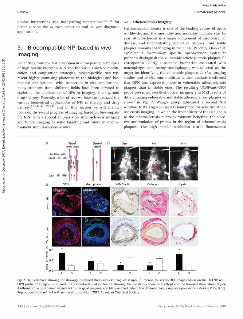
Cardiovascular disease is one of the leading causes of deathworldwide, and the morbidity and mortality increase year byyear. Atherosclerosis is a major component of cardiovasculardisease, and differentiating vulnerable plaques from stableplaques remains challenging in the clinic. Recently, Qiao et al.exploited a macrophage specific upconversion molecularprobe to distinguish the vulnerable atherosclerotic plaques.141
Osteopontin (OPN), a secreted biomarker associated withmacrophages and foamy macrophages, was selected as thetarget for identifying the vulnerable plaques. In vivo imagingstudies and ex vivo immunohistochemical analysis confirmedthat OPN was expressed more in vulnerable atheroscleroticplaques than in stable ones. The resulting UCNP-anti-OPNprobe presented excellent optical imaging and MRI results ofdifferentiating vulnerable and stable atherosclerotic plaques asshown in Fig. 7. Wang’s group fabricated a second NIRwindow (NIR-II) Ag2S-PEG@ICG nanoprobe for sensitive ather-osclerosis imaging, in which the lipophilicity of the C18 chainto the atherosclerosis microenvironment benefited the selec-tive accumulation of probes in the region of atherosclerosisplaques. The high spatial resolution NIR-II fluorescence
Fig. 7 (a) Schematic drawing for showing the varied stress-induced plagues in ApoE−/− mouse, (b) in vivo UCL images based on the UCLNP-anti-OPN probe (the region of interest is encircled with red circles for showing the oscillatory shear stress (top) and the lowered shear stress region(bottom) of the constrained vessel), (c) histological analyses, and (d) quantified data of the different plaque regions upon various staining (*P < 0.05).Reproduced from ref. 141 with permission, copyright 2017, American Chemical Society.
Review Biomaterials Science
736 | Biomater. Sci., 2018, 6, 726–745 This journal is © The Royal Society of Chemistry 2018
Publ
ishe
d on
14
Dec
embe
r 20
17. D
ownl
oade
d by
Ins
titut
e of
Che
mis
try,
CA
S on
27/
03/2
018
10:1
2:37
. View Article Online
imaging of Ag2S QDs combining with real-time PA imaging ofICG proved the feasibility of the nanoprobe for atherosclerosistargeting in an ApoE−/− mouse model.142
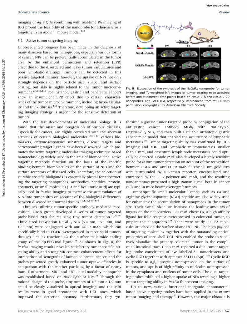
5.2 Active tumor targeting imaging
Unprecedented progress has been made in the diagnosis ofmany diseases based on nanoprobes, especially various formsof cancer. NPs can be preferentially accumulated in the tumorarea by the enhanced permeation and retention (EPR)effect due to the disordered and leaky tumor vasculatures andpoor lymphatic drainage. Tumors can be detected in thispassive targeted manner, however, the uptake of NPs not onlystrongly depends on the particle size, shape, and surfacecoating, but also is highly related to the tumor microenvi-ronment.27,143,144 For instance, gastric and pancreatic cancersshow an insufficient EPR effect due to certain character-istics of the tumor microenvironment, including hypovascular-ity and thick fibrosis.144 Therefore, developing an active target-ing imaging strategy is urgent for the sensitive detection oftumors.
With the fast developments of molecular biology, it isfound that the onset and progression of various diseases,especially for cancer, are highly correlated with the aberrantactivities of certain biological molecules.145–147 Various bio-markers, enzyme-responsive substrates, disease targets andcorresponding target ligands have been discovered, which pro-motes the active targeting molecular imaging technique-basednanotechnology widely used in the area of biomedicine. Activetargeting methods function on the basis of the specificbinding between biomolecules on the surface of NPs and thesurface receptors of diseased cells. Therefore, the selection ofsuitable specific bioligands is essentially pivotal for construct-ing the targeting nanoprobes. Antibodies, peptides, protein,aptamers, or small molecules (FA and hyaluronic acid) are typi-cally used in in vivo imaging to increase the accumulation ofNPs into tumor sites on account of the biological differencesbetween diseased and normal tissues.31,121,148–150
Through utilizing tumor-specific antibody mediated reco-gnition, Gao’s group developed a series of tumor targetedprobe-based NPs for realizing tiny tumor detection.31,85,86
Three sized PEGylation NaGdF4 NPs (5.4 nm, 15.1 nm, and19.8 nm) were conjugated with anti-EGFR mAb, which canspecifically bind to EGFR overexpressed in most solid tumorsthrough a “click reaction” via the surface maleimide endinggroup of the dp-PEG-mal ligand.86 As shown in Fig. 8, thein vivo imaging results revealed satisfactory tumor-specific tar-geting ability and strong MR contrast enhancement effects forintraperitoneal xenografts of human colorectal cancer, and theprobes presented greatly enhanced tumor uptake efficacies incomparison with the mother particles, by a factor of nearlyfour. Furthermore, MRI and UCL dual-modality nanoprobewas established based on NaGdF4:Yb,Er NPs.31 Through therational design of the probe, tiny tumors of 1.7 mm × 1.9 mmcould be clearly visualized in optical imaging, and the MRIresults were in good accordance with UCL ones, whichimproved the detection accuracy. Furthermore, they syn-
thesized a gastric tumor targeted probe by conjugation of theanti-gastric cancer antibody MGb2 with NaGdF4:Yb,Er@NaGdF4 NPs, and then built a reliable orthotopic gastriccancer mice model that enabled the occurrence of lymphaticmetastasis.85 Tumor targeting ability was confirmed by UCLimaging and MRI, and lymphatic micrometastasis smallerthan 1 mm, and omentum lymph node metastasis could opti-cally be detected. Conde et al. also developed a highly sensitiveprobe for in vivo tumor detection on account of the recognitionbetween EGFR and anti-EGFR mAb.151 The 90 nm gold NPswere surrounded by a Raman reporter, encapsulated andentrapped by the PEG polymer and mAb, and the resultingnanoantennas presented a high Raman signal both in cancercells and in mice bearing xenograft tumors.
Tumor-specific small molecular ligands such as FA andarginine-glycine-aspartate (RGD) peptide are also widely usedfor enhancing the accumulation of nanoprobes in the tumorsite. Their “small size” can increase the loading amounts oftargets on the nanocarriers. Liu et al. chose FA, a high affinityligand for folic receptor overexpressed in colorectal tumor, toprepare the nanoprobes.121 There were nearly 500 FA mole-cules attached on the surface of one UCL NP. The high payloadof targeting molecules together with the outstanding opticalproperties of core–shell UCL NPs enabled the probe to sensi-tively visualize the primary colorectal tumor in the compli-cated intestinal tract. Chen et al. reported a dual tumor target-ing probe constituted of dye labelled-Au nanoclusters andcyclic RGD together with aptamer AS1411 (Apt).148 Cyclic RGDis specific to αvβ3 integrins overexpressed on the surface oftumors, and Apt is of high affinity to nucleolin overexpressedin the cytoplasm and nucleus of tumor cells. The dual target-ing probes exhibited a higher uptake of NPs revealing a highertumor targeting ability in in vivo fluorescent imaging.
Up to now, various functional inorganic nanomaterial-based active targeting probes have been applied in the in vivotumor imaging and therapy.27 However, the major obstacle is
Fig. 8 Illustration of the synthesis of the NaGdF4 nanoprobe for tumorimaging, and T1-weighted MR images of tumor-bearing mice acquiredbefore and at different time points based on NaGdF4-5 and NaGdF4-20nanoprobes, and Gd-DTPA, respectively. Reproduced from ref. 86 withpermission, copyright 2013, American Chemical Society.
Biomaterials Science Review
This journal is © The Royal Society of Chemistry 2018 Biomater. Sci., 2018, 6, 726–745 | 737
Publ
ishe
d on
14
Dec
embe
r 20
17. D
ownl
oade
d by
Ins
titut
e of
Che
mis
try,
CA
S on
27/
03/2
018
10:1
2:37
. View Article Online
the poor uptake efficiency of the nanoprobes to the tumor siteeven through the active targeting delivery strategy (less than10% ID g−1). The ability of target binding of the active probesis of some dispute, for example, most studies show that target-ing ligands increase the total accumulation of NPs in thetumor, while some studies can’t observe the increasing totalaccumulation but instead show influence on the distributionwithin the tumor tissue.27,152 In fact, the specific ligand–recep-tor interaction alone does not necessarily ensure that thenanoprobes could be effectively delivered to the lesion. Tomanipulate the nanobiointerface interplay between the NPsand biological environment remains the main issue ofconcern for the nanoprobe design.
5.3 Stimuli-responsive tumor imaging
Researchers gradually realize that the tumor microenvi-ronment is strongly correlated with the growth, invasion, andmetastasis of malignant tumor.84,153,154 For example, the aber-rant physicochemical features such as the overexpressed pro-teases destroy extracellular matrix integrity and correlate withan advanced tumor stage, while the reduced pH and loweredoxygen pressure are critical to the initiation and maintenanceof tumorigenesis.84,153–157 Therefore, developing noninvasivemethods for visualizing the tumor microenvironment is criti-cal for tumor diagnostics, therapy, and prognostics.Tremendous studies have been devoted to design stimuli-responsive and intelligent nanoprobes for tumor and micro-environment imaging.
5.3.1 Tumor microenvironment imaging. As is known,tumors are heterogeneous due to the dynamics and the diver-sity of the tumor microenvironment.158 Acquiring the detailedinformation of different regions of tumor is helpful for diagno-sis and therapy. Commonly, tumor microenvironment imagingcan be realized via the signal changes, such as optical, MRI,PA, and so on, caused by tumor environmental characteristicsincluding overexpressed proteins, acid, redox, hypoxic sur-rounding, which allow us to gain a new perspective on tumor-specific detection and imaging.84,159–164
Normally, matrix metalloproteinases (MMPs) occur in theunactivated zymogen form, whereas they are activated andupregulated in almost all types of human cancers.164,165 Liu’sgroup reported a novel activatable PA imaging nanoprobe forin vivo detection of cancer-related MMPs for the first time.164
CuS NPs with strong NIR absorbance were conjugated with ared-light-absorbing dye, black hole quencher 3 (BHQ3), via aMMP cleavable peptide. The obtained CuS-peptide-BHQ3probes exhibited strong PA signals at 680 nm and 930 nmbefore enzyme cleavage owing to the absorption of BHQ3 andCuS NPs, respectively. Once the probes encountered MMPs intumor areas, free BHQ3 would be released and washed outfrom the tumor, and the PA signal at 680 nm quickly dimin-ished. The PA signal ratio of 680 nm/930 nm could thus serveas an indicator of MMP activity inside the tumor. The in vivoPA imaging results demonstrated that the designed probecould be used for the detection of a specific enzyme activity.
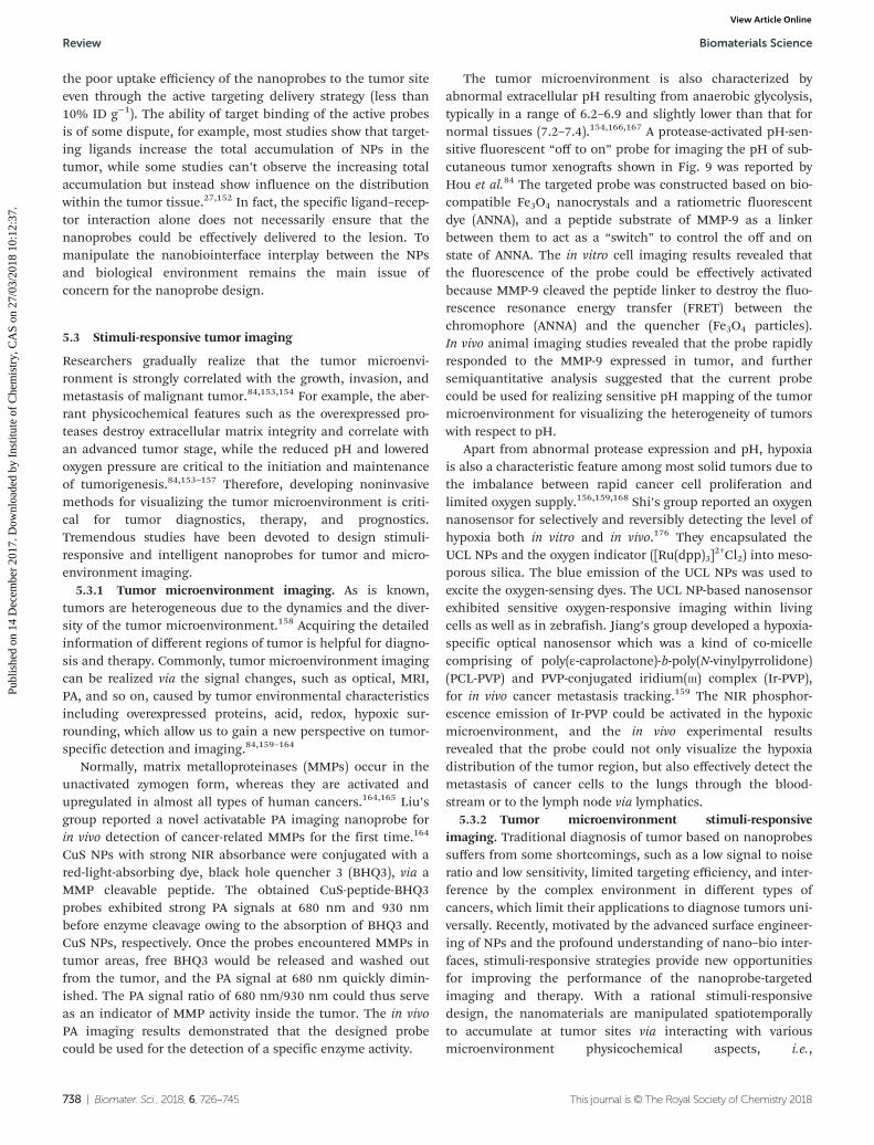
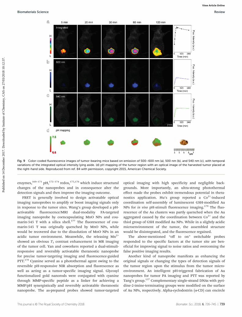
The tumor microenvironment is also characterized byabnormal extracellular pH resulting from anaerobic glycolysis,typically in a range of 6.2–6.9 and slightly lower than that fornormal tissues (7.2–7.4).154,166,167 A protease-activated pH-sen-sitive fluorescent “off to on” probe for imaging the pH of sub-cutaneous tumor xenografts shown in Fig. 9 was reported byHou et al.84 The targeted probe was constructed based on bio-compatible Fe3O4 nanocrystals and a ratiometric fluorescentdye (ANNA), and a peptide substrate of MMP-9 as a linkerbetween them to act as a “switch” to control the off and onstate of ANNA. The in vitro cell imaging results revealed thatthe fluorescence of the probe could be effectively activatedbecause MMP-9 cleaved the peptide linker to destroy the fluo-rescence resonance energy transfer (FRET) between thechromophore (ANNA) and the quencher (Fe3O4 particles).In vivo animal imaging studies revealed that the probe rapidlyresponded to the MMP-9 expressed in tumor, and furthersemiquantitative analysis suggested that the current probecould be used for realizing sensitive pH mapping of the tumormicroenvironment for visualizing the heterogeneity of tumorswith respect to pH.
Apart from abnormal protease expression and pH, hypoxiais also a characteristic feature among most solid tumors due tothe imbalance between rapid cancer cell proliferation andlimited oxygen supply.156,159,168 Shi’s group reported an oxygennanosensor for selectively and reversibly detecting the level ofhypoxia both in vitro and in vivo.176 They encapsulated theUCL NPs and the oxygen indicator ([Ru(dpp)3]
2+Cl2) into meso-porous silica. The blue emission of the UCL NPs was used toexcite the oxygen-sensing dyes. The UCL NP-based nanosensorexhibited sensitive oxygen-responsive imaging within livingcells as well as in zebrafish. Jiang’s group developed a hypoxia-specific optical nanosensor which was a kind of co-micellecomprising of poly(ε-caprolactone)-b-poly(N-vinylpyrrolidone)(PCL-PVP) and PVP-conjugated iridium(III) complex (Ir-PVP),for in vivo cancer metastasis tracking.159 The NIR phosphor-escence emission of Ir-PVP could be activated in the hypoxicmicroenvironment, and the in vivo experimental resultsrevealed that the probe could not only visualize the hypoxiadistribution of the tumor region, but also effectively detect themetastasis of cancer cells to the lungs through the blood-stream or to the lymph node via lymphatics.
5.3.2 Tumor microenvironment stimuli-responsiveimaging. Traditional diagnosis of tumor based on nanoprobessuffers from some shortcomings, such as a low signal to noiseratio and low sensitivity, limited targeting efficiency, and inter-ference by the complex environment in different types ofcancers, which limit their applications to diagnose tumors uni-versally. Recently, motivated by the advanced surface engineer-ing of NPs and the profound understanding of nano–bio inter-faces, stimuli-responsive strategies provide new opportunitiesfor improving the performance of the nanoprobe-targetedimaging and therapy. With a rational stimuli-responsivedesign, the nanomaterials are manipulated spatiotemporallyto accumulate at tumor sites via interacting with variousmicroenvironment physicochemical aspects, i.e.,
Review Biomaterials Science
738 | Biomater. Sci., 2018, 6, 726–745 This journal is © The Royal Society of Chemistry 2018
Publ
ishe
d on
14
Dec
embe
r 20
17. D
ownl
oade
d by
Ins
titut
e of
Che
mis
try,
CA
S on
27/
03/2
018
10:1
2:37
. View Article Online
enzymes,169–171 pH,172–174 redox,175,176 which induce structuralchanges of the nanoprobes and in consequence alter thedetection signals and then improve the imaging outcome.
FRET is generally involved to design activatable opticalimaging nanoprobes to amplify or boost imaging signals onlyin response to the tumor sites. Wang’s group developed a pH-activatable fluorescence/MRI dual-modality FA-targetedimaging nanoprobe by coencapsulating MnO NPs and cou-marin-545 T with a silica shell.177 The fluorescence of cou-marin-545 T was originally quenched by MnO NPs, whilewould be recovered due to the dissolution of MnO NPs in anacidic tumor environment. Meanwhile, the releasing Mn2+
showed an obvious T1 contrast enhancement in MR imagingof the tumor cell. Yan and coworkers reported a dual-stimuli-responsive and reversibly activatable theranostic nanoprobefor precise tumor-targeting imaging and fluorescence-guidedPTT.178 Cyanine served as a photothermal agent owing to thereversible pH-responsive NIR absorption and fluorescence aswell as acting as a tumor-specific imaging signal. Glycosylfunctionalized gold nanorods were conjugated with cyaninethrough MMP-specific peptide as a linker for achieving aMMP/pH synergistically and reversibly activatable theranosticnanoprobe. The as-prepared probes showed tumor-targeted
optical imaging with high specificity and negligible back-grounds. More importantly, an ultra-strong photothermaleffect made the probes exhibit tremendous potential in thera-nostics application. Hu’s group reported a Co2+-inducedcoordination self-assembly of luminescent GSH-modified AuNPs for in vivo pH-stimuli fluorescence imaging.179 The fluo-rescence of the Au clusters was partly quenched when the Auaggregated caused by the coordination between Co2+ and thethiol group of GSH modified Au NPs. While in a slightly acidicmicroenvironment of the tumor, the assembled structurewould be disintegrated, and the fluorescence regained.
The above-mentioned “off to on” switchable probesresponded to the specific factors at the tumor site are ben-eficial for improving signal to noise ratios and overcoming thefalse positive imaging results.
Another kind of nanoprobe manifests as enhancing theoriginal signals or changing the types of detection signals ofthe tumor region upon the stimulus from the tumor micro-environment. An intelligent pH-triggered fabrication of Aunanoprobes for tumor PA imaging and PTT was reported byTang’s group.137 Complementary single-strand DNAs with pyri-dine-2-imine-terminating groups were modified on the surfaceof Au NPs, respectively. Alpha-cyclodextrin (α-CD) can encircle
Fig. 9 Color-coded fluorescence images of tumor-bearing mice based on emission of 500–600 nm (a), 500 nm (b), and 540 nm (c), with temporalvariations of the integrated optical intensity lying aside. (d) pH mapping of the tumor region with an optical image of the harvested tumor placed atthe right-hand side. Reproduced from ref. 84 with permission, copyright 2015, American Chemical Society.
Biomaterials Science Review
This journal is © The Royal Society of Chemistry 2018 Biomater. Sci., 2018, 6, 726–745 | 739
Publ
ishe
d on
14
Dec
embe
r 20
17. D
ownl
oade
d by
Ins
titut
e of
Che
mis
try,
CA
S on
27/
03/2
018
10:1
2:37
. View Article Online
pyridine-2-imine to prevent hybridization of DNA strands onAu NPs under neutral pH. When the nanoprobes reached thetumor area, α-CD was separated from the ends of DNA due tothe protonation of pyridine-2-imine in the decreased pH.Consequently, Au NPs self-aggregated through complementarybase pairing. The in vivo results indicated that the obtainedprobes could act as an efficient agent for tumor-targeted PAimaging and PTT. Gao and coworkers designed a legumain-triggered aggregatable Au NPs for enhanced PA imaging andretention of chemotherapeutics in brain tumors.180 Thesurface of Au NPs was modified with Ala-Ala-Asn-Cys-Lys (Au-AK) and 2-cyano-6-amino-benzothiazole (Au-CABT), respect-ively. In the presence of legumain, a click cycloaddition reac-tion occurred between 1,2-thiolamino groups on hydrolyzedAu-AK and the cyano group on Au-CABT, which caused theaggregation of Au NPs. Doxorubicin (DOX) was further loadedon Cy5.5 labelled Au NPs through a pH-sensitive linker. Theobtained probes could not only enhance the retention of NPsin glioma cells for sensitive in vivo tumor detection by fluo-rescence and PA imaging, but also showed a positive pre-clinical significance in improving the therapeutic outcome ofglioma with reduced systemic toxicity of DOX. Wen et al.reported a novel ultrasmall biocompatible WO3−x nanodot forPA/CT imaging and therapy of tumor. WO3−x nanodots couldact as a potential radiosensitizer, and show a strong pH/O2-responsive localized surface plasmon resonance (LSPR) in theNIR region. Such properties were well aligned with the weaklyacidic and hypoxic microenvironment of tumors. Thus, WO3−x
nanodots revealed enhanced PA imaging of tumor with thecombination of excellent PTT and radiotherapy.181
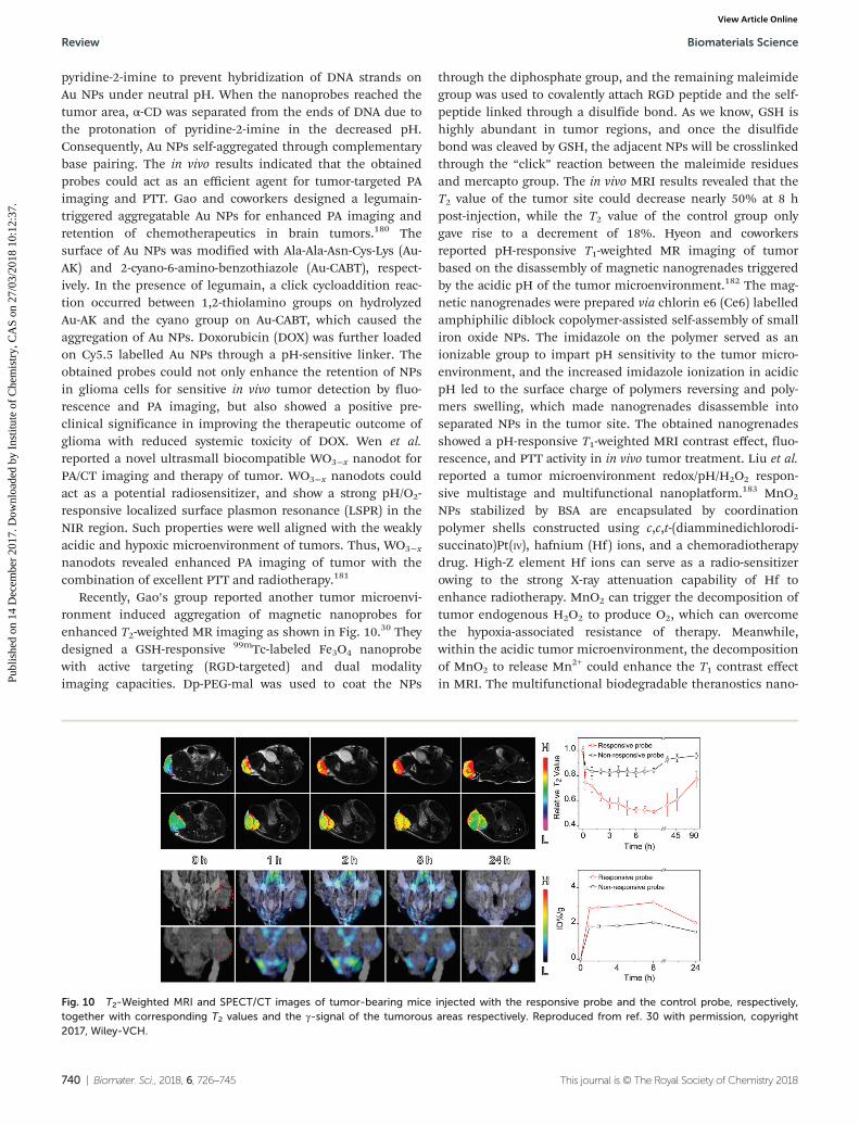
Recently, Gao’s group reported another tumor microenvi-ronment induced aggregation of magnetic nanoprobes forenhanced T2-weighted MR imaging as shown in Fig. 10.30 Theydesigned a GSH-responsive 99mTc-labeled Fe3O4 nanoprobewith active targeting (RGD-targeted) and dual modalityimaging capacities. Dp-PEG-mal was used to coat the NPs
through the diphosphate group, and the remaining maleimidegroup was used to covalently attach RGD peptide and the self-peptide linked through a disulfide bond. As we know, GSH ishighly abundant in tumor regions, and once the disulfidebond was cleaved by GSH, the adjacent NPs will be crosslinkedthrough the “click” reaction between the maleimide residuesand mercapto group. The in vivo MRI results revealed that theT2 value of the tumor site could decrease nearly 50% at 8 hpost-injection, while the T2 value of the control group onlygave rise to a decrement of 18%. Hyeon and coworkersreported pH-responsive T1-weighted MR imaging of tumorbased on the disassembly of magnetic nanogrenades triggeredby the acidic pH of the tumor microenvironment.182 The mag-netic nanogrenades were prepared via chlorin e6 (Ce6) labelledamphiphilic diblock copolymer-assisted self-assembly of smalliron oxide NPs. The imidazole on the polymer served as anionizable group to impart pH sensitivity to the tumor micro-environment, and the increased imidazole ionization in acidicpH led to the surface charge of polymers reversing and poly-mers swelling, which made nanogrenades disassemble intoseparated NPs in the tumor site. The obtained nanogrenadesshowed a pH-responsive T1-weighted MRI contrast effect, fluo-rescence, and PTT activity in in vivo tumor treatment. Liu et al.reported a tumor microenvironment redox/pH/H2O2 respon-sive multistage and multifunctional nanoplatform.183 MnO2
NPs stabilized by BSA are encapsulated by coordinationpolymer shells constructed using c,c,t-(diamminedichlorodi-succinato)Pt(IV), hafnium (Hf) ions, and a chemoradiotherapydrug. High-Z element Hf ions can serve as a radio-sensitizerowing to the strong X-ray attenuation capability of Hf toenhance radiotherapy. MnO2 can trigger the decomposition oftumor endogenous H2O2 to produce O2, which can overcomethe hypoxia-associated resistance of therapy. Meanwhile,within the acidic tumor microenvironment, the decompositionof MnO2 to release Mn2+ could enhance the T1 contrast effectin MRI. The multifunctional biodegradable theranostics nano-
Fig. 10 T2-Weighted MRI and SPECT/CT images of tumor-bearing mice injected with the responsive probe and the control probe, respectively,together with corresponding T2 values and the γ-signal of the tumorous areas respectively. Reproduced from ref. 30 with permission, copyright2017, Wiley-VCH.
Review Biomaterials Science
740 | Biomater. Sci., 2018, 6, 726–745 This journal is © The Royal Society of Chemistry 2018
Publ
ishe
d on
14
Dec
embe
r 20
17. D
ownl
oade
d by
Ins
titut
e of
Che
mis
try,
CA
S on
27/
03/2
018
10:1
2:37
. View Article Online
platform not only enabled tumor specific imaging, but alsoexhibited a satisfactory therapeutic effect in vivo. Xu’s groupalso reported a pH-ultrasensitive Mn-based layered doublehydroxide (Mn-LDH) NPs for tumor T1-weighted MRI.184 Theobtained Mn-LDH exhibited an ultrasensitive pH response andenhanced T1 imaging for at least 2 days in vivo.
Apart from the reinforcing of the MRI effect, intelligentnanoprobes with the T1–T2 switchable MRI contrast effect havealso been designed for tumor imaging. By means of the degra-dation of hyaluronic acid (HA) by hyaluronidase (HAase), atumor-associated enzyme, Chen’s group developed a HAase-responsive aggregation of Fe3O4 NPs for in vivo tumor MRimaging, which exhibited transition between T1 and T2 relax-ation character.185 Ultrasmall Fe3O4 NPs coated with HA werefirst prepared, which showed obvious T1 relaxation. Howeverupon the degradation of HA on the surface of NPs, the Fe3O4
NPs were assembled and exhibited enhanced T2 relaxation.The in vitro results showed that the T2 signal was enhanced by36%, and the T1 signal decreased by 22% in the presence ofHAase and acidic environment. Further in vivo results revealedthat the enzyme-induced self-assembling nanoprobes tunedthe T1- and T2-weighted MRI in the tumor site. Li et al.reported a bioeliminable magneto plasmonic nanoassemblyconstituted by Fe3O4 nanoclusters and a gold nanoshell forCT, PA, and MRI trimodal imaging-guided tumor PTT.169
Superparamagnetic Fe3O4 NPs were fabricated into nano-clusters with the mediator of gelatin which could be degradedby MMPs such as MMP-2 and MMP-9. The obtained NPs werecollapsed into small ones in response to the tumor localmicroenvironment of acidic pH and enzyme, and showedobvious transition from the bright T1 signal to the dark T2signal together with enhanced PA and CT imaging effects.
6. Conclusion and outlook
Bearing excellent intrinsic physical properties and versatilesurfaces, inorganic NPs have shown great potential for con-structing multifunctional nanoprobes for in vivo applicationscovering both imaging and therapy, which in turn raise indis-pensable requirements for the particles including biocompat-ibility, colloidal stability and high efficacy. Even though the in-organic NP-based probes for biomedical applications havebeen investigated for many years, the studies on how theybehave in vivo especially the pharmacokinetics and uninten-tionally formed protein corona are still in their infancy, andremain challenging for improving the treatment efficacy.Biocompatibility of NPs has been achieved by either in situcoating during the synthesis or post surface modificationusing various ligands, and biofunctionality can meanwhile berealized by conjugation of biomolecules. Nonetheless, develop-ing reliable strategies for suitable surface coatings to constructspecific nanoprobes needs more efforts, because most of thereported NP-based probes have very poor delivery efficiencytowards the site of interest. A multi-targeting NP system thatconsists of two or more targeting stages or stimuli-responsive
targeting moieties represents a promising targeting strategy.The ultimate goal of developing nanomedicine is clinicaltranslation; therefore, sufficient supply of high quality NPs isessential, and flow chemistry emerges as a promising way toachieve large-scale production without sacrificing the NPs’quality, yet the reports on mass production are currentlylimited and still underway to be further developed for the prac-tical applications of NPs.
Conflicts of interest
There are no conflicts to declare.
Acknowledgements
The authors acknowledge the financial support from the NSFC(21403250, 81771902, 81671754, 81530057, 81471726,81571746, 81671755), the Natural Science Foundation ofShandong Province of China (ZR2017BB009), and the CAS(2016YZ01, PY-2015-32).
Notes and references
1 R. R. Qiao, C. H. Yang and M. Y. Gao, J. Mater. Chem.,2009, 19, 6274.
2 J. Nam, N. Won, J. Bang, H. Jin, J. Park, S. Jung, S. Jung,Y. Park and S. Kim, Adv. Drug Delivery Rev., 2013, 65, 622.
3 L. Jing, S. V. Kershaw, Y. Li, X. Huang, Y. Li, A. L. Rogachand M. Gao, Chem. Rev., 2016, 116, 10623.
4 Z. Gao, T. Ma, E. Zhao, D. Docter, W. Yang, R. H. Stauberand M. Gao, Small, 2016, 12, 556.
5 K. Zarschler, L. Rocks, N. Licciardello, L. Boselli, E. Polo,K. P. Garcia, L. De Cola, H. Stephan and K. A. Dawson,Nanomedicine, 2016, 12, 1663.
6 S. V. Kershaw, L. Jing, X. Huang, M. Gao and A. L. Rogach,Mater. Horiz., 2017, 4, 155.
7 Y. Wei, X. Mo, P. Zhang, Y. Li, J. Liao, Y. Li, J. Zhang,C. Ning, S. Wang, X. Deng, et al., ACS Nano, 2017, 11,5915.
8 C. Liu, Y. Hou and M. Gao, Adv. Mater., 2014, 26, 6922.9 L. Jing, K. Ding, S. V. Kershaw, I. M. Kempson,
A. L. Rogach and M. Gao, Adv. Mater., 2014, 26, 6367.10 M. Zhu, G. Nie, H. Meng, T. Xia, A. Nel and Y. Zhao, Acc.
Chem. Res., 2013, 46, 622.11 Y. Liu, X. Zhang, C. Cao, Y. Zhang, J. Wei, Y. j. Li,
W. Liang, Z. Hu, J. Zhang, Y. Wei, et al., Adv. Funct. Mater.,2017, 1703771.
12 J. Lynch, J. Zhuang, T. Wang, D. LaMontagne, H. Wu andY. C. Cao, J. Am. Chem. Soc., 2011, 133, 12664.
13 N. Lee and T. Hyeon, Chem. Soc. Rev., 2012, 41, 2575.14 Q. J. Jia, J. F. Zeng, R. R. Qjao, L. H. Jing, L. Peng, F. L. Gu
and M. Y. Gao, J. Am. Chem. Soc., 2011, 133, 19512.15 X. Cheng, R. Sun, L. Yin, Z. Chai, H. Shi and M. Gao, Adv.
Mater., 2017, 29, 1604894.
Biomaterials Science Review
This journal is © The Royal Society of Chemistry 2018 Biomater. Sci., 2018, 6, 726–745 | 741
Publ
ishe
d on
14
Dec
embe
r 20
17. D
ownl
oade
d by
Ins
titut
e of
Che
mis
try,
CA
S on
27/
03/2
018
10:1
2:37
. View Article Online
16 Y. C. Wang, K. C. L. Black, H. Luehmann, W. Y. Li,Y. Zhang, X. Cai, D. H. Wan, S. Y. Liu, M. Li, P. Kim, et al.,ACS Nano, 2013, 7, 2068.
17 E. Y. Zhao, Z. X. Zhao, J. C. Wang, C. H. Yang, C. J. Chen,L. Y. Gao, Q. Feng, W. J. Hou, M. Y. Gao and Q. Zhang,Nanoscale, 2012, 4, 5102.
18 K. Ding, L. H. Jing, C. Y. Liu, Y. Hou and M. Y. Gao,Biomaterials, 2014, 35, 1608.
19 J. H. Gao, K. Chen, R. G. Xie, J. Xie, S. Lee, Z. Cheng,X. G. Peng and X. Y. Chen, Small, 2010, 6, 256.
20 X. Y. Sun, K. Ding, Y. Hou, Z. Y. Gao, W. S. Yang,L. H. Jing and M. Y. Gao, J. Phys. Chem. C, 2013, 117,21014.
21 J. Huang, Y. Hou, C. Liu, L. Jing, T. Ma, X. Sun andM. Gao, Chem. Mater., 2015, 27, 7918.
22 C. Gao, Z. Lin, Z. Wu, X. Lin and Q. He, ACS Appl. Mater.Interfaces, 2016, 8, 34252.
23 R. A. Sperling and W. J. Parak, Philos. Trans. R. Soc., A,2010, 368, 1333.
24 R. Bilan, F. Fleury, I. Nabiev and A. Sukhanova,Bioconjugate Chem., 2015, 26, 609.
25 K. P. García, K. Zarschler, L. Barbaro, J. A. Barreto,W. O’Malley, L. Spiccia, H. Stephan and B. Graham, Small,2014, 10, 2516.
26 D. Bobo, K. J. Robinson, J. Islam, K. J. Thurecht andS. R. Corrie, Pharm. Res., 2016, 33, 2373.
27 B. R. Smith and S. S. Gambhir, Chem. Rev., 2017, 117, 901.28 N. Feliu, D. Docter, M. Heine, P. del Pino, S. Ashraf,
J. Kolosnjaj-Tabi, P. Macchiarini, P. Nielsen, D. Alloyeau,F. Gazeau, et al., Chem. Soc. Rev., 2016, 45, 2440.
29 Z. Lin, N. A. Monteiro-Riviere and J. E. Riviere, WileyInterdiscip. Rev.: Nanomed. Nanobiotechnol., 2015,7, 189.
30 Z. Y. Gao, Y. Hou, J. F. Zeng, L. Chen, C. Y. Liu, W. S. Yangand M. Y. Gao, Adv. Mater., 2017, 29, 1701095.
31 C. Y. Liu, Z. Y. Gao, J. F. Zeng, Y. Hou, F. Fang, Y. L. Li,R. R. Qiao, L. Shen, H. Lei, W. S. Yang, et al., ACS Nano,2013, 7, 7227.
32 Y. Dai, C. Xu, X. Sun and X. Chen, Chem. Soc. Rev., 2017,46, 3830.
33 B. Wang, X. He, Z. Zhang, Y. Zhao and W. Feng, Acc.Chem. Res., 2013, 46, 761.
34 E. Blanco, H. Shen and M. Ferrari, Nat. Biotechnol., 2015,33, 941.
35 D. Docter, S. Strieth, O. Hayden, M. Y. Gao, S. K. Knauerand R. H. Stauber, Nanomedicine, 2015, 10, 503.
36 M. Mahmoudi, N. Bertrand, H. Zope and O. C. Farokhzad,Nano Today, 2016, 11, 817.
37 Z. Li, L. Wei, M. Y. Gao and H. Lei, Adv. Mater., 2005, 17,1001.
38 A. S. Karakoti, S. Das, S. Thevuthasan and S. Seal, Angew.Chem., Int. Ed., 2011, 50, 1980.
39 G. Palui, F. Aldeek, W. Wang and H. Mattoussi, Chem. Soc.Rev., 2015, 44, 193.
40 W. Gao, C.-M. J. Hu, R. H. Fang, B. T. Luk, J. Su andL. Zhang, Adv. Mater., 2013, 25, 3549.
41 T. Kang, F. Li, S. Baik, W. Shao, D. Ling and T. Hyeon,Biomaterials, 2017, 136, 98.
42 S. Kaur, C. Prasad, B. Balakrishnan and R. Banerjee,Biomater. Sci., 2015, 3, 955.
43 L. Zhu and V. P. Torchilin, Integr. Biol., 2013, 5, 96.44 Y. Sun, W. Feng, P. Yang, C. Huang and F. Li, Chem. Soc.
Rev., 2015, 44, 1509.45 S. A. Love, M. A. Maurer-Jones, J. W. Thompson, Y. S. Lin
and C. L. Haynes, in Annual Review of Analytical Chemistry,eds. R. G. Cooks and E. S. Yeung, 2012, vol. 5, pp. 181.
46 J. X. Li, X. L. Chang, X. X. Chen, Z. J. Gu, F. Zhao,Z. F. Chai and Y. L. Zhao, Biotechnol. Adv., 2014, 32, 727.
47 M. Mahmoudi, H. Hofmann, B. Rothen-Rutishauser andA. Petri-Fink, Chem. Rev., 2012, 112, 2323.
48 A. Gnach, T. Lipinski, A. Bednarkiewicz, J. Rybka andJ. A. Capobianco, Chem. Soc. Rev., 2015, 44, 1561.
49 R. D. Brohi, L. Wang, H. S. Talpur, D. Wu, F. A. Khan,D. Bhattarai, Z. U. Rehman, F. Farmanullah and L. J. Huo,Front. Pharmacol., 2017, 8, 606.
50 M. Z. Ahmad, B. A. Abdel-Wahab, A. Alam, S. Zafar,J. Ahmad, F. J. Ahmad, P. Midoux, C. Pichon andS. Akhter, J. Nanosci. Nanotechnol., 2016, 16, 7873.
51 O. T. Bruns, T. S. Bischof, D. K. Harris, D. Franke, Y. Shi,L. Riedemann, A. Bartelt, F. B. Jaworski, J. A. Carr,C. J. Rowlands, et al., Nat. Biomed. Eng., 2017, 1, 0056.
52 M. Zhou, J. J. Li, S. Liang, A. K. Sood, D. Liang and C. Li,ACS Nano, 2015, 9, 7085.
53 Q. J. He, Z. W. Zhang, F. Gao, Y. P. Li and J. L. Shi, Small,2011, 7, 271.
54 C. Zhou, M. Long, Y. P. Qin, X. K. Sun and J. Zheng,Angew. Chem., Int. Ed., 2011, 50, 3168.
55 G. D. Zhang, Z. Yang, W. Lu, R. Zhang, Q. Huang,M. Tian, L. Li, D. Liang and C. Li, Biomaterials, 2009, 30,1928.
56 K. Ding, J. Zeng, L. Jing, R. Qiao, C. Liu, M. Jiao, Z. Li andM. Gao, Nanoscale, 2015, 7, 11075.
57 J. B. Liu, M. X. Yu, X. H. Ning, C. Zhou, S. Y. Yang andJ. Zheng, Angew. Chem., Int. Ed., 2013, 52, 12572.
58 Z. W. Li, C. Wang, L. Cheng, H. Gong, S. N. Yin,Q. F. Gong, Y. G. Li and Z. Liu, Biomaterials, 2013, 34,9160.
59 H. S. Choi, B. I. Ipe, P. Misra, J. H. Lee, M. G. Bawendiand J. V. Frangioni, Nano Lett., 2009, 9, 2354.
60 W. Guo, X. Sun, O. Jacobson, X. Yan, K. Min, A. Srivatsan,G. Niu, D. O. Kiesewetter, J. Chang and X. Chen, ACSNano, 2015, 9, 488.
61 X. D. Zhang, D. Wu, X. Shen, P. X. Liu, F. Y. Fan andS. J. Fan, Biomaterials, 2012, 33, 4628.
62 L. Rao, B. Cai, L.-L. Bu, Q.-Q. Liao, S.-S. Guo, X.-Z. Zhao,W.-F. Dong and W. Liu, ACS Nano, 2017, 11, 3496.
63 H. Arami, A. Khandhar, D. Liggitt and K. M. Krishnan,Chem. Soc. Rev., 2015, 44, 8576.
64 S. G. Elci, Y. Jiang, B. Yan, S. T. Kim, K. Saha,D. F. Moyano, G. Yesilbag Tonga, L. C. Jackson,V. M. Rotello and R. W. Vachet, ACS Nano, 2016, 10,5536.
Review Biomaterials Science
742 | Biomater. Sci., 2018, 6, 726–745 This journal is © The Royal Society of Chemistry 2018
Publ
ishe
d on
14
Dec
embe
r 20
17. D
ownl
oade
d by
Ins
titut
e of
Che
mis
try,
CA
S on
27/
03/2
018
10:1
2:37
. View Article Online
65 S. T. Kim, K. Saha, C. Kim and V. M. Rotello, Acc. Chem.Res., 2013, 46, 681.
66 S. Hirn, M. Semmler-Behnke, C. Schleh, A. Wenk, J. Lipka,M. Schaffler, S. Takenaka, W. Moller, G. Schmid,U. Simon, et al., Eur. J. Pharm. Biopharm., 2011, 77, 407.
67 R. R. Arvizo, O. R. Miranda, D. F. Moyano, C. A. Walden,K. Giri, R. Bhattacharya, J. D. Robertson, V. M. Rotello,J. M. Reid and P. Mukherjee, PLoS One, 2011, 6,e24374.
68 H. S. Choi, W. Liu, P. Misra, E. Tanaka, J. P. Zimmer,B. I. Ipe, M. G. Bawendi and J. V. Frangioni, Nat.Biotechnol., 2007, 25, 1165.
69 X. W. Liang, H. L. Wang, Y. Zhu, R. Zhang, V. C. Cogger,X. Liu, Z. P. Xu, J. E. Grice and M. S. Roberts, ACS Nano,2016, 10, 387.
70 H. L. Ma, Y. F. Xu, X. R. Qi, Y. Maitani and T. Nagai,Int. J. Pharm., 2008, 354, 217.
71 K. Saha, M. Rahimi, M. Yazdani, S. T. Kim, D. F. Moyano,S. Hou, R. Das, R. Mout, F. Rezaee, M. Mahmoudi, et al.,ACS Nano, 2016, 10, 4421.
72 M. Kopp, S. Kollenda and M. Epple, Acc. Chem. Res., 2017,50, 1383.
73 S. J. Liu, Y. C. Han, R. R. Qiao, J. F. Zeng, Q. J. Jia,Y. L. Wang and M. Y. Gao, J. Phys. Chem. C, 2010, 114,21270.
74 L. H. Jing, C. H. Yang, R. R. Qiao, M. Niu, M. H. Du,D. Y. Wang and M. Y. Gao, Chem. Mater., 2010, 22, 420.
75 F. Q. Hu, L. Wei, Z. Zhou, Y. L. Ran, Z. Li and M. Y. Gao,Adv. Mater., 2006, 18, 2553.
76 F. Q. Hu, Z. Li, C. F. Tu and M. Y. Gao, J. Colloid InterfaceSci., 2007, 311, 469.
77 S. J. Liu, B. Jia, R. R. Qiao, Z. Yang, Z. L. Yu, Z. F. Liu,K. Liu, J. Y. Shi, O. Y. Han, F. Wang, et al., Mol. Pharm.,2009, 6, 1074.
78 J. Zeng, B. Jia, R. Qiao, C. Wang, L. Jing, F. Wang andM. Gao, Chem. Commun., 2014, 50, 2170.
79 A. V. Kroll, R. H. Fang and L. F. Zhang, BioconjugateChem., 2017, 28, 23.
80 R. H. Fang, Y. Jiang, J. C. Fang and L. Zhang, Biomaterials,2017, 128, 69.
81 Y. I. Park, Y. Piao, N. Lee, B. Yoo, B. H. Kim, S. H. Choiand T. Hyeon, J. Mater. Chem., 2011, 21, 11472.
82 H. Bin Na, I. S. Lee, H. Seo, Y. I. Park, J. H. Lee, S. W. Kimand T. Hyeon, Chem. Commun., 2007, 48, 5167.
83 Y. M. Huh, Y. W. Jun, H. T. Song, S. Kim, J. S. Choi,J. H. Lee, S. Yoon, K. S. Kim, J. S. Shin, J. S. Suh, et al.,J. Am. Chem. Soc., 2005, 127, 12387.
84 Y. Hou, J. Zhou, Z. Y. Gao, X. Y. Sun, C. Y. Liu,D. H. Shangguan, W. S. Yang and M. Y. Gao, ACS Nano,2015, 9, 3199.
85 R. R. Qiao, C. H. Liu, M. H. Liu, H. Hu, C. Y. Liu, Y. Hou,K. C. Wu, Y. N. Lin, J. M. Liang and M. Y. Gao, ACS Nano,2015, 9, 2120.
86 Y. Hou, R. R. Qiao, F. Fang, X. X. Wang, C. Y. Dong,K. Liu, C. Y. Liu, Z. F. Liu, H. Lei, F. Wang, et al., ACSNano, 2013, 7, 330.
87 J. Zeng, L. Jing, Y. Hou, M. Jiao, R. Qiao, Q. Jia, C. Liu,F. Fang, H. Lei and M. Gao, Adv. Mater., 2014, 26, 2694.
88 W. Wang, A. Kapur, X. Ji, B. Zeng, D. Mishra andH. Mattoussi, Bioconjugate Chem., 2016, 27, 2024.
89 H. Wei, N. Insin, J. Lee, H.-S. Han, J. M. Cordero, W. Liuand M. G. Bawendi, Nano Lett., 2012, 12, 22.
90 A. K. Murthy, R. J. Stover, W. G. Hardin, R. Schramm,G. D. Nie, S. Gourisankar, T. M. Truskett, K. V. Sokolovand K. P. Johnston, J. Am. Chem. Soc., 2013, 135, 7799.
91 W. Yang, S. Liu, T. Bai, A. J. Keefe, L. Zhang, J.-R. Ella-Menye, Y. Li and S. Jiang, Nano Today, 2014, 9, 10.
92 L. Zhang, H. Xue, C. L. Gao, L. Carr, J. N. Wang, B. C. Chuand S. Y. Jiang, Biomaterials, 2010, 31, 6582.
93 M. Jawanda, B. F. L. Lai, J. N. Kizhakkedathu, K. Ishiharaand R. Narain, Polym. Chem., 2013, 4, 3140.
94 Y. Zhai, J. Su, W. Ran, P. Zhang, Q. Yin, Z. Zhang, H. Yuand Y. Li, Theranostics, 2017, 7, 2575.
95 X. Ren, R. Zheng, X. Fang, X. Wang, X. Zhang, W. Yangand X. Sha, Biomaterials, 2016, 92, 13.
96 M. Xuan, J. Shao, L. Dai, Q. He and J. Li, Adv. HealthcareMater., 2015, 4, 1645.
97 Z. Wu, T. Li, W. Gao, T. Xu, B. Jurado-Sanchez, J. Li,W. Gao, Q. He, L. Zhang and J. Wang, Adv. Funct. Mater.,2015, 25, 3881.
98 W. Gao and L. Zhang, J. Drug Targeting, 2015, 23, 619.99 J. G. Piao, L. M. Wang, F. Gao, Y. Z. You, Y. J. Xiong and
L. H. Yang, ACS Nano, 2014, 8, 10414.100 L. Rao, L.-L. Bu, J.-H. Xu, B. Cai, G.-T. Yu, X. Yu, Z. He,
Q. Huang, A. Li, S.-S. Guo, et al., Small, 2015, 11, 6225.101 P. L. Rodriguez, T. Harada, D. A. Christian, D. A. Pantano,
R. K. Tsai and D. E. Discher, Science, 2013, 339, 971.102 M. Jiao, J. Zeng, L. Jing, C. Liu and M. Gao, Chem. Mater.,
2015, 27, 1299.103 R. L. Hartman, J. P. McMullen and K. F. Jensen, Angew.
Chem., Int. Ed., 2011, 50, 7502.104 S. Marre and K. F. Jensen, Chem. Soc. Rev., 2010, 39, 1183.105 A. Shavel, D. Cadavid, M. Ibáñez, A. Carrete and A. Cabot,
J. Am. Chem. Soc., 2012, 134, 1438.106 P. Laurino, R. Kikkeri and P. H. Seeberger, Nat. Protocols,
2011, 6, 1209.107 Z. Huang, H. Jiang, P. Liu, J. Sun, D. Guo, J. Shan and
N. Gu, J. Mater. Chem. A, 2015, 3, 1925.108 V. Sebastian, S.-K. Lee, C. Zhou, M. F. Kraus,
J. G. Fujimoto and K. F. Jensen, Chem. Commun., 2012, 48,6654.
109 E. J. Roberts, S. E. Habas, L. Wang, D. A. Ruddy,E. A. White, F. G. Baddour, M. B. Griffin, J. A. Schaidle,N. Malmstadt and R. L. Brutchey, ACS Sustainable Chem.Eng., 2017, 5, 632.
110 M. Jiao, L. Jing, C. Liu, Y. Hou, J. Huang, X. Wei andM. Gao, Chem. Commun., 2016, 52, 5872.
111 K. E. Sapsford, W. R. Algar, L. Berti, K. B. Gemmill,B. J. Casey, E. Oh, M. H. Stewart and I. L. Medintz, Chem.Rev., 2013, 113, 1904.
112 J. V. Staros, R. W. Wright and D. M. Swingle, Anal.Biochem., 1986, 156, 220.
Biomaterials Science Review
This journal is © The Royal Society of Chemistry 2018 Biomater. Sci., 2018, 6, 726–745 | 743
Publ
ishe
d on
14
Dec
embe
r 20
17. D
ownl
oade
d by
Ins
titut
e of
Che
mis
try,
CA
S on
27/
03/2
018
10:1
2:37
. View Article Online
113 R. R. Qiao, Q. J. Jia, S. Huwel, R. Xia, T. Liu, F. B. Gao,H. J. Galla and M. Y. Gao, ACS Nano, 2012, 6, 3304.
114 A. Rifai and S. S. Wong, J. Immunol. Methods, 1986, 94, 25.115 T. M. Rana and C. F. Meares, Bioconjugate Chem., 1990, 1,
357.116 T. M. Rana and C. F. Meares, J. Am. Chem. Soc., 1990, 112,
2457.117 M. D. Partis, D. G. Griffiths, G. C. Roberts and
R. B. Beechey, J. Protein Chem., 1983, 2, 263.118 G. Gorin, P. A. Martic and G. Doughty, Arch. Biochem.
Biophys., 1966, 115, 593.119 D. G. Smyth, O. O. Blumenfeld and W. Konigsberg,
Biochem. J., 1964, 91, 589.120 R. Jue, J. M. Lambert, L. R. Pierce and R. R. Traut,
Biochemistry, 1978, 17, 5399.121 C. Y. Liu, Y. F. Qi, R. R. Qiao, Y. Hou, K. Y. Chan, Z. Q. Li,
J. Y. Huang, L. H. Jing, J. Du and M. Y. Gao, Nanoscale,2016, 8, 12579.
122 Z. M. Saiyed, S. Sharma, R. Godawat, S. D. Telang andC. N. Ramchand, J. Biotechnol., 2007, 131, 240.
123 M. Koneracká, P. Kopčanský, M. Timko, C. N. Ramchand,A. de Sequeira and M. Trevan, J. Mol. Catal. B: Enzym.,2002, 18, 13.
124 R. V. Mehta, R. V. Upadhyay, S. W. Charles andC. N. Ramchand, Biotechnol. Tech., 1997, 11, 493.
125 X. M. Miao, X. Ning, Z. B. Li and Z. Y. Cheng, Sci. Rep.,2016, 6, 32358.
126 A. Vincent, S. Babu, E. Heckert, J. Dowding, S. M. Hirst,T. M. Inerbaev, W. T. Self, C. M. Reilly, A. E. Masunov,T. S. Rahman, et al., ACS Nano, 2009, 3, 1203.
127 S. Patil, A. Sandberg, E. Heckert, W. Self and S. Seal,Biomaterials, 2007, 28, 4600.
128 M. Wilchek and E. A. Bayer, Trends Biochem. Sci., 1989, 14,408.
129 M. Wilchek and E. A. Bayer, Anal. Biochem., 1988, 171, 1.130 N. M. Green, Adv. Protein Chem., 1975, 29, 85.131 Y. Li, L. Jing, K. Ding, J. Gao, Z. Peng, Y. Li, L. Shen and
M. Gao, RSC Adv., 2014, 4, 22545.132 A. P. Tiwari, S. J. Ghosh and S. H. Pawar, Anal. Methods,
2015, 7, 10109.133 C. A. Mirkin, R. L. Letsinger, R. C. Mucic and
J. J. Storhoff, Nature, 1996, 382, 607.134 L. L. Li, P. Wu, K. Hwang and Y. Lu, J. Am. Chem. Soc.,
2013, 135, 2411.135 J. Zhang, Y. Fu, Y. Mei, F. Jiang and J. R. Lakowicz, Anal.
Chem., 2010, 82, 4464.136 R. Hu, X. B. Zhang, R. M. Kong, X. H. Zhao, J. H. Jiang
and W. H. Tan, J. Mater. Chem., 2011, 21, 16323.137 Z. Yu, M. Wang, W. Pan, H. Wang, N. Li and B. Tang,
Chem. Sci., 2017, 8, 4896.138 J. N. Liu, W. B. Bu and J. L. Shi, Chem. Rev., 2017, 117,
6160.139 T. Thambi, J. H. Park and D. S. Lee, Chem. Commun.,
2016, 52, 8492.140 A. Jhaveri, P. Deshpande and V. Torchilin, J. Controlled
Release, 2014, 190, 352.
141 R. Qiao, H. Qiao, Y. Zhang, Y. Wang, C. Chi, J. Tian,L. Zhang, F. Cao and M. Gao, ACS Nano, 2017, 11, 1816.
142 C. Wu, Y. Zhang, Z. Li, C. Li and Q. Wang, Nanoscale,2016, 8, 12531.
143 Z. Cheng, A. Al Zaki, J. Z. Hui, V. R. Muzykantov andA. Tsourkas, Science, 2012, 338, 903.
144 M. R. Kano, Y. Bae, C. Iwata, Y. Morishita, M. Yashiro,M. Oka, T. Fujii, A. Komuro, K. Kiyono, M. Kaminishi,et al., Proc. Natl. Acad. Sci. U. S. A., 2007, 104, 3460.
145 E. Simon, Meas. Sci. Technol., 2010, 21, 112002.146 I. E. Tothill, Semin. Cell Dev. Biol., 2009, 20, 55.147 R. Weissleder and M. J. Pittet, Nature, 2008, 452, 580.148 X. Sun, Y. Li, T. Liu, Z. Li, X. Zhang and X. Chen, Adv.
Drug Delivery Rev., 2017, 110, 38.149 D. Chen, B. W. Li, S. H. Cai, P. Wang, S. W. Peng,
Y. Z. Sheng, Y. Y. He, Y. Q. Gu and H. Y. Chen,Biomaterials, 2016, 100, 1.
150 J. C. Li, Y. Hu, J. Yang, P. Wei, W. J. Sun, M. W. Shen,G. X. Zhang and X. Y. Shi, Biomaterials, 2015, 38, 10.
151 J. Conde, C. C. Bao, D. X. Cui, P. V. Baptista andF. R. Tian, J. Controlled Release, 2014, 183, 87.
152 C. H. Choi, C. A. Alabi, P. Webster and M. E. Davis, Proc.Natl. Acad. Sci. U. S. A., 2010, 107, 1235.
153 B. Davies, J. Waxman, H. Wasan, P. Abel, G. Williams,T. Krausz, D. Neal, D. Thomas, A. Hanby and F. Balkwill,Cancer Res., 1993, 53, 5365.
154 B. A. Webb, M. Chimenti, M. P. Jacobson andD. L. Barber, Nat. Rev. Cancer, 2011, 11, 671.
155 D. Yue, Y. Wang, J. Y. Xiao, P. Wang and C. S. Ren, AsianJ. Androl., 2009, 11, 541.
156 R. Kumar, E.-J. Kim, J. Han, H. Lee, W. S. Shin,H. M. Kim, S. Bhuniya, J. S. Kim and K. S. Hong,Biomaterials, 2016, 104, 119.
157 X. Zheng, X. Wang, H. Mao, W. Wu, B. Liu and X. Jiang,Nat. Commun., 2015, 6, 5834.
158 M. G. Sikkandhar, A. M. Nedumaran, R. Ravichandar,S. Singh, I. Santhakumar, Z. C. Goh, S. Mishra,G. Archunan, B. Gulyas and P. Padmanabhan, Int. J. Mol.Sci., 2017, 18, 1036.
159 X. Zheng, H. Tang, C. Xie, J. Zhang, W. Wu and X. Jiang,Angew. Chem., Int. Ed., 2015, 54, 8094.
160 S. Lee, E. J. Cha, K. Park, S. Y. Lee, J. K. Hong, I. C. Sun,S. Y. Kim, K. Choi, I. C. Kwon, K. Kim, et al., Angew.Chem., Int. Ed., 2008, 47, 2804.
161 R. Weissleder, C. H. Tung, U. Mahmood and A. Bogdanov,Nat. Biotechnol., 1999, 17, 375.
162 X. Z. Ai, C. J. H. Ho, J. Aw, A. B. E. Attia, J. Mu, Y. Wang,X. Y. Wang, Y. Wang, X. G. Liu, H. B. Chen, et al., Nat.Commun., 2016, 7, 10432.
163 K. W. Y. Chan, G. S. Liu, X. L. Song, H. Kim, T. Yu,D. R. Arifin, A. A. Gilad, J. Hanes, P. Walczak, P. C. M. vanZijl, et al., Nat. Mater., 2013, 12, 268.
164 K. Yang, L. Zhu, L. Nie, X. Sun, L. Cheng, C. Wu, G. Niu,X. Chen and Z. Liu, Theranostics, 2014, 4, 134.
165 C. Gialeli, A. D. Theocharis and N. K. Karamanos, FEBS J.,2011, 278, 16.
Review Biomaterials Science
744 | Biomater. Sci., 2018, 6, 726–745 This journal is © The Royal Society of Chemistry 2018
Publ
ishe
d on
14
Dec
embe
r 20
17. D
ownl
oade
d by
Ins
titut
e of
Che
mis
try,
CA
S on
27/
03/2
018
10:1
2:37
. View Article Online
166 L. Wang and C. Li, J. Mater. Chem., 2011, 21, 15862.167 Y. Wang, K. Zhou, G. Huang, C. Hensley, X. Huang, X. Ma,
T. Zhao, B. D. Sumer, R. J. DeBerardinis and J. Gao, Nat.Mater., 2014, 13, 204.
168 J. N. Liu, Y. Liu, W. B. Bu, J. W. Bu, Y. Sun, J. L. Du andJ. L. Shi, J. Am. Chem. Soc., 2014, 136, 9701.
169 L. Li, S. Fu, C. Chen, X. Wang, C. Fu, S. Wang, W. Guo,X. Yu, X. Zhang, Z. Liu, et al., ACS Nano, 2016, 10,7094.
170 X. Wang, D. Niu, P. Li, Q. Wu, X. Bo, B. Liu, S. Bao, T. Su,H. Xu and Q. Wang, ACS Nano, 2015, 9, 5646.
171 J. Gallo, N. Kamaly, I. Lavdas, E. Stevens, Q. D. Nguyen,M. Wylezinska-Arridge, E. O. Aboagye and N. J. Long,Angew. Chem., Int. Ed., 2014, 53, 9550.
172 R. Ge, M. Lin, X. Li, S. Liu, W. Wang, S. Li, X. Zhang,Y. Liu, L. Liu, F. Shi, et al., ACS Appl. Mater. Interfaces,2017, 9, 19706.
173 L. Zhu, Y. Yang, K. Farquhar, J. Wang, C. Tian, J. Ranvilleand S. G. Boyes, ACS Appl. Mater. Interfaces, 2016, 8,5040.
174 C. Wang, L. Cheng, Y. Liu, X. Wang, X. Ma, Z. Deng, Y. Liand Z. Liu, Adv. Funct. Mater., 2013, 23, 3077.
175 S. Gao, G. Wang, Z. Qin, X. Wang, G. Zhao, Q. Ma andL. Zhu, Biomaterials, 2017, 112, 324.
176 J. N. Liu, Y. Liu, W. B. Bu, J. W. Bu, Y. Sun, J. L. Du andJ. L. Shi, J. Am. Chem. Soc., 2014, 136, 9701.
177 B. Y. W. Hsu, M. Ng, A. Tan, J. Connell, T. Roberts,M. Lythgoe, Y. Zhang, S. Y. Wong, K. Bhakoo,A. M. Seifalian, X. Li and J. Wang, Adv. Healthcare Mater.,2016, 5, 721.
178 X. Zhao, C. X. Yang, L. G. Chen and X. P. Yan, Nat.Commun., 2017, 8, 14998.
179 X. Lai, L. Tan, X. Deng, J. Liu, A. Li, J. Liu and J. Hu, ACSAppl. Mater. Interfaces, 2017, 9, 5118.
180 S. Ruan, C. Hu, X. Tang, X. Cun, W. Xiao, K. Shi, Q. Heand H. Gao, ACS Nano, 2016, 10, 10086.
181 L. Wen, L. Chen, S. Zheng, J. Zeng, G. Duan, Y. Wang,G. Wang, Z. Chai, Z. Li and M. Gao, Adv. Mater., 2016, 28,5072.
182 D. Ling, W. Park, S. J. Park, Y. Lu, K. S. Kim, M. J. Hackett,B. H. Kim, H. Yim, Y. S. Jeon, K. Na and T. Hyeon, J. Am.Chem. Soc., 2014, 136, 5647.
183 J. Liu, Q. Chen, W. Zhu, X. Yi, Y. Yang, Z. Dong and Z. Liu,Adv. Funct. Mater., 2017, 27, 1605926.
184 B. Li, Z. Gu, N. Kurniawan, W. Chen and Z. P. Xu, Adv.Mater., 2017, 1700373.
185 H. Zhou, J. Tang, J. Li, W. Li, Y. Liu and C. Chen,Nanoscale, 2017, 9, 3040.
Biomaterials Science Review
This journal is © The Royal Society of Chemistry 2018 Biomater. Sci., 2018, 6, 726–745 | 745
Publ
ishe
d on
14
Dec
embe
r 20
17. D
ownl
oade
d by
Ins
titut
e of
Che
mis
try,
CA
S on
27/
03/2
018
10:1
2:37
. View Article Online

![Hirata et al., J Biotechnol Biomater 2015, 5:3 iotechnooy · PDF file · 2015-10-01Hirata et al., J Biotechnol Biomater 2015, 5:3 ... (TSEs) [2]. PrP is distinguished ... were sliced](https://img.pdfslide.us/doc/110x75/5aa2eba57f8b9a84398daca3/hirata-et-al-j-biotechnol-biomater-2015-53-iotechnooy-et-al-j-biotechnol.jpg)


![Journal of Biomaterials Applications ‘Green’ biocompatible ... Biomater Appl-20… · PVA/ chitosan/nano-ZnO composite nanofibrous membranes Antibacterial and antifungal [16]](https://img.pdfslide.us/doc/110x75/605be37fd9239d416832e8c2/journal-of-biomaterials-applications-agreena-biocompatible-biomater-appl-20.jpg)