Embed Size (px)
Citation preview

CroniconO P E N A C C E S S EC ORTHOPAEDICS
Review Article
The Value of B-Mode Ultrasound in Acute Trauma of Lower ExtremitiesPetra Margetić*
Radiologist in Clinic for Traumatology Zagreb, University Hospital Sisters of Mercy, Croatia
*Corresponding Author: Petra Margetić, Radiologist in Clinic for Traumatology Zagreb, University Hospital Sisters of Mercy, Croatia.
Citation: Petra Margetić. “The Value of B-Mode Ultrasound in Acute Trauma of Lower Extremities”. EC Orthopaedics 5.5 (2017): 194-206.
Received: March 19, 2017; Published: April 08, 2017
AbstractThe purpose of our study retrospective scientific review was to compare Ultrasound (US) with conventional MR findings in pa-
tients with acute trauma of the hip, knee and ankle without visible bone fracture on conventional radiograms and to evaluate the accuracy of B-mode high frequency US in the diagnosis of soft tissue lesions.
Our retrospective study included 85 patients with a history of acute trauma of hip, 207 patients with a history of acute trauma of knee and 128 patients with a history of acute trauma of ankle. All patients were referred by orthopaedic surgeons.
Inclusion criterion for the study was acute trauma without bone fracture as stated in the surgical records. Patients were submitted to full history taking, clinical examination, standard radiograms, ultrasound 7-15 MHz using linear probe in B-mode and conventional MR.
We found no statistically significant difference between US and MR findings using NPar Tests and the McNemar Test in intraarticu-lar effusion, complete and partial muscle, tendon and ligament rupture.
So, we can conclude that both US and MR were equally sensitive in detecting the presence (or absence) of the intraarticular ef-fusion, muscle, tendon and ligament injury. Only in the case clinical suspicion of bone marrow edema and cartilage lesion we prefer MR imaging.
Keywords: B-Mode Ultrasound; Acute Hip Trauma; Acute Knee Trauma; Acute Ankle Trauma
IntroductionUltrasound (US) is a simple, non-invasive imaging modality which allows high-resolution imaging of the musculoskeletal (MSK) sys-
tem. Its increasing popularity is due to the fact that it does not involve radiation, has an ability to visualize non-ossified cartilaginous and vascular structures, and allows dynamic imaging and quick contralateral comparison. US is the primary imaging modality in hip joint ef-fusion, trauma and degenerative changes of joints, muscle, tendons and ligament changes. US has a sensitivity equivalent to MRI in evalu-ation of incipient traumatic changes in experienced hands. In other MSK applications, it is often used for the initial diagnosis or in addition to other imaging modalities. In trauma and infections, US can often detect early and subtle soft tissue abnormalities and a quick compari-son with the contralateral side aids in diagnoses. Dynamic imaging is crucial in evaluating congenital instabilities and dislocations, soft tissue and ligamentous injuries, epiphyseal injuries and fracture separations. High-resolution imaging along with colour Doppler (CD) is useful in the characterization of soft tissue masses.
Ultrasound imaging uses sound waves to produce pictures of muscles, tendons, ligaments and joints throughout the body. It is used to help diagnose sprains, strains, tears and other soft tissue conditions. Despite its many strengths, however, musculoskeletal ultrasound also has some limitations in the complete evaluation of musculoskeletal disorders. Radiography and CT provide much better evaluation of mineralization and the spatial relationship of fractures. MRI is invaluable for assessment of bone marrow, bone tumours, and for evalu-ation of joints and muscles that aren’t accessible to high resolution ultrasound probes (e.g. the spine, the sacroiliac joints, the cruciate

195
The Value of B-Mode Ultrasound in Acute Trauma of Lower Extremities
Citation: Petra Margetić. “The Value of B-Mode Ultrasound in Acute Trauma of Lower Extremities”. EC Orthopaedics 5.5 (2017): 194-206.
ligaments). Musculoskeletal ultrasound also encounters its own set of artefacts, such as anisotropy, and requires a solid knowledge base and background in ultrasound technique for safe and accurate results [1].
The aim of our study was to show the applications of US in MSK with emphasis on conditions where it is a primary modality. Limita-tions of US include inability to penetrate bone, hence, limited diagnosis of intraosseous pathology and operator dependency.
The anatomical areas selected included: hip, knee and ankle/foot. These areas corresponded to the MSK-DUSI guidelines identified by the European Society of Musculoskeletal Radiology (ESMR) and the American College of Radiology (ACR) [2].
Material and Methods
This study was approved by the Ethic Committee of the Clinic of Traumatology, following the principles of the Declaration of Helsinki guiding research on human subjects. Every subject approved their participation in the study with his/her written consent.
Hip
Our study included 85 patients with history of acute or chronic hip pain. All patients (44 male and 41 female; age range, 25 - 83 years; mean age 39 years; 36 right and 52 left hips) were referred by orthopaedic surgeons. Patients had their full medical history taken, and were submitted to clinical examination, standard radiograms and ultrasound. Patients were examined according to the accepted standard Musculoskeletal Ultrasound Technical Guidelines published by the European Society of Musculoskeletal Radiology.
All patients signed a consent form before undergoing the procedure.
All image interpretations were evaluated by the same experienced musculoskeletal radiologist who had 12 years of experience in musculoskeletal radiology.
Knee
We examined 207 patients (110 male and 97 female; age range, 17 - 64 years; mean age 29 years; 84 right and 123 left knees) in the period from January 2011 to July 2014, who presented with the history of acute knee trauma. Standard radiograms reveal no bony injury. Patients had their full medical history taken, and were submitted to clinical examination, standard radiograms and ultrasound. Patients were examined according to the accepted standard Musculoskeletal Ultrasound Technical Guidelines published by the European Society of Musculoskeletal Radiology. After 7 to 10 days and after 1 month control standard radiograms were done to analyze healing and align-ment without displacement.
All patients signed a consent form before undergoing the procedure.
All image interpretations were evaluated by the same experienced musculoskeletal radiologist who had 12 years of experience in musculoskeletal radiology.
Ankle
The study involved 128 patients (105 male and 23 female; age range, 21 - 76 years; mean age 36 years; 57 right and 71 left ankles) who suffered from acute ankle joint injury without visible bone fractures on conventional radiographs. Coincidentally, half of the subjects had right ankle joint injury and the other half the left ankle joint injury. Patients had their full medical history taken, and were submitted to clinical examination, standard radiograms and ultrasound.
All patients signed a consent form before undergoing the procedure.
All image interpretations were evaluated by the same experienced musculoskeletal radiologist who had 12 years of experience in musculoskeletal radiology.

Citation: Petra Margetić. “The Value of B-Mode Ultrasound in Acute Trauma of Lower Extremities”. EC Orthopaedics 5.5 (2017): 194-206.
The Value of B-Mode Ultrasound in Acute Trauma of Lower Extremities196
ResultsHip
We examined 85 patients in the period from January 2011 to July 2014, who came to the Clinic of Traumatology in Zagreb, Croatia with the history of acute or chronic hip pain. All 85 patients underwent standard radiograms which turned out negative.
US was conducted on all patients. In 7 cases (8%) we found intraarticular effusion. In 10 (12%) we found m. biceps femoris partial rup-ture. In 8 patients (9%) we found m. aductor magnus partial rupture and in 17 cases (20%) we found m.vastus lateralis partial rupture. In 3 cases (3%) we found m. vastus medialis partialis rupture. Mm. hamstring partial rupture was found in 21 cases (25%). We performed conventional MR in all 85 cases. In we found intraarticular effusion in 9 cases (10%). In 11 (13%) we found m. biceps femoris partial rup-ture. In 8 patients (9%) we found m. aductor magnus partial rupture and in 16 cases (19%) we found m.vastus lateralis partial rupture. In 4 cases (5%) we found m.vastus medialis partial rupture. Mm. hamstring partial rupture was found in 23 cases (27%).
For statistical analysis, we used exact McNemar test with the small sample size for searching marginal homogeneity. With paired binary response data, we searched for statistically significant difference in distribution. Using a p-value of less than 0.05, the difference in marginal distribution is statistically significant and one method is more successful than other. Following these diagnostics patients underwent arthroscopy which confirmed our diagnosis (Table 1, 2), (Figure 1- 3).
Diagnosis Ultrasound (number of participants)
MR (number of participants)
Intraarticular effusion 7 9m. biceps femoris partial rupture 10 11m. aductor magnus partial rupture 8 8m. vastus lateralis partial rupture 17 16m. vastus medialis partialis rupture 3 4mm. hamstring partial rupture 21 23Total 85 85
Table 1: Comparison of US and MR findings in 85 patients - hip and thigh.
Type of lesion US Findings (n) YES US found lesion
NO US found no lesion
McNemar exact test p-valueMR Findings (n)
Intraarticular effusion (n) YES MR found lesion
NO MR found no lesion
7
0
2
76
< 0.001
m. biceps femoris partial rupture (n)
YESMR found lesion
NOMR found no lesion
10
0
1
74
< 0.001
m. aductor magnus partial rupture (n)
YESMR found lesion
NOMR found no lesion
8
0
0
77
< 0.001

Citation: Petra Margetić. “The Value of B-Mode Ultrasound in Acute Trauma of Lower Extremities”. EC Orthopaedics 5.5 (2017): 194-206.
The Value of B-Mode Ultrasound in Acute Trauma of Lower Extremities197
m. vastus lateralis partial rupture (n)
YESMR found lesion
NOMR found no lesion
16
1
0
68
< 0.001
m. vastus medialis partialis rupture (n)
YESMR found lesion
NOMR found no lesion
3
0
1
81
< 0.001
mm. hamstring partial rup-ture (n)
YESMR found lesion
NOMR found no lesion
21
0
2
62
< 0.001
Number of participants 85
N: Number of participants
Table 2: Comparison of US and MR findings in 85 patients - hip and thigh.
Figure 1: US finding of rectus femoris muscle rupture.
Figure 2: US finding of muscle tendon quadricipitis rupture.

Citation: Petra Margetić. “The Value of B-Mode Ultrasound in Acute Trauma of Lower Extremities”. EC Orthopaedics 5.5 (2017): 194-206.
The Value of B-Mode Ultrasound in Acute Trauma of Lower Extremities
198
Figure 3: US finding of intraarticular effusion, hip.
Knee
We examined 207 patients in the period from January 2011 to July 2014, who came to the Clinic of Traumatology in Zagreb, Croatia with the history of acute knee trauma in anamnesis. All 207 patients underwent standard radiograms which turned out negative.
US was conducted on all patients. In 121 cases (58%) we found intraarticular effusion. In 132 (64%) we found medial collateral liga-ment rupture and in 79 patients (38%) lateral collateral ligament rupture. We found patellar tendon rupture in 17 cases (8%) and patellar tendinitis in 17 cases (8%). In 48 cases (23%) we found m. quadriceps tendinitis calcificans. M. quadriceps tendon rupture was found in 4 cases (2%). We performed conventional MR in all 207 cases. In 122 cases (59%) we found intraarticular effusion. In 130 (63%) we found medial collateral ligament rupture and in 79 patients (38%) lateral collateral ligament rupture. We found patellar tendon rupture in 17 cases (8%) and patellar tendinitis in 12 cases (6%). In 46 cases (22%) we found m. quadriceps tendinitis calcificans. M. quadriceps tendon rupture was found in 6 cases (3%).
For statistical analysis, we used exact McNemar test with the small sample size for searching marginal homogeneity. With paired bi-nary response data, we searched for statistically significant difference in distribution. Using a p-value of less than 0.05, the difference in marginal distribution is statistically significant and one method is more successful than other.
Following these diagnostics patients underwent arthroscopy which confirmed our diagnosis (Table 3, 4), (Figure 4, 5).
Diagnosis Ultrasound (number of participants)
MR (number of participants)
Intraarticular effusion 121 122Medial collateral ligament rupture 132 130Lateral collateral ligament rupture 79 79Patellar tendon rupture 17 17Patellar tendinitis 17 12m. quadriceps tendinitis calcificans 48 46m. quadriceps tendon rupture 4 6Total 207 207
Table 3: Comparison of US and MR findings in 207 patients – knee.

199
The Value of B-Mode Ultrasound in Acute Trauma of Lower Extremities
Citation: Petra Margetić. “The Value of B-Mode Ultrasound in Acute Trauma of Lower Extremities”. EC Orthopaedics 5.5 (2017): 194-206.
Type of lesion US Findings (n) YES US found
lesion
NO US found no
lesion
McNemar exact test p-valueMR Findings (n)
Intraarticular effusion (n)
YESMR found lesion
NOMR found no lesion
121
0
1
85
< 0.001
Medial collateral ligamentrupture (n) YESMR found lesion
NOMR found no lesion
130
2
0
75
< 0.001
Lateral collateral ligament Rupture (n)
YESMR found lesion
NOMR found no lesion
79
0
0
128
< 0.001
Patellar tendon rupture
(n)
YESMR found lesion
NOMR found no lesion
17
0
0
190
< 0.001
Patellar tendinitis
(n)
YESMR found lesion
NOMR found no lesion
12
5
0
190
< 0.001
m. quadriceps tendinitis
calcificans (n)
YESMR found lesion
NOMR found no lesion
46
2
0
159
< 0.001
m. quadriceps tendon
rupture (n)
YESMR found lesion
NOMR found no lesion
4
0
2
201
< 0.001
Number of participants 207
n: number of participants
Table 4: Comparison of US and MR findings in 207 patients – knee.

200
The Value of B-Mode Ultrasound in Acute Trauma of Lower Extremities
Citation: Petra Margetić. “The Value of B-Mode Ultrasound in Acute Trauma of Lower Extremities”. EC Orthopaedics 5.5 (2017): 194-206.
Figure 4: US findings of lateral collateral ligament rupture in longitudinal scan, knee.
Figure 5: US findings of medial collateral ligament rupture in longitudinal scan, knee.
Ankle
We examined 128 patients in the period from January 2011 to July 2014, who came to the Clinic of Traumatology in Zagreb, Croatia with the history of acute or chronic hip pain. All 128 patients underwent standard radiograms which turned out negative.
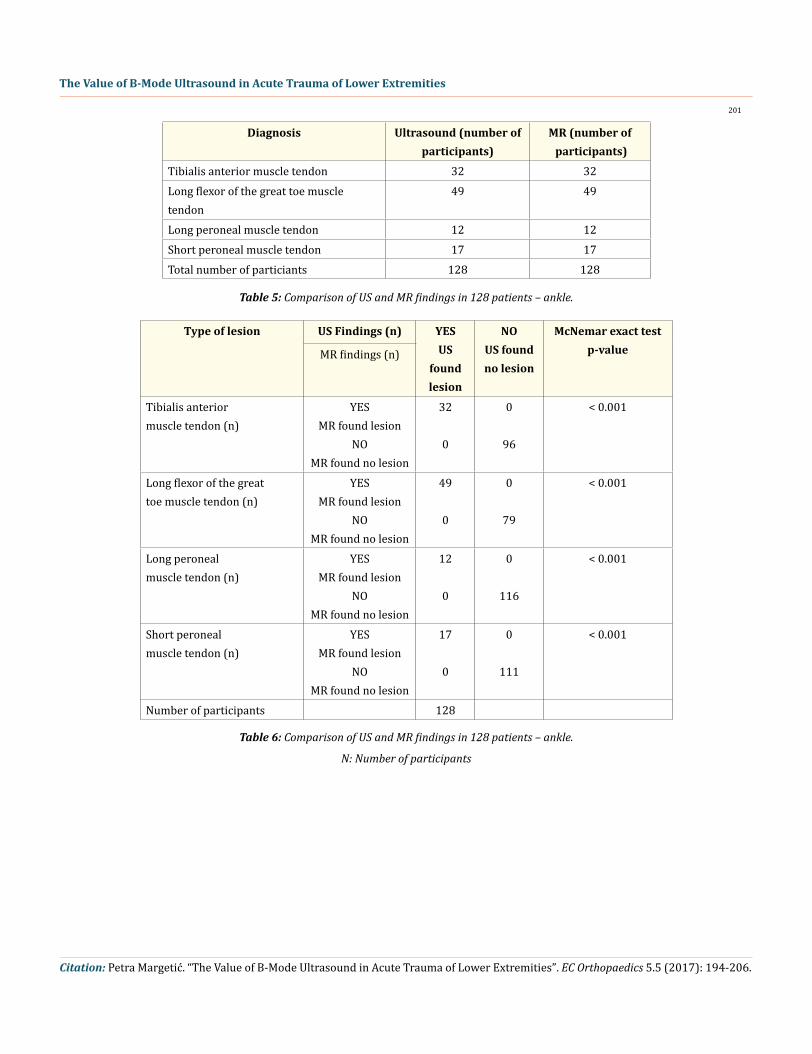
US was conducted on all patients. In 32 cases (25%) we found tibialis anterior muscle tendon. In 49 (38%) we found long flexor of the great toe muscle tendon. In 12 patients (9%) we found long peroneal muscle tendon and in 17 cases (13%) we found short peroneal muscle tendon. We performed conventional MR in all 128 cases and MR confirmed diagnosis in all cases.
For statistical analysis, we used exact McNemar test with the small sample size for searching marginal homogeneity. With paired binary response data, we searched for statistically significant difference in distribution. Using a p-value of less than 0.05, the difference in marginal distribution is statistically significant and one method is more successful than other. Following these diagnostics patients underwent arthroscopy which confirmed our diagnosis (Table 5, 6), (Figure 6-8).
Citation: Petra Margetić. “The Value of B-Mode Ultrasound in Acute Trauma of Lower Extremities”. EC Orthopaedics 5.5 (2017): 194-206.
The Value of B-Mode Ultrasound in Acute Trauma of Lower Extremities
201
Diagnosis Ultrasound (number of participants)
MR (number of participants)
Tibialis anterior muscle tendon 32 32Long flexor of the great toe muscle tendon
49 49
Long peroneal muscle tendon 12 12Short peroneal muscle tendon 17 17Total number of particiants 128 128
Table 5: Comparison of US and MR findings in 128 patients – ankle.
Type of lesion US Findings (n) YES US
found lesion
NO US found no lesion
McNemar exact test p-valueMR findings (n)
Tibialis anteriormuscle tendon (n)
YESMR found lesion
NOMR found no lesion
32
0
0
96
< 0.001
Long flexor of the greattoe muscle tendon (n)
YESMR found lesion
NOMR found no lesion
49
0
0
79
< 0.001
Long peronealmuscle tendon (n)
YESMR found lesion
NOMR found no lesion
12
0
0
116
< 0.001
Short peronealmuscle tendon (n)
YESMR found lesion
NOMR found no lesion
17
0
0
111
< 0.001
Number of participants 128
Table 6: Comparison of US and MR findings in 128 patients – ankle.
N: Number of participants

Citation: Petra Margetić. “The Value of B-Mode Ultrasound in Acute Trauma of Lower Extremities”. EC Orthopaedics 5.5 (2017): 194-206.
The Value of B-Mode Ultrasound in Acute Trauma of Lower Extremities
202
Figure 6: US finding of intraarticular effusion, ankle.
Figure 7: US findings of anterior fibulotalar ligament rupture in longitudinal scan, ankle.
Figure 8: US finding of short peroneal muscle lesion, longitudinal scan.

Citation: Petra Margetić. “The Value of B-Mode Ultrasound in Acute Trauma of Lower Extremities”. EC Orthopaedics 5.5 (2017): 194-206.
The Value of B-Mode Ultrasound in Acute Trauma of Lower Extremities
203
DiscussionHip
The hip is a big joint and that is why it could be rather difficult to approach by ultrasound [3]. In some patients with more subcutane-ous soft tissue ultrasound examination can be rather challenging especially in inexperienced hands [4]. We can easily detect intraarticular effusion, soft tissue masses and traumatic conditions which include muscle tear, tendon tear, or tendonitis and fracture. For labral pa-thology, we have to perform MR. Bancroft et al. conclude that MRI and sonography are both useful imaging methods to directly evaluate suspected abnormalities of the pelvic tendons, although tendinous mineralization and associated osseous injuries can also be detected with radiography and CT [5]. We compare US with MR and found no statistically significant difference between these two diagnostic methods in diagnosing abnormalities of the pelvic tendons. MR can easily detect bone marrow edema in the case of bone avulsion of ten-don insertion. Chang and al. concluded that sonography is a more rapidly performed examination; it has greater resolution than that of MRI; it allows dynamic evaluation of tendons and muscles [6]. Moreover, the advent of sonographic extended-field-of-view imaging allows the demonstration of the entire length of a tendon, matching MRI’s ability to display a large anatomic region. Sonography should best be considered for a focused examination, concentrating on the area of pain and clinical suspicion of pathology, whereas MRI can provide a global assessment of the region of concern. Both modalities demonstrate high accuracy for abnormalities of various tendons. We analyzed 85 patients with hip trauma without bony fracture by US an MR and we found that ultrasound is great diagnostic method for evaluated muscle and tendon posttraumatic changes and intraarticular effusion. For other hip posttraumatic changes, we prefer MR examination to evaluate cartilage and labral pathology, as well as bone marrrow [7]. Our study has some limitations. We analysed mainly muscle and tendon of upper leg more than hip joint. In hip joint we analyzed only effusion. We analysed only posttraumatic pathology and we used random patients with hip pain caused by trauma without visible bone fractures [8].
Knee
There are only few structures of the knee easily analysed by ultrasound as most structures are situated inside joint. Ultrasound is the best tool for detecting fluid in the knee joint. The demonstration of joint effusion is of paramount importance because the presence of fluid is a sign of articular disorder, resulting from injury to different components of the knee joint [9]. Ultrasound has been used to detect lesions such as ligament tear, tendon tear, plica disease or loose bodies. In addition to articular structures, muscle running across the knee should also be evaluated. The status of menisci, cartilage and bone cannot be accurately demonstrated by US and must be evaluated by other imaging techniques such as MR. So, that’s why we just evaluated medial and lateral collateral ligament, patellar retinacula, pes anse-rinus, insertions of muscle - m. quadriceps tendon, patellar tendon and joint effusion. Capo., et al. analyzed ultrasonographic visualization and assessment of the anterolateral ligament (ALL) and they concluded that ultrasound was unable to reliably identify the anterolateral structure from its femoral to tibial attachment sites [10]. Distinguishing it from the posterior iliotibial band and anterolateral capsule was challenging, and it is possible that the structure is a thickened band of fascia rather than a true ligament. As a clinical diagnostic tool, ultrasound likely offers little utility in the evaluation of the ALL for injury. ALL is a distinct structure with a consistent origin and insertion sites, is an extra-articular structure with a clear course from the lateral femoral epicondyle region, running anteroinferiorly, to the proxi-mal tibia at a site midway between the Gerdy tubercle and the head of the fibula [11]. In our study, we also found difficult to distinguish anterolateral capsula but we managed to visualised clearly the lateral collateral ligament and iliotibial band. Indirect signs such as soft tissue edema and effusion can tell us if it is a case of ALL injury. Saarakkala., et al. [12] and Kawaguchi., et al. [13] described diagnostic performance of knee ultrasonography for detecting degenerative changes of articular cartilage and medial radial displacement of the medial meniscus in knee osteoarthritis. We used similar performance but we analysed only patients with acute injury without visible changes on standard radiograms.
Ankle
US helped in diagnosis of considerably more sprain injuries (G1) than MR, whereas the MR helped in diagnosis of considerably more complete ligament ruptures (G3) than the US [14]. Morvan., et al. [15], Milz., et al. [16], and Peetrons., et al. [17] analysed ultrasound of the

Citation: Petra Margetić. “The Value of B-Mode Ultrasound in Acute Trauma of Lower Extremities”. EC Orthopaedics 5.5 (2017): 194-206.
The Value of B-Mode Ultrasound in Acute Trauma of Lower Extremities
204
ankle, lateral ankle ligaments and tibiofibular syndesmosis and they all conclude that ultrasound is routinely used to assess the disorders of the muscle-skeletal system since the size of the superficially located muscle tendons and ligaments (mt&l) can be easily visualized. In our study, we detected partial tears (G2) without loss of rectilinear appearance during dynamic sonography [18-20]. In the case of a ruptured ligament (G3), the site of the lesion is best visualized with the subject in supine position because the torn ends of the ligament are separated from each other [21-23]. When tears occurred at the level of ligament insertion, a cortical avulsion may be demonstrated by the ultrasound [24-26]. It was not possible to confirm the US findings of our subjects surgically, since closed treatment of G1-G3 injuries is considered to be appropriate for the most cases of the acute ankle injury in this clinical hospital. Therefore, it is reasonable to assume that the minor injuries followed with the post traumatic increase of the fluid in the ligament would change the MR signal whereas the ligament itself may still appear to be normal on US examination [27,28].
ConclusionTo generate high quality images of adequate size and proper annotation it is imperative to accurately assess the superficial structure
of muscle tendons and ligaments. To achieve that aim, a working knowledge of anatomy and relevant pathological conditions is required, together with the high level diagnostic equipment, precise positioning of the subject on the examination table and skilful manipulation of the diagnostic probe.
US is a valuable method for joint effusion, soft tissue and bone surface and is very important for the early detection of occult or missed fractures. Ultrasonography is a cost-effective, easy-to-use and radiation-free method which we recommend for early detection of ligament lesion in emergency room.
US is more challenging to perform, especially for beginners because it is rather subjective method depending on specialist experience. But in an experienced hand ultrasound is very accurate and sensitive method. US is faster, cheaper and more comfortable than MRI.
So, we can conclude that US is a valuable diagnostic method for muscle, tendons and ligament injury in the case of complete or incom-plete rupture. Ultrasonography is widely accessible and well tolerated by patients, making it a perfect method for establishing an initial diagnosis and monitoring the healing process. US cannot visualize intra-osseous abnormalities. US is a method of choice in diagnosing posttraumatic and degenerative changes of tendons and muscles and also in diagnosing ligamentous and capsule changes. In a case of cartilage and labral pathology, MR is better choice.
Conflict of Interest
All authors declare they have no conflicts of interest.
Bibliography
1. Bushberg JT., et al. “The Essential Physics of Medical Imaging”. LWW.
2. Nazarian LN. “The top 10 reasons musculoskeletal sonography is an important complementary or alternative technique to MRI”. American Journal of Roentgenology 190.6 (2008): 1621-1626.
3. Czyrny Z. “Muscles - histology, micro/macroanatomy and US anatomy, a brand new perspective”. Ultrasonografia 12.50 (2012): 9-27.
4. Fantino O., et al. “Conflicts, snapping and instability of the tendons. Pictorial essay”. Journal of Ultrasound 15.1 (2012): 42-49.
5. Bancroft LW and Blankenbaker DG. “Imaging of the tendons about the pelvis”. American Journal of Roentgenology 195.3 (2010): 605-617.
6. Chang A and Miller TT. “Imaging of tendons“. Sports Health 1.4 (2009): 293-300.

Citation: Petra Margetić. “The Value of B-Mode Ultrasound in Acute Trauma of Lower Extremities”. EC Orthopaedics 5.5 (2017): 194-206.
The Value of B-Mode Ultrasound in Acute Trauma of Lower Extremities
205
7. Azizi HF., et al. “Ultrasonography of snapping hip syndrome”. American Journal of Physical Medicine and Rehabilitation 94.1 (2015): e10-e11.
8. Jacobson JA., et al. “Ultrasound of the groin: techniques, pathology, and pitfalls”. American Journal of Roentgenology 205.3 (2015): 513-523.
9. Alves TI., et al. “US of the Knee: Scanning Techniques, Pitfalls, and Pathologic Conditions”. Radiographics 36.6 (2016): 1759-1775.
10. Capo J., et al. “Ultrasonographic visualization and assessment of the anterolateral ligament”. Knee Surgery, Sports Traumatology, Arthroscopy (2016).
11. Podlipská J., et al. “Comparison of Diagnostic Performance of Semi-Quantitative Knee Ultrasound and KneeRadiography with MRI: Oulu Knee Osteoarthritis Study”. Scientific Reports 6 (2016): 22365.
12. Saarakkala S., et al. “Diagnostic performance of knee ultrasonography for detecting degenerative changes of articular cartilage”. Osteoarthritis Cartilage 20.5 (2012): 376-381.
13. Kawaguchi K., et al. “Ultrasonographic evaluation of medial radial displacement of the medial meniscus in knee osteoarthritis”. Ar-thritis and Rheumatism 64.1 (2012): 173-180.
14. Margetić P and Pavić R. “Comparative assessment of the acute ankle injury by ultrasound and magnetic resonance”. Collegium Antro-pologicum 36.2 (2012): 605-610.
15. Morvan G., et al. “Ultrasound of the ankle”. European Journal of Ultrasound 14.1 (2001): 73-82.
16. Milz P., et al. “Lateral ankle ligaments and tibiofibular syndesmosis”. Acta Orthopaedica Scandinavica 69.1 (1998): 51-55.
17. Peetrons PA., et al. “Ultrasonography of ankle ligaments”. Canadian Association of Radiologists Journal 53.1 (2002): 6-13.
18. Beltran J., et al. “Ligaments of the lateral aspect of the ankle and sinus tarsi: an MR imaginig study”. Radiology 177.2 (1990): 455-458.
19. Rasmussen OS. “Sonography of tendons”. Scandinavian Journal of Medicine and Science in Sports 10.6 (2000): 360-364.
20. Striepling E., et al. “Ultrasonic assesment to the ankle joint in supination trauma”. Aktuelle Traumatologie 21.5 (1991): 194-196.
21. Rawool NK and Nazarian LN. “Ultrasound of the ankle and foot”. Seminars in Ultrasound, CT and MRI 21.3 (2000): 275-284.
22. Simanowski JH. “Ultrasound examination of fibular ligament ruptures”. Orthopade 31.3 (2002): 317-318.
23. Geusens E., et al. “Ultrasound in acute trauma of the ankle and hindfoot”. Emergency Radiology 9.5 (2002): 283-288.
24. Brasseur JL. “Ligament pathology of the ankle joint”. JBR-BTR 86 (2003): 96-101.
25. Williams GN., et al. “Syndesmotic ankle sprains in athletes”. American Journal of Sports Medicine 35.7 (2007): 1197-1207.
26. Lin CF., et al. “Ankle syndesmosis injuries: anatomy, biomechanics, mechanism of injury and clinical guidelines for diagnosis and intervention”. Journal of Orthopaedic and Sports Physical Therapy 36.6 (2006): 372-384.
27. Grass R., et al. “Injuries of the inferior tibiofibular syndesmosis”. Unfallchirurg 103.7 (2000): 520-532.

Citation: Petra Margetić. “The Value of B-Mode Ultrasound in Acute Trauma of Lower Extremities”. EC Orthopaedics 5.5 (2017): 194-206.
The Value of B-Mode Ultrasound in Acute Trauma of Lower Extremities
206
28. Ericsson E. “Treatment of lateral ankle sprains”. Knee Surgery Sports Traumatology Arthroscopy 10 (2002): 329.
Volume 5 Issue 5 April 2017© All rights reserved by Petra Margetić.

![Comparison Of Salient Feature Descriptorshh.diva-portal.org/smash/get/diva2:238363/FULLTEXT01.pdfsalient features will be studied, the SIFT features [1] and the Orientation Radiograms](https://img.pdfslide.us/doc/110x75/5f39319ab9491b31a247dcf2/comparison-of-salient-feature-238363fulltext01pdf-salient-features-will-be-studied.jpg)