Embed Size (px)
Citation preview

Diagnostic imaging of sacroiliac joints and the spine in the course of spondyloarthropathiesIwona Sudoł-Szopinska1, Andrzej Urbanik2
1 Department of Radiology, Institute of Rheumatology, Warsaw, Poland2 Department of Radiology, Jagiellonian University Medical College, Cracow, Poland
Author’s address: Iwona Sudol-Szopinska, Department of Radiology, Institute of Rheumatology, Spartanska 1 St., Warsaw, Poland, e-mail: [email protected]
Summary Spondyloarthropathies belong to a group of rheumatic diseases, in which inflammatory changes
affect mainly the sacroiliac joints, spine, peripheral joints, tendon, ligaments and capsule attachments (entheses). This group includes 6 entities: ankylosing spondylitis, arthritis associated with inflammatory bowel disease, reactive arthritis, undifferentiated spondyloarthropathy, psoriatic arthritis and juvenile spondyloarthropathy.
In 2009, ASAS (Assessment in SpondyloArthritis international Society) association, published classification criteria for spondyloarthropathies, which propose standardization of clinical-diagnostic approach in the case of sacroiliitis, spondylitis and arthritis.
Radiological diagnosis of inflammatory changes of sacroiliac joints is based on a 4 step radiographic grading method from 1966. According to modified New York criteria, the diagnosis of ankylosing spondylitis is made based on the presence of advanced lesions, sacroiliitis of at least 2 grade bilaterally or 3–4 unilaterally. In case of other types of spondyloarthropathies diagnosis is made based on presence of at least grade 1 changes.
In MRI, active inflammation of sacroiliac joints is indicated by the presence of subchondral bone marrow edema, synovitis, bursitis, or enthesitis.
ASAS discusses only the classic form of axial spondyloarthropathies, which is ankylosing spondylitis. To quantify radiological inflammatory changes in the course of the disease, Stoke Ankylosing spondylitis classification Spinal Score (SASSS) is recommended. The signs of inflammation and scarrying of the spinal cord in the course of ankylosing spondylitis, present in MRI include: bone marrow edema, sclerosis, fat metaplasia, formation of syndesmophytes, and ankylosis.
Key words: sacroiliitis • spondyloartropathies • diagnostics • radiograms • magnetic resonance imaging
PDF fi le: http://www.polradiol.com/download/index/idArt/889039
Received: 2013.02.11 Accepted: 2013.04.08
Background
Spondyloarthropathies (SpA) constitute a group of rheu-matic diseases, in which inflammatory lesions involve sac-roiliac joints, vertebral joints, peripheral joints and sites of insertion of tendons, ligaments or joint capsule [1,2]. Because of insufficient knowledge about this group of dis-eases, the diagnosis of SpA is often delayed, even up to sev-eral years, leading to disease progression.
For many years spondyloarthropathies were called “seronegative spondyloarthropathies”, but a new name – spondyloarthropathies – was introduced based on the presence of RF in some patients suffering from SpA. Spondyloarthropathies include diseases listed in Table 1. They usually affect young people. Frequency of occurrence of SpA is similar to that of rheumatoid arthritis and reaches 0.15–1.8% in general population [1–3].
Signature: © Pol J Radiol, 2013; 78(2): 43-49DOI: 10.12659/PJR.889039
43
R E V I E W A R T I C L E
SpA may be divided into 2 groups depending on dominat-ing clinical symptoms [3–6]. In the case of prevalence of symptoms indicating involvement of sacroiliac and verte-bral joints fulfilling clinical criteria of so-called inflamma-tory back pain [7] we speak of axial spondyloarthropathy. In patients with symptom prevalence suggesting inflam-mation of tendon, ligament or joint capsule attachments (enthesitis) and peripheral joints (arthritis) we talk about peripheral spondyloparthropathy.
In 2009 and 2011 the ASAS (Assessment in Spondylo-Arthritis international Society) group published modi-fied classification criteria for the two above forms of SpA [4–6,8]. They were presented in a document “The Assessment of SpondyloArthritis international Society (ASAS) handbook: a guide to assess spondyloarthritis” [6]. The ASAS classification criteria for axial and peripheral spondyloarthropathy are presented in Tables 2 and 3.
For many years, diagnostic imaging of spondyloarthropathy has been based on plain radiography (x-ray). ASAS classi-fication criteria introduced magnetic resonance (MR) into the diagnostic algorithm of axial SpA [6]. Moreover, there are multicenter studies currently in progress designed to compile standards of ultrasonographic examination, which became one of the fundamental imaging studies in the
diagnostics of locomotor system disorders due to introduc-tion of high-frequency heads and proper software [9–12].
In both forms of SpA – axial and peripheral – sacroilitis is a common clinical feature. In axial SpA disease usually begins with sacroilitis. Sporadically, inflammation of the vertebral column coexists with sacroilitis from the disease onset and rarely vertebrae are involved initially, while the radiographic picture of sacroiliac joints remains normal. Only single publication [13] reported up to 27% of such cases of SpA. According to ASAS guidelines [6] sacroilitis can be diagnosed based on x-ray and MR.
X-ray Examination of Sacroiliac Joints in SpA
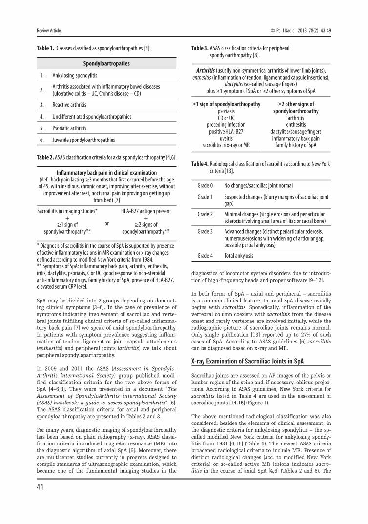
Sacroiliac joints are assessed on AP images of the pelvis or lumbar region of the spine and, if necessary, oblique projec-tions. According to ASAS guidelines, New York criteria for sacroilitis listed in Table 4 are used in the assessment of sacroiliac joints [14,15] (Figure 1).
The above mentioned radiological classification was also considered, besides the elements of clinical assessment, in the diagnostic criteria for ankylosing spondylitis – the so-called modified New York criteria for ankylosing spondy-litis from 1984 [6,16] (Table 5). The newest ASAS criteria broadened radiological criteria to include MR. Presence of distinct radiological changes (acc. to modified New York criteria) or so-called active MR lesions indicates sacro-ilitis in the course of axial SpA [4,6] (Tables 2 and 6). The
Spondyloartropaties
1. Ankylosing spondylitis
2. Arthritis associated with inflammatory bowel diseases (ulcerative colitis – UC, Crohn’s disease – CD)
3. Reactive arthritis
4. Undifferentiated spondyloarthropathies
5. Psoriatic arthritis
6. Juvenile spondyloarthropathies
Table 1. Diseases classified as spondyloarthropathies [3].
* Diagnosis of sacroilitis in the course of SpA is supported by presence of active inflammatory lesions in MR examination or x-ray changes defined according to modified New York criteria from 1984.** Symptoms of SpA: inflammatory back pain, arthritis, enthesitis, iritis, dactylitis, psoriasis, C or UC, good response to non-steroidal anti-inflammatory drugs, family history of SpA, presence of HLA-B27, elevated serum CRP level.
Table 2. ASAS classification criteria for axial spondyloarthropathy [4,6].
Inflammatory back pain in clinical examination (def.: back pain lasting ≥3 months that first occurred before the age of 45, with insidious, chronic onset, improving after exercise, without
improvement after rest, nocturnal pain improving on getting up from bed) [7]
Sacroiliitis in imaging studies*+
≥1 sign of spondyloarthropathy**
or
HLA-B27 antigen present+
≥2 signs of spondyloarthropathy**
Arthritis (usually non-symmetrical arthritis of lower limb joints), enthesitis (inflammation of tendon, ligament and capsule insertions),
dactylitis (so-called sausage fingers)plus ≥1 symptom of SpA or ≥2 other symptoms of SpA
≥1 sign of spondyloarthropathy psoriasisCD or UC
preceding infectionpositive HLA-B27
uveitissacroilitis in x-ray or MR
≥2 other signs of spondyloarthropathy
arthritisenthesitis
dactylitis/sausage fingersinflammatory back pain
family history of SpA
Table 3. ASAS classification criteria for peripheral spondyloarthropathy [8].
Grade 0 No changes/sacroiliac joint normal
Grade 1 Suspected changes (blurry margins of sacroiliac joint gap)
Grade 2 Minimal changes (single erosions and periarticular sclerosis involving small area of iliac or sacral bone)
Grade 3 Advanced changes (distinct periarticular sclerosis, numerous erosions with widening of articular gap, possible partial ankylosis)
Grade 4 Total ankylosis
Table 4. Radiological classification of sacroilitis according to New York criteria [13].
Review Article
44
© Pol J Radiol, 2013; 78(2): 43-49
fulfillment of a radiological criterion and ≥1 clinical cri-terion allows for a diagnosis of anylosing spondylitis. Diagnosis of ankylosing spondylitis is probable if single radiological criterion or 3 clinical criteria are met.
X-ray Examination of the Spine in SpA
Radiological changes in the vertebral column are not cur-rently included in the SpA classification, since the dis-ease usually begins with involvement of sacroiliac joints. However, x-ray remains superior to MR in visualization of syndesmophytes and ankylosis (joint stiffness). Patients
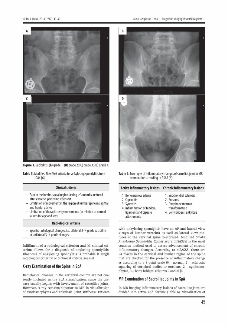
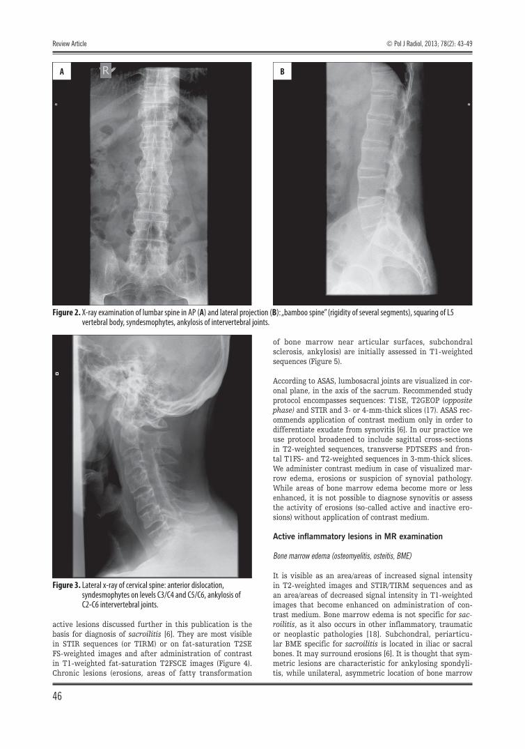
with ankylosing spondylitis have an AP and lateral view x-ray's of lumbar vertebra as well as lateral view pic-tures of the cervical spine performed. Modified Stroke Ankylosing Spondylitis Spinal Score (mSASSS) is the most common method used to assess advancement of chronic inflammatory changes. According to mSASSS, there are 24 places in the cervical and lumbar region of the spine that are checked for the presence of inflammatory chang-es according to a 3-point scale (0 – normal, 1 – sclerosis, squaring of vertebral bodies or erosions, 2 – syndesmo-phytes, 3 – bony bridges) (Figures 2 and 3) [6].
MR Examination of Sacroiliac Joints in SpA
In MR imaging inflammatory lesions of sacroiliac joint are divided into active and chronic (Table 6). Visualization of
Figure 1. Sacroilitis: (A) grade 1; (B) grade 2; (C) grade 3; (D) grade 4.
A
C
B
D
Clinical criteria
– Pain in the lumbo-sacral region lasting ≥3 months, reduced after exercise, persisting after rest
– Limitation of movement in the region of lumbar spine in sagittal and frontal planes
– Limitation of thoracic cavity movements (in relation to normal values for age and sex)
Radiological criteria
– Specific radiological changes, i.e. bilateral 2–4 grade sacroilitis or unilateral 3–4 grade changes
Table 5. Modified New York criteria for ankylosing spondylitis from 1984 [6].
Active inflammatory lesions Chronic inflammatory lesions
1. Bone marrow edema2. Capsulitis3. Synovitis4. Inflammation of tendon,
ligament and capsule attachments
1. Subchondral sclerosis2. Erosions3. Fatty bone marrow
transformation4. Bony bridges, ankylosis
Table 6. Two types of inflammatory changes of sacroiliac joint in MR examination according to ASAS (6).
© Pol J Radiol, 2013; 78(2): 43-49 Sudoł-Szopinska I. et al. – Diagnostic imaging of sacroiliac joints…
45
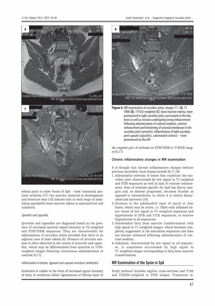
active lesions discussed further in this publication is the basis for diagnosis of sacroilitis [6]. They are most visible in STIR sequences (or TIRM) or on fat-saturation T2SE FS-weighted images and after administration of contrast in T1-weighted fat-saturation T2FSCE images (Figure 4). Chronic lesions (erosions, areas of fatty transformation
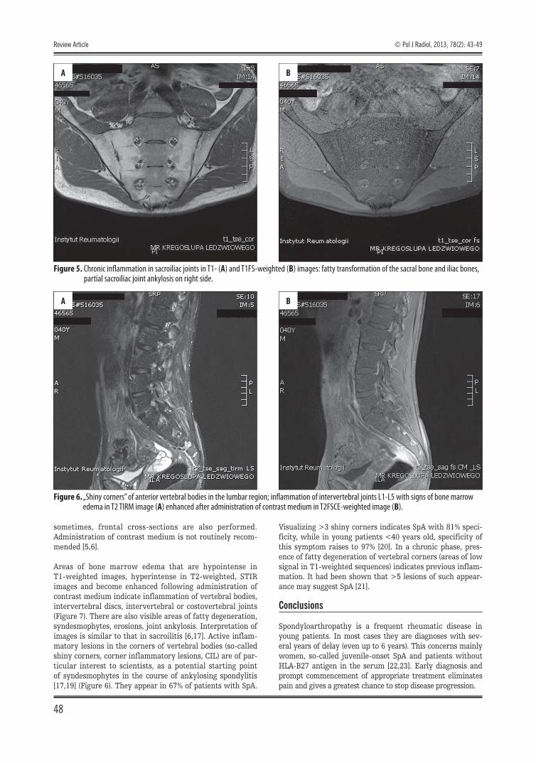
of bone marrow near articular surfaces, subchondral sclerosis, ankylosis) are initially assessed in T1-weighted sequences (Figure 5).
According to ASAS, lumbosacral joints are visualized in cor-onal plane, in the axis of the sacrum. Recommended study protocol encompasses sequences: T1SE, T2GEOP (opposite phase) and STIR and 3- or 4-mm-thick slices (17). ASAS rec-ommends application of contrast medium only in order to differentiate exudate from synovitis [6]. In our practice we use protocol broadened to include sagittal cross-sections in T2-weighted sequences, transverse PDTSEFS and fron-tal T1FS- and T2-weighted sequences in 3-mm-thick slices. We administer contrast medium in case of visualized mar-row edema, erosions or suspicion of synovial pathology. While areas of bone marrow edema become more or less enhanced, it is not possible to diagnose synovitis or assess the activity of erosions (so-called active and inactive ero-sions) without application of contrast medium.
Active inflammatory lesions in MR examination
Bone marrow edema (osteomyelitis, osteitis, BME)
It is visible as an area/areas of increased signal intensity in T2-weighted images and STIR/TIRM sequences and as an area/areas of decreased signal intensity in T1-weighted images that become enhanced on administration of con-trast medium. Bone marrow edema is not specific for sac-roilitis, as it also occurs in other inflammatory, traumatic or neoplastic pathologies [18]. Subchondral, periarticu-lar BME specific for sacroilitis is located in iliac or sacral bones. It may surround erosions [6]. It is thought that sym-metric lesions are characteristic for ankylosing spondyli-tis, while unilateral, asymmetric location of bone marrow
Figure 2. X-ray examination of lumbar spine in AP (A) and lateral projection (B): „bamboo spine” (rigidity of several segments), squaring of L5 vertebral body, syndesmophytes, ankylosis of intervertebral joints.
A B
Figure 3. Lateral x-ray of cervical spine: anterior dislocation, syndesmophytes on levels C3/C4 and C5/C6, ankylosis of C2-C6 intervertebral joints.
Review Article
46
© Pol J Radiol, 2013; 78(2): 43-49
edema point to other forms of SpA – most commonly pso-riatic arthritis [17]. Our practice (material in development) and literature data [18] indicate that at early stage of anky-losing spondylitis bone marrow edema is asymmetrical and unilateral.
Synovitis and capsulitis
Synovitis and capsulitis are diagnosed based on the pres-ence of increased synovial signal intensity in T2-weighted and STIR/TIRM sequences. They are characteristic for inflammation of sacroiliac joints provided that there is an adjacent area of bone edema [6]. Presence of articular exu-date is often observed in the course of synovitis and capsu-litis, which may be differentiated from synovitis in T1FS-weighted images following intravenous administration of contrast [6,17].
Inflammation of tendon, ligament and capsule insertions (enthesitis)
Enthesitis is visible in the form of increased signal intensity of bony or tendinous (either ligamentous or fibrous layer of
the capsule) part of enthesis on STIR/TIRM or T1FSCE imag-es [6,17].
Chronic inflammatory changes in MR examination
It is thought that chronic inflammatory changes indicate previous sacroilitis. Such lesions include [6,17,18]:1. Subchondral sclerosis of bones that constitute the sac-
roiliac joint characterized by low signal in T1-weighted and STIR sequences as well as lack of contrast enhance-ment. Area of sclerosis specific for SpA has blurry mar-gins and, as disease progresses, becomes broader as opposed to osteoarthritis, in which it is clearly demar-cated and narrower [18].
2. Erosions in the subchondral layer of sacral or iliac bones, which may be active, i.e. filled with inflamed tis-sue (areas of low signal in T1-weighted sequences and hyperintense in STIR and T1CE sequences), or inactive (hypointense in all sequences).
3. Subchondral fatty bone marrow transformation with high signal in T1-weighted images, which becomes com-pletely suppressed in fat-saturation sequences and does not become enhanced following administration of con-trast medium.
4. Ankylosis, characterized by low signal in all sequenc-es, is sometimes surrounded by high signal in T1-weighted images corresponding to fatty bone marrow transformation.
MR Examination of the Spine in SpA
Study protocol includes sagittal cross-sections and T1SE and T2SEFS-weighted or STIR images. Transverse or,
Figure 4. MR examination of sacroiliac joints, images: T1- (A), T2 TIRM (B), T1FSCE-weighted (C): bone marrow edema, more pronounced in right sacroiliac joint, particularly in the iliac bone as well as erosions undergoing strong enhancement following administration of contrast medium, contrast enhancement and thickening of synovial membrane in the sacroiliac joint (synovitis), inflammation of right sacroiliac joint capsule (capsulitis), subchondral sclerosis – more pronounced on the left.
A
C
B
© Pol J Radiol, 2013; 78(2): 43-49 Sudoł-Szopinska I. et al. – Diagnostic imaging of sacroiliac joints…
47
sometimes, frontal cross-sections are also performed. Administration of contrast medium is not routinely recom-mended [5,6].
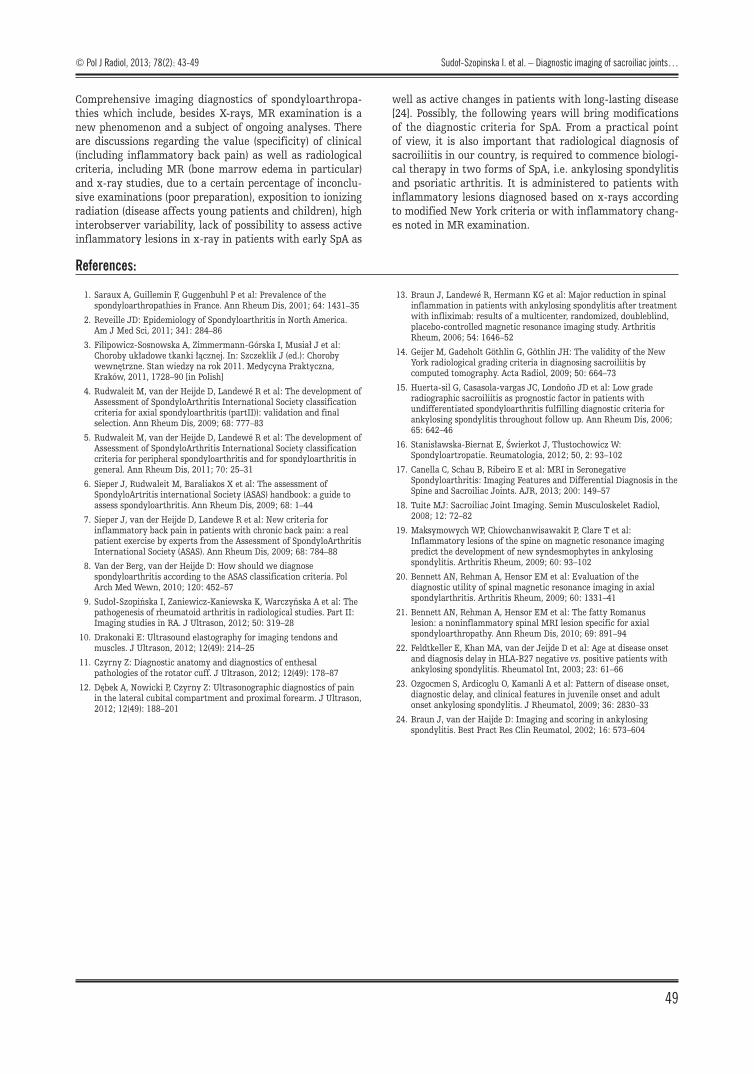
Areas of bone marrow edema that are hypointense in T1-weighted images, hyperintense in T2-weighted, STIR images and become enhanced following administration of contrast medium indicate inflammation of vertebral bodies, intervertebral discs, intervertebral or costovertebral joints (Figure 7). There are also visible areas of fatty degeneration, syndesmophytes, erosions, joint ankylosis. Interpretation of images is similar to that in sacroilitis [6,17]. Active inflam-matory lesions in the corners of vertebral bodies (so-called shiny corners, corner inflammatory lesions, CIL) are of par-ticular interest to scientists, as a potential starting point of syndesmophytes in the course of ankylosing spondylitis [17,19] (Figure 6). They appear in 67% of patients with SpA.
Visualizing >3 shiny corners indicates SpA with 81% speci-ficity, while in young patients <40 years old, specificity of this symptom raises to 97% [20]. In a chronic phase, pres-ence of fatty degeneration of vertebral corners (areas of low signal in T1-weighted sequences) indicates previous inflam-mation. It had been shown that >5 lesions of such appear-ance may suggest SpA [21].
Conclusions
Spondyloarthropathy is a frequent rheumatic disease in young patients. In most cases they are diagnoses with sev-eral years of delay (even up to 6 years). This concerns mainly women, so-called juvenile-onset SpA and patients without HLA-B27 antigen in the serum [22,23]. Early diagnosis and prompt commencement of appropriate treatment eliminates pain and gives a greatest chance to stop disease progression.
Figure 5. Chronic inflammation in sacroiliac joints in T1- (A) and T1FS-weighted (B) images: fatty transformation of the sacral bone and iliac bones, partial sacroiliac joint ankylosis on right side.
A B
Figure 6. „Shiny corners” of anterior vertebral bodies in the lumbar region; inflammation of intervertebral joints L1-L5 with signs of bone marrow edema in T2 TIRM image (A) enhanced after administration of contrast medium in T2FSCE-weighted image (B).
A B
Review Article
48
© Pol J Radiol, 2013; 78(2): 43-49
Comprehensive imaging diagnostics of spondyloarthropa-thies which include, besides X-rays, MR examination is a new phenomenon and a subject of ongoing analyses. There are discussions regarding the value (specificity) of clinical (including inflammatory back pain) as well as radiological criteria, including MR (bone marrow edema in particular) and x-ray studies, due to a certain percentage of inconclu-sive examinations (poor preparation), exposition to ionizing radiation (disease affects young patients and children), high interobserver variability, lack of possibility to assess active inflammatory lesions in x-ray in patients with early SpA as
well as active changes in patients with long-lasting disease [24]. Possibly, the following years will bring modifications of the diagnostic criteria for SpA. From a practical point of view, it is also important that radiological diagnosis of sacroiliitis in our country, is required to commence biologi-cal therapy in two forms of SpA, i.e. ankylosing spondylitis and psoriatic arthritis. It is administered to patients with inflammatory lesions diagnosed based on x-rays according to modified New York criteria or with inflammatory chang-es noted in MR examination.
1. Saraux A, Guillemin F, Guggenbuhl P et al: Prevalence of the spondyloarthropathies in France. Ann Rheum Dis, 2001; 64: 1431–35
2. Reveille JD: Epidemiology of Spondyloarthritis in North America. Am J Med Sci, 2011; 341: 284–86
3.Filipowicz-SosnowskaA,Zimmermann-GórskaI,MusiałJetal:Chorobyukładowetkankiłącznej.In:SzczeklikJ(ed.):Chorobywewnętrzne.Stanwiedzynarok2011.MedycynaPraktyczna,Kraków, 2011, 1728–90 [in Polish]
4. Rudwaleit M, van der Heijde D, Landewé R et al: The development of Assessment of SpondyloArthritis International Society classification criteria for axial spondyloarthritis (partII)): validation and final selection. Ann Rheum Dis, 2009; 68: 777–83
5. Rudwaleit M, van der Heijde D, Landewé R et al: The development of Assessment of SpondyloArthritis International Society classification criteria for peripheral spondyloarthritis and for spondyloarthritis in general. Ann Rheum Dis, 2011; 70: 25–31
6. Sieper J, Rudwaleit M, Baraliakos X et al: The assessment of SpondyloArtritis international Society (ASAS) handbook: a guide to assess spondyloarthritis. Ann Rheum Dis, 2009; 68: 1–44
7. Sieper J, van der Heijde D, Landewe R et al: New criteria for inflammatory back pain in patients with chronic back pain: a real patient exercise by experts from the Assessment of SpondyloArthritis International Society (ASAS). Ann Rheum Dis, 2009; 68: 784–88
8. Van der Berg, van der Heijde D: How should we diagnose spondyloarthritis according to the ASAS classification criteria. Pol Arch Med Wewn, 2010; 120: 452–57
9.Sudoł-SzopińskaI,Zaniewicz-KaniewskaK,WarczyńskaAetal:Thepathogenesis of rheumatoid arthritis in radiological studies. Part II: Imaging studies in RA. J Ultrason, 2012; 50: 319–28
10. Drakonaki E: Ultrasound elastography for imaging tendons and muscles. J Ultrason, 2012; 12(49): 214–25
11. Czyrny Z: Diagnostic anatomy and diagnostics of enthesal pathologies of the rotator cuff. J Ultrason, 2012; 12(49): 178–87
12.DębekA,NowickiP,CzyrnyZ:Ultrasonographicdiagnosticsofpainin the lateral cubital compartment and proximal forearm. J Ultrason, 2012; 12(49): 188–201
References:
13. Braun J, Landewé R, Hermann KG et al: Major reduction in spinal inflammation in patients with ankylosing spondylitis after treatment with infliximab: results of a multicenter, randomized, doubleblind, placebo-controlled magnetic resonance imaging study. Arthritis Rheum, 2006; 54: 1646–52
14. Geijer M, Gadeholt Göthlin G, Göthlin JH: The validity of the New York radiological grading criteria in diagnosing sacroiliitis by computed tomography. Acta Radiol, 2009; 50: 664–73
15. Huerta-sil G, Casasola-vargas JC, Londoño JD et al: Low grade radiographic sacroiliitis as prognostic factor in patients with undifferentiated spondyloarthritis fulfilling diagnostic criteria for ankylosing spondylitis throughout follow up. Ann Rheum Dis, 2006; 65: 642–46
16.Stanisławska-BiernatE,ŚwierkotJ,TłustochowiczW:Spondyloartropatie. Reumatologia, 2012; 50, 2: 93–102
17. Canella C, Schau B, Ribeiro E et al: MRI in Seronegative Spondyloarthritis: Imaging Features and Differential Diagnosis in the Spine and Sacroiliac Joints. AJR, 2013; 200: 149–57
18. Tuite MJ: Sacroiliac Joint Imaging. Semin Musculoskelet Radiol, 2008; 12: 72–82
19. Maksymowych WP, Chiowchanwisawakit P, Clare T et al: Inflammatory lesions of the spine on magnetic resonance imaging predict the development of new syndesmophytes in ankylosing spondylitis. Arthritis Rheum, 2009; 60: 93–102
20. Bennett AN, Rehman A, Hensor EM et al: Evaluation of the diagnostic utility of spinal magnetic resonance imaging in axial spondylarthritis. Arthritis Rheum, 2009; 60: 1331–41
21. Bennett AN, Rehman A, Hensor EM et al: The fatty Romanus lesion: a noninflammatory spinal MRI lesion specific for axial spondyloarthropathy. Ann Rheum Dis, 2010; 69: 891–94
22. Feldtkeller E, Khan MA, van der Jeijde D et al: Age at disease onset and diagnosis delay in HLA-B27 negative vs. positive patients with ankylosing spondylitis. Rheumatol Int, 2003; 23: 61–66
23. Ozgocmen S, Ardicoglu O, Kamanli A et al: Pattern of disease onset, diagnostic delay, and clinical features in juvenile onset and adult onset ankylosing spondylitis. J Rheumatol, 2009; 36: 2830–33
24. Braun J, van der Haijde D: Imaging and scoring in ankylosing spondylitis. Best Pract Res Clin Reumatol, 2002; 16: 573–604
© Pol J Radiol, 2013; 78(2): 43-49 Sudoł-Szopinska I. et al. – Diagnostic imaging of sacroiliac joints…
49