Embed Size (px)
Citation preview
Recapitulating maladaptive, multiscale remodelingof failing myocardium on a chipMegan L. McCain, Sean P. Sheehy, Anna Grosberg1, Josue A. Goss, and Kevin Kit Parker2
Disease Biophysics Group, Wyss Institute for Biologically Inspired Engineering, School of Engineering and Applied Sciences, Harvard University, Cambridge,MA 02138
Edited by Robert Langer, Massachusetts Institute of Technology, Cambridge, MA, and approved May 02, 2013 (received for review March 14, 2013)
The lack of a robust pipeline of medical therapeutic agents forthe treatment of heart disease may be partially attributed to thelack of in vitro models that recapitulate the essential structure–function relationships of healthy and diseased myocardium. Wedesigned and built a system to mimic mechanical overload in vitroby applying cyclic stretch to engineered laminar ventricular tissueon a stretchable chip. To test our model, we quantified changes ingene expression, myocyte architecture, calcium handling, and con-tractile function and compared our results vs. several decades ofanimal studies and clinical observations. Cyclic stretch activatedgene expression profiles characteristic of pathological remodeling,including decreased α- to β-myosin heavy chain ratios, and inducedmaladaptive changes to myocyte shape and sarcomere alignment.In stretched tissues, calcium transients resembled those reportedin failing myocytes and peak systolic stress was significantly re-duced. Our results suggest that failing myocardium, as definedgenetically, structurally, and functionally, can be replicated in anin vitro microsystem by faithfully recapitulating the structural andmechanical microenvironment of the diseased heart.
organs on chips | mechanotransduction | muscular thin films | microarray |contraction
Maladaptive cardiac hypertrophy occurs in response to a va-riety of stimuli, including myocardial infarction, genetic
mutations, or hypertension (1). Initially, hypertrophic remodelingis compensatory, as myocytes counteract losses in cardiac outputby adding myofibrils in parallel (2, 3). Over time, remodelingtransitions from adaptive to maladaptive, which is characterizedstructurally by sarcomere disarray (4), fibrosis (5), myocyte elon-gation (2, 6, 7), and ventricular dilation (3). Gene expressionreverts to an immature state, including down-regulation ofα-myosin heavy chain (MHC) concurrent with up-regulation ofβ-MHC, reminiscent of the relative expression levels of thesemotor proteins in the embryonic heart (8, 9). Functionally,myocytes isolated from failing hearts show defective excitation–contraction coupling (10), including reduced calcium uptake intothe sarcoplasmic reticulum (11). Contractile function is also re-duced, as shown by decreased cardiac output in dogs subjected tochronic rapid pacing (12) and decreased tension in left ventricularstrips isolated from failing human myocardium (13, 14). Thus,dilated and failing hearts are characterized by multiscale malad-aptive remodeling, best studied in an experimental system capa-ble of replicating and quantifying a broad range of these effects.Drug discovery, safety pharmacology, and mechanistic studies
have traditionally been performed in animal models. For exam-ple, cardiac hypertrophy and heart failure are reproduced in therat by ligating coronary arteries or by using the spontaneouslyhypertensive rat strain (15). In vitro, heart disease has beenmodeled by exposing primary cardiac myocytes to chemical (16)or exogenous mechanical stimuli (17–21), which increases cellsize, activates pathological gene expression, and remodels ionchannel currents. However, cell culture systems often fail topredict the efficacy or toxicity of therapeutic candidates, poten-tially because they do not replicate the complex structure–function relationships of native cardiac tissue (22). Traditionalcell culture systems also fail to quantify pathological changes incontractile function, instead focusing on endpoints such as gene
expression or electrophysiology, which are difficult to justify asdirect indicators of reduced cardiac output.We reasoned that we could reproduce years of published an-
imal studies on dilated and failing myocardium with a carefullydesigned in vitro microsystem. Our goal was to build failingmyocardium “on a chip” by engineering arrays of laminar ven-tricular muscle on a substrate amenable to cyclic stretch to mimicmechanical overload. To test our system, we measured genetic,structural, and functional responses, including contractile stressgeneration. Cyclic stretch activated markers of pathologicalcardiac hypertrophy, disrupted myocyte shape and sarcomerealignment with directional specificity, remodeled calcium tran-sients, and decreased stress generation in a manner similar toanimal studies and clinical observations. These data suggestthat we can recapitulate failing myocardium on a chip, whichhas potential value as a new in vitro platform for early stagedrug discovery.
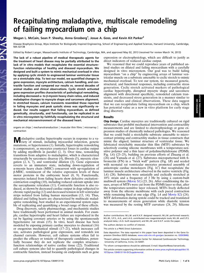
ResultsChip Design. Cardiac myocytes are traditionally cultured on rigidsubstrates that prohibit mechanical intervention and contractilitymeasurements and are limited to electrophysiology or gene ex-pression studies of chemically induced pathologies. We reasonedthat we could build a stretchable substrate amenable to micro-contact printing and contractility studies in tissue constructs thatmimic the aligned, laminar structure of ventricular tissue. Wefabricated stretchable muscular thin film (MTF) substrates byselectively coating silicone membranes with a temperature-sen-sitive polymer and a thin layer of polydimethylsiloxane (PDMS;Fig. 1A) (23–25), building on previous efforts of Zhuang et al.(26) and Yamada et al. (27). Substrates micropatterned with fi-bronectin (FN) in a “brick wall” pattern (Fig. 1B) and seededwith neonatal rat ventricular myocytes promoted assembly ofanisotropic cardiac monolayers (Fig. 1C) that recapitulate thelaminar muscle architecture observed in the native ventricle (Fig.1A) (28). Substrates were uniaxially and cyclically stretched at10% strain and a frequency of 3 Hz by using a custom-builtmultiwell system (Movie S1) (23, 26). After conditioning the tis-sues, arrays of MTFs were cut from the PDMS layer so that, afterthe temperature-sensitive layer released, MTFs freely deflectedaway from the silicone membrane with each paced contractionwhile remaining fixed at one edge (Fig. 1A and Movie S2) (24).MTF deformation during systole could be tracked and translatedto measurements of stress generation while diastolic tensionwas measured by the resting MTF curvature (24, 29). Silicone
Author contributions: M.L.M. and K.K.P. designed research; M.L.M. performed research;M.L.M., S.P.S., A.G., and J.A.G. contributed new reagents/analytic tools; M.L.M. and S.P.S.analyzed data; J.A.G. built equipment; and M.L.M. and K.K.P. wrote the paper.
The authors declare no conflict of interest.
This article is a PNAS Direct Submission.
Data deposition: The data reported in this paper have been deposited in the Gene Ex-pression Omnibus (GEO) database, www.ncbi.nlm.nih.gov/geo (accession no. GSE43846).1Present address: Edwards Lifesciences Center for Advanced Cardiovascular Technology,University of California, Irvine, CA 92697.
2To whom correspondence should be addressed. E-mail: [email protected].
This article contains supporting information online at www.pnas.org/lookup/suppl/doi:10.1073/pnas.1304913110/-/DCSupplemental.
www.pnas.org/cgi/doi/10.1073/pnas.1304913110 PNAS Early Edition | 1 of 6
BIOPH
YSICSAND
COMPU
TATIONALBIOLO
GY
membranes could also be micropatterned directly and seededwith myocytes for optical measurements of calcium transients,staining of tissue structure, and collection of lysates for gene ex-pression analysis, similar to our previous work (24). Thus, ourchip recapitulates native tissue architecture and asynchronousmechanical stretch in the ventricular wall, and is amenable tomeasurements of contractile stress generation, calcium transients,cytoskeletal structures, and gene expression.Because many cardiomyopathies are associated with me-
chanical overload (4), we reasoned that cyclic stretch would in-duce pathological subcellular, cellular, and tissue remodeling.We seeded neonatal rat ventricular myocytes onto silicone mem-branes coated with isotropic FN and initiated cyclic stretch 1 hafter seeding, when myocytes were adhered to the substrate buthad not yet formed a tissue (Fig. 1D). Myocytes spontaneouslyaligned parallel to the axis of stretch (Fig. 1E). Myocytes seededon micropatterned FN maintained uniaxial alignment when cy-clic stretch was parallel to the direction of patterning (Fig. 1F).However, transverse stretch of patterned tissues disrupted tissuearchitecture observed in isolated regions where myocytes real-igned with the exogenous stretch, creating a “parquet floor”pattern (Fig. 1G). This is reminiscent of clinical reports in which
tissue sections taken from human hearts with hypertrophic car-diomyopathy displayed similar misalignment (4). Thus, our failingmyocardium chip appears to recapitulate the form of laminarventricular tissue in the healthy and diseased heart.
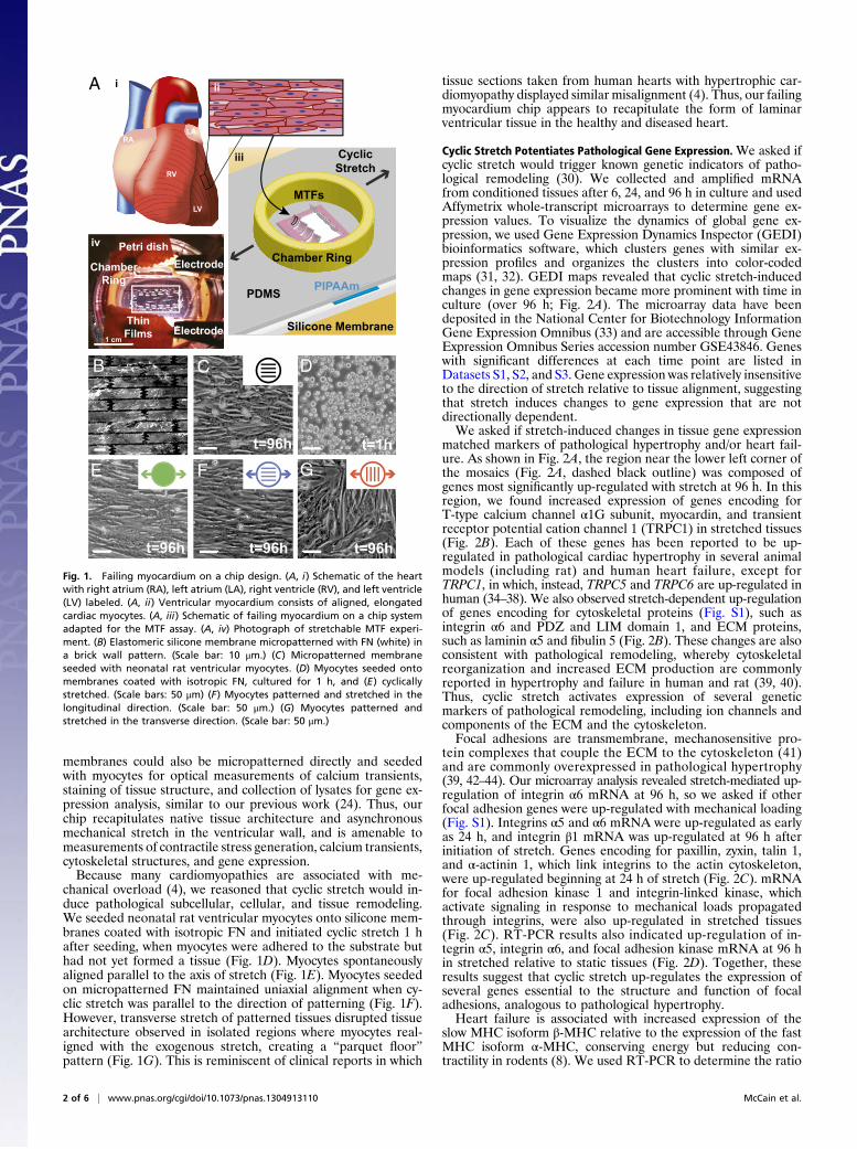
Cyclic Stretch Potentiates Pathological Gene Expression.We asked ifcyclic stretch would trigger known genetic indicators of patho-logical remodeling (30). We collected and amplified mRNAfrom conditioned tissues after 6, 24, and 96 h in culture and usedAffymetrix whole-transcript microarrays to determine gene ex-pression values. To visualize the dynamics of global gene ex-pression, we used Gene Expression Dynamics Inspector (GEDI)bioinformatics software, which clusters genes with similar ex-pression profiles and organizes the clusters into color-codedmaps (31, 32). GEDI maps revealed that cyclic stretch-inducedchanges in gene expression became more prominent with time inculture (over 96 h; Fig. 2A). The microarray data have beendeposited in the National Center for Biotechnology InformationGene Expression Omnibus (33) and are accessible through GeneExpression Omnibus Series accession number GSE43846. Geneswith significant differences at each time point are listed inDatasets S1, S2, and S3. Gene expression was relatively insensitiveto the direction of stretch relative to tissue alignment, suggestingthat stretch induces changes to gene expression that are notdirectionally dependent.We asked if stretch-induced changes in tissue gene expression
matched markers of pathological hypertrophy and/or heart fail-ure. As shown in Fig. 2A, the region near the lower left corner ofthe mosaics (Fig. 2A, dashed black outline) was composed ofgenes most significantly up-regulated with stretch at 96 h. In thisregion, we found increased expression of genes encoding forT-type calcium channel α1G subunit, myocardin, and transientreceptor potential cation channel 1 (TRPC1) in stretched tissues(Fig. 2B). Each of these genes has been reported to be up-regulated in pathological cardiac hypertrophy in several animalmodels (including rat) and human heart failure, except forTRPC1, in which, instead, TRPC5 and TRPC6 are up-regulated inhuman (34–38). We also observed stretch-dependent up-regulationof genes encoding for cytoskeletal proteins (Fig. S1), such asintegrin α6 and PDZ and LIM domain 1, and ECM proteins,such as laminin α5 and fibulin 5 (Fig. 2B). These changes are alsoconsistent with pathological remodeling, whereby cytoskeletalreorganization and increased ECM production are commonlyreported in hypertrophy and failure in human and rat (39, 40).Thus, cyclic stretch activates expression of several geneticmarkers of pathological remodeling, including ion channels andcomponents of the ECM and the cytoskeleton.Focal adhesions are transmembrane, mechanosensitive pro-
tein complexes that couple the ECM to the cytoskeleton (41)and are commonly overexpressed in pathological hypertrophy(39, 42–44). Our microarray analysis revealed stretch-mediated up-regulation of integrin α6 mRNA at 96 h, so we asked if otherfocal adhesion genes were up-regulated with mechanical loading(Fig. S1). Integrins α5 and α6 mRNA were up-regulated as earlyas 24 h, and integrin β1 mRNA was up-regulated at 96 h afterinitiation of stretch. Genes encoding for paxillin, zyxin, talin 1,and α-actinin 1, which link integrins to the actin cytoskeleton,were up-regulated beginning at 24 h of stretch (Fig. 2C). mRNAfor focal adhesion kinase 1 and integrin-linked kinase, whichactivate signaling in response to mechanical loads propagatedthrough integrins, were also up-regulated in stretched tissues(Fig. 2C). RT-PCR results also indicated up-regulation of in-tegrin α5, integrin α6, and focal adhesion kinase mRNA at 96 hin stretched relative to static tissues (Fig. 2D). Together, theseresults suggest that cyclic stretch up-regulates the expression ofseveral genes essential to the structure and function of focaladhesions, analogous to pathological hypertrophy.Heart failure is associated with increased expression of the
slow MHC isoform β-MHC relative to the expression of the fastMHC isoform α-MHC, conserving energy but reducing con-tractility in rodents (8). We used RT-PCR to determine the ratio
C
A
B
E
t=96h
G
t=96h
F
t=96h
D
t=1ht=96h
Electrode
ThinFilms Electrode
Petri dish
ChamberRing
RALA
RV
LV
PDMS PIPAAm
Silicone Membrane
MTFs
Chamber Ring
CyclicStretch
ii
iii
iv
i
1 cm
Fig. 1. Failing myocardium on a chip design. (A, i) Schematic of the heartwith right atrium (RA), left atrium (LA), right ventricle (RV), and left ventricle(LV) labeled. (A, ii) Ventricular myocardium consists of aligned, elongatedcardiac myocytes. (A, iii) Schematic of failing myocardium on a chip systemadapted for the MTF assay. (A, iv) Photograph of stretchable MTF experi-ment. (B) Elastomeric silicone membrane micropatterned with FN (white) ina brick wall pattern. (Scale bar: 10 μm.) (C) Micropatterned membraneseeded with neonatal rat ventricular myocytes. (D) Myocytes seeded ontomembranes coated with isotropic FN, cultured for 1 h, and (E) cyclicallystretched. (Scale bars: 50 μm) (F) Myocytes patterned and stretched in thelongitudinal direction. (Scale bar: 50 μm.) (G) Myocytes patterned andstretched in the transverse direction. (Scale bar: 50 μm.)
2 of 6 | www.pnas.org/cgi/doi/10.1073/pnas.1304913110 McCain et al.
of α-/β-MHC mRNA at 96 h and found that this ratio was lowestfor all stretched tissues (Fig. 2D). Interestingly, we also observedincreased α-/β-MHC gene expression in patterned tissues relativeto isotropic tissues, indicating that alignment alone potentiallyinduces maturation of MHC expression, but cyclic stretch poten-tiates pathological MHC expression.
Cyclic Stretch Induces Orientation-Dependent Structural Remodeling.Remodeling of myocyte shape, specifically length-to-width aspectratio (AR), is a hallmark of cardiomyopathies across manyspecies, including human and rat (2, 6, 7). Healthy ventricularmyocytes have an average AR of 7:1, and myocytes isolated fromhearts with eccentric and concentric hypertrophy have a higher(11:1) and lower (5:1) AR, respectively (3). We asked if cyclicstretch induces myocyte shape changes that correlate to patho-logical cellular remodeling. We stained cell membranes with di-8-ANEPPS, manually measured myocyte dimensions, and foundthat AR was higher in patterned tissues (7:1) compared withisotropic tissues (4.5:1) as a result of myocyte elongation onthe FN pattern (Fig. 3 A and B and Fig. S2), which has been re-ported previously (45). Cyclic stretch elongated myocytes beyond
7:1, which was most prominent in patterned tissues stretchedlongitudinally (10:1; Fig. 3C and Fig. S2). Myocytes stretchedtransversely were longer and wider (Fig. S2), and thus did not havea change in overall AR relative to static, patterned tissues (Fig.3D). Thus, longitudinal stretch increases the cellular AR of healthytissues to values typically observed in dilated cardiomyopathy (2).In mature cardiac tissue, ECM fibers are oriented parallel to
the long axis of the cell and the direction of contraction (46).However, pathological hypertrophy is associated with fibrosisand ECM reorganization (47), which potentially disrupts the co-operative relationship between ECM alignment and the myocytecytoskeleton (4). We asked how different orientations of cyclicstretch relative to ECM alignment alter sarcomere alignment.We immunostained for sarcomeric α-actinin in stretched andunstretched tissues, collected epifluorescent images, and usedimage processing techniques to threshold the immunosignal, de-tect pixel orientation angles (48), and calculate the orientationalorder parameter (OOP). The OOP ranges from zero for isotropicsystems to one for perfectly aligned systems (24). Compared withisotropic tissues (Fig. 3E), tissues that were patterned (Fig. 3F),stretched on isotropic FN, or longitudinally stretched on patterned
6h
24h
96h+0.34
-0.63
FC
Calcium channel, voltage-dependent, T type,α1G subunit
Myocardin
Integrin, α6
PDZ and LIM domain 1
Laminin, α5
Fibulin 5
Transient receptorpotential cation channel, subfamily C, member 1
A B
Integrin, α5
Integrin, α6 Zyxin
Paxillin
Integrin, β1
C
6h
24h
96h
6h
24h
96h
6h
24h
96h
6h
24h
96h
6h
24h
96h
Integrin-linked kinaseTalin 1
Actinin, α1
Focal adhesion kinase
6h
24h
96h
6h
24h
96h
6h
24h
96h
6h
24h
96h
D
0.0
0.4
0.8
1.2
1.6
Nor
m. α
-/β-M
HC
mR
NA
Expr
essi
on
* #* #
*
#
#
2
4
6
Nor
m. F
ocal
Adh
esio
n K
inas
em
RN
AEx
pres
sion
Nor
m. I
nteg
rin, α
6m
RN
AEx
pres
sion
0
2
4
6
Nor
m. I
nteg
rin, α
5m
RN
AEx
pres
sion
0
2# * #
* #
*
#
##
* #
* #
+1.5
-1.5
FC
+1
-1
FC
4
Fig. 2. Cyclic stretch promotes pathological gene expression. (A) Time-course GEDI maps of microarray data filtered to include any gene that showeda significant fold change (FC) at any time point in any condition after 1 h plus the indicated hours in culture. Color bar indicates FC differences in geneexpression relative to samples collected 1 h after seeding (n = 3). (B) Heat maps of microarray data for indicated genes, which are located in the dashed boxesin A. Color bar indicates FC differences relative to static, isotropic tissues at the same time point (n = 3). (C) Heat maps of microarray data for indicated genes.Color bar indicates FC differences relative to static, isotropic tissues at the same time point (n = 3). (D) Normalized mRNA expression as measured by RT-PCRfor indicated genes (mean ± SE; n = 3 tissues; *P < 0.05 vs. static, isotropic tissues; #P < 0.05 vs. static, patterned tissues).
McCain et al. PNAS Early Edition | 3 of 6
BIOPH
YSICSAND
COMPU
TATIONALBIOLO
GY
FN had higher OOPs. For transversely stretched tissues (Fig. 3G),the OOPwas lower than in other aligned tissues (Fig. 3H) becauseECM patterning and cyclic stretch competed for tissue alignmentand induced the parquet-floor architecture described earlier (Fig.1G). Together, these results demonstrate that sarcomere align-ment is compromised when ECM is not aligned coincident to thedirection of mechanical loading.
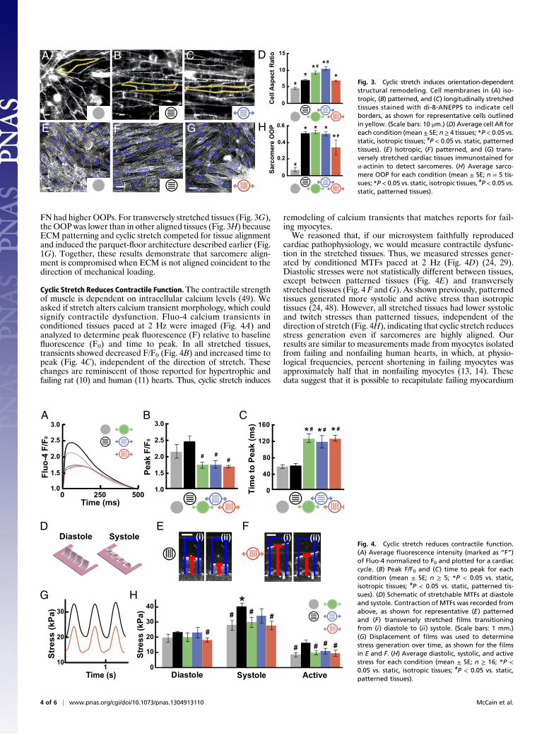
Cyclic Stretch Reduces Contractile Function.The contractile strengthof muscle is dependent on intracellular calcium levels (49). Weasked if stretch alters calcium transient morphology, which couldsignify contractile dysfunction. Fluo-4 calcium transients inconditioned tissues paced at 2 Hz were imaged (Fig. 4A) andanalyzed to determine peak fluorescence (F) relative to baselinefluorescence (F0) and time to peak. In all stretched tissues,transients showed decreased F/F0 (Fig. 4B) and increased time topeak (Fig. 4C), independent of the direction of stretch. Thesechanges are reminiscent of those reported for hypertrophic andfailing rat (10) and human (11) hearts. Thus, cyclic stretch induces
remodeling of calcium transients that matches reports for fail-ing myocytes.We reasoned that, if our microsystem faithfully reproduced
cardiac pathophysiology, we would measure contractile dysfunc-tion in the stretched tissues. Thus, we measured stresses gener-ated by conditioned MTFs paced at 2 Hz (Fig. 4D) (24, 29).Diastolic stresses were not statistically different between tissues,except between patterned tissues (Fig. 4E) and transverselystretched tissues (Fig. 4 F andG). As shown previously, patternedtissues generated more systolic and active stress than isotropictissues (24, 48). However, all stretched tissues had lower systolicand twitch stresses than patterned tissues, independent of thedirection of stretch (Fig. 4H), indicating that cyclic stretch reducesstress generation even if sarcomeres are highly aligned. Ourresults are similar to measurements made from myocytes isolatedfrom failing and nonfailing human hearts, in which, at physio-logical frequencies, percent shortening in failing myocytes wasapproximately half that in nonfailing myocytes (13, 14). Thesedata suggest that it is possible to recapitulate failing myocardium
D
0
5
10
15
Cel
l Asp
ect R
atio
*#
**
# * #A B C
0
0.2
0.4
0.6
Sarc
omer
e O
OP *
#
**
#*HE F G
Fig. 3. Cyclic stretch induces orientation-dependentstructural remodeling. Cell membranes in (A) iso-tropic, (B) patterned, and (C) longitudinally stretchedtissues stained with di-8-ANEPPS to indicate cellborders, as shown for representative cells outlinedin yellow. (Scale bars: 10 μm.) (D) Average cell AR foreach condition (mean ± SE; n≥ 4 tissues; *P< 0.05 vs.static, isotropic tissues; #P < 0.05 vs. static, patternedtissues). (E) Isotropic, (F) patterned, and (G) trans-versely stretched cardiac tissues immunostained forα-actinin to detect sarcomeres. (H) Average sarco-mere OOP for each condition (mean ± SE; n = 5 tis-sues; *P< 0.05 vs. static, isotropic tissues, #P< 0.05 vs.static, patterned tissues).
A
0
10
20
30
40
Diastole Systole Active
Stre
ss (k
Pa)
*
#
# # #
# # ##
1Time (s)
10
20
30
Str
ess
(kP
a)
Diastole Systole
# ##
1.0
1.5
2.0
2.5
3.0
Peak
F/F
0
0
40
80
120
160
Tim
e to
Pea
k (m
s) * # * # * #
1.0
1.5
2.0
2.5
3.0
500
Fluo
-4 F
/F0
2500Time (ms)
B C
E F
G H
(i) (ii) (i) (ii)D
Fig. 4. Cyclic stretch reduces contractile function.(A) Average fluorescence intensity (marked as “F”)of Fluo-4 normalized to F0 and plotted for a cardiaccycle. (B) Peak F/F0 and (C) time to peak for eachcondition (mean ± SE; n ≥ 5; *P < 0.05 vs. static,isotropic tissues; #P < 0.05 vs. static, patterned tis-sues). (D) Schematic of stretchable MTFs at diastoleand systole. Contraction of MTFs was recorded fromabove, as shown for representative (E) patternedand (F) transversely stretched films transitioningfrom (i) diastole to (ii) systole. (Scale bars: 1 mm.)(G) Displacement of films was used to determinestress generation over time, as shown for the filmsin E and F. (H) Average diastolic, systolic, and activestress for each condition (mean ± SE; n ≥ 16; *P <0.05 vs. static, isotropic tissues; #P < 0.05 vs. static,patterned tissues).
4 of 6 | www.pnas.org/cgi/doi/10.1073/pnas.1304913110 McCain et al.

on a 2D chip that is amenable to comparison with animal andclinical studies.
DiscussionIn this study, we modeled failing myocardium on a chip by ap-plying cyclic stretch to engineered, anisotropic cardiac tissues.Gene expression in stretched tissues was consistent with patho-logical remodeling, including up-regulation of focal adhesiongenes and a switch to the immature myosin isoform. Sarcomerealignment and cell shape were uniquely sensitive to the directionof stretch and matched clinical reports of pathological structuralremodeling. Stretch-induced changes in calcium cycling and sys-tolic stress generation were also comparable with measurementsfrom patients with heart failure. Together, these results suggestthat our in vitro microsystem can be used to study the genetic,structural, and functional aspects of failing myocardium.Maladaptive gene expression is a commonly used test for ex-
perimental models of heart disease because it can be easilycompared with clinical studies. Similarly, we observed a lowerratio of α- to β-MHC mRNA in stretched tissues, a commonlyaccepted indicator of heart disease (8, 9). Wemeasured increasedexpression of genes encoding for several focal adhesion pro-teins, such as focal adhesion kinase and integrin α5, in responseto stretch, which matches in vivo studies demonstrating up-regulation of focal adhesion expression and signaling duringthe hypertrophic response to mechanical overload (39, 42–44).Although our results do not identify molecular mechanisms, theydo motivate future studies to discover and validate therapeutictargets, such as focal adhesion signaling. Thus, our chip matchesthe genetic criteria of a valid model of failing myocardium andcould be used to further explore signaling and protein expression.Heart failure is also characterized by significant remodeling of
tissue architecture. Because disease progression often affects thedistribution of biomechanical forces and ECM organization, wetook advantage of our in vitro microsystem to address how thesetwo extracellular cues coregulate structural remodeling. Withtransverse cyclic stretch, local regions of sarcomere misalignmentresulted in disorganized tissue reminiscent of hypertrophic car-diomyopathy (4). Myocyte AR was also sensitive to the direction ofstretch, as myocytes stretched longitudinally had an AR of 10:1,compared with patternedmyocytes with anAR of 7:1. These valuesare similar to those reported formyocytes isolated from dilated andhealthy hearts, respectively (2). Interestingly, transverse stretch didnot affect AR, suggesting that structural remodeling is sensitive tothe direction of stretch, similar to previous in vitro reports (18, 50).These data suggest that our in vitro microsystem replicates themaladaptive remodeling of the diseased heart.Because heart failure is a broadly defined disease, it has been
associatedwith diastolic and systolic dysfunction (51). Our stretchedtissues exhibited reduced systolic stress, suggesting that excessivestretch affects myocyte shortening, but not relaxation. One po-tential explanation is that pathological hypertrophy and heartfailure are also associated with increased fibrosis, which stiffensthe cellular microenvironment (52). These pathological changesin compliance likely impede myocyte relaxation and could beessential to diastolic dysfunction. One limitation of our system isthat we cannot measure the rate of diastolic relaxation, whichcould correlate to isovolumetric relaxation, because the materialproperties of PDMSmake it difficult to separate the relaxation ofour engineered tissues from the elastic recoil of the PDMS sub-strate. Thus, our system recapitulates the systolic dysfunction offailing myocardium, but does not reveal a mechanism for diastolicdysfunction in the heart.Organs on chips have promising potential as substitutes for in
vivo animal studies with improved predictability compared withcurrent in vitro systems (53, 54). In this study, we used neonatal ratmyocytes because rat is the pharmaceutical and biotechnology in-dustry standard for the study of heart disease in vitro and in vivo.Our model could be further developed as failing myocardium ona chip technology with applications for testing therapeutic agentsagainst specific diseases and patient populations in combination
with in vitro models that focus on other cardiac diseases (55). Theuniqueness of our model lies in its ability to recapitulate nativetissue architecture and its capacity to quantify not only genetic andstructural remodeling, but also functional remodeling, includingcontractile force generation. As the quality and consistency ofhuman stem cell-derived cardiomyocytes improves, we hope toextend our model to these cells that will potentially be more pre-dictive than rodent cells (56).We also appreciate the importance ofincorporating other cell types found in the myocardium, such asfibroblasts and endothelial cells, which contribute to the multicel-lular architecture of the heart but are difficult to coculture withmyocytes. Further development of organs on chips with specificphysiological and pathological phenotypes has the potential tosignificantly reduce drug development costs while also improvingthe fidelity and predictability of efficacy and toxicity studies.
Materials and MethodsExperimental methods are described in detail in SI Materials and Methods;a brief description is included here.
Chip Fabrication. Elastic silicone membranes (Specialty Manufacturing) wereclamped into stainless steel brackets (23, 26) and affixed with 25-mm-diameter rings of silicon tubing. Membranes treated in a UV-ozone cleaner(Jelight) were micropatterned with PDMS stamps coated with 50 μg/mLFN (BD Biosciences) or coated uniformly with 50 μg/mL FN. To fabricatestretchable MTFs (23, 24), silicone membranes were temporarily cured to glassplatforms with 200 proof ethanol tomaintain baseline tension and provide rigidsurfaces for spin-coating. Two pieces of tape were applied across the length orwidth of the membrane separated by 8 mm. Poly(N-isopropylacrylamide) (Pol-ysciences) was spin-coated onto membranes followed by tape removal. PDMScured at room temperature for 4 to 5 h was then spin-coated onto membranes,which were reclamped into brackets after fully curing.
Cell Culture. All animal protocols were approved by the Harvard UniversityAnimal Care and Use Committee. Cardiac myocytes were isolated by enzy-matically digesting ventricles from 2-d-old Sprague–Dawley rats by usingpreviously published protocols (24, 48, 57). A custom bioreactor based ona previous design (26) was built to apply cyclic strain to tissues cultured onsilicone membranes using a linear motor (LinMot) to stretch as many as 10samples simultaneously in an incubator (23).
Gene Expression Analysis. RNA was isolated by using Stratagene AbsolutelyRNAMiniprep Kit (Agilent Technologies). mRNA was amplified and hybridizedto Affymetrix GeneChip Rat Gene 1.0 ST Arrays following manufacturer’sinstructions, and scanned with Affymetrix GeneChip Scanner 3000 7G. Probecell intensity data files were normalized using the robust multichip averagemethod (58) in Affymetrix Expression Console Software. Signal values werelog2-transformed and analyzed with Bioconductor open-source software andthe limma package in R (59). Average expression values for each conditionwere fit to a linear model and compared using Bayes statistics. Gene expres-sion values were analyzed with GEDI bioinformatics software package (31, 32).Differentially regulated genes were categorized by selected Gene Ontologyterms by inputting filtered probe set IDs into the AmiGO! Slimmer from theGeneOntology project (http://amigo.geneontology.org/cgi-bin/amigo/slimmer).
Sarcomere Alignment Quantification. Tissues were fixed, immunostained forsarcomeric α-actinin (A7811; Sigma-Aldrich), and imaged on an invertedfluorescent microscope (DMI 6000B; Leica). Custom MATLAB software(MathWorks) was used to calculate the sarcomere OOP of each tissue (24,48), which were statistically compared by Student t test.
Cell Shape Measurements. Tissues were incubated with 10 μM di-8-ANEPPS(Invitrogen) for 10 min at 37 °C, rinsed with Tyrode solution (1.8 mM CaCl2,5 mM glucose, 5 mM Hepes, 1 mM MgCl2, 5.4 mM KCl, 135 mM NaCl, 0.33 mMNaH2PO4, pH 7.4), moved to a confocal microscope maintained at 37 °C (LSM510; Zeiss), and imaged with a 40× objective. Custom MATLAB software(MathWorks) was used to manually outline cell borders of multiple cells pertissue, which were fit to an ellipse. The average major (cell length) andminor (cell width) axes and ARs of fitted ellipses for each condition werestatistically compared by Student t test.
Calcium Transient Measurements. Tissues were incubatedwith 10 μg/mL Fluo-4acetoxymethyl ester calcium indicator (Invitrogen) for 30 min at 37 °C, rinsed
McCain et al. PNAS Early Edition | 5 of 6
BIOPH
YSICSAND
COMPU
TATIONALBIOLO
GY

with Tyrode solution, and transferred to a confocal microscope maintainedat 37 °C (Zeiss LSM 510). Tissues were paced with a point stimulation elec-trode operating at 2 to 5 V and 2 Hz for at least 5 min before data acqui-sition with a 40× objective. For multiple fields, 10 × 10-μm2 regions wereselected in the cytoplasm of a cell and change in F over time for four beatswas averaged to determine F over F0 and time to peak (60). Average F/F0 andtime to peak for each condition were statistically compared by Student t test.
Contractile Stress Measurements. Stretchable MTFs were incubated in Tyrodesolution and moved to a stereomicroscope (Leica Microsystems). The PDMSlayer was cut to release arrays of MTFs that remained attached to the sub-strate at one longitudinal end. As the temperature decreased to less than32 °C, poly(N-isopropylacrylamide) transitioned to the aqueous phase, allow-ing MTF release (24). Temperature was restored to 37 °C, and MTFs werepaced with a field stimulation electrode operating at 5 to 10 V and 2 Hz.
Movies were acquired at 120 to 150 Hz. PDMS film thickness was measuredwith a profilometer (P-16+ Contact Stylus Profiler; KLA-Tencor). MATLABsoftware (MathWorks) was used to calculate stress generation based on theprojection of the radius of curvature and film thickness (24, 29). Average di-astolic, systolic, and twitch stresses for each condition were statistically com-pared by Student t test.
ACKNOWLEDGMENTS. We thank André G. Kléber for design of the initialcyclic stretch system and the Harvard Center for Nanoscale Systems for useof cleanroom facilities. This work was funded by American Heart AssociationPredoctoral Fellowship 0815729D, National Institutes of Health Grants 1 R01HL079126 and 1 UH2 TR000522-01, the Harvard Materials Research Scienceand Engineering Center as supported by National Science Foundation Divisionof Materials Research Grant DMR-0213805, the Harvard Stem Cell Institute andGlaxoSmithKline, and the Harvard School of Engineering and Applied Sciences.
1. Lorell BH, Carabello BA (2000) Left ventricular hypertrophy: Pathogenesis, detection,and prognosis. Circulation 102(4):470–479.
2. Gerdes AM (2002) Cardiac myocyte remodeling in hypertrophy and progression tofailure. J Card Fail 8(6, suppl):S264–S268.
3. Grossman W, Jones D, McLaurin LP (1975) Wall stress and patterns of hypertrophy inthe human left ventricle. J Clin Invest 56(1):56–64.
4. Ho CY (2009) Hypertrophic cardiomyopathy: Preclinical and early phenotype. J Car-diovasc Transl Res 2(4):462–470.
5. Jane-Lise S, Corda S, Chassagne C, Rappaport L (2000) The extracellular matrix and thecytoskeleton in heart hypertrophy and failure. Heart Fail Rev 5(3):239–250.
6. Gerdes AM, Onodera T, Wang X, McCune SA (1996) Myocyte remodeling during theprogression to failure in rats with hypertension. Hypertension 28(4):609–614.
7. Gerdes AM (1992) Remodeling of ventricular myocytes during cardiac hypertrophyand heart failure. J Fla Med Assoc 79(4):253–255.
8. Gupta MP (2007) Factors controlling cardiac myosin-isoform shift during hypertrophyand heart failure. J Mol Cell Cardiol 43(4):388–403.
9. Miyata S, Minobe W, Bristow MR, Leinwand LA (2000) Myosin heavy chain isoformexpression in the failing and nonfailing human heart. Circ Res 86(4):386–390.
10. Gómez AM, et al. (1997) Defective excitation-contraction coupling in experimentalcardiac hypertrophy and heart failure. Science 276(5313):800–806.
11. Piacentino V, 3rd, et al. (2003) Cellular basis of abnormal calcium transients of failinghuman ventricular myocytes. Circ Res 92(6):651–658.
12. Wilson JR, et al. (1987) Experimental congestive heart failure produced by rapidventricular pacing in the dog: cardiac effects. Circulation 75(4):857–867.
13. Mulieri LA, Hasenfuss G, Leavitt B, Allen PD, Alpert NR (1992) Altered myocardialforce-frequency relation in human heart failure. Circulation 85(5):1743–1750.
14. Davies CH, et al. (1995) Reduced contraction and altered frequency response of isolatedventricular myocytes from patients with heart failure. Circulation 92(9):2540–2549.
15. Carll AP, Willis MS, Lust RM, Costa DL, Farraj AK (2011) Merits of non-invasive ratmodels of left ventricular heart failure. Cardiovasc Toxicol 11(2):91–112.
16. Kelso EJ, et al. (1996) Mechanical effects of ET-1 in cardiomyocytes isolated fromnormal and heart-failed rabbits. Mol Cell Biochem 157(1-2):149–155.
17. Frank D, et al. (2008) Gene expression pattern in biomechanically stretched car-diomyocytes: evidence for a stretch-specific gene program. Hypertension 51(2):309–318.
18. Gopalan SM, et al. (2003) Anisotropic stretch-induced hypertrophy in neonatal ven-tricular myocytes micropatterned on deformable elastomers. Biotechnol Bioeng 81(5):578–587.
19. Blaauw E, et al. (2010) Stretch-induced hypertrophy of isolated adult rabbit car-diomyocytes. Am J Physiol Heart Circ Physiol 299(3):H780–H787.
20. Kensah G, et al. (2011) A novel miniaturized multimodal bioreactor for continuous insitu assessment of bioartificial cardiac tissue during stimulation and maturation.Tissue Eng Part C Methods 17(4):463–473.
21. Rana OR, et al. (2008) A simple device to apply equibiaxial strain to cells cultured onflexible membranes. Am J Physiol Heart Circ Physiol 294(1):H532–H540.
22. Bhadriraju K, Chen CS (2002) Engineering cellular microenvironments to improve cell-based drug testing. Drug Discov Today 7(11):612–620.
23. Balachandran K, et al. (2011) Cyclic strain induces dual-mode endothelial-mesenchy-mal transformation of the cardiac valve. Proc Natl Acad Sci USA 108(50):19943–19948.
24. Grosberg A, Alford PW, McCain ML, Parker KK (2011) Ensembles of engineered car-diac tissues for physiological and pharmacological study: Heart on a chip. Lab Chip11(24):4165–4173.
25. Alford PW, et al. (2011) Blast-induced phenotypic switching in cerebral vasospasm.Proc Natl Acad Sci USA 108(31):12705–12710.
26. Zhuang J, Yamada KA, Saffitz JE, Kléber AG (2000) Pulsatile stretch remodels cell-to-cell communication in cultured myocytes. Circ Res 87(4):316–322.
27. Yamada K, Green KG, Samarel AM, Saffitz JE (2005) Distinct pathways regulate ex-pression of cardiac electrical and mechanical junction proteins in response to stretch.Circ Res 97(4):346–353.
28. Greenbaum RA, Ho SY, Gibson DG, Becker AE, Anderson RH (1981) Left ventricularfibre architecture in man. Br Heart J 45(3):248–263.
29. Alford PW, Feinberg AW, Sheehy SP, Parker KK (2010) Biohybrid thin films for mea-suring contractility in engineered cardiovascular muscle. Biomaterials 31(13):3613–3621.
30. Barry SP, Davidson SM, Townsend PA (2008) Molecular regulation of cardiac hyper-trophy. Int J Biochem Cell Biol 40(10):2023–2039.
31. Eichler GS, Huang S, Ingber DE (2003) Gene Expression Dynamics Inspector (GEDI): Forintegrative analysis of expression profiles. Bioinformatics 19(17):2321–2322.
32. Sheehy SP, Huang S, Parker KK (2009) Time-warped comparison of gene expression inadaptive and maladaptive cardiac hypertrophy. Circ Cardiovasc Genet 2(2):116–124.
33. Edgar R, Domrachev M, Lash AE (2002) Gene Expression Omnibus: NCBI gene expressionand hybridization array data repository. Nucleic Acids Res 30(1):207–210.
34. Eder P, Molkentin JD (2011) TRPC channels as effectors of cardiac hypertrophy. CircRes 108(2):265–272.
35. Martínez ML, Heredia MP, Delgado C (1999) Expression of T-type Ca(2+) channels inventricular cells from hypertrophied rat hearts. J Mol Cell Cardiol 31(9):1617–1625.
36. Xing W, et al. (2006) Myocardin induces cardiomyocyte hypertrophy. Circ Res 98(8):1089–1097.
37. Torrado M, et al. (2003) Myocardin mRNA is augmented in the failing myocardium:expression profiling in the porcine model and human dilated cardiomyopathy. J MolMed (Berl) 81(9):566–577.
38. Cribbs L (2010) T-type calcium channel expression and function in the diseased heart.Channels (Austin) 4(6):447–452.
39. Heling A, et al. (2000) Increased expression of cytoskeletal, linkage, and extracellularproteins in failing human myocardium. Circ Res 86(8):846–853.
40. LeGrice IJ, et al. (2012) Progression of myocardial remodeling and mechanical dys-function in the spontaneously hypertensive rat. Am J Physiol Heart Circ Physiol303(11):H1353–H1365.
41. Parker KK, Ingber DE (2007) Extracellular matrix, mechanotransduction and structuralhierarchies in heart tissue engineering. Philos Trans R Soc Lond B Biol Sci 362(1484):1267–1279.
42. Terracio L, et al. (1991) Expression of collagen binding integrins during cardiac de-velopment and hypertrophy. Circ Res 68(3):734–744.
43. Clemente CF, et al. (2012) Focal adhesion kinase governs cardiac concentric hypertrophicgrowth by activating the AKT and mTOR pathways. J Mol Cell Cardiol 52(2):493–501.
44. Lu H, et al. (2006) Integrin-linked kinase expression is elevated in human cardiac hyper-trophy and induces hypertrophy in transgenic mice. Circulation 114(21):2271–2279.
45. Pong T, et al. (2011) Hierarchical architecture influences calcium dynamics in en-gineered cardiac muscle. Exp Biol Med (Maywood) 236(3):366–373.
46. Pope AJ, Sands GB, Smaill BH, LeGrice IJ (2008) Three-dimensional transmural orga-nization of perimysial collagen in the heart. Am J Physiol Heart Circ Physiol 295(3):H1243–H1252.
47. Strijkers GJ, et al. (2009) Diffusion tensor imaging of left ventricular remodeling inresponse to myocardial infarction in the mouse. NMR Biomed 22(2):182–190.
48. Feinberg AW, et al. (2012) Controlling the contractile strength of engineered cardiacmuscle by hierarchal tissue architecture. Biomaterials 33(23):5732–5741.
49. Bers DM (2008) Calcium cycling and signaling in cardiac myocytes. Annu Rev Physiol70:23–49.
50. Simpson DG, Majeski M, Borg TK, Terracio L (1999) Regulation of cardiac myocyteprotein turnover and myofibrillar structure in vitro by specific directions of stretch.Circ Res 85(10):e59–e69.
51. Gaasch WH (1994) Diagnosis and treatment of heart failure based on left ventricularsystolic or diastolic dysfunction. JAMA 271(16):1276–1280.
52. Berry MF, et al. (2006) Mesenchymal stem cell injection after myocardial infarction im-proves myocardial compliance. Am J Physiol Heart Circ Physiol 290(6):H2196–H2203.
53. Huh D, et al. (2010) Reconstituting organ-level lung functions on a chip. Science 328(5986):1662–1668.
54. Huh D, Torisawa YS, Hamilton GA, Kim HJ, Ingber DE (2012) Microengineered phys-iological biomimicry: Organs-on-chips. Lab Chip 12(12):2156–2164.
55. Hirt MN, et al. (2012) Increased afterload induces pathological cardiac hypertrophy:a new in vitro model. Basic Res Cardiol 107(6):307.
56. Davis RP, van den Berg CW, Casini S, Braam SR, Mummery CL (2011) Pluripotent stemcell models of cardiac disease and their implication for drug discovery and de-velopment. Trends Mol Med 17(9):475–484.
57. Bray MA, Sheehy SP, Parker KK (2008) Sarcomere alignment is regulated by myocyteshape. Cell Motil Cytoskeleton 65(8):641–651.
58. Irizarry RA, et al. (2003) Summaries of Affymetrix GeneChip probe level data. NucleicAcids Res 31(4):e15.
59. Smyth GK (2004) Linear models and empirical bayes methods for assessing differentialexpression in microarray experiments. Stat Appl Genet Mol Biol 3:Article3.
60. Howlett SE, Grandy SA, Ferrier GR (2006) Calcium sparkproperties in ventricularmyocytesare altered in aged mice. Am J Physiol Heart Circ Physiol 290(4):H1566–H1574.
6 of 6 | www.pnas.org/cgi/doi/10.1073/pnas.1304913110 McCain et al.