Embed Size (px)
Citation preview

DEVELOPMENT OF IMMUNOTHERAPIES AND VACCINES AGAINST VISCERAL
LEISHMANIASIS.
Rebecca Jacinto Faleiro
M.Sc. Molecular Biology, B.Sc. Biotechnology
Submitted in fulfilment of the requirements for the degree of
Doctor of Philosophy
Institute of Health and Biomedical Innovation (IHBI), School of Biomedical Sciences, Faculty of Health
Queensland University of Technology
2016

QUT Verified Signature

Page | ii
Acknowledgements
First and foremost I would like to thank my primary supervisor and mentor Dr
Christian R. Engwerda, thank you for taking me on as a student. Thank you for your patience
with me and the constant guidance, mentorship and support you’ve provided throughout the
last four years. I am very grateful for all the opportunities you have given me. I would also
like to thank my associate supervisor Professor Louise Hafner. Thank you for making the
time for me every month, your positive outlook and confidence in my research has been a
source of reassurance to keep going.
I would like to thank all the members of the “Engwerda Lab” and “Haque Lab” past
and present. Specifically Fabian Riviera, for your guidance with all the laboratory help, I
would have drowned in a sea of spleens and livers without it. Fiona Amante and Lynette
Beattie, thank you for helping with my drafts. Susanna Ng, thank you for your creative input.
I would also like to thank members of QIMR Berghofer Animal Facility and the Flow
cytometry Lab who have helped me during the course of this project.
A huge thanks to my fellow PhD students Marcela Montes De Oca and Mariska
Miranda and to Dr Winnie Fernando, thank you for helping me keep my sanity though the
last four years. I must also thank all my friends, thank you for putting up with me, for always
being there for me and cheering me up in your own ways.
Lastly I wish to say thank you to my family, mum and dad you’ll are the best parents a
kid could have. Your un-wavering love and faith in me has helped me reach peaks I thought

Page | iii
impossible. This thesis is dedicated to you, mum and dad. Rajiv, thank you for being a “little
brother” your antics have provided a much needed distraction, when needed.

Page | iv
Abstract
Visceral leishmaniasis (VL) is a chronic parasitic disease prevalent in tropical and sub-
tropical countries caused by the protozoan parasites Leishmania donovani and Leishmania
infantum (chagasi). VL is associated with severe immune dysfunction and clinical outcomes
of infection depend on the infecting parasite species and the host immune response. Immunity
against invading pathogens requires strong innate and adaptive host immune responses, but
Leishmania parasites can elude these defence mechanisms to persist and survive in the host.
Treatment options are limited to relatively toxic drugs and no vaccine for humans is
available. Identifying and understanding the host immune responses is of paramount
importance to better understand disease pathogenesis and for the development of vaccines
and therapies. This study has focused on the development of immune-based therapy with
immune checkpoint inhibitors and/or activators, as well as cytokines as a way to treat disease
either alone or in combination with conventional drugs. In addition, I developed a platform
for a live attenuated whole parasite vaccine against experimental VL.
The first aim of this study focused on combination immunotherapy as a way to treat VL
either alone or with conventional drugs. Previous studies have shown that activation of
glucocorticoid-induced TNF receptor family-related protein (GITR) in L. donovani -infected
mice boosted CD4+ T cell activation and reduced liver parasite burden. Similarly, IL-10
blockade has previously been shown to enhance host resistance against L. donovani. I
investigated whether combined GITR stimulation and IL-10 blockade would act
synergistically to improve anti-parasitic immunity in mice infected with L. donovani. Infected
mice were treated with a combination of an agonist anti-GITR mAb and a blocking anti-IL-

Page | v
10R mAb, and parasite burdens were assessed. Mice treated with this combination did not
control parasite growth any better than mice treated with a single form of immune
modulation. However, combination immune therapy in mice infected with a low dose of
parasites was detrimental, similar to what has been observed in humans, while no such effect
was seen in mice with high parasite burdens. Nevertheless, combined anti-IL-10 and anti-
GITR mAb treatment could improve anti-parasitic immunity when used with sub-optimal
doses of anti-parasitic drug. These results have implications for the use of immune therapies
in patients, and suggest that the outcomes may differ depending on the stage of disease, the
immune modulators used and use of anti-parasitic drug.
The second aim of the study focused on the use of cytokine therapy, by testing the
effect of IL-2/anti-IL-2 mAb complexes to treat experimental VL. IL-2/anti-IL-2 mAb
complexes have significant effects on the immune system, and have been studied extensively
in various disease settings, including cancer treatment and various infections. However, the
impact of IL-2/anti-IL-2 mAb complex treatment on L. donovani infection has not been
previously investigated. In my study, two doses of the IL-2/anti-IL-2 mAb complexes (IL-2Jc
or the IL-2Sc) resulted in a significant reduction in parasite burdens in mice infected with L.
donovani. However, no expansion of targeted cell populations was observed, as previously
reported. Further investigations with transgenic mice and cell depleting antibodies revealed
that CD4+ T cell were required for the maintenance of anti-parasitic immunity generated by
the IL-2/anti-IL-2 mAb complex treatments. This study has therefore provided evidence for
the efficacy of cytokine-based IL-2/anti-IL-2 mAb complex therapy for treating VL and
highlights that timing and dose of treatment should be considered carefully before treating.

Page | vi
The final aim of the study focused on developing a live attenuated, whole parasite
vaccine to protect against experimental VL. I evaluated the potential of both irradiation and
chemical attenuation of L. donovani parasites as a vaccine strategy. L. donovani amastigotes
or in-vitro cultured promastigotes were irradiated at 500 Gys or treated with tafurmycin, an
alkylating agent that irreversibly alters the parasite DNA, thus inhibiting parasite growth. I
found that irradiated L. donovani promastigotes provided better protection compared to
irradiated amastigotes. However, irradiated parasites were still able to expand in
immunocompromised animals, while this did not appear to be the case for chemically
attenuated parasites. Furthermore, addition of adjuvants CpG-DNA or Poly (I:C) did not
further improve vaccine mediated protection. Although this vaccine has not yet been
optimised, it did generate potent anti-parasitic CD4+ T cell responses and reduced parasite
burdens in infected tissue sites. Since many chronic infectious diseases share mechanisms of
immune suppression, these findings may have broader implications for other infectious
diseases, such as HIV, tuberculosis and malaria.
Keywords
CD4+ T cells, Leishmania, liver, spleen, immune therapy, vaccines, visceral
leishmaniasis,

Page | vii
Table of Contents
Statement of Original Authorship ............................................................................................. i
Acknowledgements .................................................................................................................. ii
Abstract ................................................................................................................................... iv
Keywords ................................................................................................................................ vi
Table of Contents ................................................................................................................... vii
List of Figures ...........................................................................................................................x
List of Tables ........................................................................................................................ xiii
Publications ........................................................................................................................... xiv
List of Abbreviations ............................................................................................................ xvi
Chapter 1: Introduction .......................................................................................1
1.1 Description of Scientific problem ...................................................................................1
1.2 Significance ....................................................................................................................3
1.3 Hypothesis ......................................................................................................................3
1.4 Aims................................................................................................................................4
Chapter 2: Literature Review ..............................................................................5
2.1 Introduction ....................................................................................................................5
2.2 Human VL ......................................................................................................................6 2.2.1 VL susceptibility ..................................................................................................7 2.2.2 Disease spectrum of human VL ...........................................................................8 2.2.3 Immune regulation during human VL ..................................................................9
2.3 The Mouse Model of VL ..............................................................................................12 2.3.1 Establishment of infection in the Liver ..............................................................13 2.3.2 Development of chronic infection in the spleen and bone marrow ....................15
2.4 The role of CD4+ T cells during infection ....................................................................20 2.4.1 The activation of CD4+ T cells by DC’s .............................................................21 2.4.2 The role of CD4+ T cells in resolving L. donovani infection in the liver ...........22 2.4.3 Organ-specific roles for CD4+ T cells during VL ..............................................24
2.5 Post Kala-azar Dermal Leishmaniasis and HIV co-infection .......................................24
2.6 Past and current Treatment Options..............................................................................27 2.6.1 Current anti-leshmania drugs available for the treatment of VL ........................28 2.6.2 Vaccines against VL ...........................................................................................31
Chapter 3: Materials and Methods ...................................................................33
3.1 Mice and parasites ........................................................................................................33 3.1.1 Mice ....................................................................................................................33 3.1.2 Parasites ..............................................................................................................33 3.1.3 Isolation of parasite for infection of mice ..........................................................34 3.1.4 In-vitro culturing of L. donovani promastigotes .................................................35 3.1.5 Irradiation of L. donovani parasites ....................................................................35 3.1.6 Chemical-attenuation of L. donovani parasites ..................................................36

Page | viii
3.1.7 Adjuvants used for immunization ......................................................................37
3.2 Sample collection..........................................................................................................37 3.2.1 Collection of blood for serum isolation ..............................................................37 3.2.2 Collection of animal organs................................................................................38 3.2.3 Assessment of parasite burdens ..........................................................................38
3.3 Cell isolation and preparation .......................................................................................40 3.3.1 Hepatic Mononuclear Cell (MNC) preparation ..................................................40 3.3.2 Splenic MNC preparation ...................................................................................40 3.3.3 Isolation of peritoneal macrophages ...................................................................41 3.3.4 Magnetic cell sorting (MACS) purification of DC’s ..........................................41
3.4 Antibodies and drugs for in-vivo administration ..........................................................42 3.4.1 In-vitro culturing of monoclonal antibodies .......................................................42 3.4.2 Intra-peritoneal administration of antibodies .....................................................43 3.4.3 Preparation and administration of IL-2/anti–IL-2 complexes ............................43 3.4.4 Preparation and administration of pentavalent antimonial drug .........................44 3.4.5 Preparation and in- vivo administration of Diphtheria toxin ..............................44
3.5 Experimental Methods ..................................................................................................45 3.5.1 In-vitro infection of Macrophages ......................................................................45 3.5.2 Detection of nitrite using Griess assay ...............................................................45 3.5.3 DC activation assay ............................................................................................46 3.5.4 Antigen-specific cellular analysis .......................................................................46 3.5.5 Fluorescence activated cell sorting (FACS) analysis of cell surface markers ....47 3.5.6 FACS analysis of intracellular cytokines and transcription factors....................48 3.5.7 Measurement of cytokines in serum and cell culture supernatants ....................49
3.6 Statistical Analysis........................................................................................................50
Chapter 4: Testing whether promoting parasite-specific CD4+ T cell function via GITR activation improves the outcome of experimental VL................................51
4.1 Introduction ..................................................................................................................51
4.2 Results ..........................................................................................................................53 4.2.1 The effect of combination immune therapy during a chronic L. donovani infection
53 4.2.2 Effect of combination immune therapy during an acute L. donovani infection .58 4.2.3 Effect of combined anti-GITR mAb and anti-IL-10 mAb therapy on immune
parameters during a low-dose L. donovani infection .........................................62 4.2.4 Effect combining immune therapy with drug treatment on an L. donovani infection
71
4.3 Discussion .....................................................................................................................78
Chapter 5: To test whether IL-2 signalling pathways are deficient in T cells during VL and to test the ability of IL-2/anti-IL-2 mAb complexes to treat and improve experimental VL outcome. .......................................................................................82
5.1 Introduction ..................................................................................................................82
5.2 Results ..........................................................................................................................85 5.2.1 The effects of IL-2/Anti-IL-2 mAb complex treatments during the chronic phase of
L. donovani infection ..........................................................................................85 5.2.2 Identification of immune cell populations expressing IL-2 receptors during an L.
donovani infection ..............................................................................................87 5.2.3 Treatment with IL-2/Anti-IL-2 mAb complexes reduced L. donovani parasite
burden .................................................................................................................88

Page | ix
5.2.4 Effect of IL-2/Anti-IL-2 mAb complex therapy on immune parameters during an L. donovani infection ..........................................................................................90
5.2.5 Treg cells do not interfere with protection mediated by IL-2J complex treatment96 5.2.6 The IL-2J complex mediates anti-parasitic effects in L. donovani -infected mice
via CD4+ T cells ...............................................................................................100 5.2.7 The IL-2S complex mediates anti-parasitic effects in L. donovani -infected mice
via CD4+ T cells, and not via CD8+ T cells or NK cells ...................................103
5.3 Discussion ...................................................................................................................107
Chapter 6: To compare different methods of parasite attenuation and establish whether a live, attenuated, whole parasite vaccine can protect against experimental VL 111
6.1 Introduction ................................................................................................................111
6.2 Results ........................................................................................................................114 6.2.1 The effects of immunization with irradiated whole L. donovani promastigotes114 6.2.2 The effect of immunization with either radio- or chemically-attenuated whole L.
donovani amastigotes .......................................................................................116 6.2.3 Immunization with irradiated promastigotes provides better protection compared
to irradiated amastigotes ...................................................................................121 6.2.4 Testing the pathogenicity of irradiated whole parasite .....................................126 6.2.5 Investigating the effect of increasing the dose of irradiation for attenuation in
immunized mice ...............................................................................................127 6.2.6 The effect of immunization with chemically-attenuated L. donovani promastigotes
in the presence of adjuvant ...............................................................................131 6.2.7 Chemically-attenuated parasites selectively inhibit pattern recognition receptors
137
6.3 Discusssion .................................................................................................................140
Chapter 7: Concluding remarks ......................................................................143
Chapter 8: Bibliography ..................................................................................149
Chapter 9: Appendix ........................................................................................164
9.1 Appendix 1 .................................................................................................................164 9.1.1 Effect of combination immune therapy on sample obtained from active human VL
patients..............................................................................................................164

Page | x
List of Figures
Figure 1.1: Current global distribution of Visceral Leishmaniasis. ..............................2
Figure 1.2 : Graphical representation of Aims..............................................................4
Figure 2.1: Life cycle of the L. donovani parasite. .......................................................6
Figure 2.2 : Overview of cellular responses during an asymptomatic L. donovani infection. .......................................................................................................15
Figure 2.3 : Overview of cellular responses during a chronic L. donovani infection. 20
Figure 4.1: Distinct effects of anti-GITR agonist antibody and blocking IL-10R and CTLA-4 interactions on anti-parasitic responses. ........................................54
Figure 4.2 : Effects of combination antibody treatment on parasite burdens in liver and spleen. ...........................................................................................................55
Figure 4.3: Effects of combined antibody treatment on parasite burdens in the liver and spleen. ...........................................................................................................57
Figure 4.4: Effects of combination antibody treatment on parasite burdens during acute infection. .......................................................................................................59
Figure 4.5: The dose of infection determines combination mAb treatment outcome.61
Figure 4.6: Representative sequential gating strategy for the isolation of Th1 cells, Tr1 cells and terminally differentiated CD4+ T cells. .........................................63
Figure 4.7: Immune modulation has little effect on Th1 responses in the liver. ........65
Figure 4.8: Immune modulation has little effect on Tr1 responses in the liver. .........66
Figure 4.9: Increased frequency and number of terminally differentiated hepatic Th1 cells in groups treated with combined anti-GITR and anti-IL-10R mAbs. ..68
Figure 4.10: Increased number and frequency of terminally differentiated hepatic Th1 cells in mice infected with low numbers of parasites. ..................................70
Figure 4.11: Immune modulation combined with sub-optimal drug therapy improved control of parasite burden. ............................................................................72
Figure 4.12: Combined mAb administration with drug treatment reduces the number of terminally differentiated Th1 cells. ...............................................................74
Figure 4.13: Antigen-specific cellular immune responses after combined mAb administration and drug treatment. ...............................................................75
Figure 4.14: Anti-parasitic immune responses after combined mAb therapy and sub-optimal drug treatment. .................................................................................77
Figure 5.1: IL-2/anti-IL-2 mAb complexes selectively stimulate lymphocyte subsets.84
Figure 5.2: The effect of IL-2/Anti-IL-2 mAb complex treatment on the chronic phase of L. donovani infection. ...............................................................................86
Figure 5.3: Representative gating strategies for the identification of IL-2 receptors on lymphocyte subsets in the liver.....................................................................87
Page | xi
Figure 5.4: Expression of IL-2 receptors is enhanced during an L. donovani infection in the Liver. .......................................................................................................88
Figure 5.5: IL-2/Anti-IL-2 mAb complexes can improve control of L. donovani growth in the spleen and liver. ..................................................................................89
Figure 5.6: Representative sequential gating strategy for the isolation of immune cells. ......................................................................................................................91
Figure 5.7: Treatment with IL-2Sc and IL-2Jc has little effect on the expansion of activated CD4+ T cell expressing IFNγ, Tr1 and Treg cell population in the liver and spleen. ............................................................................................92
Figure 5.8: Representative gating strategy for the isolation of immune cells. ...........93
Figure 5.9: Treatment with IL-2Sc and IL-2Jc has little effect on the expansion of CD8+ T cells in the liver or spleen. .........................................................................94
Figure 5.10: Treatment with IL-2Sc and IL-2Jc has little effect on the expansion of NK1.1 cells in the liver and spleen. ..............................................................95
Figure 5.11: Foxp3-GFP-DTR mice treated with DT have a reduced frequency of Treg cells. ..............................................................................................................97
Figure 5.12: Tregs do not impair IL-2J complex-mediated protection. ......................98
Figure 5.13: The impact of Treg cell depletion in IL-2J complex treated animals. ...99
Figure 5.14: Administration of anti-CD4 mAb results in efficient CD4+ T cell depletion. ....................................................................................................101
Figure 5.15: CD4+ T cells are required for IL-2J complex mediated protection. .....102
Figure 5.16: Depletion of CD4+ T cells in IL-2J complex treated animals increased the frequency of CD8+ T cell and NK1.1 cells. ................................................103
Figure 5.17: CD8+ T cells and NK cells do not contribute to IL-2S complex-mediated protection. ...................................................................................................105
Figure 5.18: CD4+ T cells are required for IL-2S complex mediated protection. ....106
Figure 6.1.1: Experimental timeline for assessment of protection against L. donovani. ....................................................................................................................114
Figure 6.2: Failure of previously reported immunisation regime to protect against VL. ....................................................................................................................115
Figure 6.3: Immunization with irradiated L. donovani amastigotes improves parasite control in the liver. ......................................................................................118
Figure 6.4: Immunization has little effect on the CD4+ T cell responses in the liver, but increased CD4+ T cell responses were observed in the spleen. ..................120
Figure 6.5: Immunization with irradiated parasites results in enhanced antigen-specific T cell responses. ..........................................................................................122
Figure 6.6: Immunization with two doses of irradiated L. donovani promastigotes resulted in lower parasite burdens in both the liver and spleen. .................124
Figure 6.7: Cellular immune responses in the livers and spleens of mice immunized with irradiated parasites and challenged with L. donovani. ........................125
Figure 6.8: Irradiated parasites caused low grade infections in B6.Rag-1-/- mice. ...127

Page | xii
Figure 6.9: Attenuation with a higher dose of radiation results in a loss of protective immune responses. ......................................................................................129
Figure 6.10: Dose of irradiation used for attenuation determines parasite viability and metabolic activity. .......................................................................................130
Figure 6.11: Immunization with chemically-attenuated parasites results in enhanced antigen-specific cellular responses. ............................................................133
Figure 6.12: Immunization with chemically-attenuated whole L. donovani promastigote results in lower parasite burden in both the liver and spleen, but the addition of adjuvant had no effect. ................................................................................135
Figure 6.13: CD4+ T cell responses in the livers and spleens of mice immunized with chemically-attenuated promastigotes and challenged with L. donovani . ..136
Figure 6.14: Chemically attenuated L. donovani promastigotes inhibit pattern recognition pathways in DC’s. ...................................................................139
Figure 9.1: GITR mRNA accumulation in PBMC is increased in VL patients. .......165
Figure 9.2: GITR activation has no significant impact on parasite growth in spleen samples and antigen-specific IFNγ production in whole blood from VL patients. .......................................................................................................166
Figure 9.3: GITR activation alone or in combination with IL-10 blockade does not improve antigen-specific IFNγ production by whole blood cells after drug treatment. ....................................................................................................167

Page | xiii
List of Tables
Table 1: Current anti-leishmanial drugs against VL. ..................................................30
Table 2 : A summary of the fluorophore-conjugated antibodies used for surface and intracellular staining .....................................................................................49

Page | xiv
Publications
Published work by the author incorporated into the thesis:
Published:
1. Immune regulation during chronic visceral leishmaniasis. Faleiro RJ, Kumar R,
Hafner LM, Engwerda CR. PLoS Negl Trop Dis. 2014 Jul 10;8(7). Noting that
parts of this review have been incorporated into Chapter 2 (Literature Review)
which also includes updated material for the thesis that is mot found in the
review.
In preparation:
1. Combination immune-therapy for the treatment of visceral leishmaniasis.
2. IL-2/anti-IL-2 complexes mediate protection against L. donovani through CD4+
T cells.
Poster Presentation
1. Rebecca Faleiro; Rajiv Kumar; Louise Hafner; Christian Engwerda.
Modulating CD4+ T cells during chronic Visceral Leishmaniasis (VL) to
improve disease outcome. Keystone Symposia on “T Cells: Regulation and
Effector Function”. Utah, USA. March 2015.

Page | xv
Additional published work by the author relevant to the thesis but not forming part of it:
1. Tissue requirements for establishing long-term CD4+ T cell-mediated immunity
following Leishmania donovani infection. Bunn PT, Stanley AC, de Labastida
Rivera F, Mulherin A, Sheel M, Alexander CE, Faleiro RJ, Amante FH,
Montes De Oca M, Best SE, James KR, Kaye PM, Haque A, Engwerda CR. J
Immunol. 2014 Apr 15;192 (8):3709-18.
Page | xvi
List of Abbreviations
Ab Antibody
AIDS Acquired Immune Deficiency Syndrome
AMC Age matched control
APC Antigen Presenting Cell
BM Bone Marrow
BSA Bovine Serum Albumin
CBA Cytometric Bead Array
CCL (C-C motif) Ligand
CD Cluster of Differentiation
CL Cutaneous Leishmaniais
CMI Cell Mediated Immunity
CO2 Carbon Dioxide
CpG ODN CpG Oligodeoxynucleotides
CTLA-4 Cytotoxic T-Lymphocyte-Associated Protein 4
CXC C-X-C chemokines
CXCL (C-X-C motif) Ligand
CXCR (C-X-C motif) Receptor
DC Dendritic Cell
DMEM Dulbecco's Modified Eagle Medium
DMSO Dimethyl Sulfoxide
dNTP Deoxynucleotide
DT Diphtheria Toxin

Page | xvii
DTR Diphtheria Toxin Receptor
EDTA Ethylenediaminetetraacetic Acid
EVL Experimental Visceral Leishmaniasis
FACS Fluorescence Activated Cell Sorting
FBS Fetal Bovine Serum
Foxp3 Forkhead Box P3
g Gram
GFP Green Fluorescent Protein
GITR Glucocorticoid-Induced TNFR Family related gene)
Gy Gray
HEPES 4-(2-hydroxyethyl)-1-piperazineethanesulfonic acid
HIV Human Immunodeficiency Virus
HLA Human Leucocyte Antigen
Hrs Hours
i.m. Intramuscularly
i.p. Intraperitoneally
i.v. Intravenously (i.v.)
iDTR Inducible Diphtheria Toxin Receptor
IFNγ Interferon Gamma
IL Interleukin
ISO Isotype Control
KC Kupffer cells
KLRG-1 Killer-cell Lectin like Receptor G1
LDU Leishmania Donovan Units
M199 Medium 199

Page | xviii
mAb Monoclonal Antibody
MACS Magnetic-Activated Cell Sorting
mg Milligrams
mL Millilitre
MLR Mixed Lymphocyte Reaction
MM Marginal Metallophilic
mM Millimolar
MMM Marginal Metallophillic Macrophages
MNCs Mononuclear Cells
MO Monocyte
mRNA Messenger RNA
MZ Marginal Zone
MZM Marginal Zone Macrophages
Mφ Macrophage
n No of Mice
NaCl Sodium Chloride
ng Nanogram
NK Natural Killer cell
NKp46 Natural killer cell p46-related protein]
NO Nitric Oxide
NO2- Nitrite
NRAMP Natural Resistance-Associated Macrophage Protein
p.c. Post Challenge
pg Pico grams
p.i. Post Infection

Page | xix
PBMCs Peripheral Blood Mononuclear Cells
PBS Phosphate Saline Buffer
PCR Polymerase Chain Reaction
PD-1 Programmed Death -1
PD-L1 Programmed Death -1 Ligand -1
PFA Paraformaldehyde
PKDL Post kala-azar dermal leishmaniasis
PMA Phorbol 12-Myristate 13-Acetate
Poly I:C Polyinosinic-polycytidylic acid
PS Penicillin/Streptomycin
RAG Recombination-Activating Gene
RBC Red blood cells
RNI Reactive Nitrogen Intermediates
ROI Reactive Oxygen Intermediates
RORγt Retinoid-Acid Receptor-related Orphan Receptor
RPMI Roswell Park Memorial Institute medium
RT Room Temperature
Sbv Pentavalent Antimonial
SEM Standard Error of Mean
SSG Sodium Stibogluconate
Sub Suboptimal
T-Bet T-box Transcription Factor
TCR T cell Receptor
TFA Tafuramycin A
TGF-β Transforming Growth Factor Beta

Page | xx
Th1 T Helper Cells
TNFα Tumour Necrosis Factor alpha
Tr1 Type 1 Regulatory T cells
Treg Regulator T cell
U Units
VL Visceral Leishmaniasis
μg Microgram
μl Microliter
μM Micromole

Page | 1
Chapter 1: Introduction
1.1 DESCRIPTION OF SCIENTIFIC PROBLEM
Leishmaniasis is caused by protozoan parasites belonging to the genus Leishmania. It
affects people and animals in many parts of the world, with higher incidence in the tropical
and sub-tropical regions. There are about 21 known species of Leishmania that can cause the
disease in humans [1]. Leishmaniasis can be broadly divided into visceral and cutaneous
forms. The focus of my study is the visceralizing form, known as Visceral Leishmaniasis
(VL).
VL is a fatal human disease if left untreated. The estimated incidence of VL is between
500,000 and 1,000,000 cases worldwide, and results in 20,000 – 40,0000 deaths each year [2]
(Figure 1.1). Six main countries: India, Ethiopia, Bangladesh, Sudan, South Sudan and
Brazil, account for 90 % of VL cases [2], and the above numbers are likely to be gross
underestimates due to poor diagnosis and reporting.

Page | 2
Figure 1.1: Current global distribution of Visceral Leishmaniasis.
(http://gamapserver.who.int/mapLibrary/Files/Maps/Leishmaniasis_2013_VL.png)
The most common treatment for VL has been pentavalent antimonials. However, there
is now considerable parasite resistance against these drugs [3]. Newer drugs against VL have
been developed in the recent years, but these are far from ideal because of their toxicity and
cost [4].
At present there is no vaccine available against human leishmaniasis [5]. Studies have
shown that immunization against leishmaniasis is achievable. “Leishmanisation”, which
involves the process of deliberately infecting people with parasites causing cutaneous
leishmaniais (CL), on hidden areas of the body, results in long term protection [6]. However
despite the solid immunity that most immunized individuals develop, the practise was largely
abandoned due to the development of complications such as large skin lesions, exacerbation

Page | 3
of skin diseases and poor responses to the vaccine [7, 8]. One of the major problems for
developing vaccines to either prevent or treat VL has been the limited understanding of the
immune mechanisms required for the control of parasite growth without causing disease. Our
current understanding of the host immune response during VL largely arises from studies
performed in L. donovani -infected, genetically susceptible mice.
1.2 SIGNIFICANCE
CD4+ T cell responses are critical for effective immunity against L. donovani infection.
This research aims to identify immune mechanisms that aid or hinder the development of
protective CD4+ T cell immune responses during VL. My research aims to identify targets for
immune modulation that can be used to improve vaccines or as therapies. Results from my
experiments will identify mechanisms of immune suppression of CD4+ T cells responses in
experimental VL, and this will have implications for helping to treat and/or prevent human
VL. In addition, because many chronic infectious diseases share mechanisms of immune
suppression, my findings may have broader implications for other infectious diseases, such as
HIV, tuberculosis and malaria.
1.3 HYPOTHESIS
My hypothesis is that during an established VL infection, organ specific CD4+ T cell
responses that govern disease outcome are suboptimal due to increased immune regulatory
activity that can be specifically targeted to improve disease outcome.

Page | 4
1.4 AIMS
To test the above hypothesis, I addressed the following aims:
1. Test whether promoting parasite-specific CD4+ T cell function via GITR
activation improves the outcome of experimental VL caused by L. donovani.
2. To test whether IL-2 signalling pathways are deficient in T cells during VL and
to test the ability of IL-2/anti-IL-2 complexes to treat and improve experimental
VL outcome.
3. To compare different methods of parasite attenuation and establish whether a
live, attenuated, whole parasite vaccine can protect against VL.
Figure 1.2 : Graphical representation of Aims.
CD4+ T cell responses in VL are Sub-optimal
Promote protective CD4+ T cell responses
Combination Immuno-therapy with an agonist antibody
Immune activation using Cytokine therapy
Generate antigen-specific CD4+ T cells
Stimulating immunity with a live, attenuated
parasite vaccine

Page | 5
Chapter 2: Literature Review
2.1 INTRODUCTION
Protozoan parasites belonging to the genus Leishmania are obligate intracellular
parasites that are transmitted by the bite of the female Phlebotomine sand fly. The injected,
flagellated metacyclic promastigote form is phagocytosed by host cell macrophages, in which
they develop into the non-flagellated, replicative amastigote form [9]. Parasite numbers
increase via binary fission, which ultimately results in the bursting of the cells, allowing the
parasite to infect other phagocytic cells and continue its life cycle [10] (Figure 2.1).
There are 21 known species of Leishmania that can cause disease in humans, and these
vary in virulence and infectivity [11]. The diseases caused by Leishmania parasites in humans
include (a) Cutaneous leishmaniasis (CL), which is characterised by cutaneous/skin lesions
which resolve in time, leaving noticeable scars. CL is caused by most Leishmania species
capable of infecting humans. (b) Mucocutaneous leishmaniasis (MCL), which starts as a
skin lesion, before spreading and causing progressive damage to tissue, especially to the areas
of mucosal tissue in the mouth and nose. This disease is caused mainly by L. braziliensis. (c)
Diffuse cutaneous leishmaniasis (DCL) is characterised by disfiguring skin lesions, which
are often mistaken for lepromatous leprosy. It is caused by the species L. aethiopica or L.
amazonensis [10] and is difficult to treat. (d) Visceral leishmaniasis (VL) is the most deadly
form of the disease, and is caused by L. donovani and L. infantum (chagasi).

Page | 6
Figure 2.1: Life cycle of the L. donovani parasite.
When an infected Phlebotomine sand fly takes a blood meal (A) it transfers metacyclic promastigotes into a
vertebrate host. (B) The parasites are phagocytosed by host macrophages and (C) change into amastigotes which
multiply and (D) rupture the macrophage, (E) infecting neighbouring macrophages. (F) The life cycle is
complete when infected macrophages are taken up by a female Phlebotomine sand fly when feeding on an
infected host.
2.2 HUMAN VL
VL, also known as Kala-azar in the Indian sub-continent, is caused by L. donovani in
humans and L. infantum (chagasi) in both humans and canines [12]. VL is the most fatal
parasitic disease after malaria and affects hundreds of thousands of the world’s poorest
people in tropical countries. The clinical spectrum ranges from asymptomatic infection to
fatal VL. The commonly targeted organs during VL are the bone marrow, liver and the spleen
[13], while macrophages in the viscera are the main host cells for VL-causing parasites [14].

Page | 7
Clinical symptoms include splenomegaly and hepatomegaly, which results in an enlarged
abdomen. Other symptoms include fever, muscle wasting, anaemia and weight loss [15, 16].
Hyper-pigmentation of warmer skin regions and the abdomen is commonly observed in
Indian patients, hence the derivation of the name kala-azar, which means black fever in Hindi
[17]. If left untreated, almost all symptomatic patients die within months of disease onset.
The diagnosis of VL is confirmed by microscopic demonstration of amastigotes in spleen or
bone marrow (BM) biopsies. Serological tests, such as rK39 dipsticks, are also used for
diagnosis, but with the limitation that they cannot differentiate between past and present
infection. PCR is another potential diagnostic option [18], but has not been validated for use
in field settings where VL is endemic.
2.2.1 VL susceptibility
The majority of the people infected with L. donovani never develop VL [19-22]. The
factors that influence susceptibility to VL are not fully understood. However, several genetic
factors have been identified, including polymorphisms in the NRAMP1/Slc11a1 gene [23,
24], but these appear to have no role in VL affecting the Indian population [25].
Polymorphisms in the CXCR2 gene, which encodes the receptor for IL-8 and other CXC
chemokines, appears to plays a role in determining VL outcome in Indian patients [26].
Polymorphisms in the IL-2Rβ gene, which is involved in T cell activation, is also implicated
in determining VL susceptibility [27]. However, polymorphisms in the HLA genes not only
play roles in susceptibility to experimental VL [28-30], but a recent study has identified
single nucleotide polymorphisms (SNPs) in this gene region that are strongly associated with
both resistance and susceptibility to VL in Indian and Brazilian populations [31]. Nutritional

Page | 8
status can also influence disease susceptibility, with malnutrition being a major risk factor for
VL, especially in rural settings [32]. Malnutrition negatively impacts on both cell-mediated
and innate immunity [33, 34]. In addition, helminth infections are very common in rural, VL-
endemic areas, which may favour Leishmania parasite replication [35, 36]. Other
epidemiological factors, such as living proximity to a previous VL patient, are also risk
factors for developing VL [37].
2.2.2 Disease spectrum of human VL
Unlike experimental VL, where there is a well-defined organ-specific course of
infection, human VL manifests as a more heterogeneous form of disease with different levels
of chronic infection observed in the spleen, liver and BM [38]. Following the course of the
infection in VL patients requires invasive techniques such as spleen and BM aspiration,
which are uncomfortable, potentially dangerous and time consuming. However, because a
better understanding of the range of disease manifestations in human VL is required and may
assist in understanding disease pathogenesis, these methods are employed in various research
projects as part of routine diagnostic procedures.
Most human Leishmania infections are subclinical or asymptomatic and this can be
attributed to the development of effective anti-parasitic, cell mediated immune responses [39,
40]. Only a small proportion of infected individuals develop disease, and VL patients that
recover from infection are usually resistant to re-infection [40, 41]. Depressed cell mediated
immunity is a characteristic of human VL and is observed by a negative Leishmania skin test
and the failure of peripheral blood mononuclear cells (PBMCs) to proliferate and produce

Page | 9
interferon gamma (IFNγ) in response to Leishmania antigen [42]. In contrast, PBMCs taken
from patients cured of VL are able to proliferate and produce IFNγ and tumour necrosis
factor alpha (TNFα) [42], suggesting that T cell responses in VL patients are refractory to
antigenic stimulation [43]. However, recent studies carried out showed that whole blood cells
taken from active VL patients and stimulated with parasite antigen were able to produce
elevated and similar levels of IFNγ as observed in cured VL patients, indicating that antigen-
specific T cells were not refractory to stimulation, but rather, other immunosuppressive
factors might contribute to unfavourable clinical outcomes [42, 44, 45]. They also showed
that significant amounts of interleukin-10 (IL-10) were produced by whole blood cells from
VL patients in response to stimulation with parasite antigens in whole blood assays [42, 44].
2.2.3 Immune regulation during human VL
Although VL initially was thought to be associated with a Th2 dominated immune
response, indicated by elevated levels of IL-4 and/or IL-13 [46, 47], more recent studies
indicate that there is not a clear Th2 bias in human VL. Typically, VL is associated with
increased production of multiple pro-inflammatory cytokines and chemokines. VL patients
have elevated plasma protein levels of IL-1, IL-6, IL-8, IL-12, IL-15, IFNγ inducible protein-
10 (IP-10), monokine induced by IFNγ (MIG), IFNγ and TNFα [46, 48]. Elevated levels of
IFNγ mRNA have been found in the spleen and bone marrow during the acute phase of
infection [46]. These observations suggest that unfavourable clinical outcomes are not related
to Th2 skewing per se, but that other mechanisms contribute to VL pathogenesis.

Page | 10
Studies on clinical samples have shown that elevated levels of IL-10 correlate with
increased incidence of several human chronic infectious diseases, such as HIV, tuberculosis
(TB) and malaria [49-52]. As mentioned earlier, IL-10 is an important regulatory cytokine
that suppresses potentially damaging inflammatory immune responses [53]. However, these
immunosuppressive properties of IL-10 can also target antigen presentation pathways in
macrophages and DC’s, thereby affecting T cell activation and cytokine production during
chronic infection, potentially promoting parasite persistence [53]. VL patients have elevated
levels of IL-10 in serum, and IL-10 mRNA accumulation was increased, relative to controls,
in BM and spleen tissue [42]. IL-10 blockade in ex vivo cell assays using spleen tissue from
VL patients, showed increased IFNγ and TNFα production associated with significantly
reduced parasite growth [54], indicating that IL-10 is a major suppressor of leishmanicidal
immune mechanisms in human VL patients (Figure 2.3). Other IL-10 neutralizing studies
also showed enhanced IFNγ production by antigen activated whole blood cells taken from
VL patients [42]. A similar result was also found in studies on PBMCs from VL patients,
where increased IFNγ production, as well as enhanced T cell proliferation, was observed
following IL-10 blockade [55-57]. The IL-10 in these human samples appeared to be
produced predominantly by highly activated IL-10-producing Th1 (Tr1) cells [46]. However,
another study recently showed that regulatory T (Treg) cells accumulated in the BM of VL
patients and were a source of IL-10 that could suppress anti-parasitic immunity [58].
Recent work by Ansari et al. showed elevated levels of circulating IL-27 and increased
IL-27 mRNA accumulation in the spleen of VL patients, as well as enhanced expression of
IL-21 mRNA [44]. IL-21 plays a role in amplifying IL-10 production by Tr1 cells induced by
IL-27 [59]. The IL-27 and IL-21 in these samples appeared to be produced mainly by CD14+
(monocytes/macrophages) cells and CD3+ T cells, respectively [44]. Thus, these studies

Page | 11
support the notion that IL-27 and IL-21 are key cytokines that promote the differentiation and
expansion of antigen specific IL-10 producing Tr1 cells during VL (Figure 2.3).
Human VL is also associated with high levels of plasma antibodies. Although
sometimes useful in diagnosis, the role of antibodies in pathogenesis of VL is not clear. The
high level of antibodies may drive the formation of immune complexes which can bind to the
Fc receptors on macrophages leading to the production of IL-10 by macrophages [60], and
thus contribute to VL pathogenesis. Another cytokine, TGF-β also has suppressive functions,
and active VL is associated with increased plasma and mRNA levels of this cytokine [61].
The parasite-derived factor Cathepsin-B, present in L. donovani, can activate TGF-β, which
then has the potential to negatively impact on macrophage activity by lowering reactive
nitrogen intermediate (RNI) production [62, 63]. A better understanding of the precise
mechanisms of TGF-β and IL-10 induction and activity during VL is required.
IL-17 has emerged as a potentially important cytokine in VL. A study in a Sudanese
village during a VL outbreak over a 6 year period found that IL-17 and IL-22 production by
PBMCs were tightly and independently associated with resistance to VL [64]. Thus, IL-17
and IL-22 may play complimentary roles to Th1 cytokines in controlling parasite growth and
preventing the development of VL (Figure 2.2). The cellular mechanisms of parasite control
induced by these cytokines remain unknown. Furthermore, the factors involved in regulating
the production of these cytokines during active VL have not been fully elucidated, although
IL-27 has been suggested to be involved in blocking Th17 expansion during infection [20].
Dissection of these processes should provide new insights into host control of parasite growth
and resistance to VL.

Page | 12
The role of CD4+ T cells and Treg cells in human VL has been widely studied, but data
on the role of CD8+ T cells is scarce. CD8+ T cells, like CD4+ cells, have immune regulatory
capacity and can also directly kill the parasite infected macrophages through cytolytic
enzymes such as granzymes, granulysin and perforin [65-67]. IL-10-producing CD8+ T cells
have been reported in human PKDL and L. guanyensis infection [68, 69], while a recent
study has shown that CD8+ T cells have an anergic or exhausted phenotype, as indicated by
high expression of CTLA-4, PD-1 and IL-10, which may affect the protective capacity of
these cells during clinical VL [70]. A better understanding of the role of CD8+ T cells in VL
may help to harness the anti-parasitic potential of these cells through vaccination or immune
therapy.
The study of VL in humans can be difficult as it often requires an invasive form of
tissue analysis, as the primary sites of infection are the spleen, liver and BM. Hence, our
current understanding of the host immune responses during VL are largely based on studies
carried out using a mouse model.
2.3 THE MOUSE MODEL OF VL
Studies in mice are carried out by establishing infection with an intravenous injections
of L. donovani amastigotes into genetically susceptible mice [71]. Resistance and
susceptibility to L. donovani infection in mice is controlled by the Slc11a1 gene (formerly
Nramp1- ‘natural resistance associated macrophage protein 1’) present in both mice and
humans [72]. This gene is involved in the activation of macrophages during infectious
disease. Genetically resistant mice have a functional Slc11a1 gene, while susceptible mice

Page | 13
have a naturally occurring Glysine → Aspartic acid amino acid mutation, resulting in a non-
functional Slc11a1 gene [13]. BALB/c and C57BL/6 mice are genetically susceptible to L.
donovani infection and are commonly used for experimental studies. Leishmania infection in
these mice is non-fatal and immune-related tissue pathology observed in these animals show
some similarity to those reported in clinical VL in humans [73].
In genetically susceptible mice infected with L. donovani, distinct organ-specific
immune responses are observed as the disease progresses [74]. The liver is the site of an
acute and resolving infection, whereas in the spleen and bone marrow (BM), a chronic
infection becomes established [73, 75].
2.3.1 Establishment of infection in the Liver
In the liver during experimental VL in genetically susceptible mice, parasitic burdens
peak between weeks 2-4 of infection and then resolve by weeks 6-8, although sterilising
immunity is not achieved [12]. Clearing of the infection in the liver depends on the formation
of inflammatory granulomas [76]. Following infection, the tissue macrophages in the liver,
known as Kupffer cells (KC), are infected by amastigotes [77]. Early cytokine production by
KC’s plays a role in recruiting more monocytes and neutrophils to the site of infection that
further amplify chemokine production [78, 79]. One study suggested that neutrophils play a
protective role early during L. donovani infection [80]. However, there is also strong
evidence from models of CL that these cells may help establish infections by acting as a safe-
haven for parasites before being taken up by monocytes [81]. An important anti-parasitic role
for monocytes in the early control of L. donovani infection has been established [78, 82],

Page | 14
although this may be more complicated than first thought, given the plasticity of these cells
and their ability to differentiate into potent APC or regulatory cells [83, 84]. The recruitment
of neutrophils and monocytes into the liver is followed by the recruitment of T cells, that are
critical for efficient granuloma formation around infected KC and control of parasite growth
[85]. In particular, activation of T cells via interleukin (IL-12) leads to their recruitment and
production of the pro-inflammatory cytokines IFNγ and TNFα [86], which further amplifies
cellular recruitment around infected KCs [13], and also activates anti-microbial mechanisms
in these cells [13]. These microbicidal mechanisms include the generation of reactive oxygen
intermediates (ROI) and RNI, that are both capable of killing parasites in infected
macrophages, although only the latter is critically important for the resolution of the disease
[87] (Figure 2.2). Recent studies have identified the C-type lectin receptors Dectin-1,
mannose receptor and specific intercellular adhesion molecule-3-grabbing non-integrin
receptor 3 (SIGNR3; a homologue of human DC-SIGN), as important pattern recognition
receptors for L. infantum [88], and also showed that early inflammasome-derived IL-1β is
critical for the induction of RNI by L. infantum-infected macrophages [89], thus identifying
critical early events in parasite recognition and control by the host.
After 4 weeks of L. donovani infection, well organised and functionally mature
granulomas are observed in the liver, associated with the control of parasite growth and a
decline in parasite burden [90]. Parasite numbers decline until 6-8 weeks post-infection, after
which, a relatively low-level persistent infection becomes established that is contained within
granulomas by CD4+ T cells [91, 92]. Following re-infection, parasite growth is controlled
within 1-2 weeks, with parasite burden only reaching a fraction of the primary infection,
indicating the development of productive, concomitant immunological memory that may
include a CD8+ T cell component [77].

Page | 15
Figure 2.2 : Overview of cellular responses during an asymptomatic L. donovani infection.
Infected macrophages produce TNFα and IL-1β in response to L. donovani infection. However, DC IL-12
production in response to L. donovani infection is required to drive the differentiation of antigen-specific
CD4+ T cells into IFNγ- and TNFα-producing Th1 cells. These cells activate infected macrophages and
monocytes to produce ROI and RNI that kill intracellular parasites. There are also reports in humans that Th17
and Th22 cells develop in asymptomatic, infected individuals, possibly driven by IL-23 and IL-6. However, the
anti-parasitic mechanism mediated by these CD4+ T cell subsets following L. donovani infection remains
unknown. Although parasite-specific antibodies are readily detected in asymptomatic individuals, their role, if
any, in control of infection and protection against reinfection is unknown. Abbreviations: MO, monocyte; Mφ,
macrophage.
2.3.2 Development of chronic infection in the spleen and bone marrow
L. donovani infection in the spleen and bone marrow is characterised by parasite
persistence and tissue damage [73, 93]. The marginal zone (MZ) and marginal metallophilic
(MM) macrophages in the spleen [94] and stromal macrophages in the BM [93] are major

Page | 16
initial targets for infection. In the spleen, subsequent infection of red pulp (RP) macrophages
by parasites also occurs [95]. Despite a small drop in parasite burden in the spleen 24 hours
after infection, parasites numbers increase and then stabilise over the following 1-2 months,
leading to a chronic infection [94]. A similar pattern of parasite growth also occurs in the BM
[96].Chronic infection in the spleen leads to splenomegaly and structural changes in the
macro-architecture of the tissue [97], such as disruptions to the MZ and loss of the follicular
dendritic cells (FDCs), which correlates to the loss of the B cell follicles in the germinal
centre (GC). These events may contribute to an inefficient immune response in the spleen
during VL [73, 97].
The MZ of the spleen plays an important role in directing cellular traffic and is located
between the macrophage beds of the red pulp and the T and B cells zones contained within
the white pulp. The marginal zone macrophages (MZM), marginal metallophillic
macrophages (MMM), sinus-lining reticular cells, marginal zone B cells and dendritic cells
(DCs) make up the MZ [98]. During a chronic L. donovani infection, widespread remodelling
of the MZ takes place, resulting in the loss of MZM and changes in cell surface molecule
expression on MMM populations [97]. The loss of MZM during VL is associated with
disrupted lymphocyte migration into the white pulp of the spleen [97]. Within the white pulp
region, remodelling causes disruption to both the gp38+ fibroblastic reticular cell (FRC)
network, which guides T cell and DC migration to the T cell zone [99] and FDC network in
the B cell follicles[73]. Studies by Dalton et al showed that by using a receptor tyrosine
kinase inhibitor (RTKI) sunitinib maleate (Sm), vascular remodelling and splenomegaly
associated with VL can be blocked and the effects can be reversed [100]. However, use of Sm
alone did not cause a reduction in parasite burden in the spleen, but when used in
combination with conventional antimonial drugs, enhanced leishmanicidal activity was

Page | 17
observed [100]. L. donovani infection also appears to promote development of regulatory
DC’s in the spleen. Examination of DC populations in the infected spleen showed an increase
in CD11clo CD45RB+ DC’s, compared to the CD11chi DC population [101]. DC’s with the
CD11clo CD45RB+ phenotype secrete IL-10 when stimulated with lipopolysaccharide (LPS)
and skew T cell development to IL-10 producing regulatory T (Tr1) cell responses [102]
(Figure 2.3). The development of these regulatory DC’s was promoted by stromal cells in
infected spleens [101]. CD11clo CD45RB+ DC’s show features of immature DC’s, indicated
by low expression of co-stimulatory molecules and intracellular MHC class II [101]. These
DC’s were capable of inhibiting mixed lymphocyte reactions (MLR’s) driven by
conventional DC’s, and this effect could be reversed by the presence of an anti-IL-10
receptor monoclonal antibody (mAb) [101]. Comparative analysis of regulatory DC’s
(CD11clo CD45RB+) generated in the presence of naive spleen stromal cells and L. donovani -
infected spleen stromal cells showed that the latter had elevated regulatory capacity, which
could overcome the effects of anti-IL-10 receptor mAb. In addition to inhibiting MLR’s,
these DC’s also had elevated levels of IL-10 mRNA accumulation, compared to CD11clo
CD45RB+ DC’s generated in the presence of naive spleen stromal cells [101].
Relatively few studies have been conducted to investigate the effect of L. donovani
infection on the BM in experimental VL. However, work by Cotterell et al. showed that in
BALB/c mice, L. donovani affects the regulation of haematopoiesis [93]. Stromal
macrophages in the BM were found to be targeted by L. donovani, and following exposure to
granulocyte macrophage colony-stimulating factor (GM-CSF) and TNFα, stromal
macrophages were able to support increased level of myelopoiesis [93]. Related changes
reported in VL patient BM include an increase in plasma cell numbers, erythroid hyperplasia
and moderate to severe megaloblostosis [103].

Page | 18
The TNFα family of cytokines and their signalling molecules have an important role to
play in the development of the splenic MZ [97]. Previous studies showed that during L.
donovani infection, TNFα is expressed throughout the spleen and plays an important role in
tissue remodelling in this organ. Studies using TNFα blockade, as well as studies in TNFα-
deficient (TNFα-/-) mice infected with L. donovani showed that the loss of MZM was
reduced, and although some structural changes in the spleen were observed, these were
greatly reduced compared with control-infected animals [97]. One of the consequences of this
overt TNFα production and subsequent impact on the MZ is thought to be that DC’s and
naive T cells fail to migrate to the periarteriolar lymphoid sheath (PALS) of the spleen,
resulting in reduced priming of naive T cells [12].
Mice deficient in IL-10 fail to establish a substantial L. donovani infection, and
blockade of IL-10 signalling during an established L. donovani infection dramatically
enhances anti-parasitic immunity [13, 104, 105]. In addition, there is strong evidence that IL-
10 plays a key role in regulating the expression of the programmed cell death (PD)-1 ligands
(PD-L1 and PD-L2) on APCs [106], and there has been a report that the splenic environment
during chronic VL is associated with the increased expression of PD-L1 on DC’s [107].
Furthermore, following ligation of PD-L1 to its receptor PD-1 found on T cells, there is
diminished T cell proliferation and cytokine production [108]. Blocking PD-L1 ligation
during L. donovani infection results in increased CD8+ T cell survival and partially
restoration of the functional capacity of these cells [12]. The partial restoration of CD8+ T
cell functionality indicates that there may be several other important immune regulators that
also suppress cytokine production by these cells.

Page | 19
IL-27 has been shown to play a major role in the induction of IL-10 producing T cells
[109]. A study in mice revealed that IL-27 drives the expansion and differentiation of IL-10-
producing Tr1 cells, promoting c-maf-mediated IL-21 production, which acts as an autocrine
growth factor for the expansion and/or maintenance of IL-27-induced Tr1 cells [110] (Figure
2.3). IL-27 belongs to the IL-12 cytokine family, and previously, IL-27Rα-deficient mice
infected with Toxoplasma gondii were found to develop a normal Th1 response, but then died
when this response became severely dysregulated [111]. IL-27 has been reported to play
critical roles experimental Leishmania infection. IL-27Rα-deficient mice infected with L.
donovani developed an enhanced Th1 responses, but severe liver pathology was also
observed in these mice [112]. In non-healing L. major infection, IL-27 was also found to
regulate IL-10 and IL-17 production by CD4+ cells [113]. Thus, IL-27 signalling appears to
be important for the generation of IL-10 during experimental leishmaniasis, and one way this
cytokine may regulate host immune responses might involve regulating expression of PD-1
and its ligands.
Although studies in the spleen and BM of L. donovani infected mice have provided a
better understanding of the immune mechanisms associated with progressive and chronic
infectious diseases, studies on disease models have limitations and ultimately discoveries
need to be validated in humans if they are going to be used to improve disease treatments or
design better vaccines.
Page | 20
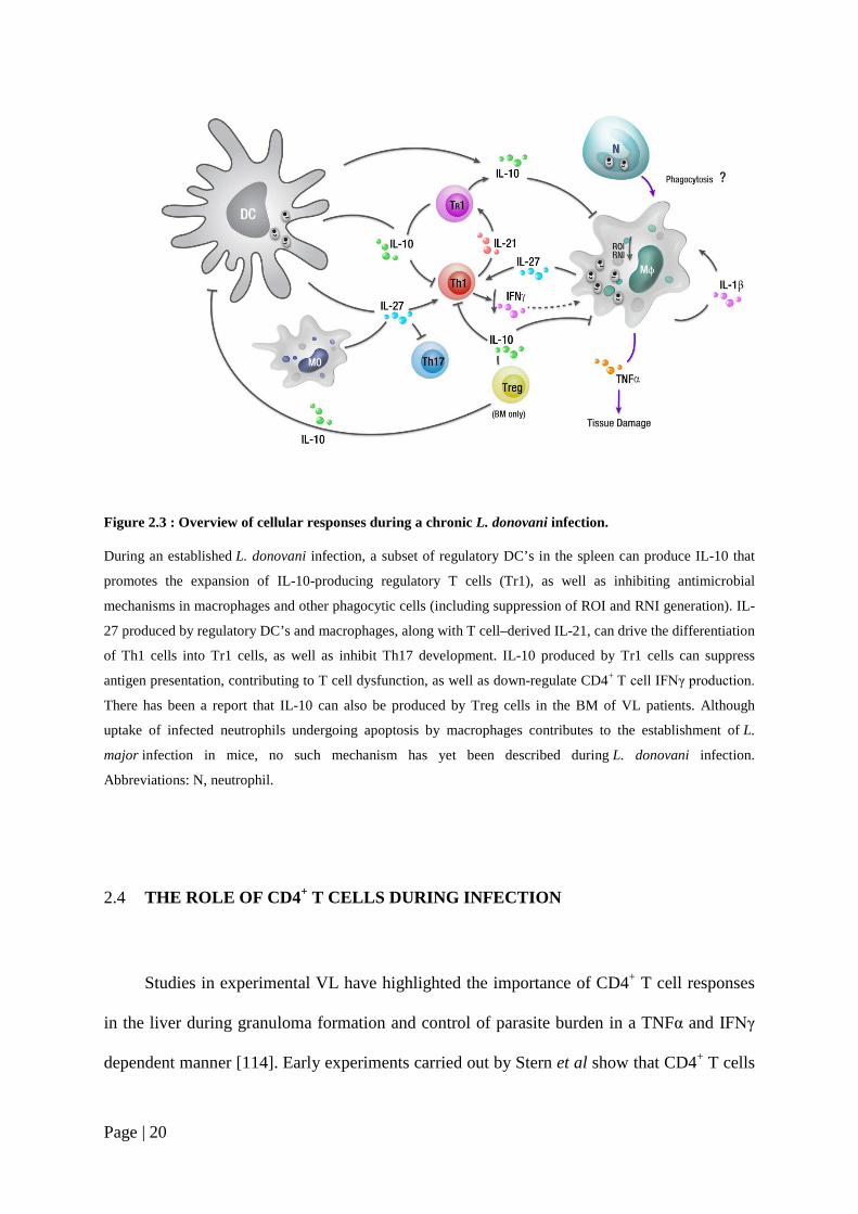
Figure 2.3 : Overview of cellular responses during a chronic L. donovani infection.
During an established L. donovani infection, a subset of regulatory DC’s in the spleen can produce IL-10 that
promotes the expansion of IL-10-producing regulatory T cells (Tr1), as well as inhibiting antimicrobial
mechanisms in macrophages and other phagocytic cells (including suppression of ROI and RNI generation). IL-
27 produced by regulatory DC’s and macrophages, along with T cell–derived IL-21, can drive the differentiation
of Th1 cells into Tr1 cells, as well as inhibit Th17 development. IL-10 produced by Tr1 cells can suppress
antigen presentation, contributing to T cell dysfunction, as well as down-regulate CD4+ T cell IFNγ production.
There has been a report that IL-10 can also be produced by Treg cells in the BM of VL patients. Although
uptake of infected neutrophils undergoing apoptosis by macrophages contributes to the establishment of L.
major infection in mice, no such mechanism has yet been described during L. donovani infection.
Abbreviations: N, neutrophil.
2.4 THE ROLE OF CD4+ T CELLS DURING INFECTION
Studies in experimental VL have highlighted the importance of CD4+ T cell responses
in the liver during granuloma formation and control of parasite burden in a TNFα and IFNγ
dependent manner [114]. Early experiments carried out by Stern et al show that CD4+ T cells

Page | 21
are critical for development of resistance against L. donovani in BALB/c mice. This study
showed that liver parasite burdens in anti-CD4 antibody treated mice were 2.6 fold higher
compared to controls [65], indicating the need for CD4+ T cells for parasite clearance.
Histological analysis of livers of mice treated with anti-CD4 antibody showed that the
granuloma formation was poorly defined, with highly infected KC surrounded by poorly
formed mononuclear cell aggregates [65]. CD8+ T cells are also required to generate an
immune response during VL [65], although they appear to play a relatively minor role.
Recent studies showed that CD8+ T cells purified from L. infantum infected mice exhibited
cytotoxic activity and expressed Th1 cytokines (IFNγ and TNFα) [115]. Another study
suggested that Leishmania parasites escape cellular responses by inducing exhaustion in
CD8+ T cells [107]. Studies by Stager et al., suggested that priming of CD8+ T cells via
vaccination can induce protection against L. donovani infection [116].
2.4.1 The activation of CD4+ T cells by DC’s
Interactions between antigen bearing DC’s and naive T cells are important for inducing
immune responses following L. donovani infection [117]. Interleukin-12 (IL-12) is a key
cytokine that plays an important role in innate and antigen-specific responses in VL [86,
118]. IL-12 activates T cells to generate IFNγ, required for the activation macrophages and
other leishmanicidal responses [119].
Neutralising IL-12 activity during L. donovani infection in BALB/c mice resulted in
delayed parasite resolution in the liver, disruption of granuloma formation and reduced IFNγ
production [86]. In the spleen, no change was observed during the first 28 days p.i. However,
Page | 22
at later time points, neutralisation of IL-12 resulted in increased parasite load, indicating that
IL-12 has no effect on early parasite replication in the spleen but is critical for the
development of immune responses required for the control of parasite burden later in the
spleen [86].
Studies carried out by Gorak et al., showed that one day after L. donovani infection,
clusters of IL-12 p40+ cells were observed in the white pulp region of the spleen. Closer
analysis of the IL-12 p40+ cells identified them as DC’s [94]. Further analysis suggested that
CD8+ DCs are the primary producers of IL-12p40, and that peak production occurred 5 hours
post infection (p.i.) in the spleen [120].
2.4.2 The role of CD4+ T cells in resolving L. donovani infection in the liver
As mentioned previously, CD4+ T cells are important for the resolution of disease
during L. donovani infection [65]. Hepatic granuloma formation is required for clearance of
parasites in the liver and is T cell dependent [121]. Early studies by Stern et al., suggested
that CD4+ T cells are the primary produces of IFNγ during an L. donovani infection. They
observed that nude mice reconstituted with immune spleen cells were able to generate
substantial IFNγ in response to mitogen and parasite antigen. Further analysis revealed that
CD4+ T cells were the major producers of IFNγ [65]. IFNγ is required for anti-leishmanicidal
activity and is vital for macrophage activation [122]. Treatment of L. donovani infected
BALB/c mice with anti-IFNγ mAb resulted in uncontrolled parasite growth and disruption of
granuloma formation [122].

Page | 23
TNFα, along with IFNγ, is crucial for macrophage activation and parasite clearance
[76, 123]. However, studies in TNFα-deficient C57BL/6 mice showed that TNFα was critical
for control of parasite growth and also contributed to granuloma assemble in the liver [76].
Interestingly, mice lacking TNFα are the only known mouse lines in which an L. donovani
infection is lethal.
IL-12, along with IFNγ and TNFα, also plays a positive role in controlling an L.
donovani infection, while Interleukin 10 (IL-10) has suppressive effects on the immune
function of T cells. In IL-10-/- mice, resolution of the L. donovani infection is achieved
quickly, compared to wild-type control mice, and treatment with anti-IL-10 receptor mAb
rapidly promotes clinical cure [104, 105].
Besides cytokines, chemokines are also important mediators of immune response
against L. donovani. Infection with L. donovani brings about a rapid T cell-independent
chemokine response followed by the amplification of this response, which is T cell-
dependent [124]. Patients with VL show high levels of CXCL9 and CXCL10 in their serum,
although the roles for these cytokines in controlling infection is not clear [125]. In the livers
of L. donovani infected mice, high levels of MIP-1, CCL2 and CXCL10 are observed [124].
MIP-1 and CCL2 produced by infected KCs are involved in attracting monocytes and
neutrophils to the liver. Resident CD4+ and CD8+ T cells in the liver promote and maintain
levels of CXCL10, which provides the stimulus for granuloma formation. Infiltrating immune
cells, along with T cells aggregate around the infected KCs to form the hepatic granuloma in
the liver [124].

Page | 24
2.4.3 Organ-specific roles for CD4+ T cells during VL
Studies carried out by Kenney et al., have shown that in the spleen of VL patients, a
mixed Th1/Th2 response is observed. Analysis of serum from these patients revealed
detectable levels of IFNγ and IL-10. Treatment with exogenous IFNγ, to assist with parasite
clearance, also resulted in increased levels of serum IL-10, suggesting an interdependent
relationship between these cytokines during disease [126]. Early studies carried out by Sacks
et al. showed that T cells taken from L. donovani infected patients were not responsive to the
Leishmania antigen and deletion of CD8+ T cells did not reverse the unresponsiveness to
parasite antigen [21]. Although our understanding of the immune mechanisms required for
resolution of infection in the liver in experimental VL is extensive, we know relatively little
about why CD4+ T cells do not control infection in humans or the spleen and bone marrow of
susceptible mice.
2.5 POST KALA-AZAR DERMAL LEISHMANIASIS AND HIV CO-INFECTION
Post kala-azar dermal leishmaniasis (PKDL) is a complication of VL characterised by
nodular, macular or a maculopapular rash on individuals who have recovered from VL [127].
PKDL appears in individuals after apparently successful VL treatment, possibly caused by
suppression of immunity in the skin to persisting parasites [128, 129]. PDKL is mainly
observed in the Indian subcontinent and East Africa, where an estimated 10-20% of cases in
India and 50-60% of cases in the Sudan progress to PKDL after VL treatment [130]. Indian
PKDL appears two to seven years, or even decades after the VL treatment, while in the Sudan
it appears earlier (six to seven months after treatment) [130]. In some cases, there may be no

Page | 25
previous history of leishmaniasis [131, 132]. PKDL cases are of epidemiological importance
because these patients can serve as parasite reservoirs [130]. PKDL is difficult to treat and
drugs used include sodium antimony gluconate (SAG), amphotericin B and miltefosine,
depending on geographical location and clinical setting. The long duration of treatment and
high drug doses required for clinical effects increases chances of drug toxicity, as well as
increasing the risk of parasites developing drug resistance [130, 133].
Immunological features of PKDL differ from VL in several ways [133]. In VL, a
suppressed CMI response is observed, which is restored on successful treatment and most
cured individuals are resistant to re-infection [134]. PDKL on the other hand arises in a
proportion of cured VL patients, due to the suppression of immune response against parasites
present in the skin [135, 136]. PKDL cases studied in Sudan show an increase in CD3+ T cell
infiltration within lesions containing Leishmania parasite or antigen, and IFNγ, IL-10 and IL-
4 are the main cytokines produced in the inflamed lesions [137]. In another Sudanese study,
Gasim et al., showed that PKDL could be predicted by assessment of IL-10, as high levels of
IL-10 were observed in plasma and keratinocytes of VL patients which developed PKDL,
compared to VL patients that did not [138]. A subsequent study by the same group also
reported a positive association between the onset of PKDL and an increase in circulating
parasite-specific PBMC, evident by the stronger parasite-specific T cell responses [139]. In
India, increased CMI responses were also observed in patients at the onset of PKDL,
compared to during chronic PKDL [140]. However, another study failed to find detectable
antigen-specific immune responses in Indian PKDL patients [141], while Ganguly et al.,
reported that CMI responses were present in Indian PKDL patients, but were dominated by
antigen-specific IL-10 production by CD8+ T cells [68]. Clearly, further studies are required
to both identify predisposing immune factors associated with PKDL development, as well as

Page | 26
to better define dysfunctional immune pathways operating during this serious disease
complication.
CD4+ CD25+ Foxp3+ regulatory T (Treg) cells are a sub-population of CD4+ T cells
involved in immune homeostasis with the potential to produce IL-10 during inflammation
[142]. Studies by Katara et al., showed that Treg cell markers and IL-10 were elevated in
tissue samples from PKDL patients, compared to tissue taken from healthy controls [143].
Furthermore, Treg cells were found to aggregate in tissue lesions of patients with PKDL,
where there was a positive association between parasite burden, certain Treg cell markers and
IL-10 levels [143]. In another recent study, elevated IL-17, IL-23 and RORγt mRNA
accumulation was found in PKDL lesions, compared with tissue after drug treatment, and this
was accompanied by increased IL-17 and IL-23 plasma levels [144]. Thus, although PKDL
is accompanied by IL-10-mediated immune suppression in many cases, the picture is not
always clear, and may also involve other deregulated inflammatory responses. Again, this is
an area requiring further investigation at the molecular and cellular level.
Recent studies have shown that secondary infections are also common in patients with
VL, possibly due to the marked immune suppression observed in infected patients [13]. L.
donovani /L. infantum co-infection with the Human Immunodeficiency Virus (HIV) has now
been recognised as a significant clinical problem [145]. A higher mortality rate has been
reported in AIDS patients co-infected with L. donovani /L. infantum, compared to other co-
infections [146]. HIV patients with VL have enhanced pro-inflammatory cytokine responses,
associated with increased HIV viral load, which can accelerate the progression from
asymptomatic HIV to AIDS [147, 148]. In addition, parasite multiplication promotes the
Page | 27
survival, proliferation and elevated levels of cellular dNTP in human monocytes which can
also accelerate HIV replication [149, 150]. Treatment of VL in HIV patients involves the use
of standard drugs, but due to the enhanced immune suppression in HIV patients and the
partial reliance on host immune mechanisms for drug efficacy [128], these treatment
strategies are often inadequate, and in most cases, patients are unresponsive to drug treatment
[128]. Furthermore, those patients who do respond to treatment often relapse, possibly due to
the low CD4+ T cells numbers associated with HIV infection and parasites persisting after
drug treatment [151]. Host T cells responses are abrogated in HIV infection, including
skewing away from Th1 responses [152] which are required for effective leishmanicidal
responses [65]. Wolday et al. showed that PBMCs from Leishmania /HIV co-infected
individuals produce low levels of IL-12 and IFNγ, and higher levels of IL-4 and IL-10
following stimulation with parasite antigen [153]. IL-15 is involved in promoting and
maintaining Th1 responses, and was also decreased in plasma from patients with Leishmania
/HIV co-infection [154]. Hence, as might be predicted, these results suggest that HIV
infection suppresses anti-parasitic Th1 immune response required for parasite clearance
[151].
2.6 PAST AND CURRENT TREATMENT OPTIONS
Prompt detection and treatment of VL is critical to achieve cure and prevent
transmission of disease. Left untreated, VL results in increased rates of morbidity and
mortality in endemic regions of the world [155].

Page | 28
2.6.1 Current anti-leshmania drugs available for the treatment of VL
The choice of treatments against VL is largely based on the region where the infection
was acquired, the local responses to treatment and the treatment resistance pattern [156].
Pentavalent antimonials, such as sodium stibogluconate, pentostam, meglumine antimonite
and glucantime, have been the mainstay of antimonial therapy for the last 60 years [157].
However, there is now considerable parasite resistance against these drugs, especially in
North-Eastern India and surrounding areas [3]. Therefore, although these drugs are still
employed to treat VL in Africa, drugs such as Amphotericin B, Miltefosine, aminosidine
(paromomycin) and sitamaquine have been developed as effective treatments against VL in
areas of antimonial drug resistance [3]. However, these drugs are still far from ideal because
of cost, toxicity, development of parasite drug resistance after prolonged use and the duration
of treatment [4]. Although some progress has been made recently in addressing this latter
issue [158], where a single dose of lipid formulation of Amphotericin B (Ambisome), was
effective in treating VL patients with lower toxicity outcomes compared to the conventional
drug treatment. Table 1 list the current anti-leishmania drugs available to treat VL.
Combination drug therapy involving administration of two anti-leishmania drugs is a
new therapeutic approach which is being tested as a way to increase efficacy, reduce
development of parasite resistance and reduce treatment duration and cost [159]. A recent
study by Sunder et al. in a cohort of 613 patients, indicated that combination of various
antimonals were more effective and less toxic than treatment with amphotericin B alone
[158]. Other interesting approaches currently being studied are targeted therapy and
immunotherapy. Targeted therapies involves blocking of essential biochemical or signalling

Page | 29
pathways that are required for Leishmania parasite growth and survival, while,
immunotherapy encompasses the use of biological molecules or compounds such as cytokine
therapy [160], alone or in combination with anti-leishmania drugs to modify immune
responses [161].
Each VL case signifies our failure to prevent leishmaniasis, and each death our failure
to treat the disease soon enough. Until disease eradication has been achieved, chemotherapy
will remain critical to prevent morbidity and mortality caused by VL. There is an urgent need
for the development of alternative and effective therapies against VL. The development of
compounds or agents that selectively inhibit key cellular pathways that
promote Leishmania parasite growth and maintenance has opened a new avenue for anti-
leishmania therapy. The use of targeted therapy and immunotherapy could address current
challenges associated with treatment and management of VL by reducing the cost of therapy,
development of drug resistance to currently available chemotherapies and decreasing the dose
or length of treatment. Therefore, a major component of this thesis is dedicated to
investigating the effect of targeted immunotherapy on the outcome of experimental VL.

Page | 30
Drugs Dosage and route of
administration Mode of action
Efficacy of
drug (%) Advantages Limitations References
Amphotericin B
0.75-1.0 mg/kg; 15-20 i.v.
infusions given daily or
every alternate day
Forms complexes with the parasite by
binding to membrane ergosterol
resulting in increased membrane
permeability, which alters the ion
balance, resulting in parasite death.
> 95% Effective in antimony
drug resistant regions
Costly, toxic and
requires longer the
duration of treatment
[162, 163]
Liposomal
Amphotericin B
(Ambisome)
10 mg/kg i.v.as a single
dose OR 3 mg/kg/day i.v.
for 5 days
Mechanism of action is similar to
Amphotericin B, although delivery of
the drug is directly to infected
macrophages.
> 96%
Extremely effective;
Shorter treatment
duration; No
documented resistance
Costly [164]
Miltefosine
50-100 mg/day for adults
depending on weight. Oral
drug.
Interacts with the surface membrane of
Leishmania parasite. Parasite death is
mediated by apoptosis
85-95%
It’s an oral drug.
Primary treatment in
the Indian subcontinent
Costly and toxic [163, 165]
Paromomycin
(a.k.a. animosidine)
11-15 mg/kg daily
intramuscularly (i.m.) for
21 days
Induces reduction in parasite
mitochondrial membrane potential and
inhibits protein synthesis
95%
Acts co-actively with
other antimonials and is
a low-cost drug for VL
Toxic and in-effect in
East Africa [166-168]
Pentavalent
antimonials
20 mg/kg/day (i.m.) for 20-
30 days
The drug is converted into the active
trivalent form in the amastigote/
macrophage vacuole and exposes the
parasite to oxidative stress of the host
cell
80-90% Low-cost drug Toxic and increased
drug resistance [169, 170]
Table 1: Current anti-leishmanial drugs against VL.
Adapted from a review by S. Sundar and colleagues [161].
Page | 31
2.6.2 Vaccines against VL
At present there is no effective VL vaccine for humans. The development of a vaccine
to prevent leishmaniasis has been a long-term goal for researchers in the field. One of the
major hurdles for developing vaccines to either prevent or treat VL, has been a limited
understanding of the precise immune mechanisms required for controlling parasite growth
without causing disease. Because of the intrusive techniques required to analyse tissue in VL
patients, our current understanding of the host immune response during VL is largely derived
from studies performed in L. donovani-infected, genetically susceptible mice.
Past studies suggest the use of whole parasite vaccines as an approach to control VL.
The process of “leishmanisation” involves the deliberate infection of people with cutaneous
leishmaniais (CL) causing parasite species on unexposed areas of the body to establish an
infection that is controlled in most individuals, resulting in long-term protection [6]. This
technique was practised for centuries throughout the Middle East and parts of Asia, and
large-scale trials were carried out in the former Soviet Union and Israel with some success
[171, 172], as long as the parasites used were viable and infective [173]. However, despite
the solid immunity that develops in most individuals, this approach has largely been
abandoned as some individuals developed complications such as large skin lesions,
exacerbation of skin diseases and poor responses to vaccines [7, 8]. To avoid this, strategies
have been shifted to attenuated or killed whole parasite vaccines. These types of vaccines are
non-pathogenic and non-replicative but have the ability to generate anti-parasitic immune
responses in immunised hosts.

Page | 32
Studies have already shown that γ-irradiated whole Leishmania parasite can be used as
a vaccine against experimental CL [174]. In a recent study carried out by Datta et al.,
prophylactic and protective immune responses were generated in mice immunised
intramuscularly (i.m.) with radio-attenuated L. donovani parasites [175, 176]. They showed
that in immunised mice, there was a higher Th1 cytokine response and suppressed Th2
responses, associated with lower parasite burdens, relative to control animals. These results
were indicative that whole attenuated parasites can be used as effective vaccines.
For the effective control of VL in endemic areas, a vaccine is deemed critical.
However, vaccine development faces several challenges either due to host evasive
mechanisms of parasites or heterogeneity in the human population resulting in poor immune
responses following vaccination. A key step in vaccine design is the identification of suitable
vaccine candidates, which stimulates a host protective immune response. Hence, another
component of this thesis is dedicated to investigating the generation of a whole parasite
vaccine to protect against experimental VL using different forms of attenuation in the
presence or absence of adjuvants.

Page | 33
Chapter 3: Materials and Methods
3.1 MICE AND PARASITES
3.1.1 Mice
Female C57BL/6 were purchased from the Animal Resources Centre (Canning Vale,
Western Australia) and the Walter and Eliza Hall Institute for Medical Research (Victoria,
Australia), and maintained under conventional conditions at the QIMR Berghofer Medical
Research Institute (QIMR Berghofer). B6.FoxP3-GFP-DTR [177], B6.NKp46Cre.iDTR
[178] and B6.RAG1-/- mice [179] were bred at the QIMR Berghofer. Unless otherwise stated,
6- to 10-week-old males and female, sex- and age-matched mice were used for all
experiments. All animal procedures were approved by the QIMR Berghofer Animal Ethics
Committee. This work was conducted under QUT Ethics Approval Number 1200000384 and
QIMR Berghofer animal ethics approval number A02-634M, in accordance with the
“Australian Code of Practice for the Care and Use of Animals for Scientific Purposes”
(Australian National Health and Medical Research Council).
3.1.2 Parasites
L. donovani (LV9) parasites were maintained by passage in B6.RAG 1-/- mice. Briefly,
B6.RAG 1-/- mice were infected intravenously (i.v.) via the lateral tail vein with 2x10⁷
amastigotes in 200 µL RPMI 1640 (QIMR Berghofer). Following the establishment of

Page | 34
chronic infection (approximately two months post infection), mice were killed by CO2
asphyxiation and amastigotes were isolated from the spleens (see 3.1.3) to set-up infections.
3.1.3 Isolation of parasite for infection of mice
Amastigotes were harvested from infected B6.RAG 1-/- mice spleens. The isolated
spleens were homogenised to a single cell suspension in RPMI 1640, supplemented with 100
µg/mL streptomycin (Invitrogen, VIC, Australia) and 100 U/mL penicillin (Invitrogen)
(RPMI/PS) and centrifuged at 128 x g (Eppendorf Centrifuge 5810R) at room temperature
(RT) for 5 minutes to remove cellular debris. The retained supernatant was centrifuged at
1800 x g at RT for 15 minutes and the pellet retained. Red blood cells (RBCs) were lysed
with RBC Lysing Buffer (Sigma-Aldrich, Castle Hill, Australia), as per the manufacturer’s
instructions. Amastigotes were pelleted by centrifugation at 1800 x g at RT for 15 minutes.
The supernatant was discarded and the pellet washed two more times by resuspending in
RPMI/PS and centrifuging at 1800 x g at RT for 15 minutes. Amastigotes were finally
resuspended in 10 mL of RPMI/PS. The amastigote suspension was passed through a blunt
26-gauge needle (Terumo Corporation, Tokyo, Japan) 10 times, to ensure lysis of all host
cells and to reduce clumping of parasites. Amastigotes were counted using the Helber
bacteria counting chamber (Weber Scientific International Ltd, Teddington, UK). Mice were
infected by injecting 2x10⁷ amastigotes i.v. via the lateral tail vein. In experiments examining
low dose infections, mice were infected with 5x10⁶ amastigotes i.v.

Page | 35
3.1.4 In-vitro culturing of L. donovani promastigotes
L. donovani amastigotes were isolated as described (in section 3.1.3). The amastigotes
were cultured in promastigote growth medium (cM199) consisting of M199 (Sigma-Aldrich)
supplemented with 20% (v/v) heat-inactivated fetal bovine serum (FBS) (Life Technologies
Australia Pty Ltd, VIC, Australia), 100 U/mL penicillin, 100 μg/mL streptomycin, 2 mM L-
glutamine, 40 mM HEPES (QIMR Berghofer), 0.1 mM adenosine (Sigma-Aldrich) (in 50
mM HEPES), 5 mg/mL hemin (Sigma-Aldrich) (in 50% triethanolamine (Sigma-Aldrich)),
and 1 mg/mL 6-biotin (Sigma-Aldrich) for 21 day at 27°C in a 5% (v/v) CO2 incubator, with
media topped up 10 days after initiating cultures. 21 days post-culture, promastigotes were
pelleted by centrifugation at 1800 x g at RT for 15 minutes. The supernatant was discarded
and the pellet washed two more times by resuspending in RPMI 1640 and centrifuging at
1800 x g at RT for 15 minutes. Promastigotes were finally resuspended in 10 mL of RPMI
1640 and passed through a blunt 26-gauge needle 10 times to reduce clumping of parasites.
Promastigotes were counted using the Helber bacteria counting chamber as described for
amastigotes above. Parasites were either irradiated or chemically-attenuated (see section 3.1.5
and 3.1.6) and injected into mice.
3.1.5 Irradiation of L. donovani parasites
Irradiation of L. donovani (LV9) parasites (amastigotes or cultured promatigotes) was
carried out by gamma irradiation (Gammacell® 40 Exactor Low Dose-Rate Research
Irradiator (Best Theratronics) with a Cesium-137 source (1.1 Gy/minute) housed at QIMR
Berghofer). LV9 parasites were irradiated at 500 (10 hrs), 1000 (20 hrs) and 1500 (30 hrs)

Page | 36
Gy, after which parasites were washed twice in RPMI/PS, counted, and the immunizing dose
calculated. Mice were immunized i.v. or intraperitoneally (i.p.) with the required number of
parasite (as described in Chapter 6).
3.1.6 Chemical-attenuation of L. donovani parasites
Chemical-attenuation of L. donovani (LV9) parasites (amastigotes or cultured
promatigotes) was carried out using TH-III-149 (tafuramycin A: TFA) (kindly provided by
Dr. Michael Good), as previously described [180]. TFA was dissolved in DMSO (Sigma-
Aldrich) at 10,000× the desired attenuating concentration. Stock solutions of PET
(polyethylene glycol 400, absolute ethanol, and Tween 80 in 6:3:1 proportions) (Sigma-
Aldrich) and 5% (w/v) glucose (Sigma-Aldrich) were made and mixed 1:2. The TFA/DMSO
solution was then added at 10% volume to the PET/glucose mix. TFA was diluted to a 20 μM
final concentration in RPMI 1640 and then L. donovani parasites were added in RPMI 1640
to give a final concentration of 2 μM. The mixture was then incubated at 37°C in a 5% (v/v)
CO2 incubator for 40 minutes with frequent agitation/rocking. The parasites were washed and
resuspended in RPMI 1640 and incubated at 37°C in a 5% (v/v) CO2 incubator for 20
minutes. The parasites were washed once with RPMI 1640, counted and the immunizing dose
was calculated. Mice were immunized i.v. with the required number of parasite (as described
in Chapter 6).

Page | 37
3.1.7 Adjuvants used for immunization
The adjuvants CpG ODN sequence 1826 (TCCATGACGTTCCTGACGTT) and
control sequence 1982 (TCCAGGACTTCTCTCAGGTT) were purchased from Sigma-
Aldrich. Poly I:C was purchased from Integrated Sciences Ltd. (NSW, Australia). Mice were
injected i.v. with 50 μg CpG ODN 1826, 50 μg CpG ODN 1982 or 50 μg Poly I:C (as
described in Chapter 6).
The immunization regime and parasite number used are described in Chapter 6. Age
matched control (AMC) mice were infected with 2x10⁷ amastigotes i.v. on the day indicated
and housed in similar conditions as the immunized mice.
3.2 SAMPLE COLLECTION
At the times indicated in the text, mice were sacrificed by CO2 asphyxiation. Body and
organ weights were recorded for each animal in grams (g).
3.2.1 Collection of blood for serum isolation
Mice were bled via cardiac puncture and the blood was collected in Minicollect
Lithium heparin tubes (Greiner Bio-One, Austria). Blood serum was obtained by centrifuging
the Minicollect tubes at 775 x g (Eppendorf Centrifuge 5418) at RT for 10 minutes and the

Page | 38
upper layer of serum was transferred into a 96 well round bottom plate (Sigma-Aldrich) and
stored at -20oC until required.
3.2.2 Collection of animal organs
After mice were bled, the spleen and livers were removed from each animal and
weights were recorded in grams (g). Spleens removed from the animals were collected in 10
mL of RPMI/PS. Livers were perfused via the hepatic portal vein using 10 ml perfusion
buffer (1X PBS (QIMR Berghofer)). Perfused livers were collected in 5 mL of 2% (v/v) FBS-
supplemented PBS.
3.2.3 Assessment of parasite burdens
Parasite burdens were determined from impression smears of spleens and livers.
Briefly, a section of the spleen and hepatic lobes were taken and blotted on filter paper
(Whatman, UK) to remove excess blood and then pressed onto a glass microscope slide
(Universal Choice Wholesaler, NSW, Australia) to produce multiple impression smears. The
smears were air dried and stained with Diff-Quik (Lab Aids, Narrabeen, Australia), according
to manufacturer’s instructions. Parasite burden was expressed as Leishman Donovan Units
(LDU), calculated by multiplying the number of parasites per 1,000 host nuclei by the organ
weight in grams [73].
LDU= no. of parasites per 1000 nuclei x organ weight (g)

Page | 39
Spleen parasite burden was also determined by limiting-dilution analysis [181]. Briefly,
Bacto beef (5 g; BD Biosciences, NSW, Australia), Neopeptone (2 g; BD Biosciences) Agar
(2 g; BD Biosciences) and NaCl (0.5 g; Sigma-Aldrich) were dissolved in 100 mL of double-
distilled H2O and autoclaved for 30 minutes. Agar heated to 56°C was dispensed into a flat-
bottom, 96-well plate (50 μl per well) (Sigma-Aldrich). Agar plates were allowed to set at a
45° angle and were stored at 4°C before use. Three hours before the addition of spleen
mononuclear cells (MNCs), the plates were placed at RT. Splenic mononuclear cells (MNCs)
were isolated, as described in Section 3.3.2. Single-cell suspensions were diluted 1/10 in
RPMI 1640 media, supplemented with 20% (v/v) heat-inactivated FBS, 100 U/mL penicillin,
100 μg/mL streptomycin, 2 mM L-glutamine, 40 mM HEPES and 10 µM Hemin (in 50%
triethanolamine). Serial dilutions of cell suspensions were made, and 100 μL was added to
each of 16 replicate wells. The plates were cultured in a humidified, 5% (v/v) CO2 incubator
at 25°C to 26°C for 9 days. The number of wells negative for parasite growth was then scored
using an inverted microscope. The logarithm of the fraction of negative wells was plotted
against the number of host cells plated at each serial dilution. The equation of the best-fit line
was generated by a χ2 minimization method, using Statistical Analysis System (SAS)
software. The cell dilution yielding a fraction of negative wells for parasite growth gives an
estimate of the reciprocal parasite frequency.

Page | 40
3.3 CELL ISOLATION AND PREPARATION
3.3.1 Hepatic Mononuclear Cell (MNC) preparation
Perfused livers collected in 5 mL of 2% (v/v) FBS-supplemented PBS (wash buffer)
were passed through a 200 µM metal mesh (Sefar Pty Ltd, QLD, Australia), placed in a tea
sieve. Cell suspension was washed twice in wash buffer by centrifugation at 338 x g for 6
minutes at RT. The cell pellet was resuspended in a 33% (v/v) Percoll (GE Healthcare
Australia Pty. Ltd., NSW, Australia) density gradient and centrifuged at 578 x g for 15
minutes (no brake). The cell pellet obtained was suspended in RBC lysing buffer and RBC
removed, as per manufacturer’s instructions. Cells were washed twice and resuspended in 5
mL RPMI/PS. Cells were counted using a haemocytometer.
3.3.2 Splenic MNC preparation
Collected spleens were passed through 100 µM mesh sieve (Corning, USA) (a single
sieve for each spleen). Cells were washed once in RPMI/PS by centrifugation at 338 x g for 6
minutes at 4oC. RBCs were lysed using RBC lysing buffer, according to the manufacturer’s
instructions. Cells were washed once and resuspended in 10 mL RPMI/PS. Cells were
counted using a haemocytometer.

Page | 41
3.3.3 Isolation of peritoneal macrophages
Peritoneal macrophages were collected by conducting a peritoneal lavage. Briefly, mice
were euthanized by CO2 asphyxiation. Using scissors and forceps the outer skin of the
peritoneum was cut and pulled back to expose the peritoneum lining the peritoneal cavity. 5
mL of ice cold D‐PBS (1X) (Sigma-Aldrich) was injected into the peritoneal cavity using 27g
needle. After injection, the peritoneum was massaged. The fluid containing peritoneal cells
was then collected using a 5 mL syringe and 25g needle. Cells collected were washed with
cold D‐PBS and resuspended in cold, complete DMEM (cDMEM), consisting of DMEM
(Sigma-Aldrich) supplemented with 10% (v/v) heat-inactivated FBS, 10 mM L‐glutamine
(Sigma-Aldrich), 100 U/mL penicillin and 100 μg/mL streptomycin. Cells were counted
using a haemocytometer.
3.3.4 Magnetic cell sorting (MACS) purification of DC’s
Mouse spleens were processed to obtain a single-cell suspension as mentioned
previously (see 3.3.2). CD11c+ DC were positively selected from single-cell suspensions
using magnetic-activated cell sorting (MACS). 108 cells from each sample were incubated in
400 µL MACS buffer (PBS, 1% BSA (Sigma-Aldrich), 5mM EDTA (Sigma-Aldrich)) with
100 µL metallo-conjugated anti-mouse anti-CD11c beads (N418) (Miltenyi Biotech,
Germany) for 15 minutes in the refrigerator (4-8°C). Cells were washed by adding 1-2 mL
MACS buffer per 107 cells and centrifuged at 338 x g for 7 minutes at 4oC. Supernatant was
discarded and cells were resuspended in ice-cold MACS buffer and passed through a 50 µM
sieve placed on a pre-wet (0.5 mL MACS buffer) MS MACS column (Miltenyi Biotec)

Page | 42
attached to a magnetic stand. The column was washed three times with 0.5 mL ice-cold
MACS buffer, removed from the magnet and positively stained cells were flushed from the
column into a collection tube using 1 mL of MACS buffer. The volume was made up to 10
mL with RPMI/PS and cells washed. Enriched DCs (30 - 40% purity checked by FACS) were
resuspended in complete RPMI (5% cRPMI), consisting of RPMI 1640 supplemented with
5% (v/v) heat-inactivated FBS, 10 mM L‐glutamine, 100 U/mL penicillin and 100 μg/mL
streptomycin. Cells were counted using a haemocytometer.
3.4 ANTIBODIES AND DRUGS FOR IN-VIVO ADMINISTRATION
3.4.1 In-vitro culturing of monoclonal antibodies
All hybridomas (anti-GITR [DTA-1], anti-IL-10R [1B1.3a], anti-IL-2 [S4B6 and JES6-
1A12] and anti-NK1.1 [PK136] mAb) were grown in-house at 37˚C in a 5% (v/v) CO2
incubator. Cell culture media consisted of RPMI 1640 supplemented with 5% (v/v) heat-
inactivated FBS, 200 µM HEPES, 2mM sodium pyruvate (Sigma-Aldrich), 100 µg/mL
streptomycin and 100 U/mL penicillin. Purified mAbs were prepared from culture
supernatants by protein G column purification (GE Healthcare Australia Pty. Ltd.), followed
by endotoxin removal (Mustang membranes, East Hills, NY). The concentration of purified
mAbs was then measured by spectrophotometry, resuspended at 2 mg/mL in 0.9% sodium
chloride (Baxter, NSW, Australia) and stored at -20˚C.

Page | 43
Anti-CD4 (GK 1.5), anti-CD8 beta (β) (53-5.8), anti-CTLA-4 (UC10-4F10-11) and
control Rat IgG mAb were purchased from BioXCell, (West Lebanon, NH, USA).
3.4.2 Intra-peritoneal administration of antibodies
All antibodies were administered i.p. by holding the mouse at a 45o angle to avoid
injuring vital organs in the peritoneal cavity. A 27-gauge insulin syringe (Terumo
Corporation) was used. For in vivo stimulation of GITR, mice were injected i.p. with 0.5 mg
anti-GITR mAb in 200 μl 0.9% sodium chloride per mouse on day 14 or day 28 post-
infection. Anti-IL-10R and anti-CTLA-4 mAbs were administered initially in 0.1mg doses
and later in 0.5 mg doses, via i.p. injection in 200 μl 0.9% sodium chloride per mouse on
days 28, 30 and 33 post infection or days 14, 19 and 24 post-infection. Anti-CD4 and anti-
CD8β mAb was administered in 0.5 mg doses, via i.p. injection in 200 μl 0.9% sodium
chloride per mouse two days prior to infection/challenge (see chapter 6), and every three days
post infection/challenge until the end of the experiment. Control mice were administered the
same quantities of control rat IgG (0.5 mg) mAb at the same time points as the stimulatory,
depleting or blocking mAbs.
3.4.3 Preparation and administration of IL-2/anti–IL-2 complexes
One and a half µg of recombinant murine IL-2 (eBioscience, San Diego, CA) was
incubated with 50 µg of either S4B6 or JES6-1A12 mAb in saline, for 30 minutes at 37°C

Page | 44
prior to i.p. administration (see 3.4.2) into each mouse in a volume of 200 µL on days 14 and
21 post infection.
3.4.4 Preparation and administration of pentavalent antimonial drug
The pentavalent antimonial, sodium stibogluconate (Sbv; Albert David Ltd, Kolkata,
India), was administered i.p. at 500 mg/kg/day doses in 200 μL 0.9% sodium chloride per
mouse on days 14 and 21 post-infection. In multiple-dosing experiments, mice were injected
i.p. on days 14, 16, 18, 20, 22, 24 and 26 post-infection with Sbv at 50 mg/kg/day doses in
200 μL 0.9% sodium chloride per mouse. The dose of Sbv used was based on drug dosing
used by others [182].
3.4.5 Preparation and in- vivo administration of Diphtheria toxin
Diphtheria toxin (DT) was purchased from Sigma-Aldrich, diluted in saline and stored
at 4°C until required. Treg cells and NK cells were depleted in B6.FoxP3-GFP-DTR and
B6.NKp46Cre.iDTR mice, respectively, by i.p. injection of DT at 8 ng/g body weight in 200
μl 0.9% sodium chloride, two days prior to infection and every three days post infection until
the end of the experiment.

Page | 45
3.5 EXPERIMENTAL METHODS
3.5.1 In-vitro infection of Macrophages
Isolated peritoneal macrophages (see 3.3.3.) were seeded (5x10⁵ cells) in 16-well, glass
chamber slides (Lab‐Tek, Rochester, NY) and incubated at 37°C in a 5% (v/v) CO2 incubator
for 24 hours. After 24 hours, non‐adherent cells were removed and adherent cells washed
with DMEM and infected with LV9 amastigotes at a multiplicity of infection (MOI) of 10:1
(in 200 μL DMEM). After incubation at 37°C in a 5% (v/v) CO2 incubator for one hour, free
amastigotes were washed off with RT DMEM and cells were cultured for another 24 hours at
37°C in a 5% (v/v) CO2 incubator, in the presence or absence of exogenous IFNγ (10 ng/mL)
(Bio-Scientific Pty. Ltd., NSW, Australia). On the following day, 50 µL of the culture
supernatant was collected for measurement of nitrite levels using the Griess assay and 100 µL
for cytokine measurement. Plates were washed twice with pre-warmed (at 37oC in a water
bath) D-PBS. Glass chambers were stripped and slides were fixed and stained using DIFF-
QUICK. Percentage infectivity was calculated as number of parasites per 100 host
macrophages.
3.5.2 Detection of nitrite using Griess assay
The Griess method was used to assess nitrite (NO2-) production in the supernatant of
cultured infected macrophages (see 3.5.1). The NO2- levels were estimated by reducing the
nitrate to nitrite with nitrate reductase and measuring the nitrite concentration

Page | 46
calorimetrically. Briefly, Griess reagent (2% Sulphanilamide and 0.2%
Napthylethylenediamide in 5% Phosphoric acid) (Sigma-Aldrich) was added to cell
supernatant (1:1 with sample) in a 96-well plate, along with the medium blank, and
absorbance was measured at 540 nm following incubation for 25 minutes at RT. The amount
of endogenous nitrite accumulated was calculated from a standard curve constructed with
different concentrations of sodium nitrite.
3.5.3 DC activation assay
Isolated DCs (see 3.3.4) were seeded (5x10⁴ cells) in 96 well round-bottom plates and
cultured with chemically-attenuated LV9 promastigotes at a MOI of 1:10 (in 200 μL cRPMI)
in the presence or absence of adjuvants CpG ODN 1826 and control CpG ODN 1982 (3
μg/mL) or Poly (I:C) (50 μg/mL). Plates were incubated at 37°C in a 5% (v/v) CO2 incubator
for 24 hours. On the following day 100 μL of culture supernatant was collected for
measurement of cytokine levels.
3.5.4 Antigen-specific cellular analysis
Mouse splenic MNCs (see 3.3.2.) were diluted to 2x10⁶ cells/mL in 5% cRPMI and
aliquoted in 96 well plates at 1x10⁵ cells/well. Cells were stimulated with fixed L. donovani
amastigotes at 2x10⁶ parasites/well. Briefly, fixed amastigotes were prepared by incubating
the parasites in 4% (w/v) paraformaldehyde (PFA; MP Biomedicals, CA, USA) for 30
minutes and washed twice with RPMI/PS. These paraformaldehyde-treated parasites were

Page | 47
then resuspended in cRPMI at 4x10⁷ parasites/mL before culturing. Cells were cultured in the
presence of parasite antigen (triplicate wells were cultured; total volume in each well was 200
µL) for a period of 24 and 72 hours at 37oC in a 5% (v/v) CO2 incubator. Intracellular
cytokine staining was performed on cultured cells at these time points and culture
supernatants were harvested for measurement of cytokine levels.
3.5.5 Fluorescence activated cell sorting (FACS) analysis of cell surface markers
Hepatic and splenic MNCs (100 µL from cell suspensions) were added to 96 well plates
and centrifuged at 338 x g for 3 minutes at 4oC (wash) once in cold FACS buffer (PBS,
0.02% (v/v) FBS). Pelleted cells were resuspended in blocking buffer (TruStain fcX™
antibody, BioLegend, CA, USA) and incubated for 15 minutes on ice. Cells were washed as
above. Dead cells were excluded from the analysis, by resuspending the pellet in 50 µL
LIVE/DEAD Fixable Aqua Stain (Invitrogen-Molecular Probes, CA, USA), and incubating at
RT for 15 minutes (protected from light). Cells were washed twice as above and the pellet
was resuspended in 50 µL of the antibody master mix (antibodies diluted in cold FACS
buffer) (Table 3.1 List of antibodies used for fluorescence-activated cell sorting (FACS)) and
incubated for 30 mins on ice (protected from light). The plate was then washed and cell
pellets were fixed in 1% PFA and stored at 4°C prior to acquisition on the LSRFortessa (BD
Biosciences). Data were analysed with FlowJo software (TreeStar, OR, USA).

Page | 48
3.5.6 FACS analysis of intracellular cytokines and transcription factors
Ex-vivo cytokine production was measured by stimulating hepatic and splenic MNCs
(100 µL from cell suspensions in 96 well plates) in 10% RPMI/PS supplemented with 10%
(v/v) heat-inactivated FBS, with phorbol 12‐myristate 13‐acetate (PMA, 25 ng/mL) (Sigma-
Aldrich) and Ionomycin (500 ng/mL) (Sigma-Aldrich) in the presence of Brefeldin A (10
mg/mL) (Sigma-Aldrich) for 3 hours at 37˚C in a 5% (v/v) CO2 incubator. The cells were
pelleted and supernatant discarded. Prior to intracellular and transcription factor staining
MNCs were stained for cell surface markers, as previously described (see 3.5.5). Cells were
then permeabilized using the Transcription Factor Staining Buffer Set (eBioscience, CA,
USA), used according to the manufacturer’s instructions. Briefly, fixation/permeabilization
working solution was made by diluting fixation/permeabilization concentrate (1 part) with
fixation/permeabilization diluent (3 parts). Working solution (100 µL) was added to each
sample and left to incubate for 30 mins on ice (protected from light). Cells were washed
twice with 200 µL permeabilization buffer (1X working solution of permeabilization buffer
by diluting the 10X concentrate with distilled water) and the pellet was resuspended in 50 µL
of the antibody master mix (intracellular and transcription factor antibodies diluted in cold
permeabilization buffer) and incubated for 45 - 60 mins on ice (protected from light). The
plate was then washed again and cell pellets were fixed in 1% PFA and stored at 4°C prior to
acquisition on the LSRFortessa. Data were analysed with FlowJo software.

Page | 49
Antigen Identifies Conjugate Clone Name Manufacturer CD11a Activated CD4+ T cells FITC M1714 Biolegend
CD122 IL-2Rβ PE 5H4 Biolegend CD132 IL-2Rγc Biotin TUGm2 BD Biosciences CD25 Tregs PE-Cy 7 PC61 Biolegend CD4 CD4+ T cells BV 605 GK1.5 BD Horizon
APC GK1.5 Biolegend PerCP-Cy 5.5 RM4-5 Biolegend CD49d Activated CD4+ T cells PE-Cy 7 R1-2 Biolegend
Biotin R1-2 Biolegend CD8α CD8+ T cells AF 700 53-6.7 Biolegend
Foxp3 Tregs FITC 150D Biolegend APC MF14 Biolegend IFNγ Protein PE XMG1.2 Biolegend APC XMG1.2 Biolegend BV 421 XMG1.2 Biolegend IL-10 Protein PE JES5-16E3 Biolegend APC JES5-16E3 Biolegend KLRG-1 Terminally
differentiated cells PerCP-Cy 5.5 2F1 eBioscience
NK1.1 NK cells BV 605 PK136 BD Horizon APC PK136 Biolegend T-Bet Th1 cells APC eBio4B10 eBioscience PE-Cy 7 eBio4B10 eBioscience TCRβ T cells BV 421 H57-597 Biolegend PerCP-Cy 5.5 H57-597 Biolegend TNFα Protein PE MP6-XT22 Biolegend
Table 2 : A summary of the fluorophore-conjugated antibodies used for surface and intracellular staining
3.5.7 Measurement of cytokines in serum and cell culture supernatants

Page | 50
Serum and tissue culture supernatants were assessed for the presence of soluble
cytokines using the Cytometric Bead Array Flex Sets (CBA) (BD Biosciences) system and
the HTS system plate reader on the LSRFortessa, as per manufactures instructions. Cytokine
quantification was determined using FCAP Array Version 3 software for Windows (BD
Biosciences).
3.6 STATISTICAL ANALYSIS
Comparisons between two groups were performed using non-parametric Mann-
Whitney tests in mouse studies. Comparisons between multiple groups were made using a
Kruskal-Wallis test and corrected using Dunn’s multiple comparisons test. Differences of p<
0.05 was considered significant (p<0.05 = *; p<0.01=**; p<0.001=***; P<0.0001=****).
Graphs depict mean values ± SEM. All statistical analyses were performed using GraphPad
Prism 6 software for Windows (GraphPad, CA, USA).

Page | 51
Chapter 4: Testing whether promoting parasite-specific CD4+ T cell function via GITR activation improves the outcome of experimental VL
4.1 INTRODUCTION
Combining different anti-parasitic drugs is one way to improve treatment of VL. This
approach aims to increase drug efficacy, as well as reduce drug toxicity, parasite resistance
and treatment duration and cost [183]. In a recent study carried out by Sunder et al. in a
cohort of 613 patients, of whom 146 were treated with amphotericin B, 156 with liposomal
amphotericin B and miltefosine, 154 with liposomal amphotericin B and paromomycin, and
157 with miltefosine and paromomycin, the combination treatments were more effective, less
toxic and better tolerated than treatment with amphotericin B alone [184]. These results
demonstrate that combination drug therapy were safer and more effective treatment options
for VL [184].
A potential extension of this work could involve combining immune therapy with
conventional anti-parasitic drug treatment to optimise control of chronic disease and better
protect against re-infection. VL is associated with suboptimal, anti-parasitic CD4+ T cell
responses [13, 185]. Experimental studies in VL have emphasized the importance of CD4+ T
cell responses in the liver for granuloma formation and control of parasite burden in a TNFα
and IFNγ dependent manner [186]. Glucocorticoid-induced TNFα receptor family-related
protein (GITR) is expressed at low levels by many immune cells, but is highly expressed on

Page | 52
Foxp3+ Treg cells, and is also up-regulated on conventional CD4+ and CD8+ T cells
following activation [187]. A previous study reported that activation of CD4+ T cells during
an established experimental L. donovani infection with a stimulatory antibody directed
against GITR resulted in greatly enhanced anti-parasitic activity and increased CD4+ T cell
polarisation to Th1 cells, with minimal effects on Treg cells [114]. Importantly, the agonist
anti-GITR mAb acted synergistically with a sub-optimal dose of anti-parasitic drug to
improve parasite burdens in both liver and spleen [114].Thus, GITR appears to be a good
target for boosting anti-parasitic CD4+ T cell responses.
IL-10 is a major regulatory cytokine produced by leukocytes in response to
inflammatory signals during VL. IL-10 production by IFNγ-producing CD4+ T (Tr1) cells
have been reported in VL patient spleen and blood samples [46], and studies in IL-10-
deficient mice showed that L. donovani infection is rapidly controlled, relative to wild type
control mice [188]. In addition, blockade of IL-10 signalling during an established infection
dramatically enhances anti-parasitic immunity [54]. CTLA-4 (CD152) is a critical T cell
regulatory molecule associated with Treg cells. CTLA-4 expression by Treg cells is critical
for their ability to suppress immune responses by inhibiting the ability of APCs to activate
other T cells, and is now a major target for anti-cancer therapy. Previous studies have shown
that antibody blockade of CTLA-4 enhances IFNγ production by T cells in L. donovani-
infected mice, resulting in better control of parasite growth, hence indicating that IL-10,
CTLA-4 and GITR are all good targets for immune modulation to improve treatment for VL.
However, these molecules modulate different arms of the host immune response, and it is
clear that targeting each of these molecules on their own does not result in complete disease
control. Hence, I hypothesise that by combining immune modulation, I can optimise immune

Page | 53
responses to clear parasites more effectively either alone or with conventional anti-parasitic
drug treatment.
4.2 RESULTS
4.2.1 The effect of combination immune therapy during a chronic L. donovani infection
As mentioned earlier (section 2.3.1), experimental VL is characterised by an acute
phase in the liver where parasite burdens peak between 2-4 weeks after infection and resolve
by weeks 6-8 post infection (p.i.). In contrast, a chronic phase develops in the spleen, with
parasite burdens peaking 4 weeks p.i., and persisting for the life of the animal. Given that VL
patients generally have chronic infections, I first aimed to improve anti-parasite immune
responses using combination immune therapy during the chronic stage of the infection in the
spleen. Hence, all treatment was started 28 days p.i.
C57BL/6 mice were infected with L. donovani and 28 days later, mice were treated
with a single dose of agonist anti-GITR mAb or inhibitory anti-IL-10R or anti-CTLA-4
mAbs. Parasite burdens and cellular analysis were carried out seven days later. Despite a
significant reduction following anti-IL-10 mAb administration in the spleen and consistent
reductions in parasite burdens following anti-IL-10 or anti-CTLA4 mAb administration in the
liver, no antibody treatments were able to significantly improve control of parasite growth in
the liver or spleen (apart from anti-IL-10 treatment) (Figure 4.1A and Figure 4.1B).
Interestingly, analysis of serum cytokines showed that the group treated with anti-GITR mAb

Page | 54
produced significantly higher amounts of the pro-inflammatory cytokine IFNγ, compared to
the other groups (Figure 4.1C).
Figure 4.1: Distinct effects of anti-GITR agonist antibody and blocking IL-10R and CTLA-4 interactions on anti-parasitic responses.
Parasite burdens were determined in the (A) livers and (B) spleens of L. donovani infected mice treated with
anti-GITR mAb or anti-IL-10R mAb or anti-CTLA-4 mAb or control rat IgG on day 28 p.i. (C) IFNγ levels
(pg/ml) in serum of treated mice. Data are represented as the mean +/- SEM at day 35 p.i. Statistical differences
of p < 0.05 (*), p < 0.01 (**) are indicated (n=5 mice per group). Results are representative of a single
experiment.
Given the above results, I next tested whether treatment with a combination of immune
modulators could improve anti-parasitic immunity in this model. C57BL/6 mice were
infected with L. donovani and 28 days later, mice were treated with various combinations of
anti-GITR, anti-IL-10R and anti-CTLA-4 mAbs, Seven days later, groups were assessed for
hepatic and splenic parasite burden, and serum cytokine levels were measured.

Page | 55
Figure 4.2 : Effects of combination antibody treatment on parasite burdens in liver and spleen.
Parasite burdens were determined in the (A) livers and (B) spleens of L. donovani infected mice treated with
mAb alone or a combination of anti-GITR, anti-IL-10R and anti-CTLA-4 mAbs on day 28 p.i. Rat IgG was used
as a control. (C) IFNγ, TNFα and IL-10 levels (pg/ml) in serum of control mice and mice treated with all 3 test
mAbs. Data are represented as the mean +/- SEM at day 35 p.i. Statistical differences of p < 0.05 (*) are
indicated (n=5 mice per group). Results are representative of a single experiment. [Clear bars = Treated with Rat
IgG; Hatched boxes = Treated with combination of anti-GITR, anti-IL-10R and anti-CTLA-4 mAbs]

Page | 56
No single or combined antibody treatment protocol was able to significantly improve
control of parasite growth in the liver or spleen, again despite consistent reductions in
parasite burden between groups treated with anti-IL-10 and anti-CTLA4 mAbs (Figure 4.2A
and 4.2B). Higher levels of the pro-inflammatory cytokine IFNγ and TNFα were measured in
the group treated with a combination of all three mAb, compared to the controls, indicative of
a strong Th1 response. However, there was a trend for elevated IL-10 levels in the group
treated with all three mAbs, compared to the control group (Figure 4.2C).
The above results indicated that no mAb alone had a significant effect on parasite
burdens at this time point. To ensure that mice had received a sufficient dose of antibody, the
quantity and the number of doses of anti-IL-10 and anti-CTLA4 mAbs was increased from
0.1mg or 0.2mg to 0.5mgs, and mice were treated with three doses of inhibitory antibody
instead of one. I still administered a single dose of the anti-GITR mAb, as previous work has
shown that multiple dosing was ineffective [114]. Also, instead of combining all three mAbs,
the inhibitory antibodies were individually combined with anti-GITR mAb so that I could
identify synergistic effects of the anti-GITR with other antibodies.

Page | 57
Figure 4.3: Effects of combined antibody treatment on parasite burdens in the liver and spleen.
Parasite burdens were determined in the (A) livers and (B) spleens of L. donovani infected mice treated with the
mAb alone or a combination of anti-GITR mAb on day 28 and anti-IL-10R or anti-CTLA-4 mAbs on days 28,
30 and 33 p.i. A dose of rat IgG equivalent to the highest antibody dose was used as a control. Data are
represented as the mean +/- SEM at day 35 p.i. Statistical differences of p < 0.05 (*) are indicated (n=5-10 mice
per group). Results are representative of two different experiments. [n.s. = not significant]
Mice treated with the combination of anti-GITR and anti-IL-10R had no improvement
in control of parasite burden in both the liver and the spleen (Figure 4.3), compared to the
control groups. Treatment with the combination of anti-GITR and anti-CTLA-4 resulted in
decreased parasite burdens in both the liver and the spleen of infected animals. However,
combined antibody treatment did not control parasite burdens any better than mice treated
with anti-CTLA-4 alone (Figure 4.3). Overall, the data shows that although combination of
anti-CTLA4 mAb with the agonist anti-GITR mAb is effective at controlling parasite burden,

Page | 58
it does not improve parasite clearance any more than anti-CTLA4 mAb treatment alone. This
data also indicates that increased dosing of anti-CTLA4 mAb improved its anti-parasitic
activity. However, the results also suggest that GITR activation has minimal therapeutic
potential during the chronic stage of experimental VL.
4.2.2 Effect of combination immune therapy during an acute L. donovani infection
From the above experiments, I found that combination mAb therapy during the chronic
stage of the L. donovani infection did not have any benefit over anti-CTLA4 or anti-IL-10
mAbs alone on control of parasite burdens in the organs of infected mice. Therefore, I
changed my attention to the acute phase of the infection, with the aim to improve immune
responses in the liver before infection became established in the spleen. As with previous
studies, I found that combining anti-CTLA-4 with anti-GITR mAbs had no additive effect on
parasite control. Therefore, I focused on using only the anti-IL-10R inhibitory mAb, since
past work suggests that IL-10 is potent suppressor of cell mediated immunity and blocking of
IL-10 improved cellular immune responses both in experimental VL and human VL [46, 54,
188].
C57BL/6 mice were infected with L. donovani for 14 days, and then treated with the
inhibitory anti-IL-10R mAb and agonistic anti-GITR mAb individually or in combination.
Parasite burdens and cellular analysis were carried out 14 days later (day 28 p.i.). The anti-
IL-10 mAb was given at a dose of 0.5mg, 3 times, while anti-GITR mAb was administered at
a single dose at day 14 p.i.

Page | 59
Figure 4.4: Effects of combination antibody treatment on parasite burdens during acute infection.
Parasite burdens were determined in the (A) livers and (B) spleens of L. donovani infected mice treated with the
mAb alone or a combination of anti-GITR mAb and anti-IL-10R mAb on day 14, 19 and 24 p.i. Rat IgG was
used as a control. Data are represented as the mean +/- SEM at day 28 p.i. Statistical differences of p < 0.01
(**), p <0.001 (***) and p < 0.0001 (****) are indicated (n=15-17 mice per group). Results are representative
of three different experiments.
Mice treated with the combination of anti-GITR and anti-IL-10R mAbs showed
statistically significant improvement in control of parasite burden in both the liver and the
spleen (Figure 4.4), compared to the control group. However blocking IL-10 alone was just
as effective, indicating no additive effect of GITR activation.
In a repeat experiment set up to confirm the results, mice were accidently infected with
a four times lower dose of parasite inoculum (5 x 10⁶ amastigotes i.v.), and results from this
experiment showed that combination anti-GITR and anti-IL-10R mAb had a detrimental

Page | 60
effect on anti-parasitic immunity in the liver (Figure 4.5). This result was confirmed by
investigating the effects of antibody treatments on mice infected with a lower parasite
inoculum (5x10⁶ parasite/mouse). Mice infected with lower parasite numbers had
consistently reduced parasite burdens following GITR activation, although this did not reach
statistical significance. IL-10 blockade resulted in significantly decreased parasite burdens,
compared to the control group, but again, combining this treatment with GITR activation
resulted in increased parasite burdens in the liver (Figure 4.5). Thus, GITR activation was
antagonistic to the anti-parasitic effects of IL-10 blockade.

Page | 61
Figure 4.5: The dose of infection determines combination mAb treatment outcome.
Parasite burdens were determined in the livers (A) and spleens (B) of mice infected with the low dose of parasite
inoculum. Infected mice were treated with mAb alone or a combination of anti-GITR mAb and anti-IL-10R
mAb, as indicated. Rat IgG was used as a control. Data are represented as the mean +/- SEM at day 28 p.i.
Statistical differences of p < 0.05 (*), p <0.001 (***) and p < 0.0001 (****) are indicated (n=15 mice per
group). Results are representative of three different experiments.
As an extension of this work, our collaborator in India also examined the effect of
combined IL-10 blockade and GITR activation in VL patient samples (Appendix 1).This data
suggested that the low dose experimental VL model in the acute stage of infection might
better represent the immune environment in human VL patients because combined antibody
treatment resulted in similar outcomes, whereby GITR activation was antagonistic to the anti-
parasitic effects of IL-10 blockade. Given this result, I focused my efforts studying the impact
of antibody treatments on liver responses in the low dose infection model.

Page | 62
4.2.3 Effect of combined anti-GITR mAb and anti-IL-10 mAb therapy on immune
parameters during a low-dose L. donovani infection
I next examined the frequency and number of Th1 (Tbet+ IFNγ-producing CD4+ T
cells) cells, Tr1 (IL-10 and IFNγ producing CD4+ T cells) cells, and terminally differentiated
Th1 (Tbet+ KLRG-1+ CD4+ T cells) cells in different treatment groups. Initially, this was
carried out as a comparative study between the high dose (2 x 10⁷ per mouse) and low dose (5
x 106 per mouse) infection, and both the liver and the spleen of infected mice were analysed
(Figure 4.6).

Page | 63
Figure 4.6: Representative sequential gating strategy for the isolation of Th1 cells, Tr1 cells and terminally differentiated CD4+ T cells.
From the TCRβ+ NK 1.1- cell
fractions, CD4+ and CD8+ cells were
gated. Activated CD4+ T cells were
selected based on CD49d and CD11a
surface markers. Intracellular staining
was carried out on activated CD4+ T
cells, to identify Th1 (Tbet+ IFNγ-
producing CD4+ T cells) cells. Tr1
(IL-10- and IFNγ-producing CD4+ T
cells) cells, and terminally
differentiated Th1 (Tbet+ KLRG-1+
CD4+ T cells) cells from the liver
CD4+ T cell population.

Page | 64
CD4+ T cell analysis carried out on liver cells showed only minor difference in the
frequency and number of Th1 (Figure 4.7 A and B) and Tr1 (Figure 4.8 A and B) cells
between mice treated with either agonistic anti-GITR or inhibitory anti-IL-10R mAbs or a
combination of both, irrespective of the dose used to establish infection (i.e., low dose
(Figure 4.7A and Figure 4.8A) or high dose (Figure 4.7B and Figure 4.8B)).

Page | 65
Figure 4.7: Immune modulation has little effect on Th1 responses in the liver.
Hepatic Th1 cellular responses were measured in mice infected with L. donovani (a low (A) or high (B) dose of
inoculum) treated with the mAb alone or a combination of anti-GITR mAb and anti-IL-10R mAb on days 14, 19
and 24 p.i. Rat IgG was used as a control. Both the frequency and total number of Th1 cells are shown
graphically. Data are represented as the mean +/- SEM at day 28 p.i. statistical differences of p < 0.0001 (****)
are indicated (n=15 mice per group). Results are representative of three different experiments.

Page | 66
Figure 4.8: Immune modulation has little effect on Tr1 responses in the liver.
Hepatic Tr1 cellular responses were measured in mice infected with L. donovani (a low (A) or high (B) dose of
inoculum) treated with the mAb alone or a combination of anti-GITR mAb and anti-IL-10R mAb on days 14, 19
and 24 p.i. Rat IgG was used as a control. Both the frequency and total number of Tr1 cells are shown
graphically. Data are represented as the mean +/- SEM at day 28 p.i. (n=15 mice per group). Results are
representative of three different experiments.

Page | 67
Antigen experienced cells that had become terminally differentiated were identified by
expression of KLRG-1 ( Killer cell lectin-like receptor G1) [189]. Cells expressing the
KLRG-1 marker have diminished proliferative capacity and are functionally exhausted [190].
Terminally differentiated Th1 cells were identified by expression of KLRG-1 and Tbet on
CD4+ T cells. In infected mice treated with the combination of anti-GITR and anti-IL-10R
mAbs, a significant increase in the frequency of KLRG-1 expressing Th1 cells was observed
in the liver, compared to the control mice given Rat IgG (Figure 4.9A and B).

Page | 68
Figure 4.9: Increased frequency and number of terminally differentiated hepatic Th1 cells in groups treated with combined anti-GITR and anti-IL-10R mAbs.
Hepatic KLRG-1+ Th1 cellular responses were measured in mice infected with L. donovani (a low (A) or high
(B) dose of inoculum) treated with the mAb alone or a combination of anti-GITR mAb and anti-IL-10R mAb on
days 14, 19 and 24 p.i. Rat IgG was used as a control. Both the frequency and total number of KLRG-1+ Th1
cells are shown graphically. Data are represented as the mean +/- SEM at day 28 p.i. statistical differences of p
< 0.05 (*), p < 0.01 (**), p < 0.001 (***) and p < 0.0001 (****) are indicated (n=15 mice per group). Results
are representative of three different experiments.

Page | 69
Further analysis revealed that mice inoculated with the lower dose of parasites had a
significant increase in the frequency and number of terminally differentiated Th1 cells
compared to mice infected with a higher dose. This difference was observed in the liver
across all treated groups (Figure 4.10 A and B).

Page | 70
Figure 4.10: Increased number and frequency of terminally differentiated hepatic Th1 cells in mice infected with low numbers of parasites.
Hepatic KLRG-1+ Th1 cellular responses were measured in mice infected with L. donovani (a low or high dose
inoculum). (A) Frequency and number of KLRG-1+ Th1 cell in mice treated with control Rat IgG. (B)
Frequency and number of KLRG-1+ Th1 cell in mice treated with both anti-GITR and anti-IL-10R mAbs. Data
are represented as the mean +/- SEM at day 28 p.i., and statistical differences of p < 0.01 (**), p < 0.001 (***)
and p < 0.0001 (****) are indicated (n=15 mice per group). Results are representative of three different
experiments.
These results show that while in a low dose infection I observed an increased Th1
response, these cells have a more functionally exhausted phenotype compared to the
corresponding groups in mice infected with higher parasite numbers. This phenotype

Page | 71
increased when infected mice were treated with the combination of anti-GITR and anti-IL-
10R mAbs (Figure 4.10 A and B).
4.2.4 Effect combining immune therapy with drug treatment on an L. donovani
infection
As mentioned earlier, combination drug therapy appears to be a promising therapeutic
method for the treatment of active VL. Hence I wanted to assess the impact of combining
immune modulation with drug therapy as a way to improve treatment outcome. A sub-
optimal dose of sodium stibogluconate (SSG Sub) was used to try and identify any positive
anti-parasitic effects from mAb treatments.
C57BL/6 mice infected with a low dose of L. donovani parasite and 14 days later were
treated with a sub-optimal dose of drug (SSG Sb) and then every two days after that for 14
days. Treatment with combination inhibitory anti-IL-10R antibody and agonistic anti-GITR
antibody was carried out as described above (e.g. Figure 4.9). The combination of drug
therapy and immune modulation significantly reduced parasite burden in both organs, when
compared to control groups (Figure 4.11 A and B).

Page | 72
Figure 4.11: Immune modulation combined with sub-optimal drug therapy improved control of parasite burden.
L. donovani infected mice were treated either with control rat IgG or with a combination of both anti-GITR
mAb and anti-IL-10R mAb in the presence or absence of SSG Sub. (A) Hepatic parasite burden and (B) Spleen
parasite burdens, respectively, are shown. Data are represented as the mean +/- SEM at day 28 p.i., and
statistical differences of p < 0.05 (*), p < 0.01 (**) and p < 0.001 (***) are indicated. (n=10 mice per group).
Results are representative of two different experiments.
Analysis of Th1 cells from the livers of mice treated with immune modulators and the
sub-optimal dose of drug showed no significant differences, compared to the control groups
(Figure 4.12 A). However the number of KLRG-1+ Th1 cells significantly decreased in
groups treated with the sub-optimal dose of drug (Figure 4.12 B). This indicated that while

Page | 73
drug treatment along with immune modulation does not alter the development of Th1 cells, it
does reduce the rate of conversion of these cells to a functionally exhausted phenotype.

Page | 74
Figure 4.12: Combined mAb administration with drug treatment reduces the number of terminally differentiated Th1 cells.
Hepatic Th1 (A) and KLRG-1+ Th1 (B) cellular responses were measured in mice infected with a low dose of L.
donovani . Mice were treated a combination of anti-GITR mAb and anti-IL-10R mAb on days 14, 19 and 24
p.i., with or without drug. Rat IgG was used as a control. Both the frequency and total number of Th1 and
KLRG-1+ Th1 cells are shown graphically. Data are represented as the mean +/- SEM at day 28 p.i., and
statistical differences of p < 0.05 (*) are indicated. (n=10 mice per group). Results are representative of two
different experiments.

Page | 75
Antigen-specific cellular immune responses were also measured after treatment, and an
increase in the frequency of antigen-specific CD4+ IFNγ+ TNFα+ cells was found in mice
treated with combination antibody and drug, compared to mice given control antibody
(Figure 4.13A). Cytokine analysis cell culture supernatants showed a significant increase in
the levels of IFNγ and TNFα, but not IL-10, in spleen cells from mice treated with
combination antibody and drug, compared to the control (Figure 4.13B).
Figure 4.13: Antigen-specific cellular immune responses after combined mAb administration and drug treatment.
Spleen cells isolated from L. donovani infected mice treated with control rat IgG or with a combination of anti-
GITR and anti-IL-10R mAbs in the presence or absence of drug were cultured with parasite antigen for 72 hrs.
(A) Frequency of CD4+ IFNγ+ TNFα+ T cells and (B) IFNγ, TNFα and IL-10 levels in cell culture supernatants
were measured. Data are represented as the mean +/- SEM at day 28 p.i., and statistical differences of p < 0.05
(*), p < 0.01 (**) and p < 0.0001 (****) are indicated. (n=10 mice per group). Results are representative of two
different experiments.

Page | 76
These results indicate that the addition of drug to combination immune therapy can
reverse the adverse effect of GITR activation on IL-10R blockade, and also significant
improve anti-parasite CD4+ T cells responses.
To establish whether combined antibody was better than single antibody treatment
when used with drug, mice infected with a low dose of L. donovani, and treated with anti-
GITR and anti-IL-10R or a combination of both, with or without a sub-optimal dose of drug.
Sub-optimal drug treatment reduced hepatic parasites burdens in all antibody treated groups,
but only reached statistical significance when combination therapy was used in conjunction
with drug treatment, as seen previously (Figure 4.14A). Assessment of antigen-specific
cellular immune responses showed that drug treatment with combination therapy as well as
with anti-IL-10R alone, resulted in significant increase in IFNγ and TNFα in cell culture
supernatants of antigen-stimulated spleen cells (Figure 4.14B).

Page | 77
Figure 4.14: Anti-parasitic immune responses after combined mAb therapy and sub-optimal drug treatment.
(A) Liver parasite burden at day 28 p.i. (14 days after the start of treatment) is shown. (B) Splenic cells isolated from L. donovani infected mice were treated either with
control rat IgG or with a combination of both anti-GITR and anti-IL-10R mAbs in the presence or absence of drug were cultured with parasite antigen for 72 hrs. IFNγ, TNFα
and IL-10 levels were measured in cell culture supernatant. Data are represented as the mean +/- SEM at day 28 p.i., and statistical differences of p < 0.05 (*), p < 0.01 (**)
and p < 0.001 (***) are indicated. (n=6 mice per group). Results are representative of a single experiment.

Page | 78
4.3 DISCUSSION
In this Chapter, the potential of combining immune modulators to improve
experimental VL disease outcome was examined. To achieve this, agnostic anti-GITR and
inhibitory anti-IL-10R and anti-CTLA-4 mAbs were used. Previous experiments have shown
that GITR is a potential therapeutic target for the treatment of VL [114], and IL-10 and
CTLA-4 blockade both improve anti-parasitic immune responses [105, 191]. Hence, L.
donovani infected mice were treated with different combinations of agnostic anti-GITR and
inhibitory anti-IL-10R and anti-CTLA-4 mAbs.
The spleen in VL infected individuals as well as in experimental VL is the site of a
chronic and persistent infection. Initially, I aimed to use combination immune therapy to treat
this targeted organ to reduce parasite load by improving anti-parasitic immune responses.
Hence treatment with the immune modulators was conducted 28 days p.i. However, I found
little indication that any mAb treatment either alone or in combination with other mAbs had
any statistically significant effect on parasite burdens. Hence, these results indicated that the
chronic stage of infection wasn’t the ideal setting to assess the potential of combining
immune modulators for the treatment of VL. Indeed, I found that the immune environment in
the livers of mice at day 14 p.i., infected with a lower parasite inoculum (Figure 4.5), seemed
to better reflect the anti-parasitic immune environment in VL patients (Appendix 1). In both
settings, GITR activation suppressed improved anti-parasitic immunity following IL-10
blockade. In the low dose mouse model, this was associated with a dramatic increase in the
frequency of functionally exhausted Th1 cells, but this negative effect of GITR activation
could be reversed by including drug treatment. Studies by our Indian collaborators showed

Page | 79
that improved anti-parasitic cellular responses to anti-IL-10 mAb treatment were no longer
suppressed by GITR activation after drug therapy, but were not improved above control-
treated samples (Appendix 1). Together, these data indicate that targeting GITR activation
offers no significant benefit over IL-10 blockade, and may actually reverse some of the
positive effects of anti-IL-10 treatment. My finding identified several factors that impacts the
effectiveness of immune modulation, including the parasite burden, tissue target and anti-
parasitic drug used. It also highlights the adverse effect of combining immune modulation
strategies.
In recent years, patients with VL generally present themselves at the clinic earlier
during disease progression, due to the increased awareness of the disease and improved
treatment programs aimed at VL eradication [192]. Thus it is possible that immune responses
observed in early clinical VL closely resembles liver anti-parasitic responses in the low
parasite burden setting of the experimental VL. Another reason the low dose liver model is
better as it may closely reflect the disease in cured VL patients that still retain persistent
parasite and asymptomatic L. donovani infected individuals. These individual have low
parasite burdens and are reservoirs of Leishmania parasites thus play a role in anthroponotic
transmission [193].
A direct correlation between parasite load and IL-10 producing Tr1 cells has been
reported in human VL [46, 194]. Tr1 cells have a suppressive effect on anti-parasitic immune
responses namely Th1 activation in both experimental and human VL [32, 105]. In my
experiments mice infected with low parasite inoculum, low Tr1 responses were observed.
This however did not result in enhanced Th1 responses, but I found evidence of increased

Page | 80
Th1 cellular exhaustion in response to combined antibody treatment. Thus it appears that
combined activation of GITR and IL-10 blockade stimulated Th1 cell expansion and
subsequent exhaustion, with no improvement in anti-parasitic immune responses.
The recent studies in the Indian subcontinent, using a single-dose of liposomal
amphotericin B treatment in VL patients, showed a 95.7 % efficiency rate, with low side
effects in treated individuals [164, 195], thus questioning the need for immune-therapy.
Although the incidence of drug-resistance developing against the single dose treatment is low
[164] it is still a possibility. Therefore I think that combining immune therapy with drug
therapy is required to further sustain immune responses once targeted elimination by drug
therapy have been meet.
As described earlier several immune check point inhibitors have been identified, that
could be targeted and combined with drug for beneficial out comes. Recent studies have
shown that combining anti-CTLA-4 and anti-PD-1 mAbs has superior anti-tumour immune
responses and drastically improves clinical outcome [196]. Alternatively PD-1 blockade
which alone improves immune responses against malaria, combined with agonistic anti-
OX40 mAb in mice infected with Plasmodium yoelii resulted in excessive T cell IFNγ
production that negatively influenced anti-parasitic antibody production [197]. These studies
together with my study indicates that development of immunotherapy strategies whether as a
single or combined treatment, have the potential to be valuable therapies in various disease
elimination programs’, however identifying the right targets without causing harm to the host
is the key.

Page | 81
In conclusion, my study shows that IL-10 blockade alone is superior to GITR activation
either alone or in combination. In in a low parasite burden setting GITR activation has an
antagonist effect when combined with IL-10 blockade and this effect can be reversed with an
anti-leishmanial drug. The low dose experimental VL model best represents the immune
environment in VL patients in the Indian subcontinent, and should considered for further
therapeutic strategies against VL.

Page | 82
Chapter 5: To test whether IL-2 signalling pathways are deficient in T cells during VL and to test the ability of IL-2/anti-IL-2 mAb complexes to treat and improve experimental VL outcome.
5.1 INTRODUCTION
Interleukin 2 (IL-2) is produced by CD4+ and CD8+ T cells following activation by
antigen and is required for proliferation, differentiation and homeostasis of CD4+ T cells,
CD8+ T cells and NK cells [198, 199]. IL-2 acts on cells by binding to either the high affinity
trimeric IL-2 receptor (IL-2R) made up of IL-2Rα (CD25), IL-2Rβ (CD122) and IL-2Rγ
(CD132) or the dimeric IL-2R (comprising β and γ chains) [198]. The trimeric IL-2R
receptor is highly expressed on activated CD4+ T cells and CD4+ T regulatory (Treg) cells
expressing the forkhead box P3 gene (FoxP3), while memory CD8+ T cells and NK cells
express high levels of the dimeric IL-2R [198].
The ability of IL-2 to promote T and NK cell responses makes it an attractive molecule
for immunotherapy. Work by Murray et al. showed that L. donovani infected mice receiving
IL-2 blocking mAb failed to control parasite growth in the liver, associated with impaired
granuloma development, compared to control mice [200]. Treating L. donovani infected mice
with exogenous IL-2, however, resulted in > 50% reduction in liver parasite burdens and an
increase in the formation of granulomas [200]. Various other studies have shown that therapy
with exogenous IL-2 resulted in expansion of CD4+ T cells in chronic viral infection [201]

Page | 83
and improved survival rates in the patients with renal carcinomas and malignant melanomas
[202]. However, the therapeutic application of IL-2 has limitations because IL-2 has a short
half-life and is rapidly cleared from circulation via the kidneys. Administering high doses of
IL-2 to overcome this drawback has serious and adverse side effects such as vascular leak
syndrome (VLS), which affects the liver and the lungs leading to liver damage and
pulmonary oedema [203].
Recent studies have shown that combining recombinant IL-2 with certain IL-2-reactive
monoclonal antibodies (mAbs) can preserve IL-2 signalling capacity and enhance the half-
life of IL-2 in-vivo (Figure 5.1) [204]. Additionally different IL-2 mAbs expose different IL-
2R binding sites when bound to recombinant IL-2 [205]. For example, injecting the S4B6
anti-IL-2 mAb in complex with IL-2 (IL-2Sc) into mice resulted in enhanced stimulation and
expansion of CD8+ T cell and NK cell populations, but had little or no effect on CD25+ T
cells [205]. In contrast, injecting IL-2 conjugated to the JES6.1A12 anti-IL-2 mAb (IL-2Jc)
into mice led to selective stimulation and expansion of CD25+ T cells, but not CD25- T cells,
and the CD25+ T cells were mainly Foxp3+ Treg cells [205].

Page | 84
Figure 5.1: IL-2/anti-IL-2 mAb complexes selectively stimulate lymphocyte subsets.
Depending on the type of IL-2 neutralizing monoclonal antibody used, recombinant IL-2 cytokine can be
selectively directed to CD25- cells, which express the low affinity IL-2Rs (A) or to CD25+ cells that express
high affinity IL-2Rs (B). The S4B6 mAb complexed with IL-2 (IL-2Sc) selectively targets cells expressing the
low affinity receptors, such as CD8+ T cells and NK cells (A), while the JES6.1A12 mAb complexed with IL-2
(IL-2Jc) targets CD25+ cells, such as activated CD4+ T cells and Treg cells (B).
Several studies have shown that IL-2/anti-IL-2 mAb complexes are potentially useful
for immunotherapy. The use of the IL-2Sc resulted in a lower incidence of VLS in treated
mice, compared to treatment with soluble IL-2 [206] and improved treatment outcomes when
used for experimental cancer immunotherapy and for treatment against mouse viral and
bacterial infections [207, 208]. Subsequently, the IL-2Jc has been shown to be efficacious in
treating various mouse models of inflammation and autoimmune disease [209, 210].
Collectively, these studies showed that IL-2/anti-IL-2 mAb complexes selectively targeted

Page | 85
distinct T cells subsets and either enhanced or suppressed immune responses, depending on
the cellular target.
The aim of work presented in this Chapter was to investigate the therapeutic potential
of IL-2/anti-IL-2 mAb complexes for the treatment of visceral leishmaniasis using a mouse
model.
5.2 RESULTS
5.2.1 The effects of IL-2/Anti-IL-2 mAb complex treatments during the chronic phase of
L. donovani infection
I first tested whether IL-2/Anti-IL-2 mAb complexes could improve anti-parasitic
immune responses during the chronic stage of L. donovani infection in the spleens of infected
mice. C57BL/6 mice were infected with L. donovani and on days 28, 30 and 33 p.i., treated
with IL-2Jc or IL-2Sc. Parasite burdens were measured on day 35 p.i. Control groups were
given saline, recombinant IL-2 (rmIL-2), JES6.1A12 (JES6) or S4B6 mAbs.

Page | 86
Figure 5.2: The effect of IL-2/Anti-IL-2 mAb complex treatment on the chronic phase of L. donovani infection.
Parasite burdens were determined in the livers (A) and spleens (B).of L. donovani infected mice treated with IL-
2Sc or IL-2Jc on days 28, 30 and 33 p.i. L. donovani infected mice treated with saline were used as controls.
Data are represented as the mean +/- SEM at day 28 p.i. (n=5 mice per group). Results are representative of a
single experiment.
Treatment with rmIL-2 or JES6 and S4B6 mAb alone no effect on parasite burdens,
compared to the control group. Furthermore, I found no significant improvement in the
control of parasite growth in the liver or spleen, compared to control groups (Figure 5.2A and
B). Overall, IL-2/anti-IL-2 mAb complex treatment during the chronic stage of an L.
donovani infection did not have a significant benefit on infection outcome, and I therefore
turned my attention to testing the therapeutic potential of this treatment during the acute
phase of infection.

Page | 87
5.2.2 Identification of immune cell populations expressing IL-2 receptors during an L.
donovani infection
Prior to testing the effect of IL-2/anti-IL-2 mAb complex treatment on the acute phase
of infection, I first examined the expression levels of IL-2Rs (CD25, CD122 and CD132), by
FACS analysis on day 14 of an established L. donovani infection. Lymphocytes were isolated
from organs of L. donovani infected mice and the frequency of NK cells, CD4+ T cells, CD8+
T cells and NK- TCRβ- cells expressing IL-2R was measured (Figure 5.3). Aged-matched,
naïve (AMC) C57BL/6 mice were used as controls.
Figure 5.3: Representative gating strategies for the identification of IL-2 receptors on lymphocyte subsets in the liver.
Live cells were gated into TCRβ- NK 1.1+, TCRβ- NK 1.1- and TCRβ+ NK 1.1- cell populations, and the TCRβ+
NK 1.1- cell population was then further gated, based on CD4 and CD8 expression. Cell surface staining was
carried out on TCRβ- NK 1.1+, TCRβ- NK 1.1- , CD4+ T cells and CD8+ T cells to identify the frequency these
cells expressing CD25, CD122 and CD132.
Naïve
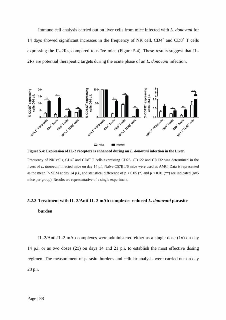
Page | 88
Immune cell analysis carried out on liver cells from mice infected with L. donovani for
14 days showed significant increases in the frequency of NK cell, CD4+ and CD8+ T cells
expressing the IL-2Rs, compared to naïve mice (Figure 5.4). These results suggest that IL-
2Rs are potential therapeutic targets during the acute phase of an L. donovani infection.
Figure 5.4: Expression of IL-2 receptors is enhanced during an L. donovani infection in the Liver.
Frequency of NK cells, CD4+ and CD8+ T cells expressing CD25, CD122 and CD132 was determined in the
livers of L. donovani infected mice on day 14 p.i. Naïve C57BL/6 mice were used as AMC. Data is represented
as the mean +/- SEM at day 14 p.i., and statistical difference of p < 0.05 (*) and p < 0.01 (**) are indicated (n=5
mice per group). Results are representative of a single experiment.
5.2.3 Treatment with IL-2/Anti-IL-2 mAb complexes reduced L. donovani parasite
burden
IL-2/Anti-IL-2 mAb complexes were administered either as a single dose (1x) on day
14 p.i. or as two doses (2x) on days 14 and 21 p.i. to establish the most effective dosing
regimen. The measurement of parasite burdens and cellular analysis were carried out on day
28 p.i.
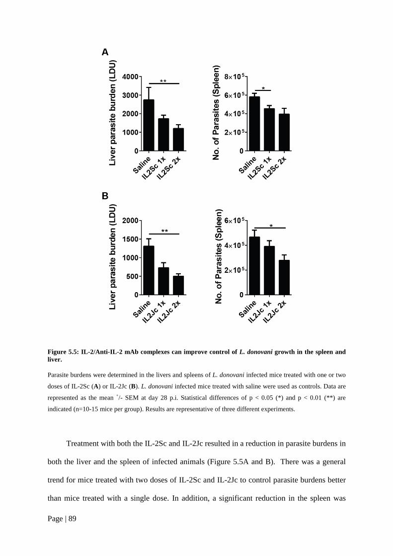
Page | 89
Figure 5.5: IL-2/Anti-IL-2 mAb complexes can improve control of L. donovani growth in the spleen and liver.
Parasite burdens were determined in the livers and spleens of L. donovani infected mice treated with one or two
doses of IL-2Sc (A) or IL-2Jc (B). L. donovani infected mice treated with saline were used as controls. Data are
represented as the mean +/- SEM at day 28 p.i. Statistical differences of p < 0.05 (*) and p < 0.01 (**) are
indicated (n=10-15 mice per group). Results are representative of three different experiments.
Treatment with both the IL-2Sc and IL-2Jc resulted in a reduction in parasite burdens in
both the liver and the spleen of infected animals (Figure 5.5A and B). There was a general
trend for mice treated with two doses of IL-2Sc and IL-2Jc to control parasite burdens better
than mice treated with a single dose. In addition, a significant reduction in the spleen was

Page | 90
only achieved with two doses of IL-2Jc. Therefore, for subsequent experiments, all
treatments were carried out with two doses of IL-2/Anti-IL-2 mAb complexes.
5.2.4 Effect of IL-2/Anti-IL-2 mAb complex therapy on immune parameters during an
L. donovani infection
As previously shown by others, IL-2Sc selectively enhances the expansion of NK cells
and CD8+ T cells, whereas the IL-2Jc selectively enhances the expansion of CD4+ T cells and
Treg cells [205]. Therefore, I wanted to determine if the control of parasite burden in the
treated groups was due to the expansion of the above mentioned cell populations. To do this,
I examined the frequency and number of activated CD4+ T cells, CD4+ IFNγ+ T cells, Tr1
(CD4+ IFNγ+ IL-10+) cells, Tregs (CD4+ Foxp3+ T cells), CD8+ T cells, CD8+ IFNγ+ T cells,
NK cells and NK IFNγ+ cells in the different treatment groups (Figure 5.6 and Figure 5.8).
This was performed as a comparative study between the IL-2Sc and IL-2Jc treatment groups,
and both the livers and the spleens from infected mice were analysed.

Page | 91
Figure 5.6: Representative sequential gating strategy for the isolation of immune cells.
From the NK 1.1- TCRβ+ cell fraction, CD4+ and CD8+ T cells were gated. Activated CD4+ T cells were
selected based on CD49d and CD11a expression. Intracellular cytokine staining was carried out to identify
IFNγ-producing CD4+ T cells, Tr1 (IL-10- and IFNγ-producing CD4+ T cells) cells in the activated cell
fractions. Tregs (CD4+ Foxp3+) cells were identified, based on nuclear FoxP3 staining on the total CD4+ T cell
population.
Surprisingly, immune cell analysis carried out on liver and spleen cells showed only
minor differences in the frequency of IFNγ-producing, activated CD4+ T cells, Tr1 and Treg
cells, between mice treated with two doses of either IL-2Jc or IL-2Sc, compared to control
groups (Figure 5.7).

Page | 92
Figure 5.7: Treatment with IL-2Sc and IL-2Jc has little effect on the expansion of activated CD4+ T cell expressing IFNγ, Tr1 and Treg cell population in the liver and spleen.
Hepatic and splenic Th1 (activated CD4+ T cells expressing IFNγ), Tr1 and Treg cell frequencies were measured in mice infected with L. donovani and treated with IL-2Sc or
IL-2Jc (separate experiments) on days 14 and 21 p.i. Mice given saline were used as controls. Data are represented as the mean +/- SEM of T cell subset frequencies, as
indicated, at day 28 p.i. Statistical differences of p < 0.05 (*) and p < 0.01 (**) are indicated (n=10-11 mice per group). Results are representative of two different
experiments.

Page | 93
Figure 5.8: Representative gating strategy for the isolation of immune cells.
NK 1.1+ TCRβ- cells, NK 1.1- TCRβ+ cells, CD4+ and CD8+ T cells were gated from live cells. NK 1.1+ TCRβ-
cells and CD8+ NK 1.1- TCRβ+ cell were gated following intracellular IFNγ staining. The frequency of IFNγ-
producing cells was then measured, as indicated.
Immune cell analysis carried out on liver and spleen cells showed only minor
differences in the frequency of CD8+ T cells and IFNγ-producing CD8+ T cells between mice
treated with two doses of either IL-2Jc or IL-2Sc, compared to control groups (Figure 5.9).
Analysis of NK1.1+ cells revealed a significant increase in the frequency of cells in the liver
of IL-2Sc treated mice (Figure 5.10). Little effect of IL-2Sc or IL-2Jc treatment on IFNγ-
producing NK1.1+ cell was observed (Figure 5.10).

Page | 94
Figure 5.9: Treatment with IL-2Sc and IL-2Jc has little effect on the expansion of CD8+ T cells in the liver or spleen.
Hepatic and splenic, CD8+ and IFNγ expressing CD8+ T cells were measured in mice infected with L. donovani
and treated with either IL-2Sc or IL-2Jc (separate experiments) on days 14 and 21 p.i. Saline treated mice were
used as controls. The frequency of CD8+ and IFNγ expressing CD8+ T cells are shown graphically. Data are
represented as the mean +/- SEM of CD8+ T cells and IFNγ-expressing CD8+ T cells at day 28 p.i. (n=10-11
mice per group). Results are representative of two different experiments.

Page | 95
Figure 5.10: Treatment with IL-2Sc and IL-2Jc has little effect on the expansion of NK1.1 cells in the liver and spleen.
Hepatic and splenic, NK1.1+ cells and IFNγ expressing NK1.1+ cells were measured in mice infected with L.
donovani treated with IL-2Sc or IL-2Jc (separate experiments) on days 14 and 21 p.i. Saline treated mice were
used as controls. Data are represented as the mean +/- SEM of the frequency of NK1.1+ and IFNγ expressing
NK1.1+ cells at day 28 p.i., and statistical differences of p < 0.05 (*) are indicated (n=10-11 mice per group).
Results are representative of two different experiments.
The above experiments indicated that two doses of IL-2/anti-IL-2 mAb complex
treatment either with IL-2Sc or IL-2Jc resulted in significantly lower parasite burden,
however, this protection was not associated with the cellular expansion of cells expressing the
IL-2Rs, suggesting that other mechanisms of protective immunity may be involved.

Page | 96
5.2.5 Treg cells do not interfere with protection mediated by IL-2J complex treatment
As mentioned earlier, IL-2Jc has been reported to selectively expand cells expressing
the high affinity heterotrimeric IL-2R expressed mainly on activated CD4+ T cells and Treg
cells. We know that conventional CD4+ T cells responses are critical for anti-parasitic
immunity against L. donovani [65], but that Treg cells may interfere with these cells [211,
212]. To test whether expansion of Treg cells impeded the effect of IL-2Jc on conventional
CD4+ T cells in L. donovani infected mice, Foxp3-GFP-DTR mice were used. These
C57BL/6 mice contain a transgene encoding human diphtheria toxin receptor (DTR) inserted
into the 3' untranslated region of Foxp3 [213]. The DTR is only found in the Foxp3+ Treg
cells of these mice, and injecting diphtheria toxin (DTx) intraperitoneally (i.p.), results in
depletion of Foxp3+ Tregs in these mice [213]. I hypothesised that eliminating Tregs in an
established L. donovani infection, while treating with IL-2Jc, would further improve parasite
control, as complex treatment would be targeted to activated CD4+ T cells.
Foxp3-GFP-DTR mice were infected with L. donovani and treated with IL-2Jc on days
14 and 21 p.i. DTx treatment (8 ng/g i.p.) was started on day 12 p.i., and then every three
days for the duration of the experiment. FACS analysis of Foxp3-GFP-DTR mice treated
with DT indicated a reduction in Treg cell frequency (Figure 5.11A and B). The combination
of IL-2Jc treatment and Treg cell depletion, however, did not further improve parasite burden
in the treated animals, thus indicating that IL-2Jc anti-parasitic effects were not being
inhibited by Treg cells (Figure 5.12A and B).

Page | 97
Figure 5.11: Foxp3-GFP-DTR mice treated with DT have a reduced frequency of Treg cells.
(A) Treg cells were identified by flow cytometry from the total CD4+ T cells population. Foxp3 expression in
CD4+ spleen cells from L. donovani infected control Foxp3-GFP-DTR mice (Saline) and Foxp3-GFP-DTR
mice treated with DT (started on day 12 p.i and then every three days for the duration of the experiment) was
analysed. (B) Data are represented as the mean +/- SEM of the frequency of hepatic and splenic Treg cells at day
28 p.i., and statistical differences of p < 0.01 (**) are indicated (n=5 mice per group). Results are representative
of a single experiment.

Page | 98
Figure 5.12: Tregs do not impair IL-2J complex-mediated protection.
Parasite burdens were determined in the livers (A) and spleens (B) of L. donovani infected Foxp3-GFP-DTR
mice treated with two doses IL-2Jc. L. donovani infected mice treated with saline were used as control. DTx
administration was started on day 12 p.i., and then every three days for the duration of the experiment. Data are
represented as the mean +/- SEM at day 28 p.i., and statistical differences of p < 0.01 (**) are indicated (n=5
mice per group). Results are representative of a single experiment.
Analysis of Treg cells from the livers and spleens of mice treated with IL-2Jc and DTx
confirmed depletion of Foxp3+ T cells (Figure 5.13A and B). The depletion of Treg cells in
IL-2Jc-treated mice did not change the frequency of Th1 and Tr1 cells in the spleen.
However, there was a significantly lower frequency of Th1 and Tr1 cells in the liver
following DT administration in IL-2Jc-treated mice, indicating different effects of Treg cell
depletion in the spleen and liver after IL-2Jc treatment (Figure 5.13 A and B). Interestingly,
although no difference in serum IFNγ and TNFα levels were observed in mice treated with
IL-2Jc and depleted of Treg cells, IL-10 levels were increased, but this did not reach
statistical significance (Figure 5.13 C).

Page | 99
Figure 5.13: The impact of Treg cell depletion in IL-2J complex treated animals.
Hepatic (A) and splenic (B) Treg, Th1and Tr1 cellular responses were measured in L. donovani infected Foxp3-
GFP-DTR mice, depleted of Tregs and treated with two doses of IL-2Jc. Saline was used as a control. The
frequency of Treg, Th1 and Tr1 cells are shown graphically. (C) Serum IFNγ, TNFα and IL-10 levels were also
measured in these mice. Data are represented as the mean +/- SEM at day 28 p.i., and statistical differences of p
< 0.01 (**) are indicated (n=5 mice per group). Results are representative of a single experiment.

Page | 100
5.2.6 The IL-2J complex mediates anti-parasitic effects in L. donovani -infected mice via
CD4+ T cells
I next tested the impact of depleting CD4+ T cells in L. donovani infected mice treated
with IL-2Jc. C57BL/6 mice were infected with L. donovani and treated with IL-2Jc on days
14 and 21 p.i. Anti-CD4 mAb (0.5 mg/mouse i.p.) treatment was started on day 12 p.i and
then every three days for the duration of the experiment. FACS analysis of total CD4+ T cells
from infected mice confirmed depletion (Figure 5.14 A and B). Depletion of CD4+ T cells in
infected mice treated with IL-2Jc resulted in a significant increase in parasite burden
compared to groups that were treated with an isotype control (ISO) mAb and IL-2Jc (Figure
5.15 A and B), thus indicating that CD4+ T cells are required for IL-2Jc-mediated anti-
parasitic immunity in experimental VL.

Page | 101
Figure 5.14: Administration of anti-CD4 mAb results in efficient CD4+ T cell depletion.
(A) CD4+ T cells were identified from NK1.1- TCRβ+ cells. CD4+ T cells from spleens of L. donovani infected
mice treated with α-CD4 mAb (started on day 12 p.i and then every three days for the duration of the
experiment) were measured and compared to mice treated with an isotype control mAb (ISO)-. (B) The
frequency of CD4+ T cells in the spleen was significantly reduced, compared with control mice. Data are
represented as the mean +/- SEM at day 28 p.i., and statistical differences of p < 0.01 (**) are indicated (n=5
mice per group). Results are representative of a single experiment.

Page | 102
Figure 5.15: CD4+ T cells are required for IL-2J complex mediated protection.
Parasite burdens were measured in the livers (A) and spleens (B) of L. donovani infected C57BL/6 mice treated
with two doses of IL-2Jc. L. donovani infected mice treated with ISO were used as controls. Anti-CD4 mAb
treatment commenced on day 12 p.i., and then every three days for the duration of the experiment. Data are
represented as the mean +/- SEM at day 28 p.i., and statistical differences of p < 0.05 (*) and p < 0.01 (**) are
indicated (n=5 mice per group). Results are representative of a single experiment.
Depletion of CD4+ T cells in IL-2Jc-treated mice resulted in decreased CD4+ T cell
frequencies, as expected, but also significant increases in CD8+ T cell and NK.1.1 cell
numbers in the spleen, however when we looked at the number of CD8+ T cell and NK.1.1
cell, there isn’t a change due to the lack of CD4+ T cells (Figure 5.16). These results indicated
that in the presence of lower CD4+ T cell, both CD8+ T cells and NK cells can expand in
response to IL-2Jc treatment. However, this expansion was not able to compensate for the
loss of CD4+ T cells in the control of parasite growth, highlighting the importance of CD4+ T
cells as anti-parasitic responder cells in mice treated with IL-2Jc.

Page | 103
Figure 5.16: Depletion of CD4+ T cells in IL-2J complex treated animals increased the frequency of CD8+ T cell and NK1.1 cells.
Splenic CD4+, CD8+ T cell and NK1.1 cells were measured in L. donovani infected C57BL/6 mice depleted of
CD4+ T cells and treated with two doses of IL-2Jc. Isotype control mAb (ISO) treated mice were used as
controls. Data are represented as the mean +/- SEM of the frequency of CD4+ T cells, CD8+ T cells and NK1.1
cells at day 28 p.i. Statistical differences of p < 0.05 (*) and p < 0.01 (**) are indicated (n=5 mice per group).
Results are representative of a single experiment.
5.2.7 The IL-2S complex mediates anti-parasitic effects in L. donovani -infected mice via
CD4+ T cells, and not via CD8+ T cells or NK cells
As mentioned earlier, IL-2Sc has been reported to selectively expand cells expressing
the low affinity heterodimeric IL-2R found mainly on activated CD8+ T cells and NK cells.
We know that CD8+ T cells aid in protection during with L. donovani [65], while studies have
shown that NK cells are insufficient and not essential to control L. donovani [214, 215]. To
test whether expansion of either CD8+ T cells or NK cells enabled the effect of IL-2Sc on
parasite burdens in L. donovani infected mice, I depleted each cell type. C57BL/6 mice were
infected with L. donovani and treated with IL-2Sc on days 14 and 21 p.i. Anti-CD8β mAb

Page | 104
(0.5 mg/mouse i.p.) treatment was started on day 12 p.i and then every three days for the
duration of the experiment. NK cell depletion was conducted using B6.NKp46Cre.iDTR mice
[178]. DTx treatment (8 ng/g i.p.) was started on day 12 p.i., and then every three days for the
duration of the experiment. FACS analysis of CD8+ T cells or NK cells from treated mice
confirmed 90% and 60% depletion, respectively. The combination of IL-2Sc treatment with
depleting either CD8+ T cells or NK cells did not alter the effects of IL-2Sc treatment on
parasite burden in the treated animals, thus indicating that the anti-parasitic effects of IL-2Sc
were not being mediated by either CD8+ T cells or NK cells (Figure 5.17 A and B).

Page | 105
Figure 5.17: CD8+ T cells and NK cells do not contribute to IL-2S complex-mediated protection.
Parasite burdens were determined in the livers and spleens of L. donovani infected C57BL/6 depleted of CD8+ T
cells (A) and B6.NKp46Cre.iDTR (B) mice treated with two doses of IL-2Sc. L. donovani infected mice treated
with isotype control mAb or saline, respectively, were used as controls. Anti-CD8β mAb and DTx
administration was commenced on day 12 p.i., and then every three days for the duration of the experiment.
Data are represented as the mean +/- SEM at day 28 p.i., (n=5 mice per group). Results are representative of a
single experiment.
Since conventional CD4+ T cells can also express the low affinity heterodimeric IL-2R,
I next tested the impact of depleting CD4+ T cells in L. donovani infected mice treated with
IL-2Sc. C57BL/6 mice were infected with L. donovani and treated with IL-2Sc on days 14
and 21 p.i. Anti-CD4 mAb (0.5 mg/mouse i.p.) treatment was started on day 12 p.i., and then
every three days for the duration of the experiment. FACS analysis of total CD4+ T cells from

Page | 106
infected mice confirmed greater than 98% depletion (Figure 5.14 A and B). Depletion of
CD4+ T cells in infected mice treated with IL-2Sc resulted in a significant increase in parasite
burden, compared to groups that were treated with an isotype control (ISO) mAb and IL-2Sc
(Figure 5.18 A and B), thus indicating that CD4+ T cells were required for IL-2Sc-mediated
anti-parasitic immunity in experimental VL.
Figure 5.18: CD4+ T cells are required for IL-2S complex mediated protection.
Parasite burdens were measured in the livers (A) and spleens (B) of L. donovani infected C57BL/6 mice treated
with two doses of IL-2Sc. L. donovani infected mice treated with an isotype control mAb (ISO) were used as
controls. Anti-CD4 mAb treatment commenced on day 12 p.i., and then every three days for the duration of the
experiment. Data are represented as the mean +/- SEM at day 28 p.i., and statistical differences of p < 0.05 (*)
and p < 0.01 (**) are indicated (n=5 mice per group). Results are representative of a single experiment.

Page | 107
5.3 DISCUSSION
In this chapter the potential of IL-2 therapy using IL-2/Anti-IL-2 mAb complexes to
improve experimental VL disease outcome was investigated. To achieve this, two commonly
used IL-2/anti-IL-2 mAb complexes, the IL-2J complex comprising rmIL-2/JES6.1A12 mAb
and the IL-2S complex comprising of rmIL-2/S4B6 mAb, were used. Previous experiments
have shown that treatment with exogenous IL-2 improved anti-parasitic immune responses
against VL [200], and several other studies have shown that IL-2/anti-IL-2 complexes
selectively target different T cell subsets to either enhance or suppress immune responses in
different disease settings [207-210]. Hence, to investigate the effect of IL-2/anti-IL-2 mAb
complex therapy during VL, L. donovani infected mice were treated with either IL-2Jc or the
IL-2Sc.
In VL, as well as in EVL, the spleen is a site of persistent and chronic infection.
Initially, I tried to use IL-2/anti-IL-2 mAb complex therapy to target this organ to improve
anti-parasitic immune responses during EVL. Treatment commenced on day 28 p.i., when a
chronic infection had established in the spleen [73, 75]. However, I found little indication that
IL-2/anti-IL-2 mAb complexes had any significant effect on parasite burdens. Overall, the
result indicated that the chronic stage of the infection wasn’t the ideal setting to assess the
therapeutic effects of these complexes. Therefore, I turned my attention to the effect of IL-
2/anti-IL-2 mAb complex therapy during the acute phase of the infection and found that mice
responded better to IL-2/anti-IL-2 mAb complex therapy given in the early stages of the
infection (from day 14 p.i.), where two doses of either IL-2Jc or IL-2Sc was better at
controlling parasite burdens than a single dose. Analysis of immune cell populations

Page | 108
associated with IL-2Jc or IL-2c treatment, however, failed to reveal any significant changes
that might account for the protective immunity initiated by this treatment.
Earlier experiments have shown that in an L. donovani infection, Treg cells inhibit
protective immune responses and the cellular recruitment required for parasite control [211,
216]. Therefore, one potential adverse effect of IL-2Jc treatment was the expansion and/or
activation of Treg cells with detrimental effects on anti-parasitic T cell responses. However,
when Treg cells were depleted by DT administration in Foxp3-GFP-DTR treated with IL-2Jc,
there was no significant improvement in the anti-parasitic effects of IL-2Jc, indicating that
this complex was not promoting Treg cell expansion of activation. To test if the IL-2Jc
mediated protection involved effects on conventional CD4+ T cells, anti-CD4 mAb was used
to deplete these cells during the IL-2Jc treatment period. This resulted in disease
exacerbation. A similar effect was observed when CD4+ T cells were depleted during the IL-
2Sc treatment period, while depletion of CD8+ T cells and NK cells had no effect on parasite
burdens of IL-2Sc treated mice. Thus, these results show the requirement of conventional
CD4+ T cells for IL-2Jc and IL-2Sc mediated protection in experimental VL.
IL-2 is a pleiotropic cytokine responsible for the development and maintenance of
Treg cells [217], expansion of Th1 [218], Th2 [219] and Th9 [220] cells, while suppressing
CD4+ T cell differentiation into Th17 cells [221]. Stimulation of NK cells and CD8+ T cells
by IL-2 results in the proliferation, increased cytotoxic activity and cytokine production
[199]. Recent studies have shown that type 2 innate lymphoid cells (ILC2) express high
levels of CD25 and can proliferate in reposes to low-dose IL-2 through activation of the
trimeric IL-2R [222]. This supplements the production of IL-5, which expands eosinophil

Page | 109
populations, resulting in eosinophilia [223]. Hence, alteration to IL-2 levels has the potential
to cause unexpected consequences, and binding of IL-2 to the IL-2Rs might lead to
unexpected changes in-vivo. In my experiments, treatment with IL-2Sc did not result in
significant expansion of NK cells or CD8+ T cells. Similarly, treatment with IL-2Jc did not
cause significant expansion of Treg cells or activated CD4+ T cells. One possible explanation
for this result is that expansion of these cell populations occurred rapidly post-treatment and
waned by the time cellular responses were measured (7 days later). However, the fact that
both IL-2Jc and IL-2Sc treatment resulted in significant reductions in parasite burden,
indicates that IL-2/anti-IL-2 mAb complexes stimulated potent anti-parasitic immunity.
In addition to IL-2/anti-IL2 mAb complexes, other IL-2-related treatments are currently
being investigated. Early studies with IL-2 fused to carrier proteins, such as the Fc domains
of IgG, improved cytokine half-life [224]. More recently, an increasing number of studies in
animals and humans have shown that multiple low doses of IL-2 have fewer adverse effects
and favourable immune outcomes [225, 226]. Several mutant forms of IL-2 have also been
developed with transformed binding sites that change interactions between the cytokine and
IL-2Rs, resulting in altered function. Shanafelt, et al., have reported the development of a
mutant form of IL-2 known as BAY 50-4798. This molecule has a higher affinity for trimeric
IL-2Rαβγ, relative to dimeric IL-2Rβγ, and promoted enhanced T cell activation, without any
effects on NK 1.1. cells [227]. Studies using IL-2 and with various check-point inhibitors
have also shown promising results for immunotherapy for cancer [228, 229]. Together with
my results, these studies demonstrate the potential of IL-2 therapy in targeting specific
immune parameters to improve disease outcomes. However, targeting the right immune cells
in the host to optimise effects and understanding the mechanism of protective immunity
remains a challenge.

Page | 110
In this Chapter, I showed that IL-2/anti-IL2 mAb complex therapy with either IL-2Jc or
IL-2Sc significantly reduced parasite burdens in experimental VL. However, surprisingly, I
observed no expansion of potential target cell populations and the mechanism of enhanced
parasite control remains unclear. Overall, IL-2 therapy warrants further investigation for
development as a treatment for clinical VL.

Page | 111
Chapter 6: To compare different methods of parasite attenuation and establish whether a live, attenuated, whole parasite vaccine can protect against experimental VL
6.1 INTRODUCTION
The most common VL treatment for the last 60 years has been antimonial
chemotherapy [230]. Pentavalent antimonials, such as sodium stibogluconate, pentostam,
meglumine antimonite and glucantime, have been the mainstay of antimonial therapy [231].
However, there is now considerable parasite resistance against these drugs, especially in
North-Eastern India and surrounding areas [232]. Therefore, although these drugs are still
employed to treat VL in Africa, drugs such as Amphotericin B, Miltefosine, aminosidine
(paromomycin) and sitamaquine have been developed as effective treatments against VL in
areas of antimonial drug resistance [232]. However, these drugs are still far from ideal
because of cost, toxicity, development of parasite drug resistance after prolonged use and
duration of treatment times [4]. Some progress has been made recently in addressing this
latter issue [164], where a single dose of lipid formulation of Amphotericin B (Ambisome),
was effective in treating VL patients with lower toxicity outcomes compared to the
conventional drug treatment. However, there are still concerns that this single dose of
treatment may eventually result in the development of drug-resistant parasites. To further
address this concern of parasite drug resistance, combination drug therapy is being developed
[158, 233]. However, studies in experimental VL suggested that L. donovani can develop
drug resistance, even to drug combination therapy [234]. Despite the arsenal of anti-

Page | 112
protozoan drugs available, it is unlikely that chemotherapy alone will control and eradicate
the disease, and thus, there is an urgent need for an effective and protective vaccine against
VL.
The development of a vaccine to prevent leishmaniasis has been a long-term goal for
researchers in the field. At present there are no licensed vaccines that prevent leishmaniasis
[235]. In theory, a vaccine to prevent leishmaniasis should be possible, as indicated by past
programs of “leishmanisation”. This process involves the deliberate infection of people with
CL-causing parasite species on unexposed areas of the body to establish an infection that is
controlled in most individuals, resulting in long-term protection [236]. This technique was
practised for centuries throughout the Middle East and parts of Asia, and large-scale trials
were carried out in the former Soviet Union and Israel with some success [171, 172] but this
relied on parasites that were viable and infective [173]. However, despite the solid immunity
that develops in most individuals, this approach has largely been abandoned due to
complications in some individuals including large skin lesions, exacerbation of skin diseases
and poor responses to the vaccine [7, 8]. The vaccines currently being developed against VL
can be divided into three groups; first generation vaccines which involve vaccination with
live-attenuated or killed parasite; second generation vaccines which involve genetically
modified parasites, subunit vaccines or recombinant virus and bacteria recombinant parasite
protein expression systems; and third generation vaccines which consist of plasmid DNA and
viral based vaccines encoding virulent Leishmania proteins [43, 235, 237]. However killed
parasite, recombinant DNA and protein vaccine appear to be ineffective due to the short-term
immunity they induce [238]. Whole irradiated or biochemically altered parasites have been
shown to confer potent protection in an experimental model of VL [239, 240], similar to the

Page | 113
results achieved by leishmanisation. Hence, for a robust protective immune response against
VL, whole parasite vaccines show promise.
Various mouse models of Leishmania infection have shown that resistance to the
parasite is mediated by cellular immune responses, primarily involving Th1 responses, which
are IL-12 dependent [241]. IFNγ and TNFα produced by parasite-specific Th1 cells drive the
effector function of macrophages to generate reactive oxygen and nitrogen species required
for intracellular parasite killing by macrophages [87]. IL-10, an immune regulatory cytokine
produce by parasite-specific CD4+ T cells, plays an active role in supressing anti-parasitic
Th1 immune responses and promotes parasite persistence in VL patients [54]. Thus, Th1
responses are important for the elimination of parasites in infected individuals. Understanding
the requirements and conditions required for the generation of protective Th1 responses
against Leishmania parasite is important for developing an effective vaccine.
The previous chapters focused on promoting CD4+ T cell responses during established
experimental VL via immune modulatory techniques such as combination immune treatment
and chemotherapy, as well as IL-2 complex therapy. These experiments showed us that sub-
optimal CD4+ T cell responses generated during VL can be improved given the right immune
conditions. In this chapter, I aimed to develop a whole parasite vaccine to generate protective
parasite-specific CD4+ T cell responses against VL.

Page | 114
6.2 RESULTS
6.2.1 The effects of immunization with irradiated whole L. donovani promastigotes
My first vaccine study was carried out to identify the vaccine regime best suited to
generate a potent immune response. I used L. donovani promastigotes as my parasite vaccine
for immunization as this form of the parasite is transmitted to the host when the sand-fly
takes its blood meal [242]. Also, L. donovani promastigotes are easy to culture in-vitro [243],
and large quantities of the parasites can be produced. The vaccine regime followed was
previously described by Scott et al. [243]. Briefly, C57BL/6 mice were immunized with four
doses of irradiated L. donovani promastigotes given either intravenously (Irr LV9 i.v.) or
intraperitoneally (Irr LV9 i.p.) and challenged with live L. donovani amastigotes two weeks
after the last immunization and sacrificed on day 14 and day 28 post challenge (p.c.) to assess
hepatic and splenic parasite burdens (Figure 6.1.1). Naïve age matched mice were used as
controls (AMC), and these were infected with L. donovani at the same time as the immunized
mice were challenged.
Figure 6.1.1: Experimental timeline for assessment of protection against L. donovani.
C57BL/6 mice were immunized with L. donovani promastigotes irradiated at 500 Gys (2x10⁷ parasites i.v. or
5x10⁷ parasites i.p.). Primary immunization was carried out on day 0 and subsequent boost on days 7, 14, and
21. Two weeks after the final booster (i.e., day 35), mice were challenged with 2 x10⁷ live L. donovani
amastigotes. Mice were sacrificed on days 14 and day 28 p.c. Naïve AMC were infected with L. donovani on
day 35 in the above scheme.

Page | 115
Analysis of hepatic and splenic parasite burdens showed no improvement in parasite
control on either day 14 or day 28 p.c., between the immunized and AMC groups (Figure 6.2
A and B). These results indicate that this immunization regime used was not effective.
Figure 6.2: Failure of previously reported immunisation regime to protect against VL.
Parasite burdens were determined in livers and spleens of L. donovani challenged mice on day 14 (A) and day
28 p.c. (B). Prior to challenge mice were immunized with irradiated (500 Gys) L. donovani promastigotes
(2x10⁷ parasites i.v. or 5x10⁷ parasites i.p.). Aged matched mice were used as controls (AMC). Data are
presented as the mean +/- SEM at day 14 and day 28 p.i. (n=5 mice per group). Results are representative of a
single experiment.

Page | 116
6.2.2 The effect of immunization with either radio- or chemically-attenuated whole L.
donovani amastigotes
Given the above results, I next investigated the effect of immunization with attenuated
amastigotes instead of promastigotes. This was done under the rationale that the challenge
infection was initiated with amastigotes, and so the immune cells should be primed for
amastigote antigens. In addition to using irradiated amastigotes, I wanted to assess the
potential of using chemically-attenuated parasites for vaccination. The chemical used to
attenuate the L. donovani parasites was TH-III-149 (tafuramycin A: TFA), which is an
alkylating agent that irreversibly alters the parasites DNA at multiple sites preventing
proliferation, but leaves the parasite viable [244]. Furthermore, to assess if multiple
immunization doses improved the control of parasite burden in vaccinated mice, C57BL/6
mice were immunized i.v. with either a single (1x) or two doses (2x) of irradiated (Irr LV9)
or chemically-attenuated (TFA LV9) L. donovani amastigotes (Figure 6.1.2). The interval
between the primary immunization and subsequent boost was increased to four weeks instead
of seven days, based on reports that longer intervals between vaccine administrations can
improve efficacy [245]. Mice were challenged with live L. donovani amastigote four weeks
after the final immunization (day 56), and they were sacrificed on day 14 p.c. Naïve AMC
were infected with L. donovani at the same time as the immunized mice were challenged.

Page | 117
Figure 6.1.2: Experimental timeline for assessment of protection against L. donovani.
C57BL/6 mice were immunized with L. donovani amastigotes irradiated at 500 Gys or chemically-attenuated
with TFA (2x10⁷ parasites i.v.). For mice receiving two immunizations (2x), the primary dose was given on day
0 and subsequent boost on day 28. Some mice received a single immunization on day 28 (1x). Four weeks post
final immunization i.e. day 56 mice were challenged with 2 x10⁷ live L. donovani amastigotes and were
sacrificed on day 14 p.c.
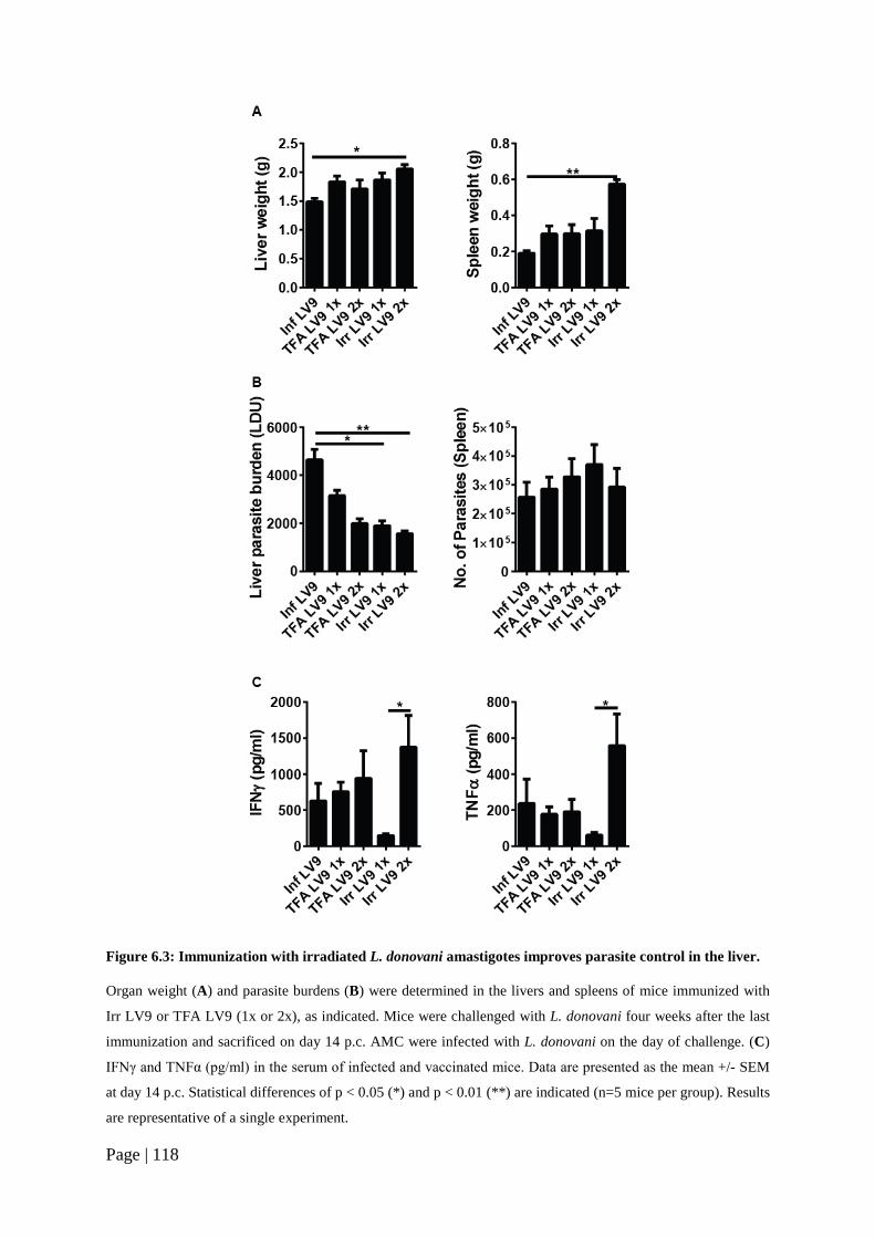
Page | 118
Figure 6.3: Immunization with irradiated L. donovani amastigotes improves parasite control in the liver.
Organ weight (A) and parasite burdens (B) were determined in the livers and spleens of mice immunized with
Irr LV9 or TFA LV9 (1x or 2x), as indicated. Mice were challenged with L. donovani four weeks after the last
immunization and sacrificed on day 14 p.c. AMC were infected with L. donovani on the day of challenge. (C)
IFNγ and TNFα (pg/ml) in the serum of infected and vaccinated mice. Data are presented as the mean +/- SEM
at day 14 p.c. Statistical differences of p < 0.05 (*) and p < 0.01 (**) are indicated (n=5 mice per group). Results
are representative of a single experiment.

Page | 119
Mice immunized with two doses of irradiated L. donovani amastigotes (Irr LV9)
showed a significant increase in liver and spleen weights (Figure 6.3 A), indicative of
increased immune cell infiltration into these organs [96]. Assessment of liver parasite
burdens showed a significant decrease in parasite burden in mice immunized with either a
single or two doses of Irr LV9, while mice immunized with two doses of chemically-
attenuated parasite (TFA LV9) appeared to be better protected compared to mice immunized
with a single dose, although not statistically different. (Figure 6.3 B). In the spleen, however,
no change in parasite burden was observed in the immunized groups compared to the AMC
group. Serum cytokine analysis revealed elevated levels of pro-inflammatory cytokines IFNγ
and TNFα in groups immunized with two doses of Irr LV9 (Figure 6.3 C).
These results indicated that immunization with irradiated LV9 provided better
protection in the liver than immunization with chemically-attenuated parasites. Moreover,
two immunizations with irradiated LV9 resulted in enhanced levels of pro-inflammatory
cytokines over the single immunization groups, as indicated by elevated levels of IFNγ and
TNFα.
I next examined the frequency of Th1 (Tbet+ IFNγ-producing CD4+ T cells) cells and
terminally differentiated Th1 (Tbet+ KLRG-1+ CD4+ T cells) cells in the different immunized
groups (please refer to previous chapters for representative gating strategy).

Page | 120
Figure 6.4: Immunization has little effect on the CD4+ T cell responses in the liver, but increased CD4+ T cell responses were observed in the spleen.
Hepatic (A) and splenic (B) Th1 and KLRG-1+ Th1 cellular responses, as indicated, were measured in mice
immunized and challenged with L. donovani amastigotes. The frequency of Th1 and KLRG-1+ Th1 cells are
shown graphically. Data are presented as the mean +/- SEM at day 14 p.c. Statistical differences of p < 0.05 (*)
are indicated (n=5 mice per group). Results are representative of a single experiment.
Immune cell analysis carried out in liver samples showed little difference in the
frequency of Th1 cells and KLRG-1+ Th1 cells between the different immunized groups.
Analysis of spleen cells, however, showed increases in the frequency of Th1 cells and
KLRG-1+ Th1 cells in immunized mice, compared to control groups. This indicated that

Page | 121
while immunized animals had a higher percentage of Th1 cells, the rate of conversion to the
functionally exhausted phenotype was higher in the spleen, and may account for the lack of
effect of immunization on parasite burden observed in this organ (Figure 6.4 A and B).
6.2.3 Immunization with irradiated promastigotes provides better protection compared
to irradiated amastigotes
To assess whether irradiated L. donovani promastigotes (Irr LV9 promastigotes) might
be a better vaccine than irradiated amastigotes, C57BL/6 mice were immunized with two
doses (2x) of irradiated L. donovani amastigotes (Irr LV9 AM) or L. donovani promastigotes
(Irr LV9 PM) (Refer to Figure 6.1.2 for regime). Mice were challenged with live L. donovani
amastigotes four weeks after the final immunization (day 56), and were sacrificed on day 14
p.c. Naïve AMC were infected with L. donovani at the same time as the immunized mice
were challenged. Antigen-specific cellular immune responses were also measured after
immunization. Briefly, 4 weeks after final immunization (i.e. day of challenge), a cohort
(n=5) of mice immunized with Irr LV9 (AM) or Irr LV9 (PM) were sacrificed, splenocytes
harvested and cultured with L. donovani parasite antigen for 72hrs. Culture supernatants were
then used to analyse antigen-specific cytokine production. Splenocytes from naïve mice were
used as controls.
Mice immunized with either Irr LV9 (Am) or Irr LV9 (Pm), but not infected, showed
no change in liver or spleen weights, compared to naïve mice (Figure 6.5 A). Measurement of
antigen-specific immune responses showed increased levels of the pro-inflammatory
cytokines IFNγ and TNFα, and the anti-inflammatory cytokine IL-10, in both immunized

Page | 122
groups, although mice immunized with Irr LV9 (Pm) showed significantly higher levels of
these cytokines (Figure 6.5 B).
Figure 6.5: Immunization with irradiated parasites results in enhanced antigen-specific T cell responses.
(A) Livers and spleens weights were measured in mice immunized with two doses of Irr LV9 (Am) or Irr LV9
(Pm) four weeks after the last immunization. Naïve, non-immunized mice were used as controls. (B) IFNγ,
TNFα and IL-10 (pg/ml) levels from an antigen-specific re-stimulation assay were measured. Data are
represented as the mean +/- SEM on the day of challenge. Statistical differences of p < 0.05 (*), p < 0.01 (**), p
< 0.001 (***) and p < 0.0001 (****) are indicated (n=5 mice per group) (NOTE: Re-stimulation assay was
performed in triplicate for biological replicates (n = 5/group). Results are representative of a single experiment.

Page | 123
At day 14 p.c., there was a significant increase in the organ weights of mice immunized
with two doses of either Irr LV9 (Am) or Irr LV9 (Pm), compared with non-immunized mice
(Figure 6.6 A). Assessment of liver parasite burdens showed a significantly lower burden in
mice immunized with either Irr LV9 (Am) or Irr LV9 (Pm) (Figure 6.6 B). However, in the
spleen, although no change in parasite burden was observed in mice immunized with Irr LV9
(Am), mice immunized with Irr LV9 (Pm) showed significantly lower parasite burdens
compared to the AMC group (Figure 6.6 B). Serum cytokine analysis revealed elevated levels
of the pro-inflammatory cytokines IFNγ and TNFα in both groups of immunized mice
(Figure 6.6 C). These results indicate that immunization with irradiated LV9 promastigotes
(Irr LV9 Pm) provides better protection than irradiated LV9 amastigotes (Irr LV9 Am).
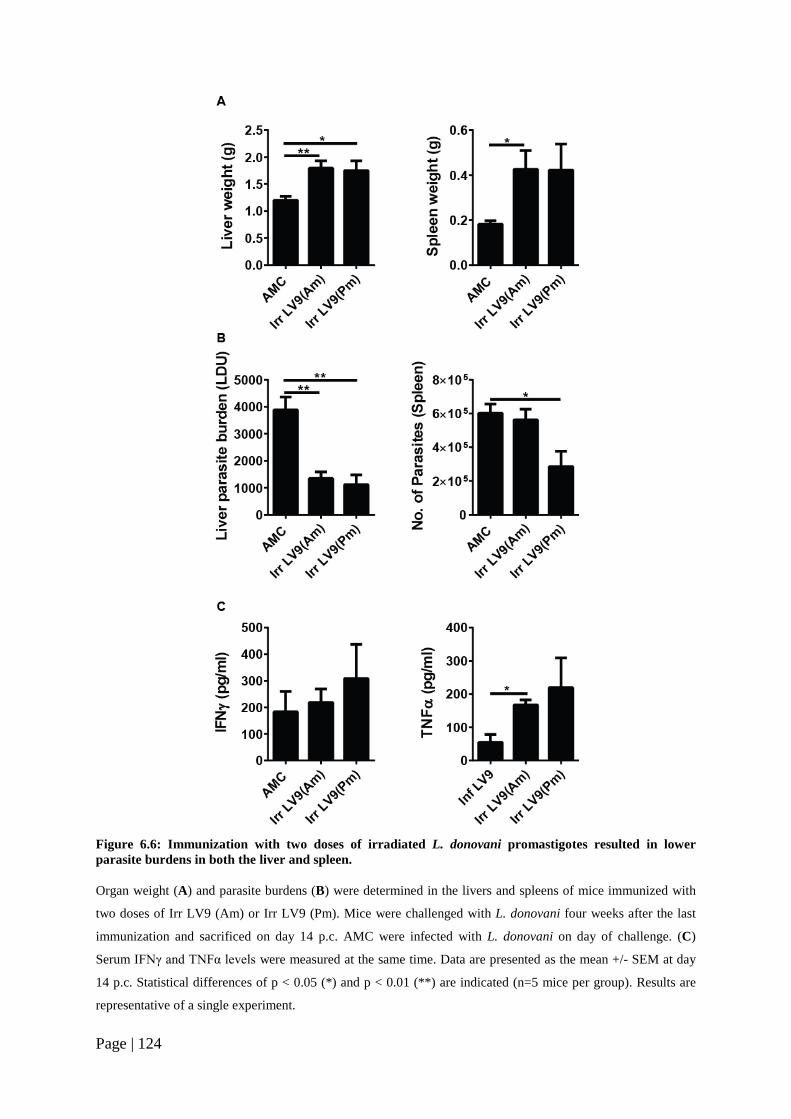
Page | 124
Figure 6.6: Immunization with two doses of irradiated L. donovani promastigotes resulted in lower parasite burdens in both the liver and spleen.
Organ weight (A) and parasite burdens (B) were determined in the livers and spleens of mice immunized with
two doses of Irr LV9 (Am) or Irr LV9 (Pm). Mice were challenged with L. donovani four weeks after the last
immunization and sacrificed on day 14 p.c. AMC were infected with L. donovani on day of challenge. (C)
Serum IFNγ and TNFα levels were measured at the same time. Data are presented as the mean +/- SEM at day
14 p.c. Statistical differences of p < 0.05 (*) and p < 0.01 (**) are indicated (n=5 mice per group). Results are
representative of a single experiment.

Page | 125
I next examined the frequency of Th1 (Tbet+ IFNγ-producing CD4+ T cells) cells and
terminally differentiated Th1 (Tbet+ KLRG-1+ CD4+ T cells) cells in different immunized
groups (refer to previous chapters for representative gating strategy).
Figure 6.7: Cellular immune responses in the livers and spleens of mice immunized with irradiated parasites and challenged with L. donovani.
Hepatic (A) and splenic (B) Th1 and KLRG-1+ Th1 cellular responses were measured in mice immunized and
challenged with L. donovani. The frequency of Th1 and KLRG-1+ Th1 cells are shown graphically. Data are
presented as the mean +/- SEM at day 14 p.c. Statistical differences of p < 0.05 (*) and p < 0.01 (**) are
indicated (n=5 mice per group). Results are representative of a single experiment.
Page | 126
Analysis of immune cells carried out on liver and spleen cells showed a significant
increase in the frequency of Th1 cells in immunized mice, compared to control groups
(Figure 6.7 A and B). However, the frequency of KLRG-1+ Th1 cells was also significantly
increased in immunized animals. This indicated that while immunized animals had a higher
percentage of Th1 cells, the rate of conversion to a functionally exhausted phenotype was
also higher (Figure 6.7 A and B).
6.2.4 Testing the pathogenicity of irradiated whole parasite
Given the significant splenomegaly observed in mice immunised with 2 doses of
irradiated amastigotes, I was concerned that parasites may not have been fully attenuated.
Therefore, I next tested whether irradiated LV9 amastigotes could grow in immune
compromised mice. B6.Rag-1-/- mice lack mature B and T lymphocytes [246], and were
infected with a single dose of either Irr LV9 or TFA LV9 amastigotes. A separate cohort of
B6.Rag-1-/- mice were infected with untreated L. donovani and 14 days later, groups were
assessed for hepatic parasite burdens. Splenic parasite burdens were not assessed since I only
wanted to detect establishment of infection.

Page | 127
Figure 6.8: Irradiated parasites caused low grade infections in B6.Rag-1-/- mice.
Hepatic weight (A) and parasite burdens (B) were determined in the livers of B6.Rag-1-/- mice infected with Irr
LV9 or TFA LV9 (1x) and sacrificed on day 14 p.i. Parasite burdens in B6.Rag-1-/- mice infected with the same
number of untreated L. donovani (AMC) were also measured. Data are presented as the mean +/- SEM at day 14
p.i. (n= 5 mice per group). Results are representative of a single experiment.
B6.Rag-1-/- mice infected with Irr LV9 or TFA LV9 amastigotes had lower parasite
burdens, compared to L. donovani infected AMC. However, mice infected Irr LV9
amastigotes had higher parasite burden compared to mice infected with TFA LV9
amastigotes (Figure 6.8 A and B), suggesting some expansion in parasite numbers in the
former group.
6.2.5 Investigating the effect of increasing the dose of irradiation for attenuation in
immunized mice
The previous results suggested that the dose of radiation being used was not sufficient
to fully attenuate L. donovani. My dosing (500 Gys) was based on previous work by Scott

Page | 128
and colleagues [243], but earlier work by Howard and colleagues showed that irradiating
promastigotes at higher dose (1500 Gys), also resulted in protective immunity [247]. Hence, I
examined the impact of increasing the radiation dose from 500 Gys to 1500 Gys. The
immunization regime followed is represented in Figure 6.1.2.
Immunized mice showed no increase in organ weights on day 14 p.c. as previously
observed (Figure 6.9 A). Significantly, no change in liver and spleen parasite burdens was
observed in immunized mice (Figure 6.9 B). Furthermore, there was no increase in serum
levels of the pro-inflammatory cytokines IFNγ and TNFα in immunized mice (Figure 6.9 C).
Together, these results indicated that immunization with LV9 promastigotes irradiated with
1500 Gys (Irr LV9 Pm) did not offer protective immunity against challenge.

Page | 129
Figure 6.9: Attenuation with a higher dose of radiation results in a loss of protective immune responses.
Organ weight (A) and parasite burdens (B) were determined in the livers and spleens of mice immunized with
two doses of Irr LV9 Pm prepared by using 1500 Gys. Mice were challenged with L. donovani four weeks after
the last immunization and sacrificed on day 14 p.c. AMC were also infected with L. donovani on the day of
challenge. (C) IFNγ and TNFα levels in the serum of AMC, immunized and naïve control mice were measured.
Data are represented as the mean +/- SEM at day 14 p.c. Statistical differences of p < 0.01 (**) are indicated
(n=5 mice per group). Results are representative of a single experiment.
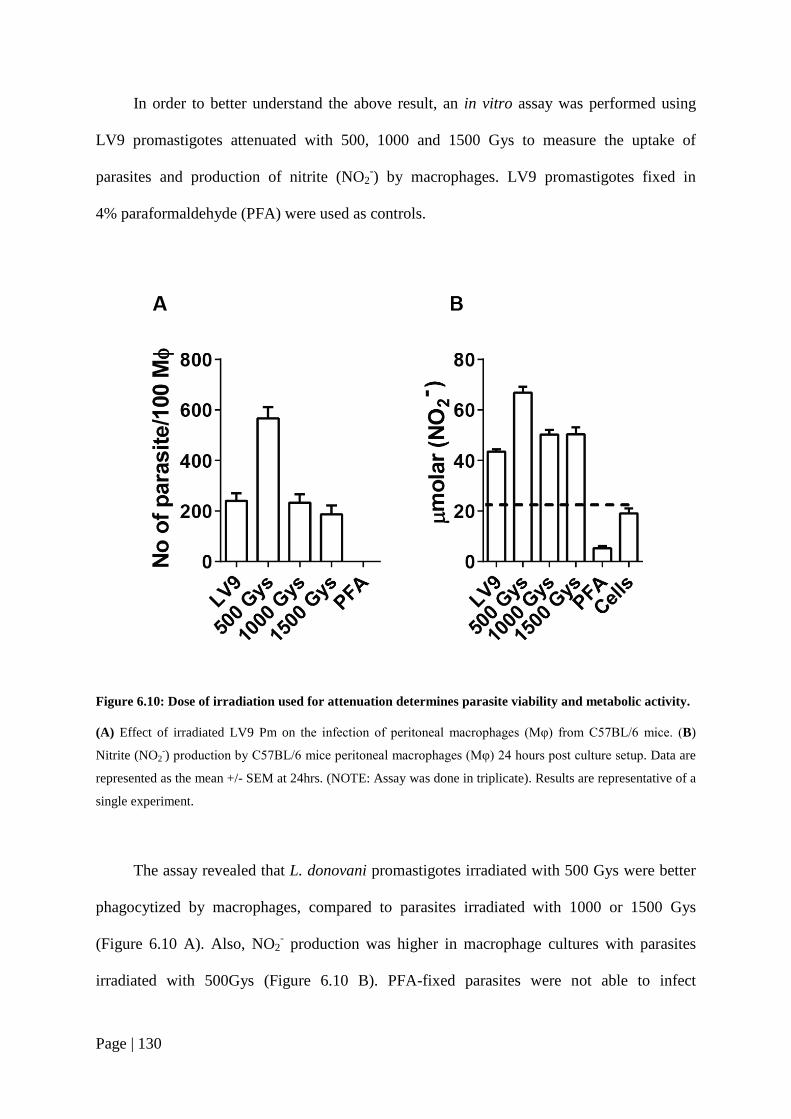
Page | 130
In order to better understand the above result, an in vitro assay was performed using
LV9 promastigotes attenuated with 500, 1000 and 1500 Gys to measure the uptake of
parasites and production of nitrite (NO2-) by macrophages. LV9 promastigotes fixed in
4% paraformaldehyde (PFA) were used as controls.
Figure 6.10: Dose of irradiation used for attenuation determines parasite viability and metabolic activity.
(A) Effect of irradiated LV9 Pm on the infection of peritoneal macrophages (Mφ) from C57BL/6 mice. (B)
Nitrite (NO2-) production by C57BL/6 mice peritoneal macrophages (Mφ) 24 hours post culture setup. Data are
represented as the mean +/- SEM at 24hrs. (NOTE: Assay was done in triplicate). Results are representative of a
single experiment.
The assay revealed that L. donovani promastigotes irradiated with 500 Gys were better
phagocytized by macrophages, compared to parasites irradiated with 1000 or 1500 Gys
(Figure 6.10 A). Also, NO2- production was higher in macrophage cultures with parasites
irradiated with 500Gys (Figure 6.10 B). PFA-fixed parasites were not able to infect

Page | 131
macrophages and the level of NO2- produced by macrophages exposed to these parasites was
minimal. These results suggest that parasites irradiated at doses higher than 1000 Gys were
less viable and metabolically active, compared to parasites irradiated at 500 Gys, and this
may offer some explanation for the results in the previous experiment. However, despite the
increased ability of L. donovani promastigotes irradiated with 500 Gys to be phagocytosed
and induce NO2- production by macrophages, these parasites do not appear to be fully
attenuated (Figure 6.8 A and B). Hence, based on these results I decided to abandon using
radiation as a means of attenuating parasites, and instead focus my efforts on chemical
attenuation of L. donovani promastigotes for development as a vaccine candidate.
6.2.6 The effect of immunization with chemically-attenuated L. donovani promastigotes
in the presence of adjuvant
To assess if immunization with chemically-attenuated L. donovani promastigotes (TFA
LV9) improved parasite control, C57BL/6 mice were immunized with two doses (2x) of TFA
LV9. Mice were challenged with live L. donovani amastigote four weeks after the final
immunization (day 56) and were sacrificed on day 14 p.c. Naïve AMC mice were infected
with L. donovani at the same time as the immunized mice were challenged (Figure 6.1.3).
Antigen-specific cellular immune responses were also measured after immunization.
Administering an adjuvant as part of the vaccine can increase its ability to induce
protective immune responses. Most adjuvants are TLR (toll-like receptor)-agonists and
several studies have demonstrated the efficacy of TLR agonists (TLR3 agonists: Poly I:C
(polyriboinosinic: polyribocytidylic acid), TLR4 agonists: MPL (Monophosphoryl Lipid),

Page | 132
TLR7 agonist: imiquimod and TLR9 agonist: CpG-DNA (immunostimulatory
oligodeoxynucleotides)) against Leishmania and several other diseases [248-251]. Therefore,
I examined the effect of immunizing with chemically-attenuated promastigotes with and
without adjuvant. I focused on using CpG-DNA and Poly I:C, since previous work suggested
that these adjuvant are potent TLR-agonists. A single dose of CpG-DNA or Poly I:C, was
given along with the primary immunization (Figure 6.1.3).
Figure 6.1.3: Experimental timeline for assessment of chemically attenuated parasites for protection against L. donovani.
C57BL/6 mice were immunized with two doses of L. donovani promastigotes that were chemically-attenuated
with TFA (2x10⁷ parasites i.v.). The primary dose was given on day 0 and subsequent boost on day 28. CpG-
DNA or Poly I:C was administered with the first immunization. Four weeks after the final immunization (i.e.,
day 56), mice were challenged with 2 x10⁷ live LV9 amastigotes and were sacrificed on day 14 p.c.
Mice immunized with TFA LV9 (Pm) alone or with CpG-DNA or Poly I:C, showed no
significant increase in liver or spleen weights, compared to naïve mice (Figure 6.11 A).
Analysis of antigen-specific immune responses after immunization, but prior to challenge,
showed increased levels of the pro-inflammatory cytokines IFNγ and TNFα, and the anti-
inflammatory cytokine IL-10, in all immunized groups (Figure 6.11 B).

Page | 133
Figure 6.11: Immunization with chemically-attenuated parasites results in enhanced antigen-specific cellular responses.
(A) Liver and spleen weights in mice immunized with two doses of TFA LV9 (Pm) with or without CpG-DNA
or Poly I:C four weeks after the last immunization. Naïve mice were used as controls. (B) IFNγ, TNFα and IL-
10 (pg/ml) levels from an antigen re-stimulation assay performed on splenocytes four weeks after the last
immunization, but before challenge. Data are represented as the mean +/- SEM at day of challenge. Statistical
differences of p < 0.05 (*) and p < 0.001 (***) are indicated (n=5 mice per group) Results are representative of
a single experiment. (NOTE: the antigen restimulation assay was performed in triplicate).

Page | 134
Mice immunized with two doses of TFA LV9 (Pm) alone or with the CpG-DNA
showed a significant increase in liver and spleen weights at day 14 p.c. (Figure 6.12 A).
However, liver parasite burdens were significantly lower in mice immunized with TFA LV9
(Pm) alone or with Poly I:C, while mice immunized with TFA LV9 (Pm) and CpG-DNA had
lower liver burdens, although these were not significantly different from controls (Figure 6.12
B). In the spleen, although there was a trend for lower parasite burdens in all immunized
groups, none were significantly different from controls (Figure 6.12 B). Hence, addition of
CpG DNA or Poly I:C as adjuvants for chemically-attenuated L. donovani promastigotes, did
not improve vaccine efficacy.

Page | 135
Figure 6.12: Immunization with chemically-attenuated whole L. donovani promastigote results in lower parasite burden in both the liver and spleen, but the addition of adjuvant had no effect.
Organ weight (A) and parasite burdens (B) were measured in the livers and spleens of mice immunized with
TFA LV9 (Pm) alone or in combination with CpG-DNA or Poly I:C. Mice were challenged with L. donovani
four weeks after the last immunization and sacrificed on day 14 p.c. AMC mice were infected with L. donovani
on the day of challenge. Data are represented as the mean +/- SEM on the day of challenge. Statistical
differences of p < 0.05 (*) and p < 0.01 (**) are indicated (n=5 mice per group). Results are representative of a
single experiment.

Page | 136
I next assessed the CD4+ T cell responses in groups of mice immunized with TFA LV9
(Pm) alone, by examining the frequency of Th1 (Tbet+ IFNγ-producing CD4+ T cells) cells
and terminally differentiated Th1 (Tbet+ KLRG-1+ CD4+ T cells) cells (please refer to
previous chapters for representative gating strategy).
Figure 6.13: CD4+ T cell responses in the livers and spleens of mice immunized with chemically-attenuated promastigotes and challenged with L. donovani .
Hepatic (A) and splenic (B) Th1 and KLRG-1+ Th1 cellular responses were measured in mice immunized and
challenged with L. donovani . The frequency of Th1 and KLRG-1+ Th1 cells are shown graphically. Data are
represented as the mean +/- SEM at day 14 p.c. Statistical differences of p < 0.01 (**) are indicated (n=5 mice
per group). Results are representative of a single experiment.

Page | 137
In the liver, I found no significant difference in the frequency of Th1 cells, but a
significant increase in KLRG-1+ Th1 cells in mice immunized with chemically-attenuated
LV9, relative to AMC mice (Figure 6.13 A). In the spleen, a significant increase in the
frequency of both Th1 and KLRG-1+ Th1 cells was observed in groups immunized with
chemically-attenuated LV9 (Figure 6.13 B). Thus, a significantly increased Th1 response was
only found in the spleens of vaccinated mice, but an increased frequency of functionally
exhausted Th1 cells was observed in both tissue sites examined. Together, these data indicate
that mice immunized with two doses of chemically-attenuated LV9 promastigotes over a
period of eight weeks are partially protected in the liver against an L. donovani challenge
infection. However, the addition of CpG-DNA or Poly I:C to the vaccine regime did not
improve vaccine efficacy. Furthermore, despite an increased frequency of Th1 cells in the
spleens of vaccinated mice, this organ remained susceptible to infection.
6.2.7 Chemically-attenuated parasites selectively inhibit pattern recognition receptors
The innate immune system is the host’s first line of defence against pathogens. It is
activated via pattern recognition receptors (PRRs) including TLRs and NLRs (nucleotide-
binding domain like receptors) that are present on and in APCs, where they identify
pathogen-associated molecular patterns (PAMPs). From the above experiment, I found that
immunizing mice with chemically-attenuated parasite along with adjuvant did not further
boost protective immune responses. I therefore hypothesised that chemically-attenuated L.
donovani parasites inhibited PRR signalling pathways, thus preventing immuno-stimulatory
effects of the TLR-agonist CpG-DNA and Poly I:C. Thus, I set up an experiment using
purified DC’s isolated from the spleens of naïve C57BL/6 mice and cultured them with CpG-
Page | 138
DNA or Poly I:C alone. In addition, chemically-attenuated parasites (TFA treated L.
donovani promatigotes) were also added to these cultures. I then measured the levels of the
pro-inflammatory cytokines IL-1β, IL-6, IL-12 and TNFα in the culture supernatants as these
cytokines play important roles in DC maturation and function during infection [252-254].
Levels of the pro-inflammatory cytokines IL-1β, IL-6, IL-12 and TNFα did not change
when TFA LV9 was added to unstimulated DC cultures. Although the addition of CpG-DNA
or Poly I:C to DC cultures resulted in increased production of IL-1β, IL-6, TNFα and IL-12
(IL-12 levels only increased in the presence of CpG-DNA), the introduction of TFA LV9
caused a significant decrease in IL-1β, IL-6, and TNFα levels, but no change to IL-12 levels.
Together, these results show that chemically-attenuated L. donovani promastigotes
selectively inhibited cytokine production stimulated by stimulation of PRRs (Figure 6.14).

Page | 139
Figure 6.14: Chemically attenuated L. donovani promastigotes inhibit pattern recognition pathways in DC’s.
IL-12p70, IL-1β, IL-6 and TNFα (pg/ml) levels from the DC cultures. Data are represented as the mean +/- SEM. Statistical differences of p < 0.05 (*), p < 0.01 (**) and p <
0.001 (***) are indicated (DCs isolated from n=5 mice and pooled) (NOTE: each cell culture condition was performed in triplicate wells). Results are representative of a
single experiment.

Page | 140
6.3 DISCUSSSION
In this chapter, I examined the effectiveness of attenuated, whole parasite vaccines, to
generate anti-parasitic immune responses against L. donovani. To accomplish this, whole L.
donovani amastigotes or promastigotes were irradiated or chemically-attenuated and used as
vaccine candidates. “Leishmaniszation”, the practice of inoculating people with live L. major
on unexposed areas of the body, resulted in long-term protection against CL infection [236].
This indicates that a vaccine against Leishmania is achievable, and I therefore tested whether
attenuated whole L. donovani parasites could be used to generate protective immunity.
Initially, I aimed to use irradiated L. donovani promastigotes as my vaccine following a
vaccine regime describe by Scott et al. [243]. However, I found little indication that the
immunization with different doses of irradiated L. donovani promastigotes given through
different immunization routes had any statistically significant effect on parasite burdens post
challenge. Hence, these results indicated that either the vaccine candidates or the vaccination
regime were not ideal. A study by Castiglione et al. [255], indicated that the interval between
the prime and the boost vaccinations affects the immune response generated. Indeed, I found
that parasite burdens were better controlled in the livers of challenged mice when irradiated
or chemically-attenuated L. donovani amastigotes were used, but the interval between the
prime and the boost injection had to be increased from one week to four weeks. However, I
discovered that when irradiated L. donovani was injected into immune compromised mice,
parasite numbers expanded indicating they were not fully attenuated. Increasing the radiation
dose, however, lead to the loss of vaccine protection, suggesting that higher doses of
radiation caused parasites to be non-viable and unable to induce a potent immune response.

Page | 141
Therefore, I focused my efforts on testing chemically-attenuated L. donovani promastigotes,
and found they could be used to reduce parasite burdens in the livers of immunized mice,
following challenge with L. donovani. However, attempts to combine adjuvant with the
attenuated parasites failed and no improvement in immunity was observed. Further testing
indicated that the live, chemically attenuated parasites were inhibiting pattern recognition
pathways on DC’s, supporting observation by others with live parasites [256, 257]. I only
tested the effects of parasites on CpG DNA- and Poly I:C-mediated activation, so cannot be
sure whether all pattern recognition pathways are affected or if this is a selective process.
Given the importance of adjuvants for vaccine efficacy, this is an important question that will
have to be addressed in the future.
An ideal vaccine is one that elicits strong protective immune responses without causing
disease. Achieving the correct level of attenuation is a challenging task, as it is likely to have
an impact on the efficacy and safety of the vaccine. If the attenuation is inefficient, there is a
chance of parasite growth, resulting in disease progression. If the level of attenuation is too
high (e.g. too high a radiation dose), my data indicates this will result in a poor vaccine
because the parasites are unable to activate innate immune responses in the host. Manna et
al. reported that 100-150 Gy was an appropriate radiation doses for parasite attenuation to
stimulate an immune response without parasite outgrowth, while 50 a dose of Gy allowed
parasite growth [258]. My results showed that attenuation with 500 Gy induced immune
responses, while increasing the dose to 1500 Gy resulted in loss a of protective immunity.
However, immune compromised experimental hosts infected L. donovani parasites irradiated
with 500 Gy had higher parasite burden compared to mice infected with the chemically
attenuated parasites. Thus, careful attention must be paid to select both the right method and
level of attenuation when using live parasites as vaccines.

Page | 142
Vaccines can either be used for prevention of disease or therapeutically. A preventative
vaccine could be deployed in VL endemic areas, and also used to protect individuals
travelling to these areas. Therapeutic vaccines can be used to treat people with an established
infection, and are generally designed to boost the body's immune response in order to better
control the infection [259]. One example where a therapeutic vaccine might be useful to
control L. donovani infection is to help treat post kala azar dermal leishmaniasis (PKDL),
which develops after treatment of visceral leishmaniasis (VL) and is observed in 50-60%
patients in Sudan and in about 0.8-8% of cases in India [260]. Most PKDL patients heal
naturally within 6 months, but those who don't often require months of daily drug treatment.
In these patients, a vaccine that boosted anti-parasitic immunity may be helpful. In VL
endemic regions, there are many asymptomatic individuals [185]. These people are potential
reservoirs of parasite that may contribute to parasite transmission. As such, therapeutic
vaccines may be used to reduce parasite loads in these individuals in an effort to reduce the
risk of transmission. In an era where VL elimination is on the WHO agenda, it is doubtful
that chemotherapy alone will achieve this goal. Therefore, new strategies, such as a
therapeutic vaccination, with or without drug treatment, may be useful by reducing
transmission of parasites and preventing the development drug resistance in parasites by
reducing reliance on chemotherapy.
In summary my study provides proof-of-principal for a chemically attenuated whole
parasite vaccine. I identify several key factors that impact on the effectiveness of an
attenuated whole parasite vaccine, including the means of parasite attenuation, the
vaccination regime, and the adjuvant used. Clearly, there is still much work to be done to
develop a chemically attenuated vaccine for testing in humans, but my work has made an
important start in this direction.

Page | 143
Chapter 7: Concluding remarks
The main aims of this thesis focused on testing new ways to improve sub-optimal
CD4+ T cell responses during L. donovani infection. The first two results Chapters (Chapters
4 and 5) of this thesis focused on improving anti-parasitic CD4+ T cells responses generated
during experimental VL by using combination immune therapy and cytokine therapy. The
third results Chapter (Chapter 6) dealt with generating anti-parasitic immune responses using
a whole parasite vaccine.
In Chapter 4, I showed that combined immune therapy with an agonistic anti-GITR
mAb and anti-IL-10R blocking mAb had no additive effect on the control of parasite burden
in experimental VL, compared to IL-10R blocking alone. However, when mice infected with
a lower dose of parasites were treated with the same combination of mAbs, GITR activation
actually reversed the positive anti-parasitic effects observed with anti-IL-10R mAb treatment
in the liver. This indicated that treatment outcomes depended on several factors, such as
parasite load at the start of treatment and the organ being targeted for treatment. Furthermore,
our Indian collaborators showed that GITR activation in combination with IL-10 blockade
had no significant impact on parasite growth in spleen samples or at improving antigen-
specific IFNγ production in whole blood from VL patients (unpublished data; appendix 1).
These results suggest that selecting an immune modulator without causing harm to the host is
critical. Haque et al. [114] showed that stimulation of GITR via an agonistic mAb resulted in
expansion of conventional CD4+ T cells. Interestingly, although treatment with the combined
mAb treatment resulted in expansion of Th1 CD4+ T cells, these cells had a functionally
exhausted phenotype. This highlights the importance of understanding the mechanism of

Page | 144
GITR activity in experimental VL so that the appropriate time to administer anti-GITR mAb
can be determined. Antimonial chemotherapy has been the primary line of VL treatment for
more than 50 years [3]. However, administration of the drugs over a long period of time
resulted in toxicity and drug resistance, and increasing cost of alternative drugs is a major
issue. Combining immune modulation that stimulates the immune system with anti-parasitic
drugs has the potential to aid in the treatment of a broad range of diseases. In the study by
Haque et al. mentioned above, administering the agonistic GITR mAb with a suboptimal
dose of anti-leishmanial drug resulted in enhanced parasite clearance. In my study, treatment
with the combination cocktail along with the drug in a low dose infection resulted in
significantly enhanced anti-parasitic immune responses and reduced parasite burdens.
Interestingly, IL-10R blockade combined with chemotherapy also resulted in significantly
enhanced anti-parasitic immune responses. Studies have shown that targeting PD-1, LAG-3
and CTLA-4 inhibitory receptors individually or in combination in VL, and various other
disease settings, have significantly improved immune responses [191, 196, 261, 262]. Further
studies should be undertaken in order to establish an ideal combination of immune
modulators that can work well with existing drugs to optimise the generation of anti-parasite
immunity in VL.
In Chapter 5, I showed that IL-2/anti-IL-2 mAb complex therapy with either IL-2Jc or
IL-2Sc significantly reduced parasite burdens in experimental VL. However, an associated
expansion of potential target cell populations was not observed. Several studies have shown
that CD4+ Foxp3+ T (Treg) cells play a central role in regulating anti-parasitic immunity
during Leishmania infection, but this is dependent on the parasite species being studied. In L.
major infection, the role of Treg cells seems to be dependent on the genetic background of
the host. In BALB/c mice, Treg cells facilitate protection by dampening detrimental Th2

Page | 145
responses [263], while in C57BL/6 infected mice, anti-parasitic Th1 response are suppressed
by Treg cells and prevents sterile cure [211]. In a study by Sacks et al. further suggested that
in an experimental L. major infection, large numbers of IFNγ+ IL-10- Th1 cells are need
relatively early in the infection to achieve sterile cure [264]. Another study by Ehrlich, A. et
al. showed that in an experimental L. (V.) panamensis infection, induction of Treg cells in-
vivo with IL-2Jc lead to resolution of active disease [265]. Treatment with IL-2Jc was carried
out i.p. over a nine day period (every third day), and resulted in significant expansion of
Treg cells at day 2 post-treatment, followed by a decrease in Treg cell numbers by 8 days
post-treatment. This study, as well as other, report a 24 hour half-life for the IL-2/anti-IL-2
mAb complex [266], and demonstrate that increased Treg cell numbers and function are
short-lived, despite continuation of IL-2/anti-IL-2 mAb complex treatment [210]. This could
explain the failure to detect any cellular expansion in my study, and suggests that optimising
the dose and timing of IL-2/anti-IL-2 mAb complex (IL-2Jc) treatment may offer a way to
improve efficacy. Further investigation into the role of IL-2/anti-IL-2 mAb complexes in
promoting the expansion of specific T cell subsets during L. donovani infection is warranted.
As mentioned previously, treatment with IL-2Sc resulted in expansion of CD8+ T cells.
Stern et al. showed that depletion of CD8+ T cells in an established L. donovani infection lead
to impaired granuloma formation, which resulted in disease exacerbation, thus [65]
highlighting the importance of CD8+ T cells for parasite control. In another study by Tsagozis
et al., isolated antigen-specific CD8+ T cells expressed high levels of IFNγ and TNFα, and
elevated cytotoxic activity against Leishmania antigens [115]. However, despite the evidence
that CD8+ T cells are strong mediators of immune responses against Leishmania, recent
findings suggest that L. donovani induces defective CD8+ T cell antigen-specific responses,
by interfering with clonal expansion of CD8+ T cells, resulting in cell dysfunction and

Page | 146
eventually exhaustion [107]. It is again possible that altering the dosing regime for IL-2Sc
treatment might better target expansion of anti-parasitic CD8+ T cells, which was not detected
in my experiments.
The development of a vaccine to protect against VL depends on generating potent
antigen-specific immune responses that are able to protect the host from the primary infection
and also provide long-term protection against re-infection. In Chapter 6, I investigated the
potential of a live, attenuated whole parasite vaccine against a L. donovani infection. I found
that vaccine efficacy depended on the interval between the prime and boost, the life cycle
stage of the L. donovani parasite and degree of parasite attenuation. Mice immunized with the
primary vaccine candidate (irradiated L. donovani amastigotes), were able to control parasite
burdens only in the liver, whereas those challenged with irradiated L. donovani promastigotes
controlled parasites in both the liver and spleen. However, when irradiated parasites were
injected into an immunocompromised mouse, parasites expanded, indicating they were only
partially attenuated. Increasing the dose of radiation resulted in loss of protective immune
responses conferred by the vaccine, suggesting that the higher dose caused rapid killing of the
parasite, and that live, attenuated parasite vaccines should be better than an inactive parasite
vaccine. A study by Sacks et al. showed that during chronic Leishmania infection short lived
effector T cells (TEF) are maintained at high frequencies and these are critical for protective
immunity against reinfection and mediate a state of concomitant immunity, however these
cells fail to survive in the absence of antigen [267]. Developing a vaccine that can provide
long term depository of antigen to generate these short lived TEF would be worth
investigating. In a study by Belshe R et al. immunizing 7,852 children with either live
attenuated influenza vaccine or an inactivated vaccine showed 54.9% fewer cases of
influenza in the group that received live attenuated vaccine, compared to the group that

Page | 147
received inactivated vaccine [268], providing further support for the idea that live attenuated
vaccines will be more effective at generating potent immune responses than killed vaccines.
However, the right method and dose of attenuation is essential to prevent the establishment of
an infection following vaccination. Chemical attenuation is a feasible alternative to radiation,
as conditions for attenuation can be strictly controlled. Indeed, we observed that chemical
attenuation of L. donovani promastigotes controlled burdens in both the liver and the spleen,
and although attenuated parasite were detected in immunocompromised mice, it is likely that
they derived from the initial inoculum of attenuated parasites. Many studies have shown that
administering an adjuvant in combination with a vaccine increases vaccine efficiency [248-
251]. In my vaccine studies, however, I found no additive effect of the adjuvants Poly (I:C)
and CpG-DNA. A further analysis of this result indicated that chemically attenuated parasites
selectively inhibited pattern recognition receptor (PRR) activation of APCs by these
molecules. This aspect of a live, attenuated L. donovani vaccines needs to be further
examined because the vaccine alone does not appear to provide optimal protection. It is
possible that adjuvants that target alternative PRR pathways might be better suited to this
type of vaccine.
The idea of combining immune modulation with vaccination has been investigated in
CL; blockade of IL-10 signalling during vaccination against L. major resulted in a significant
decrease in inflammation and parasite burden, compared to vaccination alone [269-271].
Based on these and various other studies, it would be interesting to investigate the outcome of
combining immune modulating antibodies or cytokine therapy strategies with the attenuated
vaccine examined in my study.

Page | 148
In conclusion, my study highlights the following: 1) the need to identify the right
targets for immune modulation for a safe and progressive therapy outcome. 2) cytokine
therapy with IL-2/anti-IL-2 complexes could be considered as treatment against VL and 3)
development of vaccines against VL requires careful consideration, including the type of
attenuation, the parasite lifecycle stage, vaccination regime, adjuvants and/or other types of
immune modulators.

Page | 149
Chapter 8: Bibliography
1. David, C. and N. Craft, Cutaneous and mucocutaneous leishmaniasis. Dermatologic Therapy, 2009. 22(6): p. 491-502.
2. Alvar, J., et al., Leishmaniasis worldwide and global estimates of its incidence. PLoS ONE, 2012. 7(5): p. e35671.
3. Croft, S., S. Sundar, and A. Fairlamb, Drug resistance in leishmaniasis. Clinical Microbiology Reviews, 2006. 19(1): p. 111-126.
4. Duncan, R., et al., Identification and Characterization of Genes Involved in Leishmania Pathogenesis: The Potential for Drug Target Selection. Molecular Biology International, 2011. 2011.
5. Kedzierski, L., Y. Zhu, and E. Handman, Leishmania vaccines: progress and problems. Parasitology, 2006. 133(SupplementS2): p. S87-S112.
6. Handman, E., Leishmaniasis: Current status of vaccine development. Clinical Microbiology Reviews, 2001. 14(2): p. 229-243.
7. Modabber, F., Vaccines against leishmaniasis. Annals of Tropical Medicine and Parasitology, 1995. 89: p. 83-88.
8. Serebryakov, V., S. Karakhodzhaeva, and M. Dzhumaev, Effect of leishmanial vaccinations on the dynamics of immunity to diphtheria in conditions of secondary revaccination with adsorbed pertussis-diphtheria-tetanus vaccine. Meditsinskaya Parazitologiya i Parazitarnye Bolezni, 1972. 41(3): p. 303-307.
9. Stanley, A.C. and C.R. Engwerda, Balancing immunity and pathology in visceral leishmaniasis. Immunology and Cell Biology, 2007. 85(2): p. 138-147.
10. Bañuls, A.-L., M. Hide, and F. Prugnolle, Leishmania and the Leishmaniases: A Parasite Genetic Update and Advances in Taxonomy, Epidemiology and Pathogenicity in Humans, in Advances in Parasitology, R.M. J.R. Baker and D. Rollinson, Editors. 2007, Academic Press. p. 1-458.
11. David, C.V. and N. Craft, Cutaneous and mucocutaneous leishmaniasis. Dermatologic Therapy, 2009. 22(6): p. 491-502.
12. Bankoti, R. and S. Stäger, Differential regulation of the immune response in the spleen and liver of mice infected with Leishmania donovani. Journal of Tropical Medicine, 2012. 2012.
13. Stanley, A. and C. Engwerda, Balancing immunity and pathology in visceral leishmaniasis. Immunology and Cell Biology, 2007. 85(2): p. 138-147.
14. Moore, K. and G. Matlashewski, Intracellular infection by Leishmania donovani inhibits macrophage apoptosis. The Journal of Immunology, 1994. 152(6): p. 2930-2937.
15. Pearson, R. and A. Sousa, Clinical spectrum of leishmaniasis. Clinical Infectious Diseases, 1996. 22(1): p. 1-11.
16. Dedet, J. and F. Pratlong, In Leishmaniasis., in Manson's Tropical Diseases, Cook GC and Z. AI., Editors. 2008, Saunders: London p. 1341-1365.
17. Murray, H., Treatment of visceral leishmaniasis (kala-azar): A decade of progress and future approaches. International Journal of Infectious Diseases, 2000. 4(3): p. 158-177.
18. Srivastava, P., et al., Diagnosis of Indian Visceral Leishmaniasis by Nucleic Acid Detection Using PCR. PLoS ONE, 2011. 6(4): p. e19304.

Page | 150
19. Ho, M., et al., Prevalence and disease spectrum in a new focus of visceral leishmaniasis in Kenya. Transactions of the Royal Society of Tropical Medicine and Hygiene, 1982. 76(6): p. 741-746.
20. Nylén, S. and R. Kumar, Immunobiology of visceral leishmaniasis. Frontiers in Immunology, 2012. 3.
21. Sacks, D., et al., An analysis of T cell responsiveness in Indian kala-azar. The Journal of Immunology, 1987. 138(3): p. 908-913.
22. Bucheton, B., et al., The interplay between environmental and host factors during an outbreak of visceral leishmaniasis in eastern Sudan. Microbes and Infection, 2002. 4(14): p. 1449-1457.
23. Bucheton, B., et al., Genetic control of visceral leishmaniasis in a Sudanese population: candidate gene testing indicates a linkage to the NRAMP1 region. 2003. 4(2): p. 104-109.
24. Mohamed, H., et al., SLC11A1 (formerly NRAMP1) and susceptibility to visceral leishmaniasis in The Sudan. Eur J Hum Genet, 2003. 12(1): p. 66-74.
25. Mehrotra, S., et al., No evidence for association between SLC11A1 and visceral leishmaniasis in India. BMC Medical Genetics, 2011. 12(1): p. 71.
26. Mehrotra, S., et al., Genetic and functional evaluation of the role of CXCR1 and CXCR2 in susceptibility to visceral leishmaniasis in north-east India. BMC Medical Genetics, 2011. 12(1): p. 162.
27. Bucheton, B., et al., Identification of a novel G245R polymorphism in the IL-2 receptor [beta] membrane proximal domain associated with human visceral leishmaniasis. Genes Immun, 2006. 8(1): p. 79-83.
28. Blackwell, J. and M. Roberts, Immunomodulation of murine visceral leishmaniasis by administration of monoclonal anti-Ia antibodies: differential effects of anti-I-A vs. anti-I-E antibodies. European Journal of Immunology, 1987. 17(11): p. 1669-1672.
29. Kaye, P., et al., Altered course of visceral leishmaniasis in mice expressing transgenic I-E molecules. European Journal of Immunology, 1992. 22(2): p. 357-364.
30. Blackwell, J., J. Freeman, and D. Bradley, Influence of H-2 complex on acquired resistance to Leishmania donovani infection in mice. Nature, 1980. 283(5742): p. 72-74.
31. Fakiola, M., et al., Common variants in the HLA-DRB1-HLA-DQA1 HLA class II region are associated with susceptibility to visceral leishmaniasis. Nat Genet, 2013. 45(2): p. 208-213.
32. Nylén, S. and D. Sacks, Interleukin-10 and the pathogenesis of human visceral leishmaniasis. Trends in Immunology, 2007. 28(9): p. 378-384.
33. Anstead, G., et al., Malnutrition Alters the Innate Immune Response and Increases Early Visceralization following Leishmania donovani Infection. Infection and Immunity, 2001. 69(8): p. 4709-4718.
34. Hughes, S. and P. Kelly, Interactions of malnutrition and immune impairment, with specific reference to immunity against parasites. Parasite Immunology, 2006. 28(11): p. 577-588.
35. O’Neal, S., et al., Influence of Helminth Infections on the Clinical Course of and Immune Response to Leishmania braziliensis Cutaneous Leishmaniasis. Journal of Infectious Diseases, 2007. 195(1): p. 142-148.
36. Maurya, R., et al., A risk of visceral leishmaniasis in case of helminths co-infection in endemic regions. Innovative Journal of Medical and Health Sciences, 2012. 2(4).
37. Ostyn, B., et al., Incidence of Symptomatic and Asymptomatic Leishmania donovani Infections in High-Endemic Foci in India and Nepal: A Prospective Study. PLoS Negl Trop Dis, 2011. 5(10): p. e1284.

Page | 151
38. Wilson, M., S. Jeronimo, and R. Pearson, Immunopathogenesis of infection with the visceralizing Leishmania species. Microbial Pathogenesis, 2005. 38(4): p. 147-160.
39. Badaro, R., et al., New Perspectives on a Subclinical Form of Visceral Leishmaniasis. The Journal of Infectious Diseases, 1986. 154(6): p. 1003-1011.
40. Ostyn, B., et al., Incidence of Symptomatic and Asymptomatic <italic>Leishmania donovani</italic> Infections in High-Endemic Foci in India and Nepal: A Prospective Study. PLoS Negl Trop Dis, 2011. 5(10): p. e1284.
41. Jeronimo, S., et al., Natural History of Leishmania (Leishmania) chagasi infection in Northeastern Brazil: Long-Term Follow-Up. Clinical Infectious Diseases, 2000. 30(3): p. 608-609.
42. Singh, O., et al., Reassessment of Immune Correlates in Human Visceral Leishmaniasis as Defined by Cytokine Release in Whole Blood. Clinical and Vaccine Immunology, 2012. 19(6): p. 961-966.
43. Das, A. and N. Ali, Vaccine development against Leishmania donovani. Frontiers in Immunology, 2012. 3.
44. Ansari, N., et al., IL-27 and IL-21 Are Associated with T Cell IL-10 Responses in Human Visceral Leishmaniasis. The Journal of Immunology, 2011. 186(7): p. 3977-3985.
45. Gidwani, K., et al., Serological Markers of Sand Fly Exposure to Evaluate Insecticidal Nets against Visceral Leishmaniasis in India and Nepal: A Cluster-Randomized Trial. PLoS Negl Trop Dis, 2011. 5(9): p. e1296.
46. Nylén, S., et al., Splenic accumulation of IL-10 mRNA in T cells distinct from CD4+CD25+ (Foxp3) regulatory T cells in human visceral leishmaniasis. The Journal of Experimental Medicine, 2007. 204(4): p. 805-817.
47. Sundar, S., et al., Circulating T Helper 1 (Th1) Cell- and Th2 Cell-Associated Cytokines in Indian Patients with Visceral Leishmaniasis. The American Journal of Tropical Medicine and Hygiene, 1997. 56(5): p. 522-525.
48. Ansari, N., S. Saluja, and P. Salotra, Elevated levels of interferon-γ, interleukin-10, and interleukin-6 during active disease in Indian kala azar. Clinical Immunology, 2006. 119(3): p. 339-345.
49. Anderson, C.F., et al., CD4+CD25−Foxp3− Th1 cells are the source of IL-10–mediated immune suppression in chronic cutaneous leishmaniasis. The Journal of Experimental Medicine, 2007. 204(2): p. 285-297.
50. Boussiotis, V.A., et al., IL-10–producing T cells suppress immune responses in anergic tuberculosis patients. The Journal of Clinical Investigation, 2000. 105(9): p. 1317-1325.
51. Plebanski, M., et al., Interleukin 10-mediated immunosuppression by a variant CD4 T cell epitope of Plasmodium falciparum. Immunity, 1999. 10(6): p. 651-660.
52. Rodríguez-García, M., et al., Expression of PD-L1 and PD-L2 on human macrophages is up-regulated by HIV-1 and differentially modulated by IL-10. Journal of Leukocyte Biology, 2011. 89(4): p. 507-515.
53. Couper, K., D. Blount, and E. Riley, IL-10: The Master Regulator of Immunity to Infection. The Journal of Immunology, 2008. 180(9): p. 5771-5777.
54. Gautam, S., et al., IL-10 neutralization promotes parasite clearance in splenic aspirate cells from patients with visceral leishmaniasis. Journal of Infectious Diseases, 2011. 204(7): p. 1134-1137.
55. Ghalib, H., et al., IL-12 enhances Th1-type responses in human Leishmania donovani infections. The Journal of Immunology, 1995. 154(9): p. 4623-9.

Page | 152
56. Carvalho, E., et al., Restoration of IFN-gamma production and lymphocyte proliferation in visceral leishmaniasis. The Journal of Immunology, 1994. 152(12): p. 5949-56.
57. Ghalib, H., et al., Interleukin 10 production correlates with pathology in human Leishmania donovani infections. The Journal of Clinical Investigation, 1993. 92(1): p. 324-329.
58. Rai, A.K., et al., Regulatory T Cells Suppress T Cell Activation at the Pathologic Site of Human Visceral Leishmaniasis. PLoS ONE, 2012. 7(2): p. e31551.
59. Spolski, R., et al., IL-21 Mediates Suppressive Effects via Its Induction of IL-10. The Journal of Immunology, 2009. 182(5): p. 2859-2867.
60. Buxbaum, L. and P. Scott, Interleukin 10- and Fcγ Receptor-Deficient Mice Resolve Leishmania mexicana Lesions. Infection and Immunity, 2005. 73(4): p. 2101-2108.
61. Caldas, A., et al., Balance of IL-10 and Interferon-gamma plasma levels in human visceral leishmaniasis: Implications in the pathogenesis. BMC Infectious Diseases, 2005. 5(1): p. 113.
62. Somanna, A., V. Mundodi, and L. Gedamu, Functional Analysis of Cathepsin B-like Cysteine Proteases fromLeishmania donovani Complex: EVIDENCE FOR THE ACTIVATION OF LATENT TRANSFORMING GROWTH FACTOR β. Journal of Biological Chemistry, 2002. 277(28): p. 25305-25312.
63. Gantt, K., et al., Activation of TGF-β by Leishmania chagasi: Importance for Parasite Survival in Macrophages. The Journal of Immunology, 2003. 170(5): p. 2613-2620.
64. Pitta, M., et al., IL-17 and IL-22 are associated with protection against human kala azar caused by Leishmania donovani. The Journal of Clinical Investigation, 2009. 119(8): p. 2379-2387.
65. Stern, J., et al., Role of L3T4+ and LyT-2+ cells in experimental visceral leishmaniasis. The Journal of Immunology, 1988. 140(11): p. 3971-3977.
66. Colmenares, M., et al., Perforin and Gamma Interferon Are Critical CD8+ T-Cell-Mediated Responses in Vaccine-Induced Immunity against Leishmania amazonensis Infection. Infection and Immunity, 2003. 71(6): p. 3172-3182.
67. Stenger, S., et al., An Antimicrobial Activity of Cytolytic T Cells Mediated by Granulysin. Science, 1998. 282(5386): p. 121-125.
68. Ganguly, S., et al., Increased Levels of Interleukin-10 and IgG3 Are Hallmarks of Indian Post-Kala-Azar Dermal Leishmaniasis. Journal of Infectious Diseases, 2008. 197(12): p. 1762-1771.
69. Bourreau, E., et al., IL-10 producing CD8+ T cells in human infection with Leishmania guyanensis. Microbes and Infection, 2007. 9(8): p. 1034-1041.
70. Gautam, S., et al., CD8 T Cell Exhaustion in Human Visceral Leishmaniasis. Journal of Infectious Diseases, 2013.
71. Kaye, P., et al., The immunopathology of experimental visceral leishmaniasis. Immunological Reviews, 2004. 201(1): p. 239-253.
72. Blackwell, J., et al., SLC11A1 (formerly NRAMP1) and disease resistance. Cellular Microbiology, 2001. 3(12): p. 773-784.
73. Smelt, S., et al., Destruction of follicular dendritic cells during chronic visceral leishmaniasis. The Journal of Immunology, 1997. 158(8): p. 3813-3821.
74. Engwerda, C. and P. Kaye, Organ-specific immune responses associated with infectious disease. Immunology today, 2000. 21(2): p. 73-78.
75. Wilson, M., et al., Local suppression of IFN-gamma in hepatic granulomas correlates with tissue-specific replication of Leishmania chagasi. The Journal of Immunology, 1996. 156(6): p. 2231-2239.

Page | 153
76. Engwerda, C., et al., Distinct roles for lymphotoxin-α and tumor necrosis factor in the control of Leishmania donovani infection. The American Journal of Pathology, 2004. 165(6): p. 2123-2133.
77. McElrath, M., H. Murray, and Z. Cohn, The dynamics of granuloma formation in experimental visceral leishmaniasis. The Journal of Experimental Medicine, 1988. 167(6): p. 1927-1937.
78. Cervia, J., H. Rosen, and H. Murray, Effector role of blood monocytes in experimental visceral leishmaniasis. Infection and Immunity, 1993. 61(4): p. 1330-1333.
79. Smelt, S., et al., B cell-deficient mice are highly resistant to Leishmania donovani infection, but develop neutrophil-mediated tissue pathology. The Journal of Immunology, 2000. 164(7): p. 3681-3688.
80. McFarlane, E., et al., Neutrophils Contribute to Development of a Protective Immune Response during Onset of Infection with Leishmania donovani. Infection and Immunity, 2008. 76(2): p. 532-541.
81. Peters, N., et al., In Vivo Imaging Reveals an Essential Role for Neutrophils in Leishmaniasis Transmitted by Sand Flies. Science, 2008. 321(5891): p. 970-974.
82. Sato, N., et al., Defects in the Generation of IFN-γ Are Overcome to Control Infection with Leishmania donovani in CC Chemokine Receptor (CCR) 5-, Macrophage Inflammatory Protein-1α-, or CCR2-Deficient Mice. The Journal of Immunology, 1999. 163(10): p. 5519-5525.
83. Sheel, M. and C. Engwerda, The diverse roles of monocytes in inflammation caused by protozoan parasitic diseases. Trends in Parasitology, 2012. 28(10): p. 408-416.
84. León, B., M. López-Bravo, and C. Ardavín, Monocyte-Derived Dendritic Cells Formed at the Infection Site Control the Induction of Protective T Helper 1 Responses against Leishmania. Immunity, 2007. 26(4): p. 519-531.
85. Kaye, P. and G. Bancroft, Leishmania donovani infection in scid mice: lack of tissue response and in vivo macrophage activation correlates with failure to trigger natural killer cell-derived gamma interferon production in vitro. Infection and Immunity, 1992. 60(10): p. 4335-4342.
86. Engwerda, C., et al., Neutralization of IL-12 demonstrates the existence of discrete organ-specific phases in the control of Leishmania donovani. European Journal of Immunology, 1998. 28(2): p. 669-680.
87. Murray, H., Z. Xiang, and X. Ma, Responses to Leishmania donovani in mice deficient in both phagocyte oxidase and inducible nitric oxide synthase. The American Journal of Tropical Medicine and Hygiene, 2006. 74(6): p. 1013-1015.
88. Lefèvre, L., et al., The C-type Lectin Receptors Dectin-1, MR, and SIGNR3 Contribute Both Positively and Negatively to the Macrophage Response to Leishmania infantum. Immunity, 2013. 38(5): p. 1038-1049.
89. Lima-Junior, D., et al., Inflammasome-derived IL-1[beta] production induces nitric oxide-mediated resistance to Leishmania. Nat Med, 2013. 19(7): p. 909-915.
90. Murray, H., et al., Experimental visceral leishmaniasis: Production of interleukin 2 and interferon-gamma, tissue immune reaction, and response to treatment with interleukin 2 and interferon-gamma. The Journal of Immunology, 1987. 138(7): p. 2290-2297.
91. Alexander, C., P. Kaye, and C. Engwerda, CD95 is required for the early control of parasite burden in the liver of Leishmania donovani-infected mice. European Journal of Immunology, 2001. 31(4): p. 1199-1210.
92. Murray, H., et al., Antimicrobial Response of a T Cell-Deficient Host to Cytokine Therapy: Effect of Interferon-γ in Experimental Visceral Leishmaniasis in Nude Mice. Journal of Infectious Diseases, 1995. 171(5): p. 1309-1316.

Page | 154
93. Cotterell, S., C. Engwerda, and P. Kaye, Leishmania donovani infection of bone marrow stromal macrophages selectively enhances myelopoiesis, by a mechanism involving GM-CSF and TNF-α. Blood, 2000. 95(5): p. 1642-1651.
94. Gorak, P.A., C. Engwerda, and P. Kaye, Dendritic cells, but not macrophages, produce IL-12 immediately following Leishmania donovani infection. European Journal of Immunology, 1998. 28(2): p. 687-695.
95. Melby, P., et al., Leishmania donovani: Evolution and Architecture of the Splenic Cellular Immune Response Related to Control of Infection. Experimental Parasitology, 2001. 99(1): p. 17-25.
96. Cotterell, S., C. Engwerda, and P. Kaye, Enhanced Hematopoietic Activity Accompanies Parasite Expansion in the Spleen and Bone Marrow of Mice Infected with Leishmania donovani. Infection and Immunity, 2000. 68(4): p. 1840-1848.
97. Engwerda, C., et al., A Role for tumor necrosis factor-α in remodeling the splenic marginal zone during Leishmania donovani infection. The American Journal of Pathology, 2002. 161(2): p. 429-437.
98. Aichele, P., et al., Macrophages of the splenic marginal zone are essential for trapping of blood-borne particulate antigen but dispensable for induction of specific T cell responses. The Journal of Immunology, 2003. 171(3): p. 1148-1155.
99. Ato, M., et al., Defective CCR7 expression on dendritic cells contributes to the development of visceral leishmaniasis. Nat Immunol, 2002. 3(12): p. 1185-1191.
100. Dalton, J., et al., Inhibition of receptor tyrosine kinases restores immunocompetence and improves immune-dependent chemotherapy against experimental leishmaniasis in mice. The Journal of Clinical Investigation, 2010. 120(4): p. 1204-1216.
101. Svensson, M., et al., Stromal Cells Direct Local Differentiation of Regulatory Dendritic Cells. Immunity, 2004. 21(6): p. 805-816.
102. Wakkach, A., et al., Characterization of Dendritic Cells that Induce Tolerance and T Regulatory 1 Cell Differentiation In Vivo. Immunity, 2003. 18(5): p. 605-617.
103. Varma, N. and S. Naseem, Hematologic Changes in Visceral Leishmaniasis/Kala Azar. Indian Journal of Hematology and Blood Transfusion, 2010. 26(3): p. 78-82.
104. Murphy, M., et al., IL-10 mediates susceptibility to Leishmania donovani infection. European Journal of Immunology, 2001. 31(10): p. 2848-2856.
105. Murray, H., et al., Interleukin-10 (IL-10) in experimental visceral leishmaniasis and IL-10 receptor blockade as immunotherapy. Infection and Immunity, 2002. 70(11): p. 6284-6293.
106. Ha, S., et al., Manipulating both the inhibitory and stimulatory immune system towards the success of therapeutic vaccination against chronic viral infections. Immunological Reviews, 2008. 223(1): p. 317-333.
107. Joshi, T., et al., B7-H1 Blockade Increases Survival of Dysfunctional CD8+ T Cells and Confers Protection against Leishmania donovani Infections. PLoS Pathog, 2009. 5(5): p. e1000431.
108. Freeman, G. and E. al., Engagement of the Pd-1 Immunoinhibitory Receptor by a Novel B7 Family Member Leads to Negative Regulation of Lymphocyte Activation. The Journal of Experimental Medicine, 2000. 192(7): p. 1027-1034.
109. Awasthi, A., et al., A dominant function for interleukin 27 in generating interleukin 10-producing anti-inflammatory T cells. Nat Immunol, 2007. 8(12): p. 1380-1389.
110. Pot, C., et al., Cutting Edge: IL-27 Induces the Transcription Factor c-Maf, Cytokine IL-21, and the Costimulatory Receptor ICOS that Coordinately Act Together to Promote Differentiation of IL-10-Producing Tr1 Cells. The Journal of Immunology, 2009. 183(2): p. 797-801.

Page | 155
111. Villarino, A., et al., The IL-27R (WSX-1) Is Required to Suppress T Cell Hyperactivity during Infection. Immunity, 2003. 19(5): p. 645-655.
112. Rosas, L., et al., Interleukin-27R (WSX-1/T-Cell Cytokine Receptor) gene-deficient mice display enhanced resistance to Leishmania donovani infection but develop severe liver immunopathology. The American Journal of Pathology, 2006. 168(1): p. 158-169.
113. Anderson, C., et al., IL-27 Regulates IL-10 and IL-17 from CD4+ Cells in Nonhealing Leishmania major Infection. The Journal of Immunology, 2009. 183(7): p. 4619-4627.
114. Haque, A., et al., Therapeutic glucocorticoid-induced TNF receptor-mediated amplification of CD4+ T cell responses enhances antiparasitic immunity. The Journal of Immunology, 2010. 184(5): p. 2583-2592.
115. Tsagozis, P., E. Karagouni, and E. Dotsika, CD8+ T cells with parasite-specific cytotoxic activity and a Tc1 profile of cytokine and chemokine secretion develop in experimental visceral leishmaniasis. Parasite Immunology, 2003. 25(11-12): p. 569-579.
116. Stager, S., et al., Natural antibodies and complement are endogenous adjuvants for vaccine-induced CD8+ T-cell responses. Nature Medicine, 2003. 9(10): p. 1287-1292.
117. Ato, M., et al., Loss of dendritic cell migration and impaired resistance to Leishmania donovani infection in mice deficient in CCL19 and CCL21. The Journal of Immunology, 2006. 176(9): p. 5486-5493.
118. Murray, H., Endogenous Interleukin-12 regulates acquired resistance in experimental visceral leishmaniasis. Journal of Infectious Diseases, 1997. 175(6): p. 1477-1479.
119. Taylor, A. and H. Murray, Intracellular antimicrobial activity in the absence of Interferon-γ: Effect of Interleukin-12 in experimental visceral leishmaniasis in Interferon-γ gene-disrupted mice. The Journal of Experimental Medicine, 1997. 185(7): p. 1231-1240.
120. Stanley, A., et al., VCAM-1 and VLA-4 modulate dendritic cell IL-12p40 production in experimental visceral leishmaniasis. PLoS Pathog, 2008. 4(9): p. e1000158.
121. Murray, H., Tissue granuloma structure-function in experimental visceral leishmaniasis. International Journal of Experimental Pathology, 2001. 82(5): p. 249-267.
122. Squires, K., et al., Experimental visceral leishmaniasis: Role of endogenous IFN-gamma in host defense and tissue granulomatous response. The Journal of Immunology, 1989. 143(12): p. 4244-4249.
123. Tumang, M., et al., Role and effect of TNF-alpha in experimental visceral leishmaniasis. The Journal of Immunology, 1994. 153(2): p. 768-775.
124. Cotterell, S.E.J., C.R. Engwerda, and P.M. Kaye, Leishmania donovani infection initiates T cell-independent chemokine responses, which are subsequently amplified in a T cell-dependent manner. European Journal of Immunology, 1999. 29(1): p. 203-214.
125. Hailu, A., et al., Elevated plasma levels of interferon (IFN)-gamma, IFN-gamma inducing cytokines, and IFN-gamma inducible CXC chemokines in visceral leishmaniasis. The American Journal of Tropical Medicine and Hygiene, 2004. 71(5): p. 561-567.
126. Kenney, R., et al., Splenic cytokine responses in Indian kala-azar before and after treatment. Journal of Infectious Diseases, 1998. 177(3): p. 815-819.
127. Zijlstra, E., et al., Post-kala-azar dermal leishmaniasis. The Lancet Infectious Diseases, 2003. 3(2): p. 87-98.

Page | 156
128. Alvar, J., et al., The Relationship between Leishmaniasis and AIDS: the Second 10 Years. Clinical Microbiology Reviews, 2008. 21(2): p. 334-359.
129. Ansari, N., V. Ramesh, and P. Salotra, Interferon (IFN)–γ, Tumor Necrosis Factor–α, Interleukin-6, and IFN-γ Receptor 1 Are the Major Immunological Determinants Associated with Post–Kala Azar Dermal Leishmaniasis. Journal of Infectious Diseases, 2006. 194(7): p. 958-965.
130. Singh, S., U. Sharma, and J. Mishra, Post-kala-azar dermal leishmaniasis: recent developments. International Journal of Dermatology, 2011. 50(9): p. 1099-1108.
131. Ramesh, V. and A. Mukherjee, Post-kala-azar dermal leishmaniasis. International Journal of Dermatology, 1995. 34(2): p. 85-91.
132. Uranw, S., et al., Post-Kala-azar Dermal Leishmaniasis in Nepal: A Retrospective Cohort Study (2000–2010). PLoS Negl Trop Dis, 2011. 5(12): p. e1433.
133. Ganguly, S., et al., Post-kala-azar dermal leishmaniasis – an overview. International Journal of Dermatology, 2010. 49(8): p. 921-931.
134. Kharazmi, A., et al., T-cell response in human leishmaniasis. Immunology Letters, 1999. 65(1–2): p. 105-108.
135. Zijlstra, E., et al., Post-kala-azar dermal leishmaniasis. The Lancet Infectious Diseases, 2003. 3(2): p. 87-98.
136. Ramesh, V., R. Singh, and P. Salotra, Short communication: Post-kala-azar dermal leishmaniasis – an appraisal. Tropical Medicine & International Health, 2007. 12(7): p. 848-851.
137. Ismail, A., et al., Immunopathology of post kala-azar dermal leishmaniasis (PKDL): T-cell phenotypes and cytokine profile. The Journal of Pathology, 1999. 189(4): p. 615-622.
138. Gasim, S., et al., High levels of plasma IL-10 and expression of IL-10 by keratinocytes during visceral leishmaniasis predict subsequent development of post-kala-azar dermal leishmaniasis. Clinical and Experimental Immunology, 1998. 111(1): p. 64-69.
139. Gasim, S., et al., The development of post-kala-azar dermal leishmaniasis (PKDL) is associated with acquisition of Leishmania reactivity by peripheral blood mononuclear cells (PBMC). Clinical & Experimental Immunology, 2000. 119(3): p. 523-529.
140. Haldar, J., et al., Cell-mediated immune response in Indian kala-azar and post-kala-azar dermal leishmaniasis. Infection and Immunity, 1983. 42(2): p. 702-707.
141. Neogy, A., et al., Modulation of the cell-mediated immune response in kala-azar and post-kala-azar dermal leishmaniasis in relation to chemotherapy. Annals of Tropical Medicine and Parasitology, 1988. 82(1): p. 27-34.
142. Rubtsov, Y.P., et al., Reaulatory T cell-derived interleukin-10 limits inflammation at environmental interfaces. Immunity, 2008. 28(4): p. 546-558.
143. Katara, G., et al., Foxp3 and IL-10 Expression Correlates with Parasite Burden in Lesional Tissues of Post Kala Azar Dermal Leishmaniasis (PKDL) Patients. PLoS Negl Trop Dis, 2011. 5(5): p. e1171.
144. Katara, G., et al., Evidence for Involvement of Th17 Type Responses in Post Kala Azar Dermal Leishmaniasis (PKDL). PLoS Negl Trop Dis, 2012. 6(6): p. e1703.
145. Evans, K. and L. Kedzierski, Development of vaccines against visceral leishmaniasis. Journal of Tropical Medicine, 2012. 2012.
146. Russo, R., et al., Visceral leishmaniasis in those infected with HIV: clinical aspects and other opportunistic infections. Annals of Tropical Medicine and Parasitology, 2003. 97(1): p. 99-105.

Page | 157
147. Bernier, R., et al., Activation of human immunodeficiency virus type 1 in monocytoid cells by the protozoan parasite Leishmania donovani. Journal of Virology, 1995. 69(11): p. 7282-5.
148. Zhao, C.Q., B. Papadopoulou, and M.J. Tremblay, Leishmania infantum enhances human immunodeficiency virus type-1 replication in primary human macrophages through a complex cytokine network. Clinical Immunology, 2004. 113(1): p. 81-88.
149. Stenzinger, A., et al., Visceral leishmaniasis in a patient with AIDS: early pathological diagnosis using conventional histology, PCR and electron microscopy is the key for adequate treatment. Virchows Archiv, 2012. 460(3): p. 357-360.
150. Mock, D., et al., Leishmania Induces Survival, Proliferation and Elevated Cellular dNTP Levels in Human Monocytes Promoting Acceleration of HIV Co-Infection. PLoS Pathog, 2012. 8(4): p. e1002635.
151. Moreno, J., HIV and Leishmania Co-infection, in Immunity to Parasitic Infection. 2012, John Wiley & Sons, Ltd. p. 353-360.
152. Clerici, M., et al., Type 1 cytokine production and low prevalence of viral isolation correlate with long-term nonprogression in HIV infection. AIDS research and human retroviruses, 1996. 12(11): p. 1053-1061.
153. Wolday, D., et al., HIV-1 alters T helper cytokines, interleukin-12 and interleukin-18 responses to the protozoan parasite Leishmania donovani. AIDS (London, England), 2000. 14(8): p. 921-929.
154. d’Ettorre, G., et al., Central role of interleukin-15 in human immunodeficiency virus (HIV)-infected patients with visceral leishmaniasis. Acta Tropica, 2006. 99(1): p. 83-87.
155. Weatherhead, J. and L. Woc-Colburn, Therapeutic options and vaccine development in the treatment of leishmaniasis. World J Pharmacol, 2015. 4(2): p. 210-218.
156. Hodiamont, C. and E. al., Species-Directed Therapy for Leishmaniasis in Returning Travellers: A Comprehensive Guide. PLoS Negl Trop Dis, 2014. 8(5): p. e2832.
157. Boelaert, M., et al., Visceral leishmaniasis control: a public health perspective. Transactions of the Royal Society of Tropical Medicine and Hygiene, 2000. 94(5): p. 465-471.
158. Sundar, S. and E. al., Comparison of short-course multidrug treatment with standard therapy for visceral leishmaniasis in India: an open-label, non-inferiority, randomised controlled trial. The Lancet, 2011. 377(9764): p. 477-486.
159. Van Griensven, J., et al., Combination therapy for visceral leishmaniasis. The Lancet Infectious Diseases, 2010. 10(3): p. 184-194.
160. Murray, H., et al., Antagonizing Deactivating Cytokines To Enhance Host Defense and Chemotherapy in Experimental Visceral Leishmaniasis. Infection and Immunity, 2005. 73(7): p. 3903-3911.
161. Singh, O. and S. Sundar, Immunotherapy and Targeted Therapies in Treatment of Visceral Leishmaniasis: current status and future prospects. Frontiers in Immunology, 2014. 5.
162. Sundar, S., et al., Amphotericin B Treatment for Indian Visceral Leishmaniasis: Response to 15 Daily versus Alternate-Day Infusions. Clinical Infectious Diseases, 2007. 45(5): p. 556-561.
163. Sundar, S., et al., Cure of antimony-unresponsive Indian post-kala-azar dermal leishmaniasis with oral miltefosine. Transactions of The Royal Society of Tropical Medicine and Hygiene, 2006. 100(7): p. 698-700.
164. Sundar, S., et al., Single-Dose Liposomal Amphotericin B for Visceral Leishmaniasis in India. New England Journal of Medicine, 2010. 362(6): p. 504-512.

Page | 158
165. Sundar, S. and E. al., Efficacy of Miltefosine in the Treatment of Visceral Leishmaniasis in India After a Decade of Use. Clinical Infectious Diseases, 2012. 55(4): p. 543-550.
166. Sundar, S., et al., Injectable Paromomycin for Visceral Leishmaniasis in India. New England Journal of Medicine, 2007. 356(25): p. 2571-2581.
167. Hailu, A. and E. al., Geographical Variation in the Response of Visceral Leishmaniasis to Paromomycin in East Africa: A Multicentre, Open-Label, Randomized Trial. PLoS Negl Trop Dis, 2010. 4(10): p. e709.
168. Jhingran, A., et al., Paromomycin: Uptake and resistance in Leishmania donovani. Molecular and Biochemical Parasitology, 2009. 164(2): p. 111-117.
169. Singh, N., R. Singh, and S. Sundar, Novel Mechanism of Drug Resistance in Kala Azar Field Isolates. Journal of Infectious Diseases, 2003. 188(4): p. 600-607.
170. Mittal, M., et al., Characterization of natural antimony resistance in Leishmania donovani isolates. The American journal of tropical medicine and hygiene, 2007. 76(4): p. 681-688.
171. Greenblatt, C., The present and future of vaccination for cutaneous leishmaniasis., in Mizrahi, A., Et Al. 1980. p. P259-286.
172. Kellina, O., Problems and current lines in investigations on the epidemiology of leishmaniasis and its control in the USSR. Bulletin De La Societe De Pathologie Exotique, 1981. 74(3): p. 306-318.
173. Kellina, O., Changes in the degree of virulence of Leishmania tropica major strains. Med Parazitol Parazitarn Bolezni, 1965. 34((6)): p. 701-708.
174. Rivier, D., et al., Vaccine development against cutaneous leishmaniasis. Subcutaneous administration of radioattenuated parasites protects CBA mice against virulent Leishmania major challenge. Parasite Immunology, 1993. 15(2): p. 75-84.
175. Datta, S., et al., Therapeutic immunization with radio-attenuated Leishmania parasites through i.m. route revealed protection against the experimental murine visceral leishmaniasis. Parasitology Research, 2012. 111(1): p. 361-369.
176. Datta, S., et al., Radio-attenuated leishmanial parasites as immunoprophylactic agent against experimental murine visceral leishmaniasis. Experimental Parasitology, 2012. 130(1): p. 39-47.
177. Fontenot, J., et al., Regulatory T Cell Lineage Specification by the Forkhead Transcription Factor Foxp3. Immunity, 2005. 22(3): p. 329-341.
178. Narni-Mancinelli, E., et al., Fate mapping analysis of lymphoid cells expressing the NKp46 cell surface receptor. Proceedings of the National Academy of Sciences, 2011. 108(45): p. 18324-18329.
179. Bunn, P.T. and E. al., Tissue Requirements for Establishing Long-Term CD4+ T Cell–Mediated Immunity following Leishmania donovani Infection. The Journal of Immunology, 2014. 192(8): p. 3709-3718.
180. Howard, T., et al., Novel furano analogues of duocarmycin C1 and C2: design, synthesis, and biological evaluation of seco-iso-Cyclopropylfurano[2,3-e]indoline (seco-iso-CFI) and seco-Cyclopropyltetrahydrofurano[2,3-f]quinoline (seco-CFQ) analogues. Bioorganic & Medicinal Chemistry, 2002. 10(9): p. 2941-2952.
181. Kumar, R., et al., Leishmania Specific CD4 T Cells Release IFNγ That Limits Parasite Replication in Patients with Visceral Leishmaniasis. PLoS Negl Trop Dis, 2014. 8(10): p. e3198.
182. Murray, H., Interleukin 10 receptor blockade—pentavalent antimony treatment in experimental visceral leishmaniasis. Acta Tropica, 2005. 93(3): p. 295-301.
183. Van Griensven, J., et al., Combination therapy for visceral leishmaniasis. The Lancet Infectious Diseases, 2010. 10(3): p. 184-194.

Page | 159
184. Sundar, S., et al., Comparison of short-course multidrug treatment with standard therapy for visceral leishmaniasis in India: an open-label, non-inferiority, randomised controlled trial. The Lancet, 2011. 377(9764): p. 477-486.
185. Sacks, D., et al., An analysis of T cell responsiveness in Indian kala-azar. The Journal of Immunology, 1987. 138(3): p. 908-13.
186. Haque, A., et al., Therapeutic Glucocorticoid-Induced TNF Receptor-Mediated Amplification of CD4+ T Cell Responses Enhances Antiparasitic Immunity. The Journal of Immunology, 2010. 184(5): p. 2583-2592.
187. Muriglan, S., et al., GITR activation induces an opposite effect on alloreactive CD4+ and CD8+ T cells in Graft-Versus-Host disease. The Journal of Experimental Medicine, 2004. 200(2): p. 149-157.
188. Stanley, A., et al., Activation of invariant NKT cells exacerbates experimental visceral leishmaniasis. PLoS Pathog, 2008. 4(2): p. e1000028.
189. Cyktor, J., et al., Killer Cell Lectin-Like Receptor G1 Deficiency Significantly Enhances Survival after Mycobacterium tuberculosis Infection. Infection and Immunity, 2013. 81(4): p. 1090-1099.
190. Reiley, W., et al., Distinct functions of antigen-specific CD4 T cells during murine Mycobacterium tuberculosis infection. Proceedings of the National Academy of Sciences, 2010. 107(45): p. 19408-19413.
191. Murphy, M., et al., Blockade of CTLA-4 enhances host resistance to the intracellular pathogen, Leishmania donovani. The Journal of Immunology, 1998. 161(8): p. 4153-4160.
192. Huda, M., et al., Active case detection in national visceral leishmaniasis elimination programs in Bangladesh, India, and Nepal: feasibility, performance and costs. BMC Public Health, 2012. 12(1): p. 1001.
193. Topno, R., et al., Asymptomatic Infection with Visceral Leishmaniasis in a Disease-Endemic Area in Bihar, India. The American Journal of Tropical Medicine and Hygiene, 2010. 83(3): p. 502-506.
194. Verma, S., et al., Quantification of Parasite Load in Clinical Samples of Leishmaniasis Patients: IL-10 Level Correlates with Parasite Load in Visceral Leishmaniasis. PLoS ONE, 2010. 5(4): p. e10107.
195. Mondal, D., et al., Efficacy and safety of single-dose liposomal amphotericin B for visceral leishmaniasis in a rural public hospital in Bangladesh: a feasibility study. The Lancet Global Health, 2014. 2(1): p. e51-e57.
196. Wolchok, J., et al., Nivolumab plus Ipilimumab in Advanced Melanoma. New England Journal of Medicine, 2013. 369(2): p. 122-133.
197. Zander, R., et al., PD-1 Co-inhibitory and OX40 Co-stimulatory Crosstalk Regulates Helper T Cell Differentiation and Anti-Plasmodium Humoral Immunity. Cell Host & Microbe, 2015. 17(5): p. 628-641.
198. Malek, T., The Biology of Interleukin-2. Annual Review of Immunology, 2008. 26(1): p. 453-479.
199. Smith, K., Interleukin-2: Inception, Impact, and Implications. Science, 1988. 240(4856): p. 1169.
200. Murray, H., et al., Role and effect of IL-2 in experimental visceral leishmaniasis. The Journal of Immunology, 1993. 151(2): p. 929-38.
201. Abrams D and et al., Interleukin-2 Therapy in Patients with HIV Infection. New England Journal of Medicine, 2009. 361(16): p. 1548-1559.
202. Rosenberg, S., et al., Treatment of 283 consecutive patients with metastatic melanoma or renal cell cancer using high-dose bolus interleukin 2. JAMA, 1994. 271(12): p. 907-913.

Page | 160
203. McDermott, D. and M. Atkins, Application of IL-2 and other cytokines in renal cancer. Expert Opinion on Biological Therapy, 2004. 4(4): p. 455-468.
204. Phelan, J., T. Orekov, and F. Finkelman, Cutting Edge: Mechanism of Enhancement of In Vivo Cytokine Effects by Anti-Cytokine Monoclonal Antibodies. The Journal of Immunology, 2008. 180(1): p. 44-48.
205. Boyman, O., et al., Selective Stimulation of T Cell Subsets with Antibody-Cytokine Immune Complexes. Science, 2006. 311(5769): p. 1924-1927.
206. Krieg, C., et al., Improved IL-2 immunotherapy by selective stimulation of IL-2 receptors on lymphocytes and endothelial cells. Proceedings of the National Academy of Sciences, 2010. 107(26): p. 11906-11911.
207. Tomala, J., et al., In Vivo Expansion of Activated Naive CD8+ T Cells and NK Cells Driven by Complexes of IL-2 and Anti-IL-2 Monoclonal Antibody As Novel Approach of Cancer Immunotherapy. The Journal of Immunology, 2009. 183(8): p. 4904-4912.
208. Hamilton, S., et al., IL-2 Complex Treatment Can Protect Naive Mice from Bacterial and Viral Infection. The Journal of Immunology, 2010. 185(11): p. 6584-6590.
209. Lee, S., et al., Interleukin-2/anti-interleukin-2 monoclonal antibody immune complex suppresses collagen-induced arthritis in mice by fortifying interleukin-2/STAT5 signalling pathways. Immunology, 2012. 137(4): p. 305-316.
210. Webster, K., et al., In vivo expansion of T reg cells with IL-2–mAb complexes: induction of resistance to EAE and long-term acceptance of islet allografts without immunosuppression. The Journal of Experimental Medicine, 2009. 206(4): p. 751-760.
211. Mendez, S., et al., Role for CD4+ CD25+ Regulatory T Cells in Reactivation of Persistent Leishmaniasis and Control of Concomitant Immunity. The Journal of Experimental Medicine, 2004. 200(2): p. 201-210.
212. Belkaid, Y., et al., CD4+CD25+ regulatory T cells control Leishmania major persistence and immunity. Nature, 2002. 420(6915): p. 502-507.
213. Kim, J., J. Rasmussen, and A. Rudensky, Regulatory T cells prevent catastrophic autoimmunity throughout the lifespan of mice. Nat Immunol, 2007. 8(2): p. 191-197.
214. Murray, H., et al., Antimicrobial Response of a T Cell-Deficient Host to Cytokine Therapy: Effect of Interferon-γ in Experimental Visceral Leishmaniasis in Nude Mice. The Journal of Infectious Diseases, 1995. 171(5): p. 1309-1316.
215. Sartori, A., et al., Increased natural killer activity does not prevent progression of experimental Kala-azar. Revista do Instituto de Medicina Tropical de São Paulo, 1999. 41: p. 215-219.
216. Gupta, G., et al., Treatment with IP-10 induces host-protective immune response by regulating the T regulatory cell functioning in Leishmania donovani-infected mice. Medical Microbiology and Immunology, 2011. 200(4): p. 241-253.
217. Malek, T., The main function of IL-2 is to promote the development of T regulatory cells. Journal of Leukocyte Biology, 2003. 74(6): p. 961-965.
218. Liao, W., et al., Modulation of cytokine receptors by IL-2 broadly regulates differentiation into helper T cell lineages. Nat Immunol, 2011. 12(6): p. 551-559.
219. Cote-Sierra, J., et al., Interleukin 2 plays a central role in Th2 differentiation. Proceedings of the National Academy of Sciences of the United States of America, 2004. 101(11): p. 3880-3885.
220. Liao, W., et al., Opposing actions of IL-2 and IL-21 on Th9 differentiation correlate with their differential regulation of BCL6 expression. Proceedings of the National Academy of Sciences, 2014. 111(9): p. 3508-3513.
221. Laurence, A., et al., Interleukin-2 Signaling via STAT5 Constrains T Helper 17 Cell Generation. Immunity, 2007. 26(3): p. 371-381.

Page | 161
222. Roediger, B. and e. al., Cutaneous immunosurveillance and regulation of inflammation by group 2 innate lymphoid cells. Nat Immunol, 2013. 14(6): p. 564-573.
223. Van Gool, F. and e. al., Interleukin-5–producing group 2 innate lymphoid cells control eosinophilia induced by interleukin-2 therapy. Vol. 124. 2014. 3572-3576.
224. Harvill, E. and S. Morrison, An IgG3-IL2 fusion protein activates complement, binds FcγRI, generates LAK activity and shows enhanced binding to the high affinity IL-2R. Immunotechnology, 1995. 1(2): p. 95-105.
225. Kennedy-Nasser, A. and e. al., Ultra Low-Dose IL-2 for GVHD Prophylaxis after Allogeneic Hematopoietic Stem Cell Transplantation Mediates Expansion of Regulatory T Cells without Diminishing Antiviral and Antileukemic Activity. Clinical Cancer Research, 2014. 20(8): p. 2215-2225.
226. Saadoun, D. and e. al., Regulatory T-Cell Responses to Low-Dose Interleukin-2 in HCV-Induced Vasculitis. New England Journal of Medicine, 2011. 365(22): p. 2067-2077.
227. Shanafelt, A. and e. al., A T-cell-selective interleukin 2 mutein exhibits potent antitumor activity and is well tolerated in vivo. Nat Biotech, 2000. 18(11): p. 1197-1202.
228. West, E. and e. al., PD-L1 blockade synergizes with IL-2 therapy in reinvigorating exhausted T cells. The Journal of Clinical Investigation, 2013. 123(6): p. 2604-2615.
229. Broucek, J. and e. al., Combination immunotherapy with anti-CTLA-4 and interleukin-2 redirects regulatory T cells into tumor-draining lymph nodes and expands anti-tumor CD8+ T cells in the tumor microenvironment. Journal for immunotherapy of cancer, 2014. 2(Suppl 3): p. P97.
230. Haldar, A., P. Sen, and S. Roy, Use of Antimony in the Treatment of Leishmaniasis: Current Status and Future Directions. Molecular Biology International, 2011. 2011.
231. Boelaert, M., et al., Visceral leishmaniasis control: a public health perspective. Transactions of the Royal Society of Tropical Medicine and Hygiene, 2000. 94(5): p. 465-471.
232. Croft, S., S. Sundar, and A. Fairlamb, Drug Resistance in Leishmaniasis. Clinical Microbiology Reviews, 2006. 19(1): p. 111-126.
233. Sundar, S. and J. Chakravarty, Leishmaniasis: an update of current pharmacotherapy. Expert Opinion on Pharmacotherapy, 2013. 14(1): p. 53-63.
234. García-Hernández, R., et al., Leishmania donovani Develops Resistance to Drug Combinations. PLoS Negl Trop Dis, 2012. 6(12): p. e1974.
235. Kedzierski, L., Y. Zhu, and E. Handman, Leishmania vaccines: progress and problems. Parasitology, 2006. 133(SupplementS2): p. S87-S112.
236. Nadim, A., et al., Effectiveness of leishmanization in the control of cutaneous leishmaniasis. Bull Soc Pathol Exot Filiales, 1983. 76(4): p. 377-83.
237. Kedzierski, L., Leishmaniasis. Human Vaccines & Immunotherapeutics, 2011. 7(11): p. 1204-1214.
238. Okwor, I. and J. Uzonna, Persistent parasites and immunologic memory in cutaneous leishmaniasis: implications for vaccine designs and vaccination strategies. Immunologic Research, 2008. 41(2): p. 123-136.
239. Anand, S. and R. Madhubala, Genetically Engineered Ascorbic acid-deficient Live Mutants of Leishmania donovani induce long lasting Protective Immunity against Visceral Leishmaniasis. Sci. Rep., 2015. 5.
240. Datta, S., et al., Therapeutic immunization with radio-attenuated Leishmania parasites through i.m. route revealed protection against the experimental murine visceral leishmaniasis. Parasitology Research, 2012. 111(1): p. 361-369.

Page | 162
241. Bacellar, O., et al., Interleukin-12 Restores Interferon-γ Production and Cytotoxic Responses in Visceral Leishmaniasis. Journal of Infectious Diseases, 1996. 173(6): p. 1515-1518.
242. Pearson, R., et al., Interaction of Leishmania donovani promastigotes with human monocyte-derived macrophages: parasite entry, intracellular survival, and multiplication. Infection and Immunity, 1981. 32(3): p. 1249-1253.
243. Scott, P., et al., Vaccination against cutaneous leishmaniasis in a murine model. I. Induction of protective immunity with a soluble extract of promastigotes. The Journal of Immunology, 1987. 139(1): p. 221-7.
244. Good, M., et al., Cross-species malaria immunity induced by chemically attenuated parasites. The Journal of Clinical Investigation, 2013. 123(8): p. 3353-3362.
245. Gilbert, S. and E. al., Synergistic DNA–MVA prime-boost vaccination regimes for malaria and tuberculosis. Vaccine, 2006. 24(21): p. 4554-4561.
246. Mombaerts, P., et al., RAG-1-deficient mice have no mature B and T lymphocytes. Cell, 1992. 68(5): p. 869-877.
247. Howard, J., et al., Prophylactic immunization against experimental leishmaniasis: I. Protection induced in mice genetically vulnerable to fatal Leishmania tropica infection. The Journal of Immunology, 1982. 129(5): p. 2206-12.
248. Tewari, K. and E. al., Poly(I:C) is an effective adjuvant for antibody and multi-functional CD4+ T cell responses to Plasmodium falciparum circumsporozoite protein (CSP) and αDEC-CSP in non human primates. Vaccine, 2010. 28(45): p. 7256-7266.
249. Buates, S. and G. Matlashewski, Treatment of Experimental Leishmaniasis with the Immunomodulators Imiquimod and S-28463: Efficacy and Mode of Action. Journal of Infectious Diseases, 1999. 179(6): p. 1485-1494.
250. Walker, P., et al., Immunostimulatory oligodeoxynucleotides promote protective immunity and provide systemic therapy for leishmaniasis via IL-12- and IFN-γ-dependent mechanisms. Proceedings of the National Academy of Sciences, 1999. 96(12): p. 6970-6975.
251. Reed, S., R. Coler, and A. Campos-Neto, Development of a leishmaniasis vaccine: the importance of MPL. Expert Review of Vaccines, 2003. 2(2): p. 239-252.
252. Carvalho, L., E. Pearce, and P. Scott, Functional Dichotomy of Dendritic Cells following Interaction with Leishmania braziliensis: Infected Cells Produce High Levels of TNF-α, whereas Bystander Dendritic Cells Are Activated to Promote T Cell Responses. The Journal of Immunology, 2008. 181(9): p. 6473-6480.
253. Wesa, A. and A. Galy, IL-1β induces dendritic cells to produce IL-12. International Immunology, 2001. 13(8): p. 1053-1061.
254. Stäger, S., et al., Distinct roles for IL-6 and IL-12p40 in mediating protection against Leishmania donovani and the expansion of IL-10+ CD4+ T cells. European Journal of Immunology, 2006. 36(7): p. 1764-1771.
255. Castiglione, F., et al., How the Interval between Prime and Boost Injection Affects the Immune Response in a Computational Model of the Immune System. Computational and Mathematical Methods in Medicine, 2012. 2012: p. 9.
256. Tuon, F., et al., Toll-Like Receptors and Leishmaniasis. Infection and Immunity, 2008. 76(3): p. 866-872.
257. Belkaid, Y., et al., A Natural Model of Leishmania major Infection Reveals a Prolonged “Silent” Phase of Parasite Amplification in the Skin Before the Onset of Lesion Formation and Immunity. The Journal of Immunology, 2000. 165(2): p. 969-977.

Page | 163
258. Datta, S., S. Roy, and M. Manna, Therapy with radio-attenuated vaccine in experimental murine visceral leishmaniasis showed enhanced T cell and inducible nitric oxide synthase levels, suppressed tumor growth factor-beta production with higher expression of some signaling molecules. Brazilian Journal of Infectious Diseases, 2015. 19: p. 36-42.
259. Guo, C., et al., Therapeutic Cancer Vaccines: Past, Present and Future. Advances in cancer research, 2013. 119: p. 421-475.
260. Ghalib, H. and F. Modabber, Consultation meeting on the development of therapeutic vaccines for post kala azar dermal leishmaniasis. Kinetoplastid Biology and Disease, 2007. 6: p. 7-7.
261. Haque, A., et al., CD4+ Natural Regulatory T Cells Prevent Experimental Cerebral Malaria via CTLA-4 When Expanded In Vivo. PLoS Pathog, 2010. 6(12): p. e1001221.
262. Butler, N. and E. al., Therapeutic blockade of PD-L1 and LAG-3 rapidly clears established blood-stage Plasmodium infection. Nat Immunol, 2012. 13(2): p. 188-195.
263. Xu, D., et al., CD4+CD25+ Regulatory T Cells Suppress Differentiation and Functions of Th1 and Th2 Cells, Leishmania major Infection, and Colitis in Mice. The Journal of Immunology, 2003. 170(1): p. 394-399.
264. Pagán, A., et al., Tracking antigen-specific CD4+ T cells throughout the course of chronic Leishmania major infection in resistant mice. European Journal of Immunology, 2013. 43(2): p. 427-438.
265. Ehrlich, A., et al., The Immunotherapeutic Role of Regulatory T Cells in Leishmania (Viannia) panamensis Infection. The Journal of Immunology, 2014. 193(6): p. 2961-2970.
266. Létourneau, S., et al., IL-2/anti-IL-2 antibody complexes show strong biological activity by avoiding interaction with IL-2 receptor α subunit CD25. Proceedings of the National Academy of Sciences, 2010. 107(5): p. 2171-2176.
267. Peters, N., et al., Chronic Parasitic Infection Maintains High Frequencies of Short-Lived Ly6C+ CD4+ Effector T Cells That Are Required for Protection against Re-infection. PLoS Pathog, 2014. 10(12): p. e1004538.
268. Belshe, R., et al., Live Attenuated versus Inactivated Influenza Vaccine in Infants and Young Children. New England Journal of Medicine, 2007. 356(7): p. 685-696.
269. Darrah, P., et al., IL-10 production differentially influences the magnitude, quality, and protective capacity of Th1 responses depending on the vaccine platform. The Journal of Experimental Medicine, 2010. 207(7): p. 1421-1433.
270. Stober, C., et al., IL-10 from Regulatory T Cells Determines Vaccine Efficacy in Murine Leishmania major Infection. The Journal of Immunology, 2005. 175(4): p. 2517-2524.
271. Tabbara , K., et al., Conditions Influencing the Efficacy of Vaccination with Live Organisms against Leishmania major Infection. Infection and Immunity, 2005. 73(8): p. 4714-4722.

Page | 164
Chapter 9: Appendix
9.1 APPENDIX 1
9.1.1 Effect of combination immune therapy on sample obtained from active human VL
patients.
To assess potential of GITR as a therapeutic target in human VL, GITR mRNA
accumulation on PBMC’s from clinical samples before and after drug treatment was
measured, this was compared to results obtained from healthy endemic controls (Figure 9.1).
Results indicated that GITR mRNA levels increases in VL patients compared to endemic
controls and one month post-drug treatment expression decline. Indicating that GITR could
be used as a potential target for immune modulation.

Page | 165
Figure 9.1: GITR mRNA accumulation in PBMC is increased in VL patients.
The relative expression of GITR mRNA in PBMC of VL patients was measured by qPCR before treatment (Pre-
drug; n = 7) and 28 days after the commencement of treatment (Post-drug), as well as in healthy endemic
control (EC; n = 5) samples. Statistical differences of p < 0.01 (**) are indicated.
To determine the effect of immune modulation as a therapy in VL patients, a whole
blood assay was carried out. PMBC’s from active VL patients were cultured with anti-GITR
or anti-IL-10 or a combination of both in the presence of parasite antigen to assess anti-
parasitic responses viz IFNγ production. No improvement in the production of IFNγ was
observed following the addition of agnostic anti-GITR, blocking of IL-10 resulted in
increased IFNγ production, a previously reported result [54] (Figure 9.2). However in
cultures that received the combination of antibodies a reverse effect was observed i.e.
decrease IFNγ production, indicating that GITR activation had no effect alone and had a
negative effect on the ability of antigen activated cells to respond to IL-10 blockade (Figure
9.2).

Page | 166
Figure 9.2: GITR activation has no significant impact on parasite growth in spleen samples and antigen-specific IFNγ production in whole blood from VL patients.
A. Spleen cells were cultured in blood agar presence of agonistic anti-GITR mAb or control IgG1, as indicated, before counting the number of viable amastigotes present
after 3 days by limiting dilution (n = 15). B. Antigen-specific IFNγ production was measured in whole blood cell cultures after 24 hours of stimulation with agonistic anti-
GITR mAb or control IgG1, as indicated (n = 19). C. Antigen-specific IFNγ production was measured in whole blood cell cultures after 24 hours of stimulation with a
blocking anti-IL-10 mAb, with or without agonistic anti-GITR mAb, and compared with samples treated with control IgG1, as indicated (n = 19). Statistical differences of p
< 0.05 (*) and p < 0.001 (***) are indicated.

Page | 167
To asses if drug treatment could influence combined immune therapy outcome.
PBMC’c from active VL patients treated 24 hours prior and 24h hours after drug treatment
with a single dose of liposomal amphotericin B (AmbisomeTM) were taken. The IFNγ
production in responses to parasite antigen was measured in these cells. Prior to drug
treatment, anti-IL-10 blockade improved IFNγ production, but activation with agonist anti-
GITR supressed the enhanced IFNγ production (Figure 9.3 A). After drug treatment IL-10
blockade improved IFNγ production, addition of anti-GITR had no effect on the level of
IFNγ production receiving IL-10 blockade (Figure 9.3 B). Hence these results indicate that
although drug treatment reduced the negative effect of GITR activation on IL-10 blockade,
targeting GITR as an immune therapy is of no benefit to VL patients over IL-10 blockade.
Figure 9.3: GITR activation alone or in combination with IL-10 blockade does not improve antigen-specific IFNγ production by whole blood cells after drug treatment.
Antigen-specific IFNγ production was measured on admission to clinic (A), and 24 hours after single-dose
ambisome treatment (B), in whole blood cells cultured for 24 hours with a blocking anti-IL-10 mAb, with or
without agonistic anti-GITR mAb, and compared with samples treated with control IgG1, as indicated (n = 10).
Statistical differences of p < 0.05 (*) and p < 0.01 (**) are indicated.