Embed Size (px)
Citation preview
Real time micro/nano particle detection and tracking with
nanosecond resolution
Feng Qian, Qi Song, En-kuang Tien, Ozdal Boyraz
Advanced Photonics and Devices Laboratory, EECS Department, University of California, Irvine, Irvine, CA, 92697, USA
Abstract: Real-time optical imaging and tracking of particles in a complex environment to understand
coordinated events has attracted researchers from various areas such as biomechanics. Here, we report a
way for real time detection and tracking of micron size particles in time-space-wavelength mapping
technology by using a single detector. Experimentally, we demonstrate real time tracking of micron size
glass particles with 50ns temporal resolution and <3µm spatial resolution. Submicron resolution and faster
temporal resolution are achievable with further optimization. The proposed technique utilizes the time-
wavelength technology, which has been proven to be very effective in real time digitization of ultra fast RF
signals, and arbitrary waveform generation by random objects. In this work we use a broad band continuum
source generated by a 20MHz fiber laser to emit 50nm short pulses at 1550nm. Following a dispersive time
wavelength mapping in a chirped fiber grating and space-time-wavelength mapping through a diffraction
grating with 600lines/mm, we generate an elliptical beam where each wavelength component corresponds
to different time and position in space. Then the generated beam is focused on an image plane by using
20X-40X microscope objectives. The presence of particles on the image plane induces amplitude
modulation on each pulse which is captured in real time by a high speed digitizing oscilloscope with
20GS/s sampling rate. The trajectory of the particle is extracted from the dynamic amplitude modulation in
a post processing. The same system has also been utilized for imaging of particles by using one
dimensional scanning.
Key words: Time domain spectroscopy, Real time imaging, Single shot imaging, Optical imaging,
Optical signal processing
1. Introduction
In the past decade, real-time optical imaging and tracking of submicron particles are proposed as
attractive approaches to obtain high-resolution optical images for in vitro biological sample imaging and
capturing transient properties of target objects, based on fluorescence or scattering mechanism [1,2].
Nanoengineering: Fabrication, Properties, Optics, and Devices VI, edited by Elizabeth A. Dobisz, Louay A. Eldada,Proc. of SPIE Vol. 7402, 74020A · © 2009 SPIE · CCC code: 0277-786X/09/$18 · doi: 10.1117/12.826587
Proc. of SPIE Vol. 7402 74020A-1
Downloaded From: http://reviews.spiedigitallibrary.org/ on 11/03/2016 Terms of Use: http://spiedigitallibrary.org/ss/termsofuse.aspx
Unfortunately, temporal resolutions of laser scanning microscopes are in the range of micro-second and
second due to the mechanical limitation of scanning methods and data acquisition rate. Recently,
Wavelength-division-multiplexing (WDM) based confocal microscopy technique has been demonstrated as
a promising tool for optical imaging by using space-wavelength mapping technique [3, 4]. The fundamental
principle of this method is to disperse the light beam to in one dimension, which simply reduce 3D
scanning to 2D scanning operation. Time-wavelength mapping can provide unique solution for improving
temporal resolution to realize real-time optical measurements using a single detector in consecutive
measurements [5-7]. Time domain profile of ultrafast signals can be mapped to wavelength domain; thus,
the spectral shape can be retrieved directly into time domain by using a real-time oscilloscope after a
dispersive time stretching process [8]. Time-wavelength mapping technique also prevails over the slow-
speed conventional spectrometers and allows real-time single-shot measurement of dynamic process, which
has also recently been implemented to detect highly reflective objects with sub-gigahertz resolution [10,
11]. Based on those advantages, time-wavelength mapping can be utilized to improve scanning rate to the
pulse repetition rate, which is on the order of nanosecond or below. In another field of interest, image
correlation spectroscopy has been proved to be a powerful tool of measuring dynamic processes and
provide spatially resolved transient information in biological systems [12, 13]. Analysis of temporal and
spatial correlation of image series can provide dynamic information such as diffusion coefficients and
velocity vectors by using correlation functions.
In this work, WDM based time-space-wavelength mapping technique has been demonstrated to integrate
space-wavelength mapping and time-wavelength mapping configuration into one system to achieve real-
time optical measurement. Using this technique, we present real-time optical imaging of a 5µm
microsphere, which is extensively employed for calibration of the system. Furthermore, fingerprint stains
are captured and imaged with up to 200µm/line spatial resolution and up to 1 line/50ns acquisition rate. We
also perform single shot imaging of real-time dynamics of micron-size objects and correlated movements in
a 20μm imaging range. The image generated by time-space-wavelength mapping system is compatible with
image correlation algorithms. Finally, the detection and tracking of objects are achieved with 50ns temporal
accuracy by applying the algorithms of correlation spectroscopy.
2. Experimental scheme
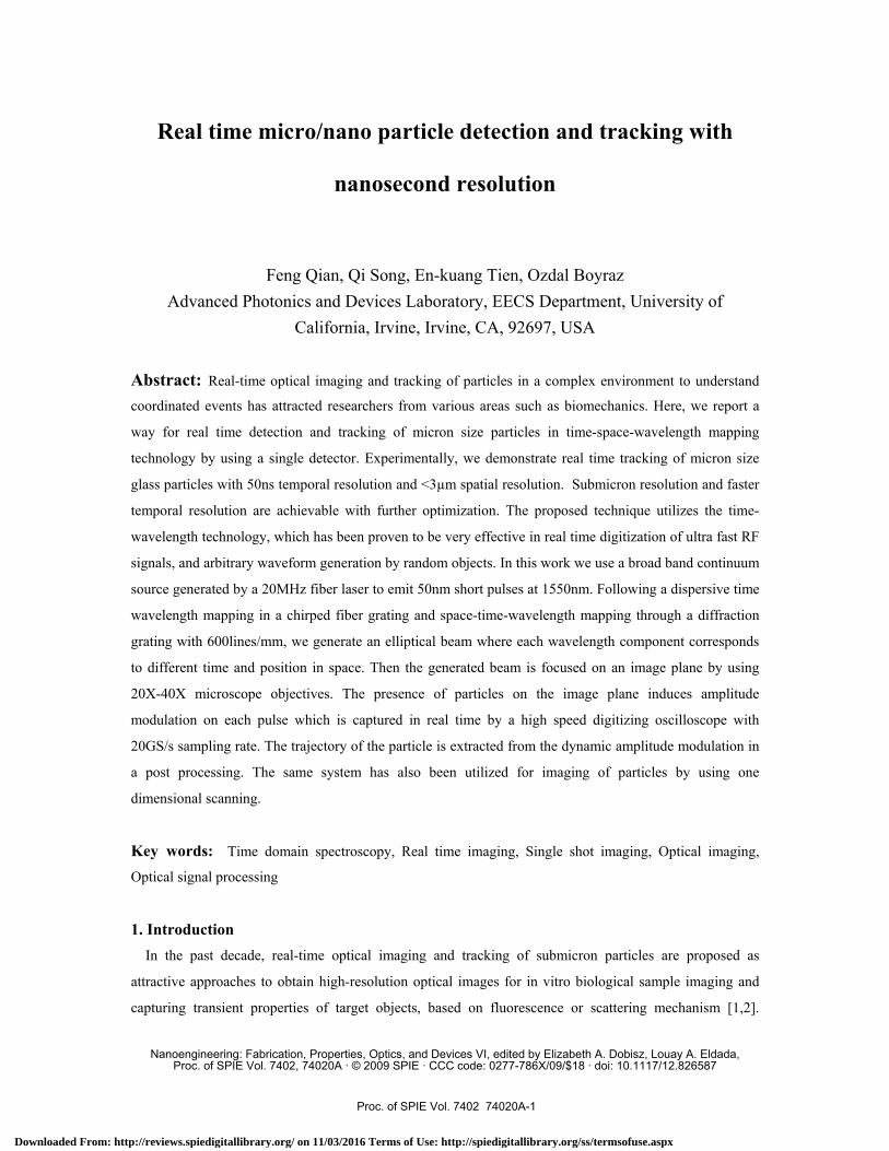
As shown conceptually in Fig. 1, the WDM based imaging system is comprised of a supercontinum light
source (SC source), diffractive components, optical focal components and a detection module. In this
scheme, a femto-second pulse laser with high power generates the broadening spectrum (~100nm) as a
supercontinum source in a highly nonlinear fiber. In the time domain, different wavelengths arrived at
different time by the dispersion unit to provide time-wavelength mapping. The light source is then
dispersed in space by a diffractive grating to generate space-wavelength mapping in one spatial dimension.
Proc. of SPIE Vol. 7402 74020A-2
Downloaded From: http://reviews.spiedigitallibrary.org/ on 11/03/2016 Terms of Use: http://spiedigitallibrary.org/ss/termsofuse.aspx
When the laterally dispersed light encounters the target object, the pulse amplitude is modulated and
transmitted to the detection part. Here, spatial information is transferred into the frequency domain by
space-wavelength mapping. So, space-time-wavelength mapping can be achieved all together in the same
configuration.
For two-dimensional (2D) pattern imaging, 2D information can be obtained by scanning only in the
dimension normal to the incident beam direction since the lateral dimensional information is extracted by a
dispersed beam. In the detection part, the spatially distributed modulated frequency domain information can
be transferred to the time domain as one consecutive pulse train by a single detector and a fast digital
oscilloscope. Prevailing over the wave front or amplitude division multiplexed system, the cross talk noise
between spatial points is significantly suppressed since different wavelengths are effectively separated and
independently detected in the time domain by a detector. By comparing the modulated signal with the
original pulse signal, position of the object and the density of the scatters can be determined.
One key advantage of time-wavelength domain conversion is the capability of dynamic monitoring of
target in a pulse period. When the displacement of objects occurs along the dispersive direction, the trace of
the target along this direction can be detected corresponding to a certain time. The temporal resolution is
determined by the pulse repetition rate of 50ns. This technique can also be incorporated with the post-
processing algorithm to observe the directional component of displacement and velocity.
Fig. 1 Conceptual Diagram of time-space-wavelength mapping system
Proc. of SPIE Vol. 7402 74020A-3
Downloaded From: http://reviews.spiedigitallibrary.org/ on 11/03/2016 Terms of Use: http://spiedigitallibrary.org/ss/termsofuse.aspx
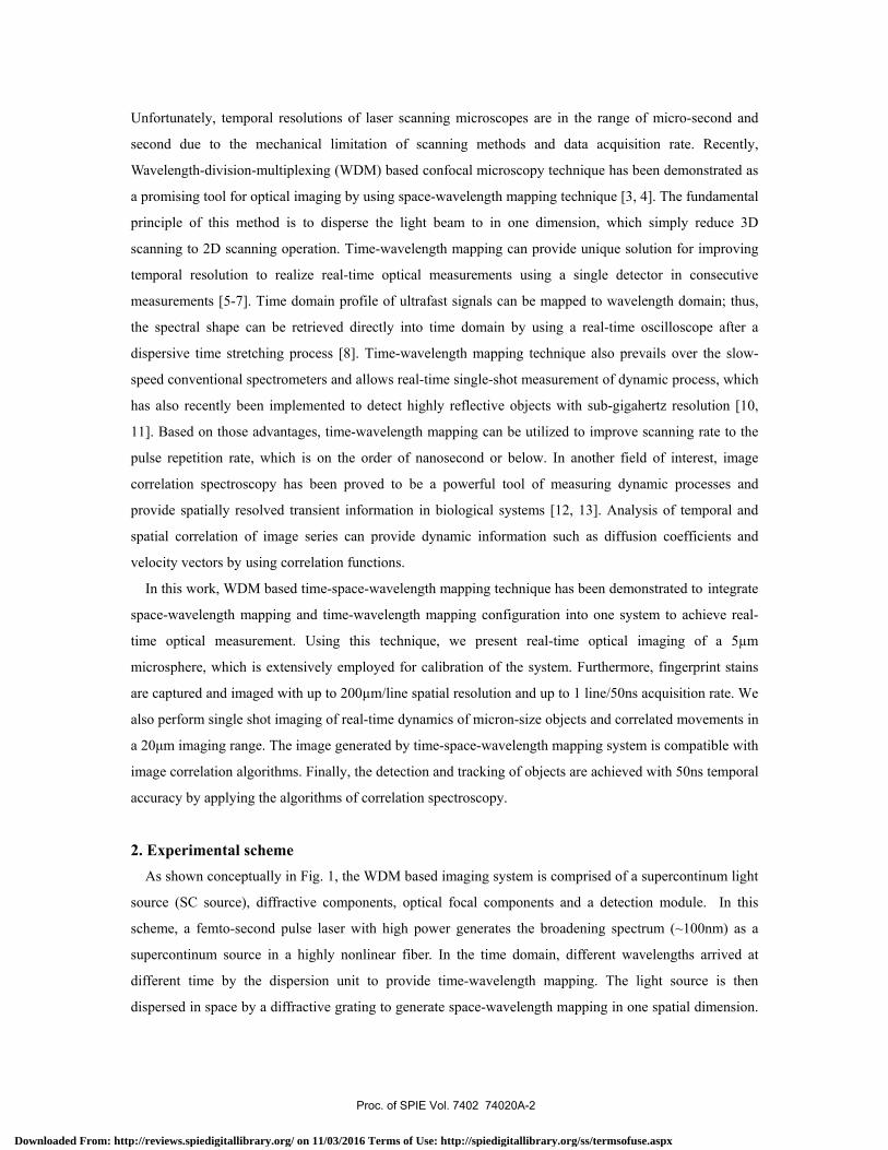
Experimental setup of the proposed optical imaging and particle tracking system is shown in Fig. 2. A
20MHz mode-locked fiber laser is used to generate 50nm continuum source with 5km of dispersion shifted
fiber. The generated continuum is then chirped by a grating based dispersion compensation module
(1300ps/nm); hence time-wavelength mapping is produced. The temporally dispersed supercontinuum is
subsequently dispersed in space by using a 600 lines/mm diffraction grating. At point “A” in Fig. 2, an
elliptical beam where each position along x axis is mapped to a different color arriving at different times.
Then the beam is focused on the sample at point “B” by using microscope objectives OL1 and OL2
(numerical apertures of 0.65 and 0.85). Polystyrene microspheres with 5µm diameter and glass particles are
used as samples in this experiment. Samples are embedded in a thin polymer film and attached to a cover
glass. Imaging is performed by monitoring the intensity of the transmitted light from the sample using an
InGaAs detector. The presence of particles on the image plane induces amplitude modulation on different
wavelengths, which is captured in time domain by a high speed real-time oscilloscope with 20GS/s
sampling rate. This setup can also be slightly modified for different sized object imaging such as fingerprint
stain imaging. The objective lenses OL1 and OL2 in Fig. 2 are replaced with two spherical lenses
(ƒ=100mm) for fingerprint object imaging.
In the imaging process, light encounter with the target object is composed of scattering light, transmitted
light and system noise. So, the light intensity at the focal point could be approximately expressed
as ),(),(),(),( yxnyxsyxtyxI ++= , where t(x,y) is transmitted intensity spectrum, s(x,y) is the
Fig .2 Experimental setup of transmitted optical imaging and particle tracking
Proc. of SPIE Vol. 7402 74020A-4
Downloaded From: http://reviews.spiedigitallibrary.org/ on 11/03/2016 Terms of Use: http://spiedigitallibrary.org/ss/termsofuse.aspx
scattering light intensity and n(x,y) is the system noise. Therefore, according to the variance of the
transmitted signal, spatial information of the object can be provided due to the presence of the scattering
light and noise induced by the object.
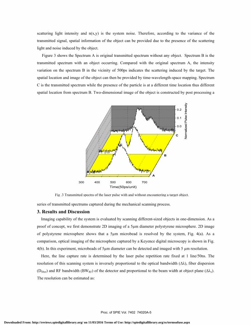
Figure 3 shows the Spectrum A is original transmitted spectrum without any object. Spectrum B is the
transmitted spectrum with an object occurring. Compared with the original spectrum A, the intensity
variation on the spectrum B in the vicinity of 500ps indicates the scattering induced by the target. The
spatial location and image of the object can then be provided by time-wavelength-space mapping. Spectrum
C is the transmitted spectrum while the presence of the particle is at a different time location thus different
spatial location from spectrum B. Two-dimensional image of the object is constructed by post processing a
series of transmitted spectrums captured during the mechanical scanning process.
3. Results and Discussion
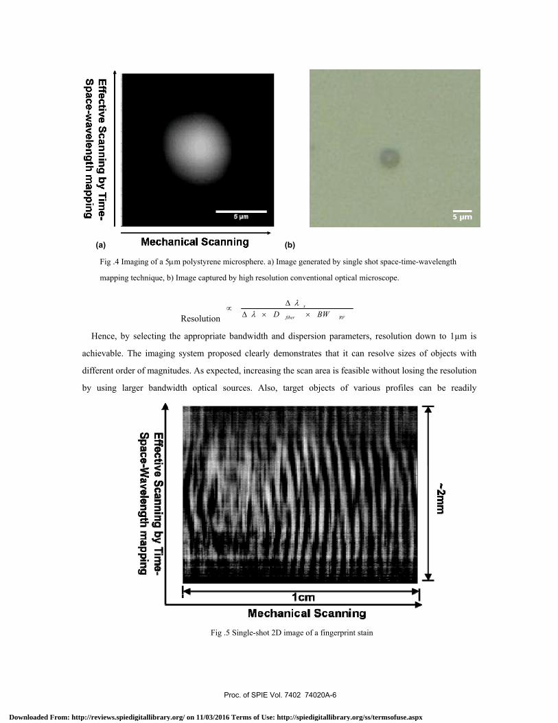
Imaging capability of the system is evaluated by scanning different-sized objects in one-dimension. As a
proof of concept, we first demonstrate 2D imaging of a 5µm diameter polystyrene microsphere. 2D image
of polystyrene microsphere shows that a 5µm microbead is resolved by the system, Fig. 4(a). As a
comparison, optical imaging of the microsphere captured by a Keyence digital microscopy is shown in Fig.
4(b). In this experiment, microbeads of 5µm diameter can be detected and imaged with 5 µm resolution.
Here, the line capture rate is determined by the laser pulse repetition rate fixed at 1 line/50ns. The
resolution of this scanning system is inversely proportional to the optical bandwidth (Δλ), fiber dispersion
(Dfiber) and RF bandwidth (BWRF) of the detector and proportional to the beam width at object plane (Δλx).
The resolution can be estimated as:
Fig .3 Transmitted spectra of the laser pulse with and without encountering a target object.
300 400 500 600 700
0.0
0.1
0.2
C
B
A
Time(50ps/unit)N
orm
aliz
ed P
ulse
Inte
nsity
Proc. of SPIE Vol. 7402 74020A-5
Downloaded From: http://reviews.spiedigitallibrary.org/ on 11/03/2016 Terms of Use: http://spiedigitallibrary.org/ss/termsofuse.aspx
Resolution RFfiber
x
BWD ××ΔΔ
∝λ
λ
Hence, by selecting the appropriate bandwidth and dispersion parameters, resolution down to 1µm is
achievable. The imaging system proposed clearly demonstrates that it can resolve sizes of objects with
different order of magnitudes. As expected, increasing the scan area is feasible without losing the resolution
by using larger bandwidth optical sources. Also, target objects of various profiles can be readily
Fig .5 Single-shot 2D image of a fingerprint stain
Fig .4 Imaging of a 5μm polystyrene microsphere. a) Image generated by single shot space-time-wavelength
mapping technique, b) Image captured by high resolution conventional optical microscope.
(a) (b)
Proc. of SPIE Vol. 7402 74020A-6
Downloaded From: http://reviews.spiedigitallibrary.org/ on 11/03/2016 Terms of Use: http://spiedigitallibrary.org/ss/termsofuse.aspx
distinguished by this configuration. The application of this proposed scheme may include detection of
objects of arbitrary shapes.
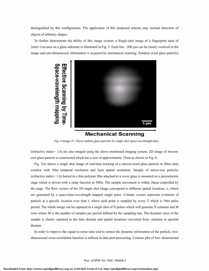
To further demonstrate the ability of this image system, a Single-shot image of a fingerprint stain of
2mm×1cm area on a glass substrate is illustrated in Fig. 5. Each line ~200 µm can be clearly resolved in the
image and one-dimensional information is acquired by mechanical scanning. Random sized glass particles
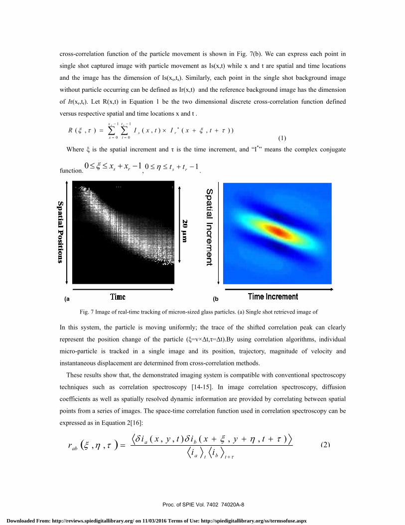
(refractive index= 1.6) are also imaged using the above mentioned imaging system. 2D image of micron-
size glass particle is constructed which has a size of approximately 25um as shown in Fig. 6.
Fig. 7(a) shows a single shot image of real-time tracking of a micron-sized glass particle in 20ms time
window with 50ns temporal resolution and 5µm spatial resolution. Sample of micro-size particles
(refractive index= 1.6) buried in a thin polymer film attached to a cover glass is mounted on a piezoelectric
stage which is driven with a ramp function at 50Hz. The sample movement is within 20μm controlled by
the stage. The Row vectors of the 2D single shot image correspond to different spatial locations, x, which
are generated by a space-time-wavelength mapped single pulse. Column vectors represent evolution of
particle at a specific location over time t, where each point is sampled by every T which is 50ns pulse
period. The whole image can be captured in a single shot of N pulses which will generate N columns and M
rows where M is the number of samples per period defined by the sampling rate. The dynamic trace of the
sample is clearly captured in the time domain and spatial locations converted from variation in spectral
domain.
In order to improve the signal to noise ratio and to extract the dynamic information of the particle, two-
dimensional cross-correlation function is utilized in data post-processing. Contour plot of two–dimensional
Fig. 6 Image of ∼25μm random glass particles by single shot space-wavelength-time
Proc. of SPIE Vol. 7402 74020A-7
Downloaded From: http://reviews.spiedigitallibrary.org/ on 11/03/2016 Terms of Use: http://spiedigitallibrary.org/ss/termsofuse.aspx
(2)
cross-correlation function of the particle movement is shown in Fig. 7(b). We can express each point in
single shot captured image with particle movement as Is(x,t) while x and t are spatial and time locations
and the image has the dimension of Is(xs,ts). Similarly, each point in the single shot background image
without particle occurring can be defined as Ir(x,t) and the reference background image has the dimension
of Ir(xr,tr). Let R(x,t) in Equation 1 be the two dimensional discrete cross-correlation function defined
versus respective spatial and time locations x and t . 1 1
0 0( , ) ( , ) ( , ) )
s sx t
s rx t
R I x t I x tξ τ ξ τ− −
∗
= =
= × + +∑ ∑ (1)
Where ξ is the spatial increment and τ is the time increment, and “I*” means the complex conjugate
function. 10 −+≤≤ rs xxξ , 10 −+≤≤ rs ttη .
In this system, the particle is moving uniformly; the trace of the shifted correlation peak can clearly
represent the position change of the particle (ξ=v×Δt,τ=Δt).By using correlation algorithms, individual
micro-particle is tracked in a single image and its position, trajectory, magnitude of velocity and
instantaneous displacement are determined from cross-correlation methods.
These results show that, the demonstrated imaging system is compatible with conventional spectroscopy
techniques such as correlation spectroscopy [14-15]. In image correlation spectroscopy, diffusion
coefficients as well as spatially resolved dynamic information are provided by correlating between spatial
points from a series of images. The space-time correlation function used in correlation spectroscopy can be
expressed as in Equation 2[16]:
( )τ
τηξδδτηξ
+
+++=
tbta
baab ii
tyxityxir
),,(),,(,,
Fig. 7 Image of real-time tracking of micron-sized glass particles. (a) Single shot retrieved image of
(a (b
Proc. of SPIE Vol. 7402 74020A-8
Downloaded From: http://reviews.spiedigitallibrary.org/ on 11/03/2016 Terms of Use: http://spiedigitallibrary.org/ss/termsofuse.aspx
Where ξia(b)(x,y,t) is the intensity fluctuation in channel a(b) at space position (x,y) and time t. The same
data can be captured by extracting the intensity variation from the image generated by space-time-
wavelength mapping algorithm.
Here, we image dynamic events occurring in a single line over long time duration. Hence, the intensity at
spatial location x expressed as I(t+nT) reveals intensity variation at the spatial point with temporal accuracy
of T which is the laser repetition rate of 50ns and n is a integer. Similarly, we can capture information about
multiple different spatial locations in a single shot image. Thus, by using the conventional correlation
algorithms such as cross correlation algorithms, this system is can identify correlated events along the
image plane and detect instantaneous correlated events with temporal accuracy of 50ns.
4. Conclusion
In summary, we present the scheme of WDM based pulse laser scanning 2D microscopy, which can
provide spatial spectrum distribution and ultrafast line refreshing rate. The feasibility of high resolution
imaging and tracking has been demonstrated by time-space-wavelength mapping technique with single-shot
single-detector. Single shot imaging of 5µm object is implemented at 20MHz line capture rate, and
correlation algorithm is applied to obtain the transient displacement of the individual particle. This system
can be further optimized for real-time imaging, tracking of multiple micro-particles or arbitrary objects. It
also shows that the conventional spectroscopy algorithms can be available to obtain the dynamic particle
information.
References
[1] Enderlein, J., Ruckstuhl, T., and Seeger, S., “Highly Efficient Optical Detection of Surface-Generated
Fluorescence”,Appl Opt. 38, 724-732(1999).
[2] Denk, W., Strickler, J. H., and Web,W. W., “Two-photon laser scanning fluorescence microscopy” Science, 248,
73-76(1990).
[3] Digman, M. A., Brown, C. M, Sengupta, P., Wiseman, P. W, Horwitz, A. R, and Gratton, E., “Measuring Fast
Dynamics in Solutions and Cells with a Laser Scanning Microscope”, Biophys J. 89, 1317-1327(2005).
[4] Shi, K. B, Nam, S. H., Li, P., Yin, S. Z., and Liu, Z. W., "Wavelength division multiplexed confocal microscopy
using supercontinuum,"Opt Commun. 263,156-162(2006).
[5] Chou, J., Han, Y., and Jalali, B.,”Time-wavelength spectroscopy for chemical sensing”, IEEE Photonics Technol.
Lett. 16, 1140-1142(2004).
[6] Chou, J., Boyraz, O., Solli,D., and Jalali , B., “Femtosecond real-time single-shot digitizer”Appl Phys Lett. 91,
161105(2007).
[7] Nuruzzaman, A., Boyraz, O., and Jalali, B., “Time-stretched short-time Fourier transform”, IEEE Trans. Instrum.
Meas. 55, 598-602(2006).
Proc. of SPIE Vol. 7402 74020A-9
Downloaded From: http://reviews.spiedigitallibrary.org/ on 11/03/2016 Terms of Use: http://spiedigitallibrary.org/ss/termsofuse.aspx
[8] Han, Y., Boyraz, O., and Jalali, B.,” Tera-sample per second real-time waveform digitizer” Appl Phys Lett. 87,
24111(2005).
[9] Solli, D. R., Chou, J., and Jalali, B., “Amplified wavelength–time transformation for real-time spectroscopy”, Nat
Photonics. 2, 48-51(2008).
[10] Goda, K., Tsia, K. K., and Jalali, B., “Amplified dispersive Fourier-transform imaging for ultrafast displacement
sensing and barcode reading”, Appl Phys Lett. 9,131109(2008).
[11] Goda, K., Tsia, K. K., and Jalali, B.,”Serial time-encoded amplified imaging for real-time observation of fast
dynamic phenomena”, Nature. 458, 1145-1149(2009).
[12] Bonnet, N., Delavoie, F., and Zahm,J. M., “Characterizing the spatio-temporal behavior of cell populations
through image auto- and cross-correlation microscopy”,Biotechniques, 43, 107-115(2007).
[13] Wiseman, P. W., Squier, J. A, Wilson, K. R, “Dynamic image correlation spectroscopy (ICS) and two-color image
cross-correlation spectroscopy (ICCS): concepts and application”, Proc. SPIE 3919, 14-20 (2000).
[14] Digman ,M. A., Sengupta ,P., Wiseman, P. W., Brown, C. M., Horwitz, A. R., and Gratton, E., “Measuring fast
dynamics in solutions and cells with a laser scanning microscope”, Biophys J. 88, L33-L36(2005).
[15] Ruan, Q. Q., Cheng, M. A., Levi, M., Gratton, E., and Mantulin, W. W., “Spatial-Temporal Studies of Membrane
Dynamics: Scanning Fluorescence Correlation Spectroscopy (SFCS)” Biophys J. vol. 87,1260-1267 (2004).
[16] Hebert,B., Costantino, S., and Wiseman, P. W., “Spatio-temporal Image Correlation Spectroscopy (SITCS):
Theory, verification and application to protein velocity mapping in living CHO cells”, Biophys J. 88,3601-
3614(2005).
Proc. of SPIE Vol. 7402 74020A-10
Downloaded From: http://reviews.spiedigitallibrary.org/ on 11/03/2016 Terms of Use: http://spiedigitallibrary.org/ss/termsofuse.aspx