Embed Size (px)
Citation preview
Validated and certified by the OIE for the purposes defined in this insert
Registration number: 20090105
TeSeE™ Western BlotReagents for in vitro confirmation of suspected TSE positive samples
Catalog # 355-1169 32 Tests

2
CONTENTS
1 - GENERAL INFORMATION
2 - ASSAY PRINCIPLE
3 - COMPOSITION OF THE KIT
4 - SAMPLES
5 - ASSAY PROCEDURE WITH MINI BLOT™ GEL5.1 Additional reagents and material required5.2 Preparation of reagents5.3 Sample purification5.4 Electrophoresis5.5 Protein transfer5.6 Immunoblotting
6 - ASSAY PROCEDURE WITH CRITERION™ XT GEL6.1 Additional reagents and material required6.2 Preparation of reagents6.3 Sample purification6.4 Electrophoresis6.5 Protein transfer6.6 Immunoblotting
7 - INTERPRETATION OF RESULTS
8 - PRECAUTIONS
9 - HYGIENE AND SAFETY INSTRUCTIONS
10 - REFERENCES
3
1 - GENERAL INFORMATION
Transmissible Spongiform Encephalopathies (TSE’s) were first reported in the eighteenth century in sheep (Scrapie) and more recently in cervids such as deer and elk (Chronic Wasting disease, CWD) and cattle (Bovine Spongiform Encephalopathy, BSE). Humans are also susceptible to certain forms of TSE such as Kuru, Creutzfeldt-Jakob Disease (CJD) or Gerstmann-Sträussler-Scheinker Syndrome (GSS). The emergence of new variant Creutzfeldt-Jakob Disease (vCJD) in the human population has been strongly linked to the dietary intake of BSE-infected meat or meat products. One of the main characteristics of TSEs is a progressive accumulation in the central nervous system of an abnormal isoform of natural or cellular prion protein (PrPc), termed PrPres. This disease specific PrPres is characterised by an increased resistance to proteases. The TeSeE™ Western Blot assay permits qualitative identification of PrPres after proteolytic treatment which results in a reduced molecular weight fragment due to ‘N’ terminus truncation.
Active/passive surveillance programs have been conducted worldwide to detect BSE, scrapie or CWD in infected animals. Those programs have resulted in the identification of increased numbers of positive cases at the screening laboratories. Those positive samples (suspected animals) are then systematically confirmed as “TSE-infected” by the demonstration of typical spongiform changes with histopathology, or with the detection of abnormal PrP by Immunohistochemistry (IHC), or of Scrapie Associated Fibrils (SAFs) by electron microscopy. These above confirmation techniques require technical expertise for the interpretation of the results and are time consuming and expensive. Western blot technique can also be considered as an alternative method for confirmation of the TSE suspected samples.
The validation data for this kit have been certified by the OIE, based on expert review, as fit for the post-mortem detection of transmissible spongiform encephalopathies (TSEs) in cattle (bovine spongiform encephalopathy, BSE), in ovines and caprines (BSE and scrapie), and in cervids (Chronic Wasting Disease, CWD), and for the following purposes:
1. To confirm TSE suspected positive samples detected at the screeninglaboratories in countries with active/passive surveillance programmes. Any sample with a negative result according to the TeSeE™ WESTERN BLOT assay interpretation criteria, following a positive rapid test result,should be tested with one of the other OIE certified confirmatory methods, Immunohistochemistry (IHC) or SAF-Immunoblot;
2. To confirm the prevalence of infection with one of the TSE associated diseases(BSE, scrapie, CWD) in the context of an epidemiological survey in a lowprevalence country;
4
3. To estimate prevalence of infection to facilitate risk analysis(e.g. surveys, implementation of disease control measures) and to assist the demonstration of the efficiency of eradication policies.
The TeSeE™ Western Blot assay is using the same assay principle as the Bio-Rad rapid assays (TeSeE™ SAP, TeSeE™ sheep/goat) that include the preliminary purification and concentration of the PrPres, associated to a highly sensitive immunoblotting. Then, it can be used efficiently for confirmation of any TSE suspected samples and for typing of TSE strains in sheep.
2 - ASSAY PRINCIPLE
The TeSeE™ Western Blot assay allows the detection of PrPres in nervous tissues (bovine, ovine, caprine, cervids, ...) or peripheral tissues (cervids) collected from infected animals.
The assay procedure begins with the digestion of cellular PrP protein (PrPc), followed by purification and concentration of disease specific PrPres. Detection of PrPres is carried out by electrophoretic migration then immunoblotting using a monoclonal antibody highly specific for PrPres.
The assay procedure includes the following steps:• Sample homogenization,• Digestion of PrPc with proteinase K,• Purification and concentration of PrPres,• Electrophoresis and transfer onto a membrane,• Immunoblotting.
5
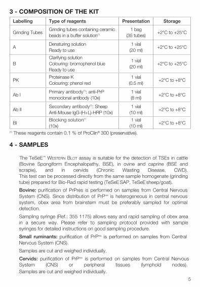
(1) These reagents contain 0.1 % of ProClin® 300 (preservative).
4 - SAMPLES
The TeSeE™ Western Blot assay is suitable for the detection of TSEs in cattle (Bovine Spongiform Encephalopathy, BSE), in ovine and caprine (BSE and scrapie), and in cervids (Chronic Wasting Disease, CWD). This test can be processed directly from the same sample homogenate (grinding tube) prepared for Bio-Rad rapid testing (TeSeE SAP, TeSeE sheep/goat).
Bovine: purification of PrPres is performed on samples from Central Nervous System (CNS). Since distribution of PrPres is heterogeneous in central nervous system, obex area from brainstem must be preferably sampled for optimal detection.
Sampling syringe (Ref.: 355 1175) allows easy and rapid sampling of obex area in a secure way. Please refer to sampling protocol provided with sample syringes for detailed instructions on good sampling procedure.
Small ruminants: purification of PrPres is performed on samples from Central Nervous System (CNS).
Samples are cut and weighed individually.
Cervids: purification of PrPres is performed on samples from Central Nervous System (CNS) or peripheral tissues (lymphoïd nodes). Samples are cut and weighed individually.
Labelling Type of reagents Presentation Storage
Grinding TubesGrinding tubes containing ceramic beads in a buffer solution(1)
1 bag(35 tubes)
+2°C to +25°C
ADenaturing solutionReady to use
1 vial(20 ml)
+2°C to +25°C
BClarifying solutionColouring: bromophenol blueReady to use
1 vial(20 ml)
+2°C to +25°C
PKProteinase KColouring: phenol red
1 vial(0.5 ml)
+2°C to +8°C
Ab IPrimary antibody(1): anti-PrP monoclonal antibody (10x)
1 vial(8 ml)
+2°C to +8°C
Ab IISecondary antibody(1): Sheep Anti-Mouse IgG-(H+L)-HRP (10x)
1 vial(10 ml)
+2°C to +8°C
BlBlocking solution(1)
(10x)1 vial
(10 ml)+2°C to +8°C
3 - COMPOSITION OF THE KIT
6
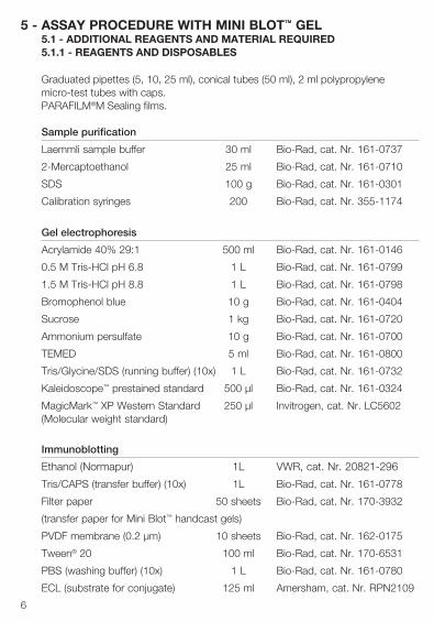
5 - ASSAY PROCEDURE WITH MINI BLOT™ GEL5.1 - ADDITIONAL REAGENTS AND MATERIAL REQUIRED5.1.1 - REAGENTS AND DISPOSABLES
Graduated pipettes (5, 10, 25 ml), conical tubes (50 ml), 2 ml polypropylene micro-test tubes with caps.PARAFILM®M Sealing films.
Sample purification
Laemmli sample buffer 30 ml Bio-Rad, cat. Nr. 161-0737
2-Mercaptoethanol 25 ml Bio-Rad, cat. Nr. 161-0710
SDS 100 g Bio-Rad, cat. Nr. 161-0301
Calibration syringes 200 Bio-Rad, cat. Nr. 355-1174
Gel electrophoresis
Acrylamide 40% 29:1 500 ml Bio-Rad, cat. Nr. 161-0146
0.5 M Tris-HCl pH 6.8 1 L Bio-Rad, cat. Nr. 161-0799
1.5 M Tris-HCl pH 8.8 1 L Bio-Rad, cat. Nr. 161-0798
Bromophenol blue 10 g Bio-Rad, cat. Nr. 161-0404
Sucrose 1 kg Bio-Rad, cat. Nr. 161-0720
Ammonium persulfate 10 g Bio-Rad, cat. Nr. 161-0700
TEMED 5 ml Bio-Rad, cat. Nr. 161-0800
Tris/Glycine/SDS (running buffer) (10x) 1 L Bio-Rad, cat. Nr. 161-0732
Kaleidoscope™ prestained standard 500 µl Bio-Rad, cat. Nr. 161-0324
MagicMark™ XP Western Standard 250 µl Invitrogen, cat. Nr. LC5602 (Molecular weight standard)
Immunoblotting
Ethanol (Normapur) 1L VWR, cat. Nr. 20821-296
Tris/CAPS (transfer buffer) (10x) 1L Bio-Rad, cat. Nr. 161-0778
Filter paper 50 sheets Bio-Rad, cat. Nr. 170-3932
(transfer paper for Mini Blot™ handcast gels)
PVDF membrane (0.2 µm) 10 sheets Bio-Rad, cat. Nr. 162-0175
Tween® 20 100 ml Bio-Rad, cat. Nr. 170-6531
PBS (washing buffer) (10x) 1 L Bio-Rad, cat. Nr. 161-0780
ECL (substrate for conjugate) 125 ml Amersham, cat. Nr. RPN2109
7
ECL Hyperfilms (18 x 24 cm) 25 films Amersham, cat. Nr. RPN2103K
Development folders 30 folders Applied Biosystems, cat Nr. T2258
Kodak developing solution LX24 to 20 L VWR or Kodak
Kodak fixative solution AL4 to 20 L VWR or Kodak
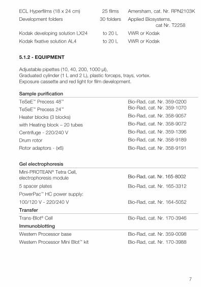
5.1.2 - EQUIPMENT
Adjustable pipettes (10, 40, 200, 1000 µl), Graduated cylinder (1 L and 2 L), plastic forceps, trays, vortex.Exposure cassette and red light for film development.
Sample purification
Bio-Rad, cat. Nr. 359-0200 Bio-Rad, cat. Nr. 359-1070
Bio-Rad, cat. Nr. 358-9057
Bio-Rad, cat. Nr. 358-9072
Bio-Rad, cat. Nr. 359-1396
Bio-Rad, cat. Nr. 358-9189
TeSeE™ Precess 48™
TeSeE™ Precess 24™
Heater blocks (3 blocks)
with Heating block – 20 tubes
Centrifuge - 220/240 V
Drum rotor
Rotor adaptors - (x6) Bio-Rad, cat. Nr. 358-9191
Gel electrophoresis
Mini-PROTEAN® Tetra Cell, electrophoresis module
5 spacer plates Bio-Rad, cat. Nr. 165-3312
PowerPac™ HC power supply:
100/120 V - 220/240 V Bio-Rad, cat. Nr. 164-5052
Transfer
Trans-Blot® Cell Bio-Rad, cat. Nr. 170-3946
Immunoblotting
Western Processor base Bio-Rad, cat. Nr. 359-0098
Western Processor Mini Blot™ kit Bio-Rad, cat. Nr. 170-3988
Bio-Rad, cat. Nr. 165-8002
8
5.2 - PREPARATION OF REAGENTS5.2.1 - SAMPLE PURIFICATION
• Proteinase KSolution of proteinase K diluted in reagent A:
u 1 ml Reagent Au 20 µl Proteinase K
Mix well by inverting until you obtain a homogeneous solution. After reconstitution, diluted proteinase K is stable 10 hours at room temperature (+18°C to +30°C).
• Laemmli solutionSolution of SDS + 2-Mercaptoethanol + Laemmli sample buffer:
u 0.6 g SDSu 1.5 ml 2-Mercaptoethanol
Mix by inverting. u 28.5 ml Laemmli sample buffer
Solution is aliquoted into 4 ml aliquots and stored at -20°C. Thawed aliquots can be re-frozen.
Note: It is recommended to prepare Laemmli solution one hour before use allowing SDS to be completely dissolved.
5.2.2 - ELECTROPHORESIS
• Hand cast discontinuous acrylamide gelThe gel must be 1.5 mm thickness.
Using the Mini Blot™ casting module, the resolving gel (13.5% acrylamide, pH8.8) is cast first, once the resolving gel is polymerized the stacking gel is added(3% acrylamide, pH 6.8).
Resolving gel (1 gel)u 2.8 ml Acrylamide 40%, 29:1u 1.7 ml 1.5 M Tris-HCl buffer, pH 8.8 / SDS (1) u 1.3 ml 50% sucrose solution (2)u 2.5 ml distilled water
Mix by inverting.u 43 µl 10% Ammonium persulfate (3)u 9 µl TEMED
9
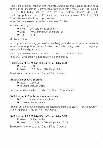
Pour 7 ml of the gel solution into the plates and retain the residual solution as a control of polymerization. Gently overlay to the top with 1 ml of 0.3 M Tris-HCl pH 8.8 / SDS buffer (4) so that the gel surface doesn’t dry out. Let the gel polymerize for 15-20 minutes at room temperature (+18°C to +30°C). Check the residual solution is polymerized. Invert the plate assembly to eliminate excess of buffer.
Stacking gel (1 gel)u 4 ml 3% Acrylamide solution (7)u 28 µl 10% Ammonium persulfate (3)u 6 µl TEMED
Mix by inverting.
Gently pour the stacking gel onto the resolving gel and retain the residual solution as a control of polymerization. Position the comb, taking care not to trap any bubble in the well positions.
Let the gel polymerize for 5-10 minutes at room temperature (+18°C to +30°C). Check the residual solution is polymerized.
(1) Solution of 1.5 M Tris-HCl buffer, pH 8.8 / SDS u 0.2 g SDSu 50 ml 1.5 M Tris-HCl buffer pH 8.8
Solution can be stored at +2°C to +8°C for 2 weeks.
(2) Solution of 50% Sucroseu 25 g Sucroseu to 50 ml Distilled water
Sucrose solution can be stored at +2°C to +8°C for 2 weeks.
(3) Solution of 10% Ammonium persulfateu 5 g Ammonium persulfateu to 50 ml Distilled water
Ammonium persulfate solution is aliquoted and stored at -20°C. Thawed solution can be stored at +2°C to +8°C for 2 weeks.
(4) Solution of 0.3 M Tris-HCl buffer, pH 8.8 / SDS u 40 ml Distilled wateru 10 ml 1.5 M Tris-HCl buffer pH 8.8 / SDS
Solution can be stored at +2°C to +8°C for 2 weeks.
10
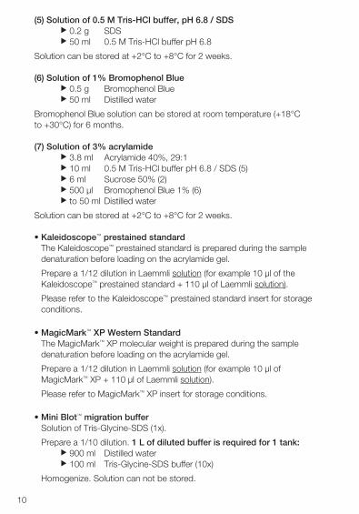
(5) Solution of 0.5 M Tris-HCl buffer, pH 6.8 / SDS u 0.2 g SDSu 50 ml 0.5 M Tris-HCl buffer pH 6.8
Solution can be stored at +2°C to +8°C for 2 weeks.
(6) Solution of 1% Bromophenol Blueu 0.5 g Bromophenol Blueu 50 ml Distilled water
Bromophenol Blue solution can be stored at room temperature (+18°C to +30°C) for 6 months.
(7) Solution of 3% acrylamideu 3.8 ml Acrylamide 40%, 29:1u 10 ml 0.5 M Tris-HCl buffer pH 6.8 / SDS (5)u 6 ml Sucrose 50% (2)u 500 µl Bromophenol Blue 1% (6)u to 50 ml Distilled water
Solution can be stored at +2°C to +8°C for 2 weeks.
• Kaleidoscope™ prestained standardThe Kaleidoscope™ prestained standard is prepared during the sampledenaturation before loading on the acrylamide gel.
Prepare a 1/12 dilution in Laemmli solution (for example 10 µl of theKaleidoscope™ prestained standard + 110 µl of Laemmli solution).
Please refer to the Kaleidoscope™ prestained standard insert for storageconditions.
• MagicMark™ XP Western StandardThe MagicMark™ XP molecular weight is prepared during the sampledenaturation before loading on the acrylamide gel.
Prepare a 1/12 dilution in Laemmli solution (for example 10 µl ofMagicMark™ XP + 110 µl of Laemmli solution).
Please refer to MagicMark™ XP insert for storage conditions.
• Mini Blot™ migration bufferSolution of Tris-Glycine-SDS (1x).
Prepare a 1/10 dilution. 1 L of diluted buffer is required for 1 tank:u 900 ml Distilled wateru 100 ml Tris-Glycine-SDS buffer (10x)
Homogenize. Solution can not be stored.
11
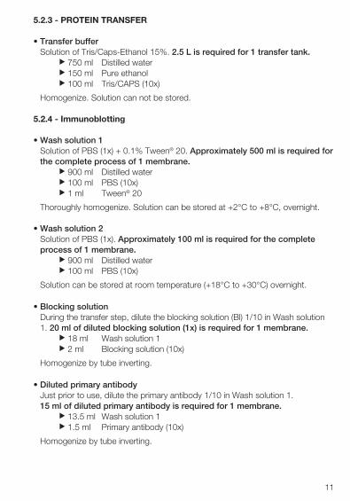
5.2.3 - PROTEIN TRANSFER
• Transfer bufferSolution of Tris/Caps-Ethanol 15%. 2.5 L is required for 1 transfer tank.
u 750 ml Distilled wateru 150 ml Pure ethanolu 100 ml Tris/CAPS (10x)
Homogenize. Solution can not be stored.
5.2.4 - Immunoblotting
• Wash solution 1Solution of PBS (1x) + 0.1% Tween® 20. Approximately 500 ml is required forthe complete process of 1 membrane.
u 900 ml Distilled wateru 100 ml PBS (10x)u 1 ml Tween® 20
Thoroughly homogenize. Solution can be stored at +2°C to +8°C, overnight.
• Wash solution 2Solution of PBS (1x). Approximately 100 ml is required for the completeprocess of 1 membrane.
u 900 ml Distilled wateru 100 ml PBS (10x)
Solution can be stored at room temperature (+18°C to +30°C) overnight.
• Blocking solutionDuring the transfer step, dilute the blocking solution (Bl) 1/10 in Wash solution1. 20 ml of diluted blocking solution (1x) is required for 1 membrane.
u 18 ml Wash solution 1u 2 ml Blocking solution (10x)
Homogenize by tube inverting.
• Diluted primary antibodyJust prior to use, dilute the primary antibody 1/10 in Wash solution 1.15 ml of diluted primary antibody is required for 1 membrane.
u 13.5 ml Wash solution 1u 1.5 ml Primary antibody (10x)
Homogenize by tube inverting.

12
• Diluted secondary antibody (conjugate)Just before use, dilute the secondary antibody 1/10 in Wash solution 1.20 ml of diluted conjugate is required for 1 membrane.
u 18 ml Wash solution 1u 2 ml Secondary antibody (10x)
Homogenize by tube inverting.
• ECLSubstrate (ECL) must be prepared just before use. 1 ml of substrate isrequired for 1 membrane.
u 0.5 ml Reagent 1u 0.5 ml Reagent 2
Homogenize the solution.
• Development solutionu 800 ml Distilled wateru 200 ml Development product
Solution can be stored at room temperature (+18°C to +30°C), in a darkroom for 15 days maximum.
• Fixative solutionu 800 ml Distilled wateru 200 ml Fixative product
Solution can be stored at room temperature (+18°C to +30°C), in a darkroom for 15 days maximum.
13
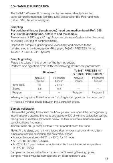
5.3 - SAMPLE PURIFICATION
The TeSeE™ Western Blot assay can be processed directly from the same sample homogenate (grinding tube) prepared for Bio-Rad rapid tests (TeSeE SAP, TeSeE sheep/goat).
Sampling For peripheral tissues (lymph nodes) insert one medium bead (Ref.: 355 1171) in the grinding tube, before to add the sample.Take a mass of 350 mg ± 40 mg of nervous tissue (preferably in the obex area) or 200 mg ± 20 mg of peripheral tissue.
Deposit the sample in grinding tube, close firmly and proceed to the grinding step in the homogenizer (Ribolyser®, TeSeE™ PRECESS 48™ or TeSeE™ PRECESS 24™ - system).
Sample grindingPlace the tubes in the crown of the homogenizer.Perform one agitation cycle with the following instrument parameters.
When grinding is insufficient, another 1 or 2 agitation cycles can be performed(2).(1) (2) Wait a 5 minutes pause between the 2 agitation cycles.
Sample calibrationRemove the grinding tubes from the homogenizer, resuspend the homogenate by inverting before opening the tubes and aspirate 500 µl with the calibration syringe taking care to immerse the needle below the level of ceramic beads to avoid sampling tissue fragments.Transfer each 500 µl sample into a 2 ml Eppendorf micro test-tube.
Note: At this stage, both grinding tubes after homogenisation and micro test-tubes after sample calibration can be stored, closed: • At room temperature (+18°C to +30°C) for 15 hours.• At +2°C to +8°C for 72 hours.• At -20°C for 1 year. Frozen samples must be thawed at room temperature
(+18°C to +30°C).
Samples can be submitted to a maximum of 3 freezing/thawing cycles. Samples must always be homogenized by inverting before use.
Ribolyser® TeSeE™ PRECESS 48™
or TeSeE™ PRECESS 24™
Nervous tissues
Peripheral tissues
Nervous tissues
Peripheral tissues
Time (sec.) 45 2 x 45(1) — —Speed 6.5 6.5 — —Program — — Program 1 Program 2
14
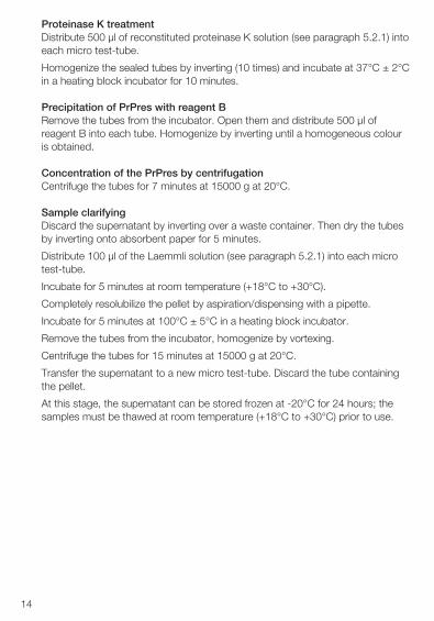
Proteinase K treatmentDistribute 500 µl of reconstituted proteinase K solution (see paragraph 5.2.1) into each micro test-tube.
Homogenize the sealed tubes by inverting (10 times) and incubate at 37°C ± 2°C in a heating block incubator for 10 minutes.
Precipitation of PrPres with reagent BRemove the tubes from the incubator. Open them and distribute 500 µl of reagent B into each tube. Homogenize by inverting until a homogeneous colour is obtained.
Concentration of the PrPres by centrifugationCentrifuge the tubes for 7 minutes at 15000 g at 20°C.
Sample clarifyingDiscard the supernatant by inverting over a waste container. Then dry the tubes by inverting onto absorbent paper for 5 minutes.
Distribute 100 µl of the Laemmli solution (see paragraph 5.2.1) into each micro test-tube.
Incubate for 5 minutes at room temperature (+18°C to +30°C).
Completely resolubilize the pellet by aspiration/dispensing with a pipette.
Incubate for 5 minutes at 100°C ± 5°C in a heating block incubator.
Remove the tubes from the incubator, homogenize by vortexing.
Centrifuge the tubes for 15 minutes at 15000 g at 20°C.
Transfer the supernatant to a new micro test-tube. Discard the tube containing the pellet.
At this stage, the supernatant can be stored frozen at -20°C for 24 hours; the samples must be thawed at room temperature (+18°C to +30°C) prior to use.
15
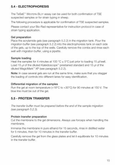
5.4 - ELECTROPHORESIS
The TeSeE™ Western Blot assay can be used for both confirmation of TSE suspected samples or for strain typing in sheep.
The following procedure is applicable for confirmation of TSE suspected samples.
Please contact your Bio-Rad representative for instruction protocol in case of strain typing application.
Gel preparationPlace the acrylamide gels (see paragraph 5.2.2) in the migration tank. Pour the migration buffer (see paragraph 5.2.2) into the electrophoresis tank on each side of the gels, up to the top of the wells. Carefully remove the combs and rinse each well with migration buffer, using a pipette.
Sample loadingHeat the samples for 4 minutes at 100 °C ± 5°C just prior to loading 15 µl/well. Load 15 µl of the diluted Kaleidoscope™ prestained standard and 15 µl of the diluted MagicMark™ XP (see paragraph 5.2.2).
Note: In case several gels are run at the same time, make sure that you stagger the loading of controls into different lanes for easy identification.
Differential migration of the samplesRun the gel at room temperature (+18°C to +30°C) for 90 minutes at 150 V. The blue line must be out of the gel.
5.5 - PROTEIN TRANSFER
The transfer buffer must be prepared before the end of the sample migration (see paragraph 5.2.3).
Protein transfer preparationCut the membrane to the gel dimensions. Always use forceps when handling the membrane.
Immerse the membrane in pure ethanol for 15 seconds, rinse in distilled water for 5 minutes, then for 10 minutes in the transfer buffer.
Carefully remove the gel from the glass plates and let it equilibrate for 10 minutes in the transfer buffer.
16
Gel sandwich preparationSoak filter paper and fibre pads in the transfer buffer.
Open the transfer cassette, with transparent side on the left. Respectively place on the transparent side a fiber pad, a filter paper, the membrane* and the gel*.
Complete with a filter paper then a fibre pad and close the cassette.
Immerse it in the transfer tank previously filled to the indicated limit with transfer buffer.
*Remove any air bubbles which may have formed.
Note: In case several membranes are processed at the same time, label eachmembrane in the corner.
Transfer onto the PVDF membraneAgitate during the transfer by using a magnetic stirring bar and run for60 minutes at 115 V.
5.6 - IMMUNOBLOTTING
a) Upon completion of the protein transfer, open the blotting assembly andremove the membrane for development. Quickly immerse the membrane inWash solution 2 (see paragraph 5.2.4), then place it in ethanol for 10 secondsbefore rinsing for 5 minutes in distilled water.
Note: At this step, the membrane can be stored overnight in distilled water at+2°C to +8°C.
Let the membrane adjust to room temperature (+18°C to +30°C) before tostart the immunoblotting.
b) Eliminate distilled water and incubate the membrane for 30 minutes in blockingsolution (see paragraph 5.2.4). Incubate under medium agitation.20 ml is sufficient for 1 membrane.
Note: from this step until the step g), the Bio-Rad Western Processor can beused for agitation and washing steps (refer to instruction manual for settings).
c) Eliminate the blocking solution and incubate the membrane in diluted primaryantibody (see paragraph 5.2.4) for 30 minutes at room temperature (+18°C to+30°C) under medium agitation.15 ml of diluted primary antibody is required for 1 membrane.
17
d) Eliminate the primary antibody solution and using Wash solution 1, briefly rinsethe membrane, then wash twice for respectively 5 and 10 minutes, under fastagitation.50 ml of Wash solution 1 is required for each cycle and for1 membrane.
e) Eliminate Wash solution 1 and incubate the membrane for 20 minutes indiluted secondary antibody (see paragraph 5.2.4) at room temperature(+18°C to +30°C) under medium agitation.20 ml of diluted secondary antibody is required for 1 membrane.
f) Eliminate the secondary antibody solution and using Wash solution 1, brieflyrinse, then wash for respectively 5, 10 and 10 minutes under fast agitation.50 ml of Wash solution 1 is required for each cycle and for 1 membrane.
g) Place the membrane in 50 ml of Wash solution 2 under slow agitation.
h) Drain the membrane on absorbent paper without blotting and place it in theplastic folder.
i) Add the ECL reagent (see paragraph 5.2.4). Eliminate the excess of reagentand air bubbles with absorbent paper. Place into the exposure cassette.
j) In a darkroom, cover the folder with a film and expose for 15 minutes. Film canbe exposed longer or shorter time for optimal signal.
k) Immerse the film in developing solution for 45 seconds (see paragraph 5.2.4).Rinse in distilled water. Immerse the film in fixative solution until the filmbecomes completely transparent.
l) Wash with distilled water and let the film dry.
18
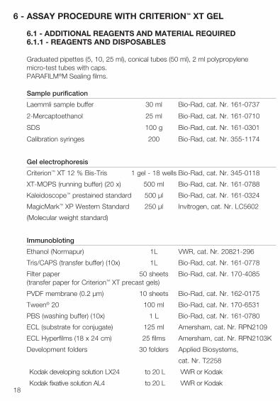
6 - ASSAY PROCEDURE WITH CRITERION™ XT GEL
6.1 - ADDITIONAL REAGENTS AND MATERIAL REQUIRED6.1.1 - REAGENTS AND DISPOSABLES
Graduated pipettes (5, 10, 25 ml), conical tubes (50 ml), 2 ml polypropylene micro-test tubes with caps.PARAFILM®M Sealing films.
Sample purification
Laemmli sample buffer 30 ml Bio-Rad, cat. Nr. 161-0737
2-Mercaptoethanol 25 ml Bio-Rad, cat. Nr. 161-0710
SDS 100 g Bio-Rad, cat. Nr. 161-0301
Calibration syringes 200 Bio-Rad, cat. Nr. 355-1174
Gel electrophoresis
Criterion™ XT 12 % Bis-Tris 1 gel - 18 wells Bio-Rad, cat. Nr. 345-0118
XT-MOPS (running buffer) (20 x) 500 ml Bio-Rad, cat. Nr. 161-0788
Kaleidoscope™ prestained standard 500 µl Bio-Rad, cat. Nr. 161-0324
MagicMark™ XP Western Standard 250 µl Invitrogen, cat. Nr. LC5602
(Molecular weight standard)
Immunobloting
Ethanol (Normapur) 1L VWR, cat. Nr. 20821-296
Tris/CAPS (transfer buffer) (10x) 1L Bio-Rad, cat. Nr. 161-0778
Filter paper 50 sheets Bio-Rad, cat. Nr. 170-4085 (transfer paper for Criterion™ XT precast gels)
PVDF membrane (0.2 µm) 10 sheets Bio-Rad, cat. Nr. 162-0175
Tween® 20 100 ml Bio-Rad, cat. Nr. 170-6531
PBS (washing buffer) (10x) 1 L Bio-Rad, cat. Nr. 161-0780
ECL (substrate for conjugate) 125 ml Amersham, cat. Nr. RPN2109
ECL Hyperfilms (18 x 24 cm) 25 films Amersham, cat. Nr. RPN2103K
Development folders 30 folders Applied Biosystems,
cat. Nr. T2258
Kodak developing solution LX24 to 20 L VWR or Kodak
Kodak fixative solution AL4 to 20 L VWR or Kodak
19
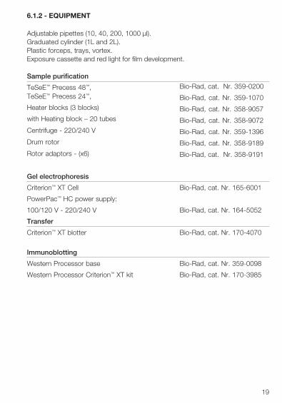
6.1.2 - EQUIPMENT
Adjustable pipettes (10, 40, 200, 1000 µl).Graduated cylinder (1L and 2L).Plastic forceps, trays, vortex.Exposure cassette and red light for film development.
Sample purification
TeSeE™ Precess 48™, TeSeE™ Precess 24™,
Heater blocks (3 blocks)
with Heating block – 20 tubes
Centrifuge - 220/240 V
Drum rotor
Rotor adaptors - (x6)
Bio-Rad, cat. Nr. 359-0200
Bio-Rad, cat. Nr. 359-1070
Bio-Rad, cat. Nr. 358-9057
Bio-Rad, cat. Nr. 358-9072
Bio-Rad, cat. Nr. 359-1396
Bio-Rad, cat. Nr. 358-9189
Bio-Rad, cat. Nr. 358-9191
Gel electrophoresis
Criterion™ XT Cell Bio-Rad, cat. Nr. 165-6001
PowerPac™ HC power supply:
100/120 V - 220/240 V Bio-Rad, cat. Nr. 164-5052
Transfer
Criterion™ XT blotter Bio-Rad, cat. Nr. 170-4070
Immunoblotting
Western Processor base Bio-Rad, cat. Nr. 359-0098
Western Processor Criterion™ XT kit Bio-Rad, cat. Nr. 170-3985
20
6.2 - PREPARATION OF REAGENTS6.2.1 - SAMPLE PURIFICATION
• Proteinase KSolution of proteinase K diluted in reagent A:
u 1 ml Reagent Au 20 µl Proteinase K
Mix well by inverting until you obtain a homogeneous solution. After reconstitution, diluted proteinase K is stable 10 hours at room temperature (+18°C to +30°C).
• Laemmli solutionSolution of SDS + 2-Mercaptoethanol + Laemmli sample buffer:
u 0.6 g SDSu 1.5 ml 2-Mercaptoethanol
Mix by inverting. u 28.5 ml Laemmli sample buffer
Solution is aliquoted into 4 ml aliquots and stored at -20°C. Thawed aliquots can be re-frozen.
Note: It is recommended to prepare Laemmli solution one hour before use allowing SDS to be correctly dissolved.
6.2.2 - ELECTROPHORESIS
• Kaleidoscope™ prestained standardThe Kaleidoscope™ prestained standard is prepared during the sampledenaturation before loading on the acrylamide gel.
Prepare a 1/12 dilution in Laemmli solution, for example 10 µl of theKaleidoscope™ prestained standard + 110 µl of Laemmli solution.
Please refer to the Kaleidoscope™ prestained standard insert for storageconditions.
• MagicMark™ XP Western StandardThe MagicMark™ XP molecular weight is prepared during the sample denaturation before loading on the acrylamide gel.
Prepare a 1/12 dilution in Laemmli solution, for example 10 µl of MagicMark™ XP + 110 µl of Laemmli solution.
Please refer to MagicMark™ XP insert for storage conditions.
21
• Criterion™ XT migration bufferSolution of MOPS (1x).
Prepare a 1/20 dilution. 1 L of diluted buffer is required for 1 tank:u 950 ml Distilled wateru 50 ml MOPS buffer (20x)
Homogenize. Solution can not be stored.
6.2.3 - PROTEIN TRANSFER
• Transfer bufferSolution of Tris/CAPS-Ethanol 15%. Approximately 2 L is required for 1migration tank.
u 750 ml Distilled wateru 150 ml Pure ethanolu 100 ml Tris/CAPS (10x)
Homogenize. Solution can not be stored.
6.2.4 - IMMUNOBLOTTING
• Wash solution 1Solution of PBS (1x) + 0.1% Tween® 20. Approximately 1 L is required for thecomplete process of 1 membrane.
u 900 ml Distilled wateru 100 ml PBS (10x)u 1 ml Tween® 20
Thorougly homogenize. Solution is stored at +2°C to +8°C, overnight.
• Wash solution 2Solution of PBS (1x). Approximately 200 ml is required for the completeprocess of 1 membrane.
u 900 ml Distilled wateru 100 ml PBS (10x)
Solution is stored at room temperature (+18°C to +30°C) overnight.
• Blocking solutionDuring the transfer step, dilute the blocking solution (Bl) 1/10 in Wash solution1. 40 ml of diluted blocking solution is required for 1 membrane.
u 36 ml Wash solution 1u 4 ml Blocking solution (10x)
Homogenize by tube inverting.
22
• Diluted primary antibodyJust prior to use, dilute the primary antibody 1/10 in Wash solution 1.30 ml of diluted antibody is required for 1 membrane.
u 27 ml Wash solution 1u 3 ml Primary antibody (10x)
Homogenize by inverting.
• Diluted secondary antibody (conjugate)Just prior to use, dilute the secondary antibody 1/10 in Wash solution 1.40 ml of diluted conjugate is required for 1 membrane.
u 36 ml Wash solution 1u 4 ml Secondary antibody (10x)
Homogenize by tube inverting.
• ECLSubstrate (ECL) must be prepared just prior to use. 2 ml of substrateis required for 1 membrane.
u 1 ml Reagent 1u 1 ml Reagent 2
Homogenize.
• Development solutionu 800 ml Distilled wateru 200 ml Development product
Solution is stored at room temperature (+18°C to +30°C), in a darkroom for 15 days maximum.
• Fixative solutionu 800 ml Distilled wateru 200 ml Fixative product
Solution is stored at room temperature (+18°C to +30°C), in a darkroom for 15 days maximum.
23
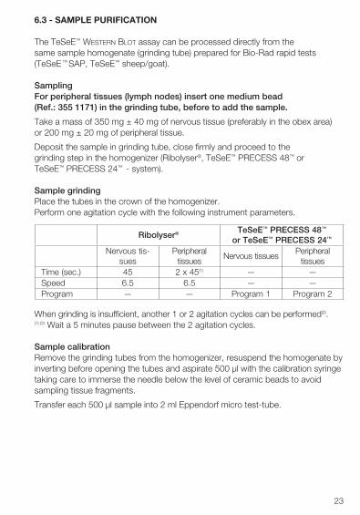
6.3 - SAMPLE PURIFICATION
The TeSeE™ Western Blot assay can be processed directly from the same sample homogenate (grinding tube) prepared for Bio-Rad rapid tests (TeSeE ™ SAP, TeSeE™ sheep/goat).
Sampling For peripheral tissues (lymph nodes) insert one medium bead (Ref.: 355 1171) in the grinding tube, before to add the sample.
Take a mass of 350 mg ± 40 mg of nervous tissue (preferably in the obex area) or 200 mg ± 20 mg of peripheral tissue.
Deposit the sample in grinding tube, close firmly and proceed to the grinding step in the homogenizer (Ribolyser®, TeSeE™ PRECESS 48™ or TeSeE™ PRECESS 24™ - system).
Sample grindingPlace the tubes in the crown of the homogenizer.Perform one agitation cycle with the following instrument parameters.
When grinding is insufficient, another 1 or 2 agitation cycles can be performed(2).(1) (2) Wait a 5 minutes pause between the 2 agitation cycles.
Sample calibrationRemove the grinding tubes from the homogenizer, resuspend the homogenate by inverting before opening the tubes and aspirate 500 µl with the calibration syringe taking care to immerse the needle below the level of ceramic beads to avoid sampling tissue fragments.
Transfer each 500 µl sample into 2 ml Eppendorf micro test-tube.
Ribolyser® TeSeE™ PRECESS 48™
or TeSeE™ PRECESS 24™
Nervous tis-sues
Peripheral tissues
Nervous tissuesPeripheral
tissuesTime (sec.) 45 2 x 45(1) — —Speed 6.5 6.5 — —Program — — Program 1 Program 2
24
Note: at this stage, both grinding tubes after homogenisation and micro test-tubes after sample calibration can be stored, closed: • At room temperature (+18°C to +30°C) for 15 hours.• At +2°C to +8°C for 72 hours.• At -20°C for 1 year. Frozen samples must be thawed at room temperature
(+18°C to +30°C).
Samples can be submitted to a maximum of 3 freezing/thawing cycles.
Samples must always be homogenized by inverting before use.
Proteinase K TreatmentDistribute 500 µl of reconstituted proteinase K solution (see paragraph 6.2.1) into each micro test-tube.
Homogenize the sealed tubes by inverting (10 times) and incubate at 37°C ± 2°C in a heating block incubator for 10 minutes.
Precipitation of PrPres with reagent BRemove the tubes from the incubator. Open and distribute 500 µl of reagent B into each tube. Homogenize by inverting until a homogeneous colour is obtained.
Concentration of the PrPres by centrifugationCentrifuge the tubes for 7 minutes at 15000 g at 20°C.
Sample clarifyingDiscard the supernatant by inverting over a waste container. Then dry the tubes by inverting onto absorbent paper for 5 minutes.
Distribute 100 µl of the Laemmli solution (see paragraph 6.2.1) into each micro test-tube.
Incubate for 5 minutes at room temperature (+18°C to +30°C).
Completely resolubilise the pellet by aspiration/dispensing with a pipette.
Incubate for 5 minutes at 100°C ± 5°C in a heating block incubator.
Remove the tubes from the incubator, homogenize by vortexing.
Centrifuge the tubes for 15 minutes at 15000 g at 20°C.
Transfer the supernatant to a new micro test-tube. Discard the tube containing the pellet.
At this stage, the supernatant can be stored frozen at -20°C for 24 hours; the samples must be thawed at room temperature (+18°C to +30°C) prior to use.
25
6.4 - ELECTROPHORESIS
The TeSeE™ Western Blot assay can be used for both confirmation of TSE suspected samples or for strain typing in sheep.
The following procedure is applicable for confirmation of TSE suspected samples.
Please contact your Bio-Rad representative for instruction protocol in case of strain typing application.
Gel preparationRemove the plastic band on the bottom of the plastic plate and place the acrylamide gels (see paragraph 6.2.2) in the migration tank. Pour the migration buffer (see paragraph 6.2.2) on each side of the gel up to the top of the wells and into the electrophoresis tank. Carefully remove the combs and rinse each well with migration buffer, using a pipette.
Sample loading
Heat the samples for 4 minutes at 100 °C ± 5°C just prior to loading 15 µl/well.
Load 15 µl of the diluted Kaleidoscope™ prestained standard and 15 µl of the diluted MagicMark™ XP (see paragraph 6.2.2).
Note: In case several gels are run at the same time, make sure that you stagger the loading of controls into different lanes for easy identification.
Differential migration of the samplesRun the gel at room temperature (+18°C to +30°C) for 50 minutes at 200 V.
6.5 - PROTEIN TRANSFER
The transfer buffer must be prepared before the end of the sample migration (see paragraph 6.2.3).
Protein transfer preparationCut the membrane to the gel dimensions. Always use forceps when handling the membrane.
Immerse the membrane in pure ethanol for 15 seconds, rinse in distilled water for 5 minutes, then for 10 minutes in the transfer buffer.
Carefully remove the gel from the plastic plates and let it equilibrate for 10 minutes in the transfer buffer.
26
Gel sandwich preparationSoak filter paper and fibre pads in the transfer buffer.
Open the transfer cassette, with red side on the left. Respectively place on the red side a fiber pad, a filter paper, the membrane* and the gel*.
Complete with a filter paper then a fibre pad and close the cassette.
Immerse it in the transfer tank, previously filled to the indicated limit with transfer buffer. A frozen ice pack is added prior to fill the tank.
*Remove any air bubbles which may have formed.
Note: In case several membranes are processed at the same time, label eachmembrane in the corner.
Transfer onto the PVDF membraneAgitate during the transfer by using a magnetic stirring bar and run for60 minutes at 115 V.
6.6 - IMMUNOBLOTTING
a) Upon completion of the protein transfer, open the blotting assembly andremove the membrane for development. Quickly immerse the membrane inWash solution 2 (see paragraph 6.2.4), then place it in ethanol for 10 secondsbefore rinsing for 5 minutes in distilled water.
Note: At this step, the membrane can be stored overnight in distilled waterat +2°C to +8°C.
Let the membrane adjust to room temperature (+18°C to +30°C) beforeto start the immunoblotting.
b) Eliminate distilled water and incubate the membrane for 30 minutes in blockingsolution (see paragraph 6.2.4). Incubate under medium agitation.
40 ml is required for 1 membrane.
Note: from this step until step g), the Bio-Rad Western Processor can be usedfor agitation and washing steps (refer to instruction manual for settings).
c) Eliminate the blocking solution and incubate the membrane in diluted primaryantibody (see paragraph 6.2.4) for 30 minutes at room temperature (+18°C to+30°C) under medium agitation.
30 ml of diluted primary antibody is required for 1 membrane.
27
d) Eliminate the primary antibody solution and using Wash solution 1, briefly rinsethe membrane, then wash twice for respectively 5 and 10 minutes, under fastagitation.
100 ml of Wash solution 1 is required for each cycle and for 1 membrane.
e) Eliminate the Wash solution 1 and incubate the membrane for 20 minutesin diluted secondary antibody (see paragraph 6.2.4) at room temperature(+18°C to +30°C) under medium agitation.
40 ml of diluted secondary antibody is required for 1 membrane.
f) Eliminate the secondary antibody solution and using Wash solution 1,briefly rinse for respectively 5, 10 and 10 minutes under fast agitation.100 ml of Wash solution 1 is required for each cycle and for 1 membrane.
g) Place the membrane in 100 ml of Wash solution 2 under slow agitation.
h) Drain the membrane on absorbent paper without blotting and place it in theplastic folder.
i) Add the ECL reagent (see paragraph 6.2.4). Eliminate the excess of reagent andair bubbles with absorbent paper. Place into the exposure cassette.
j) In a darkroom, cover the folder with a film and expose for 15 minutes. Film canbe exposed longer or shorter time for optimal signal.
k) Immerse the film in developing solution for 45 seconds (see paragraph 6.2.4).Rinse in distilled water. Immerse the film in fixative solution until the filmbecomes completely transparent.
l) Wash with distilled water and let the film dry.
28
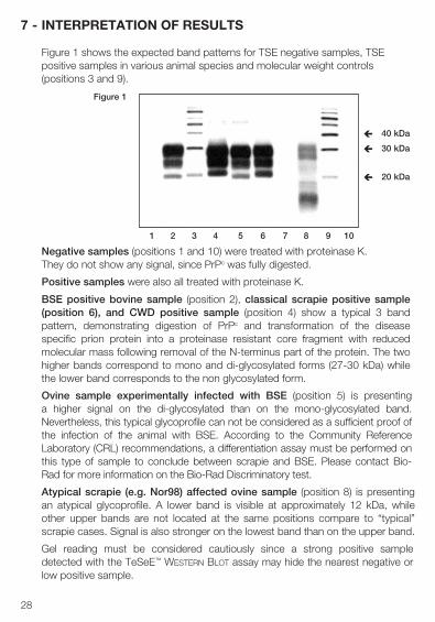
7 - INTERPRETATION OF RESULTS
Figure 1 shows the expected band patterns for TSE negative samples, TSE positive samples in various animal species and molecular weight controls (positions 3 and 9).
Negative samples (positions 1 and 10) were treated with proteinase K. They do not show any signal, since PrPc was fully digested.
Positive samples were also all treated with proteinase K.
BSE positive bovine sample (position 2), classical scrapie positive sample (position 6), and CWD positive sample (position 4) show a typical 3 band pattern, demonstrating digestion of PrPc and transformation of the disease specific prion protein into a proteinase resistant core fragment with reduced molecular mass following removal of the N-terminus part of the protein. The two higher bands correspond to mono and di-glycosylated forms (27-30 kDa) while the lower band corresponds to the non glycosylated form.
Ovine sample experimentally infected with BSE (position 5) is presenting a higher signal on the di-glycosylated than on the mono-glycosylated band. Nevertheless, this typical glycoprofile can not be considered as a sufficient proof of the infection of the animal with BSE. According to the Community Reference Laboratory (CRL) recommendations, a differentiation assay must be performed on this type of sample to conclude between scrapie and BSE. Please contact Bio-Rad for more information on the Bio-Rad Discriminatory test.
Atypical scrapie (e.g. Nor98) affected ovine sample (position 8) is presenting an atypical glycoprofile. A lower band is visible at approximately 12 kDa, while other upper bands are not located at the same positions compare to “typical” scrapie cases. Signal is also stronger on the lowest band than on the upper band.
Gel reading must be considered cautiously since a strong positive sample detected with the TeSeE™ Western Blot assay may hide the nearest negative or low positive sample.
1 2 3 4 5 6 7 8 9 10
Figure 1
40 kDa è
20 kDa è
30 kDa è
29
Limits of the test:A negative result means that the test sample does not contain detectable PrPres by TeSeE™ Western Blot assay. However, as very low levels of PrPres may not be detected, such a result does not exclude the possibility of infection.
Any sample with a negative result according to the TeSeE™ Western Blot assay interpretation criteria, following a positive rapid test result, should be tested with one of the other OIE certified confirmatory methods, Immunohistochemistry (IHC) or SAF-Immunoblot.
Any sample with a reproducible positive result according to the test interpretation critera must be verified in accordance with current legal regulation.
30
8 - PRECAUTIONS
The quality of the data obtained depends on compliance with the following good laboratory practices:
• Reagents must be stored at the appropiate temperature (refer to supplier’sindications).
• Do not use reagents whose shelf-life has expired.
• Do not use reconstituted proteinase K after 10 hours storage at roomtemperature (+18°C to +30°C).
• Do not mix or combine reagents derived from different batches of theTeSeE™ Western Blot assay during the same manipulation, with the exceptionof grinding tubes, reagent A, reagent B and proteinase K.
• Allow the reagents and buffers to adjust to room temperature (+18°C to +30°C)for 30 minutes before use.
• Thoroughly reconstitute reagents, avoiding any contamination.
• Do not perform the test in the presence of reactive vapors (acids, alkalines,aldehydes) or dust, which could alter the enzymatic activity of the conjugate.
• The enzymatic reaction is very sensitive to all metals or metallic ions.Consequently, no metallic element must be in contact with the conjugate.
• Only use polypropylene tubes.
• Use clean glassware, rinsed in distilled water, or preferably disposable material.
• Use a new pipette tip for each sample.
• When starting electrophoresis and transfer, check that the 2 electrodes are incontact with buffer.
• All the rinsing times must be respected to avoid any excess background noiseduring final staining with ECL reagent.
31
9 - HYGIENE AND SAFETY INSTRUCTIONS
Generally, hygiene conditions, biosafety measures and good laboratory practices must be in agreement with the recommendations of national regulatory authorities.
• All reagents of the kit are intended for use in “in vitro” diagnosis.
• Wear disposable gloves when handling reagents and samples and wash yourhands thoroughly after handling them.
• Do not pipette by mouth.
• Use polypropylene containers to avoid broken glass.
• All the materials directly in contact with the samples and the wash solutionsmust be considered as contaminated.
• Avoid splashing samples or solutions containing samples.
• Contaminated surfaces must be cleaned with 20 000 ppm sodium hypochloritesolution (bleach). When the contaminating liquid is an acid, contaminatedsurfaces must be first neutralized with sodium hydroxide before using bleach.Surfaces must be rinsed with distilled water, dried with ethanol and wiped withabsorbent paper. The material used for cleaning must be discarded in a specificcontainer for contaminated waste.
• Samples, material and contaminated products must be eliminated afterdecontamination:- either by soaking in 1 M sodium hydroxide (final concentration) for at least
1 hour at room temperature (+18°C to +30°C),- or by soaking in 20 000 ppm sodium hypochlorite solution for at least
1 hour at room temperature (+18°C to +30°C),- or by autoclaving at 134°C minimum for at least 18 minutes, under 3 bars of
pressure.
Note: never autoclave solutions containing bleach or reagent B.
• All operations involved in Transmissible Spongiform Encephalopathy (TSE)screening tests are subject to regulations and must be performed in an isolated,limited and controlled access laboratory devoted exclusively to this activity. Alaboratory coat, overshoes, gloves, mask with visor or simple mask with safetyglasses are required to ensure the operator’s safety.
• Operators must receive specific training concerning the risks related to TSEsagents or prions and the validated modes of decontamination forunconventional agents. Biosafety measures must be in agreement withrecommendations of regular authorities of the country.
32
• Neutralize and/or autoclave all wash solutions or wash wastes or any liquidcontaining biological samples prior to their elimination.
• Reagent B is a dangerous substance classified as nocive (> 25% alcohol)according to European legislation.
• Reagents containing 0.1% ProClin® 300 are classified as irritating preparationsaccording to European legislation.
Xn(Alcohol > 25%)(0.1% ProClin® 300)
R : 10-22-37/38-41-43-67 Flammable. Harmful if swallowed. Irritating to respiratory system and skin. Risk of serious damage to eyes. May cause sensitisation by skin contact. Inhalation of vapour may cause drowsiness and dizziness.
S : 7/9-13-26-28-37/39-46 Keep container tightly closed and in a well ventilated place. Keep away from food, drink and animal feed. In case of contact with eyes, rinse immediately with plenty of water and seek medical advice. After contact with skin wash immediately with plenty of water. Wear suitable protecting clothings, gloves and eye/face protection. If swallowed, seek medical advice immediately and show this container or label.
33
10 - REFERENCES
1. S.B. PRUSINER (1991) Molecular biology of prion diseases - Science 252:1515-1522.
2. J.B. KATZ, J.G. PEDERSEN, A.L. JENNY and W.D. TAYLOR (1992) Assessment of Western immunoblotting for the confirmatory diagnosis of ovine scrapie and bovine spongiform encephalopathy (BSE) - Journal of Veterinary Diagnostic Investigations,4, 447-449.
3. G.A.H. WELLS, Y.I. SPENCER and M. HARITANI (1994) Configuration and topographic distribution of PrP in the central nervous system in bovine spongiform encephalopathy: an immonohistochemical stydy. In: Slow Infections of theCentral Nervous System, J. BJORNSSON, R.I. CARP, A. LOVE and M. WISNIEWSKI - EdsThe New York Academy of Sciences, pp 350-352.
4. J.P. DESLYS, E. COMOY, S. HAWKINS, S. SIMON, H. SCHIMMEL, G. WELLS, J. GRASSI and J. MOYNAGH (2001)Screening slaughtered cattle for BSE - Nature: 409; 476-477.
5. S. BENESTAD, P. SARRADIN, B. THU, J. SCHÖNHEIT, M. TRANULIS and B. BRATBERG (2003) Cases of scrapie with unusual features in Norway and designation of new type, Nor98. Veterinary Record 153, 202-208.
6. A. BUSCHMANN, G. LÜHKEN, J. SCHULTZ, G. ERHARDT and M.-H. GROSCHUP (2004) Neuronal accumulation of abnormal prion protein in sheep carrying a scrapie-resistant genotype (PrPARR/ARR). Journal of General Virology 85, 2727-2733.
7. L. ORGE, A.GALO, C.MACHADO, C. LIMA, C. OCHOA, J. SILVA, M. RAMOS and J.-P. SIMAS (2004) Identification of putative atypical scrapie in sheep in Portugal. Journal of General Virology 85,3487-3491.
8. A. BUSCHMANN, A.-G. BIACABE, U. ZIEGLER, A. BENCSIK, J.-Y. MADEC, G. ERHARDT, G. LÜHKEN, T. BARON and M.-H. GROSCHUP (2004) Atypical scrapie cases in Germany and France are identified by discrepant reaction patterns in BSE rapid tests. Journal of Virological Methods 117, 27-36.
34
9. H. DE BOSSCHERE, S. ROELS, S. BENESTAD, E. VANOPDENBOSCH (2004). Scrapie case similar to Nor98 diagnosed in Belgium via active surveillance. Veterinary Record 155, 707-708.
10. H. ONNASCH, H. M. GUNN, B.J. BRADSHAW, S. BENESTAD, H.F. BASSETT (2004). Two Irish cases of scrapie resembling Nor98. Veterinary Record 155, 636-637.
11. S. BENESTAD, P. SARRADIN, J.-M. BILHEUDE, J. GRASSI , H. LAUDE, O. ANDREOLETTI, T. MOLDAL and B. BRATBERG. Are there gold standard methods for the diagnosis of TSE ? Nor98 scrapie cases: atypical cases and their challenging diagnosis. Second International Chronic Wasting Disease Symposium, Madison - Wisconsin, USA.
12. BIACABE, A-G., LAPLANCHE, J-L., RYDER,S. AND BARON, T. (2004). Distinct molecular phenotypes in bovine prion diseases, EMBO Reports 5, 110-114.
13. CASALONE, C., ZANUSSO, G., ACUTIS, P., FERRARI, S., CAPUCCI, I., TAGLIAVINI, F., MONACO, S. and CARAMELLI, M. (2004). Identification of a second bovine amyloidotic spongiform encephalopathy: Molecular similarities with sporadic Creutzfeldt-Jakob disease. PNAS 101, 3065-70.
14. BUSCHMANN A; GRETZSCHEL A; BIACABE A-G; SCHIEBEL K; CORONA C; HOFFMANN C; EIDEN M; BARON T; CASALONE C; GROSCHUP M H (2006). Atypical BSE in Germany-proof of transmissibility and biochemical characterization. Veterinary microbiology 2006;117(2-4):103-16.
15. ARSAC, J.-N., ANDREOLETTI, O., BILHEUDE, J.-M., LACROUX, C., BENESTAD, S. L., AND BAARON, T. (2007). Similar biochemical signatures and prion protein genotypes in atypical scrapie and Nor98 cases, France and Norway. Emerging Infectious Diseases 13(1), 58-65.
16. E. URO-COSTE, H CASSARD, S. SIMON, S. LUGAN, J.-M. BILHEUDE, A. PERRET-LIAUDET, J.-W. IRONSIDE, S. HAIK, C. BASSET-LEOBON, C. LACROUX, K. PEOCH, N. STREICHENBERGER, J. LANGEVELD, M.-W. HEAD, J. GRASSI, J.-J.HAUW, F. SCHELCHER, M.-B. DELISLE, O. ANDREOLETTI (2008) Beyond PrPres Type 1/Type 2 Dichotomy in Creutzfeldt-Jakob disease. Plos Pathogens volume 4, issue 2, e1000029.
Validé et certifié par l’OIE comme étant
apte aux emplois prévus dans cette notice Numéro d'enregistrement: 20090105
TeSeE™ Western BlotRéactifs pour la confirmation in vitro des échantillons suspectés positifs est
Catalog # 355-1169 32 Tests
36
SOMMAIRE
1 - INFORMATIONS GÉNÉRALES
2 - PRINCIPE DU TEST
3 - COMPOSITION DE LA TROUSSE
4 - ÉCHANTILLONS
5 - MODE OPÉRATOIRE AVEC LE GEL MINI BLOT™
5.1 Réactifs et matériels supplémentaires5.2 Préparation des réactifs5.3 Purification des échantillons5.4 Électrophorèse5.5 Transfert des protéines
5.6 Immunoblotting
6 - MODE OPÉRATOIRE AVEC LE GEL CRITERION™ XT6.1 Réactifs et matériels supplémentaires6.2 Préparation des réactifs6.3 Purification des échantillons6.4 Électrophorèse6.5 Transfert des protéines
6.6 Immunoblotting
7 - INTERPRÉTATION DES RÉSULTATS
8 - PRÉCAUTIONS
9 - MESURES D’HYGIÈNE ET DE SÉCURITÉ
10 - BIBLIOGRAPHIE
37
1 - INFORMATIONS GÉNÉRALES
Les encéphalopathies spongiformes transmissibles (EST) ont été décrites pour la première fois au dix-huitième siècle, chez les moutons (tremblante) et, plus récemment, chez les cervidés comme le cerf et l’élan (maladie du dépérissement chronique) et les bovins (encéphalopathie spongiforme bovine, ESB). L’homme est également sensible à certaines formes d’EST comme le kuru, la maladie de Creutzfeldt-Jakob (MCJ) ou le syndrome de Gerstmann-Sträussler-Scheinker (SGS). L’émergence d’une nouvelle forme de la maladie de Creutzfeldt-Jakob, dite variante, (MCJv) chez l’homme a été fortement liée à la consommation alimentaire de viandes ou de produits carnés infectés par l’agent de l’ESB. L’une des principales caractéristiques de l’encéphalopathie spongiforme transmissible (EST) est une accumulation progressive, dans le système nerveux central, d’une isoforme anormale de la protéine prion naturelle ou cellulaire (PrPc), dite PrPres. Cette protéine PrPres spécifique de la maladie se caractérise par une plus grande résistance aux protéases. Le test TeSeE™ Western Blot permet l’identification qualitative de la protéine PrPres, après un traitement protéolytique aboutissant à un fragment de poids moléculaire réduit suite à la coupure de l’extrémité N-terminale.
Des programmes de surveillance active/passive ont été lancés dans le monde entier pour détecter les animaux infectés par l’ESB, la tremblante ou par la maladie du dépérissement chronique (CWD). Ces programmes ont permis d’identifier un nombre croissant de cas positifs dans les laboratoires de dépistage. Ces échantillons positifs (animaux suspects) sont alors systématiquement confirmés comme “infectés par une EST” par une histopathologie montrant des altérations spongiformes caractéristiques, ou par la détection de la PrP anormale par immunohistochimie (IHC) ou encore par la détection de Fibrilles Associées à la Tremblante (Examen SAF) par microscopie électronique. Ces méthodes de confirmation requièrent une grande expertise pour l’interprétation des résultats et sont souvent longues et coûteuses. La technique western blot peut également être utilisée comme une méthode alternative pour la confirmation des échantillons soupçonnés d’être porteurs d’EST.
Les données de validation de ce kit ont été certifiées par l’OIE, sur la base d’un examen d’experts, comme étant conformes à l’usage qui leur est assigné à savoir la détection post mortem des encéphalopathies spongiformes transmissibles (EST) chez les bovins (encéphalopathie spongiforme bovine, ESB), chez les ovins et les caprins (ESB et tremblante) et chez les cervidés (cachexie chronique) pour les emplois suivants: 1. Confirmer une suspicion d’EST sur des prélèvements positifs détectés dans
des laboratoires de dépistage de pays appliquant des programmes de
38
surveillance active/passive. Tout prélèvement donnant un résultat négatif selon les critères d’interprétation du TeSeE™ Western Blot, après un résultat positif à un test rapide, doit être soumis à l’une des autres épreuves de confirmation certifiées par l’OIE, l’immunohistochimie ou l’Immunoblot-SAF ;
2. Confirmer la prévalence de l’infection par l’une des maladies associées auxEST (ESB, tremblante, cachexie chronique) dans le cadre d’une étudeépidémiologique menée dans un pays à faible prévalence ;
3. Estimer la prévalence de l’infection pour faciliter l’analyse de risque (par ex.pour des enquêtes ou pour la mise en place de mesures de prophylaxie) etcontribuer à démontrer l’efficacité des politiques d’éradication.
Le test TeSeE™ Western Blot utilise le même principe de dosage que les tests rapides Bio-Rad (TeSeE™ SAP, TeSeE™ sheep/goat), notamment la purification préliminaire et l’étape de concentration de la PrPres suivies d’une détection très sensible par immunoblot. Ce test constitue donc un outil efficace pour la confirmation du diagnostic d’échantillons soupçonnés d’être porteurs d’une EST, ainsi que pour le typage des souches d’EST chez les moutons.
2 - PRINCIPE DU TEST
Le test TeSeE™ Western Blot permet la détection de la PrPres dans des échantillons de tissu nerveux (bovins, ovins, caprins, cervidés…) ou périphériques (cervidés) prélevés chez des animaux infectés.
Le mode opératoire commence par la digestion de la protéine prion cellulaire (PrPc), suivie par la purification et la concentration de la protéine prion PrPres spécifique de la maladie. La détection de la PrPres est réalisée par électrophorèse puis par une technique d’immunoblotting utilisant un anticorps monoclonal hautement spécifique de la PrPres.
Le mode opératoire comprend les étapes suivantes :• Homogénéisation de l’échantillon,• Digestion de la PrPc par la protéinase K,• Purification et concentration de la PrPres,• Électrophorèse et tranfert sur une membrane,• Immunoblotting.
39
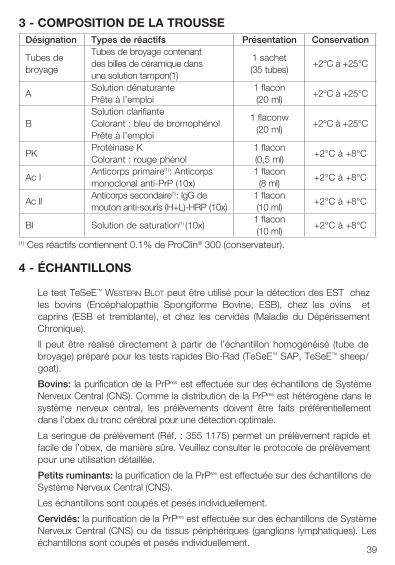
3 - COMPOSITION DE LA TROUSSEDésignation Types de réactifs Présentation Conservation
Tubes de broyage
Tubes de broyage contenant des billes de céramique dans une solution tampon(1)
1 sachet(35 tubes)
+2°C à +25°C
ASolution dénaturante Prête à l’emploi
1 flacon(20 ml)
+2°C à +25°C
BSolution clarifiante Colorant : bleu de bromophénol Prête à l’emploi
1 flaconw(20 ml)
+2°C à +25°C
PKProtéinase KColorant : rouge phénol
1 flacon(0,5 ml)
+2°C à +8°C
Ac IAnticorps primaire(1): Anticorps monoclonal anti-PrP (10x)
1 flacon(8 ml)
+2°C à +8°C
Ac IIAnticorps secondaire(1): IgG de mouton anti-souris (H+L)-HRP (10x)
1 flacon(10 ml)
+2°C à +8°C
Bl Solution de saturation(1) (10x)1 flacon(10 ml)
+2°C à +8°C
(1) Ces réactifs contiennent 0.1% de ProClin® 300 (conservateur).
4 - ÉCHANTILLONS
Le test TeSeE™ Western Blot peut être utilisé pour la détection des EST chez les bovins (Encéphalopathie Spongiforme Bovine, ESB), chez les ovins et caprins (ESB et tremblante), et chez les cervidés (Maladie du Dépérissement Chronique).
Il peut être réalisé directement à partir de l’échantillon homogénéisé (tube de broyage) préparé pour les tests rapides Bio-Rad (TeSeE™ SAP, TeSeE™ sheep/goat).
Bovins: la purification de la PrPres est effectuée sur des échantillons de Système Nerveux Central (CNS). Comme la distribution de la PrPres est hétérogène dans le système nerveux central, les prélèvements doivent être faits préférentiellement dans l’obex du tronc cérébral pour une détection optimale.
La seringue de prélèvement (Réf. : 355 1175) permet un prélèvement rapide et facile de l’obex, de manière sûre. Veuillez consulter le protocole de prélèvement pour une utilisation détaillée.
Petits ruminants: la purification de la PrPres est effectuée sur des échantillons de Système Nerveux Central (CNS).
Les échantillons sont coupés et pesés individuellement.
Cervidés: la purification de la PrPres est effectuée sur des échantillons de Système Nerveux Central (CNS) ou de tissus périphériques (ganglions lymphatiques). Les échantillons sont coupés et pesés individuellement.
40
5 - PROCÉDURE AVEC LE GEL MINI BLOT™
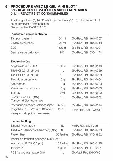
5.1 - RÉACTIFS ET MATÉRIELS SUPPLÉMENTAIRES5.1.1 - RÉACTIFS ET CONSOMMABLES
Pipettes graduées (5, 10, 25 ml), tubes coniques (50 ml), micro-tubes (2 ml) en polypropylène avec bouchon.Film protecteur PARAFILM® M.
Purification des échantillons
Tampon Laemmli 30 ml Bio-Rad, Réf. 161-0737
2-Mercaptoethanol 25 ml Bio-Rad, Réf. 161-0710
SDS 100 g Bio-Rad, Réf. 161-0301
Seringues de calibration 200 Bio-Rad, Réf. 355-1174
Électrophorèse
Acrylamide 40% 29:1 500 ml Bio-Rad, Réf. 161-0146
Tris-HCl 0,5 M, pH 6.8 1 L Bio-Rad, Réf. 161-0799
Tris-HCl 1,5 M, pH 8.8 1 L Bio-Rad, Réf. 161-0798
Bleu de bromophénol 10 g Bio-Rad, Réf. 161-0404
Saccharose 1 kg Bio-Rad, Réf. 161-0720
Persulfate d’ammonium 10 g Bio-Rad, Réf. 161-0700
TEMED 5 ml Bio-Rad, Réf. 161-0800
Tris/Glycine/SDS (10x) 1 L (Tampon d’électrophorèse)
Bio-Rad, Réf. 161-0732
Bio-Rad, Réf. 161-0324
Invitrogen, Réf. LC5602
Marqueur précoloré Kaleidoscope™ 500 µl MagicMark™ XP Western Standard 250 µl (marqueur de poids moléculaire)
Immunoblotting
Éthanol (Normapur) 1L VWR, Réf. 2821-296
Tris/CAPS (tampon de transfert) (10x) 1L Bio-Rad, Réf. 161-0778
Papier filtre 50 feuilles Bio-Rad, Réf. 170-3932
(papier de transfert pour gels Mini Blot™)
Membrane PVDF (0,2 µm) 10 feuilles Bio-Rad, Réf. 162-0175
Tween® 20 100 ml Bio-Rad, Réf. 170-6531
PBS (tampon de lavage) (10x) 1 L Bio-Rad, Réf. 161-0780
41
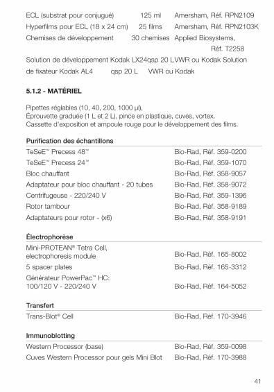
ECL (substrat pour conjugué) 125 ml Amersham, Réf. RPN2109
Hyperfilms pour ECL (18 x 24 cm) 25 films Amersham, Réf. RPN2103K
Chemises de développement 30 chemises Applied Biosystems,
Réf. T2258
Solution de développement Kodak LX24 qsp 20 L VWR ou Kodak Solution
de fixateur Kodak AL4 qsp 20 L VWR ou Kodak
5.1.2 - MATÉRIEL
Pipettes réglables (10, 40, 200, 1000 µl),Éprouvette graduée (1 L et 2 L), pince en plastique, cuves, vortex.Cassette d’exposition et ampoule rouge pour le développement des films.
Purification des échantillons
TeSeE™ Precess 48™ Bio-Rad, Réf. 359-0200
TeSeE™ Precess 24™ Bio-Rad, Réf. 359-1070
Bloc chauffant Bio-Rad, Réf. 358-9057
Adaptateur pour bloc chauffant - 20 tubes Bio-Rad, Réf. 358-9072
Centrifugeuse - 220/240 V Bio-Rad, Réf. 359-1396
Rotor tambour Bio-Rad, Réf. 358-9189
Adaptateurs pour rotor - (x6) Bio-Rad, Réf. 358-9191
Électrophorèse
Mini-PROTEAN® Tetra Cell, electrophoresis module Bio-Rad, Réf. 165-8002
5 spacer plates Bio-Rad, Réf. 165-3312
Générateur PowerPac™ HC: 100/120 V - 220/240 V Bio-Rad, Réf. 164-5052
Transfert
Trans-Blot® Cell Bio-Rad, Réf. 170-3946
Immunoblotting
Western Processor (base) Bio-Rad, Réf. 359-0098
Cuves Western Processor pour gels Mini Blot Bio-Rad, Réf. 170-3988
42
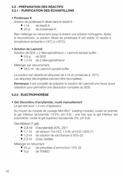
5.2 - PRÉPARATION DES RÉACTIFS5.2.1 - PURIFICATION DES ÉCHANTILLONS
• Protéinase KSolution de protéinase K diluée dans le réactif A :
u 1 ml de réactif Au 20 µl de protéinase K
Bien mélanger en retournant jusqu’à obtenir une solution homogène. Après la reconstitution, la solution diluée de protéinase K est stable 10 heures à température ambiante (+18°C à +30°C).
• Solution de LaemmliSolution de SDS + 2-Mercaptoéthanol + Laemmli sample buffer :
u 0,6 g de SDSu 1,5 ml de 2-Mercaptoéthanol
Mélanger par retournement.u 28,5 ml de Laemmli sample buffer
La solution est répartie en aliquotes de 4 ml et conservée à -20°C. Les aliquotes décongelées peuvent être recongelées.
Remarque: Il est conseillé de préparer la solution de Laemmli une heure avant utilisation pour permettre une dissolution complète du SDS.
5.2.2 - ÉLECTROPHORÈSE
• Gel discontinu d’acrylamide, coulé manuellementLe gel doit avoir 1,5 mm d’épaisseur.
Au moyen du module de coulage Mini Blot™ (casting module), couler en premierle gel inférieur (acrylamide 13,5%, pH 8,8) ; une fois que le gel inférieur estpolymérisé, couler le gel supérieur (acrylamide 3%, pH 6,8).
Gel inférieur (1 gel)u 2,8 ml d’acrylamide 40%, 29:1u 1,7 ml de tampon Tris-HCl, 1,5 M, pH 8,8 / SDS (1) u 1,3 ml de solution de saccharose à 50% (2)u 2,5 ml d’eau distillée
Mélanger en retournant.u 43 µl de persulfate d’ammonium 10% (3)u 9 µl de TEMED

43
Verser 7 ml de la solution entre les plaques et garder le reste de la solution comme témoin de polymérisation. Recouvrir délicatement jusqu’en haut avec 1 ml du tampon Tris-HCl 0,3 M pH 8,8 / SDS (4) de sorte que la surface du gel ne sèche pas.
Laisser le gel polymériser pendant 15-20 minutes à température ambiante (+18°C à +30°C). Vérifier que le reste de solution est polymérisé. Retourner le système pour éliminer l’excès de tampon.
Gel supérieur (1 gel)u 4 ml d’acrylamide 3% (7)u 28 µl de persulfate d’ammonium 10% (3)u 6 µl de TEMED
Mélanger en retournant.
Verser doucement le gel inférieur sur le gel supérieur et garder le reste de solution comme témoin de polymérisation. Positionner le peigne, en veillant à ne pas piéger de bulle d’air dans les positions des puits. Laisser le gel se polymériser pendant 5-10 minutes à température ambiante (+18°C à +30°C). Vérifier que le reste de solution est polymérisé.
(1) Solution de tampon Tris-HCl 1,5 M, pH 8,8 / SDS u 0.2 g de SDSu 50 ml de tampon Tris-HCl 1,5 M, pH 8,8
La solution peut être conservée de +2°C à +8°C pendant 2 semaines.
(2) Solution de saccharose 50%u 25 g de saccharoseu qsp 50 ml d’eau distillée
La solution de saccharose peut être conservée de +2°C à +8°C, pendant 2 semaines.
(3) Solution de persulfate d’ammonium 10%u 5 g de persulfate d’ammoniumu qsp 50 ml d’eau distillée
La solution de persulfate d’ammonium est répartie en aliquotes et conservée à -20°C. La solution décongelée peut être conservée de +2°C à +8°C, pendant 2 semaines.
(4) Solution de tampon Tris-HCl 0,3 M, pH 8,8 / SDS u 40 ml d’eau distilléeu 10 ml de tampon Tris-HCl 1,5 M, pH 8.8 / SDS
La solution peut être conservée de +2°C à +8°C, pendant 2 semaines.
44
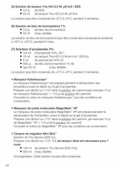
(5) Solution de tampon Tris-HCl 0,5 M, pH 6,8 / SDSu 0,2 g de SDSu 50 ml de tampon Tris-HCl 0,5 M, pH 6,8
La solution peut être conservée de +2°C à +8°C, pendant 2 semaines.
(6) Solution de bleu de bromophénol 1%u 0,5 g de bleu de bromophénolu 50 ml d’eau distillée
La solution de bleu de bromophénol peut être conservée à température ambiante (+18°C à +30°C), pendant 6 mois.
(7) Solution d’acrylamide 3%u 3,8 ml d’acrylamide 40%, 29:1u 10 ml de tampon Tris-HCl 0,5 M pH 6,8 / SDS (5)u 6 ml de saccharose 50% (2)u 500 µl de bleu de bromophénol 1% (6)u qsp 50 ml d’eau distillée
La solution peut être conservée de +2°C à +8°C, pendant 2 semaines.
• Marqueur Kaleidoscope™
Le marqueur Kaleidoscope™ est préparé pendant la dénaturation deséchantillons avant le dépôt sur le gel d’acrylamide.Préparer une dilution au 1/12e dans la solution de Laemmli (par exemple 10 µlde marqueur Kaleidoscope™ + 110 µl de solution de Laemmli).Consulter la notice du marqueur Kaleidoscope™ pour les conditions deconservation.
• Marqueur de poids moléculaire MagicMark™ XPLe marqueur de poids moléculaire MagicMark™ XP est préparé pendant ladénaturation de l’échantillon, avant le dépôt sur le gel d’acrylamide.Préparer une dilution au 1/12e dans la solution de Laemmli, par exemple 10 µlde MagicMark™ XP + 110 µl de la solution de Laemmli.Consulter la notice du MagicMark™ XP pour les conditions de conservation.
• Tampon de migration Mini Blot™
Solution de Tris-Glycine-SDS (1x).Préparer une dilution au 1/10e. 1 L de tampon dilué est nécessaire pour 1cuve:
u 100 ml de tampon Tris-Glycine-SDS (10x)u 900 ml d’eau distillée
Homogénéiser. Cette solution ne peut être conservée.
45
5.2.3 - TRANSFERT DES PROTÉINES
• Tampon de transfertSolution de Tris/CAPS-éthanol 15%. 2,5 L sont nécessaires pour 1 cuve de transfert.
u 750 ml d’eau distilléeu 150 ml d’éthanol puru 100 ml de Tris/CAPS (10x)
Homogénéiser. Cette solution ne peut être conservée.
5.2.4 - IMMUNOBLOTTING
• Solution de lavage 1Solution de PBS (1x) + 0,1% Tween® 20. Environ 500 ml sont nécessairespour le traitement complet d’une membrane.
u 900 ml d’eau distilléeu 100 ml de PBS (10x)u 1 ml de Tween® 20
Bien homogénéiser. La solution peut être conservée une nuit de +2°C à +8°C.
• Solution de lavage 2Solution de PBS (1x). Environ 100 ml sont nécessaires pour le traitementcomplet d’une membrane.
u 900 ml d’eau distilléeu 100 ml de PBS (10x)
La solution peut être conservée une nuit à température ambiante (+18°C à +30°C).
• Solution de saturationPendant l’étape de tranfert, diluer la solution de saturation (Bl) au 1/10e dansla solution de lavage 1. 20 ml de solution de saturation diluée (1x) sontnécessaires pour 1 membrane.
u 18 ml de solution de lavage 1u 2 ml de solution de saturation (10x)
Homogénéiser en retournant le tube.
• Anticorps primaire diluéJuste avant usage, diluer l’anticorps primaire au 1/10e dans la solution delavage 1. 15 ml de l’anticorps dilué sont nécessaires pour 1 membrane.
u 13,5 ml de solution de lavage 1u 1,5 ml d’anticorps primaire (10x)
Homogénéiser en retournant le tube.
46
• Anticorps secondaire dilué (conjugué)Juste avant usage, diluer l’anticorps secondaire au 1/10e dans la solutionde lavage 1. 20 ml de conjugué dilué sont nécessaires pour1 membrane.
u 18 ml de solution de lavage 1u 2 ml d’anticorps secondaires (10x)
Homogénéiser en retournant le tube.
• ECLLe substrat (ECL) doit être préparé juste avant usage. 1 ml de substrat estnécessaire pour 1 membrane.
u 0,5 ml de réactif 1u 0,5 ml de réactif 2
Homogénéiser la solution.
• Solution de développementu 800 ml d’eau distilléeu 200 ml de produit de développement
La solution peut être conservée à température ambiante (+18°C à +30°C), en chambre noire, pendant 15 jours maximum.
• Solution de fixationu 800 ml d’eau distilléeu 200 ml de produit fixateur
La solution peut être conservée à température ambiante (+18°C à +30°C), en chambre noire, pendant 15 jours maximum.
47
5.3 - PURIFICATION DES ÉCHANTILLONS
Le test TeSeE™ Western Blot peut être réalisé directement à partir de l’échantillon homogénéisé (tube de broyage) préparé pour les tests rapides Bio-Rad (TeSeE™ SAP, TeSeE™ sheep/goat).
PrélèvementPour l’analyse des tissus périphériques (ganglions lymphatiques) introduire une bille de taille moyenne (“Medium bead”, Réf. 355 1171) dans le tube de broyage avant d’y ajouter l’échantillon.Prélever une masse de 350 mg ± 40 mg de tissu nerveux (de préférence au niveau de l’obex) ou 200 mg ± 20 mg de tissu périphérique. Déposer l’échantillon dans un tube de broyage, fermer hermétiquement et procéder au broyage dans l’homogénéiseur (Ribolyser®, TeSeE™ PRECESS 48™ ou TeSeE™ PRECESS 24™ ).
Broyage de l’échantillonPlacer les tubes dans la couronne de l’homogénéiseur.Effectuer un cycle d’agitation avec les paramètres suivants:
Si le broyage est insuffisant, 1 ou 2 autres cycles d’agitation peuvent être effectués(2).(1) (2) Une pause de 5 minutes est nécessaire entre 2 cycles d’agitation.
Calibration de l’échantillonRetirer les tubes de broyage de l’homogénéiseur, remettre en suspension l’homogénat en retournant avant d’ouvrir les tubes et aspirer 500 µl avec la seringue de calibration, en veillant à plonger l’aiguille au-dessous du niveau des billes en céramique pour éviter de prélever des fragments de tissus.
Transférer chaque échantillon de 500 µl dans un micro-tube Eppendorf de 2 ml.
Remarque: à ce stade, les tubes de broyage après homogénéisation et les micro-tubes après calibration de l’échantillon peuvent être conservés, fermés :• A température ambiante (+18°C à +30°C) pendant 15 heures.• De +2°C à +8°C pendant 72 heures.• A -20°C pendant 1 an. Les échantillons congelés doivent être décongelés à
température ambiante (+18°C à +30°C).
Les échantillons peuvent être soumis à un maximum de 3 cycles de congélation/décongélation. Les échantillons doivent toujours être homogénéisés par retournement avant usage.
Ribolyser® TeSeE™ PRECESS 48™
ou TeSeE™ PRECESS 24™
Tissus nerveux
Tissus périphériques
Tissus nerveux
Tissus périphériques
Temps (sec.) 45 2 x 45(1) — —Vitesse 6.5 6.5 — —Programme — — Programme 1 Programme 2
48
Traitement par la protéinase KDistribuer 500 µl de solution de protéinase K reconstituée (voir paragraphe 5.2.1) dans chaque micro-tube.
Homogénéiser les tubes fermés en les retournant (10 fois) et incuber à 37°C ± 2°C dans un bloc chauffant pendant 10 minutes.
Précipitation de la PrPres avec le réactif BSortir les tubes de l’incubateur. Les ouvrir et distribuer 500 µl de réactif B dans chaque tube. Homogénéiser en retournant les tubes jusqu’à obtenir une couleur homogène.
Concentration de la PrPres par centrifugationCentrifuger les tubes pendant 7 minutes à 15 000 g à 20°C.
Clarification de l’échantillonJeter le surnageant dans un récipient pour déchets. Sécher ensuite les tubes en les retournant sur du papier absorbant pendant 5 minutes.
Distribuer 100 µl de solution de Laemmli (voir paragraphe 5.2.1) dans chaque micro-tube.
Incuber pendant 5 minutes à température ambiante (+18°C à +30°C).
Resolubiliser complètement le culot par aspiration/rejet avec une pipette.
Incuber pendant 5 minutes à 100°C ± 5°C dans un bloc chauffant.
Sortir les tubes de l’incubateur, homogénéiser au vortex.
Centrifuger les tubes pendant 15 minutes à 15000 g à 20°C.
Transférer le surnageant dans un nouveau micro-tube. Jeter le tube contenant le culot.
À ce stade, le surnageant peut être conservé congelé à -20°C pendant 24 heures ; les échantillons doivent être décongelés à température ambiante (+18°C à +30°C) avant usage.
49
5.4 - ÉLECTROPHORÈSE
Le test TeSeE™ Western Blot peut être à la fois utilisé pour confirmer un échantillon suspecté infecté d’EST et pour le typage de souches chez le mouton.
La procédure suivante est destinée à une analyse de confirmation d’échantillons suspectés infectés d’EST.
Pour toute information sur le protocole à suivre pour le typage des souches, merci de prendre contact avec votre représentant Bio-Rad.
Préparation du gelPlacer les gels d’acrylamide (voir paragraphe 5.2.2) dans la cuve de migration. Verser le tampon de migration (voir paragraphe 5.2.2) dans la cuve d’électrophorèse de chaque côté des gels, jusqu’en haut des puits. Retirer délicatement les peignes et rincer chaque puits avec du tampon de migration, à l’aide d’une pipette.
Dépôt des échantillonsChauffer les échantillons pendant 4 minutes à 100 °C ± 5°C juste avant de déposer 15 µl/puits.
Déposer 15 µl du marqueur Kaleidoscope™ dilué et 15 µl du marqueur MagicMark™ XP dilué (voir paragraphe 5.2.2).
Remarque: au cas où plusieurs gels seraient traités en même temps, déposer les marqueurs dans différents puits pour faciliter l’identification.
Migration différentielle des échantillonsLancer la migration à température ambiante (+18°C à +30°C) pendant 90 minutes à 150 V. Le front de migration doit être sorti du gel.
5.5 - TRANSFERT DES PROTÉINES
Le tampon de transfert doit être préparé avant la fin de la migration des échantillons (voir paragraphe 5.2.3).
Préparation du transfert des protéinesDécouper la membrane aux dimensions du gel. La membrane doit toujours être manipulée avec des pinces.
Plonger la membrane dans l’éthanol pur pendant 15 secondes, rincer dans l’eau distillée pendant 5 minutes, puis dans le tampon de transfert pendant 10 minutes. Retirer délicatement le gel des plaques de verre et le laisser s’équilibrer pendant 10 minutes dans le tampon de transfert.
50
Préparation du sandwichTremper les papiers filtre et les coussins de fibres dans le tampon de transfert.
Ouvrir la cassette de transfert, côté transparent à gauche. Placer sur le côté transparent, dans l’ordre, un coussin de fibres, un papier filtre, la membrane* et le gel*. Compléter avec un papier filtre puis un coussin de fibres et fermer la cassette.
Plonger la cassette dans la cuve de transfert, préalablement remplie jusqu’à la limite indiquée avec du tampon de transfert.
*Eliminer les éventuelles bulles d’air formées.
Remarque: Au cas où plusieurs membranes seraient traitées en même temps,identifier chaque membrane dans un coin.
Transfert sur la membrane PVDFTransférer pendant 60 minutes à 115 V, sans agitation (barreau magnétique).
5.6 - IMMUNOBLOTTING
a) Au terme du transfert des protéines, ouvrir la cassette et retirer la membranepour la révéler. Plonger rapidement la membrane dans la solution de lavage2 (voir paragraphe 5.2.4), puis la mettre dans l’éthanol pendant 10 secondesavant de rincer pendant 5 minutes dans l’eau distillée.
Note: A ce stade, la membrane peut être conservée pendant une nuit dansde l’eau distillée de +2°C à +8°C.
Laisser revenir à température ambiante (+18°C à +30°C) avant de commencerl’immunoblotting.
b) Eliminer l’eau distillée et incuber la membrane pendant 30 minutes dans lasolution de saturation (voir paragraphe 5.2.4). Incuber sous agitation modérée.
20 ml suffisent pour 1 membrane.
Remarque: à partir de cette étape jusqu’à l’étape g), le Western ProcessorBio-Rad peut être utilisé pour les étapes d’agitation et de lavage (voir le manuelpour les paramètres).
c) Eliminer la solution de saturation et incuber la membrane dans l’anticorpsprimaire dilué (voir paragraphe 5.2.4) pendant 30 minutes à températureambiante (+18°C à +30°C) sous agitation modérée.
15 ml d’anticorps primaire dilué sont nécessaires pour 1 membrane.
51
d) Eliminer la solution d’anticorps primaire et avec la solution de lavage 1, rincerrapidement la membrane, puis laver deux fois respectivement pendant 5 et10 minutes sous agitation rapide.
50 ml de solution de lavage 1 sont nécessaires pour chaque cycle et pour1 membrane.
e) Eliminer la solution de lavage 1 et incuber la membrane pendant 20 minutesdans l’anticorps secondaire dilué (voir paragraphe 5.2.4) à températureambiante (+18°C à +30°C) sous agitation modérée.
20 ml d’anticorps secondaire dilué sont nécessaires pour 1 membrane.
f) Eliminer la solution d’anticorps secondaire et avec la solution de lavage 1, rincerrapidement, puis laver respectivement pendant 5, 10 et 10 minutes, sousagitation rapide.
50 ml de solution de lavage 1 sont nécessaires pour chaque cycle et pour1 membrane.
g) Placer la membrane dans 50 ml de la solution de lavage 2 sous agitation lente.
h) Laisser s’égoutter la membrane au-dessus du papier absorbant, en évitant toutcontact direct, et la placer dans la chemise en plastique.
i) Ajouter le réactif ECL (voir paragraphe 5.2.4). Eliminer l’excès de réactif et lesbulles d’air à l’aide de papier absorbant. Mettre dans la cassette d’exposition.
j) Dans une chambre noire, recouvrir la chemise d’un film et exposer pendant 15minutes. Le film peut être exposé plus ou moins longtemps, pour unsignal optimal.
k) Plonger le film dans la solution de développement pendant 45 secondes (voirparagraphe 5.2.4). Rincer dans l’eau distillée. Plonger le film dans le fixateurjusqu’à ce que le film devienne totalement transparent.
l) Laver à l’eau distillée et laisser sécher le film.
52
6 - PROCEDURE AVEC LE GEL CRITERION™ XT6.1 - RÉACTIFS ET MATÉRIELS NÉCESSAIRES6.1.1 - RÉACTIFS ET CONSOMMABLES
Pipettes graduées (5, 10, 25 ml), tubes coniques (50 ml), micro-tubes (2 ml) en polypropylene avec bouchon.
Film de protection PARAFILM® M.
Purification des échantillons
Tampon Laemmli 30 ml Bio-Rad, Réf. 161-0737
2-Mercaptoéthanol 25 ml Bio-Rad, Réf. 161-0710
SDS 100 g Bio-Rad, Réf. 161-0301
Seringues de calibration 200 Bio-Rad, Réf. 355-1174
Electrophorèse
Criterion™ XT 12 % Bis-Tris 1 gel-18 puits Bio-Rad, Réf. 345-0118
XT-MOPS (20 x) 500 ml Bio-Rad, Réf. 161-0788(tampon d’électrophorèse)
Marqueur précoloré Kaleidoscope™ 500 µl
MagicMark™ XP Western Standard 250 µl
Bio-Rad, Réf. 161-0324
Invitrogen, Réf. LC5602 (marqueur de poids moléculaire)
Immunobloting
Ethanol (Normapur) 1L VWR, Réf. 20821-296
Tris/CAPS (tampon de transfert) (10x) 1L Bio-Rad, Réf. 161-0778
Papier filtre 50 feuilles Bio-Rad, Réf. 170-4085(papier de transfert pour gels Criterion™ XT)
Membrane PVDF (0,2 µm) 10 feuilles Bio-Rad, Réf. 162-0175
Tween® 20 100 ml
PBS (tampon de lavage) (10x) 1 L
ECL (substrat pour conjugué) 125 ml
Bio-Rad, Réf. 170-6531
Bio-Rad, Réf. 161-0780
Amersham, Réf. RPN2109
Hyperfilms pour ECL (18 x 24 cm) 25 films Amersham, Réf. RPN2103K
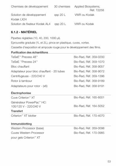
53
Chemises de développement 30 chemises Applied Biosystems, Réf. T2258
Solution de développement qsp 20 L VWR ou Kodak
Kodak LX24
Solution de fixateur Kodak AL4 qsp 20 L VWR ou Kodak
6.1.2 - MATÉRIEL
Pipettes réglables (10, 40, 200, 1000 µl).
Eprouvette graduée (1L et 2L), pince en plastique, cuves, vortex.
Cassette d’exposition et ampoule rouge pour le développement des films.
Purification des échantillons
TeSeE™ Precess 48™ Bio-Rad, Réf. 359-0200
TeSeE ™Precess 24™ Bio-Rad, Réf. 359-1070
Bloc chauffant Bio-Rad, Réf. 358-9057
Adaptateur pour bloc chauffant - 20 tubes Bio-Rad, Réf. 358-9072
Centrifugeuse - 220/240 V Bio-Rad, Réf. 359-1396
Rotor à tambour Bio-Rad, Réf. 358-9189
Adaptateurs pour rotor - (x6) Bio-Rad, Réf. 358-9191
Électrophorèse
Cuve Criterion™ XT
Générateur PowerPac™ HC:100/120 V - 220/240 V
Bio-Rad, Réf. 165-6001
Bio-Rad, Réf. 164-5052
Transfert
Criterion™ XT blotter Bio-Rad, Réf. 170-4070
Immunoblotting
Western Processor (base) Bio-Rad, Réf. 359-0098
Cuves Western Processor Bio-Rad, Réf. 170-3985
pour gels Criterion™ XT
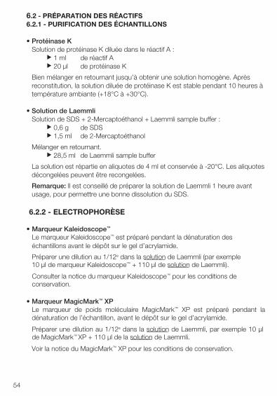
54
6.2 - PRÉPARATION DES RÉACTIFS6.2.1 - PURIFICATION DES ÉCHANTILLONS
• Protéinase KSolution de protéinase K diluée dans le réactif A :
u 1 ml de réactif Au 20 µl de protéinase K
Bien mélanger en retournant jusqu’à obtenir une solution homogène. Après reconstitution, la solution diluée de protéinase K est stable pendant 10 heures à température ambiante (+18°C à +30°C).
• Solution de LaemmliSolution de SDS + 2-Mercaptoéthanol + Laemmli sample buffer :
u 0,6 g de SDSu 1,5 ml de 2-Mercaptoéthanol
Mélanger en retournant.u 28,5 ml de Laemmli sample buffer
La solution est répartie en aliquotes de 4 ml et conservée à -20°C. Les aliquotes décongelées peuvent être recongelées.
Remarque: Il est conseillé de préparer la solution de Laemmli 1 heure avant usage, pour permettre une bonne dissolution du SDS.
6.2.2 - ELECTROPHORÈSE
• Marqueur Kaleidoscope™
Le marqueur Kaleidoscope™ est préparé pendant la dénaturation deséchantillons avant le dépôt sur le gel d’acrylamide.
Préparer une dilution au 1/12e dans la solution de Laemmli (par exemple10 µl de marqueur Kaleidoscope™ + 110 µl de solution de Laemmli).
Consulter la notice du marqueur Kaleidoscope™ pour les conditions deconservation.
• Marqueur MagicMark™ XPLe marqueur de poids moléculaire MagicMark™ XP est préparé pendant ladénaturation de l’échantillon, avant le dépôt sur le gel d’acrylamide.
Préparer une dilution au 1/12e dans la solution de Laemmli, par exemple 10 µlde MagicMark™ XP + 110 µl de la solution de Laemmli.
Voir la notice du MagicMark™ XP pour les conditions de conservation.
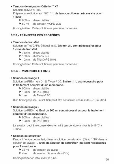
55
• Tampon de migration Criterion™ XTSolution de MOPS (1x).Préparer une dilution au 1/20e. 1 L de tampon dilué est nécessaire pour1 cuve:
u 950 ml d’eau distilléeu 50 ml de tampon MOPS (20x)
Homogénéiser. Cette solution ne peut être conservée.
6.2.3 - TRANSFERT DES PROTÉINES
• Tampon de transfertSolution de Tris/CAPS-Éthanol 15%. Environ 2 L sont nécessaires pour1 cuve de transfert.
u 750 ml d’eau distilléeu 150 ml d’éthanol puru 100 ml de Tris/CAPS (10x)
Homogénéiser. Cette solution ne peut être conservée.
6.2.4 - IMMUNOBLOTTING
• Solution de lavage 1Solution de PBS (1x) + 0,1% Tween® 20. Environ 1 L est nécessaire pourle traitement complet d’une membrane.
u 900 ml d’eau distiléeu 100 ml de PBS (10x)u 1 ml de Tween® 20
Bien homogénéiser. La solution peut être conservée une nuit de +2°C à +8°C.
• Solution de lavage 2Solution de PBS (1x). Environ 200 ml sont nécessaires pour le traitementcomplet d’une membrane.
u 900 ml d’eau distilléeu 100 ml de PBS (10x)
La solution peut être conservée une nuit à température ambiante (+18°C à +30°C).
• Solution de saturationPendant l’étape de tranfert, diluer la solution de saturation (Bl) au 1/10e dans lasolution de lavage 1. 40 ml de solution de saturation (1x) sont nécessairespour 1 membrane.
u 36 ml de solution de lavage 1u 4 ml de solution de saturation (10x)
Homogénéiser en retournant le tube.
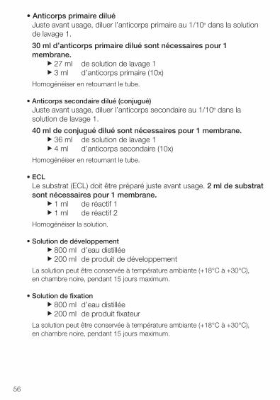
• Anticorps primaire diluéJuste avant usage, diluer l’anticorps primaire au 1/10e dans la solutionde lavage 1.
30 ml d’anticorps primaire dilué sont nécessaires pour 1membrane.
u 27 ml de solution de lavage 1u 3 ml d’anticorps primaire (10x)
Homogénéiser en retournant le tube.
• Anticorps secondaire dilué (conjugué)Juste avant usage, diluer l’anticorps secondaire au 1/10e dans lasolution de lavage 1.
40 ml de conjugué dilué sont nécessaires pour 1 membrane.u 36 ml de solution de lavage 1u 4 ml d’anticorps secondaire (10x)
Homogénéiser en retournant le tube.
• ECLLe substrat (ECL) doit être préparé juste avant usage. 2 ml de substratsont nécessaires pour 1 membrane.
u 1 ml de réactif 1u 1 ml de réactif 2
Homogénéiser la solution.
• Solution de développementu 800 ml d’eau distilléeu 200 ml de produit de développement
La solution peut être conservée à température ambiante (+18°C à +30°C), en chambre noire, pendant 15 jours maximum.
• Solution de fixationu 800 ml d’eau distilléeu 200 ml de produit fixateur
La solution peut être conservée à température ambiante (+18°C à +30°C), en chambre noire, pendant 15 jours maximum.
56
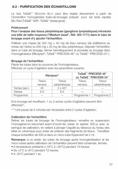
6.3 - PURIFICATION DES ÉCHANTILLONS
Le test TeSeE™ Western Blot peut être réalisé directement à partir de l’échantillon homogénéisé (tube de broyage) préparé pour les tests rapides Bio-Rad (TeSeE™ SAP, TeSeE™ sheep/goat).
PrélèvementPour l’analyse des tissus périphériques (ganglions lymphatiques) introduire une bille de taille moyenne (“Medium bead”, Réf. 355 1171) dans le tube de broyage avant d’y ajouter l’échantillon.
Prélever une masse de 350 mg ± 40 mg de tissu nerveux (de préférence au niveau de l’obex) ou 200 mg ± 20 mg de tissu périphérique. Déposer l’échantillon dans un tube de broyage, fermer hermétiquement et procéder au broyage dans l’homogénéiseur (Ribolyser®, TeSeE™ PRECESS 48™ ou TeSeE™ PRECESS 24™ ).
Broyage de l’échantillonPlacer les tubes dans la couronne de l’homogénéiseur.Effectuer un cycle d’agitation avec les paramètres suivants:
Si le broyage est insuffisant, 1 ou 2 autres cycles d’agitation peuvent être effectués(2).(1) (2) Une pause de 5 minutes est nécessaire entre 2 cycles d’agitation.
Calibration de l’échantillonRetirer les tubes de broyage de l’homogénéiseur, remettre en suspension l’homogénat en retournant avant d’ouvrir les tubes et aspirer 500 µl avec la seringue de calibration, en veillant à plonger l’aiguille au-dessous du niveau des billes en céramique pour éviter de prélever des fragments de tissus. Transférer chaque échantillon de 500 µl dans un micro-tube Eppendorf de 2 ml.
Remarque: à ce stade, les tubes de broyage après homogénéisation et les micro-tubes après calibration de l’échantillon peuvent être conservés, fermés:• A température ambiante (+18°C à +30°C) pendant 15 heures.• De +2°C à +8°C pendant 72 heures.• A -20°C pendant 1 an.
57
Ribolyser® TeSeE™ PRECESS 48™
ou TeSeE™ PRECESS 24™
Tissus nerveux
Tissus périphériques
Tissus nerveux
Tissus périphériques
Temps (sec.) 45 2 x 45(1) — —Vitesse 6.5 6.5 — —Programme — — Programme 1 Programme 2
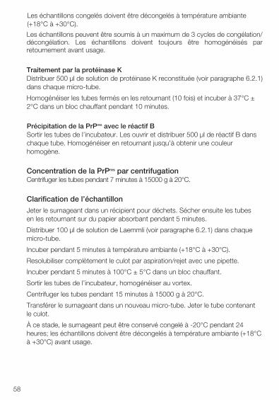
58
Les échantillons congelés doivent être décongelés à température ambiante (+18°C à +30°C).
Les échantillons peuvent être soumis à un maximum de 3 cycles de congélation/décongélation. Les échantillons doivent toujours être homogénéisés par retournement avant usage.
Traitement par la protéinase KDistribuer 500 µl de solution de protéinase K reconstituée (voir paragraphe 6.2.1) dans chaque micro-tube.
Homogénéiser les tubes fermés en les retournant (10 fois) et incuber à 37°C ± 2°C dans un bloc chauffant pendant 10 minutes.
Précipitation de la PrPres avec le réactif BSortir les tubes de l’incubateur. Les ouvrir et distribuer 500 µl de réactif B dans chaque tube. Homogénéiser en retournant jusqu’à obtenir une couleur homogène.
Concentration de la PrPres par centrifugationCentrifuger les tubes pendant 7 minutes à 15000 g à 20°C.
Clarification de l’échantillon
Jeter le surnageant dans un récipient pour déchets. Sécher ensuite les tubes en les retournant sur du papier absorbant pendant 5 minutes.
Distribuer 100 µl de solution de Laemmli (voir paragraphe 6.2.1) dans chaque micro-tube.
Incuber pendant 5 minutes à température ambiante (+18°C à +30°C).
Resolubiliser complètement le culot par aspiration/rejet avec une pipette.
Incuber pendant 5 minutes à 100°C ± 5°C dans un bloc chauffant.
Sortir les tubes de l’incubateur, homogénéiser au vortex.
Centrifuger les tubes pendant 15 minutes à 15000 g à 20°C.
Transférer le surnageant dans un nouveau micro-tube. Jeter le tube contenant le culot.
À ce stade, le surnageant peut être conservé congelé à -20°C pendant 24 heures; les échantillons doivent être décongelés à température ambiante (+18°C à +30°C) avant usage.
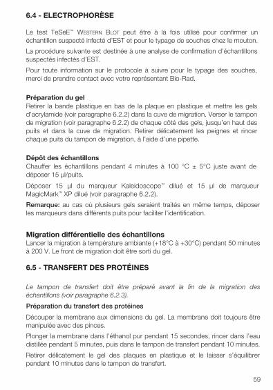
59
6.4 - ELECTROPHORÈSE
Le test TeSeE™ Western Blot peut être à la fois utilisé pour confirmer un échantillon suspecté infecté d’EST et pour le typage de souches chez le mouton.
La procédure suivante est destinée à une analyse de confirmation d’échantillons suspectés infectés d’EST.
Pour toute information sur le protocole à suivre pour le typage des souches, merci de prendre contact avec votre représentant Bio-Rad.
Préparation du gelRetirer la bande plastique en bas de la plaque en plastique et mettre les gels d’acrylamide (voir paragraphe 6.2.2) dans la cuve de migration. Verser le tampon de migration (voir paragraphe 6.2.2) de chaque côté des gels, jusqu’en haut des puits et dans la cuve de migration. Retirer délicatement les peignes et rincer chaque puits du tampon de migration, à l’aide d’une pipette.
Dépôt des échantillonsChauffer les échantillons pendant 4 minutes à 100 °C ± 5°C juste avant de déposer 15 µl/puits.
Déposer 15 µl du marqueur Kaleidoscope™ dilué et 15 µl de marqueur MagicMark™ XP dilué (voir paragraphe 6.2.2).
Remarque: au cas où plusieurs gels seraient traités en même temps, déposer les marqueurs dans différents puits pour faciliter l’identification.
Migration différentielle des échantillonsLancer la migration à température ambiante (+18°C à +30°C) pendant 50 minutes à 200 V. Le front de migration doit être sorti du gel.
6.5 - TRANSFERT DES PROTÉINES
Le tampon de transfert doit être préparé avant la fin de la migration des échantillons (voir paragraphe 6.2.3).
Préparation du transfert des protéines
Découper la membrane aux dimensions du gel. La membrane doit toujours être manipulée avec des pinces.
Plonger la membrane dans l’éthanol pur pendant 15 secondes, rincer dans l’eau distillée pendant 5 minutes, puis dans le tampon de transfert pendant 10 minutes.
Retirer délicatement le gel des plaques en plastique et le laisser s’équilibrer pendant 10 minutes dans le tampon de transfert.
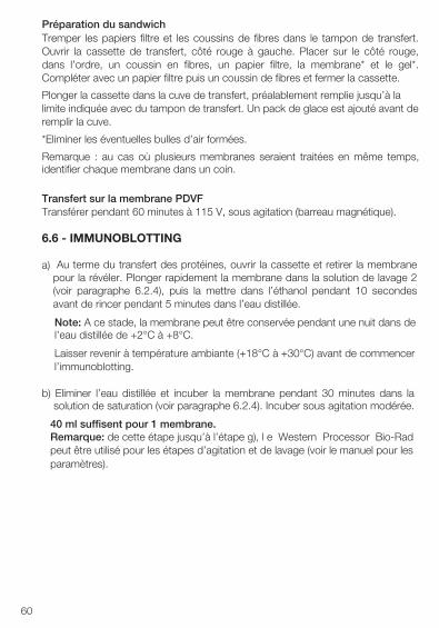
60
Préparation du sandwichTremper les papiers filtre et les coussins de fibres dans le tampon de transfert. Ouvrir la cassette de transfert, côté rouge à gauche. Placer sur le côté rouge, dans l’ordre, un coussin en fibres, un papier filtre, la membrane* et le gel*. Compléter avec un papier filtre puis un coussin de fibres et fermer la cassette.
Plonger la cassette dans la cuve de transfert, préalablement remplie jusqu’à la limite indiquée avec du tampon de transfert. Un pack de glace est ajouté avant de remplir la cuve.
*Eliminer les éventuelles bulles d’air formées.
Remarque : au cas où plusieurs membranes seraient traitées en même temps,identifier chaque membrane dans un coin.
Transfert sur la membrane PDVFTransférer pendant 60 minutes à 115 V, sous agitation (barreau magnétique).
6.6 - IMMUNOBLOTTING
a) Au terme du transfert des protéines, ouvrir la cassette et retirer la membranepour la révéler. Plonger rapidement la membrane dans la solution de lavage 2(voir paragraphe 6.2.4), puis la mettre dans l’éthanol pendant 10 secondesavant de rincer pendant 5 minutes dans l’eau distillée.
Note: A ce stade, la membrane peut être conservée pendant une nuit dans del’eau distillée de +2°C à +8°C.
Laisser revenir à température ambiante (+18°C à +30°C) avant de commencerl’immunoblotting.
b) Eliminer l’eau distillée et incuber la membrane pendant 30 minutes dans lasolution de saturation (voir paragraphe 6.2.4). Incuber sous agitation modérée.
40 ml suffisent pour 1 membrane. Remarque: de cette étape jusqu’à l’étape g), l e Western Processor Bio-Radpeut être utilisé pour les étapes d’agitation et de lavage (voir le manuel pour lesparamètres).
61
c) Eliminer la solution de saturation et incuber la membrane dans l’anticorpsprimaire dilué (voir paragraphe 6.2.4) pendant 30 minutes à températureambiante (+18°C à +30°C) sous agitation modérée.
30 ml d’anticorps primaire dilué sont nécessaires pour 1 membrane.
d) Eliminer la solution d’anticorps primaire et avec la solution de lavage 1, rincerrapidement la membrane, puis laver deux fois respectivement pendant 5 et 10minutes, sous agitation rapide.
100 ml de solution de lavage 1 sont nécessaires pour chaque cycle etpour 1 membrane.
e) Eliminer la solution de lavage 1 et incuber la membrane pendant 20 minutesdans l’anticorps secondaire dilué (voir paragraphe 6.2.4) à températureambiante (+18°C à +30°C) sous agitation modérée.
40 ml d’anticorps secondaire dilué sont nécessaires pour 1 membrane.
f) Eliminer la solution d’anticorps secondaire et avec la solution de lavage 1, rincerrapidement, puis laver respectivement pendant 5, 10 et 10 minutes, sousagitation rapide.
100 ml de solution de lavage 1 sont nécessaires pour chaque cycle etpour 1 membrane.
g) Placer la membrane dans 100 ml de la solution de lavage 2 sous agitationlente.
h) Laisser s’égoutter la membrane au-dessus du papier absorbant, en évitant toutcontact direct, et la placer dans la chemise en plastique.
i) Ajouter le réactif ECL (voir paragraphe 6.2.4). Eliminer l’excès de réactif et lesbulles d’air à l’aide de papier absorbant. Mettre dans la cassette d’exposition.
j) Dans une chambre noire, recouvrir la chemise d’un film et exposer pendant 15minutes. Le film peut être exposé plus ou moins longtemps, pour un signaloptimal.
k) Plonger le film dans la solution de développement pendant 45 secondes (voirparagraphe 6.2.4). Rincer dans l’eau distillée. Plonger le film dans le fixateurjusqu’à ce que le film devienne totalement transparent.
l) Laver à l’eau distillée et laisser sécher le film.
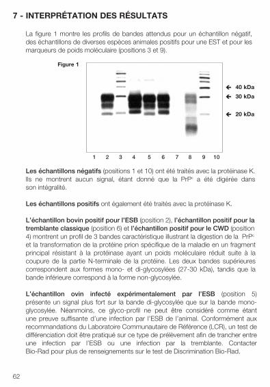
62
7 - INTERPRÉTATION DES RÉSULTATS
La figure 1 montre les profils de bandes attendus pour un échantillon négatif, des échantillons de diverses espèces animales positifs pour une EST et pour les marqueurs de poids moléculaire (positions 3 et 9).
Les échantillons négatifs (positions 1 et 10) ont été traités avec la protéinase K. Ils ne montrent aucun signal, étant donné que la PrPc a été digérée dans son intégralité.
Les échantillons positifs ont également été traités avec la protéinase K.
L’échantillon bovin positif pour l’ESB (position 2), l’échantillon positif pour la tremblante classique (position 6) et l’échantillon positif pour le CWD (position 4) montrent un profil de 3 bandes caractéristique illustrant la digestion de la PrPc
et la transformation de la protéine prion spécifique de la maladie en un fragment principal résistant à la protéinase ayant un poids moléculaire réduit suite à la coupure de la partie N-terminale de la protéine. Les deux bandes supérieures correspondent aux formes mono- et di-glycosylées (27-30 kDa), tandis que la bande inférieure correspond à la forme non-glycosylée.
L’échantillon ovin infecté expérimentalement par l’ESB (position 5) présente un signal plus fort sur la bande di-glycosylée que sur la bande mono-glycosylée. Néanmoins, ce glyco-profil ne peut être considéré comme étant une preuve suffisante d’une infection par l’ESB de l’animal. Conformément aux recommandations du Laboratoire Communautaire de Référence (LCR), un test de différenciation doit être pratiqué sur ce type de prélèvement afin de trancher entre une infection par l’ESB ou une infection par la tremblante. Contacter Bio-Rad pour plus de renseignements sur le test de Discrimination Bio-Rad.
1 2 3 4 5 6 7 8 9 10
Figure 1
40 kDa è
20 kDa è
30 kDa è
63
L’échantillon infecté par une souche de tremblante atypique (ex. Nor98) (position 8) présente un glyco-profil atypique. Une bande inférieure est visible à approximativement 12 kDa, tandis que les autres bandes supérieures ne sont pas situées aux mêmes positions que dans les cas de tremblante “classique”. Le signal est également plus fort sur la bande inférieure que sur la bande supérieure.
La lecture du gel devra être effectuée avec précaution, étant donné qu’un échantillon fortement positif détecté par le test TeSeE™ Western Blot peut en réalité cacher un échantillon proche négatif ou faiblement positif.
Limites du test:Un résultat négatif signifie que l’échantillon testé ne contient pas de quantité mesurable de PrPres par la méthode TeSeE™ Western Blot. Toutefois, comme la présence de très faibles quantités de PrPres ne peut être détectée, un tel résultat n’exclut pas la possibilité d’une infection.
Tout échantillon interprété négativement d’après les critères d’interprétation du test TeSeE™ Western Blot, et qui a été précedement interprété positif répétable par un test de détection rapide, devra être testé avec l’une des autres méthodes de confirmation certifiée par l’OIE telle que l’Immuno-Histo Chimie (IHC) ou la méthode SAF-Immunoblot.
Tout échantillon donnant un résultat positif reproductible selon les critères d’interprétation du test doit être vérifié, conformément aux dispositions légales en vigueur.
64
8 - PRÉCAUTIONS
La qualité des données obtenues dépend du respect des règles de bonnes pratiques de laboratoire suivantes :
• Les réactifs doivent être conservés à la température appropriée (voir lesindications des fournisseurs).
• Ne pas utiliser de réactifs au-delà de la date de péremption.
• La solution de protéinase K reconstituée et conservée à température ambiante(+18°C à +30°C) doit être utilisée dans les 10 heures.
• Ne pas mélanger ou associer pendant la même manipulation, des réactifsprovenant de différents lots de kits TeSeE™ Western Blot, à l’exception destubes de broyage, du réactif A, du réactif B et de la proteinase K.
• Laisser les réactifs et les tampons revenir à température ambiante (+18°C à+30°C) pendant 30 minutes avant usage.
• Reconstituer soigneusement les réactifs, en évitant toute contamination.
• Ne pas effectuer le test en présence de vapeurs reactives (acides, bases,aldéhydes) ou de poussière, car cela pourrait altérer l’activité enzymatique duconjugué.
• La réaction enzymatique est très sensible à tous les métaux ou ionsmétalliques. En conséquence, aucun élément métallique ne doit entrer encontact avec le conjugué.
• Utiliser uniquement des tubes en polypropylène.
• Utiliser une verrerie parfaitement propre, rincée à l’eau distillée, ou depréférence du matériel à usage unique.
• Utiliser un nouvel embout de pipette pour chaque échantillon.
• Au début de l’électrophorèse et du transfert, vérifier que les 2 électrodes sonten contact avec le tampon.
• Tous les temps de rinçage doivent être scrupuleusement respectés pour éviterun bruit de fond excessif pendant la coloration finale avec le réactif ECL.
65
9 - MESURES D’HYGIÈNE ET DE SÉCURITÉ
De façon générale, les conditions d’hygiène, les mesures de sécurité biologique et les bonnes pratiques de laboratoire doivent être conformes aux recommandations des autorités réglementaires nationales.
• Tous les réactifs de la trousse sont exclusivement destinés au diagnostic “invitro”.
• Porter des gants à usage unique pour manipuler les réactifs et les échantillonset se laver soigneusement les mains après manipulation.
• Ne jamais pipetter avec la bouche.
• Utiliser des récipients en polypropylène pour éviter les bris de verre.
• Tout le matériel directement en contact avec des échantillons et des solutionsde lavage doit être considéré comme contaminé.
• Eviter d’éclabousser les échantillons ou les solutions contenant les échantillons.
• Les surfaces contaminées doivent être nettoyées avec une solution à20 000 p.p.m. d’hypochlorite de sodium (eau de Javel). Lorsque le liquidecontaminant est un acide, les surfaces contaminées doivent d’abord êtreneutralisées avec de l’hydroxyde de sodium (soude) avant d’utiliser l’eau deJavel. Les surfaces doivent être rincées à l’eau distillée, séchées avec del’éthanol et essuyées avec du papier absorbant. Le matériel utilisé pour lenettoyage doit être jeté dans un récipient spécial pour déchets contaminés.
• Les échantillons, le matériel et les produits contaminés doivent être éliminésaprès décontamination :- soit par trempage dans une solution d’hydroxyde de sodium 1 M
(concentration finale) pendant au moins 1 heure à température ambiante (+18°C à +30°C),
- soit par trempage dans une solution à 20 000 p.p.m. d’hypochlorite de sodiumpendant au moins 1 heure à température ambiante (+18°C à +30°C),
- soit par autoclavage à 134°C minimum pendant au moins 18 minutes, sous une pression de 3 bars.
Remarque: ne jamais autoclaver les solutions contenant de l’eau de Javel et le réactif B.
66
• Toutes les opérations relative à la réalisation des tests de dépistage d’uneencéphalopathie spongiforme transmissible (EST) font l’objet d’uneréglementation et doivent être conduites dans un laboratoire isolé, réservéexclusivement à cette usage et dont l’accès est limité et contrôlé.
L’opérateur doit porter une combinaison, des surbottes, des gants et un masque à visière ou un masque simple avec lunettes de sécurité.
• Les opérateurs doivent recevoir une formation spécifique concernant lesrisques liés aux agents des EST ou prions et aux modes de décontaminationvalidés pour les agents infectieux “non conventionnels”. Les mesures desécurité biologique doivent être conformes aux recommandations des autoritésréglementaires nationales.
• Avant élimination, neutraliser et/ou autoclaver toutes les solutions de lavage oueaux usées de lavage ou tout liquide contenant des échantillons biologiques.
• Le réactif B est une substance dangereuse classée comme nocive (> 25%alcool), au sens de la réglementation européenne.
• Les réactifs contenant 0,1% de ProClin® 300 sont classés comme préparationsirritantes, au sens de la réglementation européenne.
Xn(Alcool > 25%)(0,1 % ProClin® 300)
R: 10-22-37/38-41-43-67 Inflammable. Nocif en cas d’ingestion. Irritant pour le système respiratoire et la peau. Risque de sérieuses lésions en cas de contact avec les yeux. Peut entraîner une sensibilisation par contact avec la peau. L’inhalation de vapeurs peut provoquer somnolence et vertiges.
S: 7/9-13-26-28-37/39-46 Conserver le récipient bien fermé et dans un endroit bien ventilé. Conserver à l’écart des aliments et boissons, y compris ceux pour animaux. En cas de contact avec les yeux, laver immédiatement et abondamment avec de l’eau, consulter un spécialiste. Après contact avec la peau se laver immédiatement et abondamment avec de l’eau. Porter des gants appropriés et un appareil de protection des yeux/du visage. En cas d’ingestion, consulter immédiatement un médecin et lui montrer l’emballage ou l’étiquette.
67
10 - BIBLIOGRAPHIE
1. S.B. PRUSINER (1991)Molecular biology of prion diseases - Science 252:1515-1522.
2. J.B. KATZ, J.G. PEDERSEN, A.L. JENNY and W.D. TAYLOR (1992) Assessment of Western immunoblotting for the confirmatory diagnosis of ovine scrapie and bovine spongiform encephalopathy (BSE) - Journal of Veterinary Diagnostic Investigations, 4, 447-449.
3. G.A.H. WELLS, Y.I. SPENCER and M. HARITANI (1994) Configuration and topographic distribution of PrP in the central nervous system in bovine spongiform encephalopathy: an immonohistochemical stydy. In: Slow Infections of the Central Nervous System, J. BJORNSSON, R.I. CARP, A. LOVE and M. WISNIEWSKI - Eds The New York Academy of Sciences, pp 350-352.
4. J.P. DESLYS, E. COMOY, S. HAWKINS, S. SIMON, H. SCHIMMEL, G. WELLS, J. GRASSI and J. MOYNAGH (2001)Screening slaughtered cattle for BSE - Nature: 409; 476-477.
5. S. BENESTAD, P. SARRADIN, B. THU, J. SCHÖNHEIT, M. TRANULIS and B. BRATBERG (2003) Cases of scrapie with unusual features in Norway and designation of new type, Nor98. Veterinary Record 153, 202-208.
6. A. BUSCHMANN, G. LÜHKEN, J. SCHULTZ, G. ERHARDT and M.-H. GROSCHUP (2004) Neuronal accumulation of abnormal prion protein in sheep carrying a scrapie-resistant genotype (PrPARR/ARR). Journal of General Virology 85, 2727-2733.
7. L. ORGE, A.GALO, C.MACHADO, C. LIMA, C. OCHOA, J. SILVA, M. RAMOS and J.-P. SIMAS (2004) Identification of putative atypical scrapie in sheep in Portugal. Journal of General Virology 85, 3487-3491.
8. A. BUSCHMANN, A.-G. BIACABE, U. ZIEGLER, A. BENCSIK, J.-Y. MADEC, G. ERHARDT, G. LÜHKEN, T. BARON and M.-H. GROSCHUP (2004) Atypical scrapie cases in Germany and France are identified by discrepant reaction patterns in BSE rapid tests. Journal of Virological Methods 117, 27-36.
68
9. H. DE BOSSCHERE, S. ROELS, S. BENESTAD, E. VANOPDENBOSCH (2004). Scrapie case similar to Nor98 diagnosed in Belgium via active surveillance. Veterinary Record 155, 707-708.
10. H. ONNASCH, H. M. GUNN, B.J. BRADSHAW, S. BENESTAD, H.F. BASSETT (2004). Two Irish cases of scrapie resembling Nor98. Veterinary Record 155, 636-637.
11. S. BENESTAD, P. SARRADIN, J.-M. BILHEUDE, J. GRASSI , H. LAUDE, O. ANDREOLETTI, T. MOLDAL and B. BRATBERG. Are there gold standard methods for the diagnosis of TSE ? Nor98 scrapie cases: atypical cases and their challenging diagnosis. Second International Chronic Wasting Disease Symposium, Madison - Wisconsin, USA.
12. BIACABE, A-G., LAPLANCHE, J-L., RYDER,S. AND BARON, T. (2004). Distinct molecular phenotypes in bovine prion diseases, EMBO Reports 5, 110-114.
13. CASALONE, C., ZANUSSO, G., ACUTIS, P., FERRARI, S., CAPUCCI, I., TAGLIAVINI, F., MONACO, S. and CARAMELLI, M. (2004). Identification of a second bovine amyloidotic spongiform encephalopathy: Molecular similarities with sporadic Creutzfeldt-Jakob disease. PNAS 101, 3065-70.
14. BUSCHMANN A; GRETZSCHEL A; BIACABE A-G; SCHIEBEL K; CORONA C; HOFFMANN C; EIDEN M; BARON T; CASALONE C; GROSCHUP M H (2006). Atypical BSE in Germany-proof of transmissibility and biochemical characterization. Veterinary microbiology 2006;117(2-4):103-16.
15. ARSAC, J.-N., ANDREOLETTI, O., BILHEUDE, J.-M., LACROUX, C., BENESTAD, S. L., AND BAARON, T. (2007). Similar biochemical signatures and prion protein genotypes in atypical scrapie and Nor98 cases, France and Norway. Emerging Infectious Diseases 13(1), 58-65.
16. E. URO-COSTE, H CASSARD, S. SIMON, S. LUGAN, J.-M. BILHEUDE, A. PERRET-LIAUDET, J.-W. IRONSIDE, S. HAIK, C. BASSET-LEOBON, C. LACROUX, K. PEOCH, N. STREICHENBERGER, J. LANGEVELD, M.-W. HEAD, J. GRASSI, J.-J.HAUW, F. SCHELCHER, M.-B. DELISLE, O. ANDREOLETTI (2008) Beyond PrPres Type 1/Type 2 Dichotomy in Creutzfeldt-Jakob disease. Plos Pathogens volume 4, issue 2, e1000029.
Validado y certificado por la OIE como apto para las finalidades definidas en este folleto
Número de registro: 20090105
TeSeE™ Western BlotReactivos para la confirmación in vitro de las muestras sospechosas positivas eet
Catalog # 355-1169 32 Pruebas
70
ÍNDICE
1 - INFORMACIÓN GENERAL
2 - PRINCIPIO DE LA PRUEBA
3 - COMPOSICIÓN DEL KIT
4 - MUESTRAS
5 - PROCEDIMIENTO CON EL GEL MINI BLOT™
5.1 Reactivos y material suplementario5.2 Preparación de los reactivos5.3 Purificación de las muestras5.4 Electroforesis5.5 Transferencia de proteínas5.6 Inmunoblotting
6 - PROCEDIMIENTO CON EL GEL CRITERION™ XT6.1 Reactivos y material suplementario6.2 Preparación de los reactivos6.3 Purificación de las muestras6.4 Electroforesis6.5 Transferencia de proteínas6.6 Inmunoblotting
7 - INTERPRETACIÓN DE LOS RESULTADOS
8 - PRECAUCIONES
9 - RECOMENDACIONES DE HIGIENE Y SEGURIDAD
10 - BIBLIOGRAFÍA
71
1 - INFORMACIÓN GENERAL
Las encefalopatías espongiformes transmisibles (EETs) se describieron por primera vez en el siglo XVIII en el cordero (tembladera) y, más recientemente,en los cérvidos como el ciervo y el alce (enfermedad debilitante crónica, EDC) y en los bovinos (encefalopatía espongiforme bovina, EEB). El hombre también es sensible a ciertas formas de EETs como el kuru, la enfermedad de Creutzfeldt-Jakob (ECJv) o el síndrome de Gerstmann-Sträussler-Scheinker (GSS). En el caso del hombre, la aparición de una nueva forma de la enfermedad de Creutzfeldt-Jakob, llamada variante (vCJD), se debe en gran medida al consumoalimentario de carne o productos cárnicos infectados por el agente de la EEB.Una de las características principales de la encefalopatía espongiforme transmisible (EETs) es la acumulación progresiva, en el sistema nervioso central, de una isoforma anormal de la proteína prión natural o celular (PrPc), denominada PrPres. Esta proteína PrPres específica de la enfermedad se caracteriza por una mayor resistencia a las proteasas. TeSeE™ Western Blot permite la identificación cualitativa de la proteína PrPres, tras un tratamiento proteolítico con el que se obtiene un fragmento de reducido peso molecular tras la ruptura en el extremo N-terminal.Programas de control activo/passivo se han realizado en el mundo entero para la detección de la EEB, trembladera o EDC in los animales infectados. Estos programas han tenido como resultado la identificación de un mayor número de casos de EET positivos en los laboratorios de detección. Las muestras positivas (animales sospechosos) identificadas durante estos programas de control son sistemáticamente confirmadas como “infectadas por EET” mediante la demostración de cambios espongiformes típicos por histopatología o mediante la detecciónde la PrP anormal mediante inmunohistoquímica (ICH) o de fibrillas asociadas al scrapie (SAF) por microscopía electrónica. Estas técnicas de confirmación requieren experiencia técnica para la interpretación de los resultados, requieren mucho tiempo y son costosas. La técnica western blot (inmunotransferencia) también se puede considerar como un método alternativo para la confirmación de muestras sospechosas de EET.
Los datos de validaciones de este kit han sido certificados por la OIE sobre la base de una revisión de expertos como son validados y aptos para una finalidad de detección post mortem de encefalopatías espongiformes transmisibles (EET) en bovinos (encefalopatía espongiforme bovina, EEB), en ovinos y caprinos (EEB y prurigo lumbar), y en cérvidos (caquexia crónica), con las siguientes finalidades:
1. Confirmar muestras positivas sospechosas de EET detectadas en loslaboratorios de criba en países con programas de vigilancia activa/pasiva. Toda muestra que dé un resultado negativo según los criterios deinterpretación del TeSeE™ WESTERN BLOT, tras un resultado positivo en unaprueba rápida, debe ser sometida a otro método de confirmación certificadopor la OIE: inmunohistoquímica (IHC) o inmunotransferencia de SAF;
2. Confirmar la prevalencia de la infección con uno de los agentes asociados a lasEET (EEB, prurigo lumbar, caquexia crónica) en el contexto de una encuestaepidemiológica en un país de baja prevalencia;
3. Calcular la prevalencia de la infección para facilitar el análisis de riesgos(encuestas, aplicación de medidas de control sanitario) y ayudar a demostrar laeficiencia de las políticas de erradicación.
El test TeSeE™ Western Blot utiliza el mismo principio de ensayo que el test rápido de Bio-Rad (TeSeE™ SAP, TeSeE™ sheep/goat), que incluye la etapa de purificación preliminar y concentración de la PrPres, seguida de inmunotransferencia. Este test se puede utilizar de manera eficaz para la confirmación de la EET en animales sospechosos y para la tipificación de la cepa de EET en las ovejas.
2 - PRINCIPIO DE LA PRUEBA
TeSeE™ Western Blot permite la detección de la PrPres en las muestras de tejidos nerviosos (bóvidos, ovejas, cabras, cérvidos...) y periféricos (cérvidos) obtenidos de animales infectadas.
El procedimiento se inicia con la digestión de la proteína prión celular (PrPc), seguida de la purificación y concentración de la proteína prión PrPres específica de la enfermedad. La detección de la PrPres se realiza por electroforesis seguida de una técnica de inmunoblotting en la que se utiliza un anticuerpo monoclonale altamente específico de la PrPres.
El procedimiento incluye las siguientes etapas:• Homogeneización de la muestra• Digestión de la PrPc con la proteinasa K• Purificación y concentración de la PrPres
• Electroforesis y transferencia sobre la membrana• Inmunoblotting
72
(1) Estos reactivos contienen 0,1% de ProClin® 300 (conservante).
4 - MUESTRASEl test TeSeE™ Western Blot conviene para la detección del EET en los bovinos (Encefalopatía Espongiforme Bovina, EEB), en las ovejas y caprinos (EEB y tembladera), y en los cervidos (Enfermedad Debilitante Crónica, EDC).
Puede utilizarse directamente a partir de la misma muestra homogenizada (tubo de trituración) para las pruebas rápidas Bio-Rad (TeSeE™ SAP, TeSeE™ sheep/goat).
Bovinos: la purificación de la PrPres se realiza en muestras del Sistema Nervioso Central (SNC). Como la distribución de la PrPres en el sistema nervioso central es heterogénea, se debe tomar preferentemente una muestra en el área del obex del tronco cerebral para una detección óptima.
La jeringa de toma de muestras (Ref.: 355 1175) permite un muestreo fácil, rápido y seguro en el área del obex. Es necessario, revisar el protocolo de toma de muestras para obtener instrucciones detalladas sobre el procedimiento de toma de muestras.
Pequeños ruminantes: la purificación de la PrPres se realiza en muestras del Sistema Nervioso Central (SNC).
Las muestras se cortan y se pesan individualmente.
Cérvidos: la purificación de la PrPres se realiza en muestras del Sistema Nervioso Central (SNC) o tejidos periféricos (nódulos linfáticos).
Las muestras se cortan y se pesan individualmente.73
Denominación Tipos de reactivos Presentación Conservación
Tubos de trituración
Tubos de trituración que contienen bolas de cerámica en una solución tampón(1)
1 sobre(35 tubos) +2°C a +25°C
A Solución desnaturalizanteLista para usar
1 frasco(20 ml) +2°C a +25°C
BSolución clarificanteColorante: azul de bromofenolLista para usar
1 frasco(20 ml) +2°C a +25°C
PK Proteinasa KColorante: rojo fenol
1 frasco(0,5 ml) +2°C a +8°C
Ac IAnticuerpo primario(1): Anticuerpo monoclonale anti-PrP (10x)
1 frasco(8 ml) +2°C a +8°C
Ac IIAnticuerpo secundario(1): IgG de oveja anti-ratón (H+L)-HRP (10x)
1 frasco(10 ml) +2°C a +8°C
Bl Solución de saturación(1)
(10 x)1 frasco(10 ml) +2°C a +8°C
3 - COMPOSICIÓN DEL KIT
74
5 - PROCEDIMIENTO CON EL GEL MINI BLOT™
5.1 - REACTIVOS Y MATERIAL SUPLEMENTARIO5.1.1 - REACTIVO Y CONSUMIBLES
Pipetas graduadas (5, 10, 25 ml), tubos cónicos (50 ml), microtubos (2 ml) de polipropileno con tapón.
Película protectora PARAFILM® M.
Purificación de las muestras
Tampón Laemmli 30 ml Bio-Rad, Ref. 161-0737
2-Mercaptoetanol 25 ml Bio-Rad, Ref. 161-0710
SDS 100 g Bio-Rad, Ref. 161-0301
Jeringas de calibración 200 Bio-Rad, Ref. 355-1174
Electroforesis
Acrilamida 40% 29:1 500 ml Bio-Rad, Ref. 161-0146
Tris-HCl 0,5 M, pH 6,8 1 L Bio-Rad, Ref. 161-0799
Tris-HCl 1,5 M, pH 8,8 1 L Bio-Rad, Ref. 161-0798
Azul de bromofenol 10 g Bio-Rad, Ref. 161-0404
Sacarosa 1 kg Bio-Rad, Ref. 161-0720
Persulfato de amonio 10 g Bio-Rad, Ref. 161-0700
TEMED 5 ml Bio-Rad, Ref. 161-0800Tris/Glicina/SDS (10x) 1 L (Tampón de electroforesis)
Bio-Rad, Ref. 161-0732
Bio-Rad, Ref. 161-0324
Invitrogen, Ref. LC5602
Marcador preteñido Kaleidoscope™ 500 µl MagicMark™ XP Western Standard 250 µl
(marcador de peso molecular)
Inmunoblotting
Etanol (Normapur) 1L VWR, Ref. 20821-296
Tris/CAPS (tampón de transferencia) (10x)1L Bio-Rad, Ref. 161-0778
Papel de filtro 50 hojas Bio-Rad, Ref. 170-3932(papel de transferencia para geles Mini Blot™)
Membrana PVDF (0,2 µm) 10 hojas Bio-Rad, Ref. 162-0175
Tween® 20 100 ml Bio-Rad, Ref. 170-6531
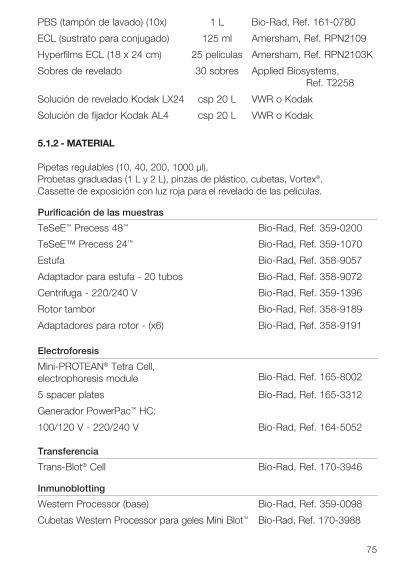
75
PBS (tampón de lavado) (10x) 1 L Bio-Rad, Ref. 161-0780
ECL (sustrato para conjugado) 125 ml Amersham, Ref. RPN2109
Hyperfilms ECL (18 x 24 cm) 25 películas Amersham, Ref. RPN2103K
Sobres de revelado 30 sobres Applied Biosystems, Ref. T2258
Solución de revelado Kodak LX24 csp 20 L VWR o Kodak
Solución de fijador Kodak AL4 csp 20 L VWR o Kodak
5.1.2 - MATERIAL
Pipetas regulables (10, 40, 200, 1000 µl),Probetas graduadas (1 L y 2 L), pinzas de plástico, cubetas, Vortex®.Cassette de exposición con luz roja para el revelado de las películas.
Purificación de las muestras
TeSeE™ Precess 48™ Bio-Rad, Ref. 359-0200
TeSeE™ Precess 24™ Bio-Rad, Ref. 359-1070
Estufa Bio-Rad, Ref. 358-9057
Adaptador para estufa - 20 tubos Bio-Rad, Ref. 358-9072
Centrifuga - 220/240 V Bio-Rad, Ref. 359-1396
Rotor tambor Bio-Rad, Ref. 358-9189
Adaptadores para rotor - (x6) Bio-Rad, Ref. 358-9191
Electroforesis
Mini-PROTEAN® Tetra Cell, electrophoresis module Bio-Rad, Ref. 165-8002
5 spacer plates Bio-Rad, Ref. 165-3312
Generador PowerPac™ HC:
100/120 V - 220/240 V Bio-Rad, Ref. 164-5052
Transferencia
Trans-Blot® Cell Bio-Rad, Ref. 170-3946
Inmunoblotting
Western Processor (base) Bio-Rad, Ref. 359-0098
Cubetas Western Processor para geles Mini Blot™ Bio-Rad, Ref. 170-3988
76
5.2 - PREPARACIÓN DE LOS REACTIVOS 5.2.1 - PURIFICACIÓN DE LAS MUESTRAS
• Proteinasa KSolución de proteinasa K diluida en reactivo A:
u 1 ml de reactivo Au 20 µl de proteinasa K
Mezclar bien por inversión del tubo hasta obtener una solución homogénea. Tras la reconstitución, la solución diluida de proteinasa K es estable 10 horas si se conserva a temperatura ambiente (+18°C a +30°C).
• Solución de LaemmliSolución de SDS + 2-Mercaptoetanol + Laemmli sample buffer:
u 0,6 g de SDSu 1,5 ml de 2-Mercaptoetanol
Mezclar por inversión del tubo.u 28,5 ml de Laemmli sample buffer
La solución se reparte en alícuotas de 4 ml y se conserva a -20°C. Las alícuotas descongeladas pueden volverse a congelar.
Nota: Se recomienda preparar la solución de Laemmli una hora antes de su uso para que el SDS se disuelva completamente.
5.2.2 - ELECTROFORESIS
• Gel discontinuo de acrilamida, vertido manualmenteEl gel tendrá un espesor de 1,5 mm.
Con el módulo de vaciado Mini Blot™ (casting module), echar en primer lugar el gel inferior (acrilamida 13,5%, pH 8,8); cuando el gel inferior haya polimerizado, añadir el gel superior (acrilamida 3% pH 6,8).Gel inferior (1 gel)
u 2,8 ml de acrilamida 40%, 29:1u 1,7 ml de tampón Tris-HCl, 1,5 M, pH 8,8 / SDS (1)u 1,3 ml de solución de sacarosa al 50% (2)u 2,5 ml de agua destilada
Mezclar por inversión del tubo.u 43 µl de persulfato de amonio 10% (3)u 9 µl de TEMED
Verter 7 ml de la solución en las placas y guardar el resto de solución como control de polimerización. Añadir con cuidado 1 ml de tampón Tris-HCl 0,3 M pH 8,8 / SDS (4) hasta arriba para que la superficie del gel no se seque.
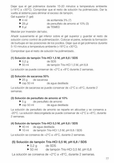
77
Dejar que el gel polimerice durante 15-20 minutos a temperatura ambiente (+18°C a +30°C). Comprobar que el resto de solución ha polimerizado. Dar la vuelta al sistema para eliminar el exceso de tampón. Gel superior (1 gel)
u 4 ml de acrilamida 3% (7)u 28 µl de persulfato de amonio al 10% (3)u 6 µl de TEMED
Mezclar por inversión del tubo.
Añadir suavemente el gel inferior sobre el gel superior y guardar el resto de solución como control de polimerización. Colocar el peine, evitando la formación de burbujas en las posiciones de los pocillos. Dejar que el gel polimerice durante 5-10 minutos a temperatura ambiente (+18°C a +30°C).
Comprobar que el resto de solución ha polimerizado.
(1) Solución de tampón Tris-HCl 1,5 M, pH 8,8 / SDSu 0,2 g de SDSu 50 ml de tampón Tris-HCl 1,5 M, pH 8,8
La solución se puede conservar de +2°C a +8°C durante 2 semanas.
(2) Solución de sacarosa 50%u 25 g de sacarosau csp 50 ml de agua destilada
La solución de sacarosa se puede conservar de +2°C a +8°C, durante 2 semanas.
(3) Solución de persulfato de amonio al 10%u 5 g de persulfato de amoniou csp 50 ml de agua destilada
La solución de persulfato de amonio se reparte en alícuotas y se conserva a -20°C. La solución descongelada se puede conservar de +2°C a +8°C, durante 2 semanas.
(4) Solución de tampón Tris-HCl 0,3 M, pH 8,8 / SDSu 40 ml de agua destiladau 10 ml de tampón Tris-HCl 1,5 M, pH 8.8 / SDS
La solución se conserva de +2°C a +8°C, durante 2 semanas.
(5) Solución de tampón Tris-HCl 0,5 M, pH 6,8 / SDSu 0,2 g de SDSu 50 ml de tampón Tris-HCl 0,5 M, pH 6,8
La solución se conserva de +2°C a +8°C, durante 2 semanas.
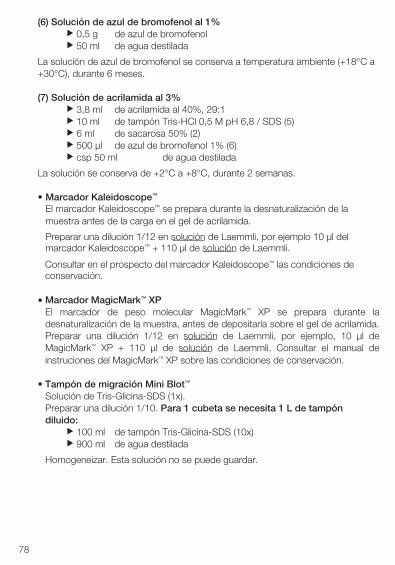
(6) Solución de azul de bromofenol al 1%u 0,5 g de azul de bromofenolu 50 ml de agua destilada
La solución de azul de bromofenol se conserva a temperatura ambiente (+18°C a +30°C), durante 6 meses.
(7) Solución de acrilamida al 3%u 3,8 ml de acrilamida al 40%, 29:1u 10 ml de tampón Tris-HCl 0,5 M pH 6,8 / SDS (5)u 6 ml de sacarosa 50% (2)u 500 µl de azul de bromofenol 1% (6)u csp 50 ml de agua destilada
La solución se conserva de +2°C a +8°C, durante 2 semanas.
• Marcador Kaleidoscope™
El marcador Kaleidoscope™ se prepara durante la desnaturalización de lamuestra antes de la carga en el gel de acrilamida.
Preparar una dilución 1/12 en solución de Laemmli, por ejemplo 10 µl delmarcador Kaleidoscope™ + 110 µl de solución de Laemmli.
Consultar en el prospecto del marcador Kaleidoscope™ las condiciones deconservación.
• Marcador MagicMark™ XPEl marcador de peso molecular MagicMark™ XP se prepara durante ladesnaturalización de la muestra, antes de depositarla sobre el gel de acrilamida.Preparar una dilución 1/12 en solución de Laemmli, por ejemplo, 10 µl deMagicMark™ XP + 110 µl de solución de Laemmli. Consultar el manual deinstruciones del MagicMark™ XP sobre las condiciones de conservación.
• Tampón de migración Mini Blot™
Solución de Tris-Glicina-SDS (1x).Preparar una dilución 1/10. Para 1 cubeta se necesita 1 L de tampóndiluido:
u 100 ml de tampón Tris-Glicina-SDS (10x)u 900 ml de agua destilada
Homogeneizar. Esta solución no se puede guardar.
78
5.2.3 - TRANSFERENCIA DE PROTEÍNAS
• Tampón de transferenciaSolución de Tris/CAPS-etanol 15%. Para 1 cubeta de transferencia senecesitan 2,5 L.
u 750 ml de agua destiladau 150 ml de etanol purou 100 ml de Tris/CAPS (10x)
Homogeneizar. Esta solución no se puede guardar.
5.2.4 - INMUNOBLOTTING
• Solución de lavado 1Solución de PBS (1x) + 0,1% Tween® 20. Para el tratamiento completode una membrana se necesitan unos 500 ml.
u 900 ml de agua destiladau 100 ml de PBS (10x)u 1 ml de Tween® 20
Homogeneizar bien. La solución se puede conservar una noche de +2°C a +8°C.
• Solución de lavado 2Solución de PBS (1x). Para el tratamiento completo de una membrana senecesitan unos 100 ml.
u 900 ml de agua destiladau 100 ml de PBS (10x)
La solución se puede conservar una noche a temperatura ambiente (+18°C a +30°C).
• Solución de saturaciónDurante la etapa de transferencia, diluir la solución de saturación (Bl) 1/10 en lasolución de lavado 1. Se necesitan 20 ml de solución de saturación diluida(1x) para 1 membrana.
u 18 ml de solución de lavado 1u 2 ml de solución de saturación (10x)
Homogeneizar por inversión del tubo.
• Anticuerpo primario diluidoJusto antes de usar, diluir la mezcla de anticuerpo primario a 1/10 en lasolución de lavado 1. Para 1 membrana se necesitan 15 ml de anticuerpodiluido.
u 13,5 ml de solución de lavado 1u 1,5 ml de anticuerpo primario (10x)
Homogeneizar por inversión del tubo.79
80
• Anticuerpo secundario diluido (conjugado)Justo antes de usar, diluir el anticuerpo secundario a 1/10 en solución de lavado 1. Para 1 membrana se necesitan 20 ml de conjugado diluido.
u 18 ml de solución de lavado 1u 2 ml de anticuerpo secundario (10x)
Homogeneizar por inversión del tubo.
• ECLEl sustrato (ECL) se debe preparar justo antes de usar. Para 1 membrana se necesita 1 ml de sustrato.
u 0,5 ml de reactivo 1u 0,5 ml de reactivo 2
Homogeneizar la solución.
• Solución de reveladou 800 ml de agua destiladau 200 ml de producto de revelado
La solución se puede conservar a temperatura ambiente (+18°C a +30°C), en cámara oscura, durante un máximo de 15 días.
• Solución de fijaciónu 800 ml de agua destiladau 200 ml de producto fijador
La solución se puede conservar a temperatura ambiente (+18°C a +30°C), en cámara oscura, durante un máximo de 15 días.
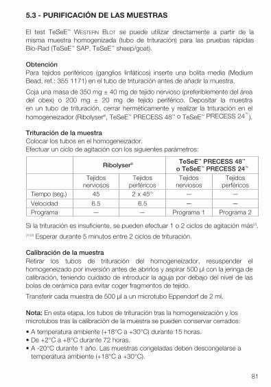
81
5.3 - PURIFICACIÓN DE LAS MUESTRAS
El test TeSeE™ Western Blot se puede utilizar directamente a partir de la misma muestra homogenizada (tubo de trituración) para las pruebas rápidas Bio-Rad (TeSeE™ SAP, TeSeE™ sheep/goat).
ObtenciónPara tejidos periféricos (ganglios linfáticos) inserte una bolita media (Medium Bead, ref.: 355 1171) en el tubo de trituración antes de añadir la muestra.
Coja una masa de 350 mg ± 40 mg de tejido nervioso (preferiblemente del área del obex) o 200 mg ± 20 mg de tejido periférico. Depositar la muestra en un tubo de trituración, cerrar herméticamente y realizar la trituración en el homogeneizador (Ribolyser®, TeSeE™ PRECESS 48™ o TeSeE™ PRECESS 24™
).
Trituración de la muestraColocar los tubos en el homogeneizador.Efectuar un ciclo de agitación con los siguientes parámetros:
Si la trituración es insuficiente, se pueden efectuar 1 o 2 ciclos de agitación más(2).(1) (2) Esperar durante 5 minutos entre 2 ciclos de trituración.
Calibración de la muestraRetirar los tubos de trituración del homogeneizador, resuspender el homogeneizado por inversión antes de abrirlos y aspirar 500 µl con la jeringa de calibración, teniendo cuidado de introducir la aguja por debajo del nivel de las bolas de cerámica para evitar coger fragmentos de tejido.
Transferir cada muestra de 500 µl a un microtubo Eppendorf de 2 ml.
Nota: En esta etapa, los tubos de trituración tras la homogeneización y los microtubos tras la calibración de la muestra se pueden conservar cerrados:
• A temperatura ambiente (+18°C a +30°C) durante 15 horas.• De +2°C a +8°C durante 72 horas.• A -20°C durante 1 año. Las muestras congeladas deben descongelarse a
temperatura ambiente (+18°C a +30°C).
Ribolyser® TeSeE™ PRECESS 48™
o TeSeE™ PRECESS 24™
Tejidos nerviosos
Tejidos perféricos
Tejidos nerviosos
Tejidos perféricos
Tiempo (seg.) 45 2 x 45(1) — —
Velocidad 6.5 6.5 — —Programa — — Programa 1 Programa 2
82
Las muestras pueden someterse a un máximo de 3 ciclos de congelación/ descongelación. Las muestras siempre deben homogeneizarse por inversión antes de usar.
Tratamiento con la proteinasa KDistribuir 500 µl de solución de proteinasa K reconstituida (ver apartado 5.2.1) en cada microtubo.
Homogeneizar los tubos cerrados por inversión (10 veces) e incubar a 37°C ± 2°C en una estufa durante 10 minutos.
Precipitación de la PrPres con el reactivo BSacar los tubos del incubador. Abrirlos y distribuir 500 µl de reactivo B en cada tubo. Homogeneizar por inversión hasta obtener un color homogéneo.
Concentración de la PrPres por centrifugaciónCentrifugar los tubos durante 7 minutos a 15.000 g a 20°C.
Aclaración de la muestra
Desechar el sobrenadante en un recipiente para residuos. A continuación, secar los tubos dándoles la vuelta sobre papel absorbente durante 5 minutos.
Distribuir 100 µl de solución de Laemmli (ver apartado 5.2.1) en cada microtubo.
Incubar durante 5 minutos a temperatura ambiente (+18°C a +30°C).
Volver a solubilizar completamente el precipitado por aspiración/dispensación con una pipeta.
Incubar durante 5 minutos a 100°C ± 5°C en una estufa.
Sacar los tubos del incubador, homogeneizar con el vórtex.
Centrifugar los tubos durante 15 minutos a 15000 g a 20°C.
Transferir el sobrenadante a un nuevo microtubo. Desechar el tubo que contiene el precipitado.
En esta etapa, el sobrenadante se puede conservar congelado a -20°C durante 24 horas; las muestras se deben descongelar a temperatura ambiente (+18°C a +30°C) antes de usar.
83
5.4 - ELECTROFORESIS
El test TeSeE™ Western Blot se puede utilizar tanto para la confirmación de muestras sospechosas de infección por EET como para el tipificado de la cepa en las ovejas.
El siguiente procedimiento es aplicable para la confirmación de muestras sospechosas de infección por EET.
En el caso de la aplicación del tipificado, póngase en contacto con su representante de Bio-Rad para solicitar el protocolo de instrucciones.
Preparación del gelColocar los geles de acrilamida (ver apartado 5.2.2) en la cubeta de migración.
Añadir el tampón de migración (ver apartado 5.2.2) en las cubetas a cada lado de los geles hasta por encima de los pocillos y en la cubeta de electroforesis. Retirar con cuidado los peines y aclarar cada pocillo con tampón de migración utilizando una pipeta.
Depósito de las muestrasCalentar las muestras durante 4 minutos a 100 °C ± 5°C justo antes de depositar 15 µl/pocillo.
Depositar 15 µl del marcador Kaleidoscope™ diluido y 15 µl de MagicMark™XP diluido (ver apartado 5.2.2).Nota: en el caso en el que se traten varios geles a la vez, depositar los marcadores en diferentes pocillos para facilitar la identificación.
Migración diferencial de las muestrasRealizar la migración a temperatura ambiente (+18°C a +30°C) durante 90 minutos a 150 V. La línea azul debe estar fuera del gel.
5.5 - TRANSFERENCIA DE LAS PROTEÍNAS
El tampón de transferencia debe prepararse antes de que finalice la migración de las muestras (ver apartado 5.2.3).
Preparación de la transferencia de las proteínasCortar la membrana con las mismas dimensiones del gel. La membrana siempre se debe manipular con pinzas.
Sumergir la membrana en etanol puro durante 15 segundos, aclarar en agua destilada durante 5 minutos y posteriormente en el tampón de transferencia durante 10 minutos.
84
Separar con cuidado el gel de las placas de vidrio y dejar que se equilibre durante 10 minutos en el tampón de transferencia.
Preparación del sandwichEmpapar los papeles de filtro y las almohadillas de fibras en el tampón de transferencia. Abrir el cassette de transferencia, con el lado transparente a la izquierda. Colocar sobre el lado transparente, siguiendo este orden, una almohadilla de fibras, un papel de filtro, la membrana* y el gel*. Completar con un papel de filtro y una almohadilla de fibras y cerrar el cassette.
Sumergir el cassette en la cubeta de transferencia, previamente llena con el tampón de transferencia hasta el límite indicado.
*Eliminar las posibles burbujas de aire que se hayan formado.
Nota: en el caso de que se traten varias membranas a la vez, identificar cadamembrana marcando en una esquina.
Transferencia sobre la membrana PVDFTransferir durante 60 minutos a 115 V, sobre agitación (barra magnética).
5.6 - INMUNOBLOTTING
a) Al finalizar la transferencia de las proteínas, abrir el cassette y sacar lamembrana para revelarla. Sumergir rápidamente la membrana en la soluciónde lavado 2 (ver apartado 5.2.4) y posteriormente ponerla en etanol durante 10segundos antes de aclarar durante 5 minutos en agua destilada.
Nota: En este paso, la membrana se puede conservar durante la noche enagua destilada en +2°C a +8°C. Deje la membrana ajustar a la temperaturaambiente (+18°C a +30°C) antes de comenzar inmunoblotting.
b) Eliminar el agua destilada e incubar la membrana durante 30 minutos en lasolución de saturación (ver apartado 5.2.4). Incubar con agitación moderada.
Para 1 membrana se necesitan 20 ml de solución de saturación.
Nota: desde esta etapa hasta la etapa g), en las etapas de agitación y delavado se puede utilizar el Western Processor Bio-Rad (ver el manual para losparámetros).
c) Eliminar la solución de saturación e incubar la membrana en el anticuerpoprimario diluido (ver apartado 5.2.4) durante 30 minutos a temperaturaambiente (+18°C a +30°C) con agitación moderada.
Para 1 membrana se necesitan 15 ml de anticuerpo primario diluido.
85
d) Eliminar la solución de anticuerpo primario y aclarar rápidamente la membranacon la solución de lavado 1 y a continuación lavar dos veces durante 5 y 10minutos respectivamente con agitación rápida.
Para cada ciclo y para 1 membrana se necesitan 50 ml de solución de lavado 1.
e) Eliminar la solución de lavado 1 e incubar la membrana durante 20 minutos enel anticuerpo secundario diluido (ver apartado 5.2.4) a temperatura ambiente(+18°C a +30°C) con agitación moderada.
Para 1 membrana se necesitan 20 ml de anticuerpo secundario diluido.
f) Eliminar la solución de anticuerpo secundario y aclarar rápidamente con lasolución de lavado 1 y a continuación lavar durante 5, 10 y 10 minutos respectivamente con agitación rápida.
Para cada ciclo y para 1 membrana se necesitan 50 ml de solución de lavado 1.
g) Colocar la membrana en 50 ml de la solución de lavado 2 con agitación lenta.
h) Dejar escurrir la membrana sosteniéndola encima del papel absorbente yevitando el contacto directo y colocar la membrana en el sobre de plástico.
i) Añadir el reactivo ECL (ver apartado 5.2.4). Eliminar el exceso de reactivo y lasburbujas de aire utilizando papel absorbente. Colocar en el cassette deexposición.
j) En una cámara oscura, cubrir el sobre con una película y exponer durante 15minutos. Este tiempo de exposición de la película se puede variar para obteneruna señal óptima.
k) Sumergir la película en la solución de revelado durante 45 segundos (verapartado 5.2.4). Aclarar en agua destilada. Sumergir la película en el fijadorhasta que la película se vuelva totalmente transparente.
l) Lavar con agua destilada y dejar secar la película.
86
6 - PROCEDIMIENTO CON EL GEL CRITERION™ XT6.1 - REACTIVOS Y MATERIAL NECESARIO6.1.1 - REACTIVOS Y CONSUMIBLES
Pipetas graduadas (5, 10, 25 ml), tubos cónicos (50 ml), microtubos (2 ml) de polipropileno con tapón.Película protectora PARAFILM® M.
Purificación de las muestras
Tampón Laemmli 30 ml Bio-Rad, Ref. 161-0737
2-Mercaptoetanol 25 ml Bio-Rad, Ref. 161-0710
SDS 100 g Bio-Rad, Ref. 161-0301
Jeringas de calibración 200 Bio-Rad, Ref. 355-1174
Electroforesis
Criterion™ XT 12 % Bis-Tris 1 gel-18 pocillos Bio-Rad, Ref. 345-0118
XT-MOPS (20 x) 500 ml Bio-Rad, Ref. 161-0788(tampón de electroforesis)
Marcador preteñido Kaleidoscope™ 500 µl Bio-Rad, Ref. 161-0324
MagicMark™ XP Western Standard 250 µl Invitrogen, Ref. LC5602(marcador de peso molecular)
Inmunobloting
Ethanol (Normapur) 1L VWR, Ref. 20821-296
Tris/CAPS (tampón de transferencia) (10x)
1L Bio-Rad, Ref. 161-0778
Papel de filtro 50 hojas Bio-Rad, Ref. 170-4085(papel de transferencia para geles Criterion™ XT)
Membrana PVDF (0,2 µm) 10 hojas Bio-Rad, Ref. 162-0175
Tween® 20 100 ml Bio-Rad, Ref. 170-6531
PBS (tampón de lavado) (10x) 1 L Bio-Rad, Ref. 161-0780
ECL (sustrato para conjugado) 125 ml Amersham, Ref. RPN2109
Hyperfilms ECL (18 x 24 cm) 25 películas Amersham, Ref. RPN2103K
Sobres de revelado 30 sobres Applied Biosystems, Ref. T2258
87
Solución de revelado Kodak LX24 csp 20 L VWR o Kodak
Solución de fijador Kodak AL4 csp 20 L VWR o Kodak
6.1.2 - MATERIAL
Pipetas regulables (10, 40, 200, 1000 µl),Probetas graduadas (1 L y 2 L), pinzas de plástico, cubetas, vortex.Cassette de exposición con luz roja para el revelado de las películas.
Purificación de las muestras
TeSeE™ Precess 48™ Bio-Rad, Ref. 359-0200 TeSeE™ Precess 24™ Bio-Rad, Ref. 359-1070
Estufa Bio-Rad, Ref. 358-9057
Adaptador para estufa - 20 tubos Bio-Rad, Ref. 358-9072
Centrifuga - 220/240 V Bio-Rad, Ref. 359-1396
Rotor tambor Bio-Rad, Ref. 358-9189
Adaptadores para rotor - (x6) Bio-Rad, Ref. 358-9191
Electroforesis
Cubeta Criterion™ XT Bio-Rad, Ref. 165-6001
Generator PowerPac™ HC :100/120 V - 220/240 V Bio-Rad, Ref. 164-5052
Transferencia
Criterion™ XT blotter Bio-Rad, Ref. 170-4070
Inmunoblotting
Bio-Rad, Ref. 359-0098 Western Processor (base)
Cuvetas Western Processor
para geles Criterion™ XT
Bio-Rad, Ref. 170-3985
88
6.2 - PREPARACIÓN DE LOS REACTIVOS 6.2.1 - PURIFICACIÓN DE LAS MUESTRAS
• Proteinasa KSolución de proteinasa K diluida en reactivo A:
u 1 ml de reactivo Au 20 µl de proteinasa K
Mezclar bien por inversión del tubo hasta obtener una solución homogénea. Tras la reconstitución, la solución diluida de proteinasa K es estable 10 horas si se conserva a temperatura ambiente (+18°C a +30°C).
• Solución de LaemmliSolución de SDS + 2-Mercaptoetanol + Laemmli sample buffer:
u 0,6 g de SDSu 1,5 ml de 2-Mercaptoetanol
Mezclar por inversión del tubo.u 28,5 ml de Laemmli sample buffer
La solución se reparte en alícuotas de 4 ml y se conserva a -20°C. Las alícuotas descongeladas pueden volverse a congelar.
Nota: Se recomienda preparar la solución de Laemmli una hora antes de su uso para que el SDS se disuelva completamente.
6.2.2 - ELECTROFORESIS
• Marcador Kaleidoscope™
El marcador Kaleidoscope™ se prepara durante la desnaturalización de la muestra antes de la carga en el gel de acrilamida.
Preparar una dilución 1/12 en solución de Laemmli, por ejemplo 10 µl del marcador Kaleidoscope™ + 110 µl de solución de Laemmli.
Consultar en el prospecto del marcador Kaleidoscope™ las condiciones de conservación.
• Marcador MagicMark™ XPEl marcador de peso molecular MagicMark™ XP se prepara durante la desnaturalización de la muestra, antes de depositarla sobre el gel de acrilamida.
Preparar una dilución 1/12 en solución de Laemmli, por ejemplo, 10 µl de MagicMark™ XP + 110 µl de solución de Laemmli.
Consultar el manual de instruciones del MagicMark™ XP sobre las condiciones de conservación.
89
• Tampón de migración Criterion™ XTSolución de MOPS (1x).Preparar una dilución 1/20. Para 1 cubeta se necesita 1 L de tampón diluido:
u 950 ml de agua destiladau 50 ml de tampón MOPS (20x)
Homogeneizar. Esta solución no se puede guardar.
6.2.3 - TRANSFERENCIA DE PROTEÍNAS
• Tampón de transferenciaSolución de Tris/CAPS-Etanol 15%. Para 1 cubeta de transferencia senecesitan unos 2 L.
u 750 ml de agua destiladau 150 ml de etanol purou 100 ml de Tris/CAPS (10x)
Homogeneizar. Esta solución no se puede guardar.
6.2.4 - INMUNOBLOTTING
• Solución de lavado 1Solución de PBS (1x) + 0,1% Tween® 20. Para el tratamiento completo de una membrana se necesitan unos 1 L.
u 900 ml de agua destiladau 100 ml de PBS (10x)u 1 ml de Tween® 20
Homogeneizar bien. La solución se puede conservar una noche de +2°C a +8°C.
• Solución de lavado 2Solución de PBS (1x). Para el tratamiento completo de una membrana se necesitan unos 200 ml.
u 900 ml de agua destiladau 100 ml de PBS (10x)
La solución se puede conservar una noche a temperatura ambiente (+18°C a +30°C).
• Solución de saturaciónDurante la etapa de transferencia, diluir la solución de saturación (Bl) 1/10 en la solución de lavado 1. Se necesitan 40 ml de solución de saturación diluida (1x) para 1 membrana.
u 36 ml de solución de lavado 1u 4 ml de solución de saturación (10x)
Homogeneizar por inversión del tubo.
90
• Anticuerpo primario diluidoJusto antes de usar, diluir el anticuerpo primario a 1/10 en la solución delavado 1.
Para 1 membrana se necesitan 30 ml de anticuerpo primario diluido.u 27 ml de la solución de lavado 1u 3 ml de anticuerpo primario (10x)
Homogeneizar por inversión del tubo.
• Anticuerpo secundario diluido (conjugado)Justo antes de usar, diluir el anticuerpo secundario a 1/10 en la solución delavado 1.
Para 1 membrana se necesitan 40 ml de conjugado diluido.u 36 ml de la solución de lavado 1u 4 ml de anticuerpo secundario (10x)
Homogeneizar por inversión del tubo.
• ECLEl sustrato (ECL) se debe preparar justo antes de usar. Para 1 membranase necesitan 2 ml de sustrato.
u 1 ml de reactivo 1u 1 ml de reactivo 2
Homogeneizar la solución.
• Solución de reveladou 800 ml de agua destiladau 200 ml de producto de revelado
La solución se puede conservar a temperatura ambiente (+18°C a +30°C), en cámara oscura, durante un máximo de 15 días.
• Solución de fijaciónu 800 ml de agua destiladau 200 ml de producto fijador
La solución se puede conservar a temperatura ambiente (+18°C a +30°C), en cámara oscura, durante un máximo de 15 días.
91
6.3 - PURIFICACIÓN DE LAS MUESTRAS
El test TeSeE™ Western Blot se puede utilizar directamente a partir de la misma muestra homogenizada (tubo de trituración) para las pruebas rápidas Bio-Rad (TeSeE™ SAP, TeSeE™ sheep/goat).
ObtenciónPara tejidos periféricos (ganglios linfáticos) inserte una bolita media (Medium Bead, ref.: 355 1171) en el tubo de trituración antes de añadir la muestra.
Coja una masa de 350 mg ± 40 mg de tejido nervioso (preferiblemente del área del obex) o 200 mg ± 20 mg de tejido periférico. Depositar la muestra en un tubo de trituración, cerrar herméticamente y realizar la trituración en el homogeneizador (Ribolyser®, TeSeE™ PRECESS 48™ o TeSeE™ PRECESS 24™
).
Trituración de la muestraColocar los tubos en el homogeneizador.Efectuar un ciclo de agitación con los siguientes parámetros:
Si la trituración es insuficiente, se pueden efectuar 1 o 2 ciclos de agitación más(2).(1) (2) Esperar durante 5 minutos entre 2 ciclos de trituración.
Calibración de la muestraRetirar los tubos de trituración del homogeneizador, resuspender el homogeneizado por inversión antes de abrirlos y aspirar 500 µl con la jeringa de calibración, teniendo cuidado de introducir la aguja por debajo del nivel de las bolas de cerámica para evitar coger fragmentos de tejido.
Transferir cada muestra de 500 µl a un microtubo Eppendorf de 2 ml.
Nota: En esta etapa, los tubos de trituración tras la homogeneización y los microtubos tras la calibración de la muestra se pueden conservar cerrados:• A temperatura ambiente (+18°C a +30°C) durante 15 horas.• De +2°C a +8°C durante 72 horas.• A -20°C durante 1 año. Las muestras congeladas deben descongelarse a
temperatura ambiente (+18°C a +30°C).
Las muestras pueden someterse a un máximo de 3 ciclos de congelación/ descongelación. Las muestras siempre deben homogeneizarse por inversión antes de usar.
Ribolyser® TeSeE™ PRECESS 48™
o TeSeE™ PRECESS 24™
Tejidos nerviosos
Tejidos perféricos
Tejidos nerviosos
Tejidos perféricos
Tiempo (seg.) 45 2 x 45(1) — —Velocidad 6.5 6.5 — —Programa — — Programa 1 Programa 2
92
Tratamiento con la proteinasa KDistribuir 500 µl de solución de proteinasa K reconstituida (ver apartado 6.2.1) en
cada microtubo.
Homogeneizar los tubos cerrados por inversión (10 veces) e incubar a 37°C ± 2°C en una estufa durante 10 minutos.
Precipitación de la PrPres con el reactivo B
Sacar los tubos del incubador. Abrirlos y distribuir 500 µl de reactivo B en cada tubo. Homogeneizar por inversión hasta obtener un color homogéneo.
Concentración de la PrPres por centrifugaciónCentrifugar los tubos durante 7 minutos a 15.000 g a 20°C.
Aclaración de la muestraDesechar el sobrenadante en un recipiente para residuos. A continuación, secar los tubos dándoles la vuelta sobre papel absorbente durante 5 minutos.
Distribuir 100 µl de solución de Laemmli (ver apartado 6.2.1) en cada microtubo.
Incubar durante 5 minutos a temperatura ambiente (+18°C a +30°C).
Volver a solubilizar completamente el precipitado por aspiración/dispensación con una pipeta.
Incubar durante 5 minutos a 100°C ± 5°C en una estufa.
Sacar los tubos del incubador, homogeneizar con el vórtex.
Centrifugar los tubos durante 15 minutos a 15000 g a 20°C.
Transferir el sobrenadante a un nuevo microtubo. Desechar el tubo que contiene el precipitado.
En esta etapa, el sobrenadante se puede conservar congelado a -20°C durante 24 horas; las muestras se deben descongelar a temperatura ambiente (+18°C a +30°C) antes de usar.
93
6.4 - ELECTROFORESIS
El test TeSeE™ Western Blot se puede utilizar tanto para la confirmación de muestras sospechosas de infección por EET como para el tipificado de la cepa en las ovejas.
El siguiente procedimiento es aplicable para la confirmación de muestras sospechosas de infección por EET.
En el caso de la aplicación del tipificado, póngase en contacto con su representante de Bio-Rad para solicitar el protocolo de instrucciones.
Preparación del gelRetirar la tira de plástico de la parte inferior de la placa de plástico e introducir los geles de acrilamida (ver apartado 6.2.2) en la cubeta de migración. Añadir el tampón de migración (ver apartado 6.2.2) en las cubetas a cada lado de los geles hasta por encima de los pocillos y en la cubeta de electroforesis. Retirar con cuidado los peines y aclarar cada pocillo con tampón de migración utilizando una pipeta.
Depósito de las muestrasCalentar las muestras durante 4 minutos a 100 °C ± 5°C justo antes de depositar 15 µl/pocillo.
Depositar 15 µl del marcador KaleidoscopeTM diluido y 15 µl de MagicMarkTM XP diluido (ver apartado 6.2.2).
Nota: en el caso en el que se traten varios geles a la vez, depositar los marcadores en diferentes pocillos para facilitar la identificación.
Migración diferencial de las muestrasRealizar la migración a temperatura ambiente (+18°C a +30°C) durante 50 minutos a 200 V.
6.5 - TRANSFERENCIA DE LAS PROTEÍNAS
El tampón de transferencia debe prepararse antes de que finalice la migración de las muestras (ver apartado 6.2.3).
Preparación de la transferencia de las proteínasCortar la membrana con las mismas dimensiones del gel. La membrana siempre se debe manipular con pinzas.
Sumergir la membrana en etanol puro durante 15 segundos, aclarar en agua destilada durante 5 minutos, y posteriormente en el tampón de transferencia durante 10 minutos.Separar con cuidado el gel de las placas plástico y dejar que se equilibre durante 10 minutos en el tampón de transferencia.
94
Preparación del sandwichEmpapar los papeles de filtro y las almohadillas de fibras en el tampón de transferencia. Abrir el cassette de transferencia, con el lado rojo a la izquierda. Colocar sobre el lado rojo, siguiendo este orden, una almohadilla de fibras, un papel de filtro, la membrana* y el gel*.
Completar con un papel de filtro y una almohadilla de fibras y cerrar el cassette.
Sumergir el cassette en la cubeta de transferencia, previamente llena con el tampón de transferencia hasta el límite indicado. Añadir hielo antes de llenar la cubeta.
*Eliminar las posibles burbujas de aire que se hayan formado.
Nota: en el caso de que se traten varias membranas a la vez, identificar cadamembrana marcando en una esquina.
Transferencia sobre la membrana PVDFTransferir durante 60 minutos a 115 V, sobre agitación (barra magnética).
6.6 - INMUNOBLOTTING
a) Al finalizar la transferencia de las proteínas, abrir el cassette y sacar lamembrana para revelarla. Sumergir rápidamente la membrana en la soluciónde lavado 2 (ver apartado 6.2.4) y posteriormente ponerla en etanol durante10 segundos antes de aclarar durante 5 minutos en agua destilada.
Nota: En este paso, la membrana se puede conservar durante la noche enagua destilada en +2°C a +8°C. Deje la membrana ajustar a la temperaturaambiente (+18°C a +30°C) antes de comenzar inmunoblotting.
b) Eliminar el agua destilada e incubar la membrana durante 30 minutos en lasolución de saturación (ver apartado 6.2.4). Incubar con agitación moderada.
Para 1 membrana se necesitan 40 ml de solución de saturación.
Nota: desde esta etapa hasta la etapa g), en las etapas de agitación y delavado se puede utilizar el Western Processor Bio-Rad (ver el manual para losparámetros).
c) Eliminar la solución de saturación e incubar la membrana en el anticuerpoprimario diluido (ver apartado 6.2.4) durante 30 minutos a temperaturaambiente (+18°C a +30°C) con agitación moderada.
Para 1 membrana se necesitan 30 ml de anticuerpo primario diluido.
95
d) Eliminar la solución de anticuerpo primario y aclarar rápidamente la membranacon la solución de lavado 1 y a continuación lavar dos veces durante5 y 10 minutos respectivamente con agitación rápida.
Para cada ciclo y para 1 membrana se necesitan 100 ml de solución delavado 1.
e) Eliminar la solución de lavado 1 e incubar la membrana durante 20 minutos enel anticuerpo secundario diluido (ver apartado 6.2.4) a temperatura ambiente(+18°C a +30°C) con agitación moderada.
Para 1 membrana se necesitan 40 ml de anticuerpo secundario diluido.
f) Eliminar la solución de anticuerpo secundario y aclarar rápidamente con lasolución de lavado 1 y a continuación lavar durante 5, 10 y 10 minutosrespectivamente con agitación rápida.
Para cada ciclo y para 1 membrana se necesitan 100 ml de solución delavado 1.
g) Colocar la membrana en 100 ml de la solución de lavado 2 con agitación lenta.
h) Dejar escurrir la membrana sosteniéndola encima del papel absorbente yevitando el contacto directo y colocar la membrana en el sobre de plástico.
i) Añadir el reactivo ECL (ver apartado 6.2.4). Eliminar el exceso de reactivo y lasburbujas de aire utilizando papel absorbente. Colocar en el cassette deexposición.
j) En una cámara oscura, cubrir el sobre con una película y exponer durante15 minutos. Este tiempo de exposición de la película se puede variar paraobtener una señal óptima.
k) Sumergir la película en la solución de revelado durante 45 segundos (verapartado 6.2.4). Aclarar en agua destilada. Sumergir la película en el fijadorhasta que la película se vuelva totalmente transparente.
l) Lavar con agua destilada y dejar secar la película.
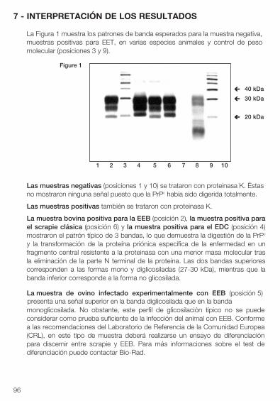
96
7 - INTERPRETACIÓN DE LOS RESULTADOS
La Figura 1 muestra los patrones de banda esperados para la muestra negativa, muestras positivas para EET, en varias especies animales y control de peso molecular (posiciones 3 y 9).
Las muestras negativas (posiciones 1 y 10) se trataron con proteinasa K. Éstas no mostraron ninguna señal puesto que la PrPc había sido digerida totalmente.
Las muestras positivas también se trataron con proteinasa K.
La muestra bovina positiva para la EEB (posición 2), la muestra positiva para el scrapie clásica (posición 6) y la muestra positiva para el EDC (posición 4) mostraron el patrón típico de 3 bandas, lo que demuestra la digestión de la PrPc y la transformación de la proteína priónica específica de la enfermedad en un fragmento central resistente a la proteinasa con una menor masa molecular tras la eliminación de la parte N terminal de la proteína. Las dos bandas superiores corresponden a las formas mono y diglicosiladas (27-30 kDa), mientras que la banda inferior corresponde a la forma no glicosilada.
La muestra de ovino infectado experimentalmente con EEB (posición 5)presenta una señal superior en la banda diglicosilada que en la bandamonoglicosilada. No obstante, este perfil de glicosilación típico no se puede considerar como prueba suficiente de la infección del animal con EEB. Conforme a las recomendaciones del Laboratorio de Referencia de la Comunidad Europea (CRL), en este tipo de muestra deberá realizarse un ensayo de diferenciación para discernir entre scrapie y EEB. Para más informaciones sobre el test de diferenciación puede contactar Bio-Rad.
1 2 3 4 5 6 7 8 9 10
Figure 1
40 kDa è
20 kDa è
30 kDa è
97
La muestra infectado por scrapie atípico (ex. Nor98) (posición 8) presenta un perfil de glicosilación atípico. A aproximadamente 12 kDa se observa una banda más baja, mientras que las otras bandas superiores no están localizadas en las mismas posiciones equivalentes a las de los casos “típicos” de scrapie. La señal también es más fuerte en la banda más inferior que en la banda superior.
La lectura del gel deberá interpretarse con cautela puesto que una muestra fuertemente positiva detectada con el test TeSeE™ Western Blot puede ocultar la muestra negativa más próxima o una muestra positiva baja.
Límites de la prueba:Un resultado negativo implica que la muestra ensayada no contiene una cantidad medible de PrPres con el método TeSeE™ Western Blot. No obstante, como no puede detectarse la presencia de muy bajas cantidades de PrPres, un resultado negativo no excluye la posibilidad de una infección.
Cualquier muestra interpretada negativamente según de los criterios d’interpretación de la prueba TeSeE™ Western Blot, y que se volvió anteriormente positivo répétable por una prueba de detección rápida, tendrà que ser probado con uno de los otros métodos de confirmación certificada por l’ OIE como la Immuno-Histo Química (IHC) o el método SAF-Immunoblot.
Todas las muestras con un resultado positivo reproductible según los criterios de interpretación de la prueba deben verificarse en conformidad con las disposiciones legales en vigor.
98
8 - PRECAUCIONES
La calidad de los resultados depende del cumplimiento de las siguientes buenas prácticas de laboratorio:
• Conservar los reactivos a la temperatura adecuada (ver las indicaciones de losfabricantes).
• No utilizar reactivos después de la fecha de caducidad.
• La solución de proteinasa K reconstituida y conservada a temperatura ambiente(+18°C a +30°C) debe utilizarse en un plazo de 10 horas.
• No mezclar ni combinar reactivos procedentes de diferentes lotes de TeSeE™
Western Blot durante el proceso, con la excepción de tubos de trituración,reactivo A, reactivo B y proteinasa K.
• Antes de la utilización, esperar 30 minutos para que los reactivos y lostampones se estabilicen a temperatura ambiente (+18°C a +30°C).
• Reconstituir cuidadosamente los reactivos evitando toda contaminación.
• No realizar la prueba en presencia de reactivos que produzcan vapores (ácidos,alcalinos, aldehídos) o polvos que puedan alterar la actividad enzimática delconjugado.
• La reacción enzimática es muy sensible a todos los metales o iones metálicos.Por tanto, ningún elemento metálico debe entrar en contacto con el conjugado.
• Utilizar únicamente tubos de polipropileno.
• Utilizar preferentemente material de uso único. En su defecto, utilizar unacristalería perfectamente lavada y enjuagada con agua destilada.
• Utilizar una punta de pipeta desechable para cada muestra.
• Antes de empezar la electroforesis y la transferencia, comprobar que los 2electrodos están en contacto con el tampón.
• Deben respetarse escrupulosamente todos los tiempos de aclarado para evitarun ruido de fondo excesivo durante la tinción final con el reactivo ECL.
99
9 - RECOMENDACIONES DE HIGIENE Y SEGURIDAD
De manera general, las condiciones de higiene, las medidas de seguridad biológica y las buenas prácticas de laboratorio deben cumplir las recomendaciones de las autoridades reglamentarias nacionales.
• Todos los reactivos del kit están destinados para uso exclusivo de diagnóstico“in vitro”.
• Utilizar guantes de un solo uso durante la manipulación de los reactivos y delas muestras y lavar cuidadosamente las manos tras la manipulación.
• No pipetear con la boca.
• Utilizar recipientes de polipropileno para evitar vidrios rotos.
• Considerar el material directamente en contacto con las muestras y lassoluciones de lavado, como productos contaminados.
• Evitar las salpicaduras de muestras o de soluciones que las contengan.
• Limpiar las superficies manchadas con una solución de 20.000 p.p.m. dehipoclorito de sodio (lejía). Si el líquido contaminante es un ácido, neutralizarpreviamente las superficies manchadas con hidróxido de sodio (sosa), ydespués limpiar con lejía. Las superficies deben aclarase con agua destilada,secarse con etanol y papel absorbente. El material utilizado para la limpiezadeberá ser desechado en un contenedor especial para residuos contaminados.
• Eliminar las muestras así como el material y los productos contaminadosdespués de su descontaminación:- ya sea sumergiéndolos en una solución de hidróxido de sodio 1 M
(concentración final) durante al menos 1 hora a temperatura ambiente (+18°C a +30°C),
- ya sea sumergiéndolos en una solución de 20.000 p.p.m. de hipoclorito de sodio durante al menos 1 hora a temperatura ambiente ( +18°C a +30°C),
- o bien por calentamiento en autoclave a 134°C mínimo durante al menos 18 minutos, con una presión de 3 bares.
Atención: no introducir en el autoclave soluciones que contengan lejía o eireactivo B.
• Todas las operaciones referentes a la realización de los tests de despistaje deuna encefalopatía espongiforme transmisible (EST) son objeto de unareglamentación y deben efectuarse en un laboratorio aislado, reservadoexclusivamente a este fin y, por tanto, con un acceso limitado y controlado.
El operador debe utilizar una bata cerrada, protector de calzado, guantes y unamascara con visera o una mascarilla simple con gafas de seguridad.
100
• Los operadores deben recibir una formación específica sobre los riesgos queconllevan los agentes de las EETs o priones y sobre los modos dedescontaminación validados para los agentes infecciosos “no convencionales”.
Las medidas de seguridad biológica deben estar en conformidad con lasrecomendaciones de las autoridades reglamentarias nacionales.
• Antes de su eliminación, neutralizar y/o autoclavar todas las soluciones delavado, el agua de lavado y cualquier líquido que contenga muestras biológicas.
• El reactivo B está una sustancia peligrosa clasificada como nociva (> 25%alcohol) en la reglamentación europea.
• Los reactivos que contengan 0.1% del ProClin® 300 son clasificados comopreparaciones irritantes en la reglamentación europea.
Xn(Alcohol > 25%)(0,1% ProClin® 300)
R : 10-22-37/38-41-43-67 Inflamable. Nocivo en caso de ingestión. Irritante para el sistema respiratorio y la piel. Riesgo de serias lesiones en caso de contacto con los ojos. Posibilidad de sensibilización en contacto con la piel. La inhalación de vapores puede provocar somnolencia y vértigos.
S : 7/9-13-26-28-37/39-46 Conservar el recipiente bien cerrado y en un lugar bien ventilado. Conservar apartado de alimentos y bebidas, incluyendo los destinados a animales. En caso de contacto con los ojos, lavar inmediatamente con abundante agua y consultar a un especialista. En caso de contacto con la piel, lávese inmediata y abundantemente con agua. Utilizar guantes apropiados y un sistema de protección de ojos/cara. En caso de ingestión, consultar inmediatamente con un médico y enseñarle el envase o la etiqueta.
101
10 - BIBLIOGRAFÍA
1. S.B. PRUSINER (1991)Molecular biology of prion diseases - Science 252:1515-1522.
2. J.B. KATZ, J.G. PEDERSEN, A.L. JENNY and W.D. TAYLOR (1992) Assessment of Western immunoblotting for the confirmatory diagnosis of ovine scrapie and bovine spongiform encephalopathy (BSE) - Journal of Veterinary Diagnostic Investigations, 4, 447-449.
3. G.A.H. WELLS, Y.I. SPENCER and M. HARITANI (1994) Configuration and topographic distribution of PrP in the central nervous system in bovine spongiform encephalopathy: an immonohistochemical stydy. In: Slow Infections of the Central Nervous System, J. BJORNSSON, R.I. CARP, A. LOVE and M. WISNIEWSKI - Eds The New York Academy of Sciences, pp 350-352.
4. J.P. DESLYS, E. COMOY, S. HAWKINS, S. SIMON, H. SCHIMMEL, G. WELLS, J. GRASSI and J. MOYNAGH (2001) Screening slaughtered cattle for BSE - Nature:
409; 476-477.
5. S. BENESTAD, P. SARRADIN, B. THU, J. SCHÖNHEIT, M. TRANULIS and B. BRATBERG (2003) Cases of scrapie with unusual features in Norway and designation of new type, Nor98. Veterinary Record 153, 202-208.
6. A. BUSCHMANN, G. LÜHKEN, J. SCHULTZ, G. ERHARDT and M.-H. GROSCHUP (2004) Neuronal accumulation of abnormal prion protein in sheep carrying a scrapie-resistant genotype (PrPARR/ARR). Journal of General Virology 85, 2727-2733.
7. L. ORGE, A.GALO, C.MACHADO, C. LIMA, C. OCHOA, J. SILVA, M. RAMOS and J.-P. SIMAS (2004) Identification of putative atypical scrapie in sheep in Portugal. Journal of General Virology 85, 3487-3491.
8. A. BUSCHMANN, A.-G. BIACABE, U. ZIEGLER, A. BENCSIK, J.-Y. MADEC, G. ERHARDT, G. LÜHKEN, T. BARON and M.-H. GROSCHUP (2004) Atypical scrapie cases in Germany and France are identified by discrepant reaction patterns in BSE rapid tests. Journal of Virological Methods 117, 27-36.
102
9. H. DE BOSSCHERE, S. ROELS, S. BENESTAD, E. VANOPDENBOSCH (2004). Scrapie case similar to Nor98 diagnosed in Belgium via active surveillance. Veterinary Record 155, 707-708.
10. H. ONNASCH, H. M. GUNN, B.J. BRADSHAW, S. BENESTAD, H.F. BASSETT (2004). Two Irish cases of scrapie resembling Nor98. Veterinary Record 155, 636-637.
11. S. BENESTAD, P. SARRADIN, J.-M. BILHEUDE, J. GRASSI , H. LAUDE, O. ANDREOLETTI, T. MOLDAL and B. BRATBERG. Are there gold standard methods for the diagnosis of TSE ? Nor98 scrapie cases: atypical cases and their challenging diagnosis. Second International Chronic Wasting Disease Symposium, Madison - Wisconsin, USA.
12. BIACABE, A-G., LAPLANCHE, J-L., RYDER,S. AND BARON, T. (2004). Distinct molecular phenotypes in bovine prion diseases, EMBO Reports 5, 110-114.
13. CASALONE, C., ZANUSSO, G., ACUTIS, P., FERRARI, S., CAPUCCI, I., TAGLIAVINI, F., MONACO, S. and CARAMELLI, M. (2004). Identification of a second bovine amyloidotic spongiform encephalopathy: Molecular similarities with sporadic Creutzfeldt-Jakob disease. PNAS 101, 3065-70.
14. BUSCHMANN A; GRETZSCHEL A; BIACABE A-G; SCHIEBEL K; CORONA C; HOFFMANN C; EIDEN M; BARON T; CASALONE C; GROSCHUP M H (2006). Atypical BSE in Germany-proof of transmissibility and biochemical characterization. Veterinary microbiology 2006;117(2-4):103-16.
15. ARSAC, J.-N., ANDREOLETTI, O., BILHEUDE, J.-M., LACROUX, C., BENESTAD, S. L., AND BAARON, T. (2007). Similar biochemical signatures and prion protein genotypes in atypical scrapie and Nor98 cases, France and Norway. Emerging Infectious Diseases 13(1), 58-65.
16. E. URO-COSTE, H CASSARD, S. SIMON, S. LUGAN, J.-M. BILHEUDE, A. PERRET-LIAUDET, J.-W. IRONSIDE, S. HAIK, C. BASSET-LEOBON, C. LACROUX, K. PEOCH, N. STREICHENBERGER, J. LANGEVELD, M.-W. HEAD, J. GRASSI, J.-J.HAUW, F. SCHELCHER, M.-B. DELISLE, O. ANDREOLETTI (2008) Beyond PrPres Type 1/Type 2 Dichotomy in Creutzfeldt-Jakob disease. Plos Pathogens volume 4, issue 2, e1000029.
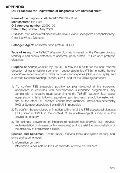
103
APPENDIXOIE Procedure for Registration of Diagnostic Kits Abstract sheet
Name of the diagnostic kit: TeSeE™ Western Blot
Manufacturer: Bio-RadOIE Approval number: 20090105Date of Registration: May 2009
Disease: Prion associated diseases (Scrapie, Bovine Spongiform Encephalopathy, Chronical Waste Disease)
Pathogen Agent: Abnormal prion protein PrPRes
Type of Assay: The TeSeE™ Western Blot kit is based on the Western blotting technique and allows detection of abnormal prion protein PrPRes after protease digestion.
Purpose of Assay: Certified by the OIE in May 2009 as fit for the post-mortem detection of transmissible spongiform encephalopathies (TSEs) in cattle (bovine spongiform encephalopathy, BSE), in ovines and caprines (BSE and scrapie), and in cervids (Chronic Wasting Disease, CWD), and for the following purposes:
1. To confirm TSE suspected positive samples detected at the screeninglaboratories in countries with active/passive surveillance programmes. Any sample with a negative result according to the TeSeE™ Western Blot assay interpretation criteria, following a positive rapid test result, should be tested with one of the other OIE certified confirmatory methods, Immunohistochemistry(IHC) or Scrapie-associated fibrils (SAF)-Immunoblot;
2. To confirm the prevalence of infection with one of the TSE associated diseases(BSE, scrapie, CWD) in the context of an epidemiological survey in a lowprevalence country;
3. To estimate prevalence of infection to facilitate risk analysis (e.g. surveys,implementation of disease control measures) and to assist the demonstration ofthe efficiency of eradication policies.
Species and Specimen: Bovine (obex), Cervids (obex and lymph nodes), and ovine and caprine (obex)
1. Information on the kitInformation is available on Bio-Rad Website, at www.bio-rad.com
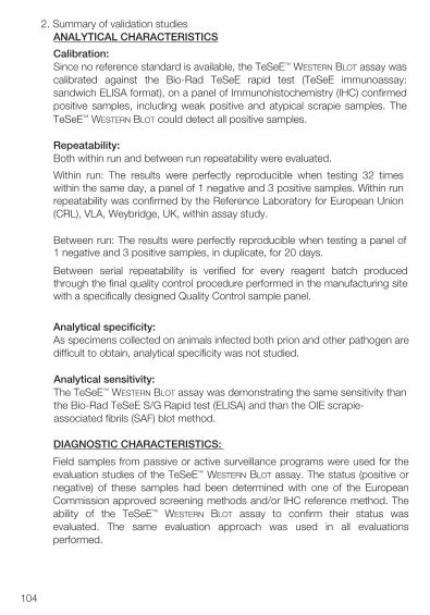
104
2. Summary of validation studiesANALYTICAL CHARACTERISTICS
Calibration:Since no reference standard is available, the TeSeE™ Western Blot assay wascalibrated against the Bio-Rad TeSeE rapid test (TeSeE immunoassay:sandwich ELISA format), on a panel of Immunohistochemistry (IHC) confirmedpositive samples, including weak positive and atypical scrapie samples. TheTeSeE™ Western Blot could detect all positive samples.
Repeatability:Both within run and between run repeatability were evaluated.
Within run: The results were perfectly reproducible when testing 32 timeswithin the same day, a panel of 1 negative and 3 positive samples. Within runrepeatability was confirmed by the Reference Laboratory for European Union(CRL), VLA, Weybridge, UK, within assay study.
Between run: The results were perfectly reproducible when testing a panel of1 negative and 3 positive samples, in duplicate, for 20 days.
Between serial repeatability is verified for every reagent batch producedthrough the final quality control procedure performed in the manufacturing sitewith a specifically designed Quality Control sample panel.
Analytical specificity: As specimens collected on animals infected both prion and other pathogen are difficult to obtain, analytical specificity was not studied.
Analytical sensitivity: The TeSeE™ Western Blot assay was demonstrating the same sensitivity than the Bio-Rad TeSeE S/G Rapid test (ELISA) and than the OIE scrapie-associated fibrils (SAF) blot method.
DIAGNOSTIC CHARACTERISTICS:
Field samples from passive or active surveillance programs were used for the evaluation studies of the TeSeE™ Western Blot assay. The status (positive or negative) of these samples had been determined with one of the European Commission approved screening methods and/or IHC reference method. The ability of the TeSeE™ Western Blot assay to confirm their status was evaluated. The same evaluation approach was used in all evaluations performed.
105
Study I Internal evaluation at Bio-Rad, France- Analytical sensitivity on ovine and bovine characterized samples
from veterinary reference laboratories
- Diagnostic specificity on ovine and bovine samples from French slaughterhouses (animal without any clinical sign) classified as negative with a rapid screening tests (TeSeE™ ELISA)
- Diagnostic sensitivity on ovine samples from French flocks with natural cases of TSE tested with the TeSeE™ ELISA and IHC;
Study II External evaluation at CRL, VLA, Weybridge, UK- Diagnostic specificity and sensitivity on bovine samples from UK
passive surveillance
- Diagnostic specificity and sensitivity retrospective study on bovine samples from UK
- Diagnostic specificity and sensitivity on ovine samples from UK active and passive surveillance;
Study III External study at CODA-CERVA, Belgium: prospective study on bovine samples from routine screening and retrospective study on previously BSE positive samples from the laboratory routine;
Study IV External study at IZW Berlin, Germany: retrospective study on 104 cervid samples (deer and elk) from Germany, USA and Canada, characterized by IHC including 64 CWD positive samples;
Study V External study at AFSSA, France: retrospective study on scrapie positive ovine samples choosen randomly among the scrapie diagnosed cases between 2002 and 2004 in France (positive with the French reference AFSSA Western blot method and with the TeSeE™ Sheep/Goat ELISA);
Study VI External retrospective study at AFSSA, France: Arsac et al, Acta Neuropathol., July 2007: retrospective study on BSE positive obex collected in France during active and passive surveillance between 2001 and 2003 (confirmed positive with the AFSSA Western blot and analysed with IHC in case of discrepancy between rapid test and Western blot);
Study VII External retrospective study at AFSSA, France: Arsac et al, EID January 2007: retrospective study on 54 atypical scrapie positive samples detected by the rapid test TeSeE™ ELISA, collected in France during the scrapie surveillance program between 2002 and 2004;
106
Study VIII External retrospective study on BSE positive samples (including atypical BSE cases) at CRL, VLA, Weybridge: Terry et Al., Veterinary Record, June 2007: retrospective study on 5 BSE positive confirmed samples (including one French H type from AFSSA and one H type from UK) and 1 scrapie positive confirmed sample;
Study IX CFIA Ottawa (external study): retrospective study on 50 positive elk brain samples, 40 positive White Tailed Deer brain, 65 positive White Tailed Deer retropharyngeal lymph node samples;
Study X USDA Ames Iowa (external study): retrospective study on 53 positive deer retropharyngeal lymph node samples.
THRESHOLD DETERMINATION
The threshold was determined according to the following criteria:
Negative samples: They do not show any signal, since PrPc was totally digested by proteinase K.
Positive samples: • Classical positive samples: They show a typical 3 band pattern, d emonstrating
digestion of PrPc and transformation of the disease specific prion protein into a proteinase resistant core fragment with reduced molecular mass following removal of N-terminus part of the protein. The two higher bands correspond to mono and di-glycosylated forms (27-30 kDa) while the lower band corresponds to the un-glycosylated form of the protein.
• Nor 98 positive samples: They show an atypical glycoprofile. A lower band isvisible at approximately 12kDa, while other upper bands are not located at thesame positions compare to classical scrapie cases.
• Other samples that should be repeated: The samples for which only the di-glycosylated band is detected and the samples for which only the di-glycosylated band and the mono-glycosylated band are detected.
Diagnostic sensitivity (DSn) and specificity (DSp) (with 95% confidence limits [Cl])
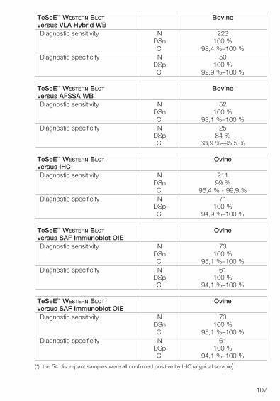
107
TeSeE™ Western Blot versus VLA Hybrid WB
Bovine
Diagnostic sensitivity NDSnCI
223100 %
98,4 %–100 %Diagnostic specificity N
DSpCI
50100 %
92,9 %–100 %
TeSeE™ Western Blot versus AFSSA WB
Bovine
Diagnostic sensitivity NDSnCI
52100 %
93,1 %–100 %Diagnostic specificity N
DSpCI
2584 %
63,9 %–95,5 %
TeSeE™ Western Blot versus IHC
Ovine
Diagnostic sensitivity NDSnCI
21199 %
96,4 % - 99,9 %Diagnostic specificity N
DSpCI
71100 %
94,9 %–100 %
TeSeE™ Western Blot versus SAF Immunoblot OIE
Ovine
Diagnostic sensitivity NDSnCI
73100 %
95,1 %–100 %Diagnostic specificity N
DSpCI
61100 %
94,1 %–100 %
TeSeE™ Western Blot versus SAF Immunoblot OIE
Ovine
Diagnostic sensitivity NDSnCI
73100 %
95,1 %–100 %Diagnostic specificity N
DSpCI
61100 %
94,1 %–100 %
(*): the 54 discrepant samples were all confirmed positive by IHC (atypical scrapie)
108
TeSeE™ Western Blot versus AFSSA WB
Ovine
Diagnostic sensitivity NDSnCI
40100 %
91,2 %–100 %Diagnostic specificity N
DSpCI
0NCNC
TeSeE™ Western Blot versus IHC
Cervids
Diagnostic sensitivity NDSnCI
272100 %
98.65 %–100 %Diagnostic specificity N
DSpCI
40100 %
91,2 %–100 %
REPRODUCTIBILITYThe CRL regularly issues proficiency testing panels for a range of TSE tests. The samples consist of homogenised brain material presented as 50% tissue/water homogenates. The summarised results for confirmatory blotting rounds issued in 2007 are given below.
- CRL Scrapie confirmatory blotting Proficiency test round A panel of 5 samples (1 strong positive, 2 weak positives [all classical scrapie] and 2 negatives) was issued to 23 European laboratories in November 2007.
10 sets of results were returned by laboratories using the BioRad TeSeE™ Western Blot. All of these 10 laboratories identified all samples correctly.
- CRL BSE confirmatory blotting Proficiency test round
A panel of 5 samples (1 strong positive, 1 medium positive, 1 weak positive [all classical BSE] and 2 negatives) was issued to 21 European laboratories in October 2007.
Eight sets of results were returned by laboratories using the BioRad TeSeE™ Western Blot. All of these 8 labs identified all samples correctly.
In addition, reproducibility of the TeSeE™ Western Blot was evaluated with the Bio-Rad control panel including 2 negative ovine samples, 1 negative bovine sample, 1 CWD positive sample, 3 scrapie positive samples and 1 BSE positive sample. The panel was provided to 3 TSE Reference laboratories: VLA in UK, CFIA in Canada and NVI in Norway. The results from the 3 sites were compared to the results obtained in the Bio-Rad facilities. All of these 3
109
laboratories identified all samples correctly (the BSE positive sample was not evaluated by the CFIA ACIA laboratory in Ottawa since this laboratory is not a Reference laboratory for BSE in Canada).
APPLICATIONSThe TeSeE™ Western Blot assay is only for use in National Reference Laboratories (NRLs) and Community Reference laboratories (CRLs). The TeSeE™ Western Blot assay can be used for different types of applications:
- as a confirmatory assay for the confirmation of TSE suspected cases, in cattle (BSE), small ruminants e.g. sheep/goat (BSE/Scrapie) and cervids (CWD). The diagnostic regimen includes several steps at both screening laboratories and Reference laboratories.
• at the screening laboratory: initial screening with one of the approved rapidtest. If the sample turns positive (= initial reactive), the test needs to berepeated in duplicate with the same assay and with the same sample(sample homogenate). If one of the two duplicates or the two turn positive,the sample is then considered as “suspect”. All remaining material (samplehomogenate + remaining tissues) are sent to the Reference laboratory forconfirmation.
• at the reference laboratory: A negative result with the TeSeE™ Western Blot
means that the test sample does not contain detectable PrPres byTeSeE™ Western Blot assay. However, as very low levels of PrPres maynot be detected, such a result does not exclude the possibility of infection.
Any sample with a negative result according to the TeSeE™ Western Blot
assay interpretation criteria, following a positive rapid test result, should betested with one of the other OIE certified confirmatory methods,Immunohistochemistry (IHC) or SAF-Immunoblot.
Any sample with a reproducible positive result according to the testinterpretation critera must be verified in accordance with current legalregulation.
- as a rapid method for the determination of the prevalence of infection withone of the TSE associated diseases (BSE, scrapie, CWD) in the context of an epidemiological survey in a low prevalence country.
Bio-Rad 3, boulevard Raymond Poincaré 92430 MARNES-LA-COQUETTE - FRANCE Tel.: +33 1 47 95 60 00 14-1032 0614Fax.: +33 1 47 41 91 33 Code : 862231
Purchase of Criterion XT Bis-Tris gels and XT MOPS running buffer is accompanied by a limited license under U.S. Patent Numbers 6,143,154; 6,096,182; 6,059,948; 5,578,180; 5,922,185; 6,162,338; and 6,783,651, and corresponding foreign patents.
ECL is a trademark of GE Healthcare. Eppendorf is a trademark of Eppendorf AG. Kodak is a trademark of Eastman Kodak Company.
REFERENCES
1. J.N. Arsac, A.G Biacabe, J.Nicollo, A.Bencsik and T. Baron. Biochemicalidentification of bovine spongiform encephalopathies in cattle. ActaNeuropathologica 2007, vol 114:509-516
2. J.N. Arsac, O. Andreoletti, J.M. Bilheude, C. Lacroux, S. L. Benestad and T.Baron. Similar Biochemical Signatures and Prion Protein Genotypes inAtypical Scrapie and Nor98 Cases, France and Norway. Emerging InfectiousDiseases –Vol. 13, No. 1, January 2007, 58-65.
3. Terry, L.A., Jenkins, R., Thorne, L., Everest, S.J., Chaplin, M.C., Davis,L.A. And Stack, M.J. (2007) First Case Of H-Type Bovine SpongiformEncephalopathy Identified In Great Britain. Veterinary Record 160: 873 – 874.
4. Biacabe A.G, Morignat E, Vulin J, Calavas D, Baron TG.M; February 2008Atypical Bovine Spongiform Encephalopathies, France, 2001-2007. Emerging Infectious Diseases Vol.14, n°2