Embed Size (px)
Citation preview
Reading in a Regular Orthography: An fMRI StudyInvestigating the Role of Visual Familiarity
Anja Ischebeck1, Peter Indefrey2,3, Nobuo Usui4, Izuru Nose5,Frauke Hellwig2,3, and Masato Taira4
Abstract
& In order to separate the cognitive processes associated withphonological encoding and the use of a visual word formlexicon in reading, it is desirable to compare the processing ofwords presented in a visually familiar form with words in avisually unfamiliar form. Japanese Kana orthography offers thispossibility. Two phonologically equivalent but visually dissim-ilar syllabaries allow the writing of, for example, foreignloanwords in two ways, only one of which is visually familiar.Familiarly written words, unfamiliarly written words, andpseudowords were presented in both Kana syllabaries (yield-ing six conditions in total) to participants during an fMRImeasurement with a silent articulation task (Experiment 1) anda phonological lexical decision task (Experiment 2) using anevent-related design. Consistent over two experimental tasks,
the three different stimulus types (familiar, unfamiliar, andpseudoword) were found to activate selectively different brainregions previously associated with phonological encoding andword retrieval or meaning. Compatible with the predictions ofthe dual-route model for reading, pseudowords and visuallyunfamiliar words, which have to be read using phonologicalassembly, caused an increase in brain activity in left inferiorfrontal regions (BA 44/47), as compared to visually familiarwords. Visually familiar and unfamiliar words were found toactivate a range of areas associated with lexico-semanticprocessing more strongly than pseudowords, such as the leftand right temporo-parietal region (BA 39/40), a region in theleft middle/inferior temporal gyrus (BA 20/21), and theposterior cingulate (BA 31). &
INTRODUCTION
Models of normal reading often assume two differentpathways to arrive at the correct pronunciation of awritten word. In the dual-route model of reading (for areview, see Coltheart, Curtis, Atkins, & Haller, 1993) it isassumed that the pronunciation of a word can be eitherassembled sequentially from the word’s letters or gra-phemes according to a set of grapheme to phonemeconversion rules (GPC rules) or retrieved directly frommemory if the visual form of the word as a whole hasbeen recognized. For example, pseudowords (pro-nounceable nonwords) can only be read by phonolog-ical assembly according to the GPC rules, whereas wordswith an irregular spelling, that is, a spelling that does notconform to the GPC rules of the respective orthography,are read more successfully via the visual word formlexicon. Orthographically regular written words on theother hand can be read via both routes.
A number of studies have ventured to identify theneural correlates of these two routes but yielded diver-
gent results. The route for phonological encoding usingthe GPC rules, also referred to as assembled phonology,has been identified with the left inferior frontal region(BA 44/45) (e.g., Fiez, Balota, Raichle, & Petersen, 1999).Other researchers have suggested that the sublexicalprocedure is localized in the left posterior superiortemporal region (e.g., Paulesu et al., 2000). The directroute of visual word form retrieval, often referred to asaccessed phonology, has been identified with a numberof diverse brain regions: the left posterior middle tem-poral gyrus, including the angular gyrus (e.g., Howardet al., 1992), areas in the left temporo-occipital lobe,especially the left fusiform gyrus (e.g., Cohen et al.,2000), and the left and right extrastriate cortices (e.g., Pe-tersen, Fox, Posner, Mintun, & Raichle, 1988).
This lack of agreement might be attributable to anumber of reasons, methodological as well as inherent.Methodologically, the incommensurability of the tasksand stimuli used in many subtraction designs in brainimaging research, as well as the assumptions of li-nearity and pure insertion, might critically impair thereproducibility of results across experiments and theirinterpretation (see, for a review, Friston et al., 1996).Furthermore, block designs, mandatory for PET experi-ments, can induce strategic effects (d’Esposito, Zarahn, &Aguirre, 1999). Inherent reasons related to the nature of
1University of Nijmegen, 2Max Planck Institute for Psycholin-guistics, 3F. C. Donders Center for Cognitive Neuroimaging,4Nihon University Graduate School of Medical Science, 5BunkyoGakuin University
D 2004 Massachusetts Institute of Technology Journal of Cognitive Neuroscience 16:5, pp. 727–741
the neural implementation of the two routes might alsoaccount for the difficulty in finding unambiguous evi-dence for them. First, the cognitive functions underscrutiny might be subserved by several functionally spe-cialized areas, highly sensitive to differences in stimulusmaterials and task requirements. Alternatively, visualword form processing might be so effortless that itremains inconspicuous given the signal-to-noise ratio ofpresent imaging methods.
The present study avoids some of the methodologicalproblems by directly comparing different script variantsof the same word and pseudoword stimuli in a silentarticulation task (Experiment 1). The randomization ofdifferent stimulus types in an event-related design pre-vents confounds due to low-frequency signal shifts(Donaldson & Buckner, 2001) and strategic adaptationby participants. Additionally, the same materials werepresented in a phonological lexical decision task (Exper-iment 2). In this variant of a lexical decision task,participants have to press one button if a letter stringsounds like a real word, and another button if not. Thistask basically requires the same processing steps as thesilent articulation task (see the Discussion section for atask analysis). It is thus possible to check the consistencyof the results over tasks and experiments and to attri-bute differences in brain activation patterns to differ-ences in the stimulus materials.
The two pathways postulated by the dual-route modelcan be investigated by comparing stimuli that can beread via one pathway to stimuli that can be read over theother or both pathways. Orthographically regular andvisually highly familiar words (i.e., words well known tothe reader) are assumed to be read over both routes,whereas the same words presented in a visually unfa-miliar way can only be read using sequential phonolog-ical encoding. A comparison of both could shed somelight on the neural basis of the dual-route model. Thereare possibilities to achieve this experimental variation inalphabetic scripts, such as mirror writing and casealternation (e.g., nIgHt). These manipulations have al-ready been employed in neuroimaging studies (mirrorwriting: Dong et al., 2000; case alternation: Polk & Farah,2002). One limitation to this approach, however, is thatthese manipulations render the sublexical units of theword, letters, and graphemes unfamiliar as well. This isan obstacle for grapheme to phoneme conversion, whena grapheme consists of more than one letter or when con-version rules apply to chunks of graphemes. Observedbrain activation differences might therefore be due to anincreased difficulty of GPC processing rather than onlyto the disruption of visual word form processing.
Japanese Kana orthography, on the other hand, allowsa more direct realization of this manipulation withoutsimultaneously rendering sublexical units unfamiliar aswell. Japanese orthography employs Chinese charactersalongside two visually distinct syllabaries called Hiraganaand Katakana, which are phonologically equivalent. Al-
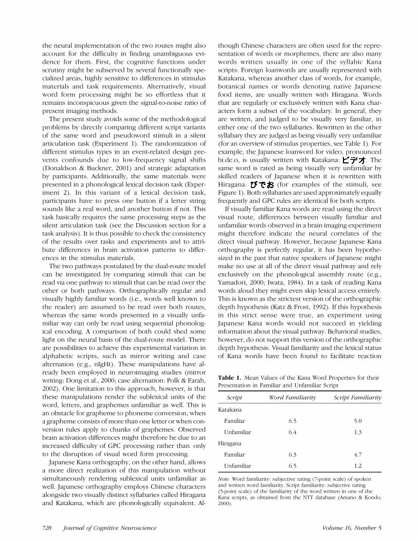
though Chinese characters are often used for the repre-sentation of words or morphemes, there are also manywords written usually in one of the syllabic Kanascripts: Foreign loanwords are usually represented withKatakana, whereas another class of words, for example,botanical names or words denoting native Japanesefood items, are usually written with Hiragana. Wordsthat are regularly or exclusively written with Kana char-acters form a subset of the vocabulary. In general, theyare written, and judged to be visually very familiar, ineither one of the two syllabaries. Rewritten in the othersyllabary they are judged as being visually very unfamiliar(for an overview of stimulus properties, see Table 1). Forexample, the Japanese loanword for video, pronouncedbi.de.o, is usually written with Katakana: . Thesame word is rated as being visually very unfamiliar byskilled readers of Japanese when it is rewritten withHiragana: (for examples of the stimuli, seeFigure 1). Both syllabaries are used approximately equallyfrequently and GPC rules are identical for both scripts.
If visually familiar Kana words are read using the directvisual route, differences between visually familiar andunfamiliar words observed in a brain imaging experimentmight therefore indicate the neural correlates of thedirect visual pathway. However, because Japanese Kanaorthography is perfectly regular, it has been hypothe-sized in the past that native speakers of Japanese mightmake no use at all of the direct visual pathway and relyexclusively on the phonological assembly route (e.g.,Yamadori, 2000; Iwata, 1984). In a task of reading Kanawords aloud they might even skip lexical access entirely.This is known as the strictest version of the orthographicdepth hypothesis (Katz & Frost, 1992). If this hypothesisin this strict sense were true, an experiment usingJapanese Kana words would not succeed in yieldinginformation about the visual pathway. Behavioral studies,however, do not support this version of the orthographicdepth hypothesis. Visual familiarity and the lexical statusof Kana words have been found to facilitate reaction
Table 1. Mean Values of the Kana Word Properties for theirPresentation in Familiar and Unfamiliar Script
Script Word Familiarity Script Familiarity
Katakana
Familiar 6.5 5.0
Unfamiliar 6.4 1.3
Hiragana
Familiar 6.3 4.7
Unfamiliar 6.5 1.2
Note. Word familiarity: subjective rating (7-point scale) of spokenand written word familiarity. Script familiarity: subjective rating(5-point scale) of the familiarity of the word written in one of theKana scripts, as obtained from the NTT database (Amano & Kondo,2000).
728 Journal of Cognitive Neuroscience Volume 16, Number 5
times in several tasks, including reading aloud (e.g.,Besner & Hildebrand, 1987; Besner & Smith, 1992) andphonological lexical decision (e.g., Usui, 1998; Kawaka-mi, 1993; Yamada, Imai, & Ikebe, 1990; Hatta, Katoh, &Kirsner, 1984; see also Table 2). The effects of word-levelvisual familiarity might therefore be interpreted as evi-dence for a visual word form lexicon for highly familiarKana words. Given the complexity and variability of theJapanese script it is possible, however, that only a smallsubset of the vocabulary, namely, extremely frequentKana words are stored in the visual word form lexicon.The dual-route model might therefore have only a li-mited range of applicability and thus is not representa-tive for Kana reading in general. It should be noted thatthe evidence cited here and the results of the presentstudy do not support or refute the more commonly used
weaker version of the orthographic depth hypothesis,namely, that readers of regular orthographies rely morestrongly on the assembly route than readers of irregularorthographies. There is indeed some evidence for be-havioral (see, for a review, Katz & Frost, 1992) as well asneural (Paulesu et al., 2000) differences in the processingof regular and irregular orthographies.
The present study is designed to address the follow-ing questions: first, whether there are stable differencesin brain activation patterns between visually familiarwords, visually unfamiliar words, and pseudowords;second, given that differences are found, if they caninterpreted in the framework of the dual-route model;additionally, whether areas can be identified that re-spond selectively to words and pseudowords. Lastly, weare interested in how much the results depend on thetask (silent articulation, phonological lexical decision).
The first fMRI experiment reported in the presentstudy employed visually familiar and unfamiliar words, aswell as pseudowords in a silent articulation task using anevent-related design. If the dual-route model is valid,pseudowords as well as unfamiliarly written words willhave to be phonologically assembled, whereas the pro-nunciation of visually highly familiar words can beaccessed directly. Stronger activations for visually unfa-miliar words and pseudowords might therefore indicateregions involved in the processing of grapheme tophoneme conversion.
It is unclear, however, what regions might be associ-ated with the direct visual route. If access to the visualword form lexicon is conceptualized as a serial searchprocess (e.g., Forster, 1976), visually unfamiliar wordsand pseudowords should yield stronger activations thanvisually familiar words. In an event-related design withrandomized materials as in the present study, it can beassumed that access to the visual word form lexicon isalways attempted. In the case of visually highly familiarwords, the search can terminate earlier. On the otherhand, it could be predicted that a specialized populationof neurons displays increased activation when a visuallyfamiliar word stimulus is encountered (see, Polk &Farah, 2002, for a similar argument). In this case, visually
Table 2. Response Times (in msec) and Error Rates asObtained in the Two Behavioral Experiments
Word Type
Task Familiar Unfamiliar Pseudoword
Naming
Hiragana 523 (25.6) 548 (26.2) 665 (50.1)
1.85% 2.78% 5.32%
Katakana 517 (18.2) 622 (43.6) 669 (46.5)
1.62% 4.17% 4.63%
pLDTa
Hiragana 510 (30.0) 569 (42.7) 760 (70.3)
0.47% 2.09% 3.49%
Katakana 492 (27.1) 715 (73.0) 780 (71.7)
0.47% 12.33% 3.95%
Note. Standard errors are given in parentheses, error percentagesbelow the response times.apLDT = phonological lexical decision task.
Figure 1. An example of theKana stimuli.
Ischebeck et al. 729
highly familiar words should activate a word form rec-ognition area more strongly than visually unfamiliarwords or pseudowords, similar to, for example, thefusiform face recognition area (Kanwisher, McDermot,& Chun, 1997).
The second fMRI experiment, conducted using thesame stimuli but a different task (phonological lexicaldecision), provided an estimate of the extent to whichbrain activations observed in Experiment 1 can beattributed to the task rather than to differences in thestimulus materials. In this task, visually familiar wordscan be judged correctly by looking them up directly inthe visual word form lexicon. In the case of visuallyunfamiliar words and pseudowords, participants have toassemble the pronunciation of the stimulus and check itagainst the auditory word form lexicon to give a correctanswer. As elaborated in the task analysis in the Dis-cussion section, both tasks require very similar pro-cessing steps. Additionally, two off-line behavioralexperiments were conducted to test the stimulus mate-rials in a task of reading aloud and phonological lexicaldecision.
RESULTS
Behavioral Experiments
In the first experiment participants were instructed toread aloud, as fast and correctly as possible, familiarlyand unfamiliarly written words and pseudowords pre-sented on a monitor. The factors script (Katakana,Hiragana) and word type (familiar, unfamiliar, and pseu-doword) yielded six conditions in total. Only naming la-tencies for correct answers between 250 and 2000 msec(2504 of 2592 data points) were analyzed (see Table 2)and entered into a repeated measures ANOVA with thetwo factors script and word type. There was a signifi-cant main effect of the factor word type, F(1,11) = 15.19,SEM = 692, p < .01, due to the slower reaction times tovisually unfamiliar words and pseudowords as comparedto visually familiar words. The significant main effect ofthe factor script, F(2,22) = 80.14, SEM = 1667, p < .001,and interaction of both factors, F(2,22) = 12.67, SEM =888, p < .001, are due to the significant difference be-tween visually unfamiliar Katakana and Hiragana words,t(11) = 6.33, p < .001. Reaction times to visually familiarwords or pseudowords did not differ significantly be-tween Hiragana and Katakana. The longer reactiontimes to visually unfamiliar Katakana are most likelycaused by our choice of stimuli. Because rewriting famil-iar Hiragana words with Katakana would have yieldedstimuli that have medium instead of very low visualfamiliarity ratings, we used words usually written withChinese characters for the Katakana unfamiliar condition(see Besner & Hildebrandt, 1987; Hatta et al., 1984, for asimilar choice of stimuli). Words usually written withChinese characters have been found to be rated visuallyhighly unfamiliar when presented in Katakana (Amano &
Kondo, 2000). However, this condition proved to bemore difficult for the Japanese participants even thoughoverall word familiarity was matched to the Katakanafamiliar condition. The overall familiarity effect is signif-icant for Katakana, t(11) = 7.39, p < .001, as well as forHiragana, t(11)= 3.94, p< .01, when analyzed separately.
In the second experiment, the same materials andprocedure were used with a phonological lexical deci-sion task. Only reaction times for correct answers be-tween 250 and 2000 msec (2494 of 2592 data points)were analyzed (see Table 2) and entered into a repeatedmeasures ANOVA with the two factors script and wordtype. Again there was a significant main effect of thefactor word type, F(1,11) = 35.72, SEM= 1238, p< .001,due to the slower reaction times to visually unfamiliarwords and pseudowords. The significant main effect ofthe factor script, F(2,22) = 63.40, SEM = 6740, p < .001,and the significant interaction of both factors, F(2,22) =24.50, SEM = 1765, p < .001, are, as in the namingexperiment, due to the longer reaction times for visuallyunfamiliar Katakana words, t(11) = 6.29, p< .001. Again,visually familiar words were judged significantly fasterthan visually unfamiliar words in both scripts, Katakana:t(11) = 7.59, p < .001; Hiragana: t(11) = 5.97, p < .001.The observed pattern of response times is very similar tothe results of the first experiment.
fMRI Experiments
Experiment 1: Silent Articulation Task
Ten participants took part in this experiment performinga silent articulation task. In an event-related design, thesame materials were used as in the behavioral experi-ments. The six conditions of the experiment werecollapsed over the factor script yielding three condi-tions, which will be referred to in short as ‘‘familiar,’’‘‘unfamiliar,’’ and ‘‘pseudoword.’’ Six contrasts werecalculated in total.
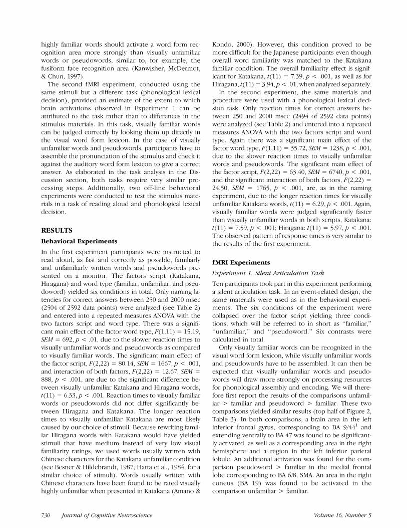
Only visually familiar words can be recognized in thevisual word form lexicon, while visually unfamiliar wordsand pseudowords have to be assembled. It can then beexpected that visually unfamiliar words and pseudo-words will draw more strongly on processing resourcesfor phonological assembly and encoding. We will there-fore first report the results of the comparisons unfamil-iar > familiar and pseudoword > familiar. These twocomparisons yielded similar results (top half of Figure 2,Table 3). In both comparisons, a brain area in the leftinferior frontal gyrus, corresponding to BA 9/441 andextending ventrally to BA 47 was found to be significant-ly activated, as well as a corresponding area in the righthemisphere and a region in the left inferior parietallobule. An additional activation was found for the com-parison pseudoword > familiar in the medial frontallobe corresponding to BA 6/8, SMA. An area in the rightcuneus (BA 19) was found to be activated in thecomparison unfamiliar > familiar.
730 Journal of Cognitive Neuroscience Volume 16, Number 5
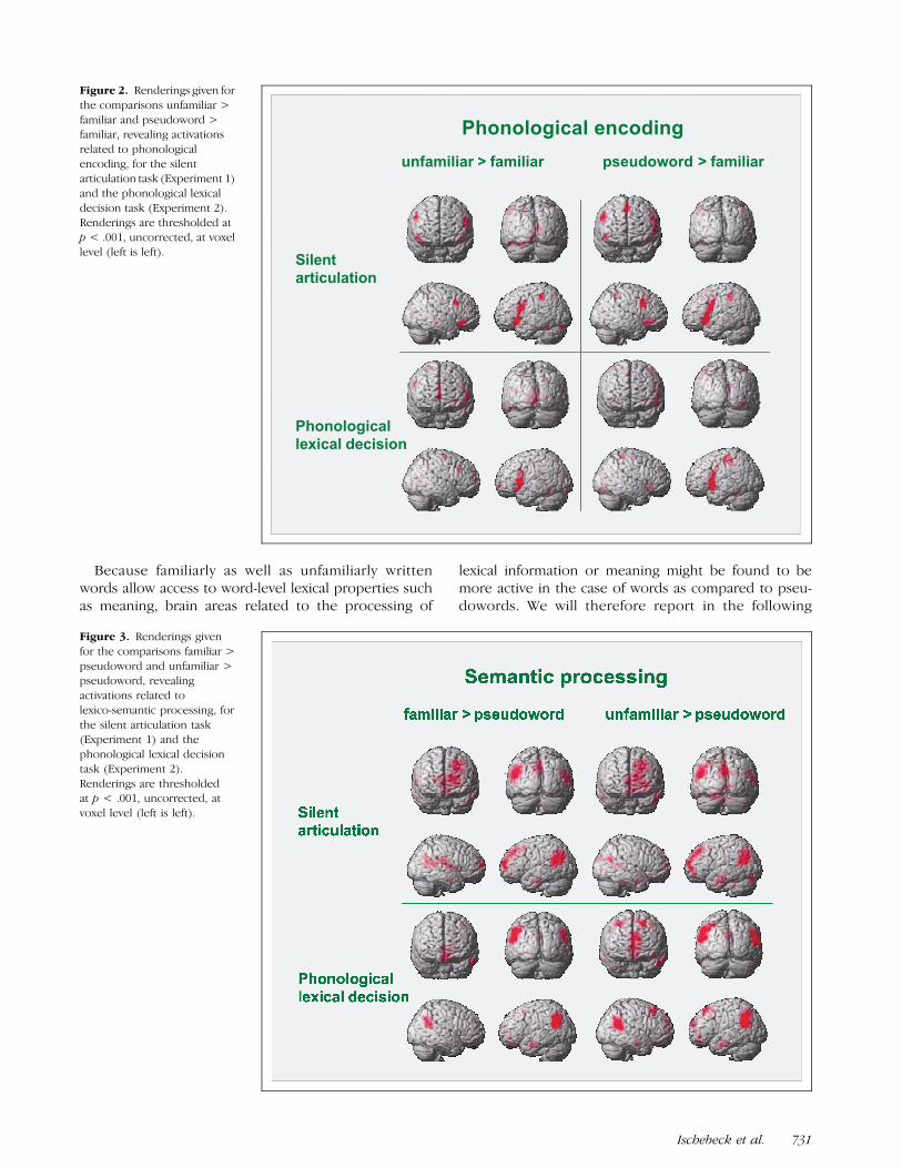
Because familiarly as well as unfamiliarly writtenwords allow access to word-level lexical properties suchas meaning, brain areas related to the processing of
lexical information or meaning might be found to bemore active in the case of words as compared to pseu-dowords. We will therefore report in the following
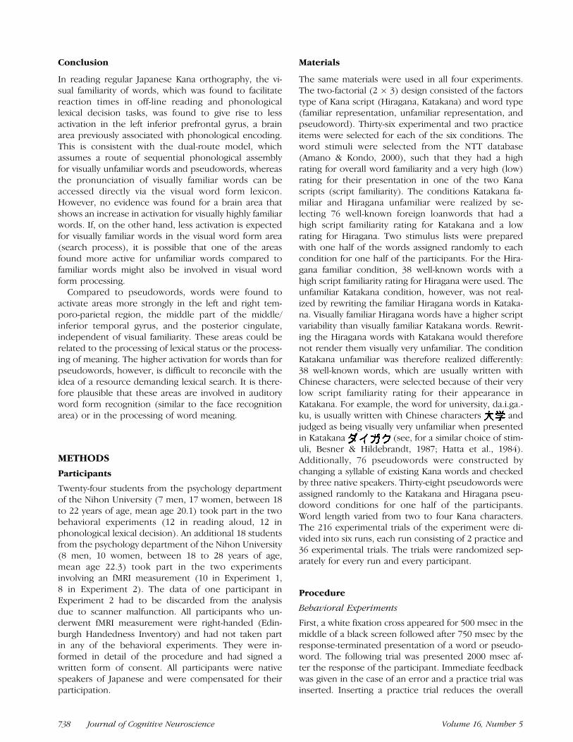
Figure 2. Renderings given for
the comparisons unfamiliar >
familiar and pseudoword >familiar, revealing activations
related to phonological
encoding, for the silentarticulation task (Experiment 1)
and the phonological lexical
decision task (Experiment 2).
Renderings are thresholded atp < .001, uncorrected, at voxel
level (left is left).
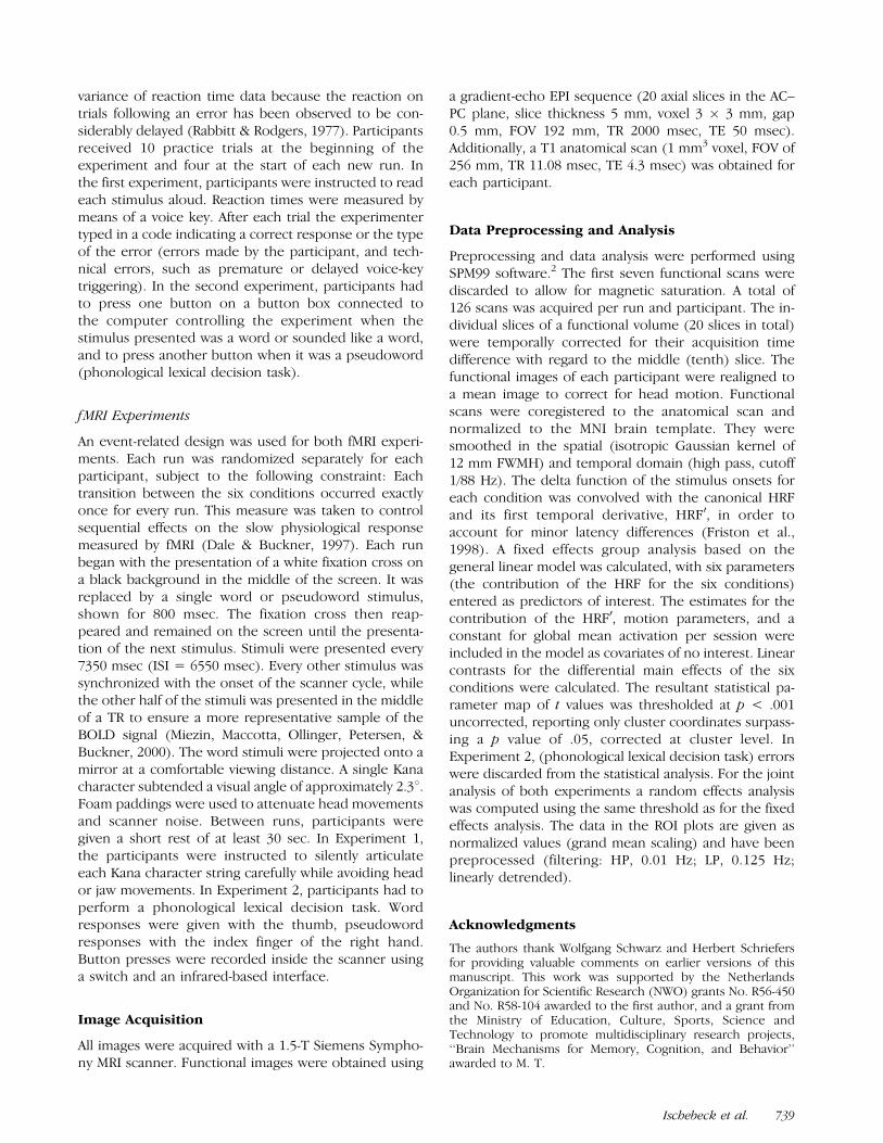
Figure 3. Renderings given
for the comparisons familiar >
pseudoword and unfamiliar >pseudoword, revealing
activations related to
lexico-semantic processing, for
the silent articulation task(Experiment 1) and the
phonological lexical decision
task (Experiment 2).
Renderings are thresholdedat p < .001, uncorrected, at
voxel level (left is left).
Ischebeck et al. 731
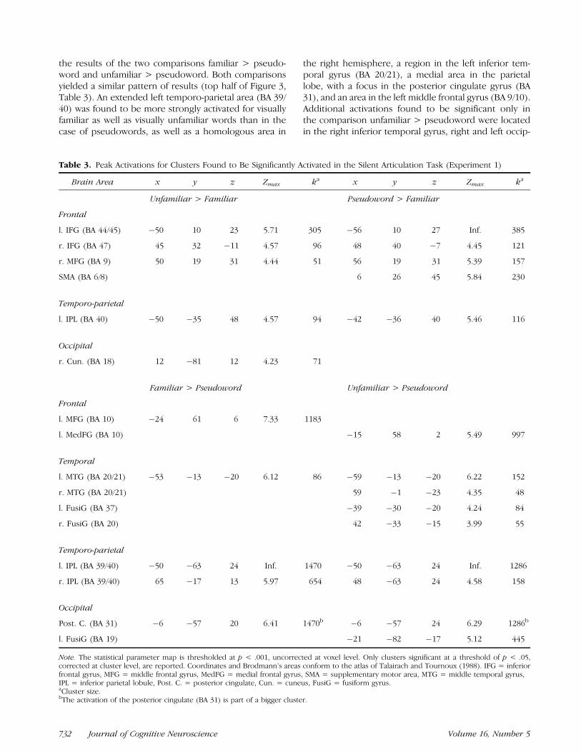
the results of the two comparisons familiar > pseudo-word and unfamiliar > pseudoword. Both comparisonsyielded a similar pattern of results (top half of Figure 3,Table 3). An extended left temporo-parietal area (BA 39/40) was found to be more strongly activated for visuallyfamiliar as well as visually unfamiliar words than in thecase of pseudowords, as well as a homologous area in
the right hemisphere, a region in the left inferior tem-poral gyrus (BA 20/21), a medial area in the parietallobe, with a focus in the posterior cingulate gyrus (BA31), and an area in the left middle frontal gyrus (BA 9/10).Additional activations found to be significant only inthe comparison unfamiliar > pseudoword were locatedin the right inferior temporal gyrus, right and left occip-
Table 3. Peak Activations for Clusters Found to Be Significantly Activated in the Silent Articulation Task (Experiment 1)
Brain Area x y z Zmax ka x y z Zmax ka
Unfamiliar > Familiar Pseudoword > Familiar
Frontal
l. IFG (BA 44/45) �50 10 23 5.71 305 �56 10 27 Inf. 385
r. IFG (BA 47) 45 32 �11 4.57 96 48 40 �7 4.45 121
r. MFG (BA 9) 50 19 31 4.44 51 56 19 31 5.39 157
SMA (BA 6/8) 6 26 45 5.84 230
Temporo-parietal
l. IPL (BA 40) �50 �35 48 4.57 94 �42 �36 40 5.46 116
Occipital
r. Cun. (BA 18) 12 �81 12 4.23 71
Familiar > Pseudoword Unfamiliar > Pseudoword
Frontal
l. MFG (BA 10) �24 61 6 7.33 1183
l. MedFG (BA 10) �15 58 2 5.49 997
Temporal
l. MTG (BA 20/21) �53 �13 �20 6.12 86 �59 �13 �20 6.22 152
r. MTG (BA 20/21) 59 �1 �23 4.35 48
l. FusiG (BA 37) �39 �30 �20 4.24 84
r. FusiG (BA 20) 42 �33 �15 3.99 55
Temporo-parietal
l. IPL (BA 39/40) �50 �63 24 Inf. 1470 �50 �63 24 Inf. 1286
r. IPL (BA 39/40) 65 �17 13 5.97 654 48 �63 24 4.58 158
Occipital
Post. C. (BA 31) �6 �57 20 6.41 1470b �6 �57 24 6.29 1286b
l. FusiG (BA 19) �21 �82 �17 5.12 445
Note. The statistical parameter map is thresholded at p < .001, uncorrected at voxel level. Only clusters significant at a threshold of p < .05,corrected at cluster level, are reported. Coordinates and Brodmann’s areas conform to the atlas of Talairach and Tournoux (1988). IFG = inferiorfrontal gyrus, MFG = middle frontal gyrus, MedFG = medial frontal gyrus, SMA = supplementary motor area, MTG = middle temporal gyrus,IPL = inferior parietal lobule, Post. C. = posterior cingulate, Cun. = cuneus, FusiG = fusiform gyrus.aCluster size.bThe activation of the posterior cingulate (BA 31) is part of a bigger cluster.
732 Journal of Cognitive Neuroscience Volume 16, Number 5
ital fusiform gyrus (BA 18/19) and in the right and lefttemporal fusiform gyrus (BA 36/37). The remainingtwo comparisons familiar > unfamiliar and pseudo-word > unfamiliar did not yield any suprathresholdclusters.
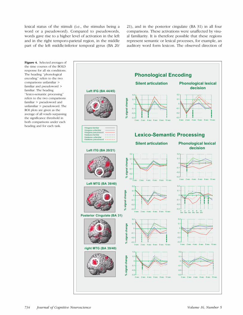
Given the significant difference between the twoscripts regarding the unfamiliar condition, it is possiblethat the pattern of results does not generalize over bothscripts. To investigate the symmetry of the results forboth scripts, selective averages of the time course of theBOLD response for all six experimental conditions aregiven in Figure 4.
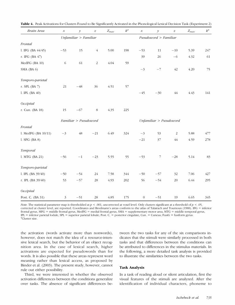
Experiment 2: Phonological Lexical Decision Task
The same experimental procedure and materials wereused as in Experiment 1, using a phonological lexical de-cision task. The six conditions were again collapsed overthe factor script yielding three conditions: familiar,unfamiliar, and pseudoword. Six linear contrasts werecalculated.
As in Experiment 1, first the results of the compar-isons unfamiliar > familiar and pseudoword > familiarare reported. These two comparisons yielded a similarpattern of results (bottom half of Figure 2, Table 4),similar to the results of Experiment 1. In both compar-isons, areas in the left inferior frontal gyrus, corre-sponding to BA 44/45 and BA 47, were found to bemore strongly activated for unfamiliarly written words ndpseudowords than in the case of familiarly written words.Additional activations were found for the comparisonunfamiliar > familiar in the anterior medial frontal gyrus,the right superior parietal lobule, and a medial occipitalarea. In the comparison pseudoword > familiar addition-al activations were found in the SMA (BA 6), and in the leftinferior parietal lobule.
As in Experiment 1, the results of the comparisonsfamiliar > pseudoword and unfamiliar > pseudowordmight indicate brain areas involved in lexico-semanticprocessing. These comparisons yielded results (bottomhalf of Figure 3, Table 4) that also resemble the results ofExperiment 1. In both comparisons, an extended lefttemporo-parietal area (BA 39/40) was found to bestrongly activated, as well as a homologous region inthe right hemisphere. Also, a region in the left inferiortemporal gyrus (BA 20/21) was found to be significantlyactivated in both comparisons, as well as areas in themedial frontal lobe (BA 10/11), and a medial area in theoccipital lobe with a focus in the posterior cingulate(BA 31). The remaining two comparisons familiar >unfamiliar and pseudoword > familiar yielded no supra-threshold clusters.
Joint Analysis of Experiment 1 and Experiment 2
To investigate possible activation differences betweenthe two tasks, a random effects analysis was calculated.
However, activation differences between the tasks didnot surpass the significance threshold for any of thesix comparisons.
DISCUSSION
An Overview over the Principal Results
The first goal of the present study was to investigatewhether the behavioral differences between visuallyhighly familiar and unfamiliar Kana words and pseudo-words give rise to a stable pattern of activation differ-ences in the brain. The very systematic differencesbetween visually familiar and unfamiliar words foundconsistently over two different tasks support the as-sumption that skilled readers of Japanese process thesestimulus types differently, possibly in a way as assumedin the dual-route model of reading.
Our second focus of interest was to identify brainregions that might correspond to the two pathwaysassumed in the dual-route model. According to thedual-route model, the pronunciation of visually unfamil-iar words and pseudowords has to be constructed se-quentially, whereas the pronunciation of visually highlyfamiliar words can be accessed directly. A coherentregion in the left inferior frontal gyrus correspondingto BA 44, 45, and 47 was found to be more stronglyactivated for unfamiliarly written words and pseudo-words than for visually highly familiar words. A corres-ponding activated area was observed in the right inferiorfrontal gyrus. These areas have been associated, amongothers, with sequential phonological encoding (e.g., Fiezet al., 1999). It is possible that the activation differencesobserved here reflect a gradual rather than a categoricaldifference with regard to the use of the phonologicalassembly route. Although the assembly route is used lessfor visually highly familiar words, it is drawn upon morestrongly in the case of visually unfamiliar words and pseu-dowords. The activation found in this area might thusalso reflect the greater difficulty to process these words,as indicated by the longer reaction times.
It seems to be more difficult, however, to identifycorrelates of the direct visual pathway in the presentstudy. If a word recognition area existed in the brainsimilar to other object recognition areas identified so far,more activation is expected in this area for visuallyfamiliar than for unfamiliar or unknown word stimuli.However, no active region was identified in the compar-ison familiar > unfamiliar. Alternatively, it is conceivablethat visually familiar words allow the early termination ofa search process and should therefore lead to less acti-vation in the visual word form area than visually unfa-miliar words and pseudowords. All areas more active forvisually unfamiliar and pseudowords are then also can-didates for a visual word form area.
Furthermore, we were interested in whether therewould be a difference in brain activations reflecting the
Ischebeck et al. 733
lexical status of the stimuli (i.e., the stimulus being aword or a pseudoword). Compared to pseudowords,words gave rise to a higher level of activation in the leftand in the right temporo-parietal region, in the middlepart of the left middle/inferior temporal gyrus (BA 20/
21), and in the posterior cingulate (BA 31) in all fourcomparisons. These activations were unaffected by visu-al familiarity. It is therefore possible that these regionsrepresent semantic or lexical processes, for example, anauditory word form lexicon. The observed direction of
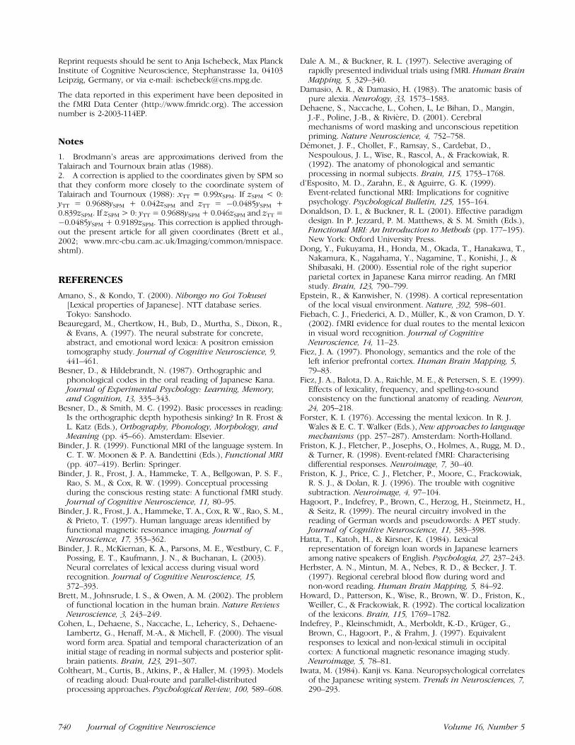
Figure 4. Selected averages of
the time courses of the BOLD
response for all six conditions.The heading ‘‘phonological
encoding’’ refers to the two
comparisons unfamiliar >
familiar and pseudoword >familiar. The heading
‘‘lexico-semantic processing’’
refers to the two comparisons
familiar > pseudoword andunfamiliar > pseudoword. The
ROI plots are given as the
average of all voxels surpassingthe significance threshold in
both comparisons under each
heading and for each task.
734 Journal of Cognitive Neuroscience Volume 16, Number 5
the activation (words activate more than nonwords),however, does not match the idea of a resource-inten-sive lexical search, but the behavior of an object recog-nition area. In the case of lexical search, higheractivations are expected for pseudowords than forwords. It is also possible that these areas represent wordmeaning rather than lexical access, as proposed byBinder et al. (2003). The present study, however, cannotrule out either possibility.
Third, we were interested in whether the observedactivation differences between the conditions generalizeover tasks. The absence of significant differences be-
tween the two tasks for any of the six comparisons in-dicates that the stimuli were similarly processed in bothtasks and that differences between the conditions canbe attributed to differences in the stimulus materials. Inthe following, a more detailed task analysis is providedto illustrate the similarities between the two tasks.
Task Analysis
In a task of reading aloud or silent articulation, first thevisual features of the stimuli are analyzed. After theidentification of individual characters, phoneme to
Table 4. Peak Activations for Clusters Found to Be Significantly Activated in the Phonological Lexical Decision Task (Experiment 2)
Brain Area x y z Zmax ka x y z Zmax ka
Unfamiliar > Familiar Pseudoword > Familiar
Frontal
l. IFG (BA 44/45) �53 15 4 5.00 198 �53 11 �10 5.39 247
r. IFG (BA 47) 39 26 �6 4.32 61
MedFG (BA 10) 6 61 2 4.04 59
SMA (BA 6) �3 �7 42 4.20 75
Temporo-parietal
r. SPL (BA 7) 21 �48 36 4.51 57
l. IPL (BA 40) �45 �30 44 4.43 141
Occipital
r. Cun. (BA 18) 15 �67 8 4.35 225
Familiar > Pseudoword Unfamiliar > Pseudoword
Frontal
l. MedFG (BA 10/11) �3 48 �21 6.49 324 �3 53 2 5.88 477
l. SFG (BA 8) �21 37 44 4.59 278
Temporal
l. MTG (BA 21) �56 �1 �23 5.55 55 �53 7 �28 5.14 83
Temporo-parietal
l. IPL (BA 39/40) �50 �54 24 7.58 344 �50 �57 32 7.06 427
r. IPL (BA 39/40) 53 �57 28 4.93 202 56 �54 20 6.44 295
Occipital
Post. C. (BA 31) 3 �51 28 4.85 175 0 �51 19 6.65 345
Note. The statistical parameter map is thresholded at p < .001, uncorrected at voxel level. Only clusters significant at a threshold of p < .05,corrected at cluster level, are reported. Coordinates and Brodmann’s areas conform to the atlas of Talairach and Tournoux (1988). IFG = inferiorfrontal gyrus, MFG = middle frontal gyrus, MedFG = medial frontal gyrus, SMA = supplementary motor area, MTG = middle temporal gyrus,IPL = inferior parietal lodule, SPL = superior parietal lobule; Post. C. = posterior cingulate, Cun. = Cuneus; FusiG = fusiform gyrus.aCluster size.
Ischebeck et al. 735
grapheme conversion can be performed. In a perfectlyregular orthography, the utterance can be initiateddirectly after grapheme to phoneme conversion. Thebehavioral data, however, suggest that skilled readers ofJapanese do not rely exclusively on the assembly routein a task of reading aloud. They attempt to access theword in the mental lexicon before utterance. Finding amatch in the visual word form lexicon then translatesinto faster word recognition and naming times forfamiliarly written words. The search in this lexicon willtime-out (Taylor & Lupker, 2001), however, for unfamil-iarly written words and pseudowords. Their pronuncia-tion has to be assembled using GPC rules. Similarly, thefaster reading times for visually unfamiliar words com-pared to pseudowords indicate that the reader alsomakes an attempt to look up the assembled candidatein the auditory word form lexicon. In the case ofpseudowords, the search in the auditory word formlexicon will finally time-out, resulting in longer naminglatencies for pseudowords. However, given that Japa-nese Kana orthography is perfectly regular, why shouldthe reader retrieve a word in the visual and the auditoryword form lexicon in a silent articulation task? A reasonfor this strategy might lie in the underspecification ofprosodic parameters by Japanese Kana orthography,such as correct tone accent. Tone accent is oftenirregular and a lexical property of the Japanese vocabu-lary (cf., Vance, 1987). An assembled articulatory approx-imation of the word might therefore not satisfy a skilledreader intending to read correctly. An additional gain ofthis strategy might be the reduction in articulatoryprocessing load when a word is recognized. Then awhole-word motor program could be used for its artic-ulation, instead of the effortful sequential assembly ofsmaller articulatory elements in the case of pseudo-words. The randomization of the stimuli and the useof both scripts in all conditions prevented the partic-ipants from skipping the search in the visual and audi-tory word form lexicon.
In the phonological lexical decision task, after pro-cessing the visual features of the stimuli, grapheme tophoneme conversion can be performed. Familiarly writ-ten words can be directly recognized in the visual wordform lexicon. In contrast to the silent articulation task,the retrieval of the correct pronunciation is not neces-sary since only a categorization response is required.Similar to the silent articulation task, pseudowords andunfamiliarly written words have to be phonologicallyassembled and checked against the auditory word formlexicon. A pseudoword response can be given only afterthe search in the auditory word form lexicon has endedunsuccessfully. This task analysis, therefore, matches theordering of phonological lexical decision times found inthe present study and elsewhere. The two tasks used inthe present study thus involve very similar processingsteps. However, there are also differences. Accessingthe pronunciation of visually familiar words is only obli-
gatory in the silent articulation task, although it mightbe accessed automatically in the phonological lexicaldecision task as well. The silent articulation task requiresan articulatory response, whereas a decision and amanual categorization response is required in the pho-nological decision task.
Visual Familiarity and Phonological Processing
Areas in the left inferior frontal gyrus (BA 44/45), includ-ing the anterior insula, have been repeatedly associatedwith phonological processing in neuroimaging studies(Fiez et al., 1999; Herbster, Mintun, Nebes, & Becker,1997). Compared to words, these areas have been foundto be activated more strongly by pseudowords (Hagoortet al., 1999; Herbster et al., 1997; Rumsey et al., 1997),words with a low frequency of occurrence (Fiebach, Frie-derici, Muller, & von Cramon, 2002; Fiez et al., 1999),words written with mirrored letters (Dong et al., 2000),and, as observed in the present study, words writtenin an unfamiliar script. This is compatible with the viewthat a common mechanism mediates the pronunciationof both pseudowords, words with a low frequency ofoccurrence and unfamiliarly written words, namely, ef-fortful sequential phonological assembly by graphemeto phoneme conversion. Another area that has beenassociated with GPC processing is the posterior part ofthe superior temporal gyrus at the temporo-parietaljunction (Paulesu et al., 2000; Price, 2000). In the presentstudy, however, this area was not found to be more ac-tive in the case of visually unfamiliar words and pseudo-words as compared to visually familiar words.
Possible Locations of the VisualWord FormLexicon
A number of very diverse brain areas have been pro-posed as possible locations of a visual word formlexicon. One of these candidate areas is the posteriorpart of the middle and superior temporal gyrus, includ-ing the angular gyrus (Paulesu et al., 2000; Beauregardet al., 1997; Menard, Kosslyn, Thompson, Alpert, &Rauch, 1996; Howard et al., 1992). Recently, however,this region has been associated with semantic ratherthan orthographic processing (see, for a review, Binder,1999; Price, Indefrey, & van Turennout, 1999). This viewis corroborated by the present study, which reports astronger activation of this region for words than forpseudowords independent of orthographic familiarity.
Other studies suggest that the visual word form lex-icon is located in the posterior part of the left inferiortemporal gyrus, including the fusiform gyrus (e.g., Co-hen et al., 2000; Paulesu et al. 2000; Price, 2000; Lawet al., 1991). Neuropsychological evidence from patientswith pure alexia suggests that this region indeed plays acrucial role in reading (Damasio & Damasio, 1983). Inthis region, brain areas have been identified that re-spond selectively to highly specific complex visual stim-
736 Journal of Cognitive Neuroscience Volume 16, Number 5
uli such as faces (Kanwisher et al., 1997) or places(Epstein & Kanwisher, 1998), and it is regarded func-tionally as a part of the ventral processing stream ofobject recognition (Mishkin, Ungerleider, & Macko,1983). Object recognition areas usually exhibit strongeractivation when a matching stimulus is presented.Therefore, a visual word form area located in this regionis expected to also display more activation when avisually familiar word is encountered, than in the caseof a pseudoword or visually unfamiliar word (Polk &Farah, 2002). Functional imaging evidence, however, isdivergent even in this respect. Some studies reportstronger activation for words than for pseudowords(Fiebach et al., 2002; Herbster et al., 1997), whereasothers report less activation (Paulesu et al., 2000; Fiezet al., 1999; Hagoort et al., 1999). In the present study,neither a difference between words and pseudowordsnor an influence of orthographic familiarity was ob-served with regard to this region. This is compatiblewith other studies, which also report no differencebetween words of low and high frequency of occurrence(Fiebach et al., 2002; Fiez et al., 1999), and between casealternated words and normally written words (Polk &Farah, 2002; Mayall, Humphreys, Mechelli, Olson, &Price, 2001; Xu et al., 2001) for any area in this region.It is possible, however, that this region has a highlyspecialized and intricate functional architecture difficultto investigate given the limited spatial resolution andsignal-to-noise ratio of present imaging methods. Areasin this region have been identified which are specializedin the processing of letters or digits (Polk et al., 2002),an abstract or case-independent representation of writ-ten words (Polk & Farah, 2002; Dehaene et al., 2001),and orthographic regularity (Cohen et al., 2000).
A third region put forward as a possible location ofthe visual word form lexicon is the extrastriate area inthe occipital lobe (e.g., Pugh et al., 1996; Petersen et al.,1988; Petersen, Fox, Snyder, & Raichle, 1990). Resultsare again diverse: Some studies report a stronger acti-vation for words than for pseudowords (Fiebach et al.,2002; Petersen et al., 1988, 1990), others report less acti-vation (Hagoort et al., 1999) or failed to find anydifference (e.g., Xu et al., 2001; Herbster et al., 1997;Howard et al., 1992). It is possible that this area is spe-cialized with regard to more basic aspects of letterprocessing: Activation seems to depend on string lengthrather than being letter specific (Indefrey et al., 1997). Inthe present study, unfamiliarly written words but notpseudowords were found to activate this area morestrongly than familiarly written words. However, in thelight of the above-mentioned evidence it might notbe justified to conclude that this area is specific for therecognition of whole word forms. It is possible that theobserved activation in this area is due to basic visualprocesses modulated by attention. In the case of ran-domized trial presentation, the visual analysis of anunfamiliar word might be reemphasized if a word is
recognized in the auditory word form lexicon but not inthe visual word form lexicon.
Lexicality and Semantic Processing
Several imaging studies have compared the processingof words to pseudowords in tasks such as reading aloud(e.g., Fiez et al., 1999; Hagoort et al., 1999; Herbsteret al., 1997; Rumsey et al., 1997; Petersen et al., 1988),lexical decision (Binder et al., 2003; Fiebach et al., 2002),or rhyming (Xu et al., 2001). Although often an activa-tion of the left inferior frontal gyrus is reported forpseudowords compared to words, results are less con-sistent for the comparison of words to pseudowords.Some studies do not report any region significantlymore activated by words than by pseudowords (Xuet al., 2001; Tagamets, Novick, Chalmers, & Friedman,2000; Fiez et al., 1999; Rumsey et al., 1997). In thepresent study, a set of areas consisting of a left andright temporo-parietal area (BA 39/40), a small region inthe middle part of the left middle/inferior temporalgyrus (BA 21/20), and the posterior cingulate (BA 31)was observed to be more active for words than forpseudowords, independent of visual familiarity. Someof these areas replicate the results of other studies usingtasks of reading or lexical decision: An activation in thetemporo-parietal region is reported by Hagoort et al.(1999), and the middle part of the left middle/inferiortemporal gyrus by Fiebach et al. (2002). The whole set ofregions reported here, including the activation in theposterior cingulate and frontal areas, has been observedin difficult semantic judgment tasks (Binder et al., 1997,1999; Demonet et al., 1992). In the recent study ofBinder et al. (2003), the same set of regions has beenidentified for a lexical decision task, using orthographi-cally controlled words and pseudowords in an event-related design.
We also found several activations in the left andmedial frontal lobe for words compared to pseudo-words. Activations in the left and medial frontal lobehave repeatedly been reported in neuroimaging studiesinvestigating semantic processing (e.g., Roskies, Fiez,Balota, Raichle, & Petersen, 2001; Wagner et al., 1998;Demonet et al., 1992). However, lesions in frontal areashave not been reported to cause semantic deficits. Thissuggests that frontal areas rather exert a control func-tion with regard to the effortful retrieval of semantic in-formation from posterior sources (e.g., Fiez, 1997). Inthe present study, however, there was little overlap withregard to the locus of activation for the two tasks. Thisdifference might be ascribable to the higher degree ofcognitive control and performance monitoring requiredin the case of the phonological lexical decision task, ascompared to the silent articulation task, because medialfrontal areas are assumed to play a major role withregard to these functions (MacDonald, Cohen, Stenger,& Carter, 2000).
Ischebeck et al. 737
Conclusion
In reading regular Japanese Kana orthography, the vi-sual familiarity of words, which was found to facilitatereaction times in off-line reading and phonologicallexical decision tasks, was found to give rise to lessactivation in the left inferior prefrontal gyrus, a brainarea previously associated with phonological encoding.This is consistent with the dual-route model, whichassumes a route of sequential phonological assemblyfor visually unfamiliar words and pseudowords, whereasthe pronunciation of visually familiar words can beaccessed directly via the visual word form lexicon.However, no evidence was found for a brain area thatshows an increase in activation for visually highly familiarwords. If, on the other hand, less activation is expectedfor visually familiar words in the visual word form area(search process), it is possible that one of the areasfound more active for unfamiliar words compared tofamiliar words might also be involved in visual wordform processing.
Compared to pseudowords, words were found toactivate areas more strongly in the left and right tem-poro-parietal region, the middle part of the middle/inferior temporal gyrus, and the posterior cingulate,independent of visual familiarity. These areas could berelated to the processing of lexical status or the process-ing of meaning. The higher activation for words than forpseudowords, however, is difficult to reconcile with theidea of a resource demanding lexical search. It is there-fore plausible that these areas are involved in auditoryword form recognition (similar to the face recognitionarea) or in the processing of word meaning.
METHODS
Participants
Twenty-four students from the psychology departmentof the Nihon University (7 men, 17 women, between 18to 22 years of age, mean age 20.1) took part in the twobehavioral experiments (12 in reading aloud, 12 inphonological lexical decision). An additional 18 studentsfrom the psychology department of the Nihon University(8 men, 10 women, between 18 to 28 years of age,mean age 22.3) took part in the two experimentsinvolving an fMRI measurement (10 in Experiment 1,8 in Experiment 2). The data of one participant inExperiment 2 had to be discarded from the analysisdue to scanner malfunction. All participants who un-derwent fMRI measurement were right-handed (Edin-burgh Handedness Inventory) and had not taken partin any of the behavioral experiments. They were in-formed in detail of the procedure and had signed awritten form of consent. All participants were nativespeakers of Japanese and were compensated for theirparticipation.
Materials
The same materials were used in all four experiments.The two-factorial (2 � 3) design consisted of the factorstype of Kana script (Hiragana, Katakana) and word type(familiar representation, unfamiliar representation, andpseudoword). Thirty-six experimental and two practiceitems were selected for each of the six conditions. Theword stimuli were selected from the NTT database(Amano & Kondo, 2000), such that they had a highrating for overall word familiarity and a very high (low)rating for their presentation in one of the two Kanascripts (script familiarity). The conditions Katakana fa-miliar and Hiragana unfamiliar were realized by se-lecting 76 well-known foreign loanwords that had ahigh script familiarity rating for Katakana and a lowrating for Hiragana. Two stimulus lists were preparedwith one half of the words assigned randomly to eachcondition for one half of the participants. For the Hira-gana familiar condition, 38 well-known words with ahigh script familiarity rating for Hiragana were used. Theunfamiliar Katakana condition, however, was not real-ized by rewriting the familiar Hiragana words in Kataka-na. Visually familiar Hiragana words have a higher scriptvariability than visually familiar Katakana words. Rewrit-ing the Hiragana words with Katakana would thereforenot render them visually very unfamiliar. The conditionKatakana unfamiliar was therefore realized differently:38 well-known words, which are usually written withChinese characters, were selected because of their verylow script familiarity rating for their appearance inKatakana. For example, the word for university, da.i.ga.-ku, is usually written with Chinese characters andjudged as being visually very unfamiliar when presentedin Katakana (see, for a similar choice of stim-uli, Besner & Hildebrandt, 1987; Hatta et al., 1984).Additionally, 76 pseudowords were constructed bychanging a syllable of existing Kana words and checkedby three native speakers. Thirty-eight pseudowords wereassigned randomly to the Katakana and Hiragana pseu-doword conditions for one half of the participants.Word length varied from two to four Kana characters.The 216 experimental trials of the experiment were di-vided into six runs, each run consisting of 2 practice and36 experimental trials. The trials were randomized sep-arately for every run and every participant.
Procedure
Behavioral Experiments
First, a white fixation cross appeared for 500 msec in themiddle of a black screen followed after 750 msec by theresponse-terminated presentation of a word or pseudo-word. The following trial was presented 2000 msec af-ter the response of the participant. Immediate feedbackwas given in the case of an error and a practice trial wasinserted. Inserting a practice trial reduces the overall
738 Journal of Cognitive Neuroscience Volume 16, Number 5
variance of reaction time data because the reaction ontrials following an error has been observed to be con-siderably delayed (Rabbitt & Rodgers, 1977). Participantsreceived 10 practice trials at the beginning of theexperiment and four at the start of each new run. Inthe first experiment, participants were instructed to readeach stimulus aloud. Reaction times were measured bymeans of a voice key. After each trial the experimentertyped in a code indicating a correct response or the typeof the error (errors made by the participant, and tech-nical errors, such as premature or delayed voice-keytriggering). In the second experiment, participants hadto press one button on a button box connected tothe computer controlling the experiment when thestimulus presented was a word or sounded like a word,and to press another button when it was a pseudoword(phonological lexical decision task).
fMRI Experiments
An event-related design was used for both fMRI experi-ments. Each run was randomized separately for eachparticipant, subject to the following constraint: Eachtransition between the six conditions occurred exactlyonce for every run. This measure was taken to controlsequential effects on the slow physiological responsemeasured by fMRI (Dale & Buckner, 1997). Each runbegan with the presentation of a white fixation cross ona black background in the middle of the screen. It wasreplaced by a single word or pseudoword stimulus,shown for 800 msec. The fixation cross then reap-peared and remained on the screen until the presenta-tion of the next stimulus. Stimuli were presented every7350 msec (ISI = 6550 msec). Every other stimulus wassynchronized with the onset of the scanner cycle, whilethe other half of the stimuli was presented in the middleof a TR to ensure a more representative sample of theBOLD signal (Miezin, Maccotta, Ollinger, Petersen, &Buckner, 2000). The word stimuli were projected onto amirror at a comfortable viewing distance. A single Kanacharacter subtended a visual angle of approximately 2.38.Foam paddings were used to attenuate head movementsand scanner noise. Between runs, participants weregiven a short rest of at least 30 sec. In Experiment 1,the participants were instructed to silently articulateeach Kana character string carefully while avoiding heador jaw movements. In Experiment 2, participants had toperform a phonological lexical decision task. Wordresponses were given with the thumb, pseudowordresponses with the index finger of the right hand.Button presses were recorded inside the scanner usinga switch and an infrared-based interface.
Image Acquisition
All images were acquired with a 1.5-T Siemens Sympho-ny MRI scanner. Functional images were obtained using
a gradient-echo EPI sequence (20 axial slices in the AC–PC plane, slice thickness 5 mm, voxel 3 � 3 mm, gap0.5 mm, FOV 192 mm, TR 2000 msec, TE 50 msec).Additionally, a T1 anatomical scan (1 mm3 voxel, FOV of256 mm, TR 11.08 msec, TE 4.3 msec) was obtained foreach participant.
Data Preprocessing and Analysis
Preprocessing and data analysis were performed usingSPM99 software.2 The first seven functional scans werediscarded to allow for magnetic saturation. A total of126 scans was acquired per run and participant. The in-dividual slices of a functional volume (20 slices in total)were temporally corrected for their acquisition timedifference with regard to the middle (tenth) slice. Thefunctional images of each participant were realigned toa mean image to correct for head motion. Functionalscans were coregistered to the anatomical scan andnormalized to the MNI brain template. They weresmoothed in the spatial (isotropic Gaussian kernel of12 mm FWMH) and temporal domain (high pass, cutoff1/88 Hz). The delta function of the stimulus onsets foreach condition was convolved with the canonical HRFand its first temporal derivative, HRF0, in order toaccount for minor latency differences (Friston et al.,1998). A fixed effects group analysis based on thegeneral linear model was calculated, with six parameters(the contribution of the HRF for the six conditions)entered as predictors of interest. The estimates for thecontribution of the HRF0, motion parameters, and aconstant for global mean activation per session wereincluded in the model as covariates of no interest. Linearcontrasts for the differential main effects of the sixconditions were calculated. The resultant statistical pa-rameter map of t values was thresholded at p < .001uncorrected, reporting only cluster coordinates surpass-ing a p value of .05, corrected at cluster level. InExperiment 2, (phonological lexical decision task) errorswere discarded from the statistical analysis. For the jointanalysis of both experiments a random effects analysiswas computed using the same threshold as for the fixedeffects analysis. The data in the ROI plots are given asnormalized values (grand mean scaling) and have beenpreprocessed (filtering: HP, 0.01 Hz; LP, 0.125 Hz;linearly detrended).
Acknowledgments
The authors thank Wolfgang Schwarz and Herbert Schriefersfor providing valuable comments on earlier versions of thismanuscript. This work was supported by the NetherlandsOrganization for Scientific Research (NWO) grants No. R56-450and No. R58-104 awarded to the first author, and a grant fromthe Ministry of Education, Culture, Sports, Science andTechnology to promote multidisciplinary research projects,‘‘Brain Mechanisms for Memory, Cognition, and Behavior’’awarded to M. T.
Ischebeck et al. 739
Reprint requests should be sent to Anja Ischebeck, Max PlanckInstitute of Cognitive Neuroscience, Stephanstrasse 1a, 04103Leipzig, Germany, or via e-mail: [email protected].
The data reported in this experiment have been deposited inthe fMRI Data Center (http://www.fmridc.org). The accessionnumber is 2-2003-114EP.
Notes
1. Brodmann’s areas are approximations derived from theTalairach and Tournoux brain atlas (1988).2. A correction is applied to the coordinates given by SPM sothat they conform more closely to the coordinate system ofTalairach and Tournoux (1988): xTT = 0.99xSPM. If zSPM < 0:yTT = 0.9688ySPM + 0.042zSPM and zTT = �0.0485ySPM +0.839zSPM. If zSPM > 0: yTT = 0.9688ySPM + 0.046zSPM and zTT =�0.0485ySPM + 0.9189zSPM. This correction is applied through-out the present article for all given coordinates (Brett et al.,2002; www.mrc-cbu.cam.ac.uk/Imaging/common/mnispace.shtml).
REFERENCES
Amano, S., & Kondo, T. (2000). Nihongo no Goi Tokusei[Lexical properties of Japanese]. NTT database series.Tokyo: Sanshodo.
Beauregard, M., Chertkow, H., Bub, D., Murtha, S., Dixon, R.,& Evans, A. (1997). The neural substrate for concrete,abstract, and emotional word lexica: A positron emissiontomography study. Journal of Cognitive Neuroscience, 9,441–461.
Besner, D., & Hildebrandt, N. (1987). Orthographic andphonological codes in the oral reading of Japanese Kana.Journal of Experimental Psychology: Learning, Memory,and Cognition, 13, 335–343.
Besner, D., & Smith, M. C. (1992). Basic processes in reading:Is the orthographic depth hypothesis sinking? In R. Frost &L. Katz (Eds.), Orthography, Phonology, Morphology, andMeaning (pp. 45–66). Amsterdam: Elsevier.
Binder, J. R. (1999). Functional MRI of the language system. InC. T. W. Moonen & P. A. Bandettini (Eds.), Functional MRI(pp. 407–419). Berlin: Springer.
Binder, J. R., Frost, J. A., Hammeke, T. A., Bellgowan, P. S. F.,Rao, S. M., & Cox, R. W. (1999). Conceptual processingduring the conscious resting state: A functional fMRI study.Journal of Cognitive Neuroscience, 11, 80–95.
Binder, J. R., Frost, J. A., Hammeke, T. A., Cox, R. W., Rao, S. M.,& Prieto, T. (1997). Human language areas identified byfunctional magnetic resonance imaging. Journal ofNeuroscience, 17, 353–362.
Binder, J. R., McKiernan, K. A., Parsons, M. E., Westbury, C. F.,Possing, E. T., Kaufmann, J. N., & Buchanan, L. (2003).Neural correlates of lexical access during visual wordrecognition. Journal of Cognitive Neuroscience, 15,372–393.
Brett, M., Johnsrude, I. S., & Owen, A. M. (2002). The problemof functional location in the human brain. Nature ReviewsNeuroscience, 3, 243–249.
Cohen, L., Dehaene, S., Naccache, L., Lehericy, S., Dehaene-Lambertz, G., Henaff, M.-A., & Michell, F. (2000). The visualword form area. Spatial and temporal characterization of aninitial stage of reading in normal subjects and posterior split-brain patients. Brain, 123, 291–307.
Coltheart, M., Curtis, B., Atkins, P., & Haller, M. (1993). Modelsof reading aloud: Dual-route and parallel-distributedprocessing approaches. Psychological Review, 100, 589–608.
Dale A. M., & Buckner, R. L. (1997). Selective averaging ofrapidly presented individual trials using fMRI. Human BrainMapping, 5, 329–340.
Damasio, A. R., & Damasio, H. (1983). The anatomic basis ofpure alexia. Neurology, 33, 1573–1583.
Dehaene, S., Naccache, L., Cohen, L, Le Bihan, D., Mangin,J.-F., Poline, J.-B., & Riviere, D. (2001). Cerebralmechanisms of word masking and unconscious repetitionpriming. Nature Neuroscience, 4, 752–758.
Demonet, J. F., Chollet, F., Ramsay, S., Cardebat, D.,Nespoulous, J. L., Wise, R., Rascol, A., & Frackowiak, R.(1992). The anatomy of phonological and semanticprocessing in normal subjects. Brain, 115, 1753–1768.
d’Esposito, M. D., Zarahn, E., & Aguirre, G. K. (1999).Event-related functional MRI: Implications for cognitivepsychology. Psychological Bulletin, 125, 155–164.
Donaldson, D. I., & Buckner, R. L. (2001). Effective paradigmdesign. In P. Jezzard, P. M. Matthews, & S. M. Smith (Eds.),Functional MRI: An Introduction to Methods (pp. 177–195).New York: Oxford University Press.
Dong, Y., Fukuyama, H., Honda, M., Okada, T., Hanakawa, T.,Nakamura, K., Nagahama, Y., Nagamine, T., Konishi, J., &Shibasaki, H. (2000). Essential role of the right superiorparietal cortex in Japanese Kana mirror reading. An fMRIstudy. Brain, 123, 790–799.
Epstein, R., & Kanwisher, N. (1998). A cortical representationof the local visual environment. Nature, 392, 598–601.
Fiebach, C. J., Friederici, A. D., Muller, K., & von Cramon, D. Y.(2002). fMRI evidence for dual routes to the mental lexiconin visual word recognition. Journal of CognitiveNeuroscience, 14, 11–23.
Fiez, J. A. (1997). Phonology, semantics and the role of theleft inferior prefrontal cortex. Human Brain Mapping, 5,79–83.
Fiez, J. A., Balota, D. A., Raichle, M. E., & Petersen, S. E. (1999).Effects of lexicality, frequency, and spelling-to-soundconsistency on the functional anatomy of reading. Neuron,24, 205–218.
Forster, K. I. (1976). Accessing the mental lexicon. In R. J.Wales & E. C. T. Walker (Eds.), New approaches to languagemechanisms (pp. 257–287). Amsterdam: North-Holland.
Friston, K. J., Fletcher, P., Josephs, O., Holmes, A., Rugg, M. D.,& Turner, R. (1998). Event-related fMRI: Characterisingdifferential responses. Neuroimage, 7, 30–40.
Friston, K. J., Price, C. J., Fletcher, P., Moore, C., Frackowiak,R. S. J., & Dolan, R. J. (1996). The trouble with cognitivesubtraction. Neuroimage, 4, 97–104.
Hagoort, P., Indefrey, P., Brown, C., Herzog, H., Steinmetz, H.,& Seitz, R. (1999). The neural circuitry involved in thereading of German words and pseudowords: A PET study.Journal of Cognitive Neuroscience, 11, 383–398.
Hatta, T., Katoh, H., & Kirsner, K. (1984). Lexicalrepresentation of foreign loan words in Japanese learnersamong native speakers of English. Psychologia, 27, 237–243.
Herbster, A. N., Mintun, M. A., Nebes, R. D., & Becker, J. T.(1997). Regional cerebral blood flow during word andnon-word reading. Human Brain Mapping, 5, 84–92.
Howard, D., Patterson, K., Wise, R., Brown, W. D., Friston, K.,Weiller, C., & Frackowiak, R. (1992). The cortical localizationof the lexicons. Brain, 115, 1769–1782.
Indefrey, P., Kleinschmidt, A., Merboldt, K.-D., Kruger, G.,Brown, C., Hagoort, P., & Frahm, J. (1997). Equivalentresponses to lexical and non-lexical stimuli in occipitalcortex: A functional magnetic resonance imaging study.Neuroimage, 5, 78–81.
Iwata, M. (1984). Kanji vs. Kana. Neuropsychological correlatesof the Japanese writing system. Trends in Neurosciences, 7,290–293.
740 Journal of Cognitive Neuroscience Volume 16, Number 5
Kanwisher, N., McDermott, J., & Chun, M. M. (1997). Thefusiform face area: A module in human extrastriate cortexspecialized for face perception. Journal of Neuroscience, 17,4302–4311.
Katz, L., & Frost, R. (1992). The reading process is differentfor different orthographies: The orthographic depthhypothesis. In R. Frost & L. Katz (Eds.), Orthography,phonology, morphology, and meaning (pp. 67–84).Amsterdam: Elsevier.
Kawakami, M. (1993). Kana-go no goi kettei kadai ni okeruhyoki no shinkinsei to shori tan’i [Script familiarity andprocessing unit in lexical decision with Japanese Kanawords]. Japanese Journal of Psychology, 64, 235–239.
Law, I., Kannao, I., Fujita, H., Lassen, N. A., Miura, S., &Uemura, K. (1991). Left supramarginal/angular gyri activationduring reading of syllabograms in the Japanese language.Journal of Neurolinguistics, 6, 243–251.
MacDonald, A. W., Cohen, J. D., Stenger, V. A., & Carter, C. S.(2000). Dissociating the role of the dorsolateral prefrontaland anterior cingulate cortex in cognitive control. Science,288, 1835–1838.
Mayall, K., Humphreys, G. W., Mechelli, A., Olson, A., & Price,C. J. (2001). The effects of case mixing on word recognition:Evidence from a PET study. Journal of CognitiveNeuroscience, 13, 844–853.
Menard, M. T., Kosslyn, S. M., Thompson, W. L., Alpert, N. M.,& Rauch, S. L. (1996). Encoding words and pictures: Apositron emission tomography study. Neuropsychologia, 34,185–194.
Miezin, F. M., Maccotta, L., Ollinger, J. M., Petersen, S. E., &Buckner, R. L. (2000). Characterizing the hemodynamicresponse: Effects of presentation rate, sampling procedure,and the possibility of ordering brain activity based on relativetiming. Neuroimage, 11, 735–759.
Mishkin, M., Ungerleider, L. G., & Macko, K. A. (1983). Objectvision and spatial vision: Two cortical pathways. Trends inNeurosciences, 6, 414–417.
Paulesu, E., McCrory, E., Fazio, F., Menoncello, L., Brunswick,N., Cappa, S. F., Cotelli, M., Cossu, G., Corte, F., Lorusso,M., Pesenti, S., Gallagher, A., Perani, D., Price, C., Frith, C. D.,& Frith, U. (2000). A cultural effect on brain function.Nature Neuroscience, 3, 91–96.
Petersen, S. E., Fox, P. T., Posner, M. I., Mintun, M., & Raichle,M. E. (1988). Positron emission tomographic studies ofcortical anatomy of single words processing. Nature, 331,585–589.
Petersen, S. E., Fox, P. T., Snyder, A. Z., & Raichle, M. E. (1990).Activation of extrastriate and frontal cortical areas by visualwords and word-like stimuli. Science, 249, 1041–1044.
Polk, T. A., & Farah, M. J. (2002). Functional MRI evidencefor an abstract, not visual, word-form area. Journalof Experimental Psychology: General, 131, 65–72.
Polk, T. A., Stallcup, M., Aguirre, G. K., Alsop, D. C., D’Esposito,M., Detre, J. A., & Farah, M. J. (2002). Neural specialization
for letter recognition. Journal of Cognitive Neuroscience,14, 145–159.
Price, C. J. (2000). The anatomy of language: Contributionsfrom functional neuroimaging. Journal of Anatomy, 197,335–359.
Price, C. J., Indefrey, P., & van Turennout, M. (1999). Theneural architecture underlying the processing of written andspoken word forms. In C. M. Brown & P. Hagoort (Eds.), Theneurocognition of language (pp. 211–240). New York:Oxford University Press.
Pugh, K. R., Shaywitz, B. A., Shaywitz, S. E., Constable, R. T.,Skudlarski, P., Fulbright, R. K., Bronen, R. A., Shankweiler,D. P., Katz, L., Fletcher, J. M., & Gore, J. C. (1996). Cerebralorganization of component processes in reading. Brain, 119,1221–1238.
Rabbitt, P., & Rodgers, B. (1977). What does a man do after hemakes an error? An analysis of response programming.Quarterly Journal of Experimental Psychology, 29, 727–743.
Roskies, A. L., Fiez, J. A., Balota, D. A., Raichle, M. E., &Petersen, S. E. (2001). Task-dependent modulation ofregions in the left inferior frontal cortex during semanticprocessing. Journal of Cognitive Neuroscience, 13, 829–843.
Rumsey, J. M., Horwitz, B., Donohue, B. C., Nace, K., Maisog,J. M., & Andreason, P. (1997). Phonological and orthographiccomponents of word recognition. A PET-rCBF study. Brain,120, 739–759.
Tagamets, M.-A., Novick, J. M., Chalmers, M. L., & Friedman,R. B. (2000). A parametric approach to orthographicprocessing in the brain: An fMRI study. Journal ofCognitive Neuroscience, 12, 281–297.
Talairach, J., & Tournoux, P. (1988). Co-planar stereotacticatlas of the human brain. New York: Thieme.
Taylor, T. E., & Lupker, S. J. (2001). Sequential effects innaming: A time-criterion account. Journal of ExperimentalPsychology: Learning, Memory, and Cognition, 27, 117–138.
Usui, N. (1998). Kana tango no ninchi ni okeru zentaitekishori no kento. [On the role of holistic processing in therecognition of Japanese Kana words]. Japanese Journal ofPsychology, 69, 105–112.
Vance, T. J. (1987). An introduction to Japanese phonology.Albany: State University of New York Press.
Wagner, A. D., Schacter, D. L., Rotte, M., Koutstaal, W., Maril,A., & Dale, A. M. (1998). Science, 281, 1188–1191.
Xu, B., Grafman, J., Gaillard, W. D., Ishii, K., Vega-Bermudez,F., Pietrini, P., Reeves-Tyler, P., DiCamillo, P., & Theodore,W. (2001). Conjoint and extended neural networks forthe computation of speech codes: The neural basis ofselective impairment in reading words and pseudowords.Cerebral Cortex, 11, 267–277.
Yamada, J., Imai, H., & Ikebe, Y. (1990). The use of theorthographic lexicon in reading. Journal of GeneralPsychology, 117, 311–323.
Yamadori, A. (2000). Neuropsychological model of readingbased on Japanese experiences. Psychologia, 43, 1–14.
Ischebeck et al. 741