Embed Size (px)
Citation preview
The Journal of Neuroscience, February 1994, 14(2): 846-856
Reactive Astrogliosis in the Neonatal Mouse Brain and Its Modulation by Cytokines
Vijayabalan Balasingam, Trevor Tejada-Berges, Erin Wright, Radka Bouckova, and Voon Wee Yong
Montreal Neurological Institute, Department of Neurology and Neurosurgery, McGill University, Montreal, Quebec H3A 2B4, Canada
Reactive astrogliosis is a characteristic response of astro- cytes to inflammation and trauma of the adult CNS. To as- sess the hypothesis that cytokines from inflammatory mono- nuclear cells that accumulate around lesion sites have a role in modulating astrogliosis, this study sought to take advan- tage of the neonatal system in which astrogliosis is reported to be minimal following injury and in which the immune sys- tem is relatively immature compared to adult animals. A ni- trocellulose membrane implant into the cortex of postnatal day 3 mice resulted in a tremendous astrogliotic response 4 d later, as measured by glial fibrillary acidic protein (GFAP) immunoreactivity and GFAP content. In contrast, a neonatal stab wound produced limited astroglial response when com- pared to the adult stab wound. Utilizing the neonatal stab wound model, cytokines were microinjected into the wound site at the time of injury. All cytokines tested (-y-IFN, IL-l, IL-2, IL-6, TNF-(Y, and M-CSF) resulted in a significantly in- creased astrogliosis. The specificity of the cytokine re- sponse was demonstrated by the inability of human T-IFN, but not mouse y-IFN, in enhancing neonatal mouse astrog- liosis, in accordance with reports that the interaction of y-IFN with its receptor occurs in a species-specific manner. We conclude that neonatal astrocytes can become reactive if an adequate injury stimulus is presented, and that the re- lease of immunoregulatory cytokines by cells around lesion sites may be a mechanism that contributes to the production of gliosis.
[Key words: astrogliosis, cytokine, gliosis, interferon, glial fibrillary acidic protein, neonates]
Reactive astrogliosis, where astrocytes undergo hypertrophy and/ or proliferation in addition to other histological and enzymatic changes, is a prominent aftermath following trauma and inflam- mation to the CNS (Latov et al., 1979; Smith et al., 1983; Mathewson and Berry, 1985; Maxwell et al., 1990a,b). A long- term result of the astrocytic reaction can be the formation of a glial scar at the lesion site (Reier et al., 1983; Liuzzi and Lasek,
Received Apr; 26, 1993; revised Aug. 4, 1993; accepted Aug. 1 I, 1993. This work was supported by the Medical Research Council of Canada. V.B. is
the recipient of a studentship from the Canadian network for neural regeneration and functional recovery, one of 15 networks of Centres of Excellence supported by the Government of Canada. We thank Dr. Larry Eng for introducing us to the technique for measuring the content of GFAP, and Ms. Jolanda Turley for skilled technical assistance.
Correspondence should be addressed to Voon Wee Yong, Ph.D., Montreal Neurological Institute, 3801 University Street, Montreal, Quebec H3A 2B4, Can- ada. Copyright 0 1994 Society for Neuroscience 0270-6474/94/140846-l 1$05.00/O
1987) which, via yet poorly understood mechanisms, may in- hibit axonal regeneration or remyelination.
Injury to the CNS also involves the recruitment of both en- dogenous and exogenous inflammatory mononuclear cells, par- ticularly when the blood-brain barrier is breached (Kitamura et al., 1972; Tsuchihashi et al., 198 1; Giulian, 1987; Giulian et al., 1989; Morshead and van der Kooy, 1990; Milligan et al., 199 1; Woodroofe et al., 199 1; Taupin et al., 1993). The cyto- kines released by the inflammatory mononuclear cells may have a role in modulating astrogliosis. This notion is supported by studies where the administration of interleukin- 1 (IL- 1) (Giulian et al., 1988) interleukin-2 (IL-2) (Watts et al., 1989), and in- terferon-y (r-IFN) (Yong et al., 1991a) into the adult rodent brain increases the extent of glial fibrillary acidic protein im- munoreactivity (GFAP-IR). Intraocular injections ofy-IFN, tu- mor necrosis factor-a (TNF-a), and IL- 1 have also been reported to evoke gliosis in rabbits (Brosnan et al., 1989). In addition, in vitro evidence for the proliferation of neonatal rat or calf bovine astrocytes in response to IL- 1, IL-6, and TNF-o( (Giulian and Lachman, 1985; Nieto-Sampedro and Berman, 1987; Sel- maj et al., 1990), and human astrocytic cell lines or primary human astrocytes to TNF-o( and y-IFN (Barna et al., 1990; Yong et al., 199 1 a, 1992) have given further credence to the direct or indirect role of cytokines in promoting astroglial reactivity.
While the presentation of astrogliosis is common to injuries occurring in the adult CNS, injuries inflicted during embryonic or neonatal periods have been observed to produce minimal astrogliosis, if any at all, in cortical stab wounds (Sumi and Hager, 1968; Bignami and Dahl, 1976; Berry et al., 1983; Max- well et al., 1990b) and spinal cord lesions (Osterberg and Wat- tenberg, 1963; Gearhart et al., 1979; Bernstein et al., 198 1; Barrett at al., 1984) although this contention has been chal- lenged (Roessmann and Gambetti, 1986; Moore et al., 1987; Trimmer and Wunderlich, 1990). Reasons postulated for the lack of astrogliosis in neonatal CNS injuries have included the relative immaturity and plasticity of neonatal astrocytes and neurons, and the lack of myelin in neonatal animals. Since the immune system in neonatal animals is relatively immature com- pared to adults (Hobbs, 1969; Abo et al., 1983; Lu and Unanue, 1985; De Paoli et al., 1988; Hannet et al., 1992) the consequent lack of cytokine production to evoke astrogliosis may constitute a probable cause of the lack of astrogliosis following neonata! CNS injuries. To explore this postulate, the aim of the present study was to inflict damage to the neonatal brain, to document the resultant extent of astrogliosis, and to determine whether this extent could be increased by exogenously administered cy- tokines.
Initial studies using the implantation of a piece of nitrocel-
The Journal of Neuroscience, February 1994, 74(2) 847
lulose membrane (NC) into the cerebral cortex of postnatal day 3 (P3) mouse resulted in extensive GFAP-IR and increased GFAP content when measured 4 d postinjury. In contrast, a neonatal stab wound resulted in minimal astrogliosis, in accor- dance with the multitude of reports that have documented min- imal astrogliosis in neonatal animals following a stab injury (Sumi and Hager, 1968; Bignami and Dahl, 1976; Berry et al., 1983; Maxwell et al., 1990b) compared to an adult stab wound (Cavanagh, 1970; Mathewson and Berry, 1985; Hozumi et al., 1990; Maxwell et al., 1990a).
Using the neonatal stab wound model, with its minimal as- trogliosis, a bolus dose of cytokines (20 U in 2 ~1) was admin- istered to the stab cavity immediately following the injury. We demonstrate that while controls had minimal astrogliosis 4 d after, cytokine-treated animals had extensive astrogliosis. All cytokines tested [y-IFN, IL- 1, IL-2, IL-6, TNF-o(, and human macrophage colony-stimulating factor (M-CSF)] provided for enhanced astrogliosis as determined by GFAP-IR. The species specificity of the cytokine effect was demonstrated by the in- ability of human y-IFN to evoke a gliotic response in accordance with reports that the interaction of r-IFN with its receptor oc- curs in a species-specific manner (Gray et al., 1989; Hemmi et al., 1989; Rubio and de Felipe, 199 1; Plata-Salaman, 1992). The results suggest that the occurrence of astrogliosis in neonatal animals is dependent on the type of injury inflicted, and that the release of immunoregulatory cytokines by cells around lesion sites could be a mechanism that contributes to the production of astrogliosis.
Materials and Methods Creation of brain injury in neonatal mouse. Postnatal day 3 CD1 mouse pups (of either sex from standard-sized litters) obtained from a com- mercial source (Charles River Canada, Montreal, Canada) were anes- thetized with inhalational methoxyflurane. An incision was made in the skin overlying the skull, and an iris scissors was used to make a 1 mm cut in the skull. Three different types of injuries were then inflicted in groups of animals. For NC stab injury, a dry 1 mm’ piece of nitrocel- lulose membrane (Schleicher and Schuell, Keene, NH) previously boiled in three changes of water to remove surfactant (Rudge et al., 1989) was inserted into the parietal cortex perpendicular to the surface and re- moved immediately. For NC implant injury, animals were treated in the same manner but the membrane was left in place for the entire duration of the experiment (4 d). For scissors stab animals, an iris scissors was used to make a 1 -mm-deep cut to the parietal cortex. The incision was closed using Krazy Glue and the pups were kept under a heat lamp for 1 hr before being returned to their nursing mothers.
Creation of scissors stab injury in adult mouse brains. Female CD1 retired breeders (Charles River Canada, Montreal, Canada) were anes- thetized with an intraperitoneal injection of chloral hydrate (150 mg/ kg) and immobilized in a stereotaxic frame. A midline incision was made and a unilateral circular (2-mm-diameter) craniectomy was per- formed over the left hemisphere by using a dental drill. The scissors stab injury was inflicted as described for neonates and the animals were kept under a heat lamp for 1 hr postsurgery.
Administration ofcytokines to neonatalanimals in vivo. Immediately following the scissors stab injury, a 22 gauge Hamilton microinjector (Hamilton Company, Reno, NV) attached to a stereotaxis instrument was used to deposit 2 ~1 of recombinant cytokine solution (10 U/fil) directly to the wound site. Injection rate was 1 Fl/min. The skin incision was closed with Krazy Glue as above. The following recombinant cy- tokines suspended in 0.2% BSA were utilized: murine -y-IFN, human y-IFN, human IL-l (a, p), human IL-2, human IL-6, human tumor necrosis factor-a (TNF-a) and human macrophage colony-stimulating factor (M-CSF). These were chosen to reflect cytokines predominantly released by T-lymphocytes (r-IFN, IL-2, and M-CSF) or microglia/ macrophages (IL- 1, IL-6, TNF-(u) that are likely to be present at lesion sites, although astroglia have the potential to make some of these cy- tokines under selective conditions (Wesselingh et al., 1990). Except for
y-IFN, all the human cytokines used are described to be effective on murine cells by the manufacturers (Genzyme, Cambridge, MA; United Biotechnology Inc., Lake Placid, NY).
Qualitative and quantitative assessment of GFAP-IR in situ. All an- imals were killed by CO, asphyxiation 4 d following surgery; this time point was chosen to reflect our findings (Moumdjian et al., 199 1; Yong et al., 199 la) and those of others (Norton et al., 1992) that the extent of GFAP-IR is likely to be extensive at this juncture, at least in adult stab iniuries. Brain was removed and quick-frozen in isopentane on dry ice. Ten micrometer coronal sections were obtained on gelatin-coated slides and subiected to immunofluorescence for GFAP (dial fibrillary acidic proteini, a cytoplasmic intermediate filament specific for astral cytes (Eng, 1985). In brief, sections were air dried for 10 min and fixed in 70% ethanol for 20 min. After washing with phosphate-buffered saline (PBS), each section was treated for 2 hr with 3% ovalbumin (Sigma) as a blocking step prior to incubation with a rabbit anti-GFAP polyclonal antibodv (1:lOO: Dako Corn.) for 6 hr at room temperature. Following a brief rinse with PBS, a goat anti-rabbit immunoglobulin conjugated to FITC (1:75; Jackson) was introduced for 1 hr. Negative control for immunohistochemistry was replacement of the primary antibody with the diluting medium for antibody, HHG (1 mM HEPES buffer, 2% horse serum, 10% goat serum in Hanks’ balanced salt solution). This was followed by a brief rinse in PBS and a final water rinse before mounting with Gelvatol. Slides were coded so that the qualitative assessment of GFAP-IR could be performed blind. Examination was restricted to the cortical regions, since astrocytes in these areas, unlike those in the ex- ternal glia limitans and corpus callosum, are normally not GFAP-IR although containing this intermediate-filament protein (Bignami and Dahl, 1974). The area of the cortex containing GFAP-IR astrocytes was qualitatively tabulated from + to + + + + in ascending order of cortical area covered by GFAP-IR astrocytes.
Quantitative assessment of GFAP-IR for the different injury models was performed using a confocal laser scanning microscope (Leica Las- ertechnik, Heidelberg, Germany) following immunolabeling for GFAP. Only the dorsal cortex ipsilateral to the lesion site and its corresponding contralateral area were scanned for the quantitative assessment. The scanner was mounted on a Leica Fluovert FS Microscope and optical excitation was achieved on the 488 nm line of an argon laser. The emitted fluorescent light was directed through a band-pass filter peaking at 535 f 7 nm before transmission to the photomultiplier. Samples were scanned with a 2.5 X, 0.08 NA objective in order to encompass the cortical ipsilateral hemisphere within the laser’s image acquisition domain. The image was reconstructed from the averages of 64 passes per raster line (scan -64) in an attempt to obtain high signal-to-noise ratio. The same area of cortex (at lesion site) was examined in all ani- mals, the criteria being the density of GFAP-IR cells and the degree of spread of reactivity as one moved away from the wound site. Areas with GFAP-IR were traced out on each section to encompass only regions contributing to a cumulative immunofluorescence intensity with a standard deviation of 30. This criterion was adopted to achieve uni- form conservative estimates on the extent of astrogliosis in all groups except the scissors stab and NC stab animals. For the latter samples, the relatively small GFAP-IR cortical areas necessitated scanning using a 10 x ,0.30 NA objective in order to facilitate image acquisition. Images were also obtained (at scan - 8) with a 40 x , 1.3 NA oil immersion objective for all groups, as a means to verify the presence of GFAP-IR astrocytes in all samples scanned with a lower-powered objective.
Quantification of GFAP content. GFAP protein extraction and anal- ysis by sodium dodecyl sulfate-polyacrylamide gel electrophoresis (SDS- PAGE) were carried out via a modified protocol as previously described (Hozumi et al., 1990). Cortical lesion site was resected 4 d following injury and quick-frozen on dry ice. In addition, areas corresponding to the lesion site on the contralateral side as well as areas ipsilaterally anterior to the lesion site were also resected. Pooled resected cortical tissue from uninjured normal animals served as controls. Each sample (approximately 10 mg) was homogenized in 50 vol of 100 mM phosphate buffer foH 7.4) containina 8 M urea at 4°C. The homogenate was boiled for 5 min on a heat block at 100°C and then analyzed for total protein content with a protein assay kit (Bio-Rad) that utilized bovine serum albumin as a standard. Six milligrams of protein extract were further diluted in sample buffer (50 mM Tris-HCI (pH 6.8), 2% SDS, 10% glycerol, -0.1-0.05% bromophenol blue) and boiled for 5 min at 100°C. Samples (in 20 ~1 vol) were electrophoresed on a 10% SDS-PAGE for 45 min at 200 V constant voltage, with different quantities of purified bovine GFAP (Boehringer-Mannheim) as standards.

After electrophoresis, the samples were transblotted to a piece or Immobilon P membrane (Millipore) at 100 V constant voltage for 1 hr at 4°C. The membrane was incubated in 5% skim milk in PBS (Blotto) followed by anti-GFAP antibody solution (Dako; diluted 1: 1000 in Blotto) for 2 hr at 4°C. The membrane was rinsed in PBS and incubated in llSI:Protein A (2 FCi diluted in 20 ml of Blotto) for 1 hr. GFAP protein content was quantified on a phosphor-imager (Molecular Dy- namics) using IMAGE QUANT software. For each membrane, the concen- tration of GFAP in each sample was determined by comparison to a standard curve constructed from the integrated volumes occupied by the different uurified bovine GFAP standards. The correlation coeffi- cient for the standard curves was always 0.98 or better. To allow for comparisons between different membranes, and because several initial Western blots on the same samples showed good reproducibility, GFAP content of test samples was expressed as a ratio of GFAP content from the pooled normal cortices on the same SDS-PAGE. Representative autoradiograms of Western blots have been included (see Figs. 4, 6).
Assessment of in vitro astrocyte pro&ration. The procedure for the culture of neonatal astrocytes from postnatal day 1 CD1 pups, and assessment ofproliferation have been described in detail elsewhere (Yong, 1992). In all experiments, cells were treated once with test agents (see Table 3) and maintained for 4 d; 1 FCi of )H-thymidine was admin- istered during the last 16 hr of the experiment.
Results Astrogliosis can occur after neonatal injury We confined our inspection ofastrogliosis to the cortical re,gions since the cortex of normal animals show no GFAP-IR astrocytes (Fig. 1) although containing this intermediate filament protein; in contrast, normal brains demonstrate GFAP staining of the corpus callosum and the glia limitans (Bignami and Dahl, 1974). Initial experiments using a piece of NC inserted into the cortex of neonatal mouse for 4 d (NC implant) evoked extensive as- trogliosis as determined by the area of GFAP-IR (Figs. 1, 3). To reconcile this observation with the multitude of reports that have documented minimal astrogliosis in neonatal animals fol- lowing a CNS stab injury (Osterberg and Wattenberg, 1963; Sumi and Hager, 1968; Bignami and Dahl, 1976; Gearhart et al., 1979; Bernstein et al., 1981; Berry et al., 1983; Barrett at al., 1984; Maxwell et al., 1990b), we performed a scissors stab injury to the cortex in P3 mice. Four days later, GFAP-IR was minimal (Fig. 2); in contrast, a similar scissors stab injury to the adult mouse brain (Fig. 2) resulted in extensive gliosis with a spatial distribution described by Mathewson and Berry (1985). Furthermore, in neonatal animals stabbed with a piece of NC membrane that was then removed (NC stab), GFAP-IR was also minimal (Fig. 2), suggesting that the increase in the GFAP- IR observed in neonatal NC implant group was likely a factor of the duration of the NC implant in vivo.
We quantitated the area of the cortex containing GFAP-IR astrocytes in the different injury models. Figure 3 confirms the extensive increase in GFAP-IR in the neonatal NC implant group, and the minimal astrogliosis following scissors stab or NC stab to the P3 pups. Similar quantitation for adult scissors stab injury indicates that the extent of gliosis in neonatal NC implant animals was even higher than that following adult scis- sors stab injury (Fig. 3).
Protein extracts from the resected areas circumscribing the
The Journal of Neuroscience, February 1994, 74(2) 849
Table 1. Astrologliosis in neonatal NC implant injury
GFAP content Area of GFAP-IR (ratio of
Injury (x 10’ pm’) normal)
Neonatal scissors stab 30 f 2 (14) 1.3 + 0.1 (8)
Neonatal NC stab 53 i 5 (14) 1.6 f 0.2 (6)
Neonatal NC implant 1016 i 37 (14)* 3.0 f 0.3 (13)*
Adult scissors stab 496 i 22 (14)* Not done
Values are mean i SEM with number of samples shown in parentheses. Two brain sections per animal were scanned to give the area of GFAP-IR. For mea- surements of GFAP content, a 10 mg piece of tissue circumscribing the lesion site was used per animal.
* p < 0.05 compared toall the othergroups, usmgone-way ANOVA with Duncan’s multiple comparisons.
lesion sites were electrophoresed on SDS-PAGE (Fig. 4). GFAP content (expressed as micrograms GFAP/mg total protein) at the lesion site was significantly increased in the neonatal NC implant injury model over both NC stab, scissors stab, and normal animals to correlate with the observed increase in GFAP- IR (Table I).
In a previous report (Moumdjian et al., 1991), we demon- strated that following a large stab wound to the adult rat brain, the extent of astroglial reactivity was extensive and involved also the contralateral hemisphere. In the present study with neonatal mice, no contralateral astrogliosis was documented for any of the injuries performed, including the NC implant group, where ipsilateral astrogliosis was extensive (Figs. 1, 2). Fur- thermore, when GFAP protein content was quantified from tissue some distance from the lesion site (corresponding con- tralateral area or ipsilateral areas at least 4 mm from the lesion), no changes could be documented from controls, even in the NC implant group (data not shown). Thus, while NC implant in neonatal mouse brains resulted in an increase in GFAP-IR (Fig. 1) and GFAP protein content (Fig. 4), this was focal and re- mained confined to the area immediately circumscribing the lesion.
The above findings of the increased GFAP-IR and GFAP content in the neonatal NC implant group, but not in the NC stab or scissors stab animals (Table l), suggest that the occur- rence of astrogliosis in the neonatal brain is clearly feasible and is dependent on the type of injury inflicted.
Cytokines can enhance neonatal astrogliosis Qualitative analysis. To assess the contribution of the immune system in producing astrogliosis, we augmented the neonatal’s immature immune system with the administration of cytokines. The scissors stab-injured animals with its minimal astrogliosis now demonstrate enhanced GFAP-IR to most cytokines. As shown in Table 2, recombinant mouse y-IFN, IL-l, IL-2, IL-6, TNF-a, and M-CSF elicited increased GFAP-IR when com- pared to vehicle (0.2% BSA)-treated controls. In contrast, hu- man y-IFN did not evoke astrogliosis over that of vehicle-treat-
t
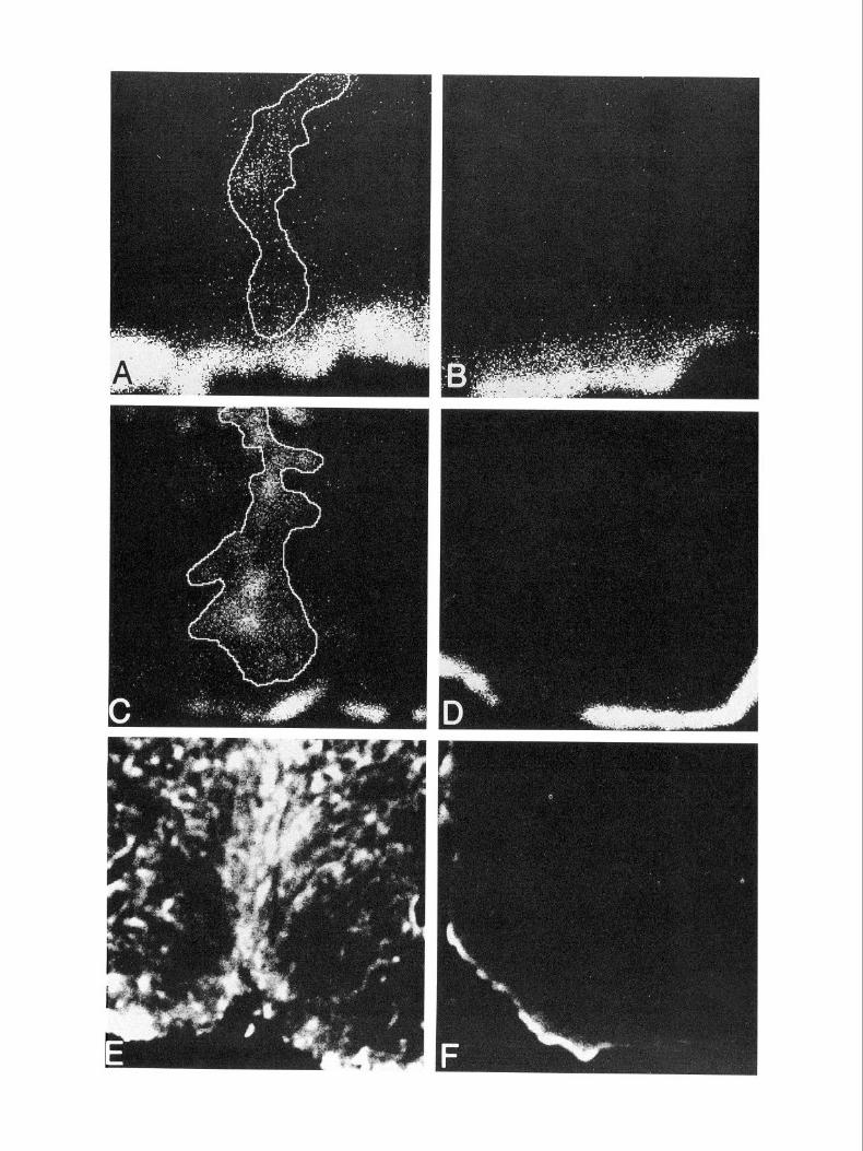
Fimre I. Implant of NC membrane into P3 mouse DUDS for 4 d increases the extent of GFAP-IR at the imulant site (0. In C. the cortical area o&pied by GFAP-IR astrocytes is encompassed with& the traced outline. In normal neonates (A and B, representing the ipsilateral and contralateral hemispheres), or in the contralateral hemisphere of NC implant pups (D), GFAP-IR was detected only in the corpus callosum (top of each frame) and glia limitans (bottom of each frame). Images in A-D were acquired by confocal laser scanning microscopy (described in detail in Materials and Methods) using a 2.5 x objective. E is a higher magnification of the traced area in C, acquired using a 40 x objective, to denote the morphology and reactive nature of the astrocytes.
The Journal of Neuroscience, February 1994, 14(2) 851
Extent Of GFAP-IR In Neonatal Mouse Brain In Response To Different Injuries
. . . . . .
. 1,000 - . .
. ; “2 . 3 8W- 0 . 25 z . : 600 2 .::: 4 ” I .:
. 400 - .-:a
. .
200 - : $1.
. ..SI.... I
0 . . . . . . .- . . . . . .
Scissors-stab NC-stab NC-implant Scissors- Scissors- stab + BSA stab +Y-IFN
@ & ,p
\* Pb 9 *
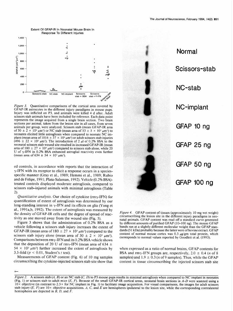
Figure 3. Quantitative comparisons of the cortical area covered by GFAP-IR astrocytes in the different injury paradigms in mouse pups. Injury was inflicted on P3, and animals were killed 4 d after. Adult scissors stab animals have been included for reference. Each data point represents the image acquired from a single brain section. Two brain sections per animal, taken from the lesion site in all cases, from seven animals per group, were analyzed. Scissors stab (mean GFAP-IR area of 30 + 2 x 10) pm2) or NC stab (mean area of 53 +- 5 x 10’ pm>) to neonates elicited little astrogliosis when compared to neonate NC im- plant (mean area of 10 16 ? 37 x 1 O3 pm2) or adult scissors stab injuries (496 & 22 x 10’ pmz). The introduction of 2 ~1 of 0.2% BSA to the neonatal scissors stab wound site resulted in increased GFAP-IR (mean area of 180 + 27 x 1 O3 pm>) compared to scissors stab alone, while 20 U of -/-IFN in 0.2% BSA enhanced astroglial reactivity even further (mean area of 634 + 54 x 10) pm2).
ed controls, in accordance with reports that the interaction of r-IFN with its receptor to elicit a response occurs in a species- specific manner (Gray et al., 1989; Hemmi et al., 1989; Rubio and de Felipe, 199 1; Plata-Salaman, 1992). Vehicle (0.2% BSA)- treated controls displayed moderate astrogliosis, compared to scissors stab-injured animals with minimal astrogliosis (Table a.
Quantitative analysis. Our choice of cytokine (rmy-IFN) for quantification of extent of astrogliosis was determined by our long-standing interest in Y-IFN and its effects on glia (Yong et al., 199 la,b, 1992). The extent of astrogliosis was measured by the density of GFAP-IR cells and the degree of spread of reac- tivity as one moved away from the wound site (Fig. 5).
Figure 3 shows that the administration of 0.2% BSA as a vehicle following a scissors stab injury increases the extent of GFAP-IR (mean area of 180 + 27 x 10) pm2) compared to the scissors stab injury alone (mean area of 30 ? 2 x lo3 hm2). Comparisons between rmy-IFN and its 0.2% BSA vehicle shows that the deposition of 20 U of rmy-IFN (mean area of 634 + 54 x lo3 pm*) further increased the extent of astrogliosis by 3.5fold (p < 0.01, Student’s t test).
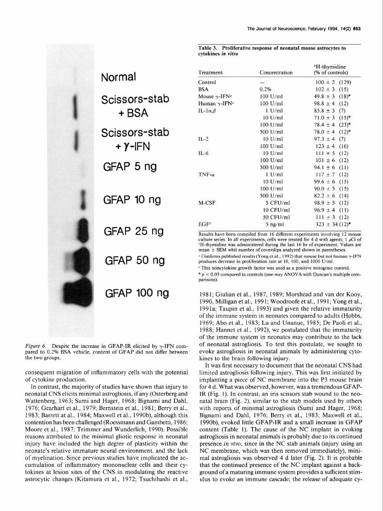
Measurements of GFAP content (Fig. 6) of 10 mg samples circumscribing the cytokine-injected scissors stab site show that
c
Normal
Scissors-stab
NC-stab
NC-implant
1 GFAP IO ng
GFAP 25 ng
GFAP 50 ng
GFAP 100 ng
Figure 4. GFAP content of tissues (approximately 10 mg wet weight) circumscribing the lesion site in the different in.jury paradigms in neo- natal animals. GFAP content was read off a standard curve generated by different amounts of purified GFAP (10-100 ng). The mouse GFAP bands ran at a slightly different molecular weight than the GFAP stan- dards (5 1 kDa) probably because the latter were ofbovine extract. GFAP content of normal mouse cortex was 0.3 pg/gm total protein, which corresponds to normal values reported by Goodlett et al. (1993).
when expressed as a ratio of normal brains, GFAP contents for BSA and rmy-IFN groups are, respectively, 2.0 f 0.4 (n of 8 samples) and 1.9 + 0.3 (n of 9 samples). Thus, while the GFAP content in tissue circumscribing the injected scissors stab site
Figure 2. A scissors stab (A, B) or an NC stab (C, D) to P3 mouse pups results in minimal astrogliosis when compared to NC implant in neonates (Fig. 1) or scissors stab to adult mice (E, r;?. Because of the small GFAP-IR cortical areas, neonatal brain sections in A-D were scanned using a 10 x objective (in contrast to 2.5 x for NC implant in Fig. 1) to facilitate image acquisition. For visual comparisons, the images for adult scissors stab injury (E, F) are 10 x objective acquisitions. A, C, and E are hemispheres ipsilateral to the lesion site, while the corresponding contralateral hemispheres are depicted in B, D, and F.
852 Balasingam et al. - Neonatal Astrogliosis and Cytokines
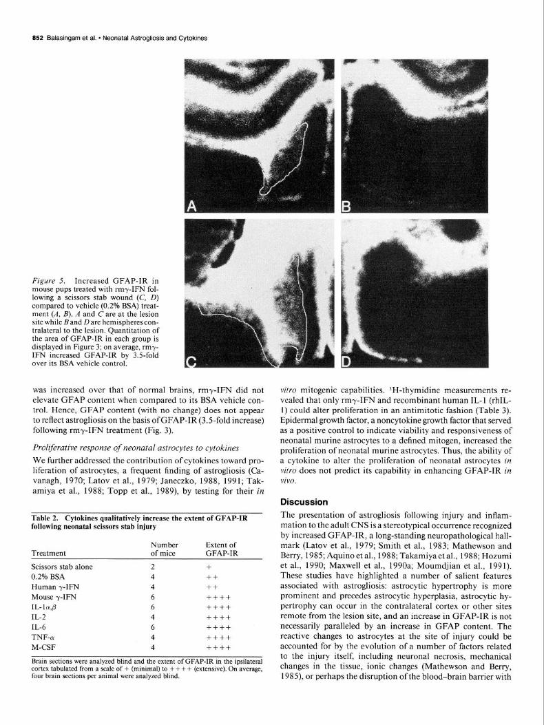
Figure 5. Increased GFAP-IR in mouse pups treated with rmy-IFN fol- lowing a scissors stab wound (C, D) compared to vehicle (0.2% BSA) treat- ment (A, B). A and C are at the lesion site while Band Dare hemispheres con- tralateral to the lesion. Quantitation of the area of GFAP-IR in each group is displayed in Figure 3; on average, rmy- IFN increased GFAP-IR by 35fold over its BSA vehicle control.
was increased over that of normal brains, rmr-IFN did not elevate GFAP content when compared to its BSA vehicle con- trol. Hence, GFAP content (with no change) does not appear to reflect astrogliosis on the basis ofGFAP-IR (3.5fold increase) following rmy-IFN treatment (Fig. 3).
Proliferative response of neonatal astrocytes to cytokines
We further addressed the contribution of cytokines toward pro- liferation of astrocytes, a frequent finding of astrogliosis (Ca- vanagh, 1970; Latov et al., 1979; Janeczko, 1988, 1991; Tak- amiya et al., 1988; Topp et al., 1989), by testing for their in
Table 2. Cytokines qualitatively increase the extent of GFAP-IR following neonatal scissors stab injury
Number Extent of Treatment of mice GFAP-IR
Scissors stab alone 2 + 0.2% BSA 4 ++ Human r-IFN 4 ++ Mouse r-IFN 6 ++++ IL- 1 cu,p 6 ++++ IL-2 4 ++++ IL-6 6 ++++ TNF-a 4 ++++ M-CSF 4 ++++
Brain sections were analyzed blind and the extent of GFAP-IR in the ipsilateral cortex tabulated from a scale of + (minimal) to + + + + (extensive). On average, four brain sections per animal were analyzed blind.
vitro mitogenic capabilities. 3H-thymidine measurements re- vealed that only rmy-IFN and recombinant human IL- 1 (rhIL- 1) could alter proliferation in an antimitotic fashion (Table 3). Epidermal growth factor, a noncytokine growth factor that served as a positive control to indicate viability and responsiveness of neonatal murine astrocytes to a defined mitogen, increased the proliferation of neonatal murine astrocytes. Thus, the ability of a cytokine to alter the proliferation of neonatal astrocytes in vitro does not predict its capability in enhancing GFAP-IR in vivo.
Discussion The presentation of astrogliosis following injury and inflam- mation to the adult CNS is a stereotypical occurrence recognized by increased GFAP-IR, a long-standing neuropathological hall- mark (Latov et al., 1979; Smith et al., 1983; Mathewson and Berry, 1985; Aquino et al., 1988; Takamiya et al., 1988; Hozumi et al., 1990; Maxwell et al., 1990a; Moumdjian et al., 199 1). These studies have highlighted a number of salient features associated with astrogliosis: astrocytic hypertrophy is more prominent and precedes astrocytic hyperplasia, astrocytic hy- pertrophy can occur in the contralateral cortex or other sites remote from the lesion site, and an increase in GFAP-IR is not necessarily paralleled by an increase in GFAP content. The reactive changes to astrocytes at the site of injury could be accounted for by the evolution of a number of factors related to the injury itself, including neuronal necrosis, mechanical changes in the tissue, ionic changes (Mathewson and Berry, 1985), or perhaps the disruption of the blood-brain barrier with
The Journal of Neuroscience, February 1994, T4(2) 853
Normal
Scissors-stab + BSA
Scissors-stab + Y-IFN
GFAP 5 ng
GFAP 10 ng M-CSF
GFAP 25 ng
GFAP 50 ng
GFAP 100 ng
Figure 6. Despite the increase in GFAP-IR elicited by y-IFN com- pared to 0.2% BSA vehicle, content of GFAP did not differ between the two groups.
consequent migration of inflammatory cells with the potential of cytokine production.
In contrast, the majority of studies have shown that injury to neonatal CNS elicits minimal astrogliosis, if any (Osterberg and Wattenberg, 1963; Sumi and Hager, 1968; Bignami and Dahl, 1976; Gearhart et al., 1979; Bernstein et al., 1981; Berry et al., 1983; Barrett at al., 1984; Maxwell et al., 1990b), although this contention has been challenged (Roessmann and Gambetti, 1986; Moore et al., 1987; Trimmer and Wunderlich, 1990). Possible reasons attributed to the minimal gliotic response in neonatal injury have included the high degree of plasticity within the neonate’s relative immature neural environment, and the lack of myelination. Since previous studies have implicated the ac- cumulation of inflammatory mononuclear cells and their cy- tokines at lesion sites of the CNS in modulating the reactive astrocytic changes (Kitamura et al., 1972; Tsuchihashi et al.,
Table 3. Proliferative response of neonatal mouse astrocytes to cytokines in vitro
Treatment
Control BSA Mouse 7-IFNQ Human y-IFW IL- 1 cu,p
IL-2
IL-6
TNF-CY
EGF”
Concentration
- 0.2% 100 U/ml 100 U/ml
1 U/ml 10 U/ml
100 U/ml 500 U/ml
10 U/ml 100 U/ml 10 U/ml
100 U/ml 500 U/ml
1 U/ml 10 U/ml
100 U/ml 500 U/ml
5 CFU/ml 10 CFU/ml 50 CFU/ml
5 @ml
‘H-thymidine (O/o of controls)
100 + 2 (129) 102 + 3 (15)
49.8 AZ 3 (18)* 98.8 t 4 (12) 85.8 + 5 (7) 71.0 + 3 (15)* 78.4 -t 4 (23)* 78.0 + 4 (12)* 97.3 +- 4 (7) 123 +- 4 (16) 111 z!z 5 (12) 101 ? 6 (12)
94.1 !z 6 (11) 117 f 7 (12)
99.6 zk 6 (15) 90.0 t 5 (15) 82.2 L 6 (14) 98.9 + 5 (12) 96.9 + 4 (11) 111 2 3 (12) 323 2 34 (12)*
Results have been compiled from 16 different experiments mvolving 12 mouse culture series. In all experiments, cells were treated for 4 d with agents; 1 pCi of ‘H-thymidine was admimstered during the last 16 hr of experiment. Values are mean f SEM whit number of coverslips analyzed shown in parentheses. “ Confirms published results (Yong et al., 1992) that mouse but not human -y-IFN produces decrease in prohferation rate at 10, 100, and 1000 U/ml.
” This noncytokine growth factor was used as a positive mitogenic control. * p < 0.05 compared to controls (one-way ANOVA with Duncan’s multiple com- parisons).
198 1; Giulian et al., 1987, 1989; Morshead and van der Kooy, 1990, Milligan et al., 199 1; Woodroofe et al., 199 1; Yong et al., 199 la; Taupin et al., 1993) and given the relative immaturity of the immune system in neonates compared to adults (Hobbs, 1969; Abo et al., 1983; Lu and Unanue, 1985; De Paoli et al., 1988; Hannet et al., 1992), we postulated that the immaturity of the immune system in neonates may contribute to the lack of neonatal astrogliosis. To test this postulate, we sought to evoke astrogliosis in neonatal animals by administering cyto- kines to the brain following injury.
It was first necessary to document that the neonatal CNS had limited astrogliosis following injury. This was first initiated by implanting a piece of NC membrane into the P3 mouse brain for 4 d. What was observed, however, was a tremendous GFAP- IR (Fig. 1). In contrast, an iris scissors stab wound to the neo- natal brain (Fig. 2), similar to the stab models used by others with reports of minimal astrogliosis (Sumi and Hager, 1968; Bignami and Dahl, 1976; Berry et al., 1983; Maxwell et al., 1990b), evoked little GFAP-IR and a small increase in GFAP content (Table 1). The cause of the NC implant in evoking astrogliosis in neonatal animals is probably due to its continued presence in viva, since in the NC stab animals (injury using an NC membrane, which was then removed immediately), mini- mal astrogliosis was observed 4 d later (Fig. 2). It is probable that the continued presence of the NC implant against a back- ground of a maturing immune system provides a sufficient stim- ulus to evoke an immune cascade; the release of adequate cy-
554 Balasingam et al. * Neonatal Astrogliosis and Cytokines
tokines at the lesion site may then contribute to the extensive astrogliosis observed (Fig. 3). Whatever the explanation, the conclusion is that reactive astrogliosis as measured by GFAP- IR and GFAP content (Fig. 4) can occur in the neonatal brain, and that it is dependent on the type of injury inflicted (Table 1).
In the NC implant model, astrogliosis was characterized by both an increased synthesis of GFAP intermediate filaments and hypertrophy of the astrocytic cytoplasmic processes. The functional role for the increase in this intermediate filament is not known. Smith et al. (1986) have reported that reactive as- trocytes could migrate on to an NC implant within 24-48 hr postimplantation in neonatal animals and that these astrocytes formed a terrain that facilitated axonal extension and regener- ation. These neonatal astrocytes also appeared to lack the ex- pression of putative growth-inhibitory molecules such as chon- droitin-6-sulfate proteoglycan and cytotactin that were present in adult astrocytes (McKeon et al., 199 1). These findings suggest that neonatal reactive astrocytes may have potential regenera- tive properties.
To test the hypothesis implicating cytokines as contributors toward astrogliosis, we chose to utilize the neonatal stab model with its inherent minimal gliotic response. A single microinjec- tion of cytokines (rmy-IFN, rhIL- 1, rhIL-2, rhIL-6, rhTNF-oc, and rhM-CSF, all ofwhich are described to be effective in mouse cells by the manufacturer) into the cerebral cortex ofthe neonatal mouse produced an astrogliotic response (Table 2) similar to that seen in adult stab wound models by GFAP immunoreac- tivity (Fig. 2) (Mathewson and Berry, 1985; Moumdjian et al., 199 1; Yong et al., 199 la). The specificity of the cytokine effect was demonstrated by the inability of rhy-IFN to evoke an as- trogliotic response beyond that of vehicle-treated controls (Ta- ble 2) an observation that is in accordance with reports indi- cating a species-specific interaction between -y-IFN and its receptor (Gray et al., 1989; Hemmi et al., 1989; Rubio and de Felipe, 199 1; Plata-Salaman, 1992). The finding that a single administration of cytokines can induce significant astrogliosis in the neonatal stab model with its inherent minimal astrogliosis is consistent with the postulate that the lack of astrogliosis fol- lowing neonatal injury is related to an immature immune sys- tem; this immature immune system would then be reconstituted by cytokine administration,
The quantification ofthe extent ofastrogliosis evoked by rmy- IFN (634 t 54 x IO3 wrn’) revealed a 3.5fold increase over that of vehicle-treated controls (180 f 27 x 10) pm’) as deter- mined by GFAP-IR (Figs. 3, 5). However, the analyses ofGFAP content from tissue circumscribing the injection sites for rmy- IFN and vehicle-treated controls did not differ but it was higher than unoperated normal controls (Fig. 6). Thus, GFAP content did not reflect the extent of astrogliosis on the basis of GFAP- IR following rmr-IFN treatment. A similar type of occurrence has been documented in animals with experimental autoim- mune encephalomyelitis (EAE), where GFAP content of the spinal cord did not differ from controls at 13-18 d postinocu- lation (dpi), a period when intense GFAP-IR was observed in the EAE groups (Smith et al., 1983; Goldmuntz et al., 1986); correlation of GFAP-IR with GFAP content was observed at later periods (35-65 dpi) (Aquino et al., 1988). The most likely interpretation for the noncorrespondence between GFAP-IR and GFAP content may be that as astrocytes swell and GFAP filament dissociate there is an increased availability of antigenic epitopes to antibodies for GFAP (Aquino et al., 1988; Eng et al., 1989). This phenomenon seen in EAE for the initial increase
in GFAP-IR before eventual increase in GFAP content appears similar to that observed in our neonatal rmy-IFN scissors stab model.
Why do all cytokines tested induce astrogliosis? While it is possible that all these cytokines have direct effects on astrocytes, an indirect phenomenon through a possible common pathway is also likely. This route may conceivably be by the recruitment of inflammatory mononuclear cells, including a final effector cell and its cytokine(s), to the lesion site. This possibility is supported by the report of Brosnan et al. (1989) who described the occurrence of astrogliosis and increased adherence of in- flammatory cells to the vasculature after intraocular injection of r-IFN, TNF-o(, and IL- 1. In addition, Watts et al. (1989) have demonstrated the disruption of the blood-brain barrier and the recruitment of inflammatory cells to the intracerebral injection site of IL-2. Furthermore, Simmons and Willenbourg (1990) have described the occurrence of a widespread inflam- matory response to a single microinjection of r-IFN or TNF-cu in the lumbosacral cord. Finally, Sethna and Lampson (199 1) have observed a that a single intracerebral injection of r-IFN resulted in the recruitment of many types of inflammatory cells to the brain. Our laboratory is currently testing the direct and indirect role of cytokines in mediating gliosis in vivo. We are examining the role of -y-IFN as a final common mediator, given the identification of a specific receptor for mouse -r-IFN on neonatal mouse astrocytes (Rubio and de Felipe, 199 l), and given the potent effects of r-IFN on astrocytes in mixed or purified cultures in vitro (Yong et al., 199 la,b, 1992).
An important question now arises: which cells are responsible for the production of cytokines? Conceivably, infiltrating mono- nuclear phagocytes (macrophages) and other cells ofthe immune system (e.g., T-lymphocytes and NK cells) are potentially in- volved. Cells intrinsic to the CNS could also be potential sources of cytokines. In this regard, microglia, astrocytes, and even neu- rons have been suggested to synthesize cytokines under selective conditions (Giulian et al., 1987; Wesselingh et al., 1990; Logan et al., 1992; Tchelingerian et al., 1993). The nature of the in vivo cellular elements contributing to cytokine(s) production fol- lowing injury remains to be elucidated.
Our investigations demonstrate that the ability of a cytokine to alter the proliferation of neonatal astrocytes in vitro does not predict its capability in enhancing GFAP-IR in vivo. While all cytokines tested in vivo increased GFAP-IR (Table 2) our in vitro studies implicate an antimitotic effect by rmy-IFN (Yong et al., 1992) and IL- 1, without any significant effects by the other cytokines (Table 3). It is also worth noting that while others have found IL- 1, IL-6, and TNF-ol to be mitogenic for neonatal rat and calf bovine astrocytes (Giulian and Lachman, 1985; Nieto-Sampedro and Berman, 1987; Selmaj et al., 1990) these cytokines were not mitogenic for neonatal mouse astrocytes. This apparent discrepancy may be due to species differences, since we have demonstrated that while rmy-IFN was inhibitory for proliferation of neonatal or adult mouse astrocytes, rhr-IFN was a mitogen for fetal and adult human astrocytes (Yong et al., 1992).
In conclusion, astrogliosis can occur in the neonatal brain if a sufficient stimulus (NC implant) is present. For the neonatal scissors stab wound model with its inherent minimal astrog- liosis, a single administration of cytokines induces extensive astrogliosis. These results implicate immunoregulatory cyto- kines as important contributing factors to the production of astrogliosis following an injury to the CNS.
The Journal of Neuroscience, February 1994, 14(2) 855
References
Abo T, Miller CA, Gartland GL, Balch CM (1983) Differentiation stages of human natural killer cells in lymphoid tissue from fetal to adult life. J Exp Med 157:273-284.
Aquino DA, Chiu FC, Brosnan CF, Norton WT (1988) Glial fibrillary acidic protein increases in the spinal cord of Lewis rats with acute experimental autoimmune encephalomyelitis. J Neurochem 5 1: 108% 1096.
Barna BP, Estes ML, Jacobs BS, Hudson S, Ransohoff RM (1990) Human astrocytes proliferate in response to tumor necrosis factor alpha. J Neuroimmunol 30:239-243.
Barrett CP, Donati EJ, Guth L (1984) Differences between adult and neonatal rats in their astroglial response to spinal injury. Exp Neural 84:374-385.
Bernstein DR, Bechard DE, Stelzner DJ (198 1) Neurite growth main- tained near the lesion site long after spinal cord transection in the newborn rat. Neurosci Lett 26;55-60.
Berry M, Maxwell WL, Logan A, Mathewson A, McConnell P, Ashburst DE, Thomas GH (1983) Deposition of scar tissue in the central nervous system. Acta Neurochir [Suppl] 32:3 l-53.
Bignami A, Dahl D (1974) Astrocyte-specific protein and neuroglial differentiation. An immunofluorescence study with antibodies to the glial fibrillary acidic protein. J Comp Neurol 153:27-38.
Bianami A. Dahl D (1976) The astronlial resnonse to stabbing: im- munofluorescence studies with antibodies to astrocyte-specific protein (GFA) in mammalian and submammalian vertebrates. Neuropathol Appl Neurobiol 2:99-l 10.
Brosnan CF, Litwak MS, Schroeder CE, Selmaj K, Raine CS, Arezzo JC (1989) Preliminary studies of cytokine-induced functional effects on the visual pathways in the rabbit. J Neuroimmunol 25:227-239.
Cavanagh JB (1970) The proliferation of astrocytes around a needle wound in the rat brain. J Anat 106:471-487.
De Paoli P, Battistin S, Santini GF (1988) Age-related changes in human lymphocyte subsets: progressive reduction ofthe CD4 CD45R (suppressor inducer) population. Clin Immunol Immunopathol 48: 290-296.
Eng LF (1985) Glial fibrillary acidic protein (GFAP): the major protein ofglial intermediate filaments in differentiated astrocytes. J Neuroim- munol 8:203-214.
Eng LF, D’Amelio FE, Smith ME (1989) Dissociation of GFAP in- termediate filaments in EAE: observations in the lumbar spinal cord. Glia 2:308-3 17.
Gearhart J, Oster-Granite ML, Guth L (1979) Histological changes after transection of the spinal cord of fetal and neonatal mice. Exp Neurol 66: 1-l 5.
Giulian D (I 987) Ameboid microglia as effecters of inflammation in the central nervous system. J Neurosci Res 18: 155-l 7 I.
Giulian D, Lachman LB (1985) Interleukin- 1 stimulation of astroglia proliferation after brain injury. Science 228:497-499.
Giulian D, Woodward J, Young DG, Krebs JF, Lachman LB (1988) Interleukin- 1 injected into mammalian brain stimulates astrogliosis and neovascularization. J Neurosci 8:2485-2490.
Giulian D, Chen J, Ingeman JE, George JK, Noponen M (1989) The role of mononuclear phagocytes in wound healing after traumatic injury to the adult mammalian brain. J Neurosci 9:44 16-4429.
Goldmuntz EA, Brosnan CF, Chiu FC, Norton WT (1986) Astrocytic reactivity and intermediate filament metabolism in experimental au- toimmune encephalomyelitis: the effect of suppression with prazosin. Brain Res 397: 16-26.
Goodlett CR, Leo JT, O’Callaghan JP, Mahoney JC, West JR (1993) Transient cortical astrogliosis induced by alcohol exposure during the neonatal brain growth spurt in rats. Dev Brain Res 72:85-97.
Gray PW, Leong S, Fennie EH, Farrar MA, Pingel JT, Fernandez-Luna J, Schreiber RD (1989) Cloning and expression of the cDNA for the murine interferon-y receptor. Proc Nat1 Acad Sci USA 86:8497- 8501.
Hannet I, Erkeller-Yuksel F, Lydyard P, Deneys V, DeBruyere M (1992) Developmental and maturational changes in human blood lympho- cyte subpopulations. Immunol Today 13:2 15-2 18.
Hemmi S, Peghini P, Metzler M, Merlin G, Dembic Z, Aguet M (1989) Cloning of murine interferon-y receptor CDNA: expression in human cells mediates high affinity binding but is not sufficient to confer sensitivity to murine interferon-y. Proc Nat1 Sci Acad USA 86:9901- 9905.
Hobbs JR (1969) Primary immune paresis. In: Immunology and de- velopment (Adinolfi M, ed), pp 117-l 18. London: Heinemann.
Hozumi I, Chiu FC, Norton WT (1990) Biochemical and immuno- cytochemical changes in glial fibrillary acidic protein after stab wounds. Brain Res 524164-7 1.
Kitamura T, Hattori H, Fujita S (1972) Autoradiographic studies of histogenesis of brain macrophages in the mouse. J Neuropathol Exp Neurol 31:502-5 18.
Janeczko K (1988) The proliferative response of astrocytes to injury in neonatal rat brain. A combined immunocytochemical and auto- radiographic study. Brain Res 456:280-285.
Janeczko K (199 1) The proliferative response of S- 100 protein positive glial cells to injury in the neonatal rat brain. Brain Res 564:86-90.
Latov N, Nilaver G, Zimmerman EA, Johnson WG, Silverman AJ, Defendini R, Cote L (1979) Fibrillary astrocytes proliferate in re- sponse to brain injury. Dev Biol 72:381-384.
Liuzzi FJ, Lasek RJ (1987) Astrocytes block axonal regeneration in mammals by activating the physiological stop pathway. Science 237: 642-645.
Logan A, Frautschy SA, Gonzalez A-M, Spom MB, Baird A (1992) Enhanced expression of transforming growth factor p 1 in the rat brain after a localized cerebral injury. Bra% Res 587:216-225.
Lu CY, Unanue ER (1985) MAcrophage ontogeny: implications for host defense, T-lymphocyte differentiation, and the acquisition of self tolerance. Clin Immunol Allergy 5:253-269.
Mathewson AJ, Berry M (1985) Observations on the astrocyte re- sponse to a cerebral stab wound in adult rats. Brain Res 327:6 l-69.
Maxwell WL, Follows R, Ashhurst DE, Berry M (1990a) The response of the cerebral hemisphere of the rat to injury. I. The mature rat. Philos Trans R Sot Lond [Biol] 328:479-500.
Maxwell WL, Follows R, Ashhurst DE, Berry M (1990b) The response of the cerebral hemisphere of the rat to injury. II. The neonatal rat. Philos Trans R Sot Lond lBiol1 328:50 l-5 13.
McKeon RJ, Schreiber RC, kudge JS, Silver J (1991) Reduction of neurite outgrowth in a model of glial scarring following CNS injury is correlated with the expression of inhibitory molecules on reactive astrocytes. J Neurosci 11:3398-3411.
Milligan CE, Levitt P, Cunningham TJ (199 1) Brain macrophages and microglia respond differently to lesions of the developing and adult visual system. J Comp Neurol 3 14: 136-l 46.
Moore IE, Bountempo JM, Weller RO (1987) Response of fetal and neonatal rat brain to injury. Neuropathol Appl Neurobiol 13:219- 228.
Morshead CM, van der Kooy D (1990) Separate blood and brain origins of proliferating cells during gliosis in adult brains. Brain Res 5351237-244.
Moumdjian R, Ante1 JP, Yong VW (1991) Origin of contralateral reactive gliosis in surgically injured rat cerebral cortex. Brain Res 547: 223-228.
Nieto-Sampedro M, Berman MA (1987) Interleukin- 1 like activity in rat brain: sources, targets, and effect of injury. J Neurosci Res 17: 214-219.
Norton WT, Aquino DA, Hozumi I, Chiu FC, Brosnan CF (1992) Quantitative aspects of reactive gliosis: a review. Neurochem Res 17: 877-885.
Osterberg KA, Wattenberg LW (1963) The age of dependency of en- zymes in reactive glia. Proc Sot Exp Biol Med 113: 145-147.
Plata-Salaman CR (1992) Interferon and central regulation of feeding. Am J Physiol 263:R1222-R1227.
Reier PJ, Stensaas LJ, Guth L (1983) The astrocytic scar as an im- pediment to regeneration in the central nervous system. In: Funda- mentals of spinal cord reconstruction (Kao CC, Bunae RP. Reier PJ. eds), p 163. New York: Raven. .
-
Roessmann U, Gambetti P (1986) Pathological reaction of astrocytes in perinatal brain injury. Acta Neuropathol (Berl) 70:302-307.
Rubio N, de Felipe C (1991) Demonstration of the presence of a specific interferon-r receptor on murine astrocyte cell surface. J Neu- roimmunol 35:ll l-l 17.
Rudge JS, Smith GM, Silver J (1989) An in vitro model of wound healing in the CNS: analysis ofcell reaction and interaction at different ages. Exp Neurol 103: l-l 6.
Selmaj KW, Farooq M, Norton WT, Raine CS, Brosnan CF (1990) Proliferation of astrocytes in vitro in response to cytokines: a primary role of tumor necrosis factor. J Immunol 144: 129-l 35.
Sethna MP, Lampson LA (199 1) Immune modulation within the brain:
656 Balasingam et al. * Neonatal Astrogliosis and Cytokines
recruitment of inflammatory cells and increased major histocompat- ibility antigen expression following intracerebral injection of inter- feron-r. J Neuroimmunol 34: 12 1-132.
Simmons RD, Willenborg DO (1990) Direct injection of cytokines into the spinal cord causes autoimmune encephalomyelitis-like in- flammation. J Neurol Sci 100:37-42.
Smith GM, Miller RH, Silver J (1986) Changing role of forebrain astrocytes during development, regenerative failure, and induced re- generation upon transplantation. J Comp Neurol 25 1:23-43.
Smith ME, Eng L (1987) Glial fibrillary acidic protein in chronic relapsing experimental allergic encephalomyelitis in SJL/J mice. J Neurosci Res 18:203-208.
Smith ME, Somera FP, Eng L (1983) Immunocytochemical staining for glial tibrillary acidic protein and the metabolism of cytoskeletal proteins in experimental allergic encephalomyelitis. Brain Res 264: 241-253.
Sumi SM, Hager H (1968) Electron microscopic study of the reaction of the newborn rat brain to injury. Acta Neuropathol (Berl) 10:324- 335.
Takamiya Y, Kohsaka S, Toya S, Otani M, Tsukada Y (1988) Im- munohistochemical studies on the proliferation of reactive astrocytes and the expression of cytoskeletal proteins following brain injury in rats. Dev Brain Res 38:20 l-2 10.
Taupin V, Toulmond S, Serrano A, Benavides J, Zavala F (1993) Increase in IL-6, IL- 1 and TNF levels in rat brain following traumatic lesion. J Neuroimmunol 42: 177-l 86.
Tchelingerian J-L, Quinonero J, Booss J, Jacque C (1993) Localization of TNFol and IL-la immunoreactivities in striatal neurons after sur- gical injury to the hippocampus. Neuron lo:21 3-224.
Topp KS, Faddis BT, Vijayan VK (1989) Trauma-induced prolifer- ation of astrocytes in the brains of young and aged rats. Glia 2:201- 21 1.
Trimmer PA, Wunderlich RE (1990) Changes in astroglial scar for- mation in rat optic nerve as a function of development. J Comp Neurol 296~359-378.
Tsuchihashi Y, Kitamura T, Fujita S (1981) Immunofluorescence studies of the monocytes in the injured rat brain. Acta Neuropathol (Berl) 53:213-219.
Watts RG, Wright JL, Atkinson LL, Merchant RE (1989) Histopatho- logical and blood-brain barrier changes in rats induced by an intra- cerebral injection ofhuman recombinant interleukin-2. Neurosurgery 25:202-208.
Wesselingh SL, Cough NM, Finlay-Jones JJ, McDonald PJ (1990) Detection of cytokine MRNA in astrocyte cultures using the poly- merase chain reaction. Lymphokine Res 9: 177-l 85.
Woodroofe MN, Sarna GS, Wadhwa M, Hayes GM, Loughlin AJ, Tin- ker A, Cuzner ML (199 1) Detection of interleukin- 1 and interleukin-6 in adult rat brain, following mechanical injury, by in viva microdi- alysis: evidence of a role for microglia in cytokine production. J Neu- roimmunol 33:227-236.
Yong VW (1992) Proliferation of human and mouse astrocytes in vitro: signalling through the protein kinase C pathway. J Neurol Sci 11 l:92-103.
Yong VW, Moumdjian R, Yong FP, Ruijs TCG, Freedman MS, Cash- man N, Ante1 JP (199 la) Gamma-interferon promotes proliferation of adult human astrocytes in vitro and reactive aliosis in the adult mouse brain in vivo. Proc Nat1 Acad Sci USA 88f70 16-7020.
Yone, VW, Yone FP. Ruiis TCG. Ante1 JP. Kim SU (199 1 b) Exnres- - -__ I , I
sion and modulation ofHLA-DR on cultured human adult astrocytes. J Neuropathol Exp Neurol 50: 16-28.
Yong VW, Tejada-Berges T, Goodyer CC, Ante1 JP, Yong FP (1992) Differential proliferative response of human and mouse astrocytes to gamma-interferon. Glia 6:269-280.