Embed Size (px)
Citation preview

pubs.acs.org/ICPublished on Web 07/12/2010r 2010 American Chemical Society
Inorg. Chem. 2010, 49, 6283–6292 6283
DOI: 10.1021/ic902319d
Reactions of HNO with Heme Proteins: New Routes to HNO-Heme Complexes and
Insight into Physiological Effects
Murugaeson R. Kumar,† Jon M. Fukuto,‡ Katrina M. Miranda,*,§ and Patrick J. Farmer*,†
†Department of Chemistry and Biochemistry, Baylor University, Waco, Texas 76706, ‡Department ofChemistry, Sonoma State University, Rohnert Park, California 94928, and §Department of Chemistry andBiochemistry, University of Arizona, Tucson, Arizona 85721
Received November 23, 2009
The formation and interconversion of nitrogen oxides has been of interest in numerous contexts for decades. Earlystudies focused on gas-phase reactions, particularly with regard to industrial and atmospheric environments, and onnitrogen fixation. Additionally, investigation of the coordination chemistry of nitric oxide (NO) with hemoglobin datesback nearly a century. With the discovery in the early 1980s that NO is biosynthesized as a molecular signaling agent,the literature has been focused on the biological effects of nitrogen oxides, but the original concerns remain relevant.For instance, hemoglobin has long been known to react with nitrite, but this reductase activity has recently beenconsidered to be important to produce NO under hypoxic conditions. The association of nitrosyl hydride (HNO; alsocommonly referred to as nitroxyl) with heme proteins can also produce NO by reductive nitrosylation. Furthermore,HNO is considered to be an intermediate in bacterial denitrification, but conclusive identification has been elusive. Theauthors of this article have approached the bioinorganic chemistry of HNO from different perspectives, which haveconverged because heme proteins are important biological targets of HNO.
HNO-Heme Adducts
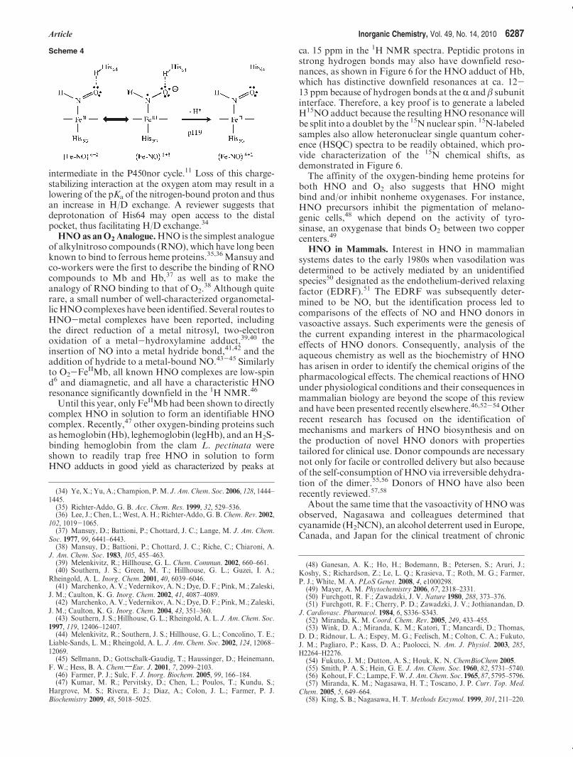
Interest in HNO in the Farmer laboratory originated fromefforts to model reductive heme-based catalysis involved inthe global nitrogen cycle (Scheme 1).1 The six-electronreduction of nitrite to ammonia can be driven by a singleenzyme, as in assimilatory nitrite reductases, or can occurstepwise via dissimilatory enzymes,which take nitrite to nitricoxide (NO), N2O, and N2. Heme nitroxyl intermediates havebeen postulated in these NOx reductions, as indicated by thedotted line in Scheme 1.A commonmechanistic question in enzymatic reduction of
nitrogen oxides is whether a nitroxyl intermediate is gener-ated by sequential electron transfer or by a two-electronprocess such as hydride transfer (Scheme 2).2,3 The conten-tion is that a ferrous nitrosyl intermediate (FeIINO) if tran-siently formed would be stable and difficult to reduce, thusacting as a catalytic dead end. For example, the single-electron reduction of NO-FeIIMb occurs at ca. -650 mV
vs NHE,4 which is at the edge of the biological reductionrange.5
An authentic nitroxyl intermediate has been observedduring turnover of the fungal NO reductase cytochromeP450nor.6 In its catalytic cycle, a ferric nitrosyl complex ofP450nor is reduced by NADH to generate an intermediate(λmax at 444 nm) that subsequently reacts with NO to giveferric heme and N2O, which is the nitrogen product ofthe HNO self-consumption pathway.7,8 Ulrich et al. showedthat the putative nitroxyl intermediate may be formedfrom the reaction of NaBH4 with the ferric nitrosyl adduct,9
suggesting that nature does indeed bypass the ther-modynamically stable FeIINO in forming a reactive nitroxylintermediate. The electronic structure, basicity, and possiblesites of protonation during turnover of this formal {FeIINO}8
species have been investigated recently.10,11 Correspondingto the free species,12-14 Lehnert et al. found an energetic
*To whom correspondence should be addressed. E-mail: [email protected] (K.M.M.), [email protected] (P.J.F.).
(1) Blair, E.; Sulc, F.; Farmer, P. J. Biomimetic NOxReductions byHemeModels and Proteins. In N4-Macrocyclic Metal Complexes; Zagal, J. H.,Bedioui, F., Dodelet, J.-P., Eds.; Springer: New York, 2006; pp 149-190.
(2) Einsle, O.; Messerschmidt, A.; Huber, R.; Kroneck, P. M. H.; Neese,F. J. Am. Chem. Soc. 2002, 124, 11737–11745.
(3) Silaghi-Dumitrescu, R. Eur. J. Inorg. Chem. 2003, 1048–1052.(4) Bayachou, M.; Lin, R.; Cho, W.; Farmer, P. J. J. Am. Chem. Soc.
1998, 120, 9888–9893.(5) Guo, M.; Sulc, F.; Ribbe, M. W.; Immoos, C. E.; Farmer, P. J.;
Burgess, B. K. J. Am. Chem. Soc. 2002, 124, 12100–12101.
(6) Shiro, Y.; Fujii,M.; Iizuka, T.; Adachi, S.; Tsukamoto,K.; Nakahara,K.; Shoun, H. J. Biol. Chem. 1995, 270, 1617–1623.
(7) Shiro, Y.; Fujii, M.; Isogai, Y.; Adachi, S.; Iizuka, T.; Obayashi, E.;Makino, R.; Nakahara, K.; Shoun, H. Biochemistry 1995, 34, 9052–9058.
(8) Nakahara, K.; Tanimoto, T.; Hatano, K.; Usuda, K.; Shoun, H.J. Biol. Chem. 1993, 268, 8350–8355.
(9) Daiber, A.; Nauser, T.; Takaya, N.; Kudo, T.; Weber, P.; Hultschig,C.; Shoun, H.; Ullrich, V. J. Inorg. Biochem. 2002, 88, 343–352.
(10) Silaghi-Dumitrescu, R. Eur. J. Inorg. Chem. 2003, 6, 1048–1052.(11) Lehnert, N.; Praneeth, V. K. K.; Paulat, F. J. Comput. Chem. 2006,
27, 1338–1351.(12) Bruna, P. J.; Marian, C. M. Chem. Phys. Lett. 1979, 67, 109–114.(13) Guadagnini, R.; Schatz, G. C. J. Chem. Phys. 1995, 102, 774–783.(14) Luna, A.;Merchan,M.; Roos, B. O.Chem. Phys. 1995, 196, 437–445.
6284 Inorganic Chemistry, Vol. 49, No. 14, 2010 Kumar et al.
preference for the Fe-N(H)O tautomer over that of Fe-NOH in the monoprotonated form.11 The axial thiolateligand to the P450 heme promoted a second protonation,yielding a catalytically active intermediate described asFeIVNHOH-.11
In a recent series of papers, we have shown that an HNOadduct of ferrousmyoglobin (HNO-FeIIMb) can be formedby the reduction of NO-FeIIMb or by the trapping of freeHNOby deoxymyoglobin (FeIIMb).15,16 TheHNO-FeIIMbadduct is relatively stable, and as a unique model for hemenitroxyl intermediates, it has been structurally characterizedby 1H NMR,17 resonance Raman, and X-ray absorptionspectroscopy/X-ray absorption near-edge structure analy-sis.18 The1H NMR spectrum is particularly diagnostic, withcharacteristic signals for HNO at 14.9 ppm and for themethyl group of Val68 at -2.5 ppm.In the Farmer laboratory,19 NO-FeIIMb is generally
synthesized by the common technique of mixing metmyoglo-bin (FeIIIMb), nitrite, and dithionite (eq 1).20 This reactionalso has physiological relevance for the biosynthesis of NO.21
Ulrich’s work9 prompted us to change the reductant fromNaNO2 to NaBH4, in the hope of obtaining the HNO-FeII
species. Indeed, the reaction of FeIIIMb with nitrite andborohydride results in a product mixture with spectra thatare consistent with the production of HNO-FeIIMb in>96% yield (see Figures S2 and S3 in the SupportingInformation). Similarly, the sequential addition of nitriteand borohydride to hemoglobin yields HNO-FeIIHb solu-
tions of ca. 65% yield (see Figures S4-S6 in the SupportingInformation).
FeII=IIIMbþNaNO2 þNa2S2O4 f NO-FeIIMb ð1Þ
FeII=IIIMbþNaNO2 þNaBH4 f HNO-FeIIMb ð2Þ
In contrast, independently prepared and purifiedNO-FeIIMb does not undergo reaction with borohydride(see Figure S8 in the Supporting Information and eqs 1 and 3).This lends credence to the supposition that NO-FeII
is avoided during catalytic turnover in P450nor. This alsoimplies that free HNO, or an HNO-releasing species, isgenerated upon reduction of nitrite by borohydride inaqueous solution.A complication is that the 1H NMR spectrum of
HNO-FeIIMb prepared by the nitrite/borohydride methodhas a doubling of the proton resonances within the hemepocket (Figure 1), suggesting a diastereomeric mixture.Neither photolysis nor heating to 60 �C affects this doubling.However, if an HNO-FeIIMb sample prepared by thismethod is air-oxidized to FeIIIMb and the HNO adduct isthen regenerated using the trapping method, only singleresonances are observed in the two characteristic regions. Asimilar doubling of resonances was previously perceived forreconstituted metmyoglobin adducts and was attributed toheme orientational disorder;22-24 the vinylic side groups ofthe heme are asymmetrical, and thus flipping of the heme
Scheme 1. Species Involved in the Nitrogen Cycle
Scheme 2. Reduction of Coordinated NOþ to NO and NO-
Figure 1. 1H NMR spectra of the HNO adduct of myoglobin (0.5-1 mM) in a 50 mMphosphate buffer at pH 7.0 either (A) prepared by theNaNO2/NaBH4 method or (B) generated by oxidation of part A, reduc-tion to FeIIMb, and subsequent reaction with the HNO donor Piloty’sacid at pH 10. H15NO-FeIIMb was prepared either by (C) DTDPreduction of NO-FeIIMb or (D) reduction with Na15NO2/NaBH4. (E)HNO adduct of FeII (2,4-dimethyldeuteroporphyrin) reconstituted myo-globin (0.1 mM) in a 50 mM phosphate buffer at pH 7.
(15) Lin, R.; Farmer, P. J. J. Am. Chem. Soc. 2000, 122, 2393–2394.(16) Sulc, F.; Immoos, C. E.; Pervitsky, D.; Farmer, P. J. J. Am. Chem.
Soc. 2004, 126, 1096–1101.(17) Sulc, F.; Fleischer, E.; Farmer, P. J.;Ma,D. J.; LaMar, G. N. J. Biol.
Inorg. Chem. 2003, 8, 348–352.(18) Immoos, C. E.; Sulc, F.; Farmer, P. J.; Czarnecki, K.; Bocian, D. F.;
Levina, A.; Aitken, J. B.; Armstrong, R. S.; Lay, P. A. J. Am. Chem. Soc.2005, 127, 814–815.
(19) Pervitsky, D.; Immoos, C.; van der Veer, W.; Farmer, P. J. J. Am.Chem. Soc. 2007, 129, 9590–9591.
(20) Arnold, E. V.; Bohle, D. S. InMethods in Enzymology, Packer, L., Ed.;Academic Press: San Diego, 1996; Vol. 268, pp 41-55.
(21) Gladwin, M. T.; Kim-Shapiro, D. B. Blood 2008, 112, 2636–2647.
(22) Tran, A. T.; Kalish, H.; Balch, A. L.; La Mar, G. N. J. Biol. Inorg.Chem. 2000, 5, 624–633.
(23) La Mar, G. N.; Hauksson, J. B.; Dugad, L. B.; Liddell, P. A.;Venkataramana, N.; Smith, K. M. J. Am. Chem. Soc. 1991, 1544–1550.
(24) Jue, T.; Krishnamoorthi, R.; LaMar, G. N. J. Am. Chem. Soc. 1983,105, 5701-5703.
Article Inorganic Chemistry, Vol. 49, No. 14, 2010 6285
within a protein pocket will produce two different diastereo-mers. To examine this hypothesis, a sample of apomyoglobinwas reconstituted with the iron complex of symmetrical 2,4-dimethyldeuteroporphyrin (Scheme 3), and its HNO adductwas prepared by the nitrite/borohydride method. The result-ing 1H NMR spectrum of the low-concentration productsolution yields single HNO and valine resonances, consistentwith the heme orientational hypothesis. Further 2D NMRstudies are underway to better characterize the structuraldifferences of these diastereomeric forms.To better understand how HNO adducts are formed in
the nitrite/borohydride procedure, the three reactants werecombined sequentially in several ways (see part S1a in theSupporting Information). HNO-FeIIMb was formed wheneither met- or deoxymyoglobin was reacted with nitrite andborohydride in any addition order. One plausible reaction ofhydride with nitrite is the generation of free HNO, whichmight then be trapped by FeIIMb (eqs 4 and 5).
HONOþH- f HNOþOH- ð4Þ
FeIIMbþHNO f HNO-FeIIMb ð5ÞThe possible generation of free HNO was examined with
the known scavengers nickel(II) tetracyanate (Ni(CN)42-)25
and iron(II) N-methyl-D-glucaminedithiocarbamate (FeII-MGD)26 (eqs 6 and 7). The characteristic 498 nm absorbanceband of Ni(CN)3NO3- was observed when Ni(CN)4
2- wasreacted with nitrite/borohydride (Figure 2) but was notapparent in the analogous reaction of Ni(CN)4
2- withnitrite/dithionite. Likewise, the reaction of FeIIMGD withnitrite/dithionite yielded a product solution whose electronparamagnetic resonance (EPR) spectrum at 77 K matchedthat of the reported NO-FeIIMGD complex (Figure 2).Substitution of dithionite by borohydride yielded a productsolution with a similar signal but at less than 5% of theintensity, suggesting that the majority of the product was thediamagnetic NO--FeIIMGD species. Both reactions clearlysuggest that HNO or an HNO-releasing species is formed inthe presence of nitrite/borohydride.
NiðCNÞ42- þNaNO2=NaBH4 f NO--NiðCNÞ33- ð6Þ
FeIIMGDþNaNO2 þNaBH4 f NO--FeIIMGD ð7ÞThe versatility of using borohydride to generate HNO
adducts inaqueous solutionswas investigatedwithnitroprusside
([Fe(CN)5NO]2-), which was recently shown to undergo twosequential reductions at high pH to generate anHNO adduct([Fe(CN)5HNO]3-) with characteristic absorbance and1H NMR spectra.27 The reaction of borohydride with[Fe(CN)5NO]2- at pH 9 (eq 8) yields a product solution witha 450 nm absorbance band and a 20.2 ppm resonance in the1H NMR spectrum (Figure 3) that are consistent with thereported [Fe(CN)5HNO]3- complex. Thus, use of borohy-dride provides a convenient route to HNO complexes in aone-flask reaction using simple chemical reagents rather thanthe laborious stepwise procedures previously utilized.
½FeðCNÞ5NO�2- þNaBH4 f ½FeðCNÞ5HNO�3- ð8ÞAcidity/Basicity of HNO-Metal Adducts. In 1970, the
pKa of HNO in solution was estimated at 4.7, but morerecent work showed the value to be∼11.5.28,29 Coordina-tion to a cationicmetal ion should lower the pKa of HNO,but this has been difficult to characterize in known small-molecule complexes. As mentioned above, Olabe and co-workers reported the generation of an HNO adduct ofnitroprusside ([FeII(CN)5(HNO)]3-) by two sequentialreductions of [FeII(CN)5(NO)]2- using dithionite at highpH.27 The adduct is unstable at pH 10; however, loweringthe pH to 7 increases the stability such that an HNOresonance can be observed at 20.02 ppm (Figure 3).Analysis of the 1H NMR signal during pH titrationindicated a pKa value of 7.7.For HNO-FeIIMb, a change in the HNO resonance in
the 1H NMR spectrum was not observed from pH 6.5 to10, suggesting that the pKa is well above the range ofthe well-known acid-alkaline transition for FeIIIMb.
Scheme 3. Heme (A) and 2,4-Dimethyldeuteroporphyrin iron (B)
Figure 2. (A) Electronic spectra of the reaction of K2[Ni(CN)4] (solidline) with Na2S2O4 (dash-dotted line) and NaNO2-NaBH4 (dashed line)in a carbonate buffer at pH9.4. (B)X-bandEPR spectra of the reactionofFeIIMGD with NaNO2/Na2S2O4 (dashed line) and NaNO2/NaBH4
(solid line) in a 50 mM phosphate buffer at pH 7.0 at 77 K.
(25) Bonner, F. T.; Akhtar, M. J. Inorg. Chem. 1981, 20, 3155–3160.(26) Xia, Y.; Cardounel, A. J.; Vanin, A. F.; Zweier, J. L. Free Radical
Biol. Med. 2000, 29, 793–797.
(27) Montenegro, A. C.; Amorebieta, V. T.; Slep, L. D.; Martin, D. F.;Roncaroli, F.; Murgida, D. H.; Bari, S. E.; Olabe, J. A. Angew. Chem., Int.Ed. 2009, 48, 4213–4216.
(28) Bartberger, M. D.; Liu, W.; Ford, E.; Miranda, K. M.; Switzer, C.;Fukuto, J. M.; Farmer, P. J.; Wink, D. A.; Houk, K. N. Proc. Natl. Acad.Sci. U.S.A. 2002, 99, 10958–10963.
(29) Shafirovich, V.; Lymar, S. V. Proc. Natl. Acad. Sci. U.S.A. 2002, 99,7340–7345.

6286 Inorganic Chemistry, Vol. 49, No. 14, 2010 Kumar et al.
Protein samples at higher pH are unstable over time andunsuitable for NMR studies.30 However, electronic ab-sorption spectra of dilute samples of HNO-FeIIMb athigh pH do show characterizable changes. As seen inFigure 4, the high-energy band blue shifts at pHs above11, whereas changes were not evident in the spectra ofNO-FeIIMb or FeIIMb. This suggests that the pKa of theHNO adduct is above 10 and likely close to 11.31
For heme-based oxidoreductases and other metallo-proteins, the influence of hydrogen-bonding residueswithin an active site is often crucial to the mechanism ofaction. A recent DFT analysis of resonance Raman dataon a variety of NO adducts of heme proteins led Xu andSpiro to postulate a differential effect of hydrogen bond-ing to the nitrogen or oxygen atoms of the coordinatednitrosyl.32 Whereas hydrogen bonding to the oxygenatom strengthens back-bonding with themetal, hydrogenbonding to the nitrogen atom weakens both the Fe-Nand N-O bonds and primes the nitrosyl adduct forreduction to the HNO-FeIIMb state. Such hydrogen-bonding interactions would likely play an important role
in NOx-reducing enzymes, such as several heme-basednitrite reductases and the P450 and binuclear iron nitricoxide reductases.33
Evidence for such interactions is observed by analysisof the deuterium exchange in HNO-FeIIMb, which canbe quantified by integration of theHNOpeak at 14.9 ppmagainst that of the methyl group of Val68 at-2.5 ppm inthe 1H NMR spectrum (Figure 5). The rate of H/Dexchange of HNO in HNO-FeIIMb is also quite distinc-tive. The exchange rate is slow at physiological pH (t1/2∼5.5 h at pH 8) but increases significantly under morealkaline conditions (t1/2 ∼16 or 9 min at pH 9 or 10,respectively). This behavior may be linked to changes inhydrogen-bonding interactions with the distal His64,which hydrogen bonds to the oxygen atom of the HNOadduct.Previous NMR analysis of HNO-FeIIMb found few
differences between spectra collected at pH 7 or 10;notable exceptions were resonances assigned to N-Hprotons on the proximal His93, which shifted from 9.32to 9.68 ppm, and that of the distal His64, which is at 8.11ppm at pH 8.5 but is not observed at pH 10. Loss of thisdistalHis64 hydrogen-bonding interactionwith theHNOadduct would explain the abrupt change inH/D exchangein the same pH region. As illustrated in Scheme 4, theHis64 hydrogen-bonding interaction promotes chargebuildup on the HNO moiety via back-bonding with theelectron-rich FeII. This may be considered as stabilizationof a resonance form with full charge delocalization ontothe ligand, i.e., anFeIIIHNO- form forwhich the nitrogen-bound proton would be more tightly held. A similardoubly protonated formwas suggested as anFeIVNHOH-
Figure 3. Absorbance spectrumof [Fe(CN)5HNO]3- preparedbyNaBH4
reduction of Na2[Fe(CN)5NO]. The inset shows the 1H NMR spectrum of[Fe(CN)5HNO]3- (ca. 1 mM) in a 50 mM phosphate buffer, pH 9.
Figure 4. Normalized electronic absorbance spectra of the Q-bandregion of HNO-FeIIMb (ca. 10 μM) at pH 7, 11, or 13.
Figure 5. 1H NMR assignment shown for HNO-FeIIMb (top) andDNO-FeIIMb (bottom) samples in the HNO (left), meso (center), andVal68 methyl (right) regions.
(30) Nagao, S.; Hirai, Y.; Suzuki, A.; Yamamoto, Y. J. Am. Chem. Soc.2005, 127, 4146–4147.
(31) Sulc, F. Dissertation, University of California, Irvine, CA, 2006.(32) Xu, C.; Spiro, T. G. J. Biol. Inorg. Chem. 2008, 13, 613–621.
(33) Ferguson, S. J.; Richardson, D. J. Adv. Photosynth. Respir. 2004, 16,169–206.
Article Inorganic Chemistry, Vol. 49, No. 14, 2010 6287
intermediate in the P450nor cycle.11 Loss of this charge-stabilizing interaction at the oxygen atom may result in alowering of the pKa of the nitrogen-bound proton and thusan increase in H/D exchange. A reviewer suggests thatdeprotonation of His64 may open access to the distalpocket, thus facilitating H/D exchange.34
HNOas anO2Analogue.HNO is the simplest analogueof alkylnitroso compounds (RNO), which have long beenknown to bind to ferrous heme proteins.35,36Mansuy andco-workers were the first to describe the binding of RNOcompounds to Mb and Hb,37 as well as to make theanalogy of RNO binding to that of O2.
38 Although quiterare, a small number of well-characterized organometal-licHNOcomplexes have been identified. Several routes toHNO-metal complexes have been reported, includingthe direct reduction of a metal nitrosyl, two-electronoxidation of a metal-hydroxylamine adduct,39,40 theinsertion of NO into a metal hydride bond,41,42 and theaddition of hydride to a metal-bound NO.43-45 Similarlyto O2-FeIIMb, all known HNO complexes are low-spind6 and diamagnetic, and all have a characteristic HNOresonance significantly downfield in the 1H NMR.46
Until this year, only FeIIMb had been shown to directlycomplex HNO in solution to form an identifiable HNOcomplex. Recently,47 other oxygen-binding proteins suchas hemoglobin (Hb), leghemoglobin (legHb), and anH2S-binding hemoglobin from the clam L. pectinata wereshown to readily trap free HNO in solution to formHNO adducts in good yield as characterized by peaks at
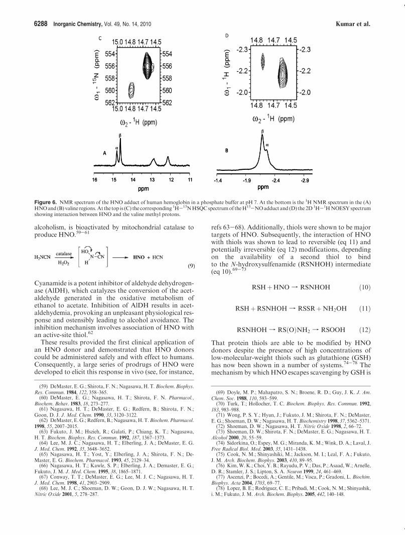
ca. 15 ppm in the 1H NMR spectra. Peptidic protons instrong hydrogen bonds may also have downfield reso-nances, as shown in Figure 6 for the HNO adduct of Hb,which has distinctive downfield resonances at ca. 12-13 ppm because of hydrogen bonds at the R and β subunitinterface. Therefore, a key proof is to generate a labeledH15NO adduct because the resulting HNO resonance willbe split into a doublet by the 15Nnuclear spin. 15N-labeledsamples also allow heteronuclear single quantum coher-ence (HSQC) spectra to be readily obtained, which pro-vide characterization of the 15N chemical shifts, asdemonstrated in Figure 6.The affinity of the oxygen-binding heme proteins for
both HNO and O2 also suggests that HNO mightbind and/or inhibit nonheme oxygenases. For instance,HNO precursors inhibit the pigmentation of melano-genic cells,48 which depend on the activity of tyro-sinase, an oxygenase that binds O2 between two coppercenters.49
HNO in Mammals. Interest in HNO in mammaliansystems dates to the early 1980s when vasodilation wasdetermined to be actively mediated by an unidentifiedspecies50 designated as the endothelium-derived relaxingfactor (EDRF).51 The EDRF was subsequently deter-mined to be NO, but the identification process led tocomparisons of the effects of NO and HNO donors invasoactive assays. Such experiments were the genesis ofthe current expanding interest in the pharmacologicaleffects of HNO donors. Consequently, analysis of theaqueous chemistry as well as the biochemistry of HNOhas arisen in order to identify the chemical origins of thepharmacological effects. The chemical reactions of HNOunder physiological conditions and their consequences inmammalian biology are beyond the scope of this reviewand have been presented recently elsewhere.46,52-54 Otherrecent research has focused on the identification ofmechanisms and markers of HNO biosynthesis and onthe production of novel HNO donors with propertiestailored for clinical use. Donor compounds are necessarynot only for facile or controlled delivery but also becauseof the self-consumption of HNO via irreversible dehydra-tion of the dimer.55,56 Donors of HNO have also beenrecently reviewed.57,58
About the same time that the vasoactivity of HNO wasobserved, Nagasawa and colleagues determined thatcyanamide (H2NCN), an alcohol deterrent used in Europe,Canada, and Japan for the clinical treatment of chronic
Scheme 4
(34) Ye, X.; Yu, A.; Champion, P. M. J. Am. Chem. Soc. 2006, 128, 1444–1445.
(35) Richter-Addo, G. B. Acc. Chem. Res. 1999, 32, 529–536.(36) Lee, J.; Chen, L.;West, A. H.; Richter-Addo, G. B.Chem. Rev. 2002,
102, 1019-1065.(37) Mansuy, D.; Battioni, P.; Chottard, J. C.; Lange, M. J. Am. Chem.
Soc. 1977, 99, 6441–6443.(38) Mansuy, D.; Battioni, P.; Chottard, J. C.; Riche, C.; Chiaroni, A.
J. Am. Chem. Soc. 1983, 105, 455–463.(39) Melenkivitz, R.; Hillhouse, G. L. Chem. Commun. 2002, 660–661.(40) Southern, J. S.; Green, M. T.; Hillhouse, G. L.; Guzei, I. A.;
Rheingold, A. L. Inorg. Chem. 2001, 40, 6039–6046.(41) Marchenko, A. V.; Vedernikov, A. N.; Dye, D. F.; Pink,M.; Zaleski,
J. M.; Caulton, K. G. Inorg. Chem. 2002, 41, 4087–4089.(42) Marchenko, A. V.; Vedernikov, A. N.; Dye, D. F.; Pink,M.; Zaleski,
J. M.; Caulton, K. G. Inorg. Chem. 2004, 43, 351–360.(43) Southern, J. S.; Hillhouse, G. L.; Rheingold, A. L. J. Am. Chem. Soc.
1997, 119, 12406–12407.(44) Melenkivitz, R.; Southern, J. S.; Hillhouse, G. L.; Concolino, T. E.;
Liable-Sands, L. M.; Rheingold, A. L. J. Am. Chem. Soc. 2002, 124, 12068–12069.
(45) Sellmann, D.; Gottschalk-Gaudig, T.; Haussinger, D.; Heinemann,F. W.; Hess, B. A. Chem.;Eur. J. 2001, 7, 2099–2103.
(46) Farmer, P. J.; Sulc, F. J. Inorg. Biochem. 2005, 99, 166–184.(47) Kumar, M. R.; Pervitsky, D.; Chen, L.; Poulos, T.; Kundu, S.;
Hargrove, M. S.; Rivera, E. J.; Diaz, A.; Colon, J. L.; Farmer, P. J.Biochemistry 2009, 48, 5018–5025.
(48) Ganesan, A. K.; Ho, H.; Bodemann, B.; Petersen, S.; Aruri, J.;Koshy, S.; Richardson, Z.; Le, L. Q.; Krasieva, T.; Roth, M. G.; Farmer,P. J.; White, M. A. PLoS Genet. 2008, 4, e1000298.
(49) Mayer, A. M. Phytochemistry 2006, 67, 2318–2331.(50) Furchgott, R. F.; Zawadzki, J. V. Nature 1980, 288, 373–376.(51) Furchgott, R. F.; Cherry, P. D.; Zawadzki, J. V.; Jothianandan, D.
J. Cardiovasc. Pharmacol. 1984, 6, S336–S343.(52) Miranda, K. M. Coord. Chem. Rev. 2005, 249, 433–455.(53) Wink, D. A.; Miranda, K. M.; Katori, T.; Mancardi, D.; Thomas,
D. D.; Ridnour, L. A.; Espey, M. G.; Feelisch, M.; Colton, C. A.; Fukuto,J. M.; Pagliaro, P.; Kass, D. A.; Paolocci, N. Am. J. Physiol. 2003, 285,H2264–H2276.
(54) Fukuto, J. M.; Dutton, A. S.; Houk, K. N. ChemBioChem 2005.(55) Smith, P. A. S.; Hein, G. E. J. Am. Chem. Soc. 1960, 82, 5731–5740.(56) Kohout, F. C.; Lampe, F.W. J. Am. Chem. Soc. 1965, 87, 5795–5796.(57) Miranda, K. M.; Nagasawa, H. T.; Toscano, J. P. Curr. Top. Med.
Chem. 2005, 5, 649–664.(58) King, S. B.; Nagasawa, H. T.Methods Enzymol. 1999, 301, 211–220.
6288 Inorganic Chemistry, Vol. 49, No. 14, 2010 Kumar et al.
alcoholism, is bioactivated by mitochondrial catalase toproduce HNO.59-61
Cyanamide is a potent inhibitor of aldehyde dehydrogen-ase (AlDH), which catalyzes the conversion of the acet-aldehyde generated in the oxidative metabolism ofethanol to acetate. Inhibition of AlDH results in acet-aldehydemia, provoking an unpleasant physiological res-ponse and ostensibly leading to alcohol avoidance. Theinhibition mechanism involves association of HNO withan active-site thiol.62
These results provided the first clinical application ofan HNO donor and demonstrated that HNO donorscould be administered safely and with effect to humans.Consequently, a large series of prodrugs of HNO weredeveloped to elicit this response in vivo (see, for instance,
refs 63-68). Additionally, thiols were shown to be majortargets of HNO. Subsequently, the interaction of HNOwith thiols was shown to lead to reversible (eq 11) andpotentially irreversible (eq 12) modifications, dependingon the availability of a second thiol to bindto the N-hydroxysulfenamide (RSNHOH) intermediate(eq 10).69-73
RSHþHNO f RSNHOH ð10Þ
RSHþRSNHOH f RSSRþNH2OH ð11Þ
RSNHOH f RSðOÞNH2 f RSOOH ð12ÞThat protein thiols are able to be modified by HNOdonors despite the presence of high concentrations oflow-molecular-weight thiols such as glutathione (GSH)has now been shown in a number of systems.74-78 Themechanism bywhichHNO escapes scavenging byGSH is
Figure 6. NMR spectrum of the HNO adduct of human hemoglobin in a phosphate buffer at pH 7. At the bottom is the 1H NMR spectrum in the (A)HNOand (B) valine regions.At the top is (C) the corresponding 1H-15NHSQCspectrumof theH15-NOadduct and (D) the 2D1H-1HNOESYspectrumshowing interaction between HNO and the valine methyl protons.
(59) DeMaster, E. G.; Shirota, F. N.; Nagasawa, H. T. Biochem. Biophys.Res. Commun. 1984, 122, 358–365.
(60) DeMaster, E. G.; Nagasawa, H. T.; Shirota, F. N. Pharmacol.,Biochem. Behav. 1983, 18, 273–277.
(61) Nagasawa, H. T.; DeMaster, E. G.; Redfern, B.; Shirota, F. N.;Goon, D. J. J. Med. Chem. 1990, 33, 3120–3122.
(62) DeMaster, E.G.; Redfern, B.; Nagasawa,H. T.Biochem. Pharmacol.1998, 55, 2007–2015.
(63) Fukuto, J. M.; Hszieh, R.; Gulati, P.; Chiang, K. T.; Nagasawa,H. T. Biochem. Biophys. Res. Commun. 1992, 187, 1367–1373.
(64) Lee, M. J. C.; Nagasawa, H. T.; Elberling, J. A.; DeMaster, E. G.J. Med. Chem. 1992, 35, 3648–3652.
(65) Nagasawa, H. T.; Yost, Y.; Elberling, J. A.; Shirota, F. N.; De-Master, E. G. Biochem. Pharmacol. 1993, 45, 2129–34.
(66) Nagasawa, H. T.; Kawle, S. P.; Elberling, J. A.; Demaster, E. G.;Fukuto, J. M. J. Med. Chem. 1995, 38, 1865–1871.
(67) Conway, T. T.; DeMaster, E. G.; Lee, M. J. C.; Nagasawa, H. T.J. Med. Chem. 1998, 41, 2903–2909.
(68) Lee, M. J. C.; Shoeman, D. W.; Goon, D. J. W.; Nagasawa, H. T.Nitric Oxide 2001, 5, 278–287.
(69) Doyle, M. P.; Mahapatro, S. N.; Broene, R. D.; Guy, J. K. J. Am.Chem. Soc. 1988, 110, 593–599.
(70) Turk, T.; Hollocher, T. C. Biochem. Biophys. Res. Commun. 1992,183, 983–988.
(71) Wong, P. S. Y.; Hyun, J.; Fukuto, J. M.; Shirota, F. N.; DeMaster,E. G.; Shoeman, D.W.; Nagasawa, H. T. Biochemistry 1998, 37, 5362–5371.
(72) Shoeman, D. W.; Nagasawa, H. T. Nitric Oxide 1998, 2, 66–72.(73) Shoeman, D. W.; Shirota, F. N.; DeMaster, E. G.; Nagasawa, H. T.
Alcohol 2000, 20, 55–59.(74) Sidorkina, O.; Espey, M. G.; Miranda, K.M.; Wink, D. A.; Laval, J.
Free Radical Biol. Med. 2003, 35, 1431–1438.(75) Cook, N. M.; Shinyashiki, M.; Jackson, M. I.; Leal, F. A.; Fukuto,
J. M. Arch. Biochem. Biophys. 2003, 410, 89–95.(76) Kim,W. K.; Choi, Y. B.; Rayudu, P. V.; Das, P.; Asaad,W.; Arnelle,
D. R.; Stamler, J. S.; Lipton, S. A. Neuron 1999, 24, 461–469.(77) Ascenzi, P.; Bocedi, A.; Gentile, M.; Visca, P.; Gradoni, L. Biochim.
Biophys. Acta 2004, 1703, 69–77.(78) Lopez, B. E.; Rodriguez, C. E.; Pribadi, M.; Cook, N. M.; Shinyashik,
i. M.; Fukuto, J. M. Arch. Biochem. Biophys. 2005, 442, 140–148.
Article Inorganic Chemistry, Vol. 49, No. 14, 2010 6289
not entirely understood but may relate to its hydropho-bicity or to the unique properties of protein thiols.In a somewhat later study, Wink and co-workers
demonstrated that an HNO donor (Angeli’s salt) elicitedsignificant cytotoxicty toward lung fibroblasts comparedto NO donors.79 This cytotoxicty is dependent upon anaerobic environment, is exacerbated by chemical deple-tion of cellular GSH, and is induced, in part, by double-stranded DNA breaks and base oxidation.79-83 More-over, this comparative analysis provided the first indica-tion that HNO could affect cellular functions by alteringthe redox status of the cell in a manner unique from thatof NO. Although HNO is capable of inducing oxidativestress, it can also act as an antioxidant via facile hydro-gen-atom donation to oxidizing radical species (akin totocopherol) and subsequent generation of NO, which isan established antioxidant.84 Significantly, the studiesdemonstrating the prooxidant effects were performed athigh levels of HNO, whereas the antioxidant propertieswere observed at much lower concentrations.Later collaborative comparisons demonstrated that the
in vitro toxicity of HNO could be replicated in vivo in amodel of ischemia-reperfusion injury in rabbits in con-trast to NO, which proved to be protective in the samemodel.85 Importantly, a subsequent study showed thatHNO could be protective toward reperfusion injuryif administered prior to the ischemic event.86 SimilarO2-dependent responses to HNO were observed in neu-ronal channel response.87
Together, these studies led to examination of the reac-tion of HNO with O2, but the product has yet to beidentified;79-83,88-90 for further discussion of this reac-tion, see ref 52. Significantly, the autoxidation of HNOis generally too slow (103 M-1 s-1) to be of kineticconsequence in many biological systems, particularly at
pharmacological levels of HNO.91,92 Furthermore, anumber of comparisons of HNO and NO donors havenow appeared in the literature (reviewed in refs 52, 53, 91,and 93-97) and nearly universally demonstrate that thephysiological properties of HNO and NO are discrete.Perhaps most importantly, analyses by Wink and
colleagues led to intensive investigation of the cardiovas-cular properties of HNO in dogs (reviewed in refs 53and 93). HNO was found to enhance myocardial con-tractility even in failing hearts.98,99 As such, HNO donorsmay act as a novel class of vasodilators and treatments forheart failure.100 This discovery substantially increasedinterest in HNO and led to an accelerated publicationrate. Ensuing analyses demonstrated that HNO targetskey regulators of normal myocyte contractile functionand increases the sensitivity ofmyofilaments to calcium ina thiol-sensitive manner.101-103
During their cardiovascular studies, Paolocci and col-leagues98 determined that the vascular effects of HNO indogs were limited to the venous side of the circulatorysystem, unlikeNOdonors, which are systemic hypotensiveagents. In vascular smooth muscle, NO leads to dilationby binding to the regulatory ferrous heme of solubleguanylyl cyclase (sGC), which increases the rate ofconversion of GTP to cGMP. The strong trans effect ofNO induces cleavage of the proximal histidine uponbinding, leading to an activating conformational change(Scheme 5). The observation that infusion ofHNOdonorsdidnot lead toelevatedcGMPlevels inplasmaboth indicatedthatHNOandNOdonot interconvert in bloodand renewedinterest in the vasoactive mechanisms of HNO.91
Several HNO donors have been observed to inducevasorelaxation in vivo or in rodent arterial/aorticring assays.63,104-106 These results instigated further
(79) Wink, D. A.; Feelisch, M.; Fukuto, J.; Christodoulou, D.;Jourd’heuil, D.; Grisham, M. B.; Vodovotz, Y.; Cook, J. A.; Krishna, M.;DeGraff, W. G.; Kim, S.; Gamson, J.; Mitchell, J. B. Arch. Biochem.Biophys. 1998, 351, 66–74.
(80) Ohshima, H.; Gilibert, I.; Bianchini, F. Free Radical Biol. Med. 1999,26, 1305–1313.
(81) Ohshima, H.; Yoshie, Y.; Auriol, S.; Gilibert, I. Free Radical Biol.Med. 1998, 25, 1057–1065.
(82) Chazotte-Aubert, L.; Oikawa, S.; Gilibert, I.; Bianchini, F.; Kawanishi,S.; Ohshima, H. J. Biol. Chem. 1999, 274, 20909–20915.
(83) Miranda, K. M.; Yamada, K.; Espey, M. G.; Thomas, D. D.;DeGraff, W.; Mitchell, J. B.; Krishna, M. C.; Colton, C. A.; Wink, D. A.Arch. Biochem. Biophys. 2002, 401, 134–144.
(84) Lopez, B. E.; Shinyashiki, M.; Han, T. H.; Fukuto, J. M. FreeRadical Biol. Med. 2007, 42, 482–491.
(85) Ma, X. L.; Gao, F.; Liu, G. L.; Lopez, B. L.; Christopher, T. A.;Fukuto, J. M.; Wink, D. A.; Feelisch, M. Proc. Natl. Acad. Sci. U.S.A. 1999,96, 14617–14622.
(86) Pagliaro, P.; Mancardi, D.; Rastaldo, R.; Penna, C.; Gattullo, D.;Miranda, K. M.; Feelisch, M.; Wink, D. A.; Kass, D. A.; Paolocci, N. FreeRadical Biol. Med. 2003, 34, 33–43.
(87) Colton, C. A.; Gbadegesin,M.;Wink,D. A.;Miranda, K.M.; Espey,M. G.; Vicini, S. J. Neurochem. 2001, 78, 1126–1134.
(88) Miranda, K.M.; Espey, M. G.; Yamada, K.; Krishna, M.; Ludwick,N.; Kim, S.; Jourd’heuil, D.; Grisham, M. B.; Feelisch, M.; Fukuto, J. M.;Wink, D. A. J. Biol. Chem. 2001, 276, 1720–1727.
(89) Miranda, K. M.; Dutton, A. S.; Ridnour, L. A.; Foreman, C. A.;Ford, E.; Paolocci, N.; Katori, T.; Tocchetti, C. G.; Mancardi, D.; Thomas,D. D.; Espey,M. G.; Houk, K. N.; Fukuto, J. M.;Wink, D. A. J. Am. Chem.Soc. 2005, 127, 722–731.
(90) Liochev, S. I.; Fridovich, I.Arch. Biochem. Biophys. 2002, 402, 166–171.(91) Miranda, K. M.; Paolocci, N.; Katori, T.; Thomas, D. D.; Ford, E.;
Bartberger, M. D.; Espey, M. G.; Kass, D. A.; Feelisch, M.; Fukuto, J. M.;Wink, D. A. Proc. Natl. Acad. Sci. U.S.A. 2003, 100, 9196–9201.
(92) Liochev, S. I.; Fridovich, I. Free Radical Biol. Med. 2003, 34, 1399–1404.
(93) Pagliaro, P. Life Sci. 2003, 73, 2137–2149.(94) Paolocci, N.; Jackson, M. I.; Lopez, B. E.; Miranda, K.; Tocchetti,
C. G.; Wink, D. A.; Hobbs, A. J.; Fukuto, J. M. Pharmacol. Ther. 2007, 113,442–458.
(95) Irvine, J. C.; Ritchie, R.H.; Favaloro, J. L.; Andrews, K. L.;Widdop,R. E.; Kemp-Harper, B. K. Trends Pharmacol. Sci. 2008, 29, 601–608.
(96) Flores-Santana, W.; Switzer, C.; Ridnour, L. A.; Basudhar, D.;Mancardi, D.; Donzelli, S.; Thomas, D. D.; Miranda, K. M.; Fukuto,J. M.; Wink, D. A. Arch. Pharm. Res. 2009, 32, 1139–1153.
(97) Switzer, C. H.; Flores-Santana, W.; Mancardi, D.; Donzelli, S.;Basudhar, D.; Ridnour, L. A.; Miranda, K.M.; Fukuto, J. M.; Paolocci, N.;Wink, D. A. Biochim. Biophys. Acta 2009, 1787, 835–840.
(98) Paolocci, N.; Saavedra, W. F.; Miranda, K. M.; Martignani, C.;Isoda, T.; Hare, J. M.; Espey, M. G.; Fukuto, J. M.; Feelisch, M.; Wink,D. A.; Kass, D. A. Proc. Natl. Acad. Sci. U.S.A. 2001, 98, 10463–10468.
(99) Paolocci, N.; Katori, T.; Champion, H. C.; St. John,M. E.;Miranda,K.M.; Fukuto, J.M.;Wink, D. A.; Kass, D. A.Proc. Natl. Acad. Sci. U.S.A.2003, 100, 5537–5542.
(100) Feelisch, M. Proc. Natl. Acad. Sci. U.S.A. 2003, 100, 4978–4980.(101) Cheong, E.; Tumbev, V.; Abramson, J.; Salama, G.; Stoyanovsky,
D. A. Cell Calcium 2005, 37, 87–96.(102) Tocchetti, C. G.; Wang, W.; Froehlich, J. P.; Huke, S.; Aon, M. A.;
Wilson, G. M.; Di Benedetto, G.; O’Rourke, B.; Gao, W. D.; Wink, D. A.;Toscano, J. P.; Zaccolo, M.; Bers, D. M.; Valdivia, H. H.; Cheng, H.; Kass,D. A.; Paolocci, N. Circ. Res. 2007, 100, 96–104.
(103) Dai, T.; Tian, Y.; Tocchetti, C. G.; Katori, T.; Murphy, A. M.;Kass, D. A.; Paolocci, N.; Gao, W. D. J. Physiol. 2007, 580, 951–960.
(104) Fukuto, J. M.; Hobbs, A. J.; Ignarro, L. J. Biochem. Biophys. Res.Commun. 1993, 196, 707–713.
(105) Murphy, M. E.; Sies, H. Proc. Natl. Acad. Sci. U.S.A. 1991, 88,10860–10864.
(106) Fukuto, J. M.; Chiang, K.; Hszieh, R.; Wong, P.; Chaudhuri, G.J. Pharmacol. Exp. Ther. 1992, 263, 546–551.

6290 Inorganic Chemistry, Vol. 49, No. 14, 2010 Kumar et al.
examination of the effects of HNO donors on sGCfunction, usually by measurement of the cGMP levelsor determination of the vasorelaxive potency in the pre-sence of sGC inhibitors.63,104,106-114 The question of thedirect activation of sGC was addressed by Dierksand Burstyn,115 who exposed partially purified bovinelung sGC to donors ofNO,NOþ, andHNOandobservedenhanced cGMP formation only upon the introductionof NO. This finding, in addition to earlier examinationsof the reaction of HNO with myoglobin and hemoglo-bin,69,116,117 led to the assumption that HNO does notreact with ferrous hemes. Kinetic analysis later suggestedthat the primary cellular targets for HNO are thiolsand oxidized metals, while NO is thought to princi-pally interact with other free radicals and with reducedmetals.91,118
To explain the vasoactivity ofHNO, the suggestionwasmade that HNO is converted to NO particularly in theaortic ring assays, for instance, by superoxide dismutase(SOD), metHb, and flavins104 or by the release of nor-mally sequestered species during tissue preparation.Furthermore, it may be that the addition of millimolardithiothreitol (DTT), which is a vital stabilizing agent forsGC because of the oxidative instability of the heme andprotein thiols under aerobic conditions, scavenged HNObefore it could bind to the heme. This possibility inconjunction with the demonstration by Farmer andco-workers16,17 of the thermally stable adduct of HNO
with deoxymyoglobin led the Fukuto and Mirandalaboratories to further investigate whether HNO candirectly enhance the activity of sGC using bovine lungsGC purified in the Burstyn laboratory.Exposure of sGC to two structurally distinct HNO
donors in thiol-free media led to a concentration-depen-dent increase in cGMP formation.119 The extent ofactivation was lower than that from NO but was signifi-cantly elevated compared to basal levels. That sGC wasnot affected by a metal chelator but was modified byDTT clearly indicated that HNO can directly interactwith sGC.Both the heme and the multiple cysteine thiols of sGC
are reasonable targets for HNO (Scheme 6). Removal ofthe heme decreased both HNO- and NO-mediated activ-ity, supporting a direct interaction with the heme for bothnitrogen oxides. Surprisingly,HNOdid not activate ferricsGC, which was expected to undergo reductive nitrosyla-tion to form the ferrous heme complex (eq 13).
FeIII þHNO f FeIINOþHþ ð13ÞFerric sGC has been previously noted to be substitution-ally inert to cyanide.120 The impact of the reactivity ofHNO with the protein thiols was investigated by sub-stitution of the hemewith themetal-free porphyrin, whichactivates the enzyme to a similar extent to the ferrousnitrosyl complex.121 In this case, HNO decreased theactivity, suggesting that cysteine thiols can function in anegative allosteric fashion when oxidized.A second recent investigation122 appeared to con-
firm104 that HNO does not affect the sGC activity unlessSOD is present.We suggest that, aswith other studies thatsuggest the requirement for oxidation of HNO to NO,buffer components may be lead to unexpected scavengingof HNO. In addition to thiols, a N-(2-hydroxyethyl)-piperazine-N’-2-ethanesulfonic acid (HEPES) buffer hasbeen shown previously to scavenge HNO,83 and relatedagents such as triethylamine (TGA), which was used inthe recent activity studies, may have a similar effect.
Conclusion. Exposure to HNO is known to affect avariety of thiol-containing proteins. That thermally stablecomplexes ofHNOwith heme proteins can nowbe readilyproduced suggests the possibility that metalloproteinsmay also be significant pharmacological targets forHNO. Because investigation of the chemical origin forthe differing cardiovascular effects observed for HNOand NO donors in the studies by Paolocci et al. demon-strated that infusion of HNO did not result in a measur-able increase in the plasma levels of cGMP,91 ques-tions remain about the role of HNO in whole orga-nisms compared to in vitro assays or studies usingexcised tissues, which may have artifactual responses toHNO donors. Furthermore, whether HNO can activatesGC or coordinate to hemoglobin or myoglobin under
Scheme 5. Activation of sGC upon Binding of NO to the RegulatoryHeme
(107) Pino, R. Z.; Feelisch, M. Biochem. Biophys. Res. Commun. 1994,201, 54–62.
(108) Ellis, A.; Lu, H.; Li, C. G.; Rand, M. J. Br. J. Pharmacol. 2001, 134,521–528.
(109) VanUffelen, B. E.; Van der Zee, J.; de Koster, B. M.; VanSteve-ninck, J.; Elferink, J. G. R. Biochem. J. 1998, 330, 719–722.
(110) Ellis, A.; Li, C. G.; Rand, M. J. Br. J. Pharmacol. 2000, 129, 315–322.
(111) Wanstall, J. C.; Jeffery, T. K.; Gambino, A.; Lovren, F.; Triggle,C. R. Br. J. Pharmacol. 2001, 134, 463–472.
(112) Costa, G.; Labadia, A.; Triguero, D.; Jimenez, E.; Garcia-Pascual,A. Naunyn-Schmiedeberg’s Arch. Pharmacol. 2001, 364, 516–523.
(113) Irvine, J. C.; Favaloro, J. L.; Kemp-Harper, B. K. Hypertension2003, 41, 1301–1307.
(114) Andrews, K. L.; Irvine, J. C.; Tare, M.; Apostolopoulos, J.;Favaloro, J. L.; Triggle, C. R.; Kemp-Harper, B. K. Br. J. Pharmacol.2009, 157, 540–550.
(115) Dierks, E.A.; Burstyn, J.N.Biochem.Pharmacol.1996, 51, 1593–1600.(116) Bazylinski, D. A.; Hollocher, T. C. J. Am. Chem. Soc. 1985, 107,
7982–7986.(117) Doyle, M. P.; Mahapatro, S. N. J. Am. Chem. Soc. 1984, 106, 3678–
3679.(118) Miranda,K.M.;Nims, R.W.; Thomas,D.D.; Espey,M.G.; Citrin,
D.; Bartberger, M. D.; Paolocci, N.; Fukuto, J. M.; Feelisch, M.; Wink,D. A. J. Inorg. Biochem. 2003, 93, 52–60.
(119) Miller, T. W.; Cherney, M. M.; Lee, A. J.; Francoleon, N. E.;Farmer, P. J.; King, S. B.; Hobbs, A. J.; Miranda, K. M.; N., B. J.; Fukuto,J. M. J. Biol. Chem. 2009, 284, 21788–21796.
(120) Stone, J. R.; Sands, R. H.; Dunham, W. R.; Marletta, M. A.Biochemistry 1996, 35, 3258–3262.
(121) Wolin, M. S.; Wood, K. S.; Ignarro, L. J. J. Biol. Chem. 1982, 257,13312–13320.
(122) Zeller, A.; Wenzl, M. V.; Beretta, M.; Stessel, H.; Russwurm, M.;Koesling, D.; Schmidt, K.; Mayer, B.Mol. Pharmacol. 2009, 76, 1115–1122.

Article Inorganic Chemistry, Vol. 49, No. 14, 2010 6291
physiological/cellular conditions remains to be deter-mined. The coordination chemistry of HNO should be afruitful area for both metalloprotein and small-moleculechemistry for the foreseeable future.
Materials and Methods
Horse skeletal muscle myoglobin (95-100%), adult hu-man hemoglobin, sodium nitrite, sodium borohydride, so-dium trimethoxyborohydride, and zinc dust were purchased
Scheme 6. Routes of Activation of sGC by HNO in Purified, O2-Free Systems
6292 Inorganic Chemistry, Vol. 49, No. 14, 2010 Kumar et al.
from Sigma-Aldrich and used as received. Piloty’s acid waspurchased from Cayman Chemicals. 4,40-Dimethyl-2,20-di-pyridyl (DTDP), tetracyanato nickelate,123 andN-methyl-D-glucaminedithiocarbamate26 were prepared and purifiedfollowing literature procedures. Stock solutions of Piloty’sacid were freshly prepared in deionized water before eachexperiment. Manipulation of various hemes and their ad-ducts of NO and HNO was performed inside an anaerobicglovebox. Purification of heme adducts was carried out on apreequilibratedSephadexG25 column ina 50mMphosphatebuffer at either pH 7 or 9.4. All absorption spectra wererecorded on a Hewlett-Packard 8453A spectrophotometer.1H NMR experiments were recorded on a Bruker Avance600 or Varian 800 MHz spectrometer. The spectra wereacquired by direct saturation of the residual water peakduring the relaxation delay. Chemical shifts were referencedto the residual water peak at 4.8 ppm. X-band EPR spectrawere recorded with a Bruker EMX spectrometer equippedwith a standard TE102 (ER 4102ST) or a high-sensitivity ER4119HS resonator.
UV-Vis Experiments. A sample of HNO-FeIIMb was con-centrated on Centricon YM10. Several microliters were addedto 2mL of the appropriate buffer (100mMphosphate buffer forpH7 and 8; 100mMcarbonate buffer for pH10; the appropriateconcentration of NaOH with 50 mM NaCl for pH 11-13).Spectra were collected in a glovebox on a USB2000 spectro-photometer.
H/D Exchange Experiments. For the NMR experiments,HNO-FeIIMb was prepared using Piloty’s acid, as describedabove. Aliquots were removed and concentrated on CentriconYM10 using a 100mM carbonate buffer at pH 10.0, 9.5, and 9.0and a 100 mM phosphate buffer at pH 8.0. Two dilutionconcentration cycles were performed, until the pH of the solu-tion in the waste reservoir was at the appropriate pH. Theprotein solution was concentrated to ∼200 μL. To the bottomof a J. Young tube was added 360 μL (60%) of D2O, and to thetop compartment was added a total of 240 μL (40%) of aHNO-FeIIMb solution diluted with a buffer. The solutionwas mixed, and time-course 1H NMR spectra were collected.The HNO peak was integrated and plotted versus time. For the95þ% D2O sample, the HNO-FeIIMb solution was fullyexchanged with D2O in a Centricon column.
Preparation of Nitrosyl Heme Proteins. To a micromolarsolution of heme (200 μL) in a 50 mM phosphate buffer at pH7 was added sodium nitrite. After standing for 5 min, sodiumdithionite was added at a ratio of sodium nitrite to sodium
dithionite of 1:3. The nitrosyl hemes were purified on a size-exclusion Sephadex G25 column and then concentrated on aAmicon YM10 membrane filters.
Nitrite/Borohydride Preparation of HNO-FeIIMb. To 60 mg
of Mb in 2 mL of a carbonate buffer at pH 9.4 was added 10-20 equiv of sodium nitrite, followed by the careful addition ofeither solid sodium borohydride (10 equiv) or 200 μL of a 1 Msolution of sodium borohydride in 1 M sodium hydroxide.Caution! Sodium borohydride is a highly reactive white solidpowder that violently reacts with water and aids combustion, soproper protection and care should be taken while handling it!Upon the addition of sodium borohydride, a visible colorchange from brown to red was observed, which indicated theformation of HNO-FeIIMb. The reaction solution was thenadded to a preequilibrated G-25 Sephadex column in a 50 mMphosphate buffer at pH 7. The fast-moving dark-red band wascollected and concentrated on an Amicon YM10 filter.
Reaction of Tetracyanatonickelate with Nitrite/Dithionite and
Nitrite/Borohydride Mixtures. Tetracyanatonickelate (5 mg)was dissolved in 1 mL of a 50 mM carbonate buffer at pH 10in a 1 cm cuvette. After the addition of sodium nitrite (1 mg),followed by sodium borohydride (2 mg), the solution immedi-ately turned dark purple. The absorbance spectrum wascollected with no attempt to isolate the reaction products.
Reaction of Iron(II) N-Methyl-D-glucaminedithiocarbamatewith Nitrite/Dithionite and Nitrite/Borohydride Mixtures. Twosamples of FeIIMGD (5 mg) were dissolved in separate 1 mLamounts of a 50mMcarbonate buffer at pH 10 in 1 cm cuvettes;to one was added sodium nitrite (1 mg), followed by sodiumborohydride (2 mg); to the other was added sodium nitrite(1 mg), followed by sodium dithionite (2 mg). Both reactionmixtures immediately turned dark green, and aliquots weretransferred to EPR tubes and frozen. No attempt was made topurify the reaction products.
Reduction of Sodium Nitroprusside by Sodium Borohydride.
Sodium nitroprusside (10mg) was dissolved in 1mL of a 50mMcarbonate buffer. Sodium nitrite and sodium borohydride werethen added, and the brick-red-colored solution was then usedfor studies without further purification.
Acknowledgment. This work was supported by grants fromthe National Science Foundation (Grant CHE-0517080 toP.J.F. and Grant CHE-0645818 to K.M.M.) and the NationalInstitutes of Health (Grant 1R21ES016441-01 to P.J.F. andGrant R01-GM076247 to K.M.M.).
Supporting Information Available: Additional experimentaldetails and figures. This material is available free of charge viathe Internet at http://pubs.acs.org.
(123) J�oszai, R.; Beszeda, I.; B�enyei, A.; Fischer, A.; Kov�acs, M.;Maliarik, M.; Nagy, P.; Shchukarev, A.; T�oth, I. Inorg. Chem. 2005, 44,9643–96451.