Embed Size (px)
Citation preview

Reaction Mechanisms of Metalloenzymes and Synthetic Model Complexes Activating
Dioxygen

Reaction Mechanisms ofMetalloenzymes and SyntheticModel Complexes Activating
DioxygenA Computational study
Valentin Georgiev

c© Valentin Georgiev, Stockholm 2009
ISSNISBN 978-91-7155-965-4
Printed in Sweden by Intellecta Docusys, Stockholm 2009
Distributor: Department of Physics, Stockholm University

I dedicate this thesis to my wife Polina and our kids Monica andFilip, who are the best thing in my life.


7
Abstract
Quantum chemistry has nowadays become a powerful and efficient tool thatcan be successfully used for studies of biosystems. It is therefore possibleto model the enzyme active-site and the reactions undergoing into it, as wellas obtaining quite accurate energetic profiles. Important conclusions can bedrawn from such profiles about the plausibility of different putative mecha-nisms.
Density Functional Theory is used in the present thesis for investigationof the catalytic mechanism of dioxygenase metallo-enzymes and syntheticmodel complexes. Three enzymes were studied – Homoprotocatechuate2,3-dioxygenase isolated fromBrevibacterium fuscum(Bf 2,3-HPCD),Manganese-Dependent Homoprotocatechuate 2,3-Dioxygenase (MndD)and Homogentisate Dioxygenase (HGD). Models consisting of 55 to208 atoms have been built from X-ray crystal structures and used in thecalculations. The computed energies were put in energy curves and were usedfor estimation of the feasibility of the suggested reaction mechanisms. Anon-heme [(L4Me4)Fe(III)]+3 complex that mimics the reactivity of intradioldioxygenases, and a heme [T(o-Cl)PPFe] complex catalyzing the stepwiseoxidation of cyclohexane to adipic acid, were also studied.
For the enzymes and the non-heme biomimetic complex the reaction wasfound to follow a mechanism that was previously suggested for extradiol andintradiol dioxygenases – ordered substrates binding and formation of peroxospecies, which further undergoes homolytic O-O bond cleavage. Different re-action steps appear to be rate limiting in the particular cases: proton transferfrom the substrate to the peroxide inBf 2,3-HPCD, the formation of the per-oxo bridge in HGD and the biomimetic complex, and notably, spin transitionin MndD.
The catalytic oxidation of cyclohexane to adipic acid in the presence ofmolecular oxygen as oxidant was studied, a reaction of great importance forthe chemical industry. Reaction mechanism is suggested, involving severalconsecutive oxidative steps. The highest calculated entalpy of activation is17.8 kcal/mol for the second oxidative step.

9
List of Papers
This thesis is based on the following papers:I Tomasz Borowski, Valentin Georgiev, and Per E. M. Siegbahn
(2005) Catalytic Reaction Mechanism of Homogentisate Dioxy-genase: A Hybrid DFT Study.J. Am. Chem. Soc., 127 (49):17303-17314
II Valentin Georgiev, Tomasz Borowski,and Per E. M. Siegbahn(2006) Theoretical study of the catalytic reaction mechanism ofMndD. J. Biol. Inorg. Chem, 11(5):571-585
III Valentin Georgiev, Tomasz Borowski, Margareta R. A.Blomberg, and Per E. M. Siegbahn (2008) A comparison ofthe reaction mechanism of iron- and manganese-containing2,3-HPCD: an important spin transition for manganese.J. Biol.Inorg. Chem, 13:929-940
IV Valentin Georgiev, Holger Noack, Margareta R. A. Blomberg andPer E. M. Siegbahn, A DFT Study on the Catalytic Reactivity of aFunctional Model Complex for Intradiol-Cleaving Dioxygenases.In manuscript
V Holger Noack, Valentin Georgiev, Johannes Adam Johannson,Margareta R. A. Blomberg and Per E. M. Siegbahn, The Con-version of Cyclohexane to Adipic Acid catalyzed by an Iron-Porphorin Complex. A theoretical study.In manuscript
Reprints were made with permission from the publishers.

10
Comments on the Contribution to the Papers
I have performed the all the calculations and prepared the manuscripts forPaper II andPaper III . For Paper I I was involved in the discussion, and Ihave performed additional calculations for testing the concerted Criegee re-arrangement. ForPaper IV I have performed all the calculations on the sug-gested intradiol mechanism and prepared the manuscript except for the Intro-duction.The calculations on the alternative extradiol path were done by HolgerNoack, who prepared the Introduction part. ForPaper V I have done all thecalculations for the first and the third major reaction steps of the suggestedmechanism, and prepared the manuscript. Holger Noack performed calcula-tions on alternative reaction paths. Johannes Johannson calculated the secondmain reaction step and contributed to discussion about entropy effects in themanuscript.
Contents
Part I: Catalysis, dioxygen, and iron1 How do the enzymes function . . . . . . . . . . . . . . . . . . . . . . . . . . . . . 15
Part II: Theory and methods2 Quantum Chemistry . . . . . . . . . . . . . . . . . . . . . . . . . . . . . . . . . . . . 213 Density Functional Theory . . . . . . . . . . . . . . . . . . . . . . . . . . . . . . . 274 Transition State Theory . . . . . . . . . . . . . . . . . . . . . . . . . . . . . . . . . 315 Technical details . . . . . . . . . . . . . . . . . . . . . . . . . . . . . . . . . . . . . . . 33
Part III: Dioxygenases6 Extradiol and intradiol catechol dioxygenases . . . . . . . . . . . . . . . . . 397 Fe- and Mn-dependent homoprotocatechuate 1,2-dioxygenases . . . 43
7.1 Background . . . . . . . . . . . . . . . . . . . . . . . . . . . . . . . . . . . . . . . . . . . . . 437.2 The model . . . . . . . . . . . . . . . . . . . . . . . . . . . . . . . . . . . . . . . . . . . . . . 467.3 Reaction mechanism of B f 2,3-HPCD . . . . . . . . . . . . . . . . . . . . . . . . . . . 467.4 Reaction mechanism of MndD. Spin transition . . . . . . . . . . . . . . . . . . . . . . 49
8 Homogentisate Dioxygenase . . . . . . . . . . . . . . . . . . . . . . . . . . . . . . 578.1 Background . . . . . . . . . . . . . . . . . . . . . . . . . . . . . . . . . . . . . . . . . . . . . 578.2 The model . . . . . . . . . . . . . . . . . . . . . . . . . . . . . . . . . . . . . . . . . . . . . . 588.3 Enzyme-Substrate Complex . . . . . . . . . . . . . . . . . . . . . . . . . . . . . . . . . . 598.4 Reaction mechanism of Homogentisate Dioxygenase . . . . . . . . . . . . . . . . . 59
9 Summary . . . . . . . . . . . . . . . . . . . . . . . . . . . . . . . . . . . . . . . . . . . . 63
Part IV: Biomimetic model complexes10 Non-heme iron model complex for intradiol-cleaving dioxygenases . . 67
10.1 Background . . . . . . . . . . . . . . . . . . . . . . . . . . . . . . . . . . . . . . . . . . . . . 6710.2 The model . . . . . . . . . . . . . . . . . . . . . . . . . . . . . . . . . . . . . . . . . . . . . . 6710.3 Reaction mechanism . . . . . . . . . . . . . . . . . . . . . . . . . . . . . . . . . . . . . . . 6910.4 Intra- vs. Extradiol bond cleavage . . . . . . . . . . . . . . . . . . . . . . . . . . . . . . 7310.5 Comparison with the biological system . . . . . . . . . . . . . . . . . . . . . . . . . . . 74
11 Heme complex catalyzing adipic acid synthesis . . . . . . . . . . . . . . . . 7711.1 Background . . . . . . . . . . . . . . . . . . . . . . . . . . . . . . . . . . . . . . . . . . . . . 7711.2 Computational technique. . . . . . . . . . . . . . . . . . . . . . . . . . . . . . . . . . . . . 7811.3 Suggested reaction mechanism . . . . . . . . . . . . . . . . . . . . . . . . . . . . . . . 80
11.3.1 Oxidation of cyclohexane to 1,2-cyclohexanediol . . . . . . . . . . . . . . . . 8011.3.2 1,2-cyclohexanediol oxidation and ring cleavage . . . . . . . . . . . . . . . . 83

CONTENTS 12
11.3.3 Oxidation of adipic aldehyde to adipic acid . . . . . . . . . . . . . . . . . . . . 85
Part V: Concluding Remarks
Part VI: Acknowledgements
Part VII: Populärvetenskaplig sammanfattning på svenskaBibliography . . . . . . . . . . . . . . . . . . . . . . . . . . . . . . . . . . . . . . . . . . . . . 97

Part I:Catalysis, dioxygen, and iron
Chemical reactions can be fairly slow at ambient conditions, and the desiredproducts are often accompanied by side products. This not only means lowefficiency, but danger as well, since the side products can be hazardous andeven toxic. Catalysts in general are substances which are able to acceleratechemical reactions without changing the chemical equilibrium and withoutbeing consumed.

15
1. How do the enzymes function
The biological catalysts found in Nature are known as enzymes. Almost allknown enzymes are protein molecules, but some RNA molecules (ribozymesand synthetic deoxyribozymes) also hold catalytic properties. The proteinmacromolecules consist of long chains of amino acids bound together bypeptide bonds [1]. These chains are furthermore folded specifically in thesecondary structure, and thus special regions in the protein structure areformed, called active sites. The active site is the part of the enzyme that isresponsible for it’s physiological role, it is the place where the catalysisoccurs. Due to the specific structural, chemical and electrostatic featuresof the active site, normally only one type of substrate molecules can bindinto it, hence the enzyme is highly selective. Having in mind the greatvariety of chemical reactions and therefore substrates, we can imagine thehuge number of enzymes Nature has designed. The enzymes are extremelyeffective, enhancing the reaction rates millions of times and acting undermild conditions. Thus they catalyze biological reactions, which under normalconditions would proceed with negligible rate. Enzymes also show impressivelevels of stereospecificity, regioselectivity and chemoselectivity [2].
A rough picture of an enzyme-catalyzed reaction can be described inthe following way: when a substrate molecule enters the active site anEnzyme-Substrate complex (ES) is formed, from which the catalytic reactionproceeds and the product is formed. In the Enzyme-Product complex (EP)the hydrogen bonds and electrostatic interactions, previously stabilizingthe ES, are destroyed and the product molecule can easily leave the activesite [3]. However, the origin of the catalytic power, the means of achievingthe reaction rate enhancement, and the specificity, are all widely debatedphenomena without straightforward explanation.
There are number of theories trying to explain how the enzymes dotheir work. One of the most widely used is thelock-and-keytheory. It wassuggested in 1894 by Emil Fisher and is based on the assumption that thesubstrate molecule corresponds perfectly to the active site, so that once itenters the enzyme, it is oriented in a particular optimal spatial position andthen “locked” into the active site with hydrogen bonds and electrostaticinteractions. This is a very illustrative picture which unfortunately providesonly a reasonable explanation of the specificity of the enzymes. Theinduced
16
fit model was suggested in 1958 by Daniel Koshland [4] as a modification tothe lock and key model. More up-to-date theories adopt the ideas of transitionstate stabilization and/or ES complex destabilization, providing an alternativepathway, reducing the reaction entropy by bringing substrates together. Allof them are based on the concept of lowering the activation energy barrier∆G‡ of the reaction. Extensive and elaborate discussions on the origin of thecatalytic power can be found in references [3, 5, 6].
Particularly interesting are the metal containing enzymes, also known asmetalloproteins. They contain a metal ion cofactor, usually coordinated bynitrogen, oxygen or sulfur atoms from the polypeptide’s amino acids. Themetal ion can be also coordinated by macrocyclic ligands incorporated in theprotein, as heme, chlorophyll, and vitamin B12. The presence of the metal ionallows these enzymes to perform oxidation and reduction, reactions that arenot easily achieved by the organic functional groups present in the proteins.Thus the metalloenzymes can catalyze biochemical reactions of greatimportance, like photosynthesis, O-O bond cleavage in cellular respiration,oxygenation, dioxygenation etc. From an industrial point of view dioxygen(and hydrogen peroxide to some extend) is the perfect oxidant - it is availablein the Earth’s atmosphere, and it doesn’t cause environmental problems.Molecular oxygen adopts an "unusual" triplet electronic configuration in itsground state, which prevents it from reacting with closed shell molecules inambient conditions. Such reactions are spin-forbidden since they involvechange of the total spin state of the system. Chemists achieve activationof dioxygen at higher temperatures and pressures, quite severe conditions,which makes the utilization of these reactions expensive for the chemicalindustry. Nature however, utilizes transition metal ions in metalloproteinsto perform the desired reactions at normal temperature and neutral pH. Inbiology, the proteins containing copper, heme-iron, and non-heme iron arethe main catalysts involved in dioxygen activation and transportation.
In order to better understand the metalloproteins, synthetic inorganic modelcomplexes [7] have been extensively studied, both experimentally and theo-retically. The use of these biomimetic complexes gives the possibility for finetuning of the catalytic reactivity. It is easier to create different biomimeticsystems by using different ligands, solvents, counterions etc., rather than per-forming mutagenesis studies on real enzymes.
Understanding the enzyme structure and reaction pathways is a directway to address the question about the nature of the enzymatic power.Such knowledge is of great importance in all areas where catalysis isinvolved, like drug design, pharmaceutics, chemical industry, etc. Eventhough there has been a great development in the science and technologyin the last decades, experimental studies are usually expensive, and canbe very slow as well. Regarding reaction mechanisms, there are also

17
limited capabilities for studying extremely short-living intermediatesand transition states. Fortunately, advances in computer technology andquantum theory provide a possibility for carrying out theoretical researchon enzyme catalysis. Using computer modeling one can investigatesuch "invisible" species as transition states, thus providing useful infor-mation which can support experimental results and help in their interpretation.

Part II:Theory and methods
The modeling of chemical reactions can be done using Quantum Mechan-ics (QM) implemented in the computational codes, i.e. Quantum Chemistry(QC). Other methods like Molecular Dynamics (MD) and Molecular Mechan-ics (MM) are based on classical mechanics, they are computationally cheapand one can use them to describe systems consisting of thousands of atoms.Thus these methods can be used for investigation of big bio-molecules likeproteins as a whole – protein structure determination and prediction. Quan-tum methods, which allow for more accurate solving of chemical problems,are quite demanding regarding computer power and hence only systems oflimited size (up to 200 atoms nowadays) can be treated.
Quantum mechanical equations give a correct description of the behavior ofelectrons, but they can only be solved exactly only for one-electron systems.Plenty of methods based on different approximations are available for solvingmany-electron problems, and choosing a relevant one for characterization ofenzyme catalytic mechanisms is an important question [8, 9].
The main physical quantity calculated in a QM study is the energy of differ-ent structures of the investigated system. These energies are used for predic-tion of intermediates, transition states and activation barriers. The connectionbetween energy barriers and reaction rates is given by Transition State Theory(TST). The reaction rates calculated in this way are compared with the cor-responding experimental observations, and should enable to exclude or favorthe suggested catalytic mechanism.


21
2. Quantum Chemistry
Quantum chemistry applies quantum mechanics and quantum field theory toaddress theoretical problems in chemistry. The quantum energy and other im-portant properties of a given system can be obtained by solving the time-independent Schrödinger equation
HΨ = EΨ (2.1)
whereH is theHamiltonian operator, which corresponds to the physical quan-tity energy,Ψ is thewave functionof the system, andE is the correspondingtotal energy. The wave function is a mathematical tool which is used for de-scribing all the properties of any physical system and it plays a central role inquantum mechanics. In generalΨ is a function of coordinates and time, butin the scope of this work we are not interested in how it evolves in time andtherefore we consider the time-independent Schrödinger equation. The wavefunction is a probability amplitude and has no physical meaning itself, butits square|Ψ(r)|2 is a probability density. Multiplied by a volume element itgives the probability|Ψ(r)|2dV that a particle can be found in the volume el-ementdV at the pointr . In order to represent a physically observable system,the wavefunction must be continuous, single-valued and finite everywhere,and its first derivative must be continuous too. The Hamiltonian of a systemconsisting ofN electrons andM nuclei has the form (in atomic units):
H = −N
∑i
12
∇2i −
M
∑A
12MA
∇2A−
N
∑i
M
∑A
ZA
r iA+
N
∑i
N
∑j>i
1r i j
+M
∑A
M
∑B>A
ZAZB
RAB(2.2)
The first two terms are the kinetic energy terms for the electrons and the nu-clei, respectively. The next three terms represent the Coulomb potential aris-ing from electron-nucleus attraction, electron-electron repulsion, and nucleus-nucleus repulsion respectively.
The wave function methods (see [8, 10] for more details and references)try to find approximate solutions directly to the Schrödinger equation. An-other approach takes the energy as a functional of the electron density. Then,instead of calculating the wave function explicitly, one has to deal with theelectron density, which depends only on 3 spatial variables, no matter howmany electrons are present in the system. This branch of quantum chemicalmethods is called Density Functional Theory (DFT) [11, 12] and it will begiven a short overview in the next section.

22
Since the wave function depends on the coordinates and the spin of theelectrons, solving the Schrödinger equation means dealing with 3 spatial and1 spin variables for each electron, 4N variables in total (where N is the num-ber of electrons). The resulting equations can be solved exactly only for one-electron systems like the H+2 molecule. All the atomic and molecular systemsthat contain more than one electron lead to complex equations that can onlybe solved by applying different approximations.
One of the main approximations that significantly simplifies thesolution of the Schrödinger equation for real many-electron systems isthe Born-Oppenheimer approximation. It is based on the fact that thenuclei are much heavier than the electrons and their velocities are muchlower. Therefore the Hamiltonian can be divided into an electronic partand a nuclear part. The electronic Hamiltonian (eq.2.3), where the nuclearpositions enter as parameters, defines the electronic Schrödinger equation,which is satisfied by the electronic wave functionΨel and the electronicenergyEel, correspondingly.
Hel = −N
∑i
12
∇2i −
N
∑i
M
∑A
ZA
r iA+
N
∑i
N
∑j>i
1r i j
(2.3)
The total energy is then a sum of the electronic energy and a term expressinga constant nuclear repulsion:
Etot = Eel +M
∑A
M
∑B>A
ZAZB
RAB(2.4)
Once the electronic problem is solved for different nuclear positions onecan construct a nuclear Hamiltonian for the motion of the nuclei in the averagefield of the electrons. In that way the electronic energy appears as a potentialon which the nuclei move – Potential Energy Surface (PES). The solutions ofthe nuclear Schrödinger equation correspond to translational, vibrational, androtational states of the molecule.
Another approximation is to construct then-particle wave function fromn one-particle wave functions. There are certain conditions, which the wavefunction must satisfy in order to properly describe some peculiar properties ofthe electrons. Electrons are indistinguishable particles, which means that thewave function must allow any of them to occupy any electronic state. A spinvariable must be included in the one-electron functions in order to describethe intrinsic angular momentum of electrons. As being fermions (particleswith half integer spin) the electrons must be described by an antisymmetricwave function – the well knownPauli exclusion principle. The spin of theelectron is taken into account by representing the one-electron wave functionas a product of a spatial orbital and a spin function, resulting in the so-calledspin-orbital:

23
χ(r,s) = ψ(r)σ(s) (2.5)
The indistinguishability and Pauli principles are satisfied by a wave func-tion constructed in the form of a Slater determinant (SD):
Ψ(x1,x2, . . . ,xn) =1√n!
χ1(x1) χ2(x1) . . . χn(x1)
χ1(x2) χ2(x2) . . . χn(x2)...
......
χ1(xn) χ2(xn) . . . χn(xn)
(2.6)
whereχi(xi) is theith spin-orbital occupied by theith electron. If two electronsoccupy the same spin-orbital two rows of the determinant will become equal,i.e. the determinant will vanish (Pauli exclusion principle). An interchange oftwo rows, which corresponds to interchange of the coordinates of two elec-trons, changes the sign of the determinantal wave function, thus satisfying therequirement for antisymmetry.
The basic method for solving the Schrödinger equation in quantum chem-istry is the Hartree-Fock (HF) method. This is the groundwork of theab ini-tio methods, those that solve the many-body time-independent Schrödingerequation from fundamental physical principles without using empirical pa-rameters. The Hartree-Fock method uses the variational principle to optimizethe Slater determinant as a trial wave function. The variational principle statesthat for a time-independent Hamiltonian operator, any trial wavefunction willhave an energy that is higher than or equal to the true ground state energycorresponding to the given Hamiltonian.
In a procedure known as theSelf-Consistent Field(SCF) asingleSlater de-terminant is optimized with respect to the energy. The optimization of the SDconsists of varying the spin-orbitals with the constraint that they should re-main orthonormal. The spin-orbitals are solutions of the Hartree-Fock equa-tions:
fiχi = εiχi i = 1,2, ...,n (2.7)
where fi is an effective one-electron operator known as the Fock operator.Apart from the already mentioned terms for the electronic kinetic energy andelectron-nuclei interactions, the third term entering the Fock operator (eq.2.8)expression is the Hartree-Fock potentialVHF
i .
fi = −12
∇2i −
N
∑i
M
∑A
ZA
r iA+VHF
i (2.8)
The Fock potential is theaveragerepulsion experienced by theith electrondue to the field created by all the remaining electrons in the system.VHF
i canbe written in the following form:

24
VHFi (x1) =
N
∑j
(Jj(x1)− K j(x1)) (2.9)
whereJ is theCoulomb operator, andK is theExchange operator. The firstone is simply the energy of the Coulomb interaction between an electron inorbital i with an electron in orbitalj . This integral is always positive, i.e.destabilizing – which is what one expects from a Coulomb repulsion betweenelectrons.
Jj(1)φi(1) =
[
∫
φ ∗j (2)
1r12
φ j(2)dx2
]
φi(1) (2.10)
The effect of the Exchange integral is to exchange electrons in differentspin orbitals, and it vanishes for electrons with opposite spin since the spinfunctions are orthogonal.
K j(1)φi(1) =
[
∫
φ ∗j (2)
1r12
φi(2)dx2
]
φ j(1) (2.11)
There is no classical interpretation for the Exchange integral. It arises fromthe antisymmetric nature of the wave function and represents purely quantumeffect.
The spin-orbitals entering the trial wave function are expanded in a finiteset of known basis functions:
ψi(r) =K
∑µ=1
cµ iφµ(r) (2.12)
The resulting wave function is a linear combination of the initialone-electron basis function, where the expansion coefficients are optimizedby the SCF procedure. Using a larger number of basis functions willgenerally lead to more accurate solution, but the computational costs scale asthe forth power of the number of basis functions in HF.
A major problem of the Hartree-Fock method is that it lacks a certain partof the electronic energy, referred to ascorrelation energy. The reason for thisis that there are two-electron terms in the Hamiltonian which are replaced byan average repulsive potential for each electron. In other words each electronfeels an average field due to the repulsion from all other electrons, insteadof the explicit electron-electron interaction. Although the correlation energyrepresents a quite small percentage of the total energy, it is rather essential forsolving chemical problems and the evaluation of relative energies.
Different more advanced wave function methods have been developed inorder to take into account the correlation energy explicitly. Some of them, like

25
Møller-Plesset perturbation theory (MPn, whenn is the order of the correc-tion), Configuration Interaction (CI) and Coupled Cluster (CC) theory, achievethis by including more determinants in the wave function. The wave functionthus becomes more flexible allowing the electrons to correlate their move-ments in different orbitals. Multi-configurational methods like MCSCF andCASSCF also add new determinants to the wave function but optimize theirorbitals together with the coefficients in front of them. CASPT2 is a mul-tireference method using additionally perturbation theory. The accuracy of thecalculations with the methods listed above is improved significantly comparedto Hartree-Fock, but the computational costs are also extremely increased. InMP2 for example the computational cost scales as the fifth power of the num-ber of basis function, and in CISD and CCSD it scales as sixth power.
The correlated wave function methods can be very accurate, and in principlecan converge to the "exact" solution, but their application is limited to 10-20atoms systems due to the enormous demands for computer power.


27
3. Density Functional Theory
A widely used method in quantum chemistry is the density functional the-ory . The foundation of this theory was laid by the works of Thomas, Fermiand Dirac on uniform electron gas (1920s). They realized that the energy ofthe system can be expressed as a functional of the electronic density, with-out dealing explicitly with the wave function. This idea was further developedand brought to success by Hohenberg and Kohn in 1964 [13, 14]. They pro-vided a new approach for solving the Schrödinger equation for many-electronsystems by proving two theorems: (i) there is a one-to-one correspondence be-tween the electronic densityρ(r) and the non-degenerate ground-state energyof the system, and (ii) the true ground-state electron density can minimize theenergy functional according to the variational principle. The second theoremmeans that it is possible to use an energy minimization procedure, similar tothe one used in wave function methods. The energy functional can be writtenas a sum of terms from the kinetic energyT[ρ], electron-electron interactionsVee[ρ], and nuclear potentialVnn:
E[ρ] = T[ρ]+Vee[ρ]+Vnn (3.1)
An important contribution to practical density functional theory was givenby Kohn and Sham when they introduced the electron density expressed as alinear combination of basis functions, similar to Hartree-Fock orbitals:
ρ(r) =N
∑i
|ψi(r)|2 (3.2)
The computational implementations of DFT are in many aspects similarto the procedures developed for wave function methods. In its use of asingle determinant and the variational principle, the Kohn-Sham approachresembles the Hartree-Fock method discussed above. Using the Slaterdeterminant constructed from the Kohn-Sham orbitals the kinetic energyTcan be expressed in the same way as in the Hartree-Fock method:
TSD[ρ] = −12 ∑
i
〈ψi |∇2i |ψi〉 (3.3)
A major principal difference is that the Kohn-Sham equation would give theexact energies and electron density, if the correct functional was used. Apply-
28
ing the Kohn-Sham approach the energy functional reduces to form in whichall the terms but one can be analytically expressed:
EDFT [ρ] = Ts[ρ]+Ene[ρ]+J[ρ]+EXC[ρ] (3.4)
whereTs[ρ] is the kinetic energy calculated from the Kohn-Sham orbitals,Ene[ρ] is the nuclei-electron interaction,J[ρ] is the classical Coulomb inte-gral, andEXC[ρ] is the exchange-correlation term. Thisexchange-correlationfunctional is not exactly known and there is no systematic way to derive it.This is actually the challenging part of DFT, since the intrinsic accuracy ofthe method depends solely on the quality of the functional. In spite of this,new functionals with surprising accuracy were constructed over the years andDFT became a useful method for chemical applications.
The simplest functionals based only on the electron density are known asLocal Density Approximation (LDA) and Local Spin Density Approximation(LSDA) for open-shell systems. The exchange-correlation energy is written as
ELDAxc [ρ] =
∫
ρ(r)εxc[ρ]dr (3.5)
whereρ(r) is the local density, which varies only slowly withr, andεxc is theexchange-correlation energy per electron.ELDA
xc [ρ] can be decomposed intoexchange and correlation terms, leading to separate expressions forεx andεc.The exchange term was formulated analytically for a uniform electron gas,but the correlation term was only estimated [15, 16]. LSDA methods sufferfrom self-interaction error and overestimate bond dissociation energies. Theygive results with similar accuracy as HF. Examples for LSDA functionalsare Vosko-Wilk-Nusair (VWN), Perdew-Zunker (PZ81), Cole-Perdew (CP),Perdew-Wang (PW92).
The next step is to include not only the density, but also its gradient:
g =|∇ρ|ρ4/3
(3.6)
in the so-called gradient corrected or Generalized Gradient Approximation(GGA). Among the most prominent GGA functionals are the Perdew’sP86 [17], the LYP functional by Lee, Yang and Parr [18], and BLYPdeveloped by Becke (exchange part) and Lee, Yang and Parr (correlationpart). The GGA approximation describes chemical bonding markedly betterthan LSDA, with accuracy similar to MP2 [19].
The most modern and successful are the hybrid functionals, which combinefunctionals from LSDA, corrections from GGA, a fraction of Hartree-FockexchangeEHF
x , and the introduction of a few empirical parameters. The most
29
prominent functional of this generation is B3LYP, which has also been usedin the present thesis. The B3LYP functional can be written as:
FB3LYPxc = (1−A)ESlater
x +AEHFx +BEBecke
x +(1−C)EVWNc +CELYP
c (3.7)
In this functional, as many others, contributions to the exchange-correlationfunctional are separated into exchange and correlation parts.ESlater
x is theDirac-Slater exchange,EHF
x is the Hartree-Fock exchange term,EBeckex is the
gradient part of the exchange functional of Becke [20, 21, 22, 23], EVWNc and
ELYPc are the correlation functionals of Vosko, Wilk, and Nusair [16] and Lee,
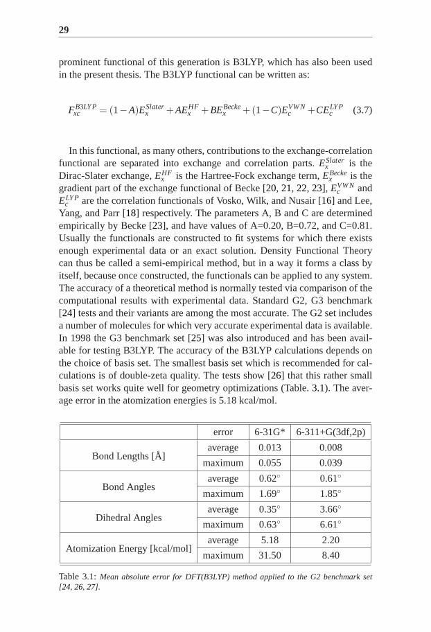
Yang, and Parr [18] respectively. The parameters A, B and C are determinedempirically by Becke [23], and have values of A=0.20, B=0.72, and C=0.81.Usually the functionals are constructed to fit systems for which there existsenough experimental data or an exact solution. Density Functional Theorycan thus be called a semi-empirical method, but in a way it forms a class byitself, because once constructed, the functionals can be applied to any system.The accuracy of a theoretical method is normally tested via comparison of thecomputational results with experimental data. Standard G2, G3 benchmark[24] tests and their variants are among the most accurate. The G2 set includesa number of molecules for which very accurate experimental data is available.In 1998 the G3 benchmark set [25] was also introduced and has been avail-able for testing B3LYP. The accuracy of the B3LYP calculations depends onthe choice of basis set. The smallest basis set which is recommended for cal-culations is of double-zeta quality. The tests show [26] that this rather smallbasis set works quite well for geometry optimizations (Table.3.1). The aver-age error in the atomization energies is 5.18 kcal/mol.
error 6-31G* 6-311+G(3df,2p)
average 0.013 0.008Bond Lengths [Å]
maximum 0.055 0.039
average 0.62◦ 0.61◦Bond Angles
maximum 1.69◦ 1.85◦
average 0.35◦ 3.66◦Dihedral Angles
maximum 0.63◦ 6.61◦
average 5.18 2.20Atomization Energy [kcal/mol]
maximum 31.50 8.40
Table 3.1:Mean absolute error for DFT(B3LYP) method applied to the G2 benchmarkset[24, 26, 27].
30
Using a large basis set, as 6-311+G(3df,2p) [24, 27] the geometry accuracyis improved only slightly, but the average error for the atomization energies ismuch lower, 2.20 kcal/mol (Table.3.1).
Based on the values presented in Table.3.1 and more recent experience[28, 29, 30] it can be concluded that results obtained from B3LYP calcula-tions have enough accuracy for studying the energetics of enzymatic reactions.The systems of interest in the present thesis however contain transition met-als, and the benchmark results discussed above do not include data for suchsystems. Investigations of M-R (M is a first row transition metal and R is H,CH2, CH3 [31] or OH [32]) bond strengths in small cationic systems show anaverage absolute error of 4.9 kcal/mol and a maximum error of 9.0 kcal/molfor B3LYP. This performance can be considered as quite good having in mindthe properties of the transition metals. The presence of degenerate 3d, 4s and4p levels makes them hard to investigate computationally. A modified versionof the B3LYP functional, which includes 15% Hartree-Fock exchange (20%in the original functional), is denoted as B3LYP∗. In cases of transition metalcomplexes such a decrease of the amount of Hartree-Fock exchange can givea better description of the system [33]. Therefore the B3LYP∗ functional hasbeen used extensively in the later work presented in this thesis. In general,states with larger exchange contribution to the total energy (high-spin states),are destabilized by B3LYP∗ due to the reduced amount of the exact exchange.Finally, B3LYP was found useful for studying enzyme reaction mechanismsdue to its adequate accuracy and the speed it provides. The problems ad-dressed in the present thesis consist of comparing alternative hypotheticalmechanisms of certain enzyme reactions. B3LYP calculations give a lot ofdata with satisfactory accuracy, therefore the different hypothetical mecha-nisms can be evaluated and those with the highest activation barriers can berejected.
31
4. Transition State Theory
The subject of investigation presented in this thesis is enzymatic chemical re-actions in which the reactants are transformed into products. The mechanismsof these transformations usually pass through several steps involving differ-ent chemically stable and unstable structures (intermediates, transition states,etc.). Each step is characterized by an unstable structure, connecting the reac-tant and the product, and known as the Transition State (TS). This TS structureis unstable by means of the higher energy it has (G‡), therefore it appears todetermine an energy barrier which the system should overcome in order tocomplete the reaction step. The activation energy barrier∆G‡ is defined as thedifference in the free energies of the reactant and the transition state. The mainapproach in the theoretical study of enzyme catalyzed reaction mechanisms isto calculate the Potential Energy Surface along the reaction coordinate, whichinvolves locating and characterizing the minima and transition states and theirrelative energies.
The Transition State Theory (TST) [34, 35, 36] makes the connection be-tween the calculated data (relative free energies) and the reaction rates whichcome out from experimental measurements. TST postulates that all stable andunstable states along the reaction coordinate obey the Boltzmann energy dis-tribution law. Based on this equilibrium energy distribution the following ex-pression gives the connection between the rate constantk, the temperature andthe reaction barrier:
k =kBT
hexp
(
−∆G‡
RT
)
, (4.1)
wherekB is the Boltzmann constant;h is the Planck constant;T is the absolutetemperature;R is the universal gas constant; and∆G‡ corresponds to the freeenergy of activation. The exponential Eyring equation presented above givesa relation between the experiment and the theory. Experiments measure rateconstants and calculations produce the energy barriers.
According to the Eyring equation, rate constant of 1 s−1 at room tempera-ture corresponds to a barrier of 18 kcal/mol. One order of magnitude of therate constant value corresponds to 1.4 kcal/mol energy barrier. Consideringthe expected error of B3LYP of 3-5 kcal/mol the picture seems not quite op-timistic. The difference in the activation barriers of the hypothetical mech-anisms however, appears to be large enough, often of the order of 10 to 30kcal/mol. Theoretical research thus gives a unique possibility to address such

32
questions, since short-living species like the transition states are accessible forstudies.
33
5. Technical details
The first step in solving the catalytic mechanism is constructing a good modelof the system under investigation. The systems studied experimentally are usu-ally too large for a quantum-chemical treatment. Therefore small models ofabout one hundred atoms need to be constructed. The goal is to build a smallmodel, that is good enough to represent a system consisting of thousands ofatoms.
X-ray crystal structures of huge number of enzymes are available at differ-ent databases, one of the most popular being the Protein Data Bank (PDB).These crystal structures can serve as a good starting point for the modelingprocess. The experience of many researchers in this area shows that in manycases including only the active site residues, the substrate molecule, and thecofactor molecules (in case there are such) into the computational model isenough for an accurate description of the chemistry occurring. The active siteon the other hand consists of a number of amino acid residues and it is of-ten impossible to include all of them in the model. In fact it is common thatonly few of them are directly involved in the catalysis. Another aspect of themodeling is that these few important amino acid residues can be modelledthemselves and thus reduced to smaller molecules. All the amino acids haveanalogous structure – a central,α-carbon atom (orCα ), amino and carboxylgroups attached to it, a hydrogen atom and a side chain (R) attached also to theα-carbon. It is enough to keep only the side chain in the model and remove therest of the residue. Usually even the side chains are modelled using suitableorganic molecules, like imidazole for Histidine, acetate for Glutamine, phenolfor Tyrosine, etc. Tyrosine is even modeled as water in the present calcula-tions for representing its hydrogen-bonding with the substrate. This techniqueis known to give quite adequate results for the purposes of studies of thistype [37].
Although such models include explicitly the part of the enzyme where thechemical reaction takes place, the effects of the rest of the protein are certainlymissing. Two techniques have been utilized in the investigations presentedhere in order to account for the effects of the protein environment: (i) geometryconstraints in the optimization, and (ii) calculations of the solvation effects ofthe protein environment. The latter will be discussed in the last subsection.During the geometry optimization constraints were applied on the terminalatoms in order to fix them to their crystallographically observed positions.This approach turned out to be quite useful especially for the second-shell

34
residues. As they are not directly coordinated to the active site metal centerthey tend to move quite a lot around their original positions, which should notbe the real situation in the enzyme. Introduced by Pelmenschikov [38], thistechnique is now widely used in our group in such kind of studies.
For the biomimetic synthetic complexes the situation is slightly different,and not at all less complicated. When the experimentally used ligand is toobig some of its substituents can be removed from the model. Usually theseare bulky hydrocarbon groups (ethyl, propyl, tert-butyl, etc.). Often the react-ing chemical species can be completely included in the model, which meansno truncation of atoms/groups, and no need for geometry constraints. Thisseems to be an advantage over the enzymatic modelling, but in fact the theo-retical study of catalytic reactions in solution is quite hard. For example, it isnot straightforward to take into account explicit interactions with the solventmolecules, couterions in the solution, base/acid molecules that are usuallypresent in the reaction environment. As a result, the biomimetic complexesmodels are often charged, which complicates the modelling of the solvent ef-fects. Processes where protons enter or leave the reaction site are also hard tomodel. In the enzymes all these characteristic chemical features are "broughttogether" in the active site by the protein structure.
Once the model is constructed, different reaction mechanisms can beprobed by finding all the intermediates and transition states along the putativereaction coordinate, and plotting the energy diagram.
For all models described in this thesis, the geometry optimizations wereperformed using the B3LYP method as implemented in the Jaguar [39] pro-gram package. The optimizations were performed in gas phase with a double-ζ quality basis set (lacvp in Jaguar), which includes an Effective Core Po-tential (ECP) on iron [40]. According to the benchmark data (Table3.1) thisdouble zeta basis set is accurate enough in terms of geometric structures. Thecomplicated and time consuming part of any theoretical study of chemicalreactions is locating the TS. By systematically freezing one or more internalcoordinates and optimizing the other degrees of freedom, the reaction coordi-nate can be scanned. The energy maximum in the scanned surface provides agood guess for the TS, and this structure is put in the TS optimization job. TheTS is defined as a structure which has a minimal energy with respect to all co-ordinates except one – the reaction coordinate. Along the reaction coordinatethe energy takes maximum at that point and thus the second derivative in theHessian is negative, which corresponds to an imaginary vibrational frequency.In this sense the imaginary frequency represents movement along the reactioncoordinate. Also, calculation of the molecular Hessian for the reactant, the TS,the intermediates, and the products, are used to estimate the Zero-Point vibra-tion Energy (ZPE), and the entropy contributions to the total energy. Entropyeffects cannot be described accurately when some coordinates in the model
35
are fixed, therefore they are not included in the results for the enzymatic sys-tems. These effects should play a minor role for the chemistry in the course ofthe reaction, but will be important when the dioxygen molecule binds at theactive site. In the present thesis, frequency calculations and transition stateop-timizations have been performed using Gaussian03 [41], at the B3LYP/lacvplevel of theory.
As it was stated above the B3LYP geometries are not very sensitive to thebasis set quality [29]. Relative energies on the other hand are particularlymuch more sensitive to the basis set, therefore it is a usual practice to ap-ply larger basis sets for obtaining accurate energies. In the present studies thisis done by performing single point calculations on the lacvp-optimized ge-ometries using triple-ζ basis set –cc-pVTZ(-f)(without f-functions) in Jaguar.This correlation-consistent polarized basis set, which is intrinsically polarizedbut does not include ECP, was used for all atoms except iron. For iron thetriple zeta quality basis setlacv3p∗∗ was explicitly used.
The DFT method is limited to a single determinant. Open-shell low-spinstates are linear combinations of different spin determinants and cannot bedescribed accurately by a single determinant. The calculated low-spin statesare therefore spin contaminated by higher spin states. The broken symmetryapproach based on the Heisenberg Hamiltonian and introduced by Noodle-man [42, 43] was used to correct the energy of the low-spin states.
Geometry optimizations and the large basis sets single point calculationsare performed in gas phase. This model is apparently missing the effects ofthe protein environment where the enzymatic reactions occur. An importantpart in such an investigation is to consider the effect of the environment. Acommon way to compute the influence of the surrounding environment is byperforming a single point calculation using the Polarizable Continuum Model(PCM), as implemented in Jaguar [44, 45].
A dielectric constant (ε) equal to 4.0 is used to set up the surrounding en-vironment in the studies of enzymatic reactions. This value has been empiri-cally determined, and it reproduces an environment resulting from a dielectricconstant of about 3 for the protein itself and about 80 for the water mediumsurrounding the protein [46]. When modeling the biomimetic complexes, thedielectric constant of the solvent used in the experiment was applied in thecalculations. For the intradiol cleaving non-heme iron complex was used avalue ofε=37.5, corresponding to acetonitrile. For the adipic acid synthesisreactionε=2.0 was used, which is the dielectric constant for cyclohexane.
Usually, the dielectric effects are not expected to change the relative en-ergies dramatically, except in cases of reactions where charge separation oc-curs [47, 48]. Large effects are considered to be a sign of a problem with thechosen model. The dielectric continuum model cannot account for short-rangesolute-solvent interactions such as hydrogen bonds [49, 50, 51, 52]. Therefore

36
one should have in mind that it gives a kind of qualitative results, and it isalways better to include hydrogen bonds explicitly in the model, if possible.
Part III:Dioxygenases
The last part of the catabolism of aromatic compounds in the environ-ment involves cleavage of the aromatic ring and insertion of both dioxygenatoms into the product, a reaction known as dioxygenation. In Nature suchspin-forbidden reactions with participation of molecular oxygen, are catalyzedby enzymes containing transition metal ions as cofactors. Transition metalshave low-lying electronic states with unpaired electrons, and very often theyhave high-spin electronic ground states. The large spin-orbit coupling of thesespecies allows for the spin changes required for such type of reactions.
The enzymes able to activate molecular oxygen and incorporate both of itsatoms in the aliphatic product are known as dioxygenases [53, 54, 55]. Theyare usually isolated from a variety of soil bacteria.


39
6. Extradiol and intradiol catecholdioxygenases
Ortho-hydroxyl dioxygenases are divided into two classes, according to thethe position at which the aromatic ring is cleaved, namely extradiol and intra-diol dioxygenases. Extradiol dioxygenases cleave one of the C-C bonds adja-cent to the hydroxyl groups of the substrate. They contain Fe(II) or Mn(II) asa cofactor, coordinated by a 2-His-1-carboxylate facial triad [56], and solventmolecules in square bipyramidial geometry. Intradiol dioxygenases cleave thering between the two adjacent hydroxyl substituents and utilize Fe(III) in theiractive site. The iron is coordinated by two histidines, two tyrosines and a hy-droxide. In both cases the organic substrate binds first to the metal ion, fol-lowed by binding of dioxygen. A subsequent attack on the aromatic ring leadsto the bridging peroxide, which is believed to be common for the extradioland intradiol catechol dioxygenases. After O-O bond cleavage one of the oxy-gen atoms is incorporated into the catechol ring via alkenyl migration in theextradiol path, or acyl migration in the intradiol path (Scheme6.1).
Fe2+
HO
H2O
His
His
Glu
H2O
Fe3+HOHis
Tyr
His
Tyr
Fe2+OHis
Tyr
His
O
R
Fe2+
His
His
GluO
HO
R
Fe3+O
His
Tyr
His
O
R
O
O
Fe3+
His
Tyr
His
OX
HO
O OH
OR
34
34
43
O
O
OR
HO
Fe2+
His
His
GluO
O
R
O
O
Fe2+
His
His
Glu
OH
O
O
R
O
O
HORO OH
3
3
3
4
4
4
Extradiol Intradiol
1 2
3
4
1 2
3
4
Scheme 6.1:Proposed catalytic cycles for extra- and intradiol cleaving dioxygenases.
This thesis presents theoretical studies of the reaction mechanisms ofthree dioxygenase enzymes: Homoprotocatechuate 2,3-dioxygenase isolatedfrom Brevibacterium fuscum, Manganese-Dependent Homoprotocatechuate2,3-Dioxygenase, and Homogentisate Dioxygenase.
40
A)
66
11
22
3344
55
OH
OH
COO-
O 2
Bf 2,3-HPCD / MndD 66
11
CHO22
COO-33
44
55
OH
COO-
HPCA 1-carboxymethyl-4-hydroxy cis-muconic semialdehyde
B)
6611
22
3344
55
OH
OH
O2HGD 66
11
22
O
334455
O
OH
HG maleylacetoacetate
O
O O
O O
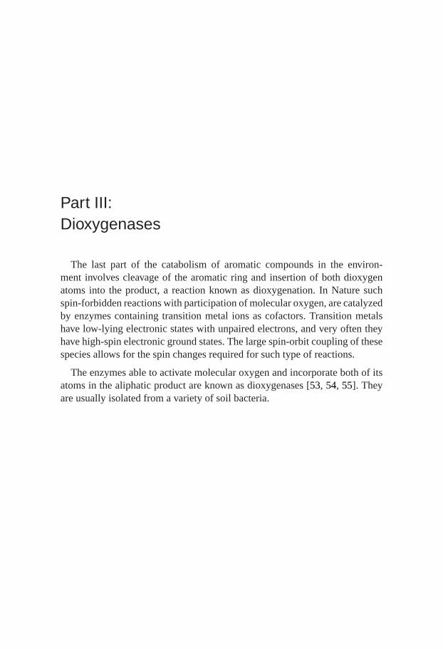
Scheme 6.2:The overall reaction catalyzed byA: homoprotocatechuate dioxygenasesBf 2,3-HPCD and MndD; andB: Homogentisate Dioxygenase HGD.
B f 2,3-HPCD and MndD are metalloenzymes that use molecular oxygen tocleave the aromatic ring of 3,4-dihydroxyphenylacetate, also known as homo-protocatechuate (HPCA), in an extradiol manner. Homogentisate dioxygenaseis an enzyme involved in the catabolism of tyrosine and phenylalanine in hu-man [57]. It catalyzes the oxidative ring scission of Homogentisate (HG) tomaleylacetoacetate. The type of ring cleavage (between the carbons C1 andC2 substituted with hydroxyl and carboxymethyl groups, see Scheme6.2 B)resembles the reaction catalyzed by Fe(III)-dependent intradiol dioxygenases.However, the dependence on Fe(II) and the structure of the active-site indicatethat HGD is more related to the extradiol dioxygenases, which cleave the cat-echol ring at a bond adjacent to the carbons binding the hydroxyl groups. Thereactions catalyzed byBf 2,3-HPCD and MndD, and by HGD are summarizedin Scheme6.2A andB, respectively.
The selectivity in the ring cleavage reaction, shown by extra- and intradioldioxygenases is still not well understood. The metal ion requirement is aninteresting issue –Bf 2,3-HPCD and MndD are highly identical in terms ofamino acid sequence and they catalyze the same reaction, but they use a dif-ferent metal as cofactor – Fe(II) and Mn(II), respectively. Studying these sys-

41
tems is important for improving our understanding of catalysis. Apart fromthat, HGD is interesting from a medical point of view. This enzyme is con-nected with two metabolic disorders – alkaptonuria (a rare hereditary humandisease [58]) and tyrosinemia type I (a life-threatening disease caused by dys-function of fumarylacetoacetase, an enzyme located downstream from HGDin the tyrosine catabolic pathway). In the studies presented in this part, hybridDFT with the B3LYP functional was used with models of different size builtfrom the crystal structures of the enzymes. The energetics of the suggestedreaction path is investigated by locating intermediates and transition statesand evaluating their relative energies, including environmental effects of thesurrounding protein.

43
7. Fe- and Mn-dependenthomoprotocatechuate 1,2-dioxygenases
This section summarizes the results originally reported inPaper II andPaperIII .
7.1 BackgroundBf 2,3-HPCD and the manganese dependent analogue MndD have 83% se-quence identity [59]. In both enzymes the active-site metal ion is coordinatedby His155, His214 and Glu267 forming the 2-His-1-carboxylate facial triadoccupying one face of an octahedron. This motif, typical for a number of non-heme Fe(II)-dependent enzymes involved in dioxygen activation [60, 61], isconsidered as a very important factor providing the right environment for thedioxygenation. The remaining three coordination positions of the octahedralcomplex are occupied by solvent molecules and are available for the sub-strates – Homoprotocatechuate (HPCA) and dioxygen. High-resolution X-raystructures for both iron- and manganese-dependent homoprotocatechuate 2,3-dioxygenases have been determined, and spectroscopic and steady state ki-netic investigations have been performed, revealing important structural andmechanistic features of these biocatalysts. The activation barrier for both en-zymes, predicted from the experimental data using transition state theory isabout 16 kcal/mol [62, 63].
According to experimental data extradiol dioxygenases use an orderedmechanism for binding substrates. The aromatic substrate first binds,which activates the metal ion for O2 binding. The O2 adduct has not beendetected and characterized up to date for the natural substrate. There ishowever evidence for NO binding [64, 65] supporting this scenario. Thecrystal structure of the enzyme with HPCA bound in the active-site (PDBcode:1QOC; Figure7.1 for Bf 2,3-HPCD) reveals that the substrate fills twoof the free coordination sites,transto the histidines, sites previously occupiedby water ligands [59]. The remaining sixth coordination site is empty inBf2,3-HPCD, while it is occupied by a water ligand in MndD. This position isopen for accommodation of O2 later. HPCA binds to the metal center by its
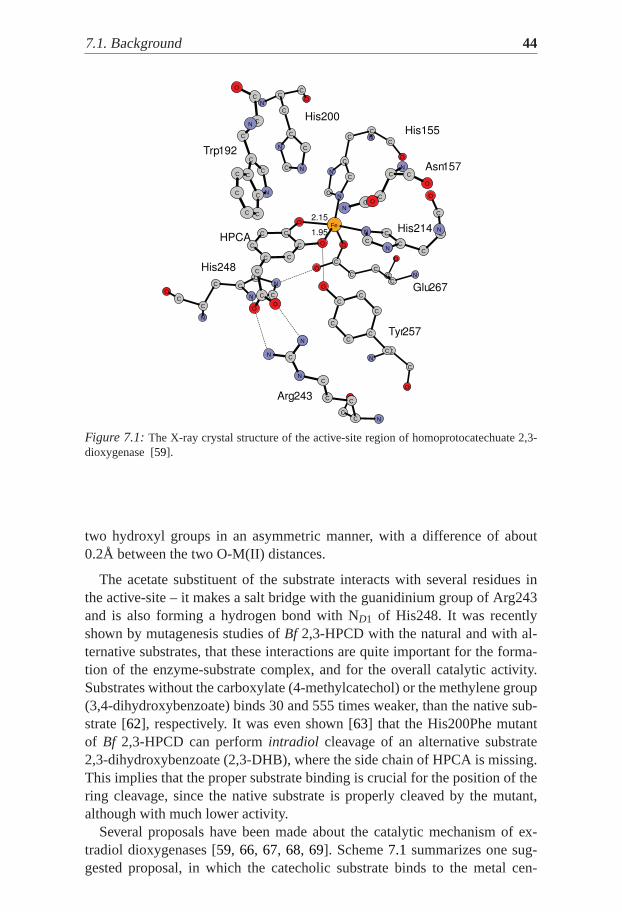
7.1. Background 44
O
C
N
CNC
N
C
N
C
C
O
C
O
C
C
O
O
O
C
N
C
C
N
O
C
C
N
C
ON
C
N
C
C
C
C C
C
C
O
C
N
C
C
Fe
C
C
N
CC
C
C
C
CN
C
C
C
C N
O
C
O
C
C
O
C
C
CCC
N
N
C
C
NC
C
O
N
N
N
C
C
C
N
C
C
C
C
C
O
C
N
C
O
O
C
C C
C
N
O
C
C
C
His200
His155
Asn157
His214
Glu267
Tyr257
Arg243
His248
Trp192
HPCA
2.15
1.95
Figure 7.1:The X-ray crystal structure of the active-site region of homoprotocatechuate 2,3-dioxygenase [59].
two hydroxyl groups in an asymmetric manner, with a difference of about0.2Å between the two O-M(II) distances.
The acetate substituent of the substrate interacts with several residues inthe active-site – it makes a salt bridge with the guanidinium group of Arg243and is also forming a hydrogen bond with ND1 of His248. It was recentlyshown by mutagenesis studies ofBf 2,3-HPCD with the natural and with al-ternative substrates, that these interactions are quite important for the forma-tion of the enzyme-substrate complex, and for the overall catalytic activity.Substrates without the carboxylate (4-methylcatechol) or the methylene group(3,4-dihydroxybenzoate) binds 30 and 555 times weaker, than the native sub-strate [62], respectively. It was even shown [63] that the His200Phe mutantof Bf 2,3-HPCD can performintradiol cleavage of an alternative substrate2,3-dihydroxybenzoate (2,3-DHB), where the side chain of HPCA is missing.This implies that the proper substrate binding is crucial for the position of thering cleavage, since the native substrate is properly cleaved by the mutant,although with much lower activity.
Several proposals have been made about the catalytic mechanism of ex-tradiol dioxygenases [59, 66, 67, 68, 69]. Scheme7.1 summarizes one sug-gested proposal, in which the catecholic substrate binds to the metal cen-
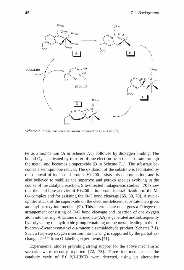
45 7.1. Background
O
C
COO-
OH
O
OH
HO
O-
COO-
His200
M2+
His214
Glu267
OO
O
O
COO-
H
His200
M2+
OO
C
O
O
COO-
H
His200
M2+
OO
O
O
COO-
H
His200
M3+
O
O
His155
C
substrate
A
product
D
B
Scheme 7.1:The reaction mechanism proposed by Que et al. [59].
ter as a monoanion (A in Scheme7.1), followed by dioxygen binding. Thebound O2 is activated by transfer of one electron from the substrate throughthe metal, and becomes a superoxide (B in Scheme7.1). The substrate be-comes a semiquinone radical. The oxidation of the substrate is facilitated bythe removal of its second proton. His200 assists this deprotonation, and isalso believed to stabilize the superoxo and peroxo species evolving in thecourse of the catalytic reaction. Site-directed mutagenesis studies [70] showthat the acid-base activity of His200 is important for stabilization of the M-O2 complex and for assisting the O-O bond cleavage [65, 69, 70]. A nucle-ophilic attack of the superoxide on the electron-deficient substrate then givesan alkyl-peroxy intermediate (C). This intermediate undergoes a Criegee re-arrangement consisting of O-O bond cleavage and insertion of one oxygenatom into the ring. A lactone intermediate (AA ) is generated and subsequentlyhydrolyzed by the hydroxide group remaining on the metal, leading to theα-hydroxy-δ -carboxymethylcis-muconic semialdehyde product (Scheme7.1).Such a two-step oxygen insertion into the ring is supported by the partial ex-change of18O from O-labeling experiments [71].
Experimental studies providing strong support for the above mechanisticscenario were recently reported [72, 73]. Three intermediates in thecatalytic cycle of B f 2,3-HPCD were detected, using an alternative

7.2. The model 46
substrate 4-nitrocatechole (4NC). The ternary complex of the enzyme withthe semiquinone substrate and side-on bound dioxygen (correspondingto intermediateB in Scheme 7.1), and the alkylperoxo intermediate(corresponding toC in Scheme7.1) were shown residing in different subunitsof a single enzyme molecule.
7.2 The model
Small models including only the most relevant parts of the residues directlycoordinated to the metal center (His155, His214, Glu267 and Tyr257), and thetruncated substrate molecule, were used initially to reduce the computationalcosts. Such models were however found insufficient for proper modeling ofthe enzymatic reaction. Although the role of His200 is not yet completelyclear, this residue is undoubtedly important for the catalytic activity. Since itis assumed that the initial proton transfer from HPCA to the superoxide is as-sisted by His200, its inclusion in the model is logical. The electron transferfrom the substrate to O2 could be better reproduced using the whole sub-strate molecule, than with the truncated model (Paper II ). Furthermore, itwas found necessary to include His248 in protonated form, in order to main-tain the neutral charge of the model. This residue also hydrogen-bonds withthe carboxylates of Glu267 and HPCA and thus keeps the whole structurecloser to the X-ray geometry. Asn157 was also added, since it provides somestabilization to the superoxide moiety. Thus a model consisting of 77 atomswas constructed and used for bothBf 2,3-HPCD and MndD. All the resultsgiven in the next sections were obtained with this model, unless the model isexplicitly specified.
7.3 Reaction mechanism of B f 2,3-HPCDThe calculatedBf 2,3-HPCD catalytic cycle is described in details inPaperIII and basically follows the experimentally suggested one in Scheme7.1.The HPCA substrate first binds at the active-site as a monoanion. Next thedioxygen molecule binds to the complex. The calculated binding energy ofO2 is +2.9 kcal/mol (solvent effects included), which would become around+13 kcal/mol if the entropy is added. A correction of about -10 kcal/mol tothe QM results was applied and thus the estimate is that dioxygen is unboundby about 3 kcal/mol. Three possible spin states were considered for the adductwith O2 – triplet, quintet and septet. The septet state (S=3) was found to bethe ground state, but the state of main interest was the quintet (S=2). In thequintet state the spin population is 3.75, 0.90 and -0.80 for iron, dioxygen andthe substrate, respectively, i.e. dioxygen is reduced with one electron to su-
47 7.3. Reaction mechanism ofB f 2,3-HPCD
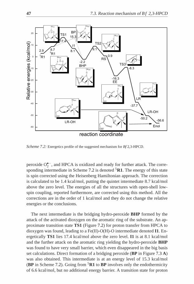
Scheme 7.2:Energetics profile of the suggested mechanism forBf 2,3-HPCD.
peroxide O•−2 , and HPCA is oxidized and ready for further attack. The corre-sponding intermediate in Scheme7.2 is denoted5R1. The energy of this stateis spin corrected using the Heisenberg Hamiltonian approach. The correctionis calculated to be 1.4 kcal/mol, putting the quintet intermediate 8.7 kcal/molabove the zero level. The energies of all the structures with open-shell low-spin coupling, reported furthermore, are corrected using this method. All thecorrections are in the order of 1 kcal/mol and they do not change the relativeenergies or the conclusions.
The next intermediate is the bridging hydro-peroxideBHP formed by theattack of the activated dioxygen on the aromatic ring of the substrate. An ap-proximate transition stateTS1 (Figure7.2) for proton transfer from HPCA todioxygen was found, leading to a Fe(II)-O(H)-O intermediate denotedI1. En-ergeticallyTS1 lies 17.4 kcal/mol above the zero level.I1 is at 8.1 kcal/moland the further attack on the aromatic ring yielding the hydro-peroxideBHPwas found to have very small barrier, which even disappeared in the big basisset calculations. Direct formation of a bridging peroxide (BP in Figure7.3A)was also obtained. This intermediate is at an energy level of 15.3 kcal/mol(BP in Scheme7.2). Going from5R1 to BP involves only the endothermicityof 6.6 kcal/mol, but no additional energy barrier. A transition state for proton
7.3. Reaction mechanism ofB f 2,3-HPCD 48
N
NC
C
C
C
C
N
C
N
O
O
C
C
C
OFe
N
C
CC
O
CN
C
N
C O
O
C
CC
C
O
C C
N
O
C
C
N
C
O
O
O1=0.42
Fe=3.75
O2=0.58
2.17
1
2
2.241.33
1.32
2.10
2.481.78
1.05
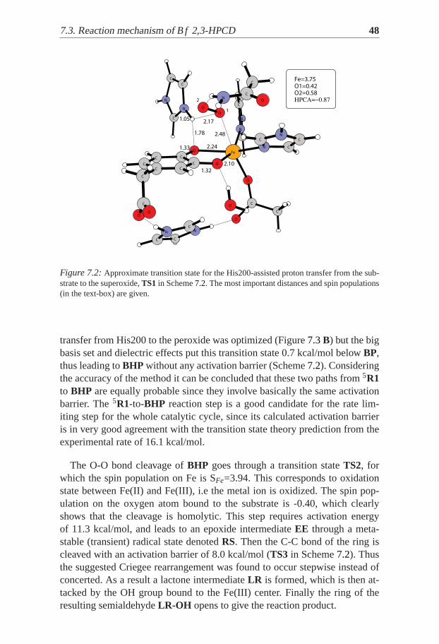
Figure 7.2:Approximate transition state for the His200-assisted proton transfer fromthe sub-strate to the superoxide,TS1 in Scheme7.2. The most important distances and spin populations(in the text-box) are given.
transfer from His200 to the peroxide was optimized (Figure7.3B) but the bigbasis set and dielectric effects put this transition state 0.7 kcal/mol belowBP,thus leading toBHP without any activation barrier (Scheme7.2). Consideringthe accuracy of the method it can be concluded that these two paths from5R1to BHP are equally probable since they involve basically the same activationbarrier. The5R1-to-BHP reaction step is a good candidate for the rate lim-iting step for the whole catalytic cycle, since its calculated activation barrieris in very good agreement with the transition state theory prediction from theexperimental rate of 16.1 kcal/mol.
The O-O bond cleavage ofBHP goes through a transition stateTS2, forwhich the spin population on Fe is SFe=3.94. This corresponds to oxidationstate between Fe(II) and Fe(III), i.e the metal ion is oxidized. The spin pop-ulation on the oxygen atom bound to the substrate is -0.40, which clearlyshows that the cleavage is homolytic. This step requires activation energyof 11.3 kcal/mol, and leads to an epoxide intermediateEE through a meta-stable (transient) radical state denotedRS. Then the C-C bond of the ring iscleaved with an activation barrier of 8.0 kcal/mol (TS3 in Scheme7.2). Thusthe suggested Criegee rearrangement was found to occur stepwise instead ofconcerted. As a result a lactone intermediateLR is formed, which is then at-tacked by the OH group bound to the Fe(III) center. Finally the ring of theresulting semialdehydeLR-OH opens to give the reaction product.

49 7.4. Reaction mechanism of MndD. Spin transition
N
C
C
C
C
O
C
O
N
C
N
C
N
N
C
C
O
Fe
C
C
CN
N
C
O
O
C
C
O
C C
O
C N
O
CC
C
C
C
C
N
O
O
2
O2=0.01O1=0.07Fe=3.74
1.30
1.50
1
2.15
1.66
2.08
1.45
1.11
N
C
C
C
C
O
O
C
N
N
C
C
N
N
C
C
O
Fe
C
C
C
N
N
CC
O
O
C
O
C C
O
CN
O
CC
C
C
C
C
N
O
O
O1=0.06Fe=3.74
O2=0.01
1
2
2.09
2.67
1.30 2.17
1.66
1.50
1.08
1.55
A B
Figure 7.3: Optimized structures ofA: bridging peroxideBP; B: transition state for protontransfer form His200 to the bridging peroxide. The most important distances and spin popula-tions (in the text-box) are given.
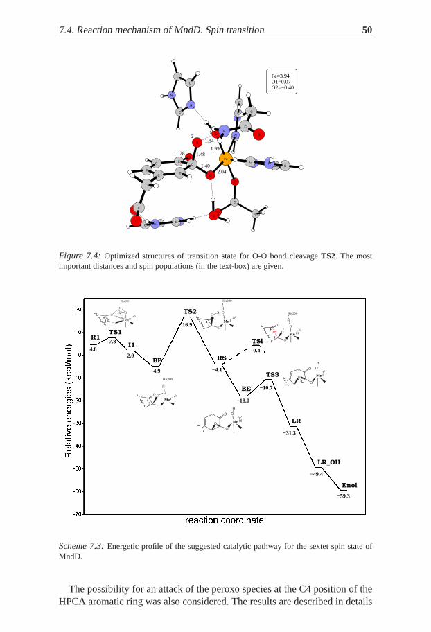
7.4 Reaction mechanism of MndD. Spin transitionThe energy profile shown in Figure7.3 was obtained in the initial study ofMndD, described inPaper II . The calculated catalytic cycle for the sextet spinstate follows the experimentally suggested path (Scheme7.1) as in the case ofBf 2,3-HPCD. In summary, the HPCA substrate first binds at the active-siteas a monoanion, followed by dioxygen. The latter is activated to a superoxoradical by an electron flow from the substrate through the metal center, andreacts with HPCA forming the bridging hydro-peroxide structureBHP at theC3 position of the ring (Figure7.5 A). Importantly, this intermediate can beformed only after the preceding protonation of O2. In the calculations this pro-tonation is modeled by a proton transfer from the C4 bound hydroxylic groupof HPCA, assisted by the second shell His200, i.e. the complete deprotona-tion of the substrate is reached. It should be noted here that based on recentmutagenesis experiments, reported by Emerson et al. [70], it was argued thatHis200 mainly stabilizes O2 in the enzyme-substrate-dioxygen complex. Bothpossibilities, that His200 can act as a base to deprotonate the second hydrox-ide group of the substrate, or as an acid protonating the reduced oxygen werefound unlikely by Emerson’s study. Those results show that the key role ofthe residue in this position is related to its ability for making hydrogen bonds,since the mutants having His200 replaced by asparagine or glutamine pre-serve 60-80% of the native activity. The calculations rather show that His200can facilitate the proton transfer and can further stabilize the Mn(II) boundhydroperoxide. In that sense, there is no conflict between the computationalresults and the experimental findings, because there is no net change of theprotonation state of His200 in the proton shift.
7.4. Reaction mechanism of MndD. Spin transition 50
N
C
C
O
N
C
C
C
N
C
C
O
C
NC
O
C
N
Fe
CN
CCC
O
N
O
C
C
O
C
O
O
C
CC
N
C
C
C
C
O
NO
O1=0.07
1
1.84
2.041.40
Fe=3.94
O2=−0.40
2
1.991.28 1.48
Figure 7.4: Optimized structures of transition state for O-O bond cleavageTS2. The mostimportant distances and spin populations (in the text-box) are given.
Mn2+
O33
44O
H
His200
OO
~
Mn2+
O33
44O
H
His200
O
O~
O
O
H
Mn2+
O
O
O
O
H
Mn2+
O
O~
Mn2+
O33
44
O
H
His200
OO
MnIII
O33
44O
H
His200
OO
4.87.8
2.0
R1TS1
I1
TS2
RS
TSi
TS3
LR
LR_OH
Enol
−59.3
16.9
−4.9 −4.1
4.5
−31.3
−49.4
BP
−18.0
EE −10.7
0.4
Mn III
MnIII
Mn III
MnII
IIMn
Scheme 7.3:Energetic profile of the suggested catalytic pathway for the sextet spin stateofMndD.
The possibility for an attack of the peroxo species at the C4 position of theHPCA aromatic ring was also considered. The results are described in details

51 7.4. Reaction mechanism of MndD. Spin transition
in Paper II . A bridging peroxide at the C4 position can be formed before,as well as after the preliminary protonation of the superoxo radical. Thesealternative reaction paths however either involve higher activation barriers,or lead to unstable products, compared with the C3-attack and formation ofBHP.
N
C
C
C
O
C
N
O
C
N
CC
C
NC
O Mn
N
C
C
C
N
C
CN
O
OC
C
O
C
O
C
O
CC
N
C
C
C
O
NC
O
*His248
Tyr257
*
Glu267
*
His214
*
Asn157
*
*
His155
*
His200
1.71
1.60 S=4.8
1.60
1.52
2.11
1.27
N
C
C
O
N
C
C
N
O
C
C
CC
N
O
C
C
N
Mn
C
CC
N
CN
O
O
O
C C C
O
O
C
C C
N
C
O
C
C
NC
O
*His248
*Tyr257
*Glu267
His214
Asn157*
*
*
His155His200
1.78
1.27 S=4.4
S=0.5 1.87
2.06
1.64
1.49
2.07
*
A B
Figure 7.5: Optimized structures ofA: the bridging hydro-peroxideBHP; B: the transitionstate for O-O bond cleavage. The most important spin populations (bold)and distances (italics)are given.
The next step – the homolytic O-O bond cleavage was found to be the ratelimiting step in the suggested mechanism, with an activation barrier of 21.8kcal/mol (Scheme7.3). It leads to a transient radical intermediateRS, whicheasily collapses to an extra epoxide structureEE) going 13.9 kcal/mol downin energy. At this stage, the competitive intra cleavage reaction was found tobe possible. Thus, the calculated barrier for intradiol cleavage is 4.5 kcal/mol(seeTSi in Scheme7.3), appearing to determine the selectivity between bothreactions. However, some uncertainty about the origin of this difference re-mains, since the removal of His248 or Asn157 separately does not change thepicture, while their simultaneous removal leads to the disappearance of theintra cleavage barrier. The C-C bond cleavage of the extra epoxideEE goesover a barrier of 7.3 kcal/mol, leading to the lactone ring intermediateLR with13.3 kcal/mol exothermicity. Next, the Mn(III) bound hydroxyl group attacksthe lactone ring without any significant barrier, going another 18.1 kcal/moldown in energy to the intermediate denoted asLR-OH in Scheme7.3. Thefinal steps of the reaction including the ring opening of theLR-OH interme-diate and the product formation do not involve any significant barriers andwere not studied in details.
The above results were obtained following the potential energy surface ofthe sextet spin state, which is the ground state of the resting enzyme. The

7.4. Reaction mechanism of MndD. Spin transition 52
relatively high calculated activation barrier of 21.8 kcal/mol with respecttothe experimentally observed 16.3 kcal/mol, was initially assigned to the usualtendency of the B3LYP method to slightly overestimate the reaction barriers.The results obtained forBf 2,3-HPCD, however, clearly suggest that additionalinvestigation should be carried out for the MndD enzyme. These studies arereported inPaper III . The attention was focused on the O-O bond cleavagestep, where a large difference between the Fe- and Mn-containing enzymes ofalmost 10 kcal/mol was obtained. A large number of calculations with differ-ent models were performed attempting to clarify the origin of this difference.The main result is that the difference of about 10 kcal/mol betweenBf 2,3-HPCD and MndD, regarding the activation barrier for O-O bond cleavage,was reproduced with all models probed. No significant geometrical differencewas found in any model between the corresponding intermediates and tran-sition states of the two enzymes. The only difference between the two caseswas the coupling of the spin of the radical substrate (subO•) with the spin ofthe metal ion. In theBf 2,3-HPCD case this spin was always antiferromagnet-ically coupled to the unpaired electrons of iron, while in MndD this couplingwas always ferromagnetic (Table7.1).
spin SM SsubO•
TS2Bf2,3−HPCD 3.94 -0.40
TS2MndD 4.40 0.50
Table 7.1:Spin populations on the metal ionM and subO• in the corresponding O-O bondcleavage transition states found for Bf 2,3-HPCD and MndD.
Antiferromagnetic coupling in MndD is possible for the quartet spin state(S=3/2). Therefore the quartet state potential energy surface for MndD wasinvestigated for the O-O bond cleavage from theBHP reactant to theEE prod-uct. The quartetBHP is 24.2 kcal/mol higher in energy than the correspondingsextet structure, while the quartetEE is as stable as the sextet product. Thus itwas assumed that the activation barrier for the homolytic O-O bond cleavagein MndD could actually be determined by a spin transition from the sextetto the quartet state, occurring at lower energy than the sextet transition state.Such spin transition should occur in a region where the two spin surfacescross. A crossing point slightly before the sextet transition state (Figure7.6A) was found in a rough scan along the O-O bond on both PESs. Thelacvpenergy of this point is around 16 kcal/mol (relative to the sextet6BHP). Withthis approximate crossing as a starting point, two interatomic distances werechosen as reaction coordinates, namely the O-O and Mn-O distances, and atwo-dimensional potential energy surface scan was done. These two distances
53 7.4. Reaction mechanism of MndD. Spin transition
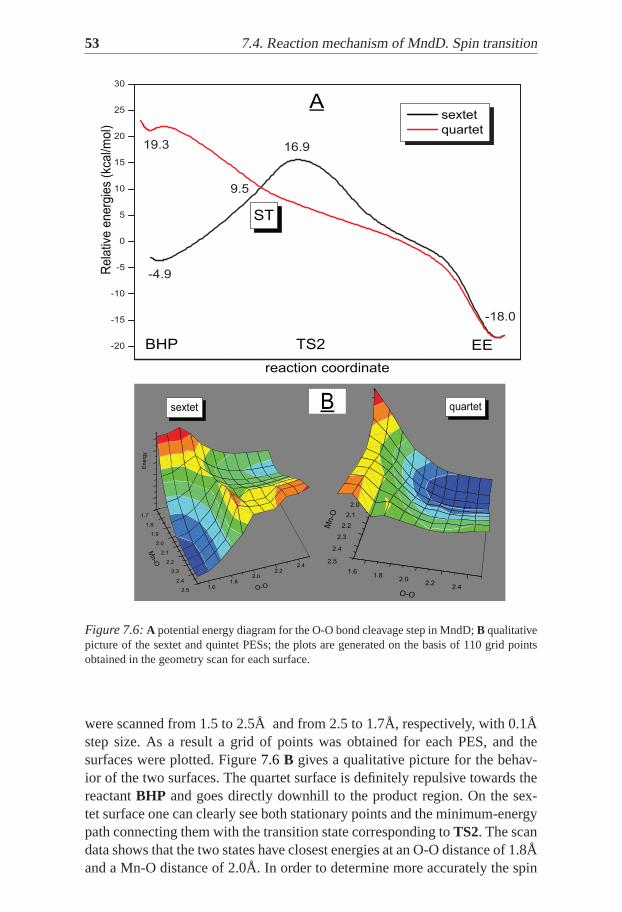
Figure 7.6:A potential energy diagram for the O-O bond cleavage step in MndD;B qualitativepicture of the sextet and quintet PESs; the plots are generated on the basisof 110 grid pointsobtained in the geometry scan for each surface.
were scanned from 1.5 to 2.5Å and from 2.5 to 1.7Å, respectively, with 0.1Åstep size. As a result a grid of points was obtained for each PES, and thesurfaces were plotted. Figure7.6 B gives a qualitative picture for the behav-ior of the two surfaces. The quartet surface is definitely repulsive towards thereactantBHP and goes directly downhill to the product region. On the sex-tet surface one can clearly see both stationary points and the minimum-energypath connecting them with the transition state corresponding toTS2. The scandata shows that the two states have closest energies at an O-O distance of 1.8Åand a Mn-O distance of 2.0Å. In order to determine more accurately the spin
7.4. Reaction mechanism of MndD. Spin transition 54
transition geometry a higher resolution geometry scan (step size 0.02Å) wasperformed around this point. Big basis set calculations were carried out foreach geometry for obtaining more accurate energies. As a result a point wasfound with an O-O distance of 1.80Å and a Mn-O distance of 2.04Å , andwith an energy of 14.4 kcal/mol relative to the reactantBHP. Attempts to findthe Minimum-Energy Crossing Point (MECP) [74] were made using a sep-arate program [75]. Since this program looks not just for any crossing pointbetween two surfaces, but for the minimum-energy crossing point, the searchactually ended up in the next crossing point, where the system comes back tothe sextet spin state. This point turned out to be theEE epoxide product of thecleavage (Figure7.6A).
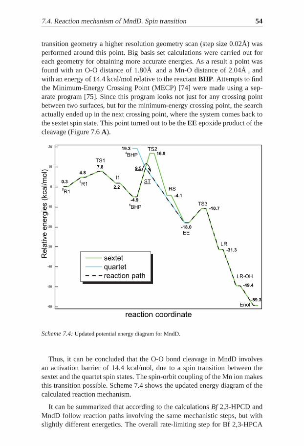
Scheme 7.4:Updated potential energy diagram for MndD.
Thus, it can be concluded that the O-O bond cleavage in MndD involvesan activation barrier of 14.4 kcal/mol, due to a spin transition between thesextet and the quartet spin states. The spin-orbit coupling of the Mn ion makesthis transition possible. Scheme7.4shows the updated energy diagram of thecalculated reaction mechanism.
It can be summarized that according to the calculationsBf 2,3-HPCD andMndD follow reaction paths involving the same mechanistic steps, but withslightly different energetics. The overall rate-limiting step for Bf 2,3-HPCA

55 7.4. Reaction mechanism of MndD. Spin transition
is the formation of the alkylperoxo intermediateBHP. For MndD a spin tran-sition from the sextet to the quartet spin state afterBHP was found to deter-mine a barrier of 14.4 kcal/mol for the homolytic O-O bond cleavage. Thusthe experimentally observed similarities between Bf 2,3-HPCD and MndDare reproduced in the calculations, and some important differences could bedetermined.

57
8. Homogentisate Dioxygenase
This section summarizes the results originally reported inPaper I.
8.1 BackgroundDespite the considerable importance of HGD, only recently was someprogress made in the research on this interesting enzyme. The crystalstructure of human HGD was solved by Titus and co-workers [76]. As inother extradiol dioxygenases, the active-site iron is coordinated by twohistidines (His335, His371) and one glutamate (Glu341), which form the2-His-1-carboxylate facial triad. The other three coordination positionsaround iron are available for Homogentisate and dioxygen or solvent ligands.Several other residues were proposed to be involved in substrate bindingand catalysis. First, His365 forms a hydrogen bond with Glu341 and maybe involved in substrate deprotonation upon binding. Second, His292 wasproposed to form a hydrogen bond with the C4-bound hydroxyl group of HG,and together with Pro295, Pro332, and Tyr333 forms a solvent-accessiblebox, which was also implicated for the substrate binding.
O
O
OH
O
O2
maleylacetoacetate
Fe2+
His292
O
O
OH
O
Fe2+
His292
OO
O
O
O
Fe2+
HisH292
OHO
O
O
O
O
Fe2+
His292
OH
OH
O
O-
O
O
OH
O
Otautomerization
1
23
45
6
Scheme 8.1:Reaction Mechanism proposed by Titus et al. for HGD.

8.2. The model 58
Recently published kinetic analysis has provided firm evidence for an or-dered mechanism, in which HG binds first to the enzyme followed by a dioxy-gen reacting with the Fe(II)-HGD-HG complex [77, 78]. Furthermore, an op-timum pH (6.2) and a turnover number (16s−1) were measured. Aside fromthis, little is known about the catalytic mechanism of HGD. The mechanismpresented in Scheme8.1 was proposed [76], and with the exception for thechemical identity of the substrate and the involvement of His292, it is verysimilar to the mechanism proposed by Lipscomb and co-workers for extradioldioxygenases [65, 69]. Thus, it is proposed that HG chelates Fe(II) with itscarboxyl and deprotonated hydroxyl groups. Molecular dioxygen reacts withthis HGD-HG complex and forms a peroxo intermediate with an O-O bridgejoining the C2 carbon and Fe(II). In the next step, the peroxo intermediateundergoes a Criegee rearrangement, yielding a seven-membered lactone ring,protonated His292, and a hydroxyl group bound to Fe(II). Finally, the lactonering is hydrolyzed to an enol form of the product, maleylacetoacetate. Thereare experimental findings supporting the formation of the lactone intermediateduring the extradiol ring cleavage [71]. However, there are also some recentexperimental data which are difficult to explain with such a generic mecha-nism for Fe(II)-dependent extradiol dioxygenases. More specifically, the dis-covery of salicylate 1,2-dioxygenase and the observed stereo-chemistry of itsproduct (transconfiguration around the C3-C4 bond) show that the substratedoes not have to possess the second hydroxyl group (or its equivalent) andthat the ring scission mechanism is probably more complicated, at least forthis particular dioxygenase [79].
8.2 The modelAvailable crystal structures for HGD were solved for the apo and holo formsof the enzyme, i.e there is currently no structure for the substrate-enzymecomplex. For this reason, the first task was to investigate the structure of theHGD-HG complex. A large model of 134 atoms was used, comprising theiron first-shell ligands (His335, His371, Glu341, and HG), two second-shellhistidines (His292 and His365), and three groups defining a substrate box(Pro295, Pro332, and Tyr333). Appropriate models are used for the differ-ent amino acid residues included. HG is assumed to be a dianion, His365 isdoubly protonated (cation), while His292 is neutral and accepts a hydrogenbond from the phenolic group of HG. The total charge of the model is zero.Smaller model (80 atoms) was used to study the reaction mechanism.

59 8.3. Enzyme-Substrate Complex
C
C
C
NN
C
C
C N
C
N
C
C
Fe
O
C
O
C
O
C
C
C
C
C
C
N
O
N
C
N
C
C
C
C
C
C
N
C
C
CO
O
C
C
C
C
N
N
C
C
NC
CN
C
C
O
N
Fe
C
C
O
C
CC
O
C
C
C
N
N
C
O
C
C
C
C
C
C
C
N
O
O
C
A B
His365Glu341
His371
His335
Pro332
HG
Tyr333
His292
Pro295
His365
Glu341
His371
His335
Pro332
HG
Tyr333
His292
Pro295
1.9
6
1.63
1.61
1.9
3
Figure 8.1:Two possible binding modes for the HG-HGD complex:A the lowest-energy con-figuration with a vacant coordination position trans to Glu341 andB the less stable configurationwith a vacancy trans to His371. Distances in Å in bold.
8.3 Enzyme-Substrate ComplexHomogentisate binds to the active-site iron through its carboxyl group andthe deprotonated C1-bound hydroxyl functionality [76]. Two arrangementsaround iron are possible. In the first one, the positiontrans to Glu341 is va-cant, and HG occupies sitestransto His335 and His371 (Figure8.1A). In thesecond arrangement, the vacancy istrans to His371, and HG is bound at sitestrans to Glu341 and His335 (Figure8.1B). The calculated energy differencefor these two optimized models is 5.2 kcal/mol in favor of the first one (A).This energy splitting remains almost unchanged when the size of the modelis reduced (4.9 kcal/mol calculated for the smaller model), therefore it is con-cluded that the difference comes from the arrangement of the ligands aroundiron.
8.4 Reaction mechanism of HomogentisateDioxygenaseThe computational results suggests that the catalytic reaction of HGD involvesthree major steps (Scheme8.2). First, molecular dioxygen binds to iron andreacts with homogentisate, forming the peroxo-bridged intermediate (1, 2,and3). Dioxygen binds to the active-site irontrans to Glu341. The groundstate of the resulting ternary complex (HGD-HG-O2) is a septet, which is 1.1

8.4. Reaction mechanism of Homogentisate Dioxygenase 60
Fe2+
OO
O
O
OH
His292
O
Glu341
His365H
His371
His335
1
TS1
2 3
TS2
4
6
5
TS4
79
TS6
TS7
Fe2+
O O
O
O
OH
O
His365H
Fe2+
HO O
O
O
OH
O
His365
Fe3+
OH O
O
O
OH
O
Fe3+
OH O
O
O
OH
O
TS3
Fe3+
HO
O
O
O
OH
OFe3+
HO
O
O
O
OH
O
10
TS5
Fe2+
HO
O
O
O
OH
O
Fe3+
OH O
O
O
O
OH
8
Fe3+
OH
O
O
O
OH
O
HG + O2
maleylacetoacetate
12
3
45
6
Scheme 8.2:Suggested mechanism for the catalytic reaction of HGD.
kcal/mol above the zero level corresponding to separate reactants. The reac-tive state however is the quintet51 which is only 3.0 kcal/mol above the septet(Scheme8.3). The attack of the dioxygen fragment on the C2 carbon of theHG ring leads through the transition stateTS1 (Figure8.2) with an activationbarrier of 18.9 kcal/mol. This is apparently the highest barrier along the sug-gested reaction path. The initial attack of the Fe-bound O2 fragment on thearomatic ring results in a peroxo-bridged structure2. Indeed, the ground statefor the peroxo-bridged structure is species2a, which lies 6.4 kcal/mol abovethe zero level. In this structure, the proton has been moved from His365 to thecarboxylate group of the substrate, and this protonated carboxylate forms astrong hydrogen bond with the distal oxygen of the peroxo group (Figure8.3).The direct proton shift from the carboxyl to the peroxo group yields interme-diate3, which is 3.1 kcal/mol less stable than2a (Scheme8.3).
The second major step is a homolytic cleavage of the O-O bond in the hy-droperoxo intermediate3, leading to the Fe(III)-OH/alkoxyl radical species4.The calculated activation barrier is 10.7 kcal/mol, and the reaction (2a→ 4) isexothermic by 1.6 kcal/mol. The O-O bond cleavage is promoted by an elec-tron transfer from Fe(II) to the peroxo group. The product of the O-O bondhomolysis, i.e the alkoxyl radical species4, has been found to be a meta-stablespecies, since it collapses to the arene oxide radical intermediate5 without anyactivation barrier.
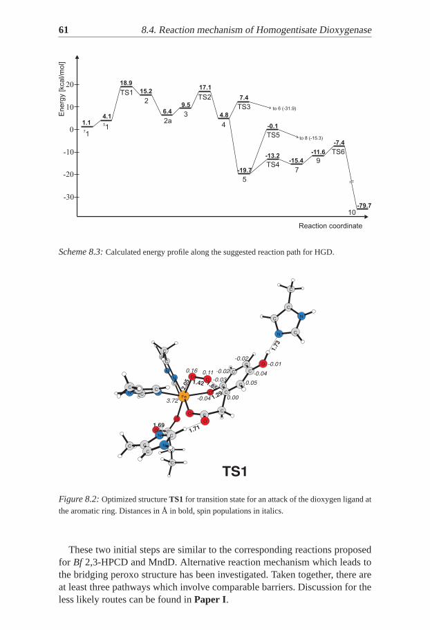
61 8.4. Reaction mechanism of Homogentisate Dioxygenase
Reaction coordinate
Energ
y [kcal/m
ol]
-10
0
10
-20
-30
1.1
15.2
9.5
4.1
18.920 17.1
4.8
7.4
-19.7
-0.1
-13.2-15.4
-11.6
-7.4
-79.7
71
51
TS1TS2
TS3
TS5
TS4
TS6
3
4
5
to 8 (-15.3)
to 6 (-31.9)
7
9
10
2
2a
6.4
Scheme 8.3:Calculated energy profile along the suggested reaction path for HGD.
N
C
C
N
C
C
O
O
C
C
C
C
Fe
C
C
O
C
C
N
C
C
O
CN
C
N
CO
N
O
C
C
C
C
C
O
C
N
C
O
C
C
C
N
3.72
2.05 1.42 1.85
1.73
0.16 0.11
-0.04 1.29
0.00
0.05-0.03
-0.02
-0.02
-0.04
-0.01
TS1
1.71
1.69
Figure 8.2:Optimized structureTS1 for transition state for an attack of the dioxygen ligand at
the aromatic ring. Distances in Å in bold, spin populations in italics.
These two initial steps are similar to the corresponding reactions proposedfor Bf 2,3-HPCD and MndD. Alternative reaction mechanism which leads tothe bridging peroxo structure has been investigated. Taken together, there areat least three pathways which involve comparable barriers. Discussion for theless likely routes can be found inPaper I.
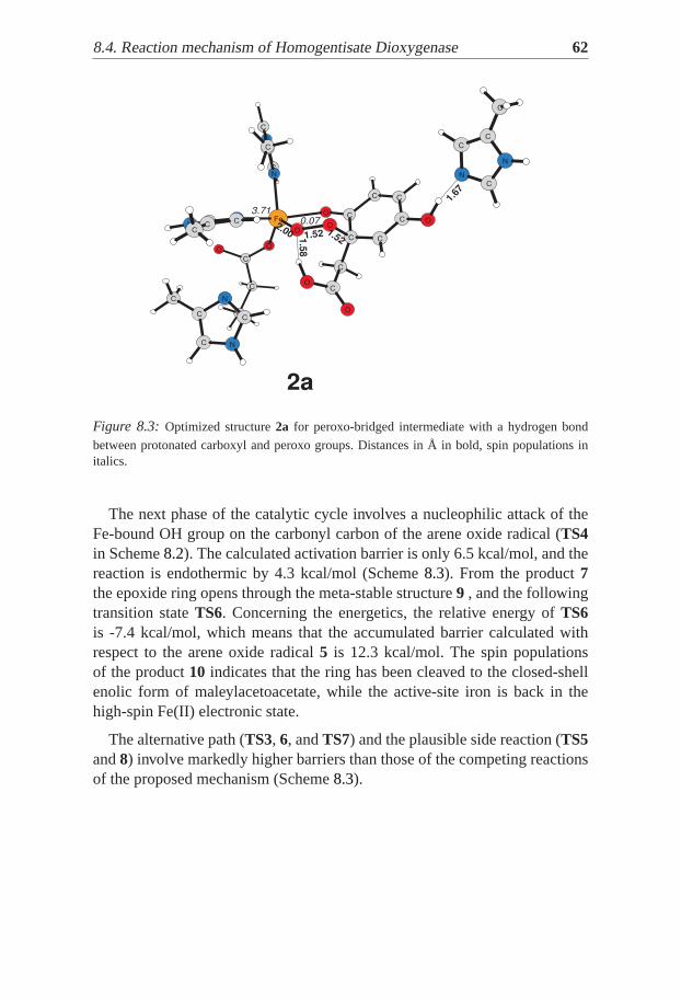
8.4. Reaction mechanism of Homogentisate Dioxygenase 62
C
C
C
N
O
C
O
O
C
N
C
CFe
C
C
C
C
C
C
N
C
C
C
C
C
O
N
N
C
O
O
N
C
O
O
C
N
C
C
C
C
N
C
C
2a
3.71
1.67
1.58
1.52
2.00 1.52
0.07
Figure 8.3: Optimized structure2a for peroxo-bridged intermediate with a hydrogen bond
between protonated carboxyl and peroxo groups. Distances in Å in bold, spin populations initalics.
The next phase of the catalytic cycle involves a nucleophilic attack of theFe-bound OH group on the carbonyl carbon of the arene oxide radical (TS4in Scheme8.2). The calculated activation barrier is only 6.5 kcal/mol, and thereaction is endothermic by 4.3 kcal/mol (Scheme8.3). From the product7the epoxide ring opens through the meta-stable structure9 , and the followingtransition stateTS6. Concerning the energetics, the relative energy ofTS6is -7.4 kcal/mol, which means that the accumulated barrier calculated withrespect to the arene oxide radical5 is 12.3 kcal/mol. The spin populationsof the product10 indicates that the ring has been cleaved to the closed-shellenolic form of maleylacetoacetate, while the active-site iron is back in thehigh-spin Fe(II) electronic state.
The alternative path (TS3, 6, andTS7) and the plausible side reaction (TS5and8) involve markedly higher barriers than those of the competing reactionsof the proposed mechanism (Scheme8.3).
63
9. Summary
All three enzymes bind the substrates in an ordered manner. After reducing thedioxygen by electron transfer from the organic substrate, a peroxo-bridged in-termediate is formed. This reaction step is found to be the rate-limiting stepfor the two Fe(II)-dependent enzymesBf 2,3-HPCD and HGD. The secondmajor step is the homolytic cleavage of the O-O bond in the peroxo interme-diate. Notably, for the Mn(II)-dependent enzyme this cleavage happens on thecrossing between the quartet and the sextet spin states, and defines the ratelimiting activation barrier.


Part IV:Biomimetic model complexes
Two synthetic Fe complexes were studied computationally using DensityFunctional Theory. The first one is a non-heme [(L-N4Me2)Fe(III)]+3 com-plex that mimics the reactivity of intradiol dioxygenases. The second one isa heme [T(o-Cl)PPFe] complex, which catalyzes the stepwise oxidation ofcyclohexane to adipic acid.


67
10. Non-heme iron model complexfor intradiol-cleaving dioxygenases
This chapter summarizes the results originally reported inPaper IV.
10.1 BackgroundTwo Fe(III)-catecholate complexes have been synthesized and their crystalstructures determined [80]. In these complexes, iron is coordinated bya tetraazamacrocyclic ligand L-N4Me2, with two adjacent coordinationsites available for the binding of the substrate. It has been experimentallyshown that the complexes react with molecular oxygen at room temperatureand exhibit catalytic activity for intradiol-cleavage of the substrate. Theexperimentally determined velocity constants for the substrates catecholand ditertbutyl catechol correspond to activation barriers of 21.0 and18.0 kcal/mol, respectively. In comparison for intradiol cleaving CatecholDioxygenase [81] the rate constant (k = 2.5x10⁵ M−1s−1) correspondsto a barrier of 10.1 kcal/mol. The basic chemical steps of the intradioldioxygenation are the following: (i) binding of dioxygen to iron, accompaniedby oxidation of the substrate to semiquinone and reduction of dioxygen tosuperoxide, (ii ) formation of peroxo-bridging intermediate after an attack ofthe superoxide on the semiquinone ring, (iii ) O-O bond cleavage assistedby protonation of the peroxide, (iv) acyl migration leading to an anhydrideintermediate, which is then hydrolyzed to the acyclic product. Even thoughthe coordination environment of the model complex and the enzyme arequite different, questions regarding the C-C cleavage step, the actual attackof the dioxygen molecule on the Fe(III)-catechol system and the intradiolspecificity can be addressed.
10.2 The modelThe computational model used in this work is based on the crystal structurereported from Koch an co-workers [80]. In terms of computational procedureand methodology the study was done according to the descriptions inChap-ter 5 of Part 2, with few differences compared to the enzymatic investiga-tions. Together with the original hybrid density functional B3LYP, B3LYP*

10.2. The model 68
was used, for which the amount of Hartree-Fock exchange is reduced from20% to 15% [33, 82]. In this study the rate limiting step was lowered by 5.4kcal/mol when using B3LYP* instead of B3LYP.
Zero-point effects, entropic and thermal corrections to the Gibbs free en-ergy were evaluated for each stationary point. It is difficult to describe cor-rectly the entropic contributions for the oxygen binding step within the presentmodeling. The calculated entropic contribution for the oxygen binding is 8.6kcal/mol, which goes up to 13.4 kcal/mol during the formation of the bridgingperoxide. Once dioxygen is bound, and the reaction has started, the relativeentropy effects vary by 1 to 2 kcal/mol. To account for compensating effectsin the oxygen binding a Van der Waals correction was evaluated with B3LYP-D using the Orca Package [83, 84]. Corrections in the range of 3 to 4 kcal/molwere calculated for these long-range dispersion interactions. The solvent wasmodeled as a macroscopic continuum with a dielectric constantε=36.6 (cor-responding to acetonitrile). Final energies of the optimized structures werecorrected for the solvent effects by employing thelacvpbasis set. As alreadymentioned, the dielectric medium should have a small effect on the reactionenergetics as long as no substantial charge separations are involved. However,some charge separation is present in this case and solvent effects thereforerange from 4 to 10 kcal/mol. Reported spin populations are used to indicatethe spin and the oxidation state of the metal ion and were derived from a Mul-liken population analysis.
O
OFe+2
O
OFe+3
O O
O
OFe+3
O O
O
OFe+3
O
OFe+2
O O
O
OFe+4
O O
R1 R2 BP
RADANHNN
N
N
Ligand: L-N4Me2
Scheme 10.1: Proposed mechanism for intradiol cleavage catalyzed bythe Fe(III)L-N4Me2 complex (where L-N4Me2 is N,N,-Dimethyl-2,11-diaza[3.3](2,6)pyridinophane) based on the calculations.

69 10.3. Reaction mechanism
10.3 Reaction mechanismThe proposed mechanism based on our calculations is given in Figure10.1.Because of the open-shell nature of the transition metal the starting Fe(III)complex (denoted asR1) can have three different spin states – sextet (spin5/2), quartet (spin 3/2) and doublet (spin 1/2). Due to possible reduction of theiron by the substrate a total number of 8 spin states exist forR1. This numberoriginates from various couplings between the unpaired electrons arising uponone electron transfer from the substrate to Fe. In order to explore the reactioncoordinate one needs to know the energetic splitting of these states for all thereaction intermediates. Some of these states were found to be energeticallyhigh and thus thermally inaccessible. A summary of the calculated relativeenergies for the lowest spin states for the reactant is given in Table10.1.
S ox. state Fe catechol ∆G
1/2 Fe(II) - 1α 0.0 (3.8)
3/2 Fe(II) 2α 1α 0.8 (3.3)
Fe(II) 4α 1β 4.5 (1.5)
Fe(III) 3α - 8.8 (10.8)
5/2 Fe(II) 4α 1α 1.2 (0.0)
Table 10.1:Spin-splitting in the reactantR1. Relative free energies given in kcal/mol.The values in parenthesis are calculated using the standardB3LYP functional.
The ground state of the reactant predicted by the calculations is a doubletstate corresponding to low-spin Fe(II) and one electron oxidized catecholateradical (semiquinonate), i.e. [(LN4Me2)Fe(II)(cat)−]+, (Figure 10.1 A). Aquartet state2α1α formed by intermediate spin on Fe(II) and semiquinonateis only 0.8 kcal/mol up in energy, and a sextet4α1α, corresponding to ahigh-spin Fe(II) ferromagnetically coupled to the one electron oxidizedsemiquinonate lies at +1.2 kcal/mol. There are two more accessible quartetstates. One is the antiferromagnetically coupled reactant Fe(II)-cat•−, 4α1β ,at 4.5 kcal/mol above the ground state. The other quartet is formed byintermediate spin on Fe(III) and catecholatecat2− and lies at +8.8 kcal/mol.However, no high spin Fe(III) catecholate complex could be found. Thus,states having an oxidized monoanionic substrate with a reduced Fe(II) ionare preferred for the reactant structure. As can be seen from Table10.1B3LYP∗ changes the spin-splitting, stabilizing the low-spin, and destabilizingthe high-spin states. The dominating ferrous-semiquinonate resonance issupposed to direct the dioxygen binding directly to the metal. In this respect
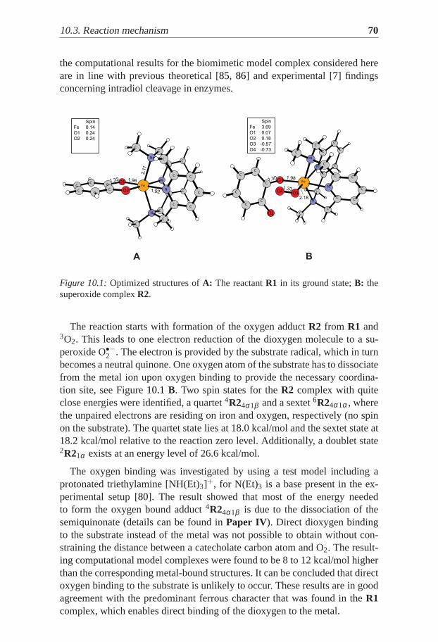
10.3. Reaction mechanism 70
the computational results for the biomimetic model complex considered hereare in line with previous theoretical [85, 86] and experimental [7] findingsconcerning intradiol cleavage in enzymes.
C
C
C
C
C
C
NO1C
C
C
N
CC Fe
C
N
C
C
C
NC O2
C
C
C
C C
C
A B
Spin
Fe 0.14
O1 0.24
O2 0.24
1.92
1.33 1.96
2.1
1
C
C
C
C
C
C
N
N
C
C
O1
O2
C
Fe
C
C
C
C
N
C
C
C
C
N
C
C C
O3
C
C
O4
1.98
2.18
Spin
Fe 3.69
O1 0.07
O2 0.18
O3 -0.57
O4 -0.73
1.33
1.30
Figure 10.1:Optimized structures ofA: The reactantR1 in its ground state;B: thesuperoxide complexR2.
The reaction starts with formation of the oxygen adductR2 from R1 and3O2. This leads to one electron reduction of the dioxygen molecule to a su-peroxide O•−2 . The electron is provided by the substrate radical, which in turnbecomes a neutral quinone. One oxygen atom of the substrate has to dissociatefrom the metal ion upon oxygen binding to provide the necessary coordina-tion site, see Figure10.1 B. Two spin states for theR2 complex with quiteclose energies were identified, a quartet4R24α1β and a sextet6R24α1α , wherethe unpaired electrons are residing on iron and oxygen, respectively (no spinon the substrate). The quartet state lies at 18.0 kcal/mol and the sextet state at18.2 kcal/mol relative to the reaction zero level. Additionally, a doublet state2R21α exists at an energy level of 26.6 kcal/mol.
The oxygen binding was investigated by using a test model including aprotonated triethylamine [NH(Et)3]+, for N(Et)3 is a base present in the ex-perimental setup [80]. The result showed that most of the energy neededto form the oxygen bound adduct4R24α1β is due to the dissociation of thesemiquinonate (details can be found inPaper IV). Direct dioxygen bindingto the substrate instead of the metal was not possible to obtain without con-straining the distance between a catecholate carbon atom and O2. The result-ing computational model complexes were found to be 8 to 12 kcal/mol higherthan the corresponding metal-bound structures. It can be concluded that directoxygen binding to the substrate is unlikely to occur. These results are in goodagreement with the predominant ferrous character that was found in theR1complex, which enables direct binding of the dioxygen to the metal.

71 10.3. Reaction mechanism
In the next step the superoxideR2 attacks the aromatic ring of the substrate.With Fe(II) providing the second electron required for the reduction of the su-peroxide, a peroxide bridge is formed, and the metal ion is oxidized to Fe(III).The transition state4TS1 (Figure10.2A) for this step lies at 23.3 kcal/mol onthe quartet surface. No corresponding transition state was found on the sextetsurface, instead, a transition state was found leading to another intermediate(see below). The formation of the bridging peroxide4BP (Figure10.2B) isslightly exergonic with a reaction free energy of 4.5 kcal/mol. The groundstate for this intermediate is a quartet, with a doublet state around 10 kcal/molhigher energy. No bridging peroxide could be found on the sextet PES.
C
C
C
C
C
O2
C
C
N
C
CO1
N
C
C
C
C
C
Fe
C
C
N
O4C
N
O3
C
C
C
C
C
A B
C
C
C
C
C
C
N
C
N
C
O2
O1
C
Fe
C
C
C
C
C
C
N
C
C
O3
N
O4
C
C
C
C
C
Spin
Fe 3.27
O1 0.08
O2 0.03
O3 -0.33
O4 -0.25
1.96
1.91
1.421.83
1.34
Spin
Fe 2.64
O1 0.11
O2 0.00
O3 0.08
O4 -0.02
1.84
1.48
1.53
1.43
1.82
Figure 10.2:Optimized structures ofA: transition state for peroxide formationTS1;B: the bridging peroxideBP.
A
C
C
C
CC
O2C
C
C
C
NO1
C
C
N
C
C
C
C
Fe
O4
C
N
C
NO3
C
C
C
C
C
Spin
Fe 2.77
O1 0.13
O2 -0.04
O3 0.62
O4 -0.66
1.83
1.49
1.53
1.45
2.09
C
C
O2
C
C
C
C
C
C
C
C
C
O1
C
N
O4
C
C
N
Fe
C
N
C
O3
C
N
C
C
C
C
C
Spin
Fe 2.86
O1 0.13
O2 -0.08
O3 0.74
O4 -0.75
1.85
1.37 1.65
1.41
B
Figure 10.3:Optimized structures ofA: transition state for the homolytic O-O bondcleavage4TS2; B: the alkoxyl radicalRAD, formed after the O-O bond cleavage.
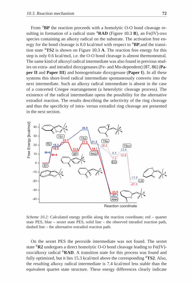
10.3. Reaction mechanism 72
From 4BP the reaction proceeds with a homolytic O-O bond cleavage re-sulting in formation of a radical state4RAD (Figure10.3B), an Fe(IV)-oxospecies containing an alkoxy radical on the substrate. The activation free en-ergy for the bond cleavage is 8.0 kcal/mol with respect to4BP,and the transi-tion state4TS2 is shown on Figure10.3A. The reaction free energy for thisstep is only 0.6 kcal/mol, i.e. the O-O bond cleavage is almost thermoneutral.The same kind of alkoxyl radical intermediate was also found in previous stud-ies on extra- and intradiol dioxygenases (Fe- and Mn-dependent) [87, 86] (Pa-per II andPaper III ) and homogentisate dioxygenase (Paper I). In all thesesystems this short-lived radical intermediate spontaneously converts into thenext intermediate. Such an alkoxy radical intermediate is absent in the caseof a concerted Criegee rearrangement (a heterolytic cleavage process). Theexistence of the radical intermediate opens the possibility for the alternativeextradiol reaction. The results describing the selectivity of the ring cleavageand thus the specificity of intra- versus extradiol ring cleavage are presentedin the next section.
O
OFe+2
R1 O
OFe+3
O O
BP
O
OFe+3
O OANH
RAD
R2
TS1TS2
LA
TSe
TScc
EO
O
OFe+2
OO
O
OFe+4
O O
O
O
Fe+3
O
O
O
OFe+4
O
O
Scheme 10.2:Calculated energy profile along the reaction coordinate; red – quartetstate PES, blue – sextet state PES, solid line – the observed intradiol reaction path,dashed line – the alternative extradiol reaction path.
On the sextet PES the peroxide intermediate was not found. The sextetstate6R2 undergoes a direct homolytic O-O bond cleavage leading to Fe(IV)-oxo/alkoxy radical6RAD. A transition state for this process was found andfully optimized, but it lies 15.3 kcal/mol above the corresponding4TS2. Also,the resulting alkoxy radical intermediate is 7.4 kcal/mol less stable than theequivalent quartet state structure. These energy differences clearly indicate
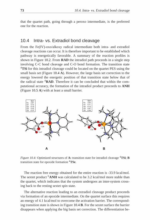
73 10.4. Intra- vs. Extradiol bond cleavage
that the quartet path, going through a peroxo intermediate, is the preferredone for the reaction.
10.4 Intra- vs. Extradiol bond cleavageFrom the Fe(IV)-oxo/alkoxy radical intermediate both intra- and extradiolcleavage reactions can occur. It is therefore important to be established whichpathway is energetically favorable. A summary of the reaction profiles isshown in Figure10.2. FromRAD the intradiol path proceeds in a single stepinvolving C-C bond cleavage and C-O bond formation. The transition state4TSi for this intradiol cleavage could be located on the quartet PES using thesmall basis set (Figure10.4A). However, the large basis set correction to theenergy lowered the energetic position of that transition state below that ofthe radical state4RAD. Therefore it can be concluded that within the com-putational accuracy, the formation of the intradiol product proceeds toANH(Figure10.5A) with at least a small barrier.
BA
C
C
C
C
C
N
C
CO2
C
C
C
CC
N
C
C
N
C
N C
O1 FeC
C
C
C
C C
O4
O3
1.99 1.29
2.47
1.87
Spin
Fe 2.87
O1 0.10
O2 -0.19
O3 0.75
O4 -0.37
C
O2
C
C
C
C
C
C
C
CC
CN
O1
C
C
N
O4
C
Fe
C
N
C
C
NO3
C
C
C
C
C
Spin
Fe 3.03
O1 0.11
O2 -0.12
O3 0.67
O4 -0.46
1.50
1.43
1.38
1.9
0 1.85
1.65
Figure 10.4:Optimized structures ofA: transition state for intradiol cleavage4TSi; Btransition state for epoxide formation4TSe.
The reaction free energy obtained for the entire reaction is -33.9 kcal/mol.The sextet product6ANH was calculated to be 3.2 kcal/mol more stable thanthe quartet, which indicates that the system undergoes an inter-system cross-ing back to the resting sextet spin state.
The alternative reaction leading to an extradiol cleavage product proceedsvia formation of an epoxide intermediate. On the quartet surface this requiresan energy of 4.1 kcal/mol to overcome the activation barrier. The correspond-ing transition state is shown in Figure10.4B. For the sextet surface the barrierdisappears when applying the big basis set correction. The differentiation be-
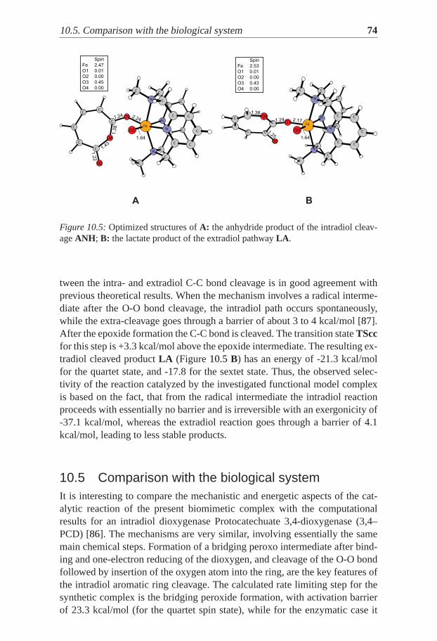
10.5. Comparison with the biological system 74
A B
C
C
C
C
C
CC
C
C
O4
C
C
C
N
O2
O1
C
C
N
Fe
C
N
C
C
NO3
C
C
C
C
C
Spin
Fe 2.47
O1 0.01
O2 0.00
O3 0.45
O4 0.00
2.24
1.3
8
1.64
1.24
1.2
2
1.43
C
C
O2
C
C
CC
C
C
C
N
C O1
N
C
C
C
O4
C
C
Fe
C
N
N
C
C
O3
C
C
C
C2.171.24
1.38
1.64
1.25
Spin
Fe 2.53
O1 0.01
O2 0.00
O3 0.43
O4 0.00
Figure 10.5:Optimized structures ofA: the anhydride product of the intradiol cleav-ageANH ; B: the lactate product of the extradiol pathwayLA .
tween the intra- and extradiol C-C bond cleavage is in good agreement withprevious theoretical results. When the mechanism involves a radical interme-diate after the O-O bond cleavage, the intradiol path occurs spontaneously,while the extra-cleavage goes through a barrier of about 3 to 4 kcal/mol [87].After the epoxide formation the C-C bond is cleaved. The transition stateTSccfor this step is +3.3 kcal/mol above the epoxide intermediate. The resulting ex-tradiol cleaved productLA (Figure10.5B) has an energy of -21.3 kcal/molfor the quartet state, and -17.8 for the sextet state. Thus, the observed selec-tivity of the reaction catalyzed by the investigated functional model complexis based on the fact, that from the radical intermediate the intradiol reactionproceeds with essentially no barrier and is irreversible with an exergonicity of-37.1 kcal/mol, whereas the extradiol reaction goes through a barrier of 4.1kcal/mol, leading to less stable products.
10.5 Comparison with the biological systemIt is interesting to compare the mechanistic and energetic aspects of the cat-alytic reaction of the present biomimetic complex with the computationalresults for an intradiol dioxygenase Protocatechuate 3,4-dioxygenase (3,4–PCD) [86]. The mechanisms are very similar, involving essentially the samemain chemical steps. Formation of a bridging peroxo intermediate after bind-ing and one-electron reducing of the dioxygen, and cleavage of the O-O bondfollowed by insertion of the oxygen atom into the ring, are the key features ofthe intradiol aromatic ring cleavage. The calculated rate limiting step for thesynthetic complex is the bridging peroxide formation, with activation barrierof 23.3 kcal/mol (for the quartet spin state), while for the enzymatic case it
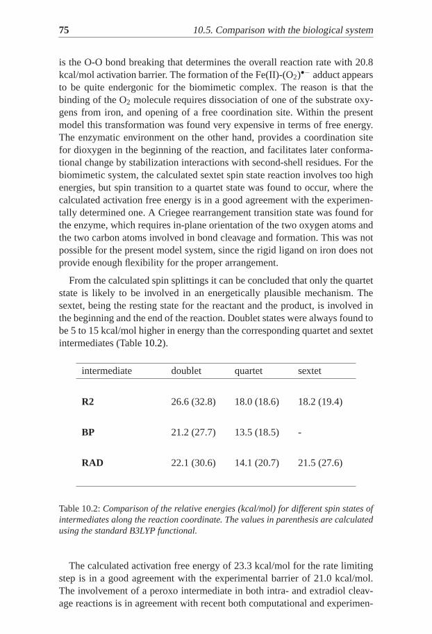
75 10.5. Comparison with the biological system
is the O-O bond breaking that determines the overall reaction rate with 20.8kcal/mol activation barrier. The formation of the Fe(II)-(O2)•− adduct appearsto be quite endergonic for the biomimetic complex. The reason is that thebinding of the O2 molecule requires dissociation of one of the substrate oxy-gens from iron, and opening of a free coordination site. Within the presentmodel this transformation was found very expensive in terms of free energy.The enzymatic environment on the other hand, provides a coordination sitefor dioxygen in the beginning of the reaction, and facilitates later conforma-tional change by stabilization interactions with second-shell residues. For thebiomimetic system, the calculated sextet spin state reaction involvestoo highenergies, but spin transition to a quartet state was found to occur, where thecalculated activation free energy is in a good agreement with the experimen-tally determined one. A Criegee rearrangement transition state was found forthe enzyme, which requires in-plane orientation of the two oxygen atoms andthe two carbon atoms involved in bond cleavage and formation. This was notpossible for the present model system, since the rigid ligand on iron does notprovide enough flexibility for the proper arrangement.
From the calculated spin splittings it can be concluded that only the quartetstate is likely to be involved in an energetically plausible mechanism. Thesextet, being the resting state for the reactant and the product, is involved inthe beginning and the end of the reaction. Doublet states were always found tobe 5 to 15 kcal/mol higher in energy than the corresponding quartet and sextetintermediates (Table10.2).
intermediate doublet quartet sextet
R2 26.6 (32.8) 18.0 (18.6) 18.2 (19.4)
BP 21.2 (27.7) 13.5 (18.5) -
RAD 22.1 (30.6) 14.1 (20.7) 21.5 (27.6)
Table 10.2:Comparison of the relative energies (kcal/mol) for different spin states ofintermediates along the reaction coordinate. The values inparenthesis are calculatedusing the standard B3LYP functional.
The calculated activation free energy of 23.3 kcal/mol for the rate limitingstep is in a good agreement with the experimental barrier of 21.0 kcal/mol.The involvement of a peroxo intermediate in both intra- and extradiol cleav-age reactions is in agreement with recent both computational and experimen-

10.5. Comparison with the biological system 76
tal studies. The peroxide O-O bond cleaves homolytically to give a radicalFe(IV)-oxo intermediateRAD. The ring cleavage and oxygen atom insertionto give the intradiol product proceed without an activation barrier. A muconicanhydride is formed, which is the final product within the scope of the presentwork. The alternative path of extradiol cleavage needs to cross an additionalbarrier that leads to the formation of an intermediate epoxide. Solely due tothis barrier intradiol cleavage is observed for this biomimetic model complex.

77
11. Heme complex catalyzing adipicacid synthesis
11.1 BackgroundAdipic acid is the most important dicarboxylic acid for the industry, with mainapplication as a precursor for the production of nylon and other polymers(PVC, Polyurethane). It is used also as a flavorant, acidulant and gelling aidin baking powders [88]. Since adipic acid occurs rarely in nature, it is syn-thesized in enormous amounts by the chemical industry. Historically, adipicacid was produced by oxidation of various fats, while the contemporary meth-ods are based on oxidation of cyclohexane. First the cyclohexane is oxidizedat 150-160◦ C with oxygen in the presence of homogeneous cobalt or man-ganese catalyst to a mixture of cyclohexanol and cyclohexanone called "KAoil" (abbreviation of "ketone-alcohol oil). The KA oils can be further oxidizedwith nitric acid, hydrogen peroxide or molecular oxygen to adipic acid. Theuse of nitric acid has disadvantages from environmental point of view, but ispreferred due the low cost of the nitric acid as oxidant. Hence, there is increas-ing interest towards usage of molecular oxygen or hydrogen peroxide, whichprovide far more environmental friendly oxidation.
The one-pot oxidation method has been reported from different groups [89,90, 91]. The one-pot synthesis is a strategy where a reactant is subjected tosuccessive chemical reactions in just one reactor. This improves the efficiencyof a chemical reaction because a lengthy separation process and purificationof the intermediates are avoided, while the yields are increased. This methodhowever, has some disadvantages that still make the process expensive – use ofexpensive radical catalyst, acidic solvent, severe reaction conditions. Utilizingmetalloporphyrins as catalysts for the one-pot synthesis of adipic acid fromcyclohexane and molecular oxygen has been reported [91, 92] as promising,but up to date it is still hard to be applied in industry due to low yields.
Porphyrin-iron complexes (hemes) are well known tightly-boundcofactors (prosthetic groups) in a number of metalloproteins – Hemoglobin,Cytochrome C, P450, Peroxidases, etc. The biological functions that theyare involved in include dioxygen transportation, electron transfer, and redoxchemistry. The heme enzymes and synthetic complexes require an additionalcofactor in order to activate the O2 molecule for substrate oxidation. TwoPorphyrin-iron complexes however can bind dioxygen as a peroxide ina Di-iron complex and cleave the O-O bond, as reported by Chin and
11.2. Computational technique. 78
co-workers [93]. The O-O bond cleavage in binuclear Porphyrin peroxocomplexes was recently studied theoretically [94] with DFT. The stability ofthe binuclear center at normal temperature was explained with the splittingsof the spin states and the way they are changed in the presence of axialligands.
In the present work, the stepwise mechanism of the catalytic oxidation ofcyclohexane to adipic acid was investigated theoretically. Hybrid density func-tional theory was used to model the putative oxidative stages in the adipic acidsynthesis. The reaction was modelled according to the conditions reported inref. [92] – solvent-free reaction system with molecular oxygen as the onlyoxidant, and a heme Fe-oxo complex (PorfFe(IV)=O) as a catalyst, at 413Ktemperature.
11.2 Computational technique.The common computational technique as described inChapter 5 of Part2 was applied in this study. The modified B3LYP* functional was used forchecking the reliability of the results. The reduction of the Hartree-Fock ex-change to 15% resulted in the typical stabilization of the low-spin states, anddestabilization of the high-spin states. The relative energies reported beloware the ones obtained with the original B3LYP.
The solvent was modeled as a macroscopic continuum with a dielectricconstantε=2.0 (corresponding to cyclohexane). Since the model used in thestudy has zero total charge and basically no charge separation happens, thesolvent effects are very small, in the range of 1–2 kcal/mol. Reported spinpopulations are used to indicate the spin- and oxidation state of the metal ion,and were derived from a Mulliken population analysis.
The complex used as a catalyst in the experiment ismeso-Tetra(o-chlorophenyl)iron Porphyrin (T(o-Cl)PPFe). Unsubstituted Porphyrin ligandwas used in the calculations. This strategy for decreasing the computationalcosts has been proven to give adequate results in recent theoretical studywhere the energetic differences between substituted and unsubstitutedPorphyrin models were found insignificant [94].
Since for loosely bound intermediates the harmonic approximation givespoor results, the thermodynamic corrections for such structures were takenfrom the infinitely separated species. Although the partition functions for anideal gas are very accurate for predicting gas phase entropies, errors are in-troduced when this method is applied to solvent molecules. In particular, thecalculated gas phase entropy of the cyclohexane molecule is higher than theentropy it has in the liquid phase. In principle, it is possible to estimate thedifference by using experimentally determined entropies, and for cyclohex-
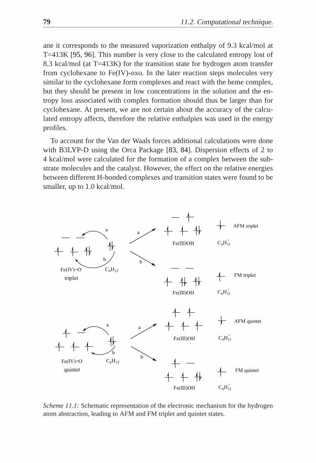
79 11.2. Computational technique.
ane it corresponds to the measured vaporization enthalpy of 9.3 kcal/mol atT=413K [95, 96]. This number is very close to the calculated entropy lost of8.3 kcal/mol (at T=413K) for the transition state for hydrogen atom transferfrom cyclohexane to Fe(IV)-oxo. In the later reaction steps molecules verysimilar to the cyclohexane form complexes and react with the heme complex,but they should be present in low concentrations in the solution and the en-tropy loss associated with complex formation should thus be larger than forcyclohexane. At present, we are not certain about the accuracy of the calcu-lated entropy affects, therefore the relative enthalpies was used in the energyprofiles.
To account for the Van der Waals forces additional calculations were donewith B3LYP-D using the Orca Package [83, 84]. Dispersion effects of 2 to4 kcal/mol were calculated for the formation of a complex between the sub-strate molecules and the catalyst. However, the effect on the relative energiesbetween different H-bonded complexes and transition states were found to besmaller, up to 1.0 kcal/mol.
Fe(IV)=O C6H12
AFM triplet
FM triplet
Fe(III)OH C6H11
Fe(III)OH C6H11
a
b
a
b
Fe(IV)=O C6H12
AFM quintet
FM quintet
Fe(III)OH C6H11
Fe(III)OH C6H11
a
b
a
b
quintet
triplet
Scheme 11.1:Schematic representation of the electronic mechanism for the hydrogenatom abstraction, leading to AFM and FM triplet and quintet states.

11.3. Suggested reaction mechanism 80
11.3 Suggested reaction mechanismThe oxidation of cyclohexane to adipic acid by the biomimetic heme complexT(o-Cl)PP-Fe(IV)-O was studied using DFT. Experimentally the reaction wasdone in cyclohexane as a solvent, and with molecular oxygen as the only oxi-dant. The suggested mechanism consists of three major steps. In the first onethe cyclohexane is hydroxylated to 1,2-cyclohexanediol. It should be notedhere that the cyclohexane molecule is in its lowest-energychair conformation,whereaxial andequatorial positions can be distinguished. The two hydrox-ylations of the cyclohexane, described below, occur at neighboring (ortho)equatorial position. In the second major step 1,2-cyclohexanediol is oxidizedto 1,6-hexanedial with opening of the ring, and in the final step both aldehydegroups of 1,6-hexanedial are oxidized, and the adipic acid is formed. Thesethree main reactions share some common features. The ground spin state ofthe reactants and the products is triplet (S=2). This is determined by the spinsplitting for the Fe(IV)-oxo and Fe(II) heme complexes, which both have atriplet ground state. For every oxidation however, the reactive state was foundto be the quintet (S=4) – the system undergoes a spin crossing from triplet toquintet state before the first transition state, and when the product is formedanother spin crossing back to the triplet occurs. Every oxidative step startswith hydrogen atom abstraction, resulting in the formation of Fe(III)-OH anda radical. Depending on the spin coupling between Fe(III)-OH and the radicalmolecule, two different triplet and quintet states exist – Antiferromagnetically(AFM) and Ferromagnetically (FM) coupled (Scheme11.1). Details about allthe different spin states that were calculated during the investigation can befound inPaper V.
11.3.1 Oxidation of cyclohexane to 1,2-cyclohexanediol
The oxidative hydroxylation starts with Hydrogen Atom Transfer (HAT)from cyclohexane to the Fe(IV)-oxo species, leading to a radical intermediate[(Porph)Fe(III)(OH)(C6H11)•]. In the next step a rebound of the OH groupoccurs to form the alcohol. The ground state of the reactant complex[(Porph)Fe(IV)(O)(C6H12)] is a triplet (Figure 11.1 A) having formallytwo unpaired electrons on iron, one of which is partially delocalized tothe oxo group (SFe=1.21 and Soxo=0.83, where SX refers to the Mullikenspin population of fragmentX). The lowest barrier for HAT, however, isfound in the quintet state. In the reactant complex, the quintet state is 7.8kcal/mol higher than the triplet ground state. But typically for the Fe(IV)-oxogroup, the system undergoes a spin crossing before the transition state isreached. In the TS for HAT the quintet state is 3.6 kcal/mol lower than theAFM triplet state. The enthalpy barrier determined by the TS in the quintetstate (Figure11.1 B) is 13.6 kcal/mol with respect to the starting complex
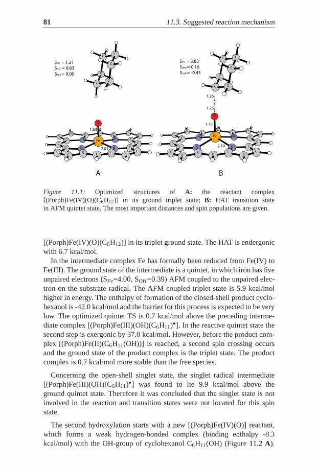
81 11.3. Suggested reaction mechanism
C C CC C
C C
N NC C
C
FeC
C
O
C
C CN N
C
C
C CC
C
C
C
C C C
SFe = 1.21
Soxo = 0.83
Ssub = 0.00
1.63
2.01
A B
C C CC
CC
C
NC
N C
C
Fe
C
C
C
O
C N C
C
N
C
C
C
C CC
C
C C C
SFe = 3.83
Soxo = 0.16
Ssub = -0.43
1.26
1.25
1.75
2.10
Figure 11.1: Optimized structures of A: the reactant complex[(Porph)Fe(IV)(O)(C6H12)] in its ground triplet state;B: HAT transition statein AFM quintet state. The most important distances and spin populations are given.
[(Porph)Fe(IV)(O)(C6H12)] in its triplet ground state. The HAT is endergonicwith 6.7 kcal/mol.
In the intermediate complex Fe has formally been reduced from Fe(IV) toFe(III). The ground state of the intermediate is a quintet, in which iron has fiveunpaired electrons (SFe=4.00, SOH=0.39) AFM coupled to the unpaired elec-tron on the substrate radical. The AFM coupled triplet state is 5.9 kcal/molhigher in energy. The enthalpy of formation of the closed-shell product cyclo-hexanol is -42.0 kcal/mol and the barrier for this process is expected to be verylow. The optimized quintet TS is 0.7 kcal/mol above the preceding interme-diate complex [(Porph)Fe(III)(OH)(C6H11)•]. In the reactive quintet state thesecond step is exergonic by 37.0 kcal/mol. However, before the product com-plex [(Porph)Fe(II)(C6H11(OH))] is reached, a second spin crossing occursand the ground state of the product complex is the triplet state. The productcomplex is 0.7 kcal/mol more stable than the free species.
Concerning the open-shell singlet state, the singlet radical intermediate[(Porph)Fe(III)(OH)(C6H11)•] was found to lie 9.9 kcal/mol above theground quintet state. Therefore it was concluded that the singlet state is notinvolved in the reaction and transition states were not located for this spinstate.
The second hydroxylation starts with a new [(Porph)Fe(IV)(O)] reactant,which forms a weak hydrogen-bonded complex (binding enthalpy -8.3kcal/mol) with the OH-group of cyclohexanol C6H11(OH) (Figure11.2 A).
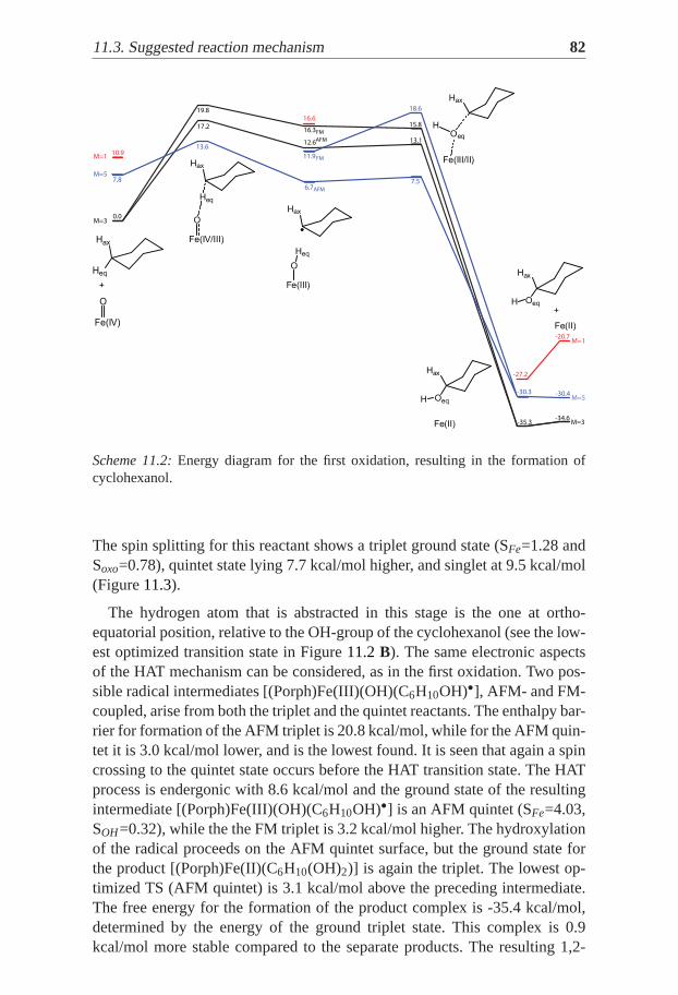
11.3. Suggested reaction mechanism 82
M=3
M=5
M=1
M=3
M=5
M=1
Fe(IV)
O
Hax
Heq
Fe(III)
O
Hax
Heq
Fe(III/II)
Oeq
Hax
H
Fe(II)
Oeq
Hax
H
Fe(IV/III)
O
Hax
Heq
0.0
-35.3
15.8
13.1
16.3
12.6
19.8
17.2
7.8
-30.3
7.56.7
11.9
13.610.9
-27.2
16.6
FM
AFM
AFM
FM
18.6
Fe(II)
Oeq
Hax
H+
-34.6
-30.4
-20.7
+
Scheme 11.2:Energy diagram for the first oxidation, resulting in the formation ofcyclohexanol.
The spin splitting for this reactant shows a triplet ground state (SFe=1.28 andSoxo=0.78), quintet state lying 7.7 kcal/mol higher, and singlet at 9.5 kcal/mol(Figure11.3).
The hydrogen atom that is abstracted in this stage is the one at ortho-equatorial position, relative to the OH-group of the cyclohexanol (see the low-est optimized transition state in Figure11.2B). The same electronic aspectsof the HAT mechanism can be considered, as in the first oxidation. Two pos-sible radical intermediates [(Porph)Fe(III)(OH)(C6H10OH)•], AFM- and FM-coupled, arise from both the triplet and the quintet reactants. The enthalpy bar-rier for formation of the AFM triplet is 20.8 kcal/mol, while for the AFM quin-tet it is 3.0 kcal/mol lower, and is the lowest found. It is seen that again a spincrossing to the quintet state occurs before the HAT transition state. The HATprocess is endergonic with 8.6 kcal/mol and the ground state of the resultingintermediate [(Porph)Fe(III)(OH)(C6H10OH)•] is an AFM quintet (SFe=4.03,SOH=0.32), while the the FM triplet is 3.2 kcal/mol higher. The hydroxylationof the radical proceeds on the AFM quintet surface, but the ground state forthe product [(Porph)Fe(II)(C6H10(OH)2)] is again the triplet. The lowest op-timized TS (AFM quintet) is 3.1 kcal/mol above the preceding intermediate.The free energy for the formation of the product complex is -35.4 kcal/mol,determined by the energy of the ground triplet state. This complex is 0.9kcal/mol more stable compared to the separate products. The resulting 1,2-
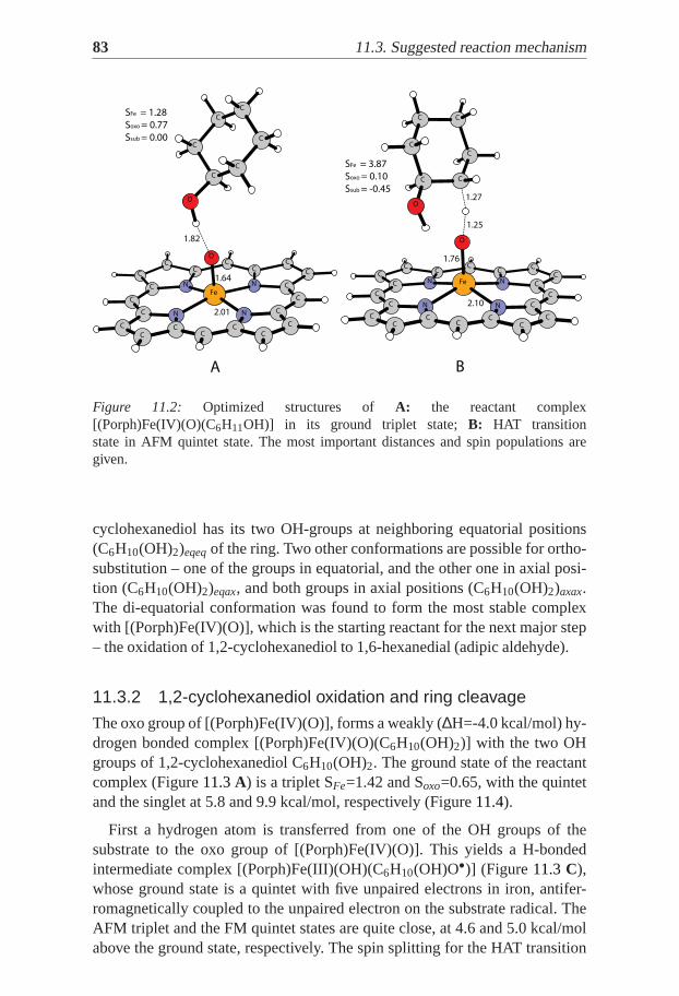
83 11.3. Suggested reaction mechanism
C C
C
C
C CC
C N N C
C
FeC
C
C
O
N
C
N C
C
C
C
O
C
C
C
C
C
C
C C
SFe = 1.28
Soxo = 0.77
Ssub = 0.00
2.01
1.64
1.82
C C CC
CC
CNC N
C
C
Fe
C
C
C
O
C
C
C
N CN
C
C
C C C
O
CC C C
SFe = 3.87
Soxo = 0.10
Ssub = -0.45
1.25
1.27
1.76
2.10
A B
Figure 11.2: Optimized structures of A: the reactant complex[(Porph)Fe(IV)(O)(C6H11OH)] in its ground triplet state;B: HAT transitionstate in AFM quintet state. The most important distances andspin populations aregiven.
cyclohexanediol has its two OH-groups at neighboring equatorial positions(C6H10(OH)2)eqeqof the ring. Two other conformations are possible for ortho-substitution – one of the groups in equatorial, and the other one in axial posi-tion (C6H10(OH)2)eqax, and both groups in axial positions (C6H10(OH)2)axax.The di-equatorial conformation was found to form the most stable complexwith [(Porph)Fe(IV)(O)], which is the starting reactant for the next major step– the oxidation of 1,2-cyclohexanediol to 1,6-hexanedial (adipic aldehyde).
11.3.2 1,2-cyclohexanediol oxidation and ring cleavage
The oxo group of [(Porph)Fe(IV)(O)], forms a weakly (∆H=-4.0 kcal/mol) hy-drogen bonded complex [(Porph)Fe(IV)(O)(C6H10(OH)2)] with the two OHgroups of 1,2-cyclohexanediol C6H10(OH)2. The ground state of the reactantcomplex (Figure11.3A) is a triplet SFe=1.42 and Soxo=0.65, with the quintetand the singlet at 5.8 and 9.9 kcal/mol, respectively (Figure11.4).
First a hydrogen atom is transferred from one of the OH groups of thesubstrate to the oxo group of [(Porph)Fe(IV)(O)]. This yields a H-bondedintermediate complex [(Porph)Fe(III)(OH)(C6H10(OH)O•)] (Figure 11.3C),whose ground state is a quintet with five unpaired electrons in iron, antifer-romagnetically coupled to the unpaired electron on the substrate radical. TheAFM triplet and the FM quintet states are quite close, at 4.6 and 5.0 kcal/molabove the ground state, respectively. The spin splitting for the HAT transition

11.3. Suggested reaction mechanism 84
M=3
M=5
M=1
M=3
M=5
M=1
0.0
-35.1
8.3
3.7
3.5
19.5
12.5
7.8
-32.0
3.4
0.3
9.5
10.9
-26.5
9.6
FM
AFM
AFM
8.6FM
Fe(IV)
OHax
Heq
Hax
OHeq
Fe(IV/III)
O
HaxHeq
Hax
OHeq
Fe(III)
O
HaxH
Hax
OHeq
Fe(III/II)
OHax H
Hax
OHeq
Fe(II)
OHeq
Hax
Hax
OHeq
+
Fe(IV)
O
Hax
Heq Hax
O
H
-8.3
-0.6
1.2
OHeq
Hax
Hax
OHeq
+
-34.2
-30.0
-20.3
Fe(II)
Scheme 11.3:Energy diagram for the second hydroxylation.
state basically follows the one of the resulting intermediate (Figure11.4). TheAFM quintet (Figure11.3B) determines the lowest path with enthalpy barrierof 10.2 kcal/mol.
The intermediate complex, in which the substrate is in a radical state,is highly unstable with respect to formation of the closed-shell product1,6-hexanedial. The free energy for this step is -39.7 kcal/mol andconsequently the barrier is very low. The electronic energy barrier for thisstep in the gas phase is only 3.5 kcal/mol. When the dielectric solventeffects and the enthalpy correction are added, the energy of the optimizedTS becomes 5.4 kcal/mol lower than the preceding intermediate complex[(Porph)Fe(III)(OH)(C6H10(OH)O•)]. The transition state for C-C bonddissociation is concerted with the HAT from the second OH group on thesubstrate radical to the OH group on iron, see Figure11.3 D. A sequentialprocess was also considered. It is not possible that the C-C bond dissociatesbefore the second HAT since this requires that the substrate enters a di-radicalstate. Both the triplet and open-shell singlet states of this di-radical are veryhigh in energy (ca. 40 kcal/mol higher than the doublet state of the monoradical in the intermediate complex). In all attempts to find a TS for thesecond HAT, the C-C bond dissociates with the OH bond. It can be concludedthat the first HAT is close to concerted with the C-C bond dissociation, andthe second HAT (Scheme11.4). In the reactive quintet state the second step isexergonic by 37.9 kcal/mol. However, before the H-bonded product complex
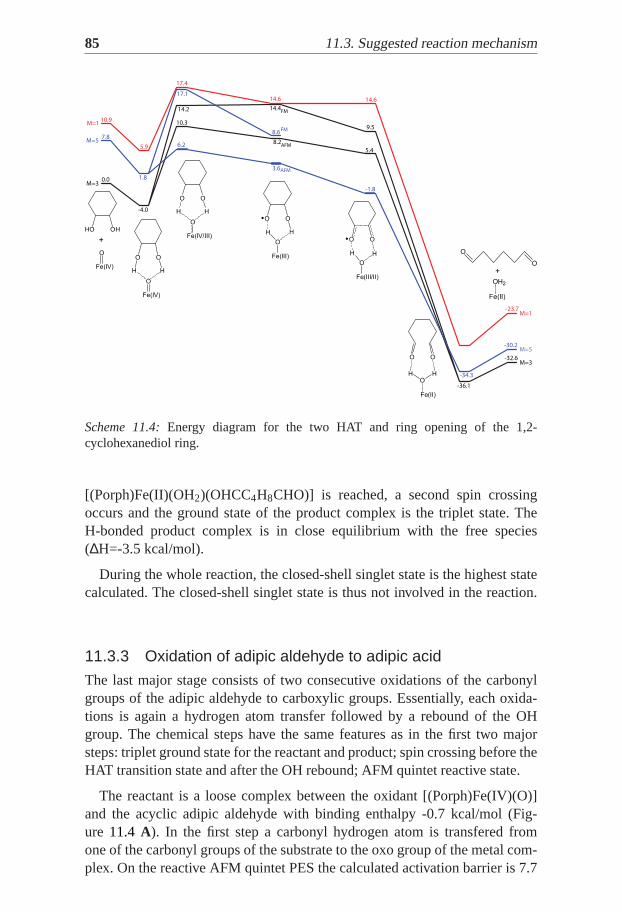
85 11.3. Suggested reaction mechanism
M=3
M=5
M=1
M=3
M=5
M=1
0.0
-36.1
5.4
8.6
8.2
17.4
10.3
7.8
-34.3
-1.8
3.6
6.2
10.9
-23.7
14.6
FM
AFM
AFM
14.4FM14.2
1.8
9.5
14.6
Fe(IV)
O
HO OH
Fe(IV)
O
O O
HH
Fe(IV/III)
O
O O
H H
Fe(III)
O
O O
H H
Fe(III/II)
O
O O
H H
Fe(II)
O
O O
HH
-4.0
5.9
17.1
-32.6
-30.2
+
Fe(II)
OH2
O
O
+
Scheme 11.4:Energy diagram for the two HAT and ring opening of the 1,2-cyclohexanediol ring.
[(Porph)Fe(II)(OH2)(OHCC4H8CHO)] is reached, a second spin crossingoccurs and the ground state of the product complex is the triplet state. TheH-bonded product complex is in close equilibrium with the free species(∆H=-3.5 kcal/mol).
During the whole reaction, the closed-shell singlet state is the highest statecalculated. The closed-shell singlet state is thus not involved in the reaction.
11.3.3 Oxidation of adipic aldehyde to adipic acid
The last major stage consists of two consecutive oxidations of the carbonylgroups of the adipic aldehyde to carboxylic groups. Essentially, each oxida-tions is again a hydrogen atom transfer followed by a rebound of the OHgroup. The chemical steps have the same features as in the first two majorsteps: triplet ground state for the reactant and product; spin crossing before theHAT transition state and after the OH rebound; AFM quintet reactive state.
The reactant is a loose complex between the oxidant [(Porph)Fe(IV)(O)]and the acyclic adipic aldehyde with binding enthalpy -0.7 kcal/mol (Fig-ure 11.4 A). In the first step a carbonyl hydrogen atom is transfered fromone of the carbonyl groups of the substrate to the oxo group of the metal com-plex. On the reactive AFM quintet PES the calculated activation barrier is 7.7

11.3. Suggested reaction mechanism 86
kcal/mol with respect to the triplet reactant, and the reaction is exergonic with7.1 kcal/mol (optimized transition state in Figure11.4B). The following OHrebound goes through an activation barrier of 3.2 kcal/mol and the exergonic-ity (determined from the triplet ground state of the product) is 50.2 kcal/mol(Scheme11.5).
In the very last HAT the activation barriers for the AFM quintet and AFMtriplet spin states for the HAT transition state differ by only 0.8 kcal/mol(Scheme11.6). Still the quintet should be considered as a reactive state withactivation barrier of 8.2 kcal/mol. The reactant and the HAT transition statestructures are shown in Figure11.5. The resulting radical intermediate lies 3.6kcal/mol below the ground state reactant, and it takes 1.8 kcal/mol for the OHrebound to occur. The adipic acid is formed with exergonicity of 51.2 kcal/mol(triplet ground state).
The catalytic formation of adipic acid from cyclohexane and dioxygen wasfound to be a strongly exergonic process. Each of the sequential steps, butone, consists of hydrogen abstraction and subsequent hydroxide rebound. Theexception is the ring cleavage step where two sub-sequential hydrogen ab-stractions occur. In all of the major steps hydrogen abstraction is rate limiting.However the overall rate limiting HAT for the whole reaction is the one forthe hydroxylation of cyclohexanol to ortho-cyclohexanediol. The calculatedenthalpy of activation for this step is 17.8 kcal/mol. The entropy effect is notexpected to increase this number significantly. The reason that this particularHAT becomes rate limiting can be found in the formation of a rather stable[(Porph)Fe(II)(C6H10(OH)2)] adduct prior to the HAT. This step is possiblycrucial in differentiating between competing reactions, that might lead to theformation ofmeta- or para-cyclohexanediol, which in turn would lead to dif-ferent final products. These alternative paths have not been considered in thiswork, and the challenge to explore them remains. The presented mechanismshows, however, that the underlying chemistry in itself is rather basic.
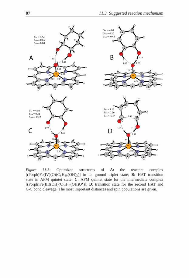
87 11.3. Suggested reaction mechanism
C C C
CC
C C
NC
N C
C
O
C
C
Fe
C
C
C
C
O
C NC
C
N
O
C CC
C
C C C
1.65
1.84
1.85
SFe = 1.42
Soxo = 0.65
Ssub = 0.00
2.01
2.01
2.01
2.01
C C CC C
C C
N NC C
C C
Fe
O
O
C
C
CN
C
N
O
C
C
C
C
C CC C
C C C
SFe = 4.00
Soxo = 0.30
Ssub = -0.43
2.10
1.77
1.20
1.18
1.65
C C CC
CC
C
NC
NC
C C
Fe
C
C
C
O
C
C
C
OO
N CN
C
C CC C
C C C
SFe = 4.03
Soxo = 0.33
Ssub = -0.72
1.77
2.10
1.84
1.62
C C CC C
C CN NC C
C C
Fe
O
O
C CN N
CC
C
O
C CC C
C
C
C
C C C
SFe = 4.13
Soxo = 0.26
Ssub = -0.94
1.74
2.46
1.33
1.88
2.10
1.11
A
C D
B
Figure 11.3: Optimized structures of A: the reactant complex[(Porph)Fe(IV)(O)(C6H10(OH)2)] in its ground triplet state;B: HAT transitionstate in AFM quintet state;C: AFM quintet state for the intermediate complex[(Porph)Fe(III)(OH)(C6H10(OH)O•)]; D: transition state for the second HAT andC-C bond cleavage. The most important distances and spin populations are given.

11.3. Suggested reaction mechanism 88
M=3
M=5
M=1
M=3
M=5
M=1
0.0
-58.0
-2.4
8.3
7.8
-56.4
-7.8
7.0
10.9
-47.8
4.9
FM
AFM
AFM
-3.1
FM
-4.6
2.2
18.1
Fe(IV/III)
O
O
O
H
Fe(III)
O
O
O
H
Fe(III/II)
O
O
O
H
Fe(II)
OH
O
O
Fe(IV)
O
O
O
H
8.9
6.6
-0.7
-58.8
-54.6
-44.9
+
Fe(IV)
O
O
O
H
Fe(II)
OH
O
O
+
Scheme 11.5:Energy diagram for the oxidation of the first carbonyl group of the adipicaldehyde.
C C C
CCC C
NC N C
C
C
O
C
Fe
C
C
O
C N
C
CN
C
C
C
C
CC
O
C C C
SFe = 1.26
Soxo = 0.78
Ssub = 0.00
1.64
2.01
O
C
C
C CC
C
C
CC
NC N
C
C
C
C
Fe
C
O
C
C
N
C
CN
O
C C CC
C C C
SFe = 3.80
Soxo = 0.18
Ssub = -0.391.32
1.22
1.74
2.10
A B
Figure 11.4: Optimized structures of A: the reactant complex[(Porph)Fe(IV)(O)(OHCC4H8CHO)] in its ground triplet state;B: HAT transi-tion state in AFM quintet state. The most important distances and spin populationsare given.

89 11.3. Suggested reaction mechanism
M=3
M=5
M=1
M=3
M=5
M=1
0.0
-58.1
-2.0
5.7
7.8
-53.9
-6.9
4.9
10.9
-46.8
1.9FM
AFM
AFM
-2.2
FM
-5.1
2.8
0.3
10.4
Fe(IV)
O
HO
O
O
H
Fe(IV/III)
O
HO
O
O
H
Fe(III)
O
HO
O
O
H
Fe(III/II)
O
HO
O
O
H
Fe(II)
OH
HO
O
O
-3.3
4.4
6.2
-57.4
-53.2
-43.5
Fe(II)
OH
HO
O
O
+
Fe(IV)
O
HO
O
O
H
+
Scheme 11.6:Energy diagram for the final oxidative step leading to adipicacid.
C C CC CC C
NC N C
CFe
C
O
C
C
C NCN
C
O
C
C
O
C
C
CC C
O
C C C
SFe = 1.25
Soxo = 0.80
Ssub = 0.00
1.64
2.01
C C CC
CC
C
NC N C
C
Fe
C
C
O
C
C N
O
C
C
N
C
C
C
C
O
C CC
C
O
C C
SFe = 3.79
Soxo = 0.18
Ssub = -0.39
1.36
1.21
1.73
2.10
A B
Figure 11.5: Optimized structures of A: the reactant complex[(Porph)Fe(IV)(O)(OHCC4H8COOH)] in its ground triplet state;B: HAT tran-sition state in AFM quintet state. The most important distances and spin populationsare given.

Part V:Concluding Remarks
The investigations presented in this thesis aim at bringing some insights andbetter understanding of the catalytic mechanism ofortho-hydroxyl dioxyge-nases, and dioxygen activation in general. A hybrid DFT method is used tomodel three enzymes (Homoprotocatechuate 2,3-dioxygenase fromBrevibac-terium fuscum Bf2,3-HPCD, Manganese-Dependent Homoprotocatechuate2,3-Dioxygenase MndD, and Homogentisate Dioxygenase HGD), and twobiomimetic complexes. The reported results are obtained with models con-sisting of up to 134 atoms.
The experimentally observed similarities betweenBf 2,3-HPCD and MndDare reproduced in the calculations, and the activation barriers obtained arein good agreement with the experiment. The suggested mechanism for thecatalytic reaction of HGD agrees with several experimental observations forextradiol dioxygenases. The alternative reaction paths were found unprobableby reason of higher barriers.
The calculations for the non-heme iron complex show that only the quartetstate is likely to be involved in an energetically plausible mechanism. Eventhough structurally different from the typical intradiol enzyme active site, thecomplex uses a mechanism with the same major features – formation of bridg-ing peroxide and homolytic O-O bond cleavage. The formation of the bridgingperoxide was found rate limiting in the proposed mechanism.
Based on the computational results, a reaction mechanism was suggestedfor the conversion of cyclohexane to adipic acid in the presence of molecu-lar oxygen, and a biomimetic heme complex as a catalyst. The mechanismconsists of several consecutive oxidative steps, where a hydrogen atom trans-fer process was found rate limiting. The overall rate limiting HAT is possiblyimportant for differentiation between the desired and the side products.


Part VI:Acknowledgements
First of all, I would like to thank my supervisors Margareta and Per foraccepting me in the group and giving me the opportunity to work in this area. Ihave found friendly atmosphere and a lot of help. I am very grateful to Tomek,for guiding my first steps, and being such a good friend.
Many thanks to all my former and present colleagues for their help andfriendship. Special thanks to Holger (from Germany) and Johannes, for thegreat times we had and for our collaboration. Thanks to Martin, Rossen andHolger for reading the thesis.
I am grateful to all my previous supervisors from the University of Sofia,Bulgaria. They introduced me to Theoretical Chemistry and helped me in dif-ficult moments.
Warm thanks to my family for the fact that I exist, and for their support allthe time, in all the difficult for us moments. Thanks to Polina’s family for theirhospitality and care.
Finally I would like to thank Polina, Monica and Filip – my present family.Thank you Polina for your love and support, for making me happy, and thankyou Monica and Filip, for bringing me all the joy! I love you!
THANK YOU ALL!


Part VII:Populärvetenskapligsammanfattning på svenska
Kvantkemi är idag ett kraftfullt och effektivt verktyg som framgångsrikt an-vänds för att studera olika biologiska system. Kvantkemisk metodik användsför att modellera enzymers aktiva säten och de reaktioner som katalyserasdär. Intermediärer och övergångstillstånd längs olika reaktionsvägar kan op-timeras och deras relativa energier kan jämföras. På så vis kan slutsatser omvilka reaktionsvägar som är mest gynnsamma dras.
I den här avhandling utnyttjas täthetsfunktionalteori (DFT) för att studeraden katalytiska funktionen hos några metallenzymer och syntetiska model-lkomplex. Tre olika enzymer som oxiderar olika varianter av den organiskamolekylen katekol har studerats. Dessa enzymer innehåller järn eller mangani det aktiva sätet. Modellerna som använts i beräkningarna består av mellan50 och 200 atomer och har konstruerats från röntgenkristallstrukturer. De syn-tetiska modellkomplex som studerats är dels ett järnkomplex som efterliknarreaktiviteten hos metallenzymerna och dels ett hem-komplex som katalyserarden stegvisa oxidationen av cyklohexan till adipinsyra. Gemensamt för allafem systemen är att aktivering av syremolekylen ingår som en viktig del. Syre-molekylens grundtillstånd är en triplett och den kan därför inte reagera medvanliga molekyler i singletttillståndet, eftersom sådana reaktioner är spinnför-bjudna. Under inverkan av metalljoner i proteiner kan sådana reaktioner ske inaturen vid normal temperatur och neutralt pH. Proteiner som innehåller kop-par eller järn hör till de vanligaste katalysatorerna för syreaktivering liksomför transport av syre.
Att studera olika enzymers struktur och deras reaktionsvägar är ett sätt attförsöka förstå deras stora katalytiska kapacitet. Sådan kunskap är av stor bety-delse inom alla områden där katalytiska egenskaper utnyttjas, som till exempeli läkemedelsindustrin och kemiindustrin.


97
Bibliography
[1] Christopher K. Mathews; K.E. van Holde; Kevin Ahern.Biochemistry. PrenticeHall, 1999.
[2] Karl-Erich Jaeger and Thorsten Eggert.Current Opinion in Biotechnology,15(4):305–313, 2004.
[3] Richard B. Silverman.The organic chemistry of enzyme-Catalyzed reac-tions. Elsevier Science, 2002.
[4] Koshland D. E. Jr.PNAS, 44:98–104, 1958.
[5] Mireia Garcia-Viloca; Jiali Gao; Martin Karplus; Donald G. Truhlar.Science,303:186–195, 2004.
[6] A. Warshel; P. K. Sharma; M. Kato; Y. Xiang; H. Liu and M. H. M. Olsson.Chem. Rev., 106:3210–3235, 2006.
[7] Costas M.; Mehn M. P.; Jensen M. P.; Que L.Chem. Rev., 104:939, 2004.
[8] Jensen F.Introduction to Computational Chemistry. John Wiley & SonsLtd, 2002.
[9] Young D. C.Computational Chemistry. John Wiley & Sons Ltd, 2001.
[10] Szabo A.; Ostlund N. S.Modern Quantum Chemistry. Dover Publications:New York, 1989.
[11] Parr R.; Yang W. Density Functional Theory of Atoms and Molecules.Oxford University Press: New York, 1989.
[12] Koch W.; Holthausen M. C.A Chemist’s Guide to Density Functional The-ory. Wiley: Weinheim, 2000.
[13] Hohenberg P.; Kohn W.Phys. Rev., 136:B864–B887, 1964.
[14] Kohn W.; Sham L.Phys. Rev., 140:A1133–A1138, 1965.
[15] D. M. Ceperly; J. B. Alder.J. Phys. Rev. Lett., 45:566, 1980.
[16] M. Nusair S. J. Vosko, L. Wilk.Can. J. Phys., 58:1200–1211, 1980.
[17] J. P. Perdew.Phys. Rev. B, 33:8822, 1986.

BIBLIOGRAPHY 98
[18] C. Lee; W. Yang; G. R. Parr.Phys. Rev. B, 37:785, 1988.
[19] J. R. Bartlett N. Oliphant.J. Chem. Phys., 100:6550, 1994.
[20] Becke A.D.Phys. Rev., A38:3098, 1988.
[21] Becke A.D.J. Chem. Phys., 96:2155, 1992.
[22] Becke A.D.J. Chem. Phys., 97:9173, 1992.
[23] Becke A.D.J. Chem. Phys., 98:5648, 1993.
[24] Curtiss L. A.; Raghavachari K.; Trucks G. W.; Pople J. A.J. Chem. Phys.,94:7221–7230, 1991.
[25] Curtiss L. A.; Raghavachari K.; Redfern P. C.; Pople J. A.J. Chem. Phys.,109:7764–7776, 1998.
[26] H. Bauschlicher, C.; Partridge.Chem. Phys. Lett., 240:533–540, 1995.
[27] C. Bauschlicher.Chem. Phys. Lett., 246:40–44, 1995.
[28] Siegbahn P. E. M.Inorg. Chem., 39(13):2923–2935, 2000.
[29] Siegbahn P. E. M.Quarterly Reviews of Biophysics., 36:91–145, 2003.
[30] M. Lundberg.Challenges in Enzyme Catalysis – Photosystem II abd Oroti-dine Decarboxylase. A Density Functional Theory Treatment. PhD thesis,Stockholm University, 2005.
[31] Blomberg M. R. A.; Siegbahn P. E. M.; Svensson M. J.J. Chem. Phys.,104:9546–9554, 1996.
[32] C. W. J. Ricca, A.; Bauschlicher.J. Chem. Phys., 101:8949–8955, 1996.
[33] Salomon O.; Hess B.A. Reiher M.Theor. Chem. Acc., 107:48–55, 2001.
[34] Eyring H.; Stern A.Chem. Rev., 24:2, 1939.
[35] J. Anderson.Adv. Chem. Phys., 91:381, 1995.
[36] Truhlar D.; Garrett B.; Klippenstein S.;.J. Phys. Chem., 100:12771, 1996.
[37] F. Himo. Biochim. Biophys. Acta - Bioenergetics, 1707:24–33, 2005.
[38] V. Pelmenschikov.Theoretical Modeling of Enzyme Catalysis with focuson radical chemistry. PhD thesis, Stockholm University, 2005.
[39] Oregon Schrödinger Inc.; Portland. Jaguar 5.5, jaguar 7.0.2007.
[40] Hay P.J. Wadt WR.J. Chem. Phys., 82:299–310, 1985.
99 BIBLIOGRAPHY
[41] T. Vreven; K. N. Kudin; J. C. Burant; J. M. Millam; S. S. Iyengar; J. Tomasi;V. Barone; B. Mennucci; M. Cossi; G. Scalmani; N. Rega; G. A. Petersson; H.Nakatsuji; M. Hada M. Ehara; K. Toyota; R. Fukuda; J. Hasegawa; M. Ishida;T. Nakajima; Y. Honda; O. Kitao; H. Nakai; M. Klene; X. Li; J. E. Knox; H.P.Hratchian; J. B. Cross; V. Bakken; C. Adamo; J. Jaramillo; R. Gomperts; R. E.Stratmann; O. Yazyev; A. J. Austin; R. Cammi; C. Pomelli; J. W. Ochterski; P.Y. Ayala; K. Morokuma; G. A. Voth; P. Salvador; J. J. Dannenberg; V. G. Za-krzewski; S. Dapprich; A. D. Daniels; M. C. Strain; O. Farkas; D. K. Malick; A.D. Rabuck; K. Raghavachari; J. B. Foresman; J. V. Ortiz; Q. Cui; A.G. Baboul;S. Clifford; J. Cioslowski; B. B. Stefanov; G. Liu; A. Liashenko; P. Piskorz; I.Komaromi; R. L. Martin; D. J. Fox; T. Keith; M. A. Al-Laham; C. Y. Peng;A. Nanayakkara; M. Challacombe; P. M. W. Gill; B. Johnson; W. Chen; M. W.Wong; C. Gonzalez; M. J. Frisch; G. W. Trucks; H. B. Schlegel; G. E. Scuseria;M. A. Robb; J. R. Cheeseman; J. A. Montgomery; Jr. and Wallingford CTJ. A.Pople; Gaussian Inc. Gaussian 03, revision d01. 2007.
[42] Noodleman L.; Case D. A.Adv. Inorg. Chem., 38:423–470, 1992.
[43] Mouesca J.-M.; Chen J. C.; Noodleman L.; Bashford D.; Case D. A. J. Am.Chem. Soc., 116:11898–11914, 1994.
[44] Tannor D.; Marten B.; Murphy R.; Friesner R.; Sitkoff D.; NickollsA.; HonigB.; Ringnalda M.; Goddart III W.J. Am. Chem. Soc., 116:11875–11882, 1994.
[45] Marten B.; Kim K.; Cortis C.; Friesner R.; Murphy R.; Ringnalda M.; SitkoffD.; Honig B. J. Phys. Chem., 100:11755–11788, 1996.
[46] Blomberg M.; Siegbahn P. E. M.; Babcock G. J. Am. Chem. Soc.,120(34):8812–8824, 1998.
[47] Siegbahn P. E. M.; Blomberg M. R. A.Chem. Rev., 100:421, 2000.
[48] Siegbahn P. E. M.J. Comput. Chem., 22:1634, 2001.
[49] Tomasi J.; Persico M.Chem. Rev., 94:2027–2094, 1994.
[50] Cramer C. J.; Truhlar D. G.Chem. Rev., 99:2161–2200, 1999.
[51] Chipman D. M.J. Phys. Chem. A, 106:7413–7422, 2002.
[52] Chipman D. M.J. Phys. Chem. A, 118:9937–9942, 2003.
[53] Ornston L. N.; Stanier R. Y.J. Biol. Chem., 241:3776–3786, 1966.
[54] Dagley S.Surv. Prog. Chem., 8:121–170, 1977.
[55] Broderick J. B.Essays Biochem., 34:173–189, 1999.
[56] Jackson T. A.; Que L. Jr.Concepts and Models in Bioinorganic Chemistry,2006.

BIBLIOGRAPHY 100
[57] Knox W. E.; Edwards S. W.J. Biol. Chem., 216:479–487, 1955.
[58] La Du B. N.; Zannoni V. G.; Laster L.; Seegmiller J. E.J. Biol. Chem.,230:251–260, 1958.
[59] Vetting M. W.; Wackett L. P.; Que L. Jr.; Lipscomb J. D.; Ohlendorf D. H. J.Bacteriol., 186:1945–1958, 2004.
[60] Bassan A.; Borowski T.; Siegbahn P. E. M.Dalton Trans., pages 3153–3162,2004.
[61] Que L. Jr.Nat. Struct. Biol., 7:182–184, 2000.
[62] Whiting A. K.; Boldt Y. R.; Hendrich L. P.; Wackett L. P.; Que L. Jr. Biochem-istry, 35:160–170, 1996.
[63] Stephanie L. Groce; John D. Lipscomb.J. Am. Chem. Soc., 125:11780–11781,2003.
[64] Sato N.; Uragami Y.; Nishizaki T.; Takahashi Y.; Sazaki G.;Sugimoto K.;Nokana T.; Masai E.; Fukuda M.; Senda T.J. Mol. Biol., 321:621–636, 2002.
[65] Arciero D. M.; Orville A. M.; Lipscomb J. D. J. Biol. Chem., 260:14035–14044, 1985.
[66] H.; Lin G. Bugg T. D.Chem. Commun., 11:941–952, 2001.
[67] Miller M. A.; Lipscomb J. D.J. Biol. Chem., 271:5524–5535, 1996.
[68] Shu L.; Chiou Y. M.; Orville A. M.; Miller M. A.; Lipscomb J. D.; Que L. Jr.Biochemistry, 34:6649–6659, 1995.
[69] Groce S. L.; Lipscomb J. D.Biochemistry, 44:7175–7188, 2005.
[70] Emerson J.; Wagner M. L.; Reynolds M. F.; Que L. Jr.; Sadowsky M. J.; WacketL.P. J. Biol. Inorg. Chem., 10:751–760, 2005.
[71] Sanvoisin J.; Langley G. J.; Bugg T. D. H.J. Am. Chem. Soc., 117:7836–7837,1995.
[72] Kovaleva E. D.; Lipscomb J. D.Science, 316:453–457, 2007.
[73] Kovaleva E. D.; Neibergall M. B.; Chakrabarty S.; Lipscomb J. D. Acc. Chem.Res., 40:475–483, 2007.
[74] Harvey J. N.; Aschi M.; Schwarz H.; Koch W.Theor. Chem. Acc., 99:347–361,2003.
[75] Harvey J. N.; Poli R.; Smith K. M.Coord. Chem. Rev., 238–239:347–361,1998.

101 BIBLIOGRAPHY
[76] Titus G.; Mueller H.; Burgner J.; Cordoba S. R. D.; Penalva M.; Timm D. Nat.Struct. Biol., 7:542–546, 2000.
[77] Amaya A. A.; Brzezinski K. T.; Farrington N.; Moran G. R.Arch. Biochem.Biophys., 421:135–142, 2004.
[78] Veldhuizen E. J. A.; Vaillancourt F. H.; Whiting C. J.; Hsiao M. M.-Y.; Gin-gras G.; Xiao Y.; Tanguay R. M.; Boukouvalas J.; Eltis L. D.Arch. Biochem.Biophys., 386:305–314, 2005.
[79] Hintner J.-P.; Reemtsma T.; Stolz A.J. Biol. Chem., 279:37250–37260, 2004.
[80] Koch W.O.; Krüger H-J.Angew. Chem. Int. Ed. Engl., 34:2671–2674, 1995.
[81] Walsh T. A.; Ballou D. P.; Mayer R.; Que L. Jr.J. Biol. Chem., 258:14422,1983.
[82] Salomon O.; Reiher M.; Hess B.A.J. Chem. Phys., 117:4729, 2002.
[83] Grimme S.J. Comp. Chem., 25:1463, 2004.
[84] Grimme S.J. Comp. Chem., 27:1787, 2004.
[85] Funabiki T.; Yamazaki T.J. Mol. Cat. A, 150:37, 1999.
[86] Borowski T.; Siegbahn P. E. M.J. Am. Chem. Soc., 128(39):12941–12953,2006.
[87] Siegbahn P. E. M.; Haeffner F.J. Am. Chem. Soc., 126(29):8919–8932, 2004.
[88] Davis D. D.; Kemp D. R. In.Kirk-Othmer Encyclopedia of Chemical Tech-nology, 4th ed.Wiley: New York, 1991.
[89] Iwahama T.; Syojyo K.; Sakaguchi S.; Ishii Y.Org. Process Res. DeV., 2:255–260, 1998.
[90] Pigamo A.; Besson M.; Blanc B.; Gallezot P.; Blackburn A.; Kozynchenko O.;Tennison S.; Crezee E.; Kapteijn F.Carbon, 40:1267–1278, 2002.
[91] Ratnasamy P.; Raja R. Ep 0784045-a1, 2002.
[92] Ying Yuan; Hongbing Ji; Yixia Chen; Yong Han; Xufeng Song; Yuanbin She;and Rugang Zhong.Org. Proc. Res. Dev., 8(3):418–420, 2004.
[93] Chin D.-H.; Del Gaudio J.; La Mar G. N.; Balch A. L.J. Am. Chem. Soc.,99:5486–5488, 1977.
[94] Blomberg M. R. A.; Johansson A. J.; Siegbahn P. E. M.Inorg. Chem., 46:7992–7997, 2007.
[95] Becket C.W.J. Am. Chem. Soc., 69:2488–2495, 1974.
[96] Aston J.G.; Szasa G.J.; Fink H.L.J. Am. Chem. Soc., 65:1135–1139, 1943.