Embed Size (px)
Citation preview
1
Imaging Procedures Manual for CT/MRI and Angiography in SITS Open, version 1 – 2015-11-06
Imaging Procedures Manual
Dear Study Coordinator and CT/MRI/DSA Technologist(s): Karolinska Institutet requires that the Study Coordinator and all DSA and CT/MRI Technologists
involved in the SITS Open protocol version 4.0 date 20151105 study read and fully understand the Manual for Ischemic Stroke Imaging.This requirement should be completed before new subjects are
imaged in SITS Open study. All new personnel, who join the study after centre initiation, are also required to read and understand this manual.
Imaging Procedures Manual for Ischemic Stroke MR/CT Imaging and Angiography
Rüdiger von Kummer
2
Imaging Procedures Manual for CT/MRI and Angiography in SITS Open, version 1 – 2015-11-06
Table of Contents
1.0 INTRODUCTION 5
2.0 STUDY INTRODUCTION 5 2.1 STUDY OVERVIEW 5 2.2 ROLE OF KAROLINSKA INSTITUTE (KI) AND IMAGING CORE LAB 6 2.3 RESPONSIBILITIES OF CLINICAL CENTRES 7 2.4 RESPONSIBILITIES OF DSA AND CT/MRI FACILITIES 8
3.0 DSA AND CT/MRI STUDY CENTRE QUALIFICATION 8 3.1 GENERAL REQUIREMENTS FOR IMAGING FACILITIES (21.8 STUDY PROTOCOL) 9 3.2 SUPPLIES PROVIDED BY SITS OPEN COORDINATION TEAM (KI) 10
4.0 SCHEDULING SUBJECT DSA AND CT/MRI SCANS 10 4.1 SUBJECT SCHEDULING 10 4.1.1 SUBJECT SCANS 10
5.0 SUBJECT CT EXAM PREPARATION 11 5.1 SUBJECT CT POSITIONING 11 5.2 LABELING DIGITAL HEADER FIELDS FOR SUBJECT CT SCANS 12 5.3 SUBJECT CT/CTA ACQUISITION 12 5.3.1 CONTRAST AGENT INJECTION TECHNIQUE 13 5.3.2 CTA DATA POST-PROCESSING 14 5.3.3 RISKS AND PROVISIONS FOR MINIMIZING RISKS 14
6.0 SUBJECT MRI EXAM PREPARATION 15 6.1 SUBJECT SAFETY AND MONITORING 16 6.2 SUBJECT MRI POSITIONING 16 6.3 LABELING DIGITAL HEADER FIELDS FOR SUBJECT MRI SCANS 17 6.4 SUBJECT MRI ACQUISITION TECHNIQUE 17 6.4.1 PRE-SCAN ADJUSTMENTS 18 6.4.2 PROTOCOL FOR MRI OF THE BRAIN 18
7.0 SUBJECT DSA ACQUISITION TECHNIQUE 20 7.1 PATIENT PREPARATION 20 7.2 DSA IMAGING PROTOCOL 20 7.2.1 BASELINE DSA 20 7.2.2 PROCEDURAL DSA 20 7.2.3 INTERVENTION 21 7.2.4 END-OF-INTERVENTION DSA 22 7.3 LABELING DIGITAL HEADER FIELDS FOR DSA 22 7.4 ARCHIVAL OF ARTERIOGRAPHY OF STUDY PATIENTS 23
8.0 PROCEDURE AND PREPARATION FOR TRANSFER OF IMAGES FOR EVALUATION 23 8.1 PREPARE CD/DVDS FOR CT/MRI AND DSA 23 8.2 SUBJECT DSA AND CT/MRI SCANS TRANSFER TO SITS OPEN TEAM (KI) AND IMAGING CORE LAB 25 8.3 SITS OPEN COORDINATION TEAM (KI) EVALUATION OF SUBJECT SCANS (QUALITY CHECK) 25
3
Imaging Procedures Manual for CT/MRI and Angiography in SITS Open, version 1 – 2015-11-06
8.3.1 REQUESTS FOR RESUBMISSION OF SUBJECT DATA 25 8.4 SITS OPEN COORDINATION TEAM (KI) SUBMISSION TO IMAGING CORE LAB 25
9.0 IMAGING CORE LAB EVALUATION OF SUBJECT DSA AND CT/MRI SCANS 26 9.1 DEFINITIONS 26
4
Imaging Procedures Manual for CT/MRI and Angiography in SITS Open, version 1 – 2015-11-06
Abbreviations
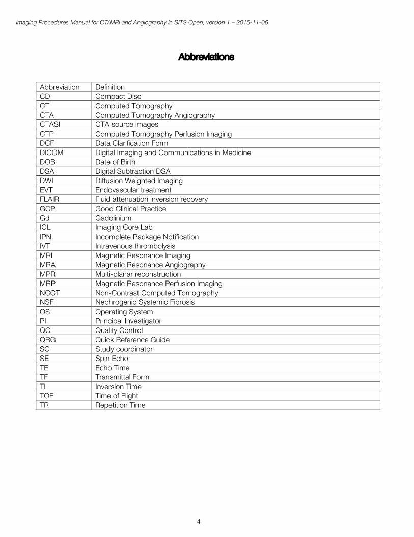
Abbreviation Definition CD Compact Disc CT Computed Tomography CTA Computed Tomography Angiography CTASI CTA source images CTP Computed Tomography Perfusion Imaging DCF Data Clarification Form DICOM Digital Imaging and Communications in Medicine DOB Date of Birth DSA Digital Subtraction DSA DWI Diffusion Weighted Imaging EVT Endovascular treatment FLAIR Fluid attenuation inversion recovery GCP Good Clinical Practice Gd Gadolinium ICL Imaging Core Lab IPN Incomplete Package Notification IVT Intravenous thrombolysis MRI Magnetic Resonance Imaging MRA Magnetic Resonance Angiography MPR Multi-planar reconstruction MRP Magnetic Resonance Perfusion Imaging NCCT Non-Contrast Computed Tomography NSF Nephrogenic Systemic Fibrosis OS Operating System PI Principal Investigator QC Quality Control QRG Quick Reference Guide SC Study coordinator SE Spin Echo TE Echo Time TF Transmittal Form TI Inversion Time TOF Time of Flight TR Repetition Time
5
Imaging Procedures Manual for CT/MRI and Angiography in SITS Open, version 1 – 2015-11-06
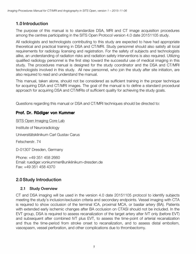
1.0 Introduction The purpose of this manual is to standardize DSA, MRI and CT image acquisition procedures among the centres participating in the SITS Open Protocol version 4.0 date 20151105 study. All radiologists and technologists contributing to this study are expected to have had appropriate theoretical and practical training in DSA and CT/MRI. Study personnel should also satisfy all local requirements for radiology licensing and registration. For the safety of subjects and technologists alike, an understanding of radiation risks and radiation safety interventions is also required. Utilizing qualified radiology personnel is the first step toward the successful use of medical imaging in this study. The procedures manual is designed for the study coordinator and the DSA and CT/MRI technologists involved in this study. All new personnel, who join the study after site initiation, are also required to read and understand the manual. This manual, taken alone, should not be considered as sufficient training in the proper technique for acquiring DSA and CT/MRI images. The goal of the manual is to define a standard procedural approach for acquiring DSA and CT/MRIs of sufficient quality for achieving the study goals. Questions regarding this manual or DSA and CT/MRI techniques should be directed to: Prof. Dr. Rüdiger von Kummer
SITS Open Imaging Core Lab Institute of Neuroradiology Universitätsklinikum Carl Gustav Carus
Fetscherstr. 74
D-01307 Dresden, Germany
Phone: +49 351 458 2660 Email: [email protected] Fax: +49 351 458 4370
2.0 Study Introduction
2.1 Study Overview CT and DSA imaging will be used in the version 4.0 date 20151105 protocol to identify subjects meeting the study’s inclusion/exclusion criteria and secondary endpoints. Vessel imaging with CTA is required to show occlusion of the terminal ICA, proximal MCA, or basilar artery (BA). Patients with extended early ischemic changes after BA occlusion on CTASI should not be included. In the EVT group, DSA is required to assess recanalization of the target artery after IVT only (before EVT) and subsequent after combined IVT plus EVT, to assess the time-point of arterial recanalization and thus the time-period from stroke onset to recanalization, and to assess distal embolism, vasospasm, vessel perforation, and other complications due to thrombectomy.
6
Imaging Procedures Manual for CT/MRI and Angiography in SITS Open, version 1 – 2015-11-06
It is anticipated that the study will involve approximately 600 subjects (maximum sample size: 2 x 300), recruited at 60 centres (30 centres for each treatment arm). Each subject will have CT/CTA scans acquired at baseline, and again at follow-up (22 – 36 hours after randomization). According to protocol, if additional irradiation is a concern, MRI and contrast-enhanced MRA may be done at follow-up instead of CT/CTA. DSA will be acquired before, during, and after the recanalization intervention for subjects in the Endovascular treatment (EVT) arm. Unscheduled follow-up examinations may be acquired for some subjects.
2.2 Role of Karolinska Institute (KI) and Imaging Core Lab
Karol inska Institutet’s Primary Responsibi l i t ies to Centre
• SITS Open Coordination team (KI) provides a SITS Open User’s Guide describing the logistics for sending images to SITS Coordination team (KI).
• SITS Open Coordination team (KI) provide centre with an Imaging Intervention Manual for subject scanning with CT, MRI, and DSA.
• SITS Open Coordination team (KI): Gathering of imaging studies from the study sites, quality check, archiving, and sending the material to the imaging core lab
Continued responsibi l i t ies of SITS Open Coordination team (KI) are to: • Verify that the sequence parameters used to acquire DSA and CT/MRI examinations are in
agreement with the Imaging Intervention manual. • Review the quality of DSA and CT/MRI images for adequate anatomical coverage. • Provide ongoing support and feedback to clinical centres and DSA and CT/MRI facilities.
• Take care that follow-up (FU) images are not sent to the Imaging Core Lab before the adjudication of images at baseline (BL) and during intervention – if applicable – has been archived.
SITS Open Coordination team (KI) personnel will review the technical quality of all DSA and CT/MRI images submitted. It is expected that the majority of examinations received will be of acceptable quality. If any problems are detected related to image quality, SITS Open Coordination team (KI) will notify the responsible clinical centres and the DSA and CT/MRI facility via email or phone, suggest possible causes of the problem, and offer potential solutions. The DSA and CT/MRI facility should try to correct these errors and avoid them in future examinations. Despite this review, the responsibility of DSA and CT/MRI scans of acceptable quality is still at the DSA and CT/MRI facility.
7
Imaging Procedures Manual for CT/MRI and Angiography in SITS Open, version 1 – 2015-11-06
Primary Responsibi l i t ies of SITS Open Coordination team (KI) to Imaging Core Lab • Provides Imaging Intervention Manual based on the definitions and requirements of the
study protocol. • Updates the Imaging Intervention Manual according to amendments of the study protocol. • Provides imaging eCRFs (Imaging evaluation from) to be filled in by the Imaging Core Lab. • Receives, archives, and performs quality checks on anonymizes subject scans. Sends the anonymized scans on CD together with a transmittal form to the Imaging Core Lab. Primary Responsibi l i t ies of Imaging Core Lab • Consults SITS Open Coordination team (KI) regarding all imaging related issues. • Reviews the Imaging Intervention Manual and imaging eCRFs (Imaging evaluation from)
before initiation of the study. • Receives, checks, and evaluates anonymized subject scans, fills in eCRFs in due time and
sends them electronically to SITS Open Coordination team (KI) • Provides support to SITS Open Coordination team (KI) to update Imaging Intervention
Manual according to amendments of the study protocol.
2.3 Responsibilities of Clinical Centres
Primary Responsibi l i t ies of Cl inical centres
• Schedule subject examinations for all visits in conjunction with the DSA and CT/MRI technologist.
• Provide subject ID (enrollment ID) and examination information to the DSA and CT/MRI technologist so that this information is entered completely and correctly on the DSA or CT/MRI Transmittal Form that is submitted with the data.
• Ensure that DSA and CT/MRI data and corresponding Transmittal Form are submitted to SITS Open Coordination team (KI) within 3 working days of acquisition. Update the study eCRF with information about the CT/MRI and DSA.
Continued responsibi l i t ies of Cl inical Centres: • Receive supplies and distribute these supplies to the appropriate study personnel, based on
defined roles in the data acquisition and submission process. • Notify SITS Open Coordination team (KI) when all subjects enrolled in the DSA and CT/MRI
study have completed their follow-up (22 - 36 hours after randomization). • Ensure that the DSA and CT/MRI personnel have a copy of this manual.
8
Imaging Procedures Manual for CT/MRI and Angiography in SITS Open, version 1 – 2015-11-06
• Notify SITS Open Coordination team (KI) about planned upgrades at the DSA and CT/MRI facility or other issues that might compromise the consistency of DSA and CT/MRI scanning over time.
2.4 Responsibilities of DSA and CT/MRI Facilities
Primary Responsibi l i t ies of Imaging Faci l i ty
• Acquire DSA and CT/MRI scans for subjects in compliance with interventions and imaging protocol detailed in this Manual.
• Verify that all subject ID (Enrollment ID) and examination information are entered completely and correctly on the DSA or MRI or CT Transmittal Form that is submitted with the image data.
• Send DSA and CT/MRI data and corresponding Transmittal Form to SITS Open Coordination team (KI) within 3 working days of acquisition.
Continued responsibi l i t ies of imaging faci l i ty: • Confirm that all DSA and CT/MRI technologists who will be performing scans for the SITS
Open Protocol are properly trained on the study-specific acquisition parameters and data submission interventions.
• Perform system calibrations at the beginning of the study. • Review exam quality and obtain repeats as necessary. • Ensure that no hardware or software upgrades are performed on the scanner being used
for this study that would compromise volumetric analysis if applicable. • Notify SITS Open Coordination team (KI) immediately as soon as they are aware that a hardware or software upgrade is scheduled. • In some cases, the responsibilities of the Clinical Centre and the DSA and CT/MRI facility overlap. We ask that the Study Coordinator work with the lead technologist to decide how overlapping responsibilities will be assigned (for example, who will fill out Transmittal Forms and mail data to SITS Open Coordination team (KI)).
3.0 DSA and CT/MRI Study Centre Qualification SITS Open Coordination team (KI) will assist in identifying and qualifying facilities for participation in the study. To be considered for participation, DSA and CT/MRI facilities must meet the following requirements listed in the box below:
9
Imaging Procedures Manual for CT/MRI and Angiography in SITS Open, version 1 – 2015-11-06
3.1 General Requirements for Imaging Facilities (21.8 Study Protocol)
• MRI scanner should have a magnetic field strength of 1.5 or 3.0 Tesla. • DSA equipment and CT/MRI scanner must meet certain hardware and/or software
requirements.
• CT/MRI and DSA (for EVT patients) must be available 24h/7days. • CT scanning should be done with a < 2,5 mm slice thickness at baseline and follow-up
(allowing measurement of clot size by imaging core lab). Angiographic images of the occlusion being treated must allow clear visualisation of the target artery in 2 planes at least. Same orientation should be used before and after treatment to allow a valid analysis of the data.
• Baseline CT/CTA and FU (CT/CTA or MRI/MRA) scans (original thin slices and 4 to 8mm reconstructions, as well as thrombectomy DSA from EVT centres) should be stored on CD/DVD and sent to SITS Open Coordination team (KI)
• In case of any ICH or suspected ICH on 24-hour assessment, imaging should be repeated on the next day, and included on CD/DVD for further assessment by core lab with regard to differentiation blood/contrast.
• All images should be saved in DICOM format including the raw data and reconstructions. • The name of the patient on images should be replaced by the subject ID (enrollment ID).
Study date and time on the image should be kept. • Evaluation of occlusion, qualifying for inclusion in the study, should be based on the first CT
angiography performed after stroke onset, even if performed at another hospital before transport to the EVT centre (with or without initiated IVT), unless the quality of the images are insufficient for evaluation according to the radiologist at the EVT centre. The first CT and CT angiography (of sufficient quality) must be included in the material sent to SITS Open Coordination team (KI)
• For IVT centres, 2 CD/DVD are required: with baseline CT/CTA, and with 24h FU CT/CTA or MRI/MRA. For EVT centres, 3 CD/DVDs should be provided: the two mentioned above, plus a separate disk with EVT records. In case of repeated follow-up imaging in suspicion of ICH, the second follow-up record may be saved together with 24-hour imaging on the same disk. Centres are also encouraged to provide any additional imaging, which may be of importance for understanding of clinical decisions case of missing images, information, or poor quality the centre will be alerted by SITS Open Coordination team (KI). Please, keep the raw data on the hospital server until you get the confirmation that CD/DVD was collected and approved for the core lab evaluation. Facilities must be able to digitally archive data on CD in uncompressed DICOM format.
10
Imaging Procedures Manual for CT/MRI and Angiography in SITS Open, version 1 – 2015-11-06
3.2 Supplies Provided By SITS Open Coordination team (KI) SITS Open Coordination team (KI) will provide the following supplies to the Clinical Centre and the DSA and CT/MRI Facility:
• Latest update of study protocol • Imaging Intervention Manual for DSA and CT/MRI of the Brain (1 copy) • Transmittal Forms for Subject CT/MRI Scans • Transmittal Forms for Subject DSA
4.0 Scheduling Subject DSA and CT/MRI Scans
4.1 Subject Scheduling 4.1.1 Subject Scans
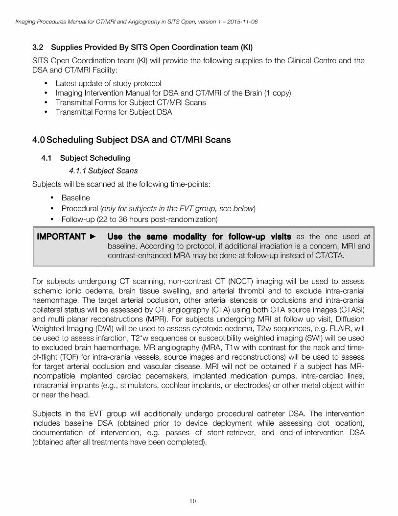
Subjects will be scanned at the following time-points: • Baseline • Procedural (only for subjects in the EVT group, see below) • Follow-up (22 to 36 hours post-randomization)
For subjects undergoing CT scanning, non-contrast CT (NCCT) imaging will be used to assess ischemic ionic oedema, brain tissue swelling, and arterial thrombi and to exclude intra-cranial haemorrhage. The target arterial occlusion, other arterial stenosis or occlusions and intra-cranial collateral status will be assessed by CT angiography (CTA) using both CTA source images (CTASI) and multi planar reconstructions (MPR). For subjects undergoing MRI at follow up visit, Diffusion Weighted Imaging (DWI) will be used to assess cytotoxic oedema, T2w sequences, e.g. FLAIR, will be used to assess infarction, T2*w sequences or susceptibility weighted imaging (SWI) will be used to excluded brain haemorrhage. MR angiography (MRA, T1w with contrast for the neck and time-of-flight (TOF) for intra-cranial vessels, source images and reconstructions) will be used to assess for target arterial occlusion and vascular disease. MRI will not be obtained if a subject has MR-incompatible implanted cardiac pacemakers, implanted medication pumps, intra-cardiac lines, intracranial implants (e.g., stimulators, cochlear implants, or electrodes) or other metal object within or near the head. Subjects in the EVT group will additionally undergo procedural catheter DSA. The intervention includes baseline DSA (obtained prior to device deployment while assessing clot location), documentation of intervention, e.g. passes of stent-retriever, and end-of-intervention DSA (obtained after all treatments have been completed).
IMPORTANT ► Use the same modal ity for fol low-up visits as the one used at baseline. According to protocol, if additional irradiation is a concern, MRI and contrast-enhanced MRA may be done at follow-up instead of CT/CTA.
11
Imaging Procedures Manual for CT/MRI and Angiography in SITS Open, version 1 – 2015-11-06
Follow-up CT/CTA or MRI/MRA will also be obtained 22 to 36 hours post randomization to assess any presence of hemorrhage, recanalization of the occluded artery, reperfusion of the ischemic region, and infarct growth.
5.0 Subject CT Exam Preparation
CT sequences to be acquired and submitted to SITS Open Coordination team (KI) at each visit are as follows (organized in the chronological order of acquisition:
• Non-Contrast Computed Tomography (NCCT) • Computed Tomography Angiography (CTA) • Computed Tomography Perfusion Imaging (CTP), optional •
The purpose of these sequences is as follows: • NCCT – detects acute cerebral ischemic (ionic) edema, detects thrombus, detects
intracranial hemorrhages • CTA – demonstrates the site of arterial occlusion and grade of collaterals, and also enables
the accurate assessment of carotid arteries • CTP – displays the dynamics of contrast agent for each voxel in selected sections
In case a CT centre performed an initial haemorrhage assessment before the patient has given consent to participate in the trial, the centre is required to perform the non-contrast CT sequence (in addition to CTA) at baseline, if the time between initial haemorrhage assessment and baseline is 60 minutes or more.
Likewise, CT centre should rely on the initial haemorrhage assessment, if the time between this initial assessment and baseline does not exceed 60 minutes. In this case, CT centre will send the initial assessment (performed before the patient has given consent to participate in the trial) to SITS Open Coordination team (KI), together with CTA. This approach seems to be ethical with respect to x-ray dosage limitation.
5.1 Subject CT Positioning
Each patient must be imaged at least 2 times during the course of the study (Baseline, 22 to 36 hours post randomization). At baseline patients should receive two IV lines to enable independent administration of diagnostic contrast medium and any medical treatment. The patient’s head movement should be limited. In the case of forced head deviation the patient’s preferred position should be stabilized without turning the head in the typical orthogonal position.
• All metal should be removed from the subject’s head and neck. This includes jewelry, removable dental work, hearing aids and hair clips.
REMINDER ► Make sure to use the same modal ity for fol low-up (22-36 hours post-randomization), as the one used at baseline. According to protocol, if additional irradiation is a concern, MRI and contrast-enhanced MRA may be done at follow-up instead of CT/CTA
12
Imaging Procedures Manual for CT/MRI and Angiography in SITS Open, version 1 – 2015-11-06
• Subject should be placed supine in the head support with appropriate pads and straps to minimize subject motion.
• The laser light should be used to assure proper subject positioning. The perpendicular, or right/ left, laser should line up with the outer canthus of the eyes and the external auditory meatus. The parallel laser should bisect the external landmarks of the glabella and the chin.
Known hypersensitivity to iodinated contrast medium of any severity, renal failure, or hyperthyroidism are contra-indications for contrast injections. If an AE occurs, the investigator’s judgment will be called on as to whether or not the imaging will be repeated. Additional imaging is strongly recommended in case of acute increase in stroke severity (increase in NIHSS by >3 points) at any time during the study.
5.2 Labeling Digital Header Fields for Subject CT Scans In compliance with privacy laws to ensure and protect the confidentiality of the subject, no subject names or identifiers should be entered into the electronic header. The information in the box below should be entered into the electronic header in lieu of subject identifiers.
FIELD: SUBJECT ID: Enter Enrollment ID
(SITSOPEN-XXXX) Example: SITSOPEN-0123
STUDY DESCRIPTION: Select the Visit name (Baseline, Intervention (EVT centres), Follow-Up)
At the end of the examination, export CT images to digital media (CD) and send to SITS Open Coordination team (KI). Facilities must be able to digitally archive data on CD in uncompressed DICOM format. CT images should be submitted to SITS Open Coordination team (KI) within 3 working days of acquisition. In addition to archiving the data to digital media, the imaging facility will need to locally archive this data.
5.3 Subject CT/CTA Acquisition CT and CTA should be implemented according to the protocol established and routinely used at each imaging centre.
Some centres may choose to perform their non-contrast CT with different slice thickness for the supratentorial and the infratentorial tissue. Some centres may choose to use axial, 1-second gantry rotation, or even helical mode, to perform the non-contrast CT (not encouraged).
REMINDER ► The safety guidelines and scan intervention for CT scans and contrast usage at centre should be followed to ensure subject safety.
13
Imaging Procedures Manual for CT/MRI and Angiography in SITS Open, version 1 – 2015-11-06
The timing method for the CT-angiogram is the one classically used in the institution (e.g.bolus-tracking methods). Both internal carotid arteries and at least one vertebral artery need to be probed selectively to perform diagnostic angiograms of all intracerebral arteries. Both anterior circulations and posterior circulation need to be imaged in frontal and in lateral view. The size of image intensifier for diagnostic series must be large enough to visualize intracerebral arteries and draining veins of the respective hemisphere or posterior circulation. All diagnostic series should be performed with an imaging rate of at least 2 images/s. Series should document contrast passage from early arterial until venous phase of perfusion.
5.3.1 Contrast Agent Injection Technique
Proper contrast medium injection technique is essential to derive meaningful information. Proper preparation (IV set up) and technique is required to reproducibly administer an adequate bolus of contrast material. An inadequate bolus results in a poor quality scan that defeats the value of the CTA examination. The patient must have an IV catheter of 18 Gauge or larger in the right antecubital vein. Smaller catheters will not be adequate. Therefore, if a smaller than 18 Gauge catheter is in place, then a larger IV catheter must be established prior to CTA examination. The IV catheter must be directly connected to the IV set-up. A test injection should be performed with approximately 10 ml of normal saline. For each CTA, a bolus of 70 ml of contrast material is administered into the right antecubital vein using a power injector at an injection rate of 4 – 6 ml/s. Non-ionic iso-osmolar or low-osmolar (300 mg/ml) iodinated contrast material must be used because of its absence of neurotoxicity for ischemic brain. The patient's right arm should lie along the patient’s side, in a relaxed not tensed or overly extended position. The saline chase to be injected is the one classically used in the institution. Injection in the left antecubital vein can be performed if catheter placement is not feasible on the right but can cause poor bolus geometry. In case of poor timing, power injector failure or reduced contrast media flow due to increased resistance in lines during the required CTA sequence, this CTA has to be repeated at the same level as where it was attempted, after identification and correction of the error. Another practical detail relates to the position of the patient’s head, with motion artefacts as a concern. The patient’s head needs not necessarily be strapped down, but head movement should be limited with foam pads. Indeed, stroke patients tend to become agitated when strapped down. In the case of forced head deviation the patient’s preferred position should be stabilized without turning the head in the typical orthogonal position. Furthermore, stroke patients at the time of the CT examination should be encouraged not to move their head prior to the CT examination. CTA scanning has to be initiated some seconds after injection of the contrast bolus (delay as used in institution). The operator has to take into account the latency between the pressing of the start button and the actual beginning of the CT data acquisition, in order to obtain the exact time delay between the beginning of contrast material injection and the beginning of CT data acquisition.
14
Imaging Procedures Manual for CT/MRI and Angiography in SITS Open, version 1 – 2015-11-06
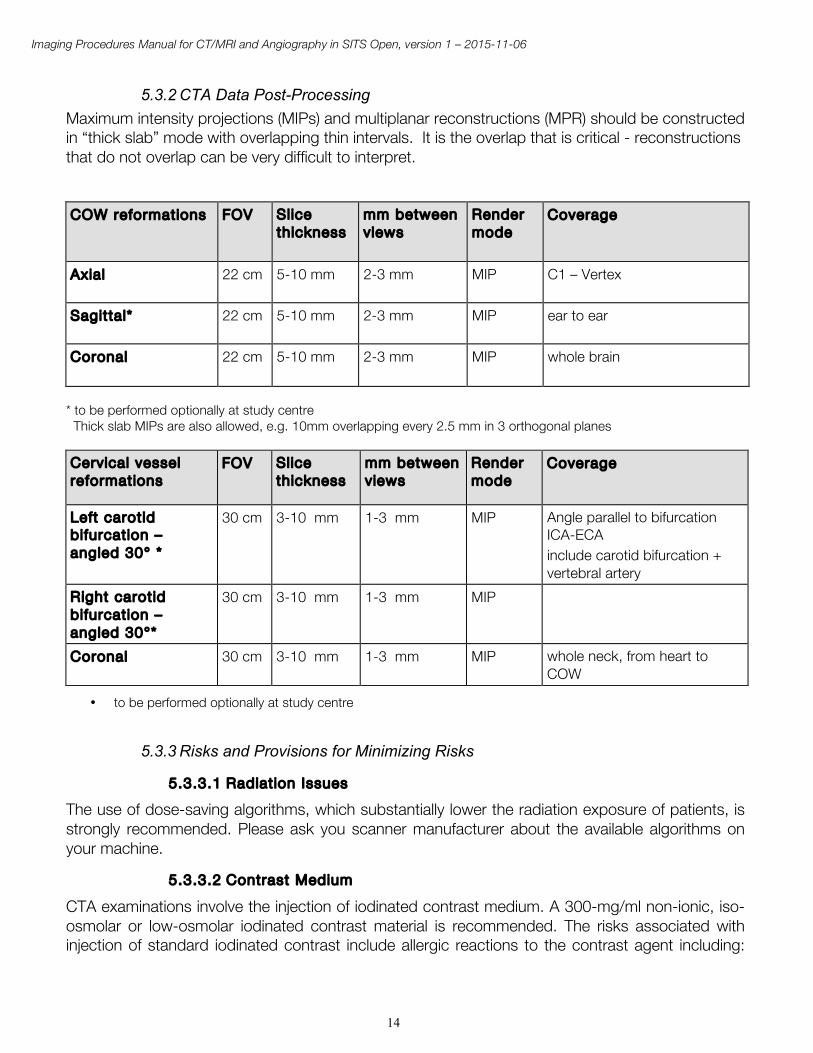
5.3.2 CTA Data Post-Processing Maximum intensity projections (MIPs) and multiplanar reconstructions (MPR) should be constructed in “thick slab” mode with overlapping thin intervals. It is the overlap that is critical - reconstructions that do not overlap can be very difficult to interpret. COW reformations FOV Sl ice
th ickness mm between v iews
Render mode
Coverage
Axia l 22 cm 5-10 mm 2-3 mm MIP C1 – Vertex
Sagitta l* 22 cm 5-10 mm 2-3 mm MIP ear to ear
Coronal 22 cm 5-10 mm 2-3 mm MIP whole brain
* to be performed optionally at study centre Thick slab MIPs are also allowed, e.g. 10mm overlapping every 2.5 mm in 3 orthogonal planes Cervical vessel reformations
FOV Sl ice th ickness
mm between v iews
Render mode
Coverage
Left carot id bi furcat ion – angled 30° *
30 cm 3-10 mm 1-3 mm MIP Angle parallel to bifurcation ICA-ECA include carotid bifurcation + vertebral artery
Right carot id bi furcat ion – angled 30°*
30 cm 3-10 mm 1-3 mm MIP
Coronal 30 cm 3-10 mm 1-3 mm MIP whole neck, from heart to COW
• to be performed optionally at study centre
5.3.3 Risks and Provisions for Minimizing Risks
5.3.3.1 Radiat ion Issues
The use of dose-saving algorithms, which substantially lower the radiation exposure of patients, is strongly recommended. Please ask you scanner manufacturer about the available algorithms on your machine.
5.3.3.2 Contrast Medium
CTA examinations involve the injection of iodinated contrast medium. A 300-mg/ml non-ionic, iso-osmolar or low-osmolar iodinated contrast material is recommended. The risks associated with injection of standard iodinated contrast include allergic reactions to the contrast agent including:
15
Imaging Procedures Manual for CT/MRI and Angiography in SITS Open, version 1 – 2015-11-06
rash, fever, agitation, confusion, headache, nausea/vomiting, difficulty with breathing, and a possible severe reaction which results in life-threatening low blood pressure and possibly death. The probability that any of these allergic reactions would occur in a patient without a history of similar reactions in the past is less than one in one thousand patients. In very few cases iodinated contrast material can lead to hyperthyroidism and in rare cases to thyrotoxemia, a life threatening condition. Iodinated contrast medium can also cause kidney dysfunction and possibly kidney failure, most commonly in patients with underlying kidney disease, but can occur in patients without prior history of kidney problems in less than one percent. Kidney dysfunction is reversible in most cases, but kidney failure represents a significant health problem during the time when the kidneys are not working. The total dose of contrast medium used in this study is limited to 70 ml for the baseline CTA examination and at 27h follow-up another 70 ml will be injected.
5.3.3.3 Contraindicat ions to Iodinated Contrast Mater ia l Administrat ion (CTA)
All usual and customary precautions taken by the centre should be employed for this study as well. Good hydration before and after contrast media administration reduces the risk of kidney failure and should be enabled whenever possible. Examples of iodinated contrast contraindications include:
• Hyperthyroidism • History of allergic reaction to iodinated contrast material • Serum creatinine measurement exceeding the standard limit for contrast injection
defined by the study centre • Known history of renal failure and/or dialysis, especially in diabetic patients • History of severe kidney disease as an adult, including tumor or transplant surgery,
or family history of kidney failure • Paraproteinemia syndromes or multiple myeloma • Collagen vascular disease • Severe cardiac insufficiency • Severely compromised liver function • Current therapy with metformin, aminoglycosides
Caution should be used in patients on current therapy with non-steroidal anti-inflammatory drugs, in patients with altered thyroid activity or nodular goiter, and in dehydrated patients. All additional contraindications, which relate to the specific product, that is administered, must be taken into consideration before usage.
6.0 Subject MRI Exam Preparation The total scan time, not including subject positioning, is approximately 11 minutes. Make certain that positioning aids are present in the intervention room, have the transmittal form on-hand for completion, and have supplies ready to label the digital media immediately after the MRI exam is complete.
16
Imaging Procedures Manual for CT/MRI and Angiography in SITS Open, version 1 – 2015-11-06
6.1 Subject Safety and Monitoring For the acquisition of perfusion and MRA sequences, which require the injection of contrast agents, patients should receive two intravenous (IV) lines to enable the independent administration of contrast medium (right antecubital vein) and any medical treatment (any other site). The patient’s head movement should be limited with foam padding. All patients should receive earplugs or other protection devices to prevent hearing loss or damage. In the case of forced head deviation the patient’s preferred position should be stabilized without turning the head in the typical orthogonal position. All centres should follow full MRI safety policies and interventions as practiced for their daily routine. Patients with contraindications to MR examination such as pacemakers, defibrillators, otic or cochlear implants, neurostimulators, intraorbital or intracranial ferromagnetic fragments (such as shrapnel), and certain aneurysm clips and prosthetic valves must be excluded from any MRI examination. Special attention should be given to tissue scars at the lower rib bow—a formerly typical location of defibrillators. A known hypersensitivity to MRI contrast medium of any severity, and impaired renal function are contraindications. Claustrophobia must also be taken into account when MRI-based patient selection is performed. Mild sedation can be considered in patients with severe claustrophobia if there are no contraindications (such as severe pulmonary or cardiac failure). Some Gadolinium-containing contrast agents (GdCAs) are reported to be associated with Nephrogenic Systemic Fibrosis (NSF), a disease formerly known as nephrogenic fibrosing dermopathy (NFD). This is a serious and life-threatening condition. All patients should be screened for renal dysfunction using laboratory tests prior to GdCA use. Use of high risk GdCAs is contraindicated in severe renal impairment and in the perioperative transplantation period. Traceability of GdCAs is essential and the name and dose of the contrast agent administered should be recorded in patient records. Patients with moderate to end-stage renal disease should not receive MRI contrast agent. If an Adverse Event occurs, the investigator’s judgement will be called on as to whether or not the imaging will be repeated. Imaging is strongly recommended in case of acute increase in stroke severity during the study and can be performed with CT or MRI.
6.2 Subject MRI Positioning Proper subject positioning is critical for obtaining high quality images. Correct, consistent and comfortable positioning of the subject within the MRI scanner will limit artifacts and maximize the acquisition of good quality images.
• As part of the normal subject pre-baseline routine, make sure all removable dental bridgework or other metallic objects (removable dental plates, belts, zippers, etc.) are removed prior to entering the scanner room. The metal (even though not ferromagnetic) may cause artifacts that can affect volumetric analysis. Consistency of the environment (e.g., no metal objects) around the subject is also very important for consistently achieving high scan quality.
17
Imaging Procedures Manual for CT/MRI and Angiography in SITS Open, version 1 – 2015-11-06
• The subject’s head must be placed in a volumetric radio frequency (RF) head coil (no surface coils are allowed).
• Maximize the subject’s comfort in the RF head coil. • Once the subject has been placed into the RF coil and comfort is maximized, ensure that
the centre of the RF coil to be used for landmarking, is approximately 1 finger width above the eyebrows.
• Ensure consistency of position of the volume of interest and field of view of the scan with respect to the magnet’s isocentre.
Maximizing comfort through proper head support will not only help to restrict head movement, but will also provide greater compliance in completing the entire exam within the allotted exam time. Proper head support can be achieved through the use of a vacuum-molded head holder, foam wedges or padding at the sides of the head, or a neck brace. Placing a Velcro strap or tape over the forehead can also provide stability and feedback to the subject and decrease movement. It is imperative that the subject’s head remains stable during acquisition. Imaging data degradation due to motion artifacts will almost always result in data rejection.
6.3 Labeling Digital Header Fields for Subject MRI Scans In compliance with privacy laws to ensure and protect the confidentiality of the subject, no subject names or identifiers should be entered into the electronic MRI header. The information in the box below should be entered into the electronic MRI header in lieu of subject identifiers. At the end of the examination, export MRI images to digital media (CD) and send to SITS Open Coordination team (KI). Facilities must be able to digitally archive data on CD in uncompressed DICOM format. MRI images should be submitted to SITS Open Coordination team (KI) within 3 working days hours of acquisition. In addition to archiving the data to digital media, the imaging facility will need to locally archive this data.
6.4 Subject MRI Acquisition Technique Image quality criteria for MRI of the brain in a clinical trial are stricter than in standard clinical practice. The assessments that will be performed on this MRI data depend highly upon the quality of images. Thus, it is imperative that the acquisition of MRI data be extremely precise and consistent. In order to achieve the most reliable evaluations of MRI of the brain, strict adherence to a uniform acquisition protocol and quality standards is required.
FIELD: SUBJECT ID: Enter Enrollment ID (SITSOPEN-XXXX)
Example: SITSOPEN-0123
STUDY DESCRIPTION: Select the Visit name (Follow-Up, Unscheduled)
18
Imaging Procedures Manual for CT/MRI and Angiography in SITS Open, version 1 – 2015-11-06
6.4.1 Pre-scan Adjustments
Most modern MRI scanners provide automated adjustment interventions for RF coil tuning and frequency adjustments after the subject is positioned in the magnet. Follow the adjustment interventions provided by the manufacturer. Image quality is usually unacceptable without proper adjustment of the RF coil and the transmit/receive equipment. Furthermore, without frequency adjustment, problems can occur with signal acquisition and proper localization of image FOV and slices.
6.4.2 Protocol for MRI of the Brain
The following is the list of the MRI sequences required for this protocol along with the approximate scan time for each sequence. Compliance with this protocol is required for all visis. This list is organized in the chronological order of acquisition:
• Localizer: 3-Plane Gradient Echo (GRE) (20 seconds) • 2D Axial Diffusion Weighted Imaging (DWI) (>1 minute) • 2D Axial T2* Gradient Echo (3 minutes) • 2D Axial Fluid-Attentuated Inversion Recovery (FLAIR) (4 minutes) • 3D TOF Magnetic Resonance Angiography (TOF-MRA) (3 minutes) • Contrast-Enhanced Magnetic Resonance Angiography (CE-MRA) (1 minute) • MRP (optional)
6.4.2.1 Local izer: 3-Plane Gradient Sequence (Scout) Localizer: 3-Plane Gradient Sequence: This shows a quick acquisition in 3 orthogonal planes for anatomical orientation. Acquire one slice in the middle of each plane (sagittal, coronal, and axial) plus additional slices as needed. Ensure that the subject is positioned properly.
6.4.2.2 Axial 2D DWI
DWI is used to detect ischemic changes on b = 1000 s/mm2 images under a standardized window setting. DWI also includes an EPI T2-weighted image with b value = 0 s/mm2. The required sequence is a diffusion weighted spin echo echoplanar sequence with a diffusion sensitivity of b = 1000 s/mm2. With 24 slices of 5 mm / 0-1 mm gap the entire brain will be imaged on axial images (parallel to AC-PC line). Window settings must be turned on a narrow width and a high window level. The aim of this window setting is to have a strong contrast between the core lesion and the surrounding tissue with normal water diffusion. Skull base associated artefacts may become more intensively visible and should not be confused with ischemic brain damage. Apparent Diffusion Coefficient (ADC) maps can be calculated to distinguish T2 shine-through (apparent in subacute infarction) from restricted diffusion. ADC maps will be used by RAPID to calculate the core of ischemic changes. For scanners with phased array coils, it is allowable to use factor of 1.6 parallel imaging for the DWI sequence ONLY.
19
Imaging Procedures Manual for CT/MRI and Angiography in SITS Open, version 1 – 2015-11-06
6.4.2.3 GRE-T2*-Weighted Images
Slice positioning must be identical to DWI.
GRE-T2*-weighted images are employed at baseline since they are the most sensitive for detection of parenchymal haemorrhage and are also helpful in excluding subarachnoid bleeding. Haemosiderin deposits caused by previous haemorrhage and/or microbleeds cause a strong signal decrease.
6.4.2.4 T2-weighted Fluid Attenuated Inversion Recovery (FLAIR)
Slice positioning must be identical to DWI. T2 FLAIR is used to image old and subacute lesions, and acute blood brain barrier disruption. These initial FLAIR images serve as a reference to differentiate the acute infarction from other lesions that are present at baseline. The FLAIR must have dark CSF to allow white matter lesions to be highlighted.
6.4.2.5 Magnetic Resonance Angiography (MRA)
MRA is used to determine the arterial occlusion or severe stenosis. A 3D time of flight (TOF) sequence centred on the circle of Willis using about 45-55 raw slices (max. 1.6 mm, FOV 22x22, 256 x 192-256 matrix) will be used. The scan time should be around 3 minutes. In case a centre prefers to perform a contrast media based MRA sequence a power injector is mandatory for the contrast media administration. The petrous section of the internal carotid artery must also be imaged in order to assess ICA flow. The M2 sections of the MCA should be visible on the unaffected hemisphere. Figure 2 gives a sample for slice positioning and three examples for 3D postprocessing.
For contrast-enhanced MRA (CE-MRA), a total of 15 mL of contrast media should be injected at a rate of 2 mL/sec, followed by a 20 mL saline flush delivered at the same rate. A tracker volume is placed in the common carotid artery as seen on the localizer image. A 3D fast spoiled gradient-echo sequence should then be acquired with 2 second delay after contrast bolus detection into the tracking region. CE-MRA images should be reconstructed using a maximum intensity projection (MIP) algorithm.
20
Imaging Procedures Manual for CT/MRI and Angiography in SITS Open, version 1 – 2015-11-06
7.0 Subject DSA Acquisition Technique Subjects in EVT group will undergo DSA at the following time points:
• Baseline—obtained prior to device deployment assessing clot location • During the intervention, at least after each pass of the device • End-of-intervention—obtained after final pass of study device • Rescue therapy with additional device
DSA requires contrast injections via guide catheter positioned in the common or internal carotid artery or vertebral artery. Additional local contrast injections via microcatheter are non-diagnostic regarding recanalization and reperfusion of target artery occlusion.
7.1 Patient Preparation Proper patient preparation is critical to obtaining quality arteriograms. Correct patient preparation includes correct, consistent positioning of the patient that limits artifacts and minimizes the number of poor quality arteriograms. Time from MR or CT scan completion to groin puncture should be less than 60 minutes, but no greater than 90 minutes. Time from groin puncture to the first deployment of the stent retriever should be less than 20 minutes. Conscious sedation should be preferred to general anaesthesia.
7.2 DSA Imaging Protocol All DSA interventions should be performed under sterile conditions according to local departmental notes. The microcatheters, guide catheters, and guide wires used in the study must have national regulatory approval in the country where they are being used. For diagnostic DSA and endovascular treatment femoral artery puncture is the recommended vessel access.
7.2.1 Baseline DSA The affected artery must be imaged in both frontal and lateral views. The size of the image intensifier should be large enough to visualize the affected artery with all branches and draining veins. The series should be performed with an imaging rate of at least 2 images/s. Series should document contrast passage from early arterial until venous phase of perfusion. The contrast agent must be injected via the guide catheter located in proximal extra-cranial artery segment.
7.2.2 Procedural DSA Should be collected following each pass of the device. For DSA collected during the intervention, the affected artery must be imaged in both frontal and lateral views. The size of the image intensifier should be large enough to visualize the affected artery with all branches and draining veins. The series should be performed with an imaging rate of at least 2 images/s. Series should document contrast passage from early arterial until venous phase of perfusion. Any of the 3 stent-retrievers identified for the study must be used. The selection of the first study device (from those defined for the study) is at the discretion of the interventionist, if the interventionist decides to

21
Imaging Procedures Manual for CT/MRI and Angiography in SITS Open, version 1 – 2015-11-06
change to another device, it should preferably be any of the other study devices. Other (non-study) devices should be avoided as far as possible as rescue therapy after incomplete effect of study devices. It is in the interest of the study that the three study devices are reasonably equally distributed, although difference in practice may occur between centres. Up to 6 passes with the stent-retriever are allowed. Additional passes or the change in stent-retrievers are regarded as “rescue therapy”. If for any reason, the thrombectomy cannot be performed, or failed to open the vessel, the patient may be treated with additional intra-arterial thrombolytics, if clinically appropriate. The dose of intra-arterial rt-PA should be recorded in eCRF. These patients will be enrolled and included in intention-to-treat analysis. If required, angioplasty and stenting of proximal artery stenosis is accepted in relation to the thrombectomy procedure. A guideline for a uniform policy of antiplatelet medications following stenting is available in Attachment 2 of the study protocol.
7.2.3 Intervention a) Physicians should follow the relevant Instructions for use for specific devices being utilised in the study. For thrombi in the anterior circulation, it is mandatory to use either balloon guide catheter or a distal access catheter (DAC). Time records are made at the beginning of the procedure and on achieving recanalization TICI grades 2b and 3 (if achieved), and with the final angiogram. The number of thrombectomy attempts should be limited to 6. b) Procedural Heparin: A heparin flush solution may be administered in the guide catheter and in the access sheath at the discretion of the physician. c) IA thrombolytics: IA thrombolytics (IAT) are permitted at the investigator’s discretion when required to achieve satisfactory recanalization. The need of IAT should be weighed against an increased risk of haemorrhage. For a patient treated with full dose IV rtPA (0.9 mg/kg), an additional dose of 0.2 mg/kg, corresponding to 14 mg in a 70 kg person, should not be exceeded. d) Anti-vasospasm agents: Prudent use of anti-vasospasm agents is permitted. e) Proximal stenosis: In order to facilitate access to the intracranial occlusion, investigators will sometimes need to traverse a proximal stenosis. Investigators may use their judgement to best address the proximal stenosis with (1) no treatment, (2) balloon angioplasty, or (3) stenting. The primary goal is to rapidly restore intracranial flow; so proximal stenosis sometimes may be best addressed at the end of the case. Care should be taken when stenting because patients must then receive antiaggregant treatment (e.g., clopidogrel) whose safety is not well established in the setting of acute stroke, in particular when undergoing IVT. Instructions for initiation of antiaggregant treatment in the study are attached to protocol (Attachment 2). Termination of the intra-arterial treatment procedure will occur if: a) Angiographic findings suggest vessel damage or extravasation of contrast. b) Neurological deterioration or alteration in function is detected leading to the suspicion of an intracranial haemorrhage. Final angiography will include ipsilateral anterior-posterior and lateral views of the involved arterial system. Aside from procedurally administered heparin, IV heparin is prohibited until after the 24 hour neuro-imaging has been performed to minimize the risk of intracranial haemorrhage. Blood pressure will be tightly controlled during the first 24 h to less than 180/105. Other drug therapy will be at the discretion of the investigator in accordance with IVT guidelines.
22
Imaging Procedures Manual for CT/MRI and Angiography in SITS Open, version 1 – 2015-11-06
IMPORTANT ► Save a copy of all patients arteriograms at your facility before sending the arteriograms to SITS Open Coordination team (KI).
7.2.4 End-of-intervention DSA
DSA must be collected at the end of the intervention. The affected artery must be imaged in both frontal and lateral views. The size of the image intensifier should be large enough to visualize the affected artery with all branches and draining veins. The series should be performed with an imaging rate of at least 2 images/s. Series should document contrast passage from early arterial until venous phase of perfusion. DSA should be collected following interim passes of the device, prior to the end of the intervention.
7.3 Labeling Digital Header Fields for DSA In compliance with privacy laws to ensure and protect the confidentiality of the subject, no subject names or identifiers should be entered into the electronic DSA header. The information in the box below should be entered into the electronic DSA header in lieu of subject identifiers.
FIELD: SUBJECT ID: Enter Enrollment ID
SITSOPEN-XXXX Example: SITSOPEN-0123
STUDY DESCRIPTION: Enter Date and Time (hh:mm) of first and last DSA run and number of runs
Please send to SITS Open Coordination team all DSA runs in subtracted mode (Digital Subtracted DSA). SITS Open Coordination team should receive the same subtracted image quality as used by investigator during intervention. If submitting images on digital media, export the data to a CD in uncompressed DICOM format. Use one CD per exam.
23
Imaging Procedures Manual for CT/MRI and Angiography in SITS Open, version 1 – 2015-11-06
7.4 Archival of Arteriography of Study patients Patient DSA must be archived at the local facility and retained for the duration of the study at least. Copies of all study images must be kept at centre in the event that images sent to SITS Open Coordination team (KI) are lost or corrupted during shipping.
8.0 Procedure and preparation for transfer of images for evaluation
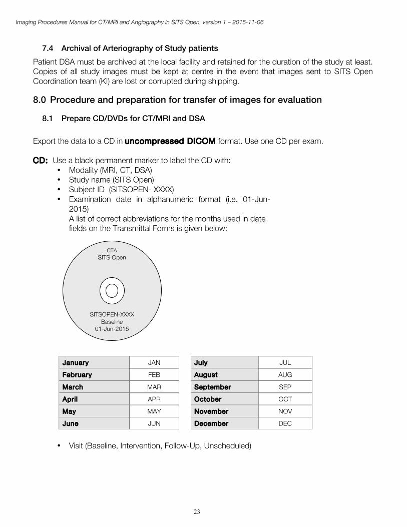
8.1 Prepare CD/DVDs for CT/MRI and DSA Export the data to a CD in uncompressed DICOM format. Use one CD per exam. CD: Use a black permanent marker to label the CD with:
• Modality (MRI, CT, DSA) • Study name (SITS Open) • Subject ID (SITSOPEN- XXXX) • Examination date in alphanumeric format (i.e. 01-Jun-
2015) A list of correct abbreviations for the months used in date fields on the Transmittal Forms is given below:
• Visit (Baseline, Intervention, Follow-Up, Unscheduled)
January JAN July JUL
February FEB August AUG
March MAR September SEP
Apri l APR October OCT
May MAY November NOV
June JUN December DEC
CTA SITS Open
SITSOPEN-XXXX Baseline
01-Jun-2015
24
Imaging Procedures Manual for CT/MRI and Angiography in SITS Open, version 1 – 2015-11-06
8.5 Transittal Form “Centre to SITS Open Coordination team (KI)” and “SITS Open Coordination team (KI) to Imaging Core Lab” for DSA and CT/MRI of the Brain When preparing to send the archived DSA and CT/MRI exam data on electronic media, complete and include the Transmittal Form.
To properly complete the transmittal form for subject CT/MRI scans and DSA, please follow the guidelines below.
Centre, Subject and Visit Information • Complete the Subject ID (Enrollment ID) • Check the appropriate box to indicate the visit and type of examination. • Each CD should be accompanied by its transmittal form.
Examination Information • Complete date and time of DSA or CT/MRI exam using the alphanumeric date format: DD-MMM-YYYY; xx:yy (24h clock) (groin puncture time) (initiation of CT/MRI) • DSA: target arterial occlusion, proximal balloon catheter, distal access catheter, aspiration,
no. of passes with stent-retriever, no. of runs, IA tPA, rescue therapy, complications
Data Shipment to SITS Open Coordination team (KI) / Imaging Core Lab • Sign and date the form after checking for completion and accuracy. • Keep a copy in study records study centre.
Print legibly and clearly.
Use blue or black ink. Make corrections according to Good Clinical Practice. Complete the study centre section of the form – missing information wil l s low the
processing of subject data.
25
Imaging Procedures Manual for CT/MRI and Angiography in SITS Open, version 1 – 2015-11-06
8.2 Subject DSA and CT/MRI Scans transfer to SITS Open team (KI) and Imaging Core Lab
IMPORTANT ► All CT/MRI/DSA data acquired for this study MUST be sent to SITS Open Coordination team (KI) within 3 working days of image acquisition. SITS Open Coordination team (KI) send these data to Imaging Core Lab after quality check.
8.3 SITS Open Coordination team (KI) Evaluation of Subject Scans (Quality check) When the subject exam data is loaded and viewed on image viewing systems at SITS Open Coordination team (KI) the routine is as follows: In case additional images are needed, SITS Open Coordination team (KI) will notify the centre via email.
8.3.1 Requests for Resubmission of Subject Data
If subject data cannot be loaded and viewed SITS Open Coordination team (KI), a Resubmission Request will be made via email to the study coordinator at centre and CT/MRI/DSA facility identifying need for resubmission. The data must be resubmitted to SITS Open Coordination team (KI) as quickly as possible.
8.4 SITS Open Coordination team (KI) submission to Imaging Core Lab After quality check and archiving of the imaging examinations (of baseline, intervention, follow-up), SITS Open Coordination team (KI) store copies as files and send CDs of the examinations together with transmittal forms to Imaging Core Lab in the following order:
1. baseline imaging (NCCT, CTA, CTP* ) 2. Intervention DSA** after the evaluation of baseline imaging has been received and archived 3. Follow-up imaging after baseline imaging and intervention DSA** has been received and
archived *optional, **for EVT group only
The CD are labelled as described under 8.1.
26
Imaging Procedures Manual for CT/MRI and Angiography in SITS Open, version 1 – 2015-11-06
9.0 Imaging Core Lab Evaluation of Subject DSA and CT/MRI Scans Imaging Core Lab confirms the delivery of imaging examinations within 3 working days by email to SITS Open Coordination team (KI). Imaging Core Lab archives the imaging examinations electronically. Imaging Core Lab evaluates the imaging examinations within 5 days after the delivery of the images and archives the findings on imaging eCRFs (imaging evaluation form) that are automatically and simultaneously archived in the study data base at Karolinska Institute.
9.1 Definitions 1. Infarct volume: Ischemic brain tissue that will not recover with reperfusion.
a. as detected by CT: Tissue with decreased x-ray attenuation (“hypodensity”). Attenuation of gray or white matter that is less than normal (comparison with unaffected brain tissue) indicates net water uptake (ionic oedema) and irreversible ischemic injury. Hypo-attenuation of gray matter causes a decrease in grey-white matter contrast and thus a loss in anatomical information.
b. as detected by MRI (DWI): Increased signal on DWI accompanied by decreased signal on apparent diffusion coefficient (ADC) maps define tissue with cytotoxic oedema that has minimal chances to recover with early reperfusion.
2. Infarct volume measurement: 0.5 x AxBxC where A is the largest diameter, B is the perpendicular diameter of the same slice, and C is the diameter in Z-axis adding the thicknesses of all slices that present tissue hypoattenuation.
3. Infarct growth: Difference in infarct volume assessed on FU imaging at 22 to 36 hours minus infarct volume at baseline.
4. Important time points a. Stroke onset: Time point last seen well b. Admission: Time documented by personal of emergency facility c. Diagnostic imaging (CT/MRI): Time of pilot scan as documented on film d. IVT: Initiation of tPA infusion e. Informed consent: documented time of signature f. Groin puncture: Time documented by interventionist g. First DSA run: Time documented on DSA film h. Treatment cervical carotid artery:
i. Start: documented time on DSA run with guide catheter in place ii. Finish: documented time on 1st DSA run showing angioplasty/stenting
i. Recanalization (DSA): Time documented on film of 1st DSA run showing mTICI 2b or 3 of target artery.
27
Imaging Procedures Manual for CT/MRI and Angiography in SITS Open, version 1 – 2015-11-06
j. Recanalization (CTA/MRA): Date and time on film showing AOL 2 or 3 of former occluded artery
5. Intra-cranial haemorrhages (ICH): see 8.5.5. of study protocol 6. Symptomatic ICH (SICH): see 8.5.7. of study protocol 7. Ischemic brain oedema: see 8.5.6. of study protocol 8. CT dense artery sign: Tubular or round shaped portion of brain artery with increased x-ray
attenuation compared to other portions of the same artery or the contra-lateral artery sometimes visible only on slices with <2.5mm thickness.
9. Thrombus length measurement: Distance measurement of hyperattenuating structure accepted as “dense artery” on thin (<2.5 mm) CT sections taking into account out of plane segments.
10. DSA-run: Repeated head imaging (2 or 3 frames/sec) before and during contrast injection into proximal brain artery via catheter in common or internal carotid artery or vertebral artery and electronical subtraction of frame before contrast injection from all images during and after contrast injection. Local contrast injection via microcatheter directly into cranial arteries cannot assess effective recanalization and reperfusion.
11. Rescue therapy: Each attempt of recanalisation after 6 passes with the study approved stent-retriever and any attempt of recanalisation with other stent-retrievers or aspiration catheters not approved by the study protocol.