Embed Size (px)
Citation preview
Razi, M. and Chan, Edmond Y. and Tooze, Sharon A. (2009) Early
endosomes and endosomal coatomer are required for autophagy.
Journal of Cell Biology, 185 (2). pp. 305-321. ISSN 0021-9525 ,
http://dx.doi.org/10.1083/jcb.200810098
This version is available at https://strathprints.strath.ac.uk/25986/
Strathprints is designed to allow users to access the research output of the University of
Strathclyde. Unless otherwise explicitly stated on the manuscript, Copyright © and Moral Rights
for the papers on this site are retained by the individual authors and/or other copyright owners.
Please check the manuscript for details of any other licences that may have been applied. You
may not engage in further distribution of the material for any profitmaking activities or any
commercial gain. You may freely distribute both the url (https://strathprints.strath.ac.uk/) and the
content of this paper for research or private study, educational, or not-for-profit purposes without
prior permission or charge.
Any correspondence concerning this service should be sent to the Strathprints administrator:
The Strathprints institutional repository (https://strathprints.strath.ac.uk) is a digital archive of University of Strathclyde research
outputs. It has been developed to disseminate open access research outputs, expose data about those outputs, and enable the
management and persistent access to Strathclyde's intellectual output.
JCB: ARTICLE
The Rockefeller University Press $30.00J. Cell Biol. Vol. 185 No. 2 305–321www.jcb.org/cgi/doi/10.1083/jcb.200810098 JCB 305
Correspondence to Sharon A. Tooze: [email protected]
E.Y.W. Chan’s present address is Strathclyde Institute of Pharmacy and Biomedical Sciences, University of Strathclyde, Glasgow G4 0NR, Scotland, UK.
Abbreviations used in this paper: ARF, ADP-ribosylation factor; Atg, autophagy related; AV, autophagic vacuole; COP, coat protein complex; EEA1, early endo-somal autoantigen 1; ERGIC-53, ER-Golgi intermediate compartment 53 kD; GM130, Golgi membrane protein 130 kD; LAMP, lysosomal-associated mem-brane protein; LTR, Lysotracker red; MPR, mannose-6-phosphate receptor; MVB, multi-vesicular body; Tfn, transferrin; Ub, ubiquitin.
Introduction
Macroautophagy, here referred to as autophagy, is a highly con-
served degradative process used for the nonselective removal of
long-lived proteins, large protein aggregates, and subcellular or-
ganelles. In contrast, selective forms of autophagy, such as pexoph-
agy and mitophagy, and chaperone-mediated autophagy, target
speciic substrates and organelles for degradation (Mizushima
et al., 2008). Successful autophagy of all types requires a response
to an initiation signal, for example starvation, followed by the ex-
pansion of the autophagosomal membrane source, a double mem-
brane called an isolation membrane or phagophore. The isolation
membrane eficiently sequesters subcellular components and
forms the nascent, or immature, autophagosome or autophagic
vacuole (AVi), which fuses with endosomes and lysosomes to ac-
quire proteases and lipases. Fusion of AVi with endosomes and
lysosomes results in the formation of amphisomes (AVi/d) and
autolysosomes, or degradative AVs (AVd).
Autophagy requires a set of proteins called the Atg (au-
tophagy related) proteins, irst identiied in yeast (Klionsky,
2007). Mammalian orthologues of many of the 31 yeast Atg pro-
teins have been identiied and characterized, and it is through the
work in yeast and mammals that the role of many of these pro-
teins is beginning to be understood. Early stages of AV forma-
tion requires ULK1, the orthologue of the Atg1 kinase, the
PtdIns-3 kinase Vps34, and two ubiquitin-like conjugation sys-
tems, whose actions result in formation of the Atg5-12-16 com-
plex, and LC3-II. LC3-II is the lipid-conjugated form of LC3-I,
an orthologue of yeast Atg8, and the most widely used marker of
AVs. This core machinery drives the initiation, expansion, and
closure of the isolation membrane (Xie and Klionsky, 2007).
The inal stages of AV maturation, the formation of AVi/ds
and AVds, occurs through fusion with endosomes and lyso-
somes, in particular multi-vesicular bodies (MVB) (Filimonenko
et al., 2007), which are formed from early endosomes by the
ESCRT machinery (Williams and Urbe, 2007; Woodman and
Futter, 2008). Recently, the ESCRT complexes were shown to
be required for formation of autolysosomes; loss of Tsg101
(ESCRTI) or Vps24 (ESCRTIII) results in an accumulation of
amphisomes (Filimonenko et al., 2007). Furthermore, the loss
or mutation of several components of the ESCRT machinery, in
Autophagy, an intracellular degradative pathway, maintains cell homeostasis under normal and stress conditions. Nascent double-membrane auto-
phagosomes sequester and enclose cytosolic components and organelles, and subsequently fuse with the endosomal pathway allowing content degradation. Autophagy re-quires fusion of autophagosomes with late endosomes, but it is not known if fusion with early endosomes is essential. We show that fusion of AVs with functional early endo-somes is required for autophagy. Inhibition of early endo-some function by loss of COPI subunits (, , or ) results
in accumulation of autophagosomes, but not an increased autophagic flux. COPI is required for ER-Golgi transport and early endosome maturation. Although loss of COPI re-sults in the fragmentation of the Golgi, this does not induce the formation of autophagosomes. Loss of COPI causes defects in early endosome function, as both transferrin re-cycling and EGF internalization and degradation are im-paired, and this loss of function causes an inhibition of autophagy, an accumulation of p62/SQSTM-1, and ubiq-uitinated proteins in autophagosomes.
Early endosomes and endosomal coatomer are required for autophagy
Minoo Razi, Edmond Y.W. Chan, and Sharon A. Tooze
London Research Institute, Cancer Research UK, London WC2A 3PX, England, UK
© 2009 Razi et al. This article is distributed under the terms of an Attribution–Noncommercial–Share Alike–No Mirror Sites license for the first six months after the publica-tion date (see http://www.jcb.org/misc/terms.shtml). After six months it is available under a Creative Commons License (Attribution–Noncommercial–Share Alike 3.0 Unported license, as described at http://creativecommons.org/licenses/by-nc-sa/3.0/).
TH
EJ
OU
RN
AL
OF
CE
LL
BIO
LO
GY
on J
uly
30, 2
012
jcb.ru
pre
ss.o
rgD
ow
nlo
aded fro
m
Published April 13, 2009
http://jcb.rupress.org/content/suppl/2009/04/14/jcb.200810098.DC1.html Supplemental Material can be found at:
JCB • VOLUME 185 • NUMBER 2 • 2009 306
number of GFP-LC3-II–positive vesicles, and suggested a role
for COPI in autophagy.
As a control for the speciicity of COP depletion we tested
if siRNA knockdown of the other coat complexes involved in
transport between the ER and Golgi, such as COPII subunits,
caused an increase in GFP-LC3-II, or GFP-LC3–positive vesicles
under the same culture conditions. 293/GFP-LC3 cells treated
with siRNAs for COPII subunits Sec23A and 23B (Fig. 1 A) or
Sec31 (not depicted) showed no increase in GFP-LC3-II (Fig. 1 B),
or GFP-LC3–positive vesicles (not depicted). The lack of in-
crease in lipidated GFP-LC3 and GFP-positive puncta after de-
pletion of COPII indicated that the effect was speciic to COPI,
and suggested that it was not a result of a general inhibition of
ER-to-Golgi transport.
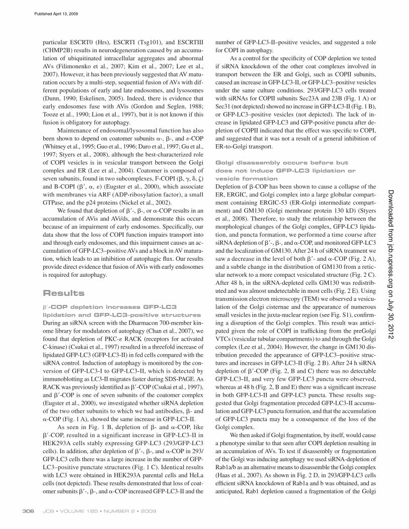
Golgi disassembly occurs before but
does not induce GFP-LC3 lipidation or
vesicle formation
Depletion of -COP has been shown to cause a collapse of the
ER, ERGIC, and Golgi complex into a large globular compart-
ment containing ERGIC-53 (ER-Golgi intermediate compart-
ment) and GM130 (Golgi membrane protein 130 kD) (Styers
et al., 2008). Therefore, to study the relationship between the
morphological changes of the Golgi complex, GFP-LC3 lipida-
tion, and puncta formation, we performed a time course after
siRNA depletion of -, -, and -COP, and monitored GFP-LC3
and the localization of GM130. After 24 h of siRNA treatment we
saw a decrease in the level of both - and -COP (Fig. 2 A),
and a subtle change in the distribution of GM130 from a retic-
ular network to a more compact vesiculated structure (Fig. 2 C).
After 48 h, in the siRNA-depleted cells GM130 was redistrib-
uted and was almost undetectable in most cells (Fig. 2 E). Using
transmission electron microscopy (TEM) we observed a vesicu-
lation of the Golgi cisternae and the appearance of numerous
small vesicles in the juxta-nuclear region (see Fig. S1), conirm-
ing a disruption of the Golgi complex. This result was antici-
pated given the role of COPI in traficking from the preGolgi
VTCs (vesicular tubular compartments) to and through the Golgi
complex (Lee et al., 2004). However, the change in GM130 dis-
tribution preceded the appearance of GFP-LC3–positive struc-
tures and increases in GFP-LC3-II (Fig. 2 B). After 24 h siRNA
depletion of -COP (Fig. 2, B and C) there was no detectable
GFP-LC3-II, and very few GFP-LC3 puncta were observed,
whereas at 48 h (Fig. 2, B and E) there was a signiicant increase
in both GFP-LC3-II and GFP-LC3 puncta. These results sug-
gested that Golgi fragmentation preceded GFP-LC3-II accumu-
lation and GFP-LC3 puncta formation, and that the accumulation
of GFP-LC3 puncta may be a consequence of the loss of the
Golgi complex.
We then asked if Golgi fragmentation, by itself, would cause
a phenotype similar to that seen after COPI depletion resulting in
an accumulation of AVs. To test if disassembly or fragmentation
of the Golgi was inducing autophagy we used siRNA-depletion of
Rab1a/b as an alternative means to disassemble the Golgi complex
(Haas et al., 2007). As shown in Fig. 2 D, in 293/GFP-LC3 cells
eficient siRNA knockdown of Rab1a and b was obtained, and as
anticipated, Rab1 depletion caused a fragmentation of the Golgi
particular ESCRT0 (Hrs), ESCRTI (Tsg101), and ESCRTIII
(CHMP2B) results in neurodegeneration caused by an accumu-
lation of ubiquitinated intracellular aggregates and abnormal
AVs (Filimonenko et al., 2007; Kim et al., 2007; Lee et al.,
2007). However, it has been previously suggested that AV matu-
ration occurs by a multi-step, sequential fusion of AVs with dif-
ferent populations of early and late endosomes, and lysosomes
(Dunn, 1990; Eskelinen, 2005). Indeed, there is evidence that
early endosomes fuse with AVis (Gordon and Seglen, 1988;
Tooze et al., 1990; Liou et al., 1997), but it is not known if this
fusion is obligatory for autophagy.
Maintenance of endosomal/lysosomal function has also
been shown to depend on coatomer subunits -, -, and -COP
(Whitney et al., 1995; Guo et al., 1996; Daro et al., 1997; Gu et al.,
1997; Styers et al., 2008), although the best-characterized role
of COPI vesicles is in vesicular transport between the Golgi
complex and ER (Lee et al., 2004). Coatomer is composed of
seven subunits, found in two subcomplexes, F-COPI (, , , )
and B-COPI (, , ) (Eugster et al., 2000), which associate
with membranes via ARF (ADP-ribosylation factor), a small
GTPase, and the p24 proteins (Nickel et al., 2002).
We found that depletion of -, -, or -COP results in an
accumulation of AVis and AVi/ds, and demonstrate this occurs
because of an impairment of early endosomes. Speciically, our
data show that the loss of COPI function impairs transport into
and through early endosomes, and this impairment causes an ac-
cumulation of GFP-LC3–positive AVs and a block in AV matura-
tion, which leads to an inhibition of autophagic lux. Our results
provide direct evidence that fusion of AVis with early endosomes
is required for autophagy.
Results
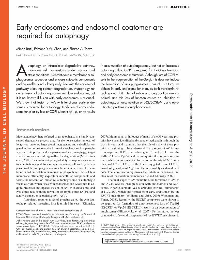
-COP depletion increases GFP-LC3
lipidation and GFP-LC3–positive structures
During an siRNA screen with the Dharmacon 700-member kin-
ome library for modulators of autophagy (Chan et al., 2007), we
found that depletion of PKC- RACK (receptors for activated
C-kinase) (Csukai et al., 1997) resulted in a threefold increase of
lipidated GFP-LC3 (GFP-LC3-II) in fed cells compared with the
siRNA control. Induction of autophagy is monitored by the con-
version of GFP-LC3-I to GFP-LC3-II, which is detected by
immunoblotting as LC3-II migrates faster during SDS-PAGE. As
RACK was previously identiied as -COP (Csukai et al., 1997),
and -COP is one of seven subunits of the coatomer complex
(Eugster et al., 2000), we investigated whether siRNA depletion
of the two other subunits to which we had antibodies, - and
-COP (Fig. 1 A), showed the same increase in GFP-LC3-II.
As seen in Fig. 1 B, depletion of - and -COP, like
-COP, resulted in a significant increase in GFP-LC3-II in
HEK293A cells stably expressing GFP-LC3 (293/GFP-LC3
cells). In addition, after depletion of -, -, and -COP in 293/
GFP-LC3 cells there was a large increase in the number of GFP-
LC3–positive punctate structures (Fig. 1 C). Identical results
with LC3 were obtained in HEK293A parental cells and HeLa
cells (not depicted). These results demonstrated that loss of coat-
omer subunits -, -, and -COP increased GFP-LC3-II and the
on J
uly
30, 2
012
jcb.ru
pre
ss.o
rgD
ow
nlo
aded fro
m
Published April 13, 2009
307ROLE OF EARLY ENDOSOMES IN AUTOPHAGY • Razi et al.
Figure 1. siRNA depletion of COPI increases autophagy. (A) siRNA depletion of -, -, and -COPI (top) and Sec23A and B (bottom) in 293/GFP-LC3 cells for 48 h. Decreased protein levels of the corresponding subunits is shown by immunoblots. (B) Loss of -, -, and -COP but not subunits of COPII (Sec 23A and B) increase GFP-LC3-II. In A and B, tubulin was the loading control. (C) GFP-LC3–positive vesicles were detectable in 293/GFP-LC3 cells after siRNA depletion of -, -, and -COP but only basal levels of GFP-LC3–positive vesicles are observed in control siRNA-depleted cells.
on J
uly
30, 2
012
jcb.ru
pre
ss.o
rgD
ow
nlo
aded fro
m
Published April 13, 2009
JCB • VOLUME 185 • NUMBER 2 • 2009 308
Figure 2. Time course of COPI depletion shows that Golgi disperses before AVs are formed, but Golgi dispersal does not cause AV formation. (A) COPI subunits were depleted in 293/GFP-LC3 cells and analyzed 24, 36, and 48 h after addition of siRNA. Immunoblotting for - and -COP confirms loss of COP subunits. (B) GFP-LC3-I and -II were monitored by immunoblots in parallel lysates using anti-GFP antibodies at the indicated times. (C) After 24 h siRNA treatment, depletion of -COP caused morphological changes and a reduction in perinuclear population of GM130. Box indicates enlarged area. (D) Rab1a/b was depleted using siRNAs for 48 h. Lysates were probed with anti-Rab1 to confirm depletion. (E) -COP or Rab1a/b were depleted as in D, and analyzed by indirect immunofluorescence using anti-GM130 and GFP fluorescence. In -COP panel, the asterisk indicates cells that did not show an accumulation of GFP-LC3–positive AVs.
on J
uly
30, 2
012
jcb.ru
pre
ss.o
rgD
ow
nlo
aded fro
m
Published April 13, 2009
309ROLE OF EARLY ENDOSOMES IN AUTOPHAGY • Razi et al.
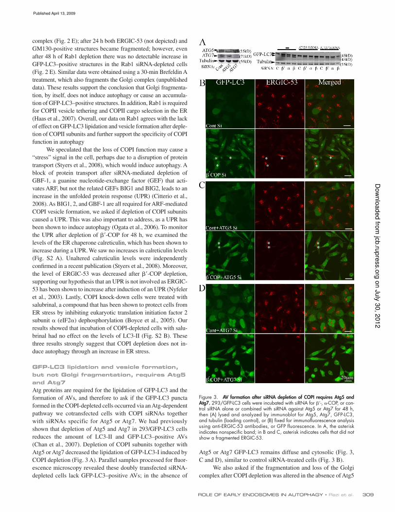
Atg5 or Atg7 GFP-LC3 remains diffuse and cytosolic (Fig. 3,
C and D), similar to control siRNA-treated cells (Fig. 3 B).
We also asked if the fragmentation and loss of the Golgi
complex after COPI depletion was altered in the absence of Atg5
complex (Fig. 2 E); after 24 h both ERGIC-53 (not depicted) and
GM130-positive structures became fragmented; however, even
after 48 h of Rab1 depletion there was no detectable increase in
GFP-LC3–positive structures in the Rab1 siRNA-depleted cells
(Fig. 2 E). Similar data were obtained using a 30-min Brefeldin A
treatment, which also fragments the Golgi complex (unpublished
data). These results support the conclusion that Golgi fragmenta-
tion, by itself, does not induce autophagy or cause an accumula-
tion of GFP-LC3–positive structures. In addition, Rab1 is required
for COPII vesicle tethering and COPII cargo selection in the ER
(Haas et al., 2007). Overall, our data on Rab1 agrees with the lack
of effect on GFP-LC3 lipidation and vesicle formation after deple-
tion of COPII subunits and further support the speciicity of COPI
function in autophagy
We speculated that the loss of COPI function may cause a
“stress” signal in the cell, perhaps due to a disruption of protein
transport (Styers et al., 2008), which would induce autophagy. A
block of protein transport after siRNA-mediated depletion of
GBF-1, a guanine nucleotide-exchange factor (GEF) that acti-
vates ARF, but not the related GEFs BIG1 and BIG2, leads to an
increase in the unfolded protein response (UPR) (Citterio et al.,
2008). As BIG1, 2, and GBF-1 are all required for ARF-mediated
COPI vesicle formation, we asked if depletion of COPI subunits
caused a UPR. This was also important to address, as a UPR has
been shown to induce autophagy (Ogata et al., 2006). To monitor
the UPR after depletion of -COP for 48 h, we examined the
levels of the ER chaperone calreticulin, which has been shown to
increase during a UPR. We saw no increases in calreticulin levels
(Fig. S2 A). Unaltered calreticulin levels were independently
conirmed in a recent publication (Styers et al., 2008). Moreover,
the level of ERGIC-53 was decreased after -COP depletion,
supporting our hypothesis that an UPR is not involved as ERGIC-
53 has been shown to increase after induction of an UPR (Nyfeler
et al., 2003). Lastly, COPI knock-down cells were treated with
salubrinal, a compound that has been shown to protect cells from
ER stress by inhibiting eukaryotic translation initiation factor 2
subunit (eIF2) dephosphorylation (Boyce et al., 2005). Our
results showed that incubation of COPI-depleted cells with salu-
brinal had no effect on the levels of LC3-II (Fig. S2 B). These
three results strongly suggest that COPI depletion does not in-
duce autophagy through an increase in ER stress.
GFP-LC3 lipidation and vesicle formation,
but not Golgi fragmentation, requires Atg5
and Atg7
Atg proteins are required for the lipidation of GFP-LC3 and the
formation of AVs, and therefore to ask if the GFP-LC3 puncta
formed in the COPI-depleted cells occurred via an Atg-dependent
pathway we cotransfected cells with COPI siRNAs together
with siRNAs specific for Atg5 or Atg7. We had previously
shown that depletion of Atg5 and Atg7 in 293/GFP-LC3 cells
reduces the amount of LC3-II and GFP-LC3–positive AVs
(Chan et al., 2007). Depletion of COPI subunits together with
Atg5 or Atg7 decreased the lipidation of GFP-LC3-I induced by
COPI depletion (Fig. 3 A). Parallel samples processed for luor-
escence microscopy revealed these doubly transfected siRNA-
depleted cells lack GFP-LC3–positive AVs; in the absence of
Figure 3. AV formation after siRNA depletion of COPI requires Atg5 and Atg7. 293/GFP-LC3 cells were incubated with siRNA for -, -COP, or con-trol siRNA alone or combined with siRNA against Atg5 or Atg7 for 48 h, then (A) lysed and analyzed by immunoblot for Atg5, Atg7, GFP-LC3, and tubulin (loading control), or (B) fixed for immunofluorescence analysis using anti-ERGIC-53 antibodies, or GFP fluorescence. In A, the asterisk indicates nonspecific band; in B and C, asterisk indicates cells that did not show a fragmented ERGIC-53.
on J
uly
30, 2
012
jcb.ru
pre
ss.o
rgD
ow
nlo
aded fro
m
Published April 13, 2009
JCB • VOLUME 185 • NUMBER 2 • 2009 310
and Atg7. We used ERGIC-53 to label the ER-Golgi intermediate
compartment and found, after COPI depletion, ERGIC-53 label-
ing was greatly diminished and appeared dispersed (Fig. 3 B).
Both the steady-state distribution of ERGIC-53 in control cells,
and the dispersal of the ERGIC-53 in COPI-depleted cells was
unaffected by co-depletion of Atg5 or Atg7 (Fig. 3, C and D).
These results demonstrate that the increase in GFP-LC3-II and
GFP-LC3 puncta after COPI depletion requires the autophagy
proteins Atg5 and 7, and show that loss of Atg7 does not affect
ERCIG-53 distribution in normal cells, or its redistribution and
dispersion after siRNA depletion of COPI.
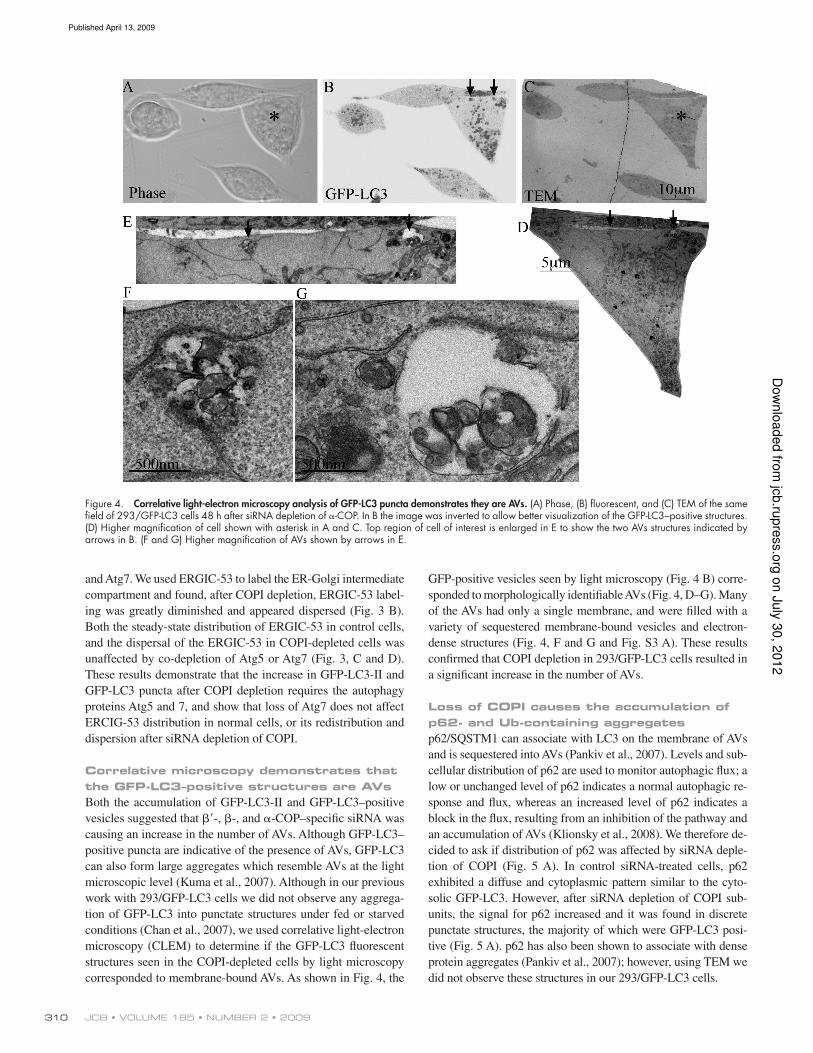
Correlative microscopy demonstrates that
the GFP-LC3–positive structures are AVs
Both the accumulation of GFP-LC3-II and GFP-LC3–positive
vesicles suggested that -, -, and -COP–speciic siRNA was
causing an increase in the number of AVs. Although GFP-LC3–
positive puncta are indicative of the presence of AVs, GFP-LC3
can also form large aggregates which resemble AVs at the light
microscopic level (Kuma et al., 2007). Although in our previous
work with 293/GFP-LC3 cells we did not observe any aggrega-
tion of GFP-LC3 into punctate structures under fed or starved
conditions (Chan et al., 2007), we used correlative light-electron
microscopy (CLEM) to determine if the GFP-LC3 luorescent
structures seen in the COPI-depleted cells by light microscopy
corresponded to membrane-bound AVs. As shown in Fig. 4, the
GFP-positive vesicles seen by light microscopy (Fig. 4 B) corre-
sponded to morphologically identiiable AVs (Fig. 4, D–G). Many
of the AVs had only a single membrane, and were illed with a
variety of sequestered membrane-bound vesicles and electron-
dense structures (Fig. 4, F and G and Fig. S3 A). These results
conirmed that COPI depletion in 293/GFP-LC3 cells resulted in
a signiicant increase in the number of AVs.
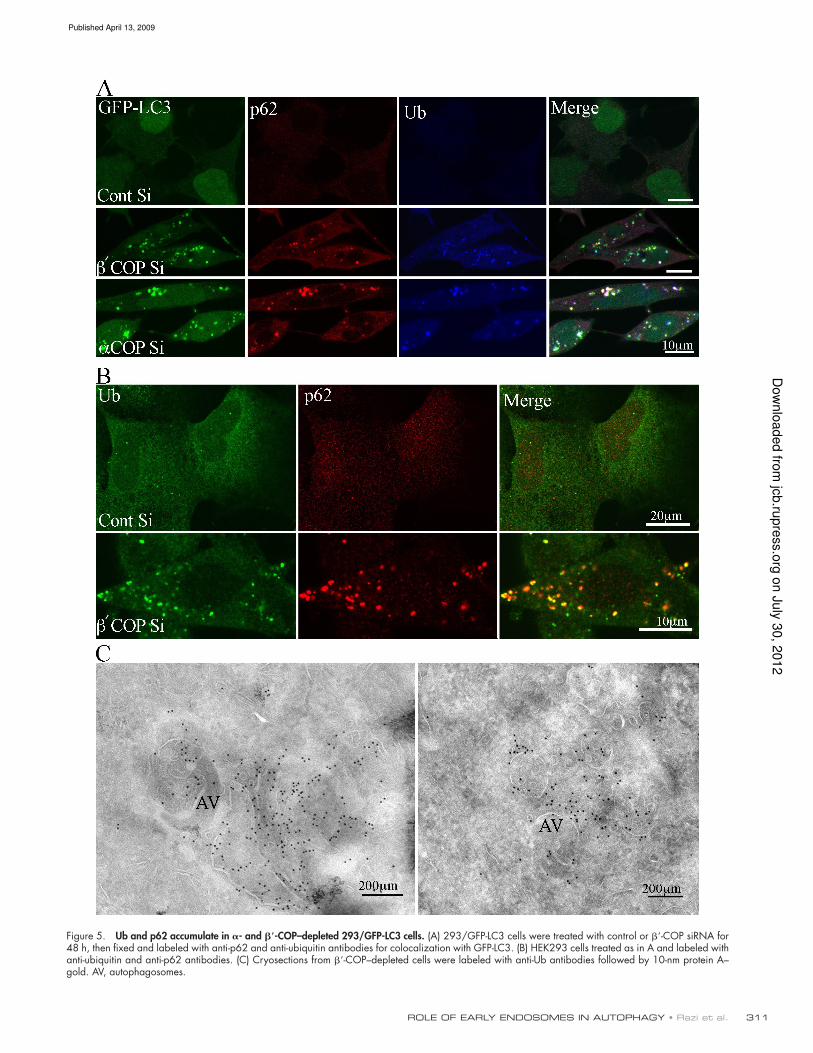
Loss of COPI causes the accumulation of
p62- and Ub-containing aggregates
p62/SQSTM1 can associate with LC3 on the membrane of AVs
and is sequestered into AVs (Pankiv et al., 2007). Levels and sub-
cellular distribution of p62 are used to monitor autophagic lux; a
low or unchanged level of p62 indicates a normal autophagic re-
sponse and lux, whereas an increased level of p62 indicates a
block in the lux, resulting from an inhibition of the pathway and
an accumulation of AVs (Klionsky et al., 2008). We therefore de-
cided to ask if distribution of p62 was affected by siRNA deple-
tion of COPI (Fig. 5 A). In control siRNA-treated cells, p62
exhibited a diffuse and cytoplasmic pattern similar to the cyto-
solic GFP-LC3. However, after siRNA depletion of COPI sub-
units, the signal for p62 increased and it was found in discrete
punctate structures, the majority of which were GFP-LC3 posi-
tive (Fig. 5 A). p62 has also been shown to associate with dense
protein aggregates (Pankiv et al., 2007); however, using TEM we
did not observe these structures in our 293/GFP-LC3 cells.
Figure 4. Correlative light-electron microscopy analysis of GFP-LC3 puncta demonstrates they are AVs. (A) Phase, (B) fluorescent, and (C) TEM of the same field of 293/GFP-LC3 cells 48 h after siRNA depletion of -COP. In B the image was inverted to allow better visualization of the GFP-LC3–positive structures. (D) Higher magnification of cell shown with asterisk in A and C. Top region of cell of interest is enlarged in E to show the two AVs structures indicated by arrows in B. (F and G) Higher magnification of AVs shown by arrows in E.
on J
uly
30, 2
012
jcb.ru
pre
ss.o
rgD
ow
nlo
aded fro
m
Published April 13, 2009
311ROLE OF EARLY ENDOSOMES IN AUTOPHAGY • Razi et al.
Figure 5. Ub and p62 accumulate in - and -COP–depleted 293/GFP-LC3 cells. (A) 293/GFP-LC3 cells were treated with control or -COP siRNA for 48 h, then fixed and labeled with anti-p62 and anti-ubiquitin antibodies for colocalization with GFP-LC3. (B) HEK293 cells treated as in A and labeled with anti-ubiquitin and anti-p62 antibodies. (C) Cryosections from -COP–depleted cells were labeled with anti-Ub antibodies followed by 10-nm protein A– gold. AV, autophagosomes.
on J
uly
30, 2
012
jcb.ru
pre
ss.o
rgD
ow
nlo
aded fro
m
Published April 13, 2009
JCB • VOLUME 185 • NUMBER 2 • 2009 312
p62 has been shown to bind ubiquitinated proteins via a
conserved UBA motif (Pankiv et al., 2007), so we next exam-
ined the distribution of ubiquitin (Ub) after siRNA knockdown
of COPI. Interestingly, after siRNA depletion of COPI we found
Ub distribution was altered, and behaved similarly to p62. Ub
had a diffuse appearance in control siRNA-treated cells, and a
punctate distribution after siRNA-mediated COP depletion. In
addition, a large number of the Ub-positive punctate structures
were also positive for GFP-LC3 (Fig. 5 A). Using HEK293A
parental cells we also observed after depletion of -COP that
p62 and Ub redistributed into punctate structures, which were
largely colocalized (Fig. 5 B).
Next we performed cryo-immuno EM to gain an under-
standing of the morphology of the GFP-LC3–positive structures
that contained p62 and Ub. Unfortunately, we were unable to label
the p62 using commercially available antibodies, but we could
detect Ub in cryosections. After siRNA-mediated depletion of -
COP we observed two major populations of Ub-positive mem-
branes: one population were AVs on which ubiquitin was detected
on the limiting cytosolic surface membrane as well as on internal
membranes, and the second consisted of small vesicles (less than
100 nm), the outer surface of which was decorated with Ub
(Fig. 5 C and Fig. S4). In -COP–depleted cells the morphology
of the Ub-labeled structures was very similar to that seen after la-
beling with GFP to detect the GFP-LC3 cells (Fig. S4). Numer-
ous small vesicles, tubules, and AVs, which resembled immature
autophagosomes (AVis), were found to be decorated with Ub and
GFP-LC3 on their cytoplasmic surface, and well as on internal
membranes. These results suggest that after COPI depletion ubiq-
uitinated proteins, and most likely p62, were present on the mem-
brane of small vesicles and tubules, as well as on the cytoplasmic
surface of AVs, and on internal membranes within the AVs.
AVs induced by COPI depletion are not
acidic or degradative
The large numbers of GFP-LC3 AVs in COPI-depleted cells
suggested that either the rate of AV formation was signiicantly
increased, or that AV maturation and consumption was inhib-
ited. An increased autophagic rate should result in an increased
degradative capacity of the cells, whereas if autophagosome
consumption or maturation is inhibited, degradation of autopha-
gic substrates should be reduced. To assay for the degradative
capacity of AVs in the COPI-depleted cells we measured long-
lived protein degradation as an indicator of autophagic activity
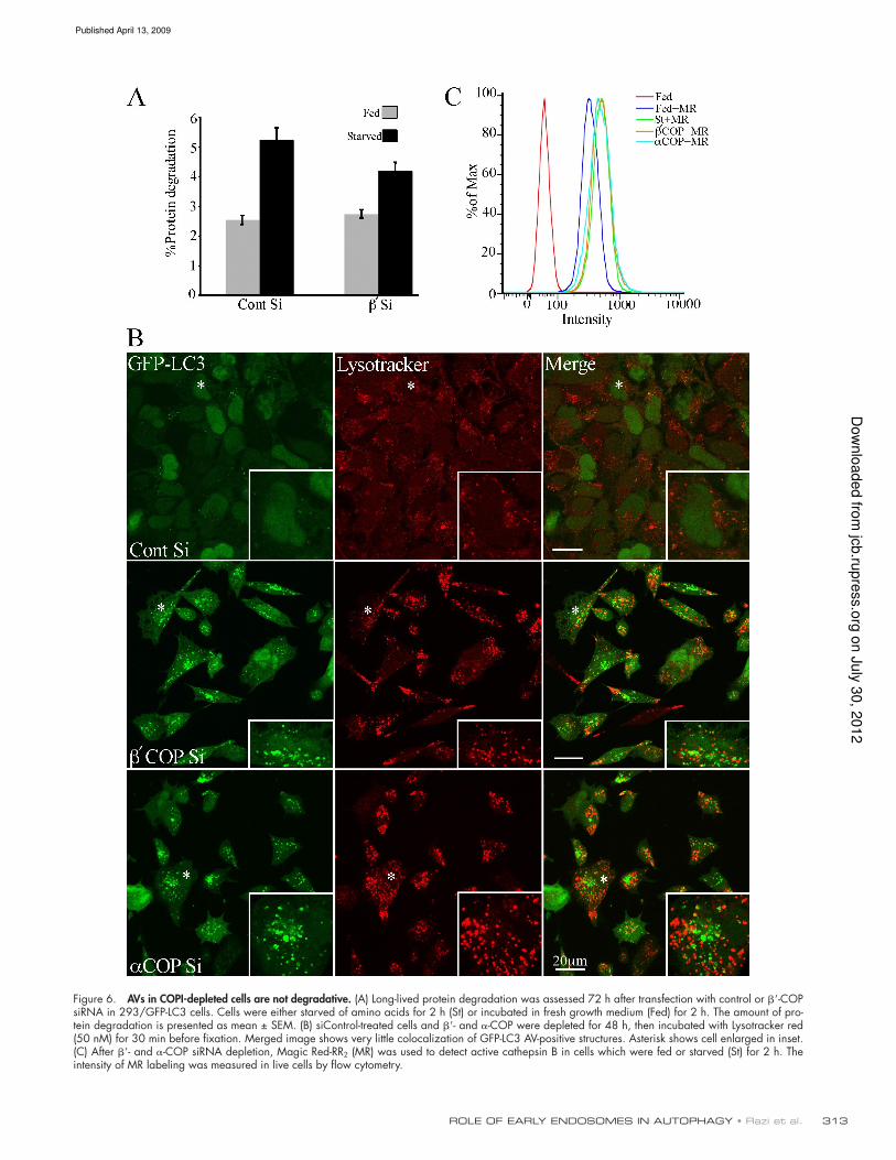
(Fig. 6 A). In siControl unstarved cells, long-lived protein deg-
radation occurred at a low basal level, and increased after a 2-h
starvation, as expected. However, after -COP depletion, de-
spite the large increase in GFP-LC3-II and GFP-LC3–positive
AVs in unstarved cells, there was no change in basal levels of
protein degradation (Fig. 6 A). This result suggests an impair-
ment in the degradative capacity of the AVs formed in COP-
depleted cells. However, the siRNA COPI-depleted cells were
still able to respond to starvation-induced autophagy as protein
degradation could be increased after 2 h starvation.
AVi/d and autolysosomes are known to be acidic compart-
ments. To understand more about the properties of the AVs in
COPI-depleted cells, in particular why they are not degradative,
we next asked if the GFP-LC3–positive AVs were acidic using
Lysotracker red (LTR), which accumulates in acidic compart-
ments. In previous studies it was shown that after 2-h amino acid
starvation 40% of AVs are LTR positive, and contain endosomal
markers (Bampton et al., 2005). Using - and -COP–depleted
cells incubated in growth medium, we found that the majority
of the GFP-LC3–positive structures did not accumulate LTR
(Fig. 6 B). Similar results were obtained using DAMP staining,
which also accumulates in acidic compartments (not depicted). To
further conirm that the lack of GFP-LC3 and LTR colocalization
was not due to quenching of the GFP-tag in acidic compartments
(which would mask the presence of accumulating GFP-LC3), we
looked at total LC3 accumulation using anti-LC3 antibodies in
COPI-depleted 293/GFP-LC3 cells. LC3 immunoreactivity should
not be sensitive to lumenal acidity. We found a complete overlap of
the two signals in COPI-depleted cells, in contrast to siControl
starved cells, which showed two immunoreactive populations of
LC3, one which overlapped with GFP-LC3, while the other did not
(Fig. S3 B). These results conirm that after COPI depletion we can
detect all the GFP-LC3–positive AVs using the GFP-tag, and that
these AVs were not degradative or acidic.
Dunn (1990) postulated that AVs become acidiied before
lysosomal enzymes are delivered by fusion with endocytotic
compartments, and this may occur by sequential fusion with
different types of endosomes; the irst to fuse would be endo-
somes containing the vacuolar (V) proton-ATPase (V-ATPase),
but without lysosomal enzymes. To understand if the lack of
degradative capacity in the accumulating AVs was only a conse-
quence of the defect in acidiication, or if lysosomal enzyme
targeting was also affected by loss of COPI, we asked if the
COPI-depleted cells had active lysosomal enzymes by examin-
ing the levels of Magic Red RR2, a luorogenic substrate which
detects active cathepsin B (Fig. 6 C). By FACS analysis, there
was no difference in the level of cathepsin B between starved
control cells and siCOPI-depleted cells. In addition, we con-
irmed that the COPI-depleted cells contained active cathepsin
D by immunoblotting cell lysates; in both control cells and
siRNA-treated cells depleted of -, -, and -COP, and Rab1a/b,
we detected similar levels of the 31-kD mature cathepsin D
heavy chain (Fig. S5 A).
Our data support the conclusion that the accumulation of
GFP-LC3 AVs in COPI-depleted cells is not a consequence of
an indirect effect on the entire endosomal–lysosomal pathway
as the COPI-depleted cells still have a degradative capacity and
normal levels of lysosomal enzymes.
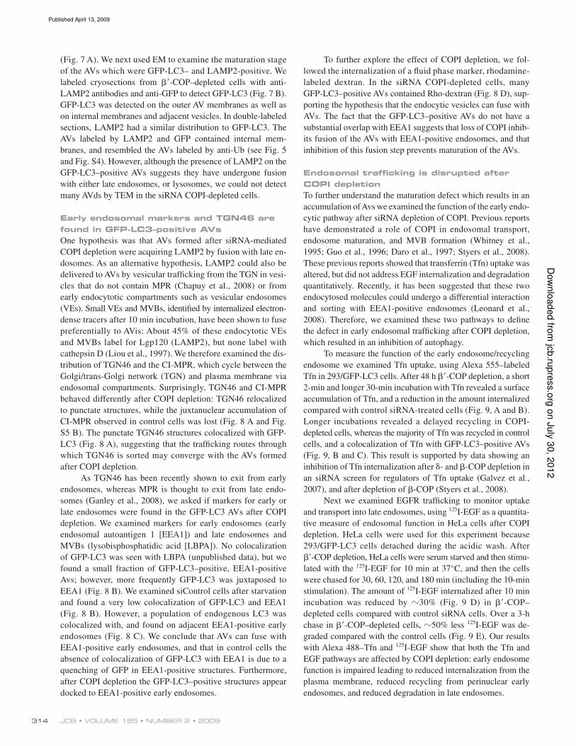
GFP-LC3 autophagosomes are LAMP1 and
2 positive
Lysosomal-associated membrane protein1 and 2 (LAMP1 and 2)
are found on late endosomes and lysosomes, and have also
been shown to be essential for the maturation of AVs (Saftig
et al., 2008). Therefore, we asked if after COPI depletion, the
distribution of LAMP1 and 2 in endosomal membranes was al-
tered, and if they were present on GFP-LC3–positive AVs. After
COPI depletion the overall distribution of LAMP1 and 2 was
not signiicantly changed. However, many of the GFP-LC3–
positive AVs contained LAMP1 (not depicted) and LAMP2
on J
uly
30, 2
012
jcb.ru
pre
ss.o
rgD
ow
nlo
aded fro
m
Published April 13, 2009
313ROLE OF EARLY ENDOSOMES IN AUTOPHAGY • Razi et al.
Figure 6. AVs in COPI-depleted cells are not degradative. (A) Long-lived protein degradation was assessed 72 h after transfection with control or -COP siRNA in 293/GFP-LC3 cells. Cells were either starved of amino acids for 2 h (St) or incubated in fresh growth medium (Fed) for 2 h. The amount of pro-tein degradation is presented as mean ± SEM. (B) siControl-treated cells and - and -COP were depleted for 48 h, then incubated with Lysotracker red (50 nM) for 30 min before fixation. Merged image shows very little colocalization of GFP-LC3 AV-positive structures. Asterisk shows cell enlarged in inset. (C) After - and -COP siRNA depletion, Magic Red-RR2 (MR) was used to detect active cathepsin B in cells which were fed or starved (St) for 2 h. The intensity of MR labeling was measured in live cells by flow cytometry.
on J
uly
30, 2
012
jcb.ru
pre
ss.o
rgD
ow
nlo
aded fro
m
Published April 13, 2009
JCB • VOLUME 185 • NUMBER 2 • 2009 314
(Fig. 7 A). We next used EM to examine the maturation stage
of the AVs which were GFP-LC3– and LAMP2-positive. We
labeled cryosections from -COP–depleted cells with anti-
LAMP2 antibodies and anti-GFP to detect GFP-LC3 (Fig. 7 B).
GFP-LC3 was detected on the outer AV membranes as well as
on internal membranes and adjacent vesicles. In double-labeled
sections, LAMP2 had a similar distribution to GFP-LC3. The
AVs labeled by LAMP2 and GFP contained internal mem-
branes, and resembled the AVs labeled by anti-Ub (see Fig. 5
and Fig. S4). However, although the presence of LAMP2 on the
GFP-LC3–positive AVs suggests they have undergone fusion
with either late endosomes, or lysosomes, we could not detect
many AVds by TEM in the siRNA COPI-depleted cells.
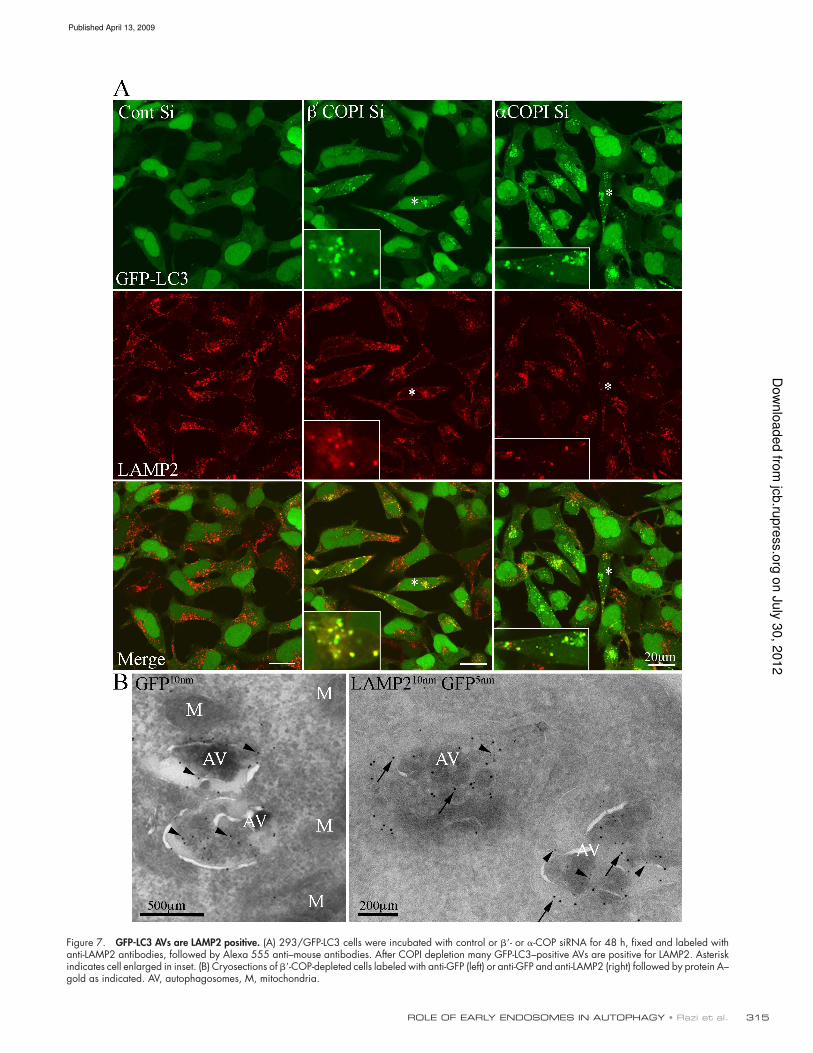
Early endosomal markers and TGN46 are
found in GFP-LC3–positive AVs
One hypothesis was that AVs formed after siRNA-mediated
COPI depletion were acquiring LAMP2 by fusion with late en-
dosomes. As an alternative hypothesis, LAMP2 could also be
delivered to AVs by vesicular traficking from the TGN in vesi-
cles that do not contain MPR (Chapuy et al., 2008) or from
early endocytotic compartments such as vesicular endosomes
(VEs). Small VEs and MVBs, identiied by internalized electron-
dense tracers after 10 min incubation, have been shown to fuse
preferentially to AVis: About 45% of these endocytotic VEs
and MVBs label for Lgp120 (LAMP2), but none label with
cathepsin D (Liou et al., 1997). We therefore examined the dis-
tribution of TGN46 and the CI-MPR, which cycle between the
Golgi/trans-Golgi network (TGN) and plasma membrane via
endosomal compartments. Surprisingly, TGN46 and CI-MPR
behaved differently after COPI depletion: TGN46 relocalized
to punctate structures, while the juxtanuclear accumulation of
CI-MPR observed in control cells was lost (Fig. 8 A and Fig.
S5 B). The punctate TGN46 structures colocalized with GFP-
LC3 (Fig. 8 A), suggesting that the traficking routes through
which TGN46 is sorted may converge with the AVs formed
after COPI depletion.
As TGN46 has been recently shown to exit from early
endosomes, whereas MPR is thought to exit from late endo-
somes (Ganley et al., 2008), we asked if markers for early or
late endosomes were found in the GFP-LC3 AVs after COPI
depletion. We examined markers for early endosomes (early
endosomal autoantigen 1 [EEA1]) and late endosomes and
MVBs (lysobisphosphatidic acid [LBPA]). No colocalization
of GFP-LC3 was seen with LBPA (unpublished data), but we
found a small fraction of GFP-LC3–positive, EEA1-positive
Avs; however, more frequently GFP-LC3 was juxtaposed to
EEA1 (Fig. 8 B). We examined siControl cells after starvation
and found a very low colocalization of GFP-LC3 and EEA1
(Fig. 8 B). However, a population of endogenous LC3 was
colocalized with, and found on adjacent EEA1-positive early
endosomes (Fig. 8 C). We conclude that AVs can fuse with
EEA1-positive early endosomes, and that in control cells the
absence of colocalization of GFP-LC3 with EEA1 is due to a
quenching of GFP in EEA1-positive structures. Furthermore,
after COPI depletion the GFP-LC3–positive structures appear
docked to EEA1-positive early endosomes.
To further explore the effect of COPI depletion, we fol-
lowed the internalization of a luid phase marker, rhodamine-
labeled dextran. In the siRNA COPI-depleted cells, many
GFP-LC3–positive AVs contained Rho-dextran (Fig. 8 D), sup-
porting the hypothesis that the endocytic vesicles can fuse with
AVs. The fact that the GFP-LC3–positive AVs do not have a
substantial overlap with EEA1 suggests that loss of COPI inhib-
its fusion of the AVs with EEA1-positive endosomes, and that
inhibition of this fusion step prevents maturation of the AVs.
Endosomal trafficking is disrupted after
COPI depletion
To further understand the maturation defect which results in an
accumulation of Avs we examined the function of the early endo-
cytic pathway after siRNA depletion of COPI. Previous reports
have demonstrated a role of COPI in endosomal transport,
endosome maturation, and MVB formation (Whitney et al.,
1995; Guo et al., 1996; Daro et al., 1997; Styers et al., 2008).
These previous reports showed that transferrin (Tfn) uptake was
altered, but did not address EGF internalization and degradation
quantitatively. Recently, it has been suggested that these two
endocytosed molecules could undergo a differential interaction
and sorting with EEA1-positive endosomes (Leonard et al.,
2008). Therefore, we examined these two pathways to deine
the defect in early endosomal traficking after COPI depletion,
which resulted in an inhibition of autophagy.
To measure the function of the early endosome/recycling
endosome we examined Tfn uptake, using Alexa 555–labeled
Tfn in 293/GFP-LC3 cells. After 48 h -COP depletion, a short
2-min and longer 30-min incubation with Tfn revealed a surface
accumulation of Tfn, and a reduction in the amount internalized
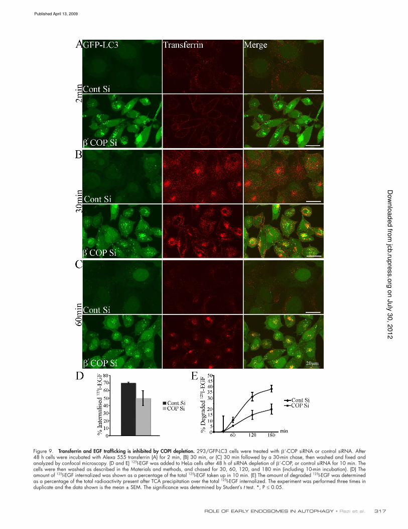
compared with control siRNA-treated cells (Fig. 9, A and B).
Longer incubations revealed a delayed recycling in COPI-
depleted cells, whereas the majority of Tfn was recycled in control
cells, and a colocalization of Tfn with GFP-LC3–positive AVs
(Fig. 9, B and C). This result is supported by data showing an
inhibition of Tfn internalization after - and -COP depletion in
an siRNA screen for regulators of Tfn uptake (Galvez et al.,
2007), and after depletion of -COP (Styers et al., 2008).
Next we examined EGFR traficking to monitor uptake
and transport into late endosomes, using 125I-EGF as a quantita-
tive measure of endosomal function in HeLa cells after COPI
depletion. HeLa cells were used for this experiment because
293/GFP-LC3 cells detached during the acidic wash. After
-COP depletion, HeLa cells were serum starved and then stimu-
lated with the 125I-EGF for 10 min at 37°C, and then the cells
were chased for 30, 60, 120, and 180 min (including the 10-min
stimulation). The amount of 125I-EGF internalized after 10 min
incubation was reduced by 30% (Fig. 9 D) in -COP–
depleted cells compared with control siRNA cells. Over a 3-h
chase in -COP–depleted cells, 50% less 125I-EGF was de-
graded compared with the control cells (Fig. 9 E). Our results
with Alexa 488–Tfn and 125I-EGF show that both the Tfn and
EGF pathways are affected by COPI depletion: early endosome
function is impaired leading to reduced internalization from the
plasma membrane, reduced recycling from perinuclear early
endosomes, and reduced degradation in late endosomes.
on J
uly
30, 2
012
jcb.ru
pre
ss.o
rgD
ow
nlo
aded fro
m
Published April 13, 2009
315ROLE OF EARLY ENDOSOMES IN AUTOPHAGY • Razi et al.
Figure 7. GFP-LC3 AVs are LAMP2 positive. (A) 293/GFP-LC3 cells were incubated with control or - or -COP siRNA for 48 h, fixed and labeled with anti-LAMP2 antibodies, followed by Alexa 555 anti–mouse antibodies. After COPI depletion many GFP-LC3–positive AVs are positive for LAMP2. Asterisk indicates cell enlarged in inset. (B) Cryosections of -COP-depleted cells labeled with anti-GFP (left) or anti-GFP and anti-LAMP2 (right) followed by protein A– gold as indicated. AV, autophagosomes, M, mitochondria.
on J
uly
30, 2
012
jcb.ru
pre
ss.o
rgD
ow
nlo
aded fro
m
Published April 13, 2009
JCB • VOLUME 185 • NUMBER 2 • 2009 316
Figure 8. GFP-LC3–positive AVs colocalize TGN46 and early endocytotic markers after COPI depletion. (A) 293/GFP-LC3 cells were incubated with siRNA for -COP or control siRNA (Cont) for 48 h, processed for immunofluorescence, and labeled with antibodies for TGN46. In (B) siRNA-treated cells were fixed and labeled, or starved for 2 h (St), and labeled with antibodies to EEA1. (C) Control-starved HEK293 cells were labeled for endogenous LC3 and EEA1 by double labeling after methanol fixation. (D) After siRNA treatment, rhodamine-labeled dextran was internalized for 20 min, followed by a 40-min chase after which the 293/GFP-LC3 cells were fixed and analyzed by confocal microscopy.
on J
uly
30, 2
012
jcb.ru
pre
ss.o
rgD
ow
nlo
aded fro
m
Published April 13, 2009
317ROLE OF EARLY ENDOSOMES IN AUTOPHAGY • Razi et al.
Figure 9. Transferrin and EGF trafficking is inhibited by COPI depletion. 293/GFP-LC3 cells were treated with -COP siRNA or control siRNA. After 48 h cells were incubated with Alexa 555 transferrin (A) for 2 min, (B) 30 min, or (C) 30 min followed by a 30-min chase, then washed and fixed and analyzed by confocal microscopy. (D and E) 125I-EGF was added to HeLa cells after 48 h of siRNA depletion of -COP, or control siRNA for 10 min. The cells were then washed as described in the Materials and methods, and chased for 30, 60, 120, and 180 min (including 10-min incubation). (D) The amount of 125I-EGF internalized was shown as a percentage of the total 125I-EGF taken up in 10 min. (E) The amount of degraded 125I-EGF was determined as a percentage of the total radioactivity present after TCA precipitation over the total 125I-EGF internalized. The experiment was performed three times in duplicate and the data shown is the mean ± SEM. The significance was determined by Student’s t test. *, P ≤ 0.05.
on J
uly
30, 2
012
jcb.ru
pre
ss.o
rgD
ow
nlo
aded fro
m
Published April 13, 2009
JCB • VOLUME 185 • NUMBER 2 • 2009 318
A consequence of altered traficking of growth factor recep-
tors could be a decrease in the uptake of nutrients and the genera-
tion of a starvation signal. Thus, COPI depletion could potentially
be causing a chronic starvation signal by down-regulating Tfn and
EGF receptors. To investigate this possibility we took three ap-
proaches. First, we mimicked growth factor receptor deprivation
by withdrawing serum from the cells. Although we did observe an
induction of GFP-LC3–positive AVs, the kinetics of formation
and consumption was different from the effects of COPI deple-
tion; a maximum response was obtained at 15 h serum starvation,
which disappeared after 24 h serum starvation. Second, we mea-
sured amino acid uptake in COPI-depleted cells in comparison to
siRNA treated control cells in full medium or after starvation.
In control starved cells we observed a 10-fold increase in [14C]-
valine uptake, but basal uptake in the COPI-depleted cells. In fact,
after 72 h COPI depletion, the levels of valine uptake per cell
were identical to control fed cells. Interestingly, COPI-depleted
cells after 2 h starvation showed a similar increase to control
starved cells. Our third approach was to use 11 mM methyl pyru-
vate (Guenther et al., 2008), shown to function as a transporter-
independent source of nutrients; addition of methyl pyruvate 24 h
before harvesting COPI-depleted cells had little effect on GFP-
LC3-II accumulation, whereas its addition inhibited the GFP-
LC3-II conversion in control starved cells (unpublished data).
Discussion
Nascent AVs (AVis) are double-membrane vesicles containing
sequestered cytoplasmic proteins and organelles. AVis only be-
come degradative (AVds) when they fuse with endosomes. It has
been proposed that a sequential fusion event between AVis and
different types of endosomal vesicles or endosomes is required
for AVi maturation to AVds or autolysosomes (Dunn, 1990;
Eskelinen, 2005). Although recent data have highlighted the im-
portance of late endocytic machinery in the autophagic response,
in particular the ESCRT complex (Filimonenko et al., 2007; Lee
et al., 2007; Rusten et al., 2007), there is no direct evidence
that fusion of early endosomes is required for AV maturation.
Through our analysis of -COP, identiied as a potential nega-
tive regulator of autophagy (Chan et al., 2007), we have shown
that functional early endosomes are required for autophagy.
We show that loss of COPI subunits , , and results in an
inhibition of AV maturation and subsequently causes an accumula-
tion of AVs. The accumulation of AVs after COPI depletion is ro-
bust, and raises the question of whether basal autophagy is also
increased by loss of COPI. This hypothesis is dificult to directly
test in our model system, as the siRNA depletion of COPI occurs
over 48 h and the levels of COP subunits decrease gradually. How-
ever, we have determined the extent of AV accumulation that could
be detected in control, untreated 293/GFP-LC3 cells after basal au-
tophagy was blocked for 5, 25, or 50 h by culturing the 293/GFP-
LC3 cells in normal culture medium in the presence of leupeptin
(unpublished data). After 25 h of leupeptin treatment there was a
signiicant accumulation of basal AVs, which was comparable to
48 h siRNA depletion of COP subunits, leading us to suggest that
the primary effect exerted by COPI depletion was an inhibition of
maturation of AVs that formed in basal conditions.
The COPI complex has been shown to function in both
anterograde and retrograde transport to and through the Golgi com-
plex, as well as in early endosome maturation. We found that loss
of either -, -, and -COP, gave identical results on both Golgi
morphology and AV accumulation. Previous data using a stable
temperature sensitive CHO cell line (ldl-F cells), in which -COP
is degraded at the nonpermissive temperature, show a loss of the
Golgi complex similar to our observations (Guo et al., 1994),
whereas siRNA depletion of -COP (Styers et al., 2008) showed
a collapse of ERGIC and Golgi markers into a common compart-
ment. The differences in Golgi morphology between our -COP
depletion experiments and those in Styers et al. [2008] are likely
to be caused by differences in the level of -COP depletion, or
differences in the levels of the other COPs: we reproducibly saw
-COP decreased after - and -COP depletion, and -COP
decreased after -COP depletion.
Having observed a profound affect on the Golgi complex
after 48 h of -COP depletion, we asked if the disruption of the
Golgi complex was responsible for the accumulation of AVs. In a
time-course experiment we observed that after 24 h siRNA de-
pletion Golgi morphology was already perturbed, suggesting that
Golgi fragmentation was the cause of the increase in AVs. We
tested this hypothesis using siRNA depletion of Rab1, which has
a similar effect of Golgi morphology and function (Haas et al.,
2007), and saw that disassembly of the Golgi, by itself, did not
cause an accumulation of AVs.
We found an increased number of p62 and Ub-positive
structures, which colocalize with GFP-LC3 after COPI depletion,
in agreement with previous data demonstrating an increase in p62
levels after a block in autophagic turnover (Pankiv et al., 2007).
Furthermore, as p62 has an ubiquitin-associated domain (UBA),
which interacts with Ub (Geetha and Wooten, 2002), the colocal-
ization we detect of Ub with p62 and GFP-LC3 in AVs represents
a pool of Ub sequestered into AVs via an interaction with p62. In-
triguingly, our data also show that some of the Ub is present on
the cytoplasmic surface of small vesicles and tubules, and these
are also sequestered into AVs. The origin of these small vesicles
and tubules is unknown, but we speculate that they originate from
compartments that, after COPI depletion, have undergone vesicu-
lation or other alterations leading to damage and recruitment of
Ub. Although we have no data to support this hypothesis, and in-
deed we did not detect a colocalization of GM130 with Ub, we
speculate that ubiquitination of the fragmented or damaged com-
partments may be a signal to target them to AVs. It is also possible
that ubiquitinated vesicles or compartments accumulated in re-
gions where AVs are forming or expanding and were nonselec-
tively sequestered. Further experiments are required to determine
the origin and fate of the ubiquitinated vesicular structures.
After siRNA depletion of COPI subunits we observed an
inhibition of Tfn internalization and recycling, in agreement
with previous reports (Guo et al., 1994; Daro et al., 1997; Styers
et al., 2008). In addition, we found -COP depletion caused a
decreased internalization and degradation of 125I-EGF, in line
with previous results showing a delayed delivery of Texas red–
labeled biotinylated EGF to lgp-95 (lysosomal glycoprotein-95)
positive structures (Daro et al., 1997). Endosomes have been
shown to have COPI coats and the early studies demonstrated a
on J
uly
30, 2
012
jcb.ru
pre
ss.o
rgD
ow
nlo
aded fro
m
Published April 13, 2009
319ROLE OF EARLY ENDOSOMES IN AUTOPHAGY • Razi et al.
possibility is that they are VEs, previously demonstrated to
undergo fusion with AVis, and are both LAMP2 positive, and
accessible to endocytotic tracers after 10 min (Liou et al., 1997).
Further experiments are required to characterize these VEs, and
for instance to investigate if they contain TGN46, before we can
fully understand the role of this compartment.
Our data demonstrate that GFP-LC3–positive AVs accumu-
lated in COPI-depleted cells due to an inhibition of early endo-
some function, resulting in the formation of AVs that cannot
mature. Previous data have shown that AV accumulation occurs
after disruption of ESCRT on late endosomes (Filimonenko et al.,
2007; Kim et al., 2007; Lee et al., 2007). Collectively, these re-
sults demonstrate a role for both early endosomes and late endo-
somes in autophagy. Our data provide direct evidence that early
endosome fusion with AVs is a prerequisite for AV maturation,
and occurs before fusion with late endosomes and MVBs.
Whether the inhibition of early endosome function and conse-
quently AVi maturation gives rise to a pathological disease state
similar to the neurodegenerative diseases associated with ESCRT
loss of function remains to be determined.
Materials and methods
Cell lines and reagentsHeLa, HEK293A, and 293/GFP-LC3 cells (Chan et al., 2007), a stable HEK293A cell line expressing rat GFP-LC3, were cultured in DMEM contain-ing 10% FCS. For indirect immunofluorescence and EM, HEK293 or 293/GFP-LC3 cells were plated on poly-D-lysine–coated coverslips. Salubrinal was from Axxora; Radionucleotides were from PerkinElmer; Oligofectamine 2000 was from Invitrogen; Tunicamycin was from Sigma-Aldrich.
Transfection and siRNA reagentssiRNAs were transfected with Oligofectamine using the manufacturer’s proto-col. All siRNAs were purchased from Thermo Fisher Scientific and the sequences are as follows: Control nontargeting siRNA duplex (D-001210-021), -COP (D-019847-01), -COP (D-017940-01), and -COP (M-011835-01). Rab1a and 1b (L-008283-00 and L-008958-01), sec23A and sec23B (M-009582-00 and M-009592-00), Atg5 (M-004374-01), and Atg7 (M-020112-01). EEA1 siRNA was as published (Leonard et al., 2008). Cells were transfected with siRNA for 48–72 h unless otherwise stated.
ImmunoblottingImmunoblotting was performed on cells lysed in 1x SDS sample buffer. Antibodies used were rabbit anti--COP (891) and rabbit anti--, --, --COP (883), both from Prof. Felix Wieland (University of Heidelberg, Heidel-berg, Germany), and rabbit anti-EAGE -COP (STO235) (Duden et al., 1991). Rabbit anti-Atg5 (Cosmo Bio Ltd); anti-Atg7 (Novus); rabbit anti-Rab1 (Santa Cruz Biotechnology, Inc.); mouse anti-Sec23 (ABR); rabbit anti-GFP [Dr. T. Hunt (CRUK)]; monoclonal anti-LC3 (5F10) (Nanotools); rabbit anti-calreticulin (Bioquote); goat anti-cathepsin D antibody (Santa Cruz Biotechnology, Inc.); rabbit anti-tubulin (Abcam). HRP-conjugated sec-ondary antibodies were revealed using ECL (GE Healthcare) according to the manufacturer’s instructions.
Indirect immunofluorescenceIndirect immunofluorescence was performed using a standard protocol on cells fixed with 3% PFA in PBS, permeabilized in 0.2% Triton X-100, and a 20-min incubation in 0.5% BSA. For endogenous LC3 (using 5F10) we fixed the cells with 20°C methanol for 5 min. Cells were then incubated with primary antibody followed by a secondary antibody in PBS contain-ing 0.1% BSA. Mouse LAMP2 (CD107B) antibody was from BD Bio-sciences, guinea pig anti-p62 from Progen, mouse anti-CI-MPR from Abcam, rabbit anti-ubiquitin from Dako, mouse anti-ERGIC-53 from Qbiogene, mouse anti-GM130 from BD Biosciences, and sheep anti-TGN46 from AbD Serotec. Rabbit anti-EEA1 antibody was a gift of M. Clague (Univer-sity of Liverpool, UK). Alexa-conjugated transferrin, secondary antibodies, and Lysotracker red were from Invitrogen. Lysotracker was used following the manufacturer’s instructions. Rhodamine-labeled dextran (Invitrogen)
role for COPI in endocytosis of virus (Whitney et al., 1995), in
endosomal carrier vesicle formation and MVB maturation using
the inhibitory M3A5 monoclonal raised against -COP (Whitney
et al., 1995; Aniento et al., 1996) and in assays using the ldlF cells
(Gu et al., 1997). More recently, in yeast COPI has been shown to
be required for endosomal transport to the vacuole, similar to the
class E yeast vacuolar protein sorting (Vps) mutants involved in
MVB formation (Gabriely et al., 2007). Finally, the endosomal
pool of COPI has been shown to be used by anthrax lethal factor
for translocation across endosomal membranes (Tamayo et al.,
2008), providing independent evidence for a role of COPI in endo-
some function. An alternative scenario that we have tested is
that COPI depletion through its inhibition of Tfn and EGF inter-
nalization may generate a starvation signal, inducing autophagy
independently of endosome function. Although we found serum
deprivation induced GFP-LC3 AV formation after a short serum
starvation, the cells adapted to the absence of serum by 24 h and
GFP-LC3 AVs were no longer detected. In addition, our assays
for amino acid uptake and sensitivity to methyl pyruvate suggest
that the COPI-depleted cells are not in a state of chronic starva-
tion due to loss of growth factor signaling.
We hypothesize that loss of the endosomal pool of COPI
results in the accumulation of GFP-LC3–positive endosomes
due to a defect in early endosome maturation. The accumulating
AVs are not acidic or LTR positive, and lack lysosomal prote-
ases such as cathepsin D (unpublished data), but are LAMP
positive and contain TGN46.
Our data demonstrating a colocalization of endogenous
LC3 with EEA1 provide strong evidence that under normal star-
vation conditions there is fusion of early endosomes with AVs,
and suggest that the EEA1-positive endosomes are involved in
AV maturation. Other evidence in hepatocytes and exocrine pan-
creatic cells supports the notion that early endosomes can fuse
with AVis (Gordon and Seglen, 1988; Tooze et al., 1990). In con-
trast, and in agreement with Fass et al., 2006 we did not see a
robust colocalization of GFP-LC3 with EEA1. We attribute this
difference to quenching of GFP in the EEA1-positive compart-
ment. However, we have tested the requirement for EEA1 in AV
maturation by transfection of an siRNA shown previously to de-
plete EEA1 (Leonard et al., 2008), and found that loss of EEA1
did not result in the accumulation of GFP-LC3–positive AVs, or
GFP-LC3-II (unpublished data). Why does loss of EEA1 not
lead to AV accumulation as seen with COPI disruption?
Recent data examining the role of EEA1 in endocytosis
suggest that EEA1-positive endosomes may have a specialized
function for sorting EGFR into a degradative compartment, and
may not be required for Tfn recycling (Leonard et al., 2008). It
was proposed that Tfn-containing endosomes only associate with
EEA1-positive endosomes, while EGF containing endocytic ves-
icles dock and fuse with the EEA1-positive endosomes due to a
requirement for Rab5. We speculate that under basal conditions,
AVi maturation can by-pass the EEA1-dependent endosome, al-
though this remains to be further demonstrated. Our data show-
ing the requirement of COPI in both Tfn and EGF traficking
indicate that COPI might have broader functions in endosomal
traficking possibly by acting upstream of the EEA1-positive
endosomes. What are these endosomes upstream of EEA1? One
on J
uly
30, 2
012
jcb.ru
pre
ss.o
rgD
ow
nlo
aded fro
m
Published April 13, 2009
JCB • VOLUME 185 • NUMBER 2 • 2009 320
References
Aniento, F., F. Gu, R.G. Parton, and J. Gruenberg. 1996. An endosomal beta COP is involved in the pH-dependent formation of transport vesicles destined for late endosomes. J. Cell Biol. 133:29–41.
Bampton, E.T.W., C.G. Goemans, D. Niranjan, N. Mizushima, and A.M. Tolkovsky. 2005. The dynamics of autophagy visualized in live cells: from autophagosome formation to fusion with endo/lysosomes. Autophagy. 1:23–36.
Boyce, M., K.F. Bryant, C. Jousse, K. Long, H.P. Harding, D. Scheuner, R.J. Kaufman, D. Ma, D.M. Coen, D. Ron, and J. Yuan. 2005. A selective inhibitor of eIF2{alpha} dephosphorylation protects cells from ER stress. Science. 307:935–939.
Chan, E.Y.W., S. Kir, and S.A. Tooze. 2007. siRNA screening of the kinome identiies ULK1 as a multi-domain modulator of autophagy. J. Biol. Chem. 282:25464–25474.
Chapuy, B., R. Tikkanen, C. Muhlhausen, D. Wenzel, K. von Figura, and S. Honing. 2008. AP-1 and AP-3 mediate sorting of melanosomal and lyso-somal membrane proteins into distinct post-Golgi traficking pathways. Trafic. 9:1157–1172.
Citterio, C., A. Vichi, G. Pacheco-Rodriguez, A.M. Aponte, J. Moss, and M. Vaughan. 2008. Unfolded protein response and cell death after deple-tion of brefeldin A-inhibited guanine nucleotide-exchange protein GBF1. Proc. Natl. Acad. Sci. USA. 105:2877–2882.
Csukai, M., C.-H. Chen, M.A. De Matteis, and D. Mochly-Rosen. 1997. The coatomer protein beta’-COP, a selective binding protein (RACK) for pro-tein kinase Cepsilon. J. Biol. Chem. 272:29200–29206.
Daro, E., D. Sheff, M. Gomez, T. Kreis, and I. Mellman. 1997. Inhibition of endo-some function in CHO cells bearing a temperature-sensitive defect in the coatomer (COPI) component epsilon-COP. J. Cell Biol. 139:1747–1759.
Duden, R., G. Grifiths, R. Frank, P. Argos, and T.E. Kreis. 1991. -COP, a 110 kDa protein associated with non-clathrin-coated vesicles and the Golgi complex, shows homology to -adaptin. Cell. 64:649–665.
Dunn, W.A. Jr. 1990. Studies on the mechanisms of autophagy: maturation of the autophagic vacuole. J. Cell Biol. 110:1935–1945.
Eskelinen, E.L. 2005. Maturation of autophagic vacuoles in mammalian cells. Autophagy. 1:1–10.
Eugster, A., G. Frigerio, M. Dale, and R. Duden. 2000. COP I domains required for coatomer integrity, and novel interactions with ARF and ARF-GAP. EMBO J. 19:3905–3917.
Filimonenko, M., S. Stuffers, C. Raiborg, A. Yamamoto, L. Malerod, E.M. Fisher, A. Isaacs, A. Brech, H. Stenmark, and A. Simonsen. 2007. Functional multivesicular bodies are required for autophagic clearance of protein aggregates associated with neurodegenerative disease. J. Cell Biol. 179:485–500.
Gabriely, G., R. Kama, and J.E. Gerst. 2007. Involvement of speciic COPI sub-units in protein sorting from the late endosome to the vacuole in yeast. Mol. Cell. Biol. 27:526–540.
Galvez, T., M.N. Teruel, W.D. Heo, J.T. Jones, M.L. Kim, J. Liou, J.W. Myers, and T. Meyer. 2007. siRNA screen of the human signaling proteome iden-tiies the PtdIns(3,4,5)P3-mTOR signaling pathway as a primary regula-tor of transferrin uptake. Genome Biol. 8:R142.
Ganley, I.G., E. Espinosa, and S.R. Pfeffer. 2008. A syntaxin 10-SNARE com-plex distinguishes two distinct transport routes from endosomes to the trans-Golgi in human cells. J. Cell Biol. 180:159–172.
Geetha, T., and M.W. Wooten. 2002. Structure and functional properties of the ubiquitin binding protein p62. FEBS Lett. 512:19–24.
Gordon, P.B., and P.O. Seglen. 1988. Prelysosomal convergence of autophagic and endocytic pathways. Biochem. Biophys. Res. Commun. 151:40–47.
Gronostajski, R.M., and A.B. Pardee. 1984. Protein degradation in 3T3 cells and tumorigenic transformed 3T3 cells. J. Cell. Physiol. 119:127–132.
Gu, F., F. Aniento, R.G. Parton, and J. Gruenberg. 1997. Functional dissection of COP-I subunits in the biogenesis of multivesicular endosomes. J. Cell Biol. 139:1183–1195.
Guenther, G.G., E.R. Peralta, K.R. Rosales, S.Y. Wong, L.J. Siskind, and A.L. Edinger. 2008. Ceramide starves cells to death by downregulating nutri-ent transporter proteins. Proc. Natl. Acad. Sci. USA. 105:17402–17407.
Guo, Q., E. Vasile, and M. Krieger. 1994. Disruptions in Golgi structure and membrane trafic in a conditional lethal mammalian cell mutant are cor-rected by epsilon-COP. J. Cell Biol. 125:1213–1224.
Guo, Q., M. Penman, B.L. Trigatti, and M. Krieger. 1996. A single point mu-tation in epsilon-COP results in temperature-sensitive, lethal defects in membrane transport in a Chinese hamster ovary cell mutant. J. Biol. Chem. 271:11191–11196.
Haas, A.K., S. Yoshimura, D.J. Stephens, C. Preisinger, E. Fuchs, and F.A. Barr. 2007. Analysis of GTPase-activating proteins: Rab1 and Rab43 are key
was used at 0.5 mg/ml in growth medium. Cells were examined and imaged with a Zeiss LSM 510 laser-scanning confocal microscope equipped with a 63x, 1.4 NA, Plan Apochromat oil-immersion objective (Carl Zeiss, Inc.). Im-ages were processed using LSM 510 software. Magic Red-(RR)2 (Immuno-chemistry Technologies, LLC) was added to the adherent cells for 20 min, after which they were removed by trypsinization, and analyzed in a LSR II FACS (Becton Dickinson) using excitation of 592 nm and emission of 628 nm.
Long-lived protein degradation and amino acid uptakeLong-lived protein degradation was performed as described previously (Chan et al., 2007). In brief, 293/GFP-LC3 cells were transfected with siRNA against -COP. 48 h later, labeling medium (DMEM, 10% dialyzed FBS containing [14C]-valine [0.2 µCi/ml, GE], and 65 µM unlabeled valine) was added to the cells. After 18 h, the labeling medium was replaced with chase medium (DMEM, 10% FCS, and 2 mM unlabeled valine) for 4 h. After the chase, cells were incubated in EBSS and 2 mM valine for 2 h to stimulate autophagy. The cells and media were collected, and separately subjected to trichloroacetic acid (TCA) precipitation by the addition of ice-cold TCA to 10%. The percentage of autophagic degradation was quanti-fied as (TCA-soluble counts from medium)/(TCA-soluble counts from medium) + (TCA-insoluble counts from the cells) (Gronostajski and Pardee, 1984).
Amino acid uptake was performed using 0.2 µCi/ml [14C]-valine in labeling medium for 3 h either to control cells, or cells after 72 h siRNA transfection. Radioactivity was measured in the cells after washing. Viable cell number was determined from duplicate wells.
EGF internalization and degradationHeLa cells were transfected with siRNA using Oligofectamine, and 72 h later cells were serum starved for at least 1 h followed by an incubation with 125I-EGF (1 ng/ml) in binding medium (0.05% BSA in serum-free DMEM) for 10 min at 37°C. Cells were surface stripped using 0.1 M glycine and 0.9% NaCl, pH 3.0, at 4°C, washed with binding medium, and placed in fresh prewarmed 37°C binding medium. After each time point the medium was collected and replaced by fresh medium for the subsequent time point. The collected media were TCA (20% final) precipitated at 4°C for 1 h. TCA- insoluble proteins were pelleted by centrifugation at 14,000 g at 4°C, and the soluble supernatant was collected. Cells were lysed in 1% Triton X-100, and the radioactivity in the chase medium, the TCA soluble supernatant, and cell lysates were counted to determine the percentage of EGF degrada-tion and recycling.
Electron microscopyCells were fixed in 0.1 M cacodylate containing 2% paraformaldehyde and 2% glutaraldehyde and were then embedded using standard procedures (Hopkins and Trowbridge, 1983). Cryo-immuno-EM was performed on cells fixed with 4% paraformaldehyde/0.1% glutaraldehyde (for ubiquitin and LAMP2 labeling cells were fixed only with PFA) in 0.1 M phosphate buffer, pH 7.4, infused with 2.3 M sucrose and supported in 10% gelatin. Sections (70 nm) were cut at 120°C and picked up in 1:1 sucrose/methylcellulose. Pri-mary antibodies were rabbit anti-ubiquitin (Dako), rabbit anti-GFP antibody (Abcam), and mouse anti-LAMP2 (BD Biosciences). Sections were then la-beled using protein A–gold as described previously (Slot and Geuze, 2007). Sections were visualized in a FEI Technai G2 Spirit Biotwin transmission elec-tron microscopic and an Ultrascan 1000 CCD camera and software.
Online supplemental materialFigure S1 shows the effect of -, -, and -COP on the structure of the Golgi complex by transmission electron microscopy. Figure S2 shows that COPI depletion does not cause ER stress. Figure S3 A shows the morphology of the AVs accumulating in COP-depleted cells by transmission electron micros-copy. Figure S3 B confirms GFP-LC3 is not quenched in acidic structures. Figure S4 shows the cryo-immuno electron microscopy of 293/GFP-LC3 cells after 48 h COPI depletion labeled for Ub and GFP. Figure S5 A shows cathepsin D is correctly stored and processed after COPI depletion, or Rab1 depletion. Figure S5 B shows that CI-MPR after COPI depletion is dispersed and not colocalized with GFP-LC3. Online supplemental material is avail-able at http://www.jcb.org/cgi/content/full/jcb.200810098/DC1.
We thank Drs. Giampietro Schiavo and Jöelle Movan for comments on the manuscript, and the Secretory Pathways laboratory, Rose Watson, Hannah Armer, and the London Research Institute EM unit for their help and advice.
We thank Cancer Research UK for support.
Submitted: 15 October 2008Accepted: 24 March 2009
on J
uly
30, 2
012
jcb.ru
pre
ss.o
rgD
ow
nlo
aded fro
m
Published April 13, 2009
321ROLE OF EARLY ENDOSOMES IN AUTOPHAGY • Razi et al.
Rabs required to maintain a functional Golgi complex in human cells. J. Cell Sci. 120:2997–3010.
Hopkins, C.R., and I.S. Trowbridge. 1983. Internalization and processing of transferrin and the transferrin receptor in human carcinoma A431 cells. J. Cell Biol. 97:508–521.
Kim, B.Y., J.A. Olzmann, G.S. Barsh, L.-S. Chin, and L. Li. 2007. Spongiform neurodegeneration-associated E3 ligase mahogunin ubiquitylates TSG101 and regulates endosomal traficking. Mol. Biol. Cell. 18:1129–1142.
Klionsky, D.J. 2007. Autophagy: from phenomenology to molecular understand-ing in less than a decade. Nat. Rev. Mol. Cell Biol. 8:931–937.
Klionsky, D.J., H. Abeliovich, P. Agostinis, D.K. Agrawal, G. Aliev, D.S. Askew, M. Baba, E.H. Baehrecke, B.A. Bahr, A. Ballabio, et al. 2008. Guidelines for the use and interpretation of assays for monitoring autophagy in higher eukaryotes. Autophagy. 4:151–175.
Kuma, A., M. Matsui, and N. Mizushima. 2007. LC3, an autophagosome marker, can be incorporated into protein aggregates independent of autophagy: caution in the interpretation of LC3 localization. Autophagy. 3:323–328.
Lee, J.A., A. Beigneux, S. Ahmad, S. Young, and F. Gao. 2007. ESCRT-III dys-function causes autophagosome accumulation and neurodegeneration. Curr. Biol. 17:1561–1567.
Lee, M.C.S., E.A. Miller, J. Goldberg, L. Orci, and R. Schekman. 2004. Bi-directional protein transport between the ER and Golgi. Annu. Rev. Cell Dev. Biol. 20:87–123.
Leonard, D., A. Hayakawa, D. Lawe, D. Lambright, K.D. Bellve, C. Standley, L.M. Lifshitz, K.E. Fogarty, and S. Corvera. 2008. Sorting of EGF and transferrin at the plasma membrane and by cargo-speciic signaling to EEA1-enriched endosomes. J. Cell Sci. 121:3445–3458.
Liou, W., H.J. Geuze, M.J.H. Geelen, and J.W. Slot. 1997. The autophagic and endocytic pathways converge at the nascent autophagic vacuoles. J. Cell Biol. 136:61–70.
Mizushima, N., B. Levine, A.M. Cuervo, and D.J. Klionsky. 2008. Autophagy ights disease through cellular self-digestion. Nature. 451:1069–1075.
Nickel, W., B. Brugger, and F.T. Wieland. 2002. Vesicular transport: the core ma-chinery of COPI recruitment and budding. J. Cell Sci. 115:3235–3240.
Nyfeler, B., O. Nufer, T. Matsui, K. Mori, and H.-P. Hauri. 2003. The cargo receptor ERGIC-53 is a target of the unfolded protein response. Biochem. Biophys. Res. Commun. 304:599–604.
Ogata, M., S.-i. Hino, A. Saito, K. Morikawa, S. Kondo, S. Kanemoto, T. Murakami, M. Taniguchi, I. Tanii, K. Yoshinaga, et al. 2006. Autophagy is activated for cell survival after endoplasmic reticulum stress. Mol. Cell. Biol. 26:9220–9231.
Pankiv, S., T.H. Clausen, T. Lamark, A. Brech, J.-A. Bruun, H. Outzen, A. Overvatn, G. Bjorkoy, and T. Johansen. 2007. p62/SQSTM1 binds di-rectly to Atg8/LC3 to facilitate degradation of ubiquitinated protein ag-gregates by autophagy. J. Biol. Chem. 282:24131–24145.
Rusten, T.E., T. Vaccari, K. Lindmo, L.M. Rodahl, I.P. Nezis, C. Sem-Jacobsen, F. Wendler, J.P. Vincent, A. Brech, D. Bilder, and H. Stenmark. 2007. ESCRTs and Fab1 regulate distinct steps of autophagy. Curr. Biol. 17:1817–1825.
Saftig, P., W. Beertsen, and E.L. Eskelinen. 2008. LAMP-2: A control step for phagosome and autophagosome maturation. Autophagy. 4:510–512.
Slot, J.W., and H.J. Geuze. 2007. Cryosectioning and immunolabeling. Nat. Protoc. 2:2480–2491.
Styers, M.L., A.K. O’Connor, R. Grabski, E. Cormet-Boyaka, and E. Sztul. 2008. Depletion of {beta}-COP reveals a role for COP-I in compartmentaliza-tion of secretory compartments and in biosynthetic transport of caveolin-1. Am. J. Physiol. Cell Physiol. 294:C1485–C1498.
Tamayo, A.G., A. Bharti, C. Trujillo, R. Harrison, and J.R. Murphy. 2008. COPI coatomer complex proteins facilitate the translocation of anthrax lethal factor across vesicular membranes in vitro. Proc. Natl. Acad. Sci. USA. 105:5254–5259.
Tooze, J., M. Hollinshead, T. Ludwig, K. Howell, B. Holack, and H. Kern. 1990. In exocrine pancreas, the basolateral endocytic pathway converges with the autophagic pathway immediately after the early endosome. J. Cell Biol. 111:329–345.
Whitney, J.A., M. Gomez, D. Sheff, T.E. Kreis, and I. Mellman. 1995. Cytoplasmic coat proteins involved in endosome function. Cell. 83:703–713.
Williams, R.L., and S. Urbe. 2007. The emerging shape of the ESCRT machin-ery. Nat. Rev. Mol. Cell Biol. 8:355–368.
Woodman, P.G., and C.E. Futter. 2008. Multivesicular bodies: co-ordinated pro-gression to maturity. Curr. Opin. Cell Biol. 20:408–414.
Xie, Z., and D.J. Klionsky. 2007. Autophagosome formation: core machinery and adaptations. Nat. Cell Biol. 9:1102–1109.
on J
uly
30, 2
012
jcb.ru
pre
ss.o
rgD
ow
nlo
aded fro
m
Published April 13, 2009
![LTD., - docshare01.docshare.tipsdocshare01.docshare.tips/files/25986/259865268.pdf · Further, "[A] section 2-1401 petition must be supported by affidavit or other showing of matters](https://img.pdfslide.us/doc/110x75/5a7376557f8b9aa7538e8d9d/ltd-a-further-a-section-2-1401-petition-must-be-supported-by-affidavit.jpg)


![Nanostructured Photocatalytic Approach to CO …...be commercially available for decades [3]–[5]. There is no full-scale CCS project that captures and sequesters CO2 from a coal-fired](https://img.pdfslide.us/doc/110x75/5f26aa76c4f8bb1b3066607d/nanostructured-photocatalytic-approach-to-co-be-commercially-available-for-decades.jpg)