Embed Size (px)
Citation preview

Aalborg Universitet
Rational Engineering of Hydratase from Lactobacillus Acidophilus Reveals CriticalResidues Directing Substrate Specificity and Regioselectivity
Eser, Bekir Engin; Poborsky, Michal; Dai, Rongrong; Kishino, Shigenobu; Ljubic, Anita;Takeuchi, Michiki; Jacobsen, Charlotte; Ogawa, Jun; Kristensen, Peter; Guo, ZhengPublished in:ChemBioChem
DOI (link to publication from Publisher):10.1002/cbic.201900389
Publication date:2020
Document VersionAccepted author manuscript, peer reviewed version
Link to publication from Aalborg University
Citation for published version (APA):Eser, B. E., Poborsky, M., Dai, R., Kishino, S., Ljubic, A., Takeuchi, M., Jacobsen, C., Ogawa, J., Kristensen, P.,& Guo, Z. (2020). Rational Engineering of Hydratase from Lactobacillus Acidophilus Reveals Critical ResiduesDirecting Substrate Specificity and Regioselectivity. ChemBioChem, 21(4), 550-563.https://doi.org/10.1002/cbic.201900389
General rightsCopyright and moral rights for the publications made accessible in the public portal are retained by the authors and/or other copyright ownersand it is a condition of accessing publications that users recognise and abide by the legal requirements associated with these rights.
- Users may download and print one copy of any publication from the public portal for the purpose of private study or research. - You may not further distribute the material or use it for any profit-making activity or commercial gain - You may freely distribute the URL identifying the publication in the public portal -
Take down policyIf you believe that this document breaches copyright please contact us at [email protected] providing details, and we will remove access tothe work immediately and investigate your claim.

www.chembiochem.org
Accepted Article
A Journal of
Title: Rational Engineering of Hydratase from Lactobacillus AcidophilusReveals Critical Residues Directing Substrate Specificity andRegioselectivity
Authors: Bekir Engin Eser, Michal Poborsky, Rongrong Dai, ShigenobuKishino, Anita Ljubic, Michiki Takeuchi, Charlotte Jacobsen,Jun Ogawa, Peter Kristensen, and Zheng Guo
This manuscript has been accepted after peer review and appears as anAccepted Article online prior to editing, proofing, and formal publicationof the final Version of Record (VoR). This work is currently citable byusing the Digital Object Identifier (DOI) given below. The VoR will bepublished online in Early View as soon as possible and may be differentto this Accepted Article as a result of editing. Readers should obtainthe VoR from the journal website shown below when it is publishedto ensure accuracy of information. The authors are responsible for thecontent of this Accepted Article.
To be cited as: ChemBioChem 10.1002/cbic.201900389
Link to VoR: http://dx.doi.org/10.1002/cbic.201900389

1
Rational Engineering of Hydratase from Lactobacillus
Acidophilus Reveals Critical Residues Directing Substrate
Specificity and Regioselectivity
Dr. Bekir Engin Eser1, Michal Poborsky1, Rongrong Dai1, Dr. Shigenobu Kishino2, Anita Ljubic3,
Dr. Michiki Takeuchi2, Prof. Dr. Charlotte Jacobsen3, Prof. Dr. Jun Ogawa2, Prof. Dr. Peter
Kristensen4, Prof. Dr. Zheng Guo1*
1 Department of Engineering, Aarhus University, 8000 Aarhus, Denmark, 2Division of Applied
Life Sciences, Graduate School of Agriculture, Kyoto University, Kyoto 606-8502, Japan,
3Division of Food Technology, National Food Institute, Technical University of Denmark, 2800
Kgs. Lyngby, Denmark, 4Faculty of Engineering and Science, Department of Chemistry and
Bioscience, Aalborg University, 9220 Aalborg, Denmark
*Corresponding Author: Zheng Guo (e-mail: [email protected])
10.1002/cbic.201900389
Acc
epte
d M
anus
crip
t
ChemBioChem
This article is protected by copyright. All rights reserved.

2
Abstract
Enzymatic conversion of fatty acids (FAs) by fatty acid hydratases (FAHs) presents a green and
efficient route for high-value hydroxy fatty acid (HFA) production. However, limited diversity was
achieved among HFAs to date with respect to chain length and hydroxy position. In this study, two
highly similar FAHs from Lactobacillus acidophilus were compared: FA-HY2 has narrow
substrate scope and strict regioselectivity, whereas FA-HY1 utilizes longer chain substrates and
hydrate various double bond positions. We reveal three active-site residues that play remarkable
role in directing substrate specificity and regioselectivity of hydration. When these residues on FA-
HY2 are mutated to the corresponding ones in FA-HY1, we observed a significant expansion of
substrate scope and a distinct enhancement in hydration of double bonds towards -end of FAs. A
three-residue mutant of FA-HY2 (TM-FA-HY2) displayed an impressive reversal of
regioselectivity towards linoleic acid, shifting ratio of the HFA regioisomers (10-OH:13-OH) from
99:1 to 12:88. Notable changes in regioselectivity were also observed for arachidonic acid and for
C18 PUFA substrates. In addition, TM-FA-HY2 converted EPA to its 12-hydroxy product with
high conversion at preparative scale. Furthermore, we demonstrated microalgae as a source of
diverse FAs for HFA production. Our study paves the way for tailor-made FAH design enabling
production of diverse HFAs for various applications from polymer industry to medical field.
10.1002/cbic.201900389
Acc
epte
d M
anus
crip
t
ChemBioChem
This article is protected by copyright. All rights reserved.

3
INTRODUCTION
Hydroxy fatty acids (HFAs) are unique fatty acid (FA) derivatives with beneficial medical
properties[1] as well as with various industrial uses[2]. HFAs can occur as gut microbial FA
metabolites and suggested to be involved in the regulation of host energy metabolism[1a]. Such a
metabolite, 10-hydroxy-cis-12-octadecenoic acid, exhibited anti-inflammatory effects in murine
enterocytes in vitro[1b] and suppressed intestinal inflammation in animal models[3]. Recently
described branched FA esters of HFAs (FAHFAs) are naturally occurring bioactive lipids with
anti-diabetic and anti-inflammatory effects[4]. More than 20 families of FAHFAs were identified
in human body that differ by composition of their FAs and position of the ester bond between HFA
and FA[4b]. Specialized proresolving mediators (SPM) are another group of HFA metabolites that
exhibit effects such as resolution of inflammation, stimulation of tissue regeneration and tumor
growth suppression[5]. SPMs include lipoxins, which are tri-hydroxy derivatives of arachidonic
acid (AA) and resolvins, which are di-, tri-, or poly-hydroxy derivatives of mainly eicosapentaenoic
acid (EPA) and docosahexaenoic acid (DHA)[5e]. Moreover, it was demonstrated that lactobacilli
used for food fermentation produce anti-fungal HFAs[6]. The potency of these HFAs against fungi
was dependent on their structure, including the position and configuration of the hydroxy group[6b].
Ricinoleic acid (12-hydroxy-9-cis-octadecenoic acid) is used in several industries for
manufacturing of various products such as surfactants, plastics (Nylon-11), plasticizers,
polyurethane resins, emulsifiers and lubricant additives[2c, 7]. 12-hydroxystearic acid is commonly
used in skin care preparations[8]. HFAs can also be used to produce polymers for durable and
flexible bioplastics with properties that can be tailored for a specific application[9]. The FA length
and the position of the hydroxyl dictate the size of the monomers and as such, the overall properties
of the material.
10.1002/cbic.201900389
Acc
epte
d M
anus
crip
t
ChemBioChem
This article is protected by copyright. All rights reserved.

4
Castor plants are used to produce the most common industrial HFA, ricinoleic acid, but they do
not provide any further product diversity and accumulate toxic ricin[2a, 2c, 10]. Due to poor reactivity
of water, chemical methods for hydration of isolated carbon-carbon double bonds often require
harsh reaction conditions such as transition metal and/or acid-base catalysts, high temperatures and
hazardous organic solvents[11]. Besides, chemical methods lead to a variety of undesired side
reactions and do not offer the necessary stereo- and regioselectivity to unlock the full potential of
HFAs[2b, 12]. There are a few chemical methods presented in literature for stereoselective addition
of water to unsaturated ketones[13] and FAs[14], however, carbon-carbon double bond needs to be
activated as an α, β-unsaturated carbonyl and transition metal catalysts are required. Fatty acid
hydratases (FAHs) present an alternative enzymatic route for synthesis of HFAs. These enzymes
attracted great interest in recent years due to their high biocatalytic potential[2a, 2b, 12, 15]. FAHs can
act on isolated carbon-carbon double bonds of the FA and produce HFAs by reversible addition of
water onto a double bond of an unsaturated FA in a regio- and stereospecific manner[12, 15b] (Scheme
1). Although other enzymatic routes are present for hydration of activated α, β-unsaturated carbonyl
compounds, FAHs can efficiently act on non-activated carbon-carbon double bonds[2b]. Moreover,
regio- and stereoselective hydration of non-FA substrates, including terminal and internal alkenes
as well as FA derivatives, have been recently demonstrated by engineered FAHs[11c, 16]. Although
no redox chemistry is involved, FAD is essential for this reaction and most likely plays a structural
or charge-stabilization role[17]. Advantageously for a biocatalytic process, FAD is required only in
catalytic amount and no recycling of the cofactor is needed. Moreover, hydratases are highly active
and relatively stable enzymes, in comparison to other hydroxylases such as P450 monooxygenases
and lipoxygenases, and can be expressed in high quantities in E. coli host[12]. Utilizing unsaturated
10.1002/cbic.201900389
Acc
epte
d M
anus
crip
t
ChemBioChem
This article is protected by copyright. All rights reserved.

5
FAs as substrates, hydratases are also well suited to convert algae or plant oil hydrolysates as well
as waste oil into valuable chemicals.
Scheme 1. Reaction catalyzed by Fatty Acid Hydratases
Most hydratases are active towards C16-C18 unsaturated FAs and are only able to add water on
Δ9 double bonds, with oleic acid (OA) being the most preferred substrate. Broadening the substrate
range and regiodiversity of hydratases will improve their applicability for industrial and medical
fields. Recently, two novel hydratases (FA-HY1 and FA-HY2) from L. acidophilus, with 76%
sequence similarity, were described[18]. Where FA-HY2 is a typical hydratase, FA-HY1 is rather
unique, with a broad substrate promiscuity, as it accepts substrates up to 22 carbons in length and
preferably hydrates double bonds other than Δ9[18a].
In this study, active-sites of FA-HY1 and FA-HY2 were compared to identify residues that
dictate the dramatic difference in substrate specificity and regioselectivity. Our analysis was based
on the structure of a closely related homolog, OhyA (oleate hydratase from Elizabethkingia
meningoseptica)[17] and sequence comparison between FA-HY1, FA-HY2 and OhyA. Through
site-directed mutagenesis, we targeted three residues of FA-HY2, located around the carboxylate
end of FA substrate, and substituted them into the corresponding amino acids from FA-HY1, either
individually or in combination. Our results demonstrate that mutations, in a stepwise manner from
a single residue mutant to a triple mutant (TM-FA-HY2), exhibit increasing degree of changes in
substrate preference and in regioselectivity, converting FA-HY2 to a more versatile catalyst with
similar properties to FA-HY1. Regioselectivity shifts were observed to some extent for all C18
10.1002/cbic.201900389
Acc
epte
d M
anus
crip
t
ChemBioChem
This article is protected by copyright. All rights reserved.

6
PUFAs and arachidonic acid (AA) with TM-FA-HY2 variant as well as a double mutant. Moreover,
TM-FA-HY2 produced 12-OH products from cis-5, cis-8, cis-11, cis-14, cis-17-eicosapentaenoic
acid (EPA) and AA more efficiently than the most active wild-type enzyme for the same products.
These two 12-OH products were detected for the first time with a hydratase. The practical utility
of FA-HY1, FA-HY2 and mutant variants for production of various HFAs through preparative
scale reactions was also demonstrated. Furthermore, our steady-state kinetic data indicate that the
effect of mutations is exhibited mostly on kcat rather than Km. Substrate docking studies further
highlight the significance of positioning of double bond for activity and regioselectivity. Lastly,
production of HFAs by hydratases from FA of two microalgae species demonstrates microalgae as
a sustainable source of FAs for production of value-added HFAs.
MATERIALS AND METHODS
Chemicals
All FAs were purchased from Sigma-Aldrich, except EPA (Calbiochem) and cis-Vaccenic acid
(MP Biomedicals). All FA substrate and standards were of the purest grade available (at least
98.5% purity). N, O-Bis(trimethylsilyl)trifluoroacetamide with trimethylchlorosilane (BSTFA +
TMCS; 99:1) used for silylation and FAD used as cofactor were also purchased from Sigma-
Aldrich. All other common chemicals were analytical grade and are commercially available.
Site-directed Mutagenesis
The genes fa-hy1(GenBank ID: LC030242.1) and fa-hy2 (genebank ID LC030243.1) from the
strain Lactobacillus acidophilus NTV001 were 1,773 and 1,776 base pairs (bp) in length,
respectively, and both genes (including stop codons) were cloned into pET-21b vector using XhoI
and NdeI endonucleases as previously described[18a]. All forward and reverse primers (see Table
S8 for sequences) required for mutagenesis were obtained from Sigma-Aldrich. Site-directed
10.1002/cbic.201900389
Acc
epte
d M
anus
crip
t
ChemBioChem
This article is protected by copyright. All rights reserved.

7
mutagenesis was carried out using QuikChange II Site-Directed Mutagenesis Kit from Agilent.
The success of the mutagenesis was confirmed by sequencing (Eurofins Genomics). For plasmid
transformation and amplification, the chemically competent E. coli strain XL1-Blue (Agilent) was
used. Plasmid purification was performed using GeneJET plasmid miniprep kit (Thermo Fisher
Scientific).
Protein Expression and Purification
For protein expression, E.coli Rosetta (DE3) cells (Merck Millipore) were transformed with a
plasmid possessing the proper gene. 0.5 L or 1 L of 2xTY or LB media (with 100 µg/mL ampicillin)
was inoculated with 5-10 mL of starter culture and the cells were grown until the optical density at
600 nm reached 0.5-1.0. At this point, the expression was induced by addition of 0.1 mM isopropyl-
β-D-thiogalactopyranoside (IPTG). Afterwards, the expression was carried at 18 °C and 180 rpm
for 16-20 hours. The cells were then harvested by centrifugation at 4,000 g and 4 °C for 30 min,
washed twice with 0.85% solution of NaCl. The cell pellet was stored at -80 °C until further use.
All the steps below were carried at 4 °C, unless otherwise stated. The previously washed cells
were resuspended in 100 mM potassium phosphate buffer, pH 6.0 (5 mL per 0.5 L of culture), and
supplemented with cOmplete EDTA-Free Protease Inhibitor (Sigma-Aldrich). After cell disruption
with sonication, the cell debris was pelleted by centrifugation at 30,000 g for 2 hours and the
supernatant was stored at -80 °C.
All proteins were purified at room temperature with ÄKTA explorer fast protein liquid
chromatography (FPLC) system (GE Healthcare) equipped with anion-exchange Mono Q HR 5/5
column (GE Healthcare). The proteins were eluted using a linear gradient of NaCl from 0 to 1 M
in 50 mM potassium phosphate buffer at pH 6.0 with a flow-rate of 2 mL/min. Only FA-HY1
required two step purification, with a subsequent loading onto cation-exchange SP Sepharose HP
10.1002/cbic.201900389
Acc
epte
d M
anus
crip
t
ChemBioChem
This article is protected by copyright. All rights reserved.

8
column (GE Healthcare) and elution under the same condition as previously. The eluted fractions
were analyzed by SDS-PAGE and those showing high purity of desired protein band were
collected. The pooled fractions were further buffer exchanged (into 100 mM potassium phosphate
at pH 6.0) and concentrated using centrifugal filters with 10 or 30 kDa cut-off. The concentration
of purified enzymes were determined with BCA Protein Assay Kit (Pierce, Thermo Fisher
Scientific). All enzyme solutions were aliquoted and stored at -80 °C.
Enzyme Activity Assays and Product Extraction
For analysis of substrate specificity and regioselectivity, a standard enzyme reaction mixture
contained (in a final volume of 1 mL), 0.5 mg of a FA substrate (1.5-2.0 mM, depending on
molecular weight of FA) and 300 µg of enzyme (4.5 μM) in 100 mM potassium phosphate buffer,
pH 6.0. The mixture also consisted of 5% ethanol to solubilize the FAs and 0.1 mM FAD cofactor.
The assays were incubated at 37 °C with a shaking speed of 120 rpm for about 16 hours. Incubation
of the assays were performed in an airtight box, in which an anaerobic atmosphere was maintained
by Oxoid AnaeroGen sachets (Thermo Fisher Scientific), to prevent oxidation of unsaturated FAs.
Reactions were quenched by addition of 1 mL of chloroform followed by rigorous mixing. Kinetic
assays were initiated by addition of enzyme and quenched by chloroform. For short time steady-
state assays (less than 30 min), reactions were carried out aerobically, but for longer time assays
anaerobic conditions were maintained. Initial velocities obtained from each substrate concentration
was fitted to Michaelis-Menten equation using Kaleidagraph Program (Synergy software). The
enzyme concentration and assay time used for each steady-state analysis is given in Supporting
Information (Table S1) along with Michaelis-Menten plots.
After the enzyme reaction is quenched, 1 mg heptadecanoic or hexadecanoic acid internal
standard was added to each reaction mixture and Bligh-Dyer method was then used to extract the
10.1002/cbic.201900389
Acc
epte
d M
anus
crip
t
ChemBioChem
This article is protected by copyright. All rights reserved.

9
lipid components. Chloroform, methanol and 1.5% KCl solution (with a ratio of 2:2:1, respectively)
in a total volume of 5 mL was added onto 1 mL of the assay mixture. Each chemical was added
separately and the mixture was vortexed at highest speed for 5-10 s after each addition. In order to
separate the phases, the mixture was centrifuged at 3,000 g for 10 min at room temperature. The
bottom chloroform phase was then collected in a fresh glass tube and subsequently, the solvent was
completely evaporated by a gentle nitrogen flow.
Preparative Scale Reactions and Product Purification
All preparative scale reactions were set at a scale of 100 mL total reaction volume in Erlenmeyer
flasks that were placed in an airtight box with AnaeroGen sachets. Initial time course experiments
at smaller scale (about 4 mL) were performed to determine optimum conditions and concentrations
for preparative scale assays (Figure S47). Reaction mixtures were incubated at 37 °C for about 24
hours with gentle shaking at 100 rpm. A standard preparative scale reaction mixture contained (in
a final volume of 100 mL), 50-100 mg of a FA substrate (1.5-3.5 mM final concentration; 0.15-
0.35 mmol scale) and 10-60 mg of enzyme (1.5-9.0 μM) in 100 mM potassium phosphate buffer,
pH 6.0. The reaction mixture also consisted of 5% ethanol and 0.10-0.15 mM FAD cofactor.
Reactions were quenched by addition of 100 mL chloroform. Extraction of the lipid components
from the reaction mixture was carried out by Bligh-Dyer method similar to 1 mL-scale assays,
except the volumes of solvents used for extraction were increased 100-fold and the extraction was
carried out in 1000 mL separatory funnel. Once the bottom chloroform phase was recovered, the
solvent was evaporated and the lipid mixture was redissolved in ethyl acetate (2-4 mL) for
purification of HFA product using Silica gel on TLC plates (L*W, 20 cm * 20 cm). The solvent
path was 18 cm and the development system was chloroform: methanol (10:1 v/v). 0.1% 2, 7-
Dichlorofluorescein solution in ethanol was used to spray TLC plates. The products on TLC plate
10.1002/cbic.201900389
Acc
epte
d M
anus
crip
t
ChemBioChem
This article is protected by copyright. All rights reserved.

10
were visualized under 365 nm UV light that showed two distinct bands, one for substrate and one
for product. The band with lower Rf value, which belonged to the product, was scraped out of the
TLC plates. The product in silica gel powder was extracted with ethyl acetate, which was
evaporated under nitrogen flow. Dried sample was dissolved in 1-2 mL of chloroform and extracted
twice with Bligh-Dyer method (water was used instead of 1.5% KCl) to remove 2, 7-
Dichlorofluorescein. Chloroform phase was collected and dried under nitrogen. Amount of dried
product was measured for yield calculation. Purity analysis of the products were carried out by GC-
FID. Detailed description of each preparative scale reaction, conversion percentages, isolated yield
values and purity levels are given in Supporting Information (Table S7).
Product Analysis, Identification and Quantification
Prior to chromatographic analysis, the extracted reaction mixture was derivatized into their
trimethylsilyl (TMS) derivatives by the addition of 150 µL of N,O-
Bis(trimethylsilyl)trifluoroacetamide with trimethylchlorosilane (BSTFA + TMCS, 99:1). The
silylation reaction was performed at 60 °C for 30 min. After derivatization, samples were injected
in splitless mode (1-2 μl) into Scion 436-GC (Bruker, Billerica, MA) system equipped with a flame
ionization detector (FID) and a non-polar fused silica capillary SPB-1 column (30 m length x 0,25
mM I.D., 0,25 µM film thickness; Sigma). The oven temperature was set to 220 °C and it was
maintained constant for 60 min of the total analysis time. Injection temperature was 250 °C and the
column flow was maintained at a constant pressure of 22 Psi throughout the analysis with helium
as the carrier gas.
For the separation of closely eluting HFA regioisomers, changes were made to the standard GC
method. In the modified method, the injection volume to the GC was typically 0.5 to 1 µL and
injection was performed in split mode, at a split ratio of 1:20. Initial oven temperature was 90 °C
10.1002/cbic.201900389
Acc
epte
d M
anus
crip
t
ChemBioChem
This article is protected by copyright. All rights reserved.

11
for 1 min and increased to 170 °C at a rate of 20 °C/min. This was immediately followed by a
further increase to 220 °C at a rate of 2 °C/min and this temperature was maintained for 16 to 62
min, depending on the chain length of products.
Controls with LA, OA and ricinoleic acid showed that the relative response factors for the TMS
derivatives of the substrate FA and its corresponding HFA product are similar (within 5%
deviation; Figure S48) with GC-FID detection under the conditions used, similar to prior
observations[19]. Thus, quantification of the product was based on the ratio of the peak area of
product peak to the total peak areas of both substrate and product. A control sample containing
only substrate was used to confirm that substrate and product total peak area are consistent between
different measurements. An internal standard (palmitic or heptadecanoic acid) was also added to
each sample before extraction, at a concentration of 1 mg/mL, to evaluate consistency of extraction
and GC measurements.
Chemical structure identification of HFA products were performed by 1H, 13C NMR and by 1H-
1H double-quantum-filtered chemical-shift correlation spectroscopy (DQF-COSY) analysis of
purified samples and by GC-MS analysis of TMS derivatives. For NMR analysis, 3-5 mg of
samples were dissolved in CDCl3 and spectra were recorded on a Bruker Avance III spectrometer
(400 MHz). GC-MS analysis was performed using Thermo Fisher DSQ II quadrupole mass
spectrometer equipped with Trace GC Ultra and TriPlus autosampler. Ionization was with electron
impact at 70 eV in positive-mode. Ion source temperature was 230 °C and transfer line was at 250
°C. The same capillary column as in GC analysis (SPB-1) was also used for GC-MS analysis with
the same temperature program. NMR spectra and GC-MS spectra of all the products identified in
this study are given in Supporting Information (Figures S6-S39).
10.1002/cbic.201900389
Acc
epte
d M
anus
crip
t
ChemBioChem
This article is protected by copyright. All rights reserved.

12
Absolute configuration of products were determined using Mosher Ester method[20], where
methylated HFA products (5-15 mg) were reacted with (S)-(+)-MTPA chloride and (R)-(-)-MTPA
chloride (see Supporting Information for details). The resulting Mosher esters were dissolved in
0.7 mL of CDCl3 for 1H NMR and DQF-COSY analysis. Enantiomeric excess (% ee) was
determined by GC-FID analysis of methylated HFA products by a chiral column (CP-Chirasil Dex
CB; 25 m length x 0,25 mM I.D., 0,25 µM film thickness; Agilent).
Microalgae Cultivation
Two microalgae species, Nannochloropsis gaditana (SAG 2.99) and Pavlova lutheri (SAG 926-
1) were obtained from Sammlung von Algenkulturen der Universität Göttingen (SAG) culture
collection in Germany. The cultivation was carried out in 1-20 L GS Schott bottles in F2P growth
medium[21]. All reactors were continuously aerated with 5% carbon dioxide/air mixture under LED
illumination with an irradiance of 100-200 μmol photon m-1s-2 and 12:12 (h) photoperiod. The
cultures were mixed by rotary shaking at 90 rpm. Cultivation temperature range was 24 ± 1 °C and
pH 7-8. pH monitoring was performed by Milwaukee MC-122-pH controller (Milwaukee
Electronics, Hungary) equipped with a solenoid valve to control CO2 addition. Biomass separation
was done by centrifugation at 6500 x g for P. lutheri and 6000 x g for N. gaditana for 10 min
followed by freeze-drying. Samples were stored at -20 °C.
Extraction of FAs from Microalgae Biomass
Mixture of chloroform:methanol:water (1.25 mL: 2.5 mL: 0.45 mL) was added onto 100 mg of
dry microalgae biomass. The resuspension was vortexed/shaken for 10 min followed by 15 min of
sonication in a bath sonicator (at 50 Hz). After a further addition of chloroform:methanol (1.25
mL:1.25 mL), the mixture was vortexed/shaken for 5 more minutes. This was followed by
centrifugation at 3,000 g for 15 min. The bottom chloroform layer (including total lipids) was
10.1002/cbic.201900389
Acc
epte
d M
anus
crip
t
ChemBioChem
This article is protected by copyright. All rights reserved.

13
recovered and transferred into new tubes before being completely dried under a flow of nitrogen.
In order to achieve hydrolysis of triglycerides, 1 mL of 0.5 M KOH in methanol was added and the
resulting mixture was incubated at 100 °C for 10 min. Afterwards, methanol:water:hexane (1.3
mL:2 mL:4 mL) was added and the mixture was vortexed vigorously for extraction of the FAs in
K+ salt form. After centrifugation, the bottom water layer was collected and acidified using 6 N
HCl to bring pH into 3-5 range. This was followed by addition of 4 mL of hexane and extraction
of hexane phase. Once the hexane was evaporated, the amount of dry FAs were determined by
weighing and proper amount of ethanol was added to achieve a stock concentration of 20 mg/mL
of total FAs. Total lipid yield from N. gaditana was 11.5% and from P. lutheri was 32.3%. Total
FA content was 33.5% and 17.9% of the total lipids for N. gaditana and P. lutheri, respectively.
FA composition analysis was performed by GC analysis of TMS derivatives of the extracted FAs.
Identification of each FA was performed by GC-MS.
Structure Analysis, Homology Modelling and Substrate Docking Studies
Crystal structure images were produced using the UCSF Chimera package from the Computer
Graphics Laboratory, University of California, San Francisco (supported by NIH P41 RR-
01081)[22]. Homology modelling was performed using Modeller 9.21 program[23]. Chain A of
OhyA (pdb id: 4uirA) was used as a template, where FAD and substrate analog was kept as rigid
bodies during homology modelling run. All six models produced for each protein were of good
quality and had a GA341 score of 1.0. The best model was decided according to molpdf and DOPE
scores. The stereochemical quality of the homology models were analyzed by the Ramachandran
Plot analysis server RAMPAGE[24], which showed less than 2.5% of residues in outlier region for
all models. Substrate docking was performed using AutoDock Vina[25], by implementation through
Chimera. Protein structures (with P6G substrate analog deleted) and FA substrates were prepared
10.1002/cbic.201900389
Acc
epte
d M
anus
crip
t
ChemBioChem
This article is protected by copyright. All rights reserved.
14
by DockPrep tool of Chimera. Analysis of the docking modes were performed through ViewDock
tool, which listed 10 docking modes according to their energy scores. Best docking model was
decided by a combination of criteria; visual inspection (correct orientation of the carboxylate and
omega ends), observed regioselectivity, reasonable distances to catalytic residues and energy
scores. More details are given in Supporting Information.
RESULTS
Design of FA-HY2 Mutants. In order to reveal the basis of substrate specificity and
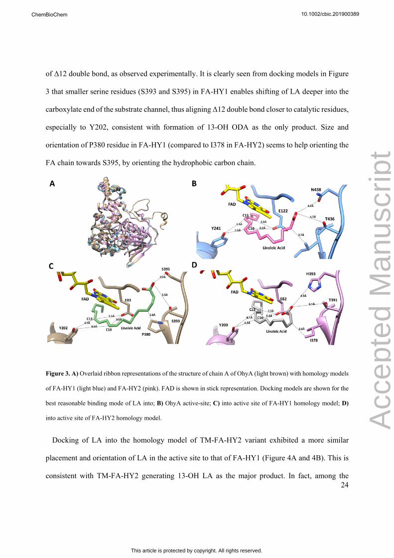
regioselectivity difference between FA-HY1 and FA-HY2, a comparative analysis of the active-
site (within ~5 Å of the substrate and FAD) of the two enzymes was performed. This analysis was
based on the FAD and substrate analog (hexaethylene glycol; P6G) bound structure of the closely
related homolog OhyA[17] (pdb id: 4uir; 62% similarity to FA-HY1; Figure 1). Three amino acids
attracted our attention in this analysis. Two of these residues were equivalent to the amino acids
that were shown to interact with the carboxylate end of the FA substrate in an OA docking model
of OhyA structure[17] (T436 and N438 in OhyA; Figure 1). Apparently, while OhyA and FA-HY2
have identical (T436 vs. T391 in FA-HY2) or similar residues (N438 vs. H393 in FA-HY2) at these
corresponding positions, FA-HY1 had less bulky serine residues at both positions. It was
previously demonstrated that mutation of these two residues to alanine abolished the activity
significantly in OhyA[17]. Moreover, an isoleucine (I423 in OhyA) was identified, which is also an
isoleucine in FA-HY2 (I378), but a proline in FA-HY1 (P380) at corresponding position (Figure
1A). This residue is in close proximity to the carboxylate-anchoring residues of T436 and N438
(OhyA numbering), as well as to the substrate analogue. To evaluate the effect of the three amino
acids on substrate specificity and regiodiversity, these residues on FA-HY2 were mutated to the
corresponding residues on FA-HY1. Single mutations of T391S (T391S-FA-HY2) and H393S
10.1002/cbic.201900389
Acc
epte
d M
anus
crip
t
ChemBioChem
This article is protected by copyright. All rights reserved.

15
(H393S-FA-HY2) were designed as well as a double mutant (DM-FA-HY2;T391S/H393S) and a
triple mutant (TM-FA-HY2; T391S/H393S/I378P). H393Q mutant (H393Q-FA-HY2) was also
generated to further evaluate the plasticity of the residue at this position (Table S2).
Figure 1. A) Partial sequence alignment of OhyA, FA-HY1 and FA-HY2 highlighting the residues (in red boxes)
mutated in this study. B) Location of the residues in the active site of OhyA (pdb id: 4uir) with respect to the substrate
analogue (P6G; ethylene units represented in blue color) and catalytic residues of E122 and Y241. FAD is shown in
yellow. Ribbon representation is shown only for mutated residues and their nearby neighbours. T436 and N438 reside
at the edge of a small loop region between two beta-sheets. I423 resides at the junction of a loop and beta-sheet. C)
Percent identity and similarity levels between OhyA, FA-HY1 and FA-HY2.
Analysis of Substrate Specificity and Regioselectivity. Activities of FA-HY1, FA-HY2 and
mutant enzymes were screened towards 12 FA substrates with various chain lengths and different
degrees of unsaturation (Scheme 2). The results are presented in Figure 2, Table 1 and Tables S10-
S12. Both wild-type and mutant hydratases displayed varying levels of hydration activity towards
10 out of 12 FAs. For trans-vaccenic acid and LA methyl ester, no activity was observed with any
10.1002/cbic.201900389
Acc
epte
d M
anus
crip
t
ChemBioChem
This article is protected by copyright. All rights reserved.

16
of the enzymes, indicating essential requirement of cis-conformation and free carboxylate group of
substrates, consistent with earlier reports[18]. Confirmation for the identity of the products have
been carried out by GC-MS analysis of TMS derivatives and by 1H and 13C NMR of purified
products (Figures S6-S39).
Scheme 2. Structures and names of all fatty acid substrates used in this study.
O
OH
O
OH
O
OH
O
OH
O
OH
O
OH
O
OH
O
OH
O
OH
O
OH
O
OH
O
OCH3
oleic acid (cis-9-octadecanoic acid)
palmitoleic acid (cis-9-hexadecanoic acid)
cis-vaccenic acid (cis-11-octadecanoic acid)
alpha-linolenic acid (cis-9,cis-12,cis-15-octadecatrienoic acid)
gamma-linolenic acid (cis-6,cis-9,cis-12-octadecatrienoic acid)
linoleic acid (cis-9,cis-12-octadecadienoic acid)
arachidonic acid (cis-5,cis-8,cis-11,cis-14-eicosatetraenoic acid)
ETA (cis-8,cis-11,cis-14-eicosatrienoic acid)
EPA (cis-5,cis-8,cis-11,cis-14,cis-17-eicosapentaenoic acid)
trans-vaccenic acid (trans-11-octadecanoic acid)
DHA (cis-4,cis-7,cis-10,cis-13,cis-16,cis-19-docosahexaenoic acid)
Linoleic acid methyl ester (methyl cis-9,cis-12-octadecadienoate)
*
*
*
*
**
**
**
**
**
**
*position of hydroxylation for the products observed.
Comparison of the activities of wild-type FA-HY1 and wild-type FA-HY2 towards OA and cis-
vaccenic acid reveals the significance of double bond position of FA. Both OA and cis-vaccenic
acid are C18:1 FAs with a single double bond in cis configuration; at Δ9 position for OA and at
Δ11 position for cis-vaccenic acid. FA-HY1 converted over 90% of cis-vaccenic acid to 12-OH
product, but had almost 20-fold lower activity towards OA (less than 5% conversion). For FA-
10.1002/cbic.201900389
Acc
epte
d M
anus
crip
t
ChemBioChem
This article is protected by copyright. All rights reserved.
17
HY2, the situation was completely reversed; over 90% conversion with OA, but no detectable
activity towards cis-vaccenic acid (Figure 2A, Table S10).
Mutant enzymes showed remarkable shift from FA-HY2 towards FA-HY1 (Figure 2, S1 and
Table 1). Our results demonstrate significant regioselectivity shift in the case of LA. TM-FA-HY2
can now convert about 60% of substrate into 13-OH product and only 8% into 10-OH, whereas
FA-HY2 almost exclusively produces 10-OH and FA-HY1 exclusively 13-OH (Figure 2B). This
indicates an almost complete reversal of regioselectivity against LA for TM-FA-HY2, shifting ratio
of the regioisomers of HFA products (10-OH:13-OH) from 99:1 to 12:88 (Table 1). DM-FA-HY2
showed almost equal split (54:46) between 10-OH and 13-OH products. Besides, total turnover
numbers were also higher for both DM-FA-HY2 and TM-FA-HY2 compared to FA-HY2, with
50% and 80% increases respectively. Both mutants also had higher total turnover numbers than
FA-HY1 (Table 1).
In the case of palmitoleic acid (POA), three mutations on FA-HY2 decreased the activity from
90% to 5% conversion, bringing down TM-FA-HY2 activity to almost same level as FA-HY1
(4.2% conversion) (Figure 2A). Both regioselectivity and total activity shift were observed in the
case of AA, where TM-FA-HY2 can carry out ~50% total conversion compared to only 2%
conversion by FA-HY2 (Figure 2D and Tables S11-S12). Regioselectivity shift was significant for
DM-FA-HY2 with AA, exhibiting a 47:53 regioisomer ratio (12-OH:15-OH) (Table S12). While
FA-HY2 can only produce 12-OH product from AA, both DM-FA-HY2 and TM-FA-HY2 exhibit
around 5% conversion to 15-OH, which is the major product of FA-HY1. TM-FA-HY2 exhibited
about 15-fold higher conversion of EPA to 12-OH product with respect to FA-HY2 and 5-fold
higher conversion with respect to FA-HY1 under identical conditions. While FA-HY2 is totally
inactive towards DHA, histidine mutants as well as DM-FA-HY2 and TM-FA-HY2 had the same
10.1002/cbic.201900389
Acc
epte
d M
anus
crip
t
ChemBioChem
This article is protected by copyright. All rights reserved.

18
level of activity as FA-HY1 (~2-3%). Hence, just two to three mutations (DM-FA-HY2 and TM-
FA-HY2) were sufficient to convert FA-HY2 to an FA-HY1-like enzyme, without sacrificing much
of their original activity against the conventional hydratase substrates of OA, alpha-linolenic and
gamma-linolenic acids (Figure 2, Tables S10-S11). Among all the variants, only I378P had very
low activities or was totally inactive. However, when this mutation is coupled with T391S and
H393S mutations, activities are rescued and even increased for some of the substrates. It is also
interesting to note that H393Q mutation did not impair the activity of FA-HY2.
Furthermore, di-hydroxy products 10,13-dihydroxy octadecanoic acid, was also observed for LA
when H393S-FA-HY2, DM-FA-HY2 or TM-FA-HY2 are used, although none has been detected
for wild-type enzymes (Figure 2B, S1 and Table S11). Overall conversion levels of LA by these
three mutant enzymes are about 50% higher compared to wild-type enzymes.
Table 1. Impact of double and triple mutations on activity and regioselectivity towards linoleic
acida.
Enzyme 10-OH ODA[b] (% conversion)
13-OH ODA[b] (% conversion)
10,13-di-OHODA[b]
(% conversion)
10-OH/13-OH ratio[c]
TTN[d]
FA-HY1 0 47.9 ± 1.7 0 0:100 190
FA-HY2 40.9 ± 6.8 0.5 ± 0.4 0 99:1 165
DM-FA-HY2
30.5 ± 1.8 26.3 ± 0.4 7.3 ± 0.5 54:46 254
TM-FA-HY2
7.9 ± 0.9 58.7 ± 2.4 8.4 ± 1.9 12:88 298
[a]Assays were carried out by shaking incubation of 4.5 μM of each enzyme and 1.79 mM of linoleic acid and100 μM FAD for 16 hours at 37 °C under anaerobic conditions. [b]10-OH ODA (10-hydroxy-cis-12-octadecenoic acid); 13-OH ODA (13-hydroxy-cis-9-octadecenoic acid) and 10,13-diOH ODA (10,13-dihydroxy octadecanoic acid). [c] Ratio of 10-hydroxy-cis-12-octadecenoic acid to 13-hydroxy-cis-9-octadecenoic acid. [d]TTN: Total turnover number.
10.1002/cbic.201900389
Acc
epte
d M
anus
crip
t
ChemBioChem
This article is protected by copyright. All rights reserved.

19
Figure 2. A) Conversion levels of wild-type and mutant fatty acid hydratases for monounsaturated FA substrates. Error
bars indicate standard error for mean value of at least two separate experiments. Conversion levels of wild-type and
mutant fatty acid hydratases; B) for conversion of linoleic acid substrate into products of 10-OH ODA (10-hydroxy-
cis-12-octadecenoic acid), 13-OH ODA (13-hydroxy-cis-9-octadecenoic acid) and 10,13-diOH ODA (10,13-
dihydroxy octadecanoic acid); C) for conversion of alpha-linolenic acid substrate into products of 10-OH ODDA (10-
hydroxy-cis-12,cis-15-octadecadienoic acid), 13-OH ODDA (13-hydroxy-cis-9,cis-15-octadecadienoic acid); D) for
conversion of arachidonic acid substrate into products of 12-OH ETrA (12-hydroxy-cis-5,cis-8,cis-14-eicosatrienoic
acid), 15-OH ETrA (15-hydroxy-cis-5,cis-8,cis-11-eicosatrienoic acid). Error bars indicate standard error for mean
value of at least two separate experiments.
Preparative Scale Reactions and Structural Identification. In order to evaluate conversion
levels at preparative scale and to isolate and determine structure of new HFA products by NMR,
select reactions were set up in 100 mL scale with a quantity of 50-100 mg (0.15-0.35 mmol scale)
of substrates and with a concentration of 0.1 mg/mL-0.5 mg/mL (1.5-9.0 μM) of enzymes.
0
20
40
60
80
100
cis-vaccenic acid product (12-OH)palmitoleic acid product (10-OH)
oleic acid product (10-OH)
FA-HY2
% C
onve
rsio
n o
f Sub
stra
te
T391S H393S H393Q I378P DM TM FA-HY1
0
20
40
60
80
10010-OH ODDA13-OH ODDA
% c
on
vers
ion
(a
lph
a-l
inol
en
ic a
cid
)
FA-HY2 T391S H393S H393Q I378P DM TM FA-HY1
0
20
40
60
80
100 10-OH ODA13-OH ODA10,13-diOH ODA
% c
on
vers
ion
(lin
olei
c a
cid)
FA-HY2 T391S H393S H393Q I378P DM TM FA-HY1
A B
C D
0
20
40
60
80
100 12-OH ETrA
15-OH ETrA
% c
on
vers
ion
(ar
ach
ido
nic
aci
d)
FA-HY2 T391S H393S H393Q I378P DM TM FA-HY1
10.1002/cbic.201900389
Acc
epte
d M
anus
crip
t
ChemBioChem
This article is protected by copyright. All rights reserved.

20
Reactions were carried out for ~24 hours at 37 °C. TM-FA-HY2 variant exhibited conversion levels
of 92% for ETA as substrate and 67% for EPA as substrate. After extraction and purification by
preparative TLC, 12-OH product of ETA ((S)-12-hydroxy-cis-8, cis-14-eicosadienoic acid) was
isolated with a 53% yield and 12-OH product of EPA ((S)-12-hydroxy-cis-5,cis-8,cis-14,cis-17-
eicosatetraenoic acid) was isolated with a yield of 31%. Both products were in (S)-configuration
with an enantiomeric excess (% ee) of at least 95%, as determined by chiral GC analysis and by
Mosher Ester method (Figures S40, S42). Preparative scale reactions were also carried out using
TM-FA-HY2 with AA and OA as substrates and using H393S with LA as substrate, in addition to
conversions with FA-HY1 (AA and LA) and with FA-HY2 (LA and OA). Varying conversion
levels were obtained, from 5% to 99% (Table S7). 13-OH LA product from FA-HY1 reaction and
10-OH LA product from H393S reaction were isolated in pure form with yields of 29% and 13%,
respectively, and with high enantiopurities (>95% ee). While 10-OH LA product was in (S)-
configuration, 13-OH LA was determined to be in (R)-configuration due to change in Cahn-Ingold-
Prelog priority rules upon double bond positions. Majority of the purified HFA products had a
purity of >95% as determined by GC-FID analysis. Structural analysis of purified samples were
confirmed by NMR (1H and 13C) analysis (Figures S7-S39). Absolute configuration of new
products were determined by Mosher Ester method (Tables S2-S6; Figures S40-S46). Details of
the conversion and yield analysis of all reactions are given in Table S7.
Steady-state Kinetics. In order to gain more insight into the activity differences between wild-
type and mutant enzymes, kcat and apparent Km values towards selected substrates were measured
(Table 2, Figures S2-S5). FA-HY1 is almost 4-orders of magnitude catalytically more efficient
(based on kcat/Km value) compared to FA-HY2 with LA as substrate (12900 vs. 2.6). This is
particularly due to the significant difference in kcat values (340 min-1 vs. 0.36 min-1). Km values are
10.1002/cbic.201900389
Acc
epte
d M
anus
crip
t
ChemBioChem
This article is protected by copyright. All rights reserved.

21
comparable within error percentage indicating that both enzymes can bind LA with similar affinity.
Moreover, double and triple mutations on FA-HY2 increased kcat significantly (about 10 to 20-fold)
for LA with respect to FA-HY2, which should also be responsible for the increased overall yield
(vide supra). However, no significant difference was observed for Km, again suggesting that
binding affinity is not affected.
Table 2. Steady-state kinetic parameters[a] for FA-HY1, FA-HY2 and selected mutants.
Enzyme Substrate Product[b,c] kcat (1/min) Km (mM) kcat/Km
(1/mMꞏmin)
FA-HY1 LA 13-OH ODA 340 ± 41 0.026 ± 0.008
12900 ± 2300
FA-HY2 LA 10-OH ODA 0.36 ± 0.06 0.139 ± 0.078
2.6 ± 1.0
DM-FA-HY2
LA (13-OH ODA +10 OH ODA)
3.04 ± 0.34 0.057 ± 0.018
54.0 ± 13.0
TM-FA-HY2
LA (13-OH ODA +10 OH ODA)
5.81 ± 0.71 0.036 ± 0.015
160 ± 50
FA-HY1 OA 10-OH SA 0.026 ± 0.001
0.028 ± 0.009
0.93 ± 0.25
FA-HY2 OA 10-OH SA 7.12 ± 0.65 0.020 ± 0.008
360 ± 110
TM-FA-HY2
OA 10-OH SA 0.69 ± 0.07 0.016 ± 0.007
43.0 ± 15.0
FA-HY1 POA 10-OH PA 0.061 ± 0.007
0.376 ± 0.095
0.16 ± 0.02
FA-HY2 POA 10-OH PA 0.89 ± 0.10 0.046 ± 0.017
19.0 ± 6.0
DM-FA-HY2
POA 10-OH PA 0.063 ± 0.003
0.030 ± 0.007
2.1 ± 0.4
TM-FA-HY2
POA 10-OH PA 0.017 ±0.001 0.039 ± 0.007
0.42 ± 0.07
FA-HY1 AA (12-OH ETrA+15 OH ETrA)
5.7 ± 0.6 0.076 ± 0.023
75.0 ± 17.0
TM-FA-HY2
AA (12-OH ETrA+15 OH ETrA)
0.89 ± 0.06 0.020 ± 0.007
45.0 ±13.0
10.1002/cbic.201900389
Acc
epte
d M
anus
crip
t
ChemBioChem
This article is protected by copyright. All rights reserved.

22
[a]Michaelis-Menten plots are given in Supporting Information. [b]Product abbreviations: 10-OH ODA (10-hydroxy-cis-12-octadecenoic acid); 13-OH ODA (13-hydroxy-cis-9-octadecenoic acid); 10-OH SA (10-hydroxy-stearic acid); 10-OH PA (10-hydroxy palmitic acid); 12-OH ETrA (12-hydroxy-cis-5,cis-8,cis-14-eicosatrienoic acid); 15-OH ETrA (15-hydroxy-cis-5,cis-8,cis-11-eicosatrienoic acid). [c]In the case where two products are formed, the total amount of products was taken into account for kinetic analysis. It was observed that the kinetics for the formation of each product separately were almost identical.
Although long-time conversion levels of both FA-HY2 and TM-FA-HY2 are close to 100% for
OA, kcat/Km for TM-FA-HY2 is about an order of magnitude less compared to that of FA-HY2 and
in a range that is almost half-way between catalytic efficiencies of FA-HY1 and FA-HY2 (Table
2). Similar difference was also observed for POA when FA-HY2 is compared to both DM-FA-
HY2 and TM-FA-HY2. As for AA, FA-HY1 exhibits higher kcat and Km values compared to TM-
FA-HY2. However, catalytic efficiencies are similar (75 vs. 45).
Overall, the effect of mutations is significantly reflected on kcat, but not on Km. All measured Km
values for mutant enzymes are within ~2-fold range of Km values of FA-HY2 for each respective
substrate. However, changes in kcat values, either increase or decrease, are almost an order of
magnitude when DM-FA-HY2 and TM-FA-HY2 are compared to FA-HY2 for all the substrates
tested.
Substrate Docking Studies. For a better understanding of the structural basis of substrate
specificity and regioselectivity of hydratases, binding modes of LA with respect to carboxylate
anchoring and catalytic residues (E82 and Y200 in FA-HY2) were compared. Docking
investigation was performed on the homology models of FA-HY1, FA-HY2 and TM-FA-HY2,
which were based on the structure of OhyA as the template (Figure 3A). OhyA is the only structure
available to date with both FAD and a substrate analog (hexaethylene glycol) bound and is most
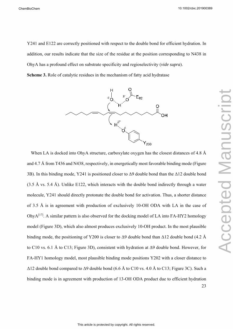
relevant to catalysis[17]. According to the mechanism proposed based on this structure, Y241 (Y200
in FA-HY2) protonates the double bond and E122 (E82 in FA-HY2) activates a water molecule to
attack the resulting carbocation at C10 position[17] (Scheme 3). This mechanism necessitates that
10.1002/cbic.201900389
Acc
epte
d M
anus
crip
t
ChemBioChem
This article is protected by copyright. All rights reserved.
23
Y241 and E122 are correctly positioned with respect to the double bond for efficient hydration. In
addition, our results indicate that the size of the residue at the position corresponding to N438 in
OhyA has a profound effect on substrate specificity and regioselectivity (vide supra).
Scheme 3. Role of catalytic residues in the mechanism of fatty acid hydratase
When LA is docked into OhyA structure, carboxylate oxygen has the closest distances of 4.8 Å
and 4.7 Å from T436 and N438, respectively, in energetically most favorable binding mode (Figure
3B). In this binding mode, Y241 is positioned closer to Δ9 double bond than the Δ12 double bond
(3.5 Å vs. 5.4 Å). Unlike E122, which interacts with the double bond indirectly through a water
molecule, Y241 should directly protonate the double bond for activation. Thus, a shorter distance
of 3.5 Å is in agreement with production of exclusively 10-OH ODA with LA in the case of
OhyA[17]. A similar pattern is also observed for the docking model of LA into FA-HY2 homology
model (Figure 3D), which also almost produces exclusively 10-OH product. In the most plausible
binding mode, the positioning of Y200 is closer to Δ9 double bond than Δ12 double bond (4.2 Å
to C10 vs. 6.1 Å to C13; Figure 3D), consistent with hydration at Δ9 double bond. However, for
FA-HY1 homology model, most plausible binding mode positions Y202 with a closer distance to
Δ12 double bond compared to Δ9 double bond (6.6 Å to C10 vs. 4.0 Å to C13; Figure 3C). Such a
binding mode is in agreement with production of 13-OH ODA product due to efficient hydration
10.1002/cbic.201900389
Acc
epte
d M
anus
crip
t
ChemBioChem
This article is protected by copyright. All rights reserved.
24
of Δ12 double bond, as observed experimentally. It is clearly seen from docking models in Figure
3 that smaller serine residues (S393 and S395) in FA-HY1 enables shifting of LA deeper into the
carboxylate end of the substrate channel, thus aligning Δ12 double bond closer to catalytic residues,
especially to Y202, consistent with formation of 13-OH ODA as the only product. Size and
orientation of P380 residue in FA-HY1 (compared to I378 in FA-HY2) seems to help orienting the
FA chain towards S395, by orienting the hydrophobic carbon chain.
Figure 3. A) Overlaid ribbon representations of the structure of chain A of OhyA (light brown) with homology models
of FA-HY1 (light blue) and FA-HY2 (pink). FAD is shown in stick representation. Docking models are shown for the
best reasonable binding mode of LA into; B) OhyA active-site; C) into active site of FA-HY1 homology model; D)
into active site of FA-HY2 homology model.
Docking of LA into the homology model of TM-FA-HY2 variant exhibited a more similar
placement and orientation of LA in the active site to that of FA-HY1 (Figure 4A and 4B). This is
consistent with TM-FA-HY2 generating 13-OH LA as the major product. In fact, among the
10.1002/cbic.201900389
Acc
epte
d M
anus
crip
t
ChemBioChem
This article is protected by copyright. All rights reserved.

25
binding modes generated by the docking program, one more plausible binding mode can also be
found, which aligns Y200 better with Δ9 double bond (4.4 Å to C10) while still keeping it close
to Δ12 double bond, with a distance of 5.0 Å (Figure 4C). Such a binding mode will explain
formation of 10-OH and 10,13-diOH as the minor products. This suggests that TM-FA-HY2 might
be attaining both binding modes.
Figure 4. A) Overlaid active site structures of FA-HY1, FA-HY2 and TM-FA-HY2 homology models. For TM-FA-
HY2, the docking model in Figure 4B is shown. B) Docking model showing LA in the active site of TM-FA-HY2
homology model in the best reasonable binding mode for C13 hydroxylation. C) Docking model showing LA in the
active site of TM-FA-HY2 homology model in the best reasonable binding mode for C10 hydroxylation.
Conversion of Microalgae FA into HFAs by Hydratases. Microalgae has great potential as a
sustainable source of various FAs[26]. Different microalgae species can accumulate a diverse range
of saturated and unsaturated long-chain FAs (C14-C22 mainly) that can reach up to 5-15% of their
dry biomass depending on cultivation conditions[27]. Nannochloropsis gaditana and Pavlova
10.1002/cbic.201900389
Acc
epte
d M
anus
crip
t
ChemBioChem
This article is protected by copyright. All rights reserved.

26
lutheri biomass were used to show the proof-of-concept for sustainable conversion of microalgae
FAs into HFAs by hydratases. N. gaditana has high content of EPA and POA as well as lower
levels of AA, DHA and C18 unsaturated FAs. P. lutheri builds up significant amounts of EPA,
DHA and POA (Figure 5A, Tables S13-S14).
Assays were carried out similar to the long-time conversion assays (vide supra). However,
instead of using pure FA solutions as substrates, total FA hydrolysate of the lipids extracted from
microalgae biomass were used. The results show that wild-type and mutant enzymes can produce
HFAs from FAs of microalgae extract almost as efficiently as from pure FA substrates and in some
cases even with better yields (Figure 5, Tables S15-S16). Moreover, regioselectivity patterns of the
enzymes are retained. There are some noticeable differences for the conversion of some FAs. For
example, while POA conversion is about 5% with FA-HY1 and TM-FA-HY2 using pure substrate,
the conversion level increased to 35-50% with microalgae extracts (Figure 5B). However,
conversion of OA and POA with FA-HY2 were decreased from ~90-95% with pure substrates to
20-45% with microalgae. GC chromatograms of FA-HY2 reaction exhibited an additional peak
that elutes close to OA peak (~60% of the starting substrate amount). According to GC-MS
analysis, this peak can be attributed to trans-isomer of the substrate (trans-10-octadecenoic acid),
as shown previously for LA[18a]. Although this peak was also observed with pure OA at much lower
levels (less than 5%), the extent of the reverse reaction generating trans-isomer is higher with
microalgae.
Another significant difference is the increased conversion levels of EPA from microalgae extract
in comparison to pure EPA under identical assay conditions. All enzymes, except FA-HY2, became
much more efficient for the conversion of EPA to 12-OH product (Figure 5D). When P. lutheri
extract (which contains ~22% of EPA) was used as a substrate, conversion of EPA to 12-OH
10.1002/cbic.201900389
Acc
epte
d M
anus
crip
t
ChemBioChem
This article is protected by copyright. All rights reserved.

27
product with ~40% yield can be achieved with TM-FA-HY2, compared to 6% conversion with
pure substrate. Similar pattern was also observed with N. gaditana, although the analysis was
complicated with peak overlap of 12-OH product of EPA and 15-OH product of AA. A less
pronounced, but a similar increase in conversion was also observed for DHA, especially for P.
lutheri extract (Figure 5C). Higher conversion levels were also obtained for 10,13-dihydroxy-
octadecanoic acid production from LA (Figure 5F). Conversion profiles with other FAs were
mostly similar to pure substrate conversion profiles (Figure 5E, Tables S15-S16).
It is important to note that microalgae FA extract contains 25-30% saturated FAs. One might
consider that such a high concentration of saturated FAs could have inhibitory effects through non-
productive binding at the active site. However, no significant inhibition for the production of HFAs,
at least in our assay time-scale, has been observed. Our results suggest that microalgae can be used
as a convenient source to produce a variety of HFA products and even more effectively than pure
substrates in the case of long chain PUFAs.
10.1002/cbic.201900389
Acc
epte
d M
anus
crip
t
ChemBioChem
This article is protected by copyright. All rights reserved.

28
10.1002/cbic.201900389
Acc
epte
d M
anus
crip
t
ChemBioChem
This article is protected by copyright. All rights reserved.

29
Figure 5. A) FA profiles (weight percentage) of P. lutheri and N. gaditana, showing the major FAs present in the FA
extract used for assays. PA, palmitic acid; SDA, stearidonic acid. Comparison for the substrate conversion levels of;
B) palmitoleic acid; C) DHA; D) EPA; and E and F) LA, when either pure substrate or microalgae extract were used
for enzymatic conversion with FA-HY1, FA-HY2, H393S-FA-HY2 and TM-FA-HY2 enzymes. Assays were carried
out for 16 hours at 37 °C by shaking at 110 rpm under anaerobic conditions. Enzyme concentration was 0.3 mg/mL
and substrate concentration was 0.5 mg/mL for pure substrate and 1 mg/mL for extracts (total FA concentration), in a
total volume of 1 mL. Percentages of each fatty acid in the total extract are given in Supporting Information (Tables
S13 and S14).
DISCUSSION
In this study, we report comparison of activity, kinetics, substrate specificity and regioselectivity
of FA-HY1, FA-HY2 and FA-HY2 mutants, where critical active-site residues of FA-HY2 were
swapped to the corresponding residues of FA-HY1. Our results clearly demonstrate that three
amino acid residues, carboxylate-anchoring residues of T391, H393 and an unexpected nearby
residue I378, have remarkable impact for modulating substrate specificity and regioselectivity.
Among single residue mutants, changes in activity and regioselectivity were observed with H393S-
FA-HY2 mutant whereas T391S-FA-HY2 was similar to FA-HY2, which can be attributed to
larger size of histidine with respect to threonine. In most cases, combination of mutations yielded
a synergistic effect. Especially, DM-FA-HY2 and TM-FA-HY2 exhibited remarkable
regioselectivity shift with LA. In addition, both mutants exhibited much higher activity levels with
C20 FAs. Besides, no substantial loss in the activity of mutants (except I378P) towards OA, alpha-
and gamma-linolenic acids was observed. Furthermore, kinetic results suggests that although
substrate binding is not affected to a great extent, positioning of the substrate with respect to
catalytic residues leads to differences in activity levels.
10.1002/cbic.201900389
Acc
epte
d M
anus
crip
t
ChemBioChem
This article is protected by copyright. All rights reserved.
30
Preparative scale reactions showed varying levels of conversion and isolated yields (Table S7).
We observed that conversions with some of the substrates, especially EPA and ETA, were much
higher than the small scale (1 mL) activity assays. Although this can be partly attributed to
increased enzyme concentration and reaction time, other effects such as more efficient mass
transfer in large shake flasks vs. small test tubes, should also be responsible. Dispersion of poorly
soluble long chain fatty acids in aqueous media are apparently enhanced by quality of mixing
leading to efficient accessibility of the substrate to the enzyme. 12-OH product of ETA and 12-OH
product of EPA, two new products only accessible by TM-FA-HY2 at a reasonable level, has been
converted at 92% and 69%, respectively. These levels are significantly higher than small scale
assays, which exhibited about 6-7% conversion. 12-OH product of EPA was isolated with 31%
yield and 12-OH product of ETA was isolated with 53% yield, both with excellent regioselectivity
and high stereoselectivity. Wild-type FA-HY2 exhibited 98.5% conversion and 82% isolated yield
for OA, while wild-type FA-HY1 converted 80% of LA to 13-OH substrate and an isolated yield
of 29% was achieved. These results show that various HFA products can be prepared from wild-
type and mutant enzymes at preparative scale and isolated with good yields in enantiopure form.
Since HFA derivatives of C20 FAs are important lipid mediator molecules with anti-inflammatory
activity and therapeutic potential[5a], further engineering of hydratases can contribute to green
stereoselective routes for enzymatic synthesis of desired hydroxy derivatives of EPA and AA for
medical research. Moreover, all C18 and C20 HFAs produced by wild-type and mutant enzymes
can be tested for authentic biological activity alone or as components of FAHFAs[4c], as well as for
their physico-mechanical properties for various bioplastic applications[9a].
Based on our results, it can be concluded that less bulky carboxylate-anchoring residues (serines
vs. threonine and histidine) will possibly pull the FA chain more towards the carboxylate end of
10.1002/cbic.201900389
Acc
epte
d M
anus
crip
t
ChemBioChem
This article is protected by copyright. All rights reserved.

31
the channel, with proline 380 helping by pushing the hydrophobic FA chain towards serine
residues. This apparently leads to a poorer alignment of the double bonds in closer proximity to the
carboxylate (such as Δ9 and Δ11), but a better alignment of the distant double bonds (such as Δ12,
Δ13 and Δ14), with respect to catalytic residues (glutamate and tyrosine). In addition, longer active-
site cavity with smaller carboxylate-anchoring residues enable acceptance of C20 FAs as efficient
substrates. Structure of the substrate also seems to be important besides the size of the channel.
This is well demonstrated in the completely swapped activity pattern of FA-HY2 and FA-HY1
towards OA and cis-vaccenic acid (vide supra). Our substrate docking studies with homology
models of wild-type enzymes and TM-FA-HY2 support our experimental observations regarding
positioning of catalytic residues with respect to double bond.
In cases where multiple products are generated, two scenarios can be considered. First, two
different binding modes might be in equilibrium, leading to a distinct product for each binding
mode. Other possibility is that there is only one dominant binding mode, but the catalytic residues
are sufficiently close to multiple double bond positions to carry out hydration to generate multiple
products. Both scenarios are well demonstrated in LA docking models of TM-FA-HY2 (Figure 4B
and 4C).
An interesting result from our studies is that I378P mutation almost completely abolished the
activity of FA-HY2 towards all substrates. I378 resides at the junction of a beta-sheet and a loop
region, which is close to carboxylate-anchoring residues and the substrate. One possibility is that
I378P mutation may cause a local conformational change due to sterical restrictions of proline[28],
that is detrimental to activity. However, our homology models with TM-FA-HY2 and with FA-
HY1 shows that mutated proline and its surrounding retains the same position as in wild-type FA-
HY2. In this model, due to sterical restrictions, proline had to point directly towards FA chain,
10.1002/cbic.201900389
Acc
epte
d M
anus
crip
t
ChemBioChem
This article is protected by copyright. All rights reserved.
32
making it closer to the carboxylate end of the substrate channel compared to isoleucine (Figure
4A). This possibly indicates that proline is involved in proper orientation of the carboxylate end of
FA substrate in the larger space created by smaller serine residues. Loss of activity upon I378P
mutation can very well be rescued in TM-FA-HY2 variant. In fact, a sequence comparison of
multiple FAH homologs from HFam2 family (1188 homologs, including FA-HY1 and FA-HY2)
of Hydratase Engineering Database[15c] indicates that homologs with a proline residue instead of
an isoleucine mostly accommodate two serines at carboxylate anchoring positions, or at least one
serine corresponding to N438 position.
Recently, two studies were published where engineering of the fatty acid hydratase OhyA
enabled hydration of carbon-carbon double bonds in non-FA substrates. In the first study, Hauer
and coworkers demonstrated hydration of short to medium chain 1-alkenes (C5 to C10) using a
combined approach of semi-rational engineering and decoy molecule strategy[11c]. When
heptadecanoic acid or similar chain length acids were used as decoy molecules (to activate
enzyme), 1-decene was hydrated efficiently by the wild-type enzyme to produce (S)-2-decanol.
However, wild-type enzyme had very low or no activity against shorter chain 1-alkenes (C5-C8).
Site-saturation mutagenesis of Alanine 248 residue (A248), which points against the omega end of
the natural FA substrate, revealed variants that can efficiently hydrate shorter chain 1-alkenes.
Mutants with larger residues of leucine or tryptophan (A248L and A248W) were able to efficiently
hydrate 1-heptene and 1-hexene, respectively, to produce corresponding secondary alcohols. This
has been attributed to pushing effect of the larger residue to shift the alkene substrate more towards
the catalytic residues, resulting in better alignment with the nucleophilic water and catalytic
residues. Moreover, A248L variant was also able to hydrate functionalized alkenes, internal
alkenes, a bulky alkene with an aromatic group and an alkyne. In both FA-HY1 and FA-HY2,
10.1002/cbic.201900389
Acc
epte
d M
anus
crip
t
ChemBioChem
This article is protected by copyright. All rights reserved.

33
A248 corresponds to a charged histidine residue (H209 in FA-HY1 and H207 in FA-HY2). In fact,
A248 in OhyA seems to have a similar role to I378 in FA-HY2 (or P380 in FA-HY1), by pushing
the FA chain to a certain direction within the active site to effect reaction outcome by changing the
alignment of double bonds with catalytic residues. In a more recent study, Pichler and coworkers
performed enzyme engineering also on OhyA, this time on the residues at the carboxylate end of
the substrate[16]. Their purpose was to convert key charged residues on the carboxylate end of the
FA into residues that will interact favorably with FA head groups other than the free carboxylates,
such as esters, amides and alcohols, thus enabling hydration of FA derivatives. Conversion of three
residues into alanines (Q265A/T436A/N438A triple mutant) had a large positive impact on the
hydration activity towards oleic acid esters compared to wild-type OhyA. T436 and N438
correspond to T391 and H393 in FA-HY2, respectively, and mutations into alanine has possibly
opened up more space for bulkier esters as well as created a hydrophobic environment favoring
binding of ester head group. Furthermore, oleamide was also more efficiently hydrated with
Q265S/N438A double mutant. Both these two recent studies and our study reveal the hot spot
residues important for substrate promiscuity. These studies together show that FA hydratases are
promiscuous enzymes which can be efficiently directed for hydration reactions of various FAs as
well as non-FA substrates to generate non-natural products. An iterative engineering approach of
the hot spots at the carboxylate end and at the omega end of FA-HY1, which is already naturally a
promiscuous enzyme, should enable access to new substrates and products. While natural FA
substrates will be advantageous for feasible production of value-added HFAs from cheap abundant
sources of FAs such as microalgae, utilization of non-FA substrates by engineered hydratases will
render new selective synthetic approaches for organic synthesis.
10.1002/cbic.201900389
Acc
epte
d M
anus
crip
t
ChemBioChem
This article is protected by copyright. All rights reserved.

34
This study is a first demonstration of microalgae oil as a sustainable source of FAs for efficient
enzymatic conversion to HFAs. Both wild-type enzymes and mutants can retain most of their
properties observed with pure substrates, including substrate specificity and regioselectivity, when
a complex mixture of FAs from a microalgae extract is supplied as the substrate. Presence of 25-
30% saturated FAs did not seem to inhibit activity to any significant extent. Moreover, conversion
levels of EPA and DHA, two abundant PUFAs in microalgae oil, are even increased for almost all
enzymes compared to activity with pure substrates. Given that no significant hydration activity has
been reported to date against EPA and DHA with a hydratase, our results render promising
opportunities to generate novel HFA derivatives from microalgae as a sustainable source of
omega-3 FAs.
CONCLUSION
Our study provides an important navigation point for generating hydratase variants with desired
activity and selectivity for production of a variety of HFAs for use in applications that range from
monomer production for bioplastics to synthesis of bioactive compounds with high therapeutic
potential. Our TM-FA-HY2 variant is a good representative of this potential with its properties that
are unlike FA-HY1 and FA-HY2. TM-FA-HY2 is the only hydratase to date that can convert AA
to 12-OH FA product. Unlike other hydratases, TM-FA-HY2 can generate three products from LA,
two monohydroxy and one dihydroxy product. Moreover, TM-FA-HY2 exhibited high conversion
rates towards EPA and ETA in preparative scale and enabled isolation of novel 12-OH products
from EPA and ETA with moderate yields, showing the practical potential of the enzyme for
production. Replacement of other amino acid residues at positions targeted in this study by semi-
rational and random enzyme engineering approaches should make it possible to create tailor-made
10.1002/cbic.201900389
Acc
epte
d M
anus
crip
t
ChemBioChem
This article is protected by copyright. All rights reserved.

35
enzymes to access specific HFA products. Optimization of reaction conditions will lead to
sustainable production of desired HFAs from microalgae as a convenient feedstock.
ASSOCIATED CONTENT
Supporting Information
Full scale representative GC chromatogram, Michaelis-Menten plots of all steady-state kinetic
data and steady-state kinetic assay conditions, GC-MS spectra of all hydroxy fatty acid products
reported in this study, NMR spectra of purified products, absolute configuration analysis, details
of preparative scale assays, primer sequences used for site-directed mutagenesis, protein
sequences, average conversion percentages of all substrates tested with standard error values,
fatty acid composition of microalgae oil, average conversion levels of microalgae fatty acids with
standard errors.
AUTHOR INFORMATION
Corresponding Author
*Zheng Guo; e-mail: [email protected]
Author Contributions
The manuscript was written through contributions of all authors. All authors have given approval
to the final version of the manuscript.
Notes
The authors declare no competing financial interest.
10.1002/cbic.201900389
Acc
epte
d M
anus
crip
t
ChemBioChem
This article is protected by copyright. All rights reserved.

36
ACKNOWLEDGMENT
This work was supported by Novo Nordisk Foundation (grant NNF16OC0021740) and by
AUFF-NOVA from Aarhus Universitets Forskningsfond (AUFF-E-2015-FLS-9-12).
Keywords: Protein Engineering, Enzyme Catalysis, Fatty Acid Hydratase, Hydroxy Fatty Acid,
Microalgae
REFERENCES
[1] a) J. Ogawa, Eur. J. Lipid Sci. Technol. 2015, 117, 577-578; b) P. Bergamo, D. Luongo, J. Miyamoto, E. Cocca, S. Kishino, J. Ogawa, S. Tanabe, M. Rossi, J. Funct. Foods 2014, 11, 192-202.
[2] a) K. R. Kim, D. K. Oh, Biotechnol. Adv. 2013, 31, 1473-1485; b) A. Hiseni, I. W. C. E. Arends, L. G. Otten, ChemCatChem 2015, 7, 29-37; c) A. Beopoulos, J. Verbeke, F. Bordes, M. Guicherd, M. Bressy, A. Marty, J. M. Nicaud, Appl. Microbiol. Biotechnol. 2014, 98, 251-262; d) D. G. Hayes, J. Am. Oil Chem. Soc. 2004, 81, 1077-1103.
[3] J. Miyamoto, T. Mizukure, S. B. Park, S. Kishino, I. Kimura, K. Hirano, P. Bergamo, M. Rossi, T. Suzuki, M. Arita, J. Ogawa, S. Tanabe, J. Biol. Chem. 2015, 290, 2902-2918.
[4] a) M. M. Yore, I. Syed, P. M. Moraes-Vieira, T. Zhang, M. A. Herman, E. A. Homan, R. T. Patel, J. Lee, S. Chen, O. D. Peroni, A. S. Dhaneshwar, A. Hammarstedt, U. Smith, T. E. McGraw, A. Saghatelian, B. B. Kahn, Cell 2014, 159, 318-332; b) O. Kuda, M. Brezinova, M. Rombaldova, B. Slavikova, M. Posta, P. Beier, P. Janovska, J. Veleba, J. Kopecky, E. Kudova, T. Pelikanova, J. Kopecky, Diabetes 2016, 65, 2580-2590; c) B. Kahn, M. Yore, I. Syed, P. Moraes-Vieira, T. Zhang, M. Herman, E. Homan, J. Lee, S. Chen, O. Peroni, A. Hammarstedt, R. Patel, T. McGraw, U. Smith, A. Saghatelian, FASEB J. 2016, 30, 504.503-504.503.
[5] a) M. L. Sulciner, C. N. Serhan, M. M. Gilligan, D. K. Mudge, J. Chang, A. Gartung, K. A. Lehner, D. R. Bielenberg, B. Schmidt, J. Dalli, E. R. Greene, Y. Gus-Brautbar, J. Piwowarski, T. Mammoto, D. Zurakowski, M. Perretti, V. P. Sukhatme, A. Kaipainen, M. W. Kieran, S. Huang, D. Panigrahy, J. Exp. Med. 2018, 215, 115-140; b) M. G. Duvall, B. D. Levy, Eur. J. Pharmacol. 2016, 785, 144-155; c) G. Fredman, M. Spite, Mol. Aspects Med. 2017, 58, 65-71; d) M. Spite, J. Claria, C. N. Serhan, Cell Metab. 2014, 19, 21-36; eC. N. Serhan, Nature 2014, 510, 92-101.
[6] a) B. A. Black, C. Sun, Y. Y. Zhao, M. G. Ganzle, J. M. Curtis, J. Agric. Food Chem. 2013, 61, 5338-5346; b) B. A. Black, E. Zannini, J. M. Curtis, M. G. Ganzle, Appl. Environ. Microbiol. 2013, 79, 1866-1873.
[7] N. d. L. da Silva, M. R. W. Maciel, C. B. Batistella, R. M. Filho, in Twenty-Seventh Symposium on Biotechnology for Fuels and Chemicals (Eds.: J. D. McMillan, W. S. Adney, J. R. Mielenz, K. T. Klasson), Humana Press, Totowa, NJ, 2006, pp. 405-414.
10.1002/cbic.201900389
Acc
epte
d M
anus
crip
t
ChemBioChem
This article is protected by copyright. All rights reserved.
37
[8] T. Mi, Y. Dong, U. Santhanam, N. Huang, Exp. Dermatol. 2018, 0. [9] a) C. Liu, F. Liu, J. Cai, W. Xie, T. E. Long, S. R. Turner, A. Lyons, R. A. Gross,
Biomacromolecules 2011, 12, 3291-3298; b) S. Huf, S. Krügener, T. Hirth, S. Rupp, S. Zibek, Eur. J. Lipid Sci. Technol. 2011, 113, 548-561; c) T. Tran, P. Kumar, H.-R. Kim, C. Hou, B. Kim, Polymers 2018, 10, 927.
[10] D. S. Ogunniyi, Bioresour. Technol. 2006, 97, 1086-1091. [11] a) V. Resch, U. Hanefeld, Catal. Sci. Technol. 2015, 5, 1385-1399; b) C. N. Cornell, M. S.
Sigman, Inorg. Chem. 2007, 46, 1903-1909; c) R. M. Demming, S. C. Hammer, B. M. Nestl, S. Gergel, S. Fademrecht, J. Pleiss, B. Hauer, Angew. Chem., Int. Ed. Engl. 2019, 58, 173-177.
[12] M. Engleder, H. Pichler, Appl. Microbiol. Biotechnol. 2018, 102, 5841-5858. [13] A. J. Boersma, D. Coquiere, D. Geerdink, F. Rosati, B. L. Feringa, G. Roelfes, Nat. Chem.
2010, 2, 991-995. [14] S.-Q. Wang, Z.-W. Wang, L.-C. Yang, J.-l. Dong, C.-Q. Chi, D.-N. Sui, Y.-Z. Wang, J.-G.
Ren, M.-Y. Hung, Y.-Y. Jiang, J. Mol. Catal. A: Chem. 2007, 264, 60-65. [15] a) L. E. Bevers, M. W. Pinkse, P. D. Verhaert, W. R. Hagen, J. Bacteriol. 2009, 191, 5010-
5012; b) R. M. Demming, M. P. Fischer, J. Schmid, B. Hauer, Curr. Opin. Chem. Biol. 2018, 43, 43-50; c) J. Schmid, L. Steiner, S. Fademrecht, J. Pleiss, K. B. Otte, B. Hauer, J. Mol. Catal. B: Enzym. 2016, 133, S243-S249; d) R. M. Demming, K. B. Otte, B. M. Nestl, B. Hauer, ChemCatChem 2017, 9, 758-766; e) K. Zorn, I. Oroz-Guinea, H. Brundiek, U. T. Bornscheuer, Prog. Lipid Res. 2016, 63, 153-164.
[16] M. Engleder, G. A. Strohmeier, H. Weber, G. Steinkellner, E. Leitner, M. Muller, D. Mink, M. Schurmann, K. Gruber, H. Pichler, Angew. Chem., Int. Ed. Engl. 2019.
[17] M. Engleder, T. Pavkov-Keller, A. Emmerstorfer, A. Hromic, S. Schrempf, G. Steinkellner, T. Wriessnegger, E. Leitner, G. A. Strohmeier, I. Kaluzna, D. Mink, M. Schurmann, S. Wallner, P. Macheroux, K. Gruber, H. Pichler, ChemBioChem 2015, 16, 1730-1734.
[18] a) A. Hirata, S. Kishino, S. B. Park, M. Takeuchi, N. Kitamura, J. Ogawa, J. Lipid Res. 2015, 56, 1340-1350; b) K. R. Kim, H. J. Oh, C. S. Park, S. H. Hong, J. Y. Park, D. K. Oh, Biotechnol. Bioeng. 2015, 112, 2206-2213.
[19] C. S. Chaurasia, T. D. Williams, C. M. Judson, R. P. Hanzlik, J. Mass Spectrom. 1995, 30, 1018-1022.
[20] T. R. Hoye, C. S. Jeffrey, F. Shao, Nat. Protoc. 2007, 2, 2451-2458. [21] R. R. Guillard, in Culture of Marine Invertebrate Animals (Eds.: W. L. Smith, M. H.
Chanley), Springer, Boston, MA, 1975, pp. 29-60. [22] E. F. Pettersen, T. D. Goddard, C. C. Huang, G. S. Couch, D. M. Greenblatt, E. C. Meng,
T. E. Ferrin, J. Comput. Chem. 2004, 25, 1605-1612. [23] B. Webb, A. Sali, Curr. Protoc. Bioinformatics 2014, 47, 5 6 1-32. [24] S. C. Lovell, I. W. Davis, W. B. Arendall, 3rd, P. I. de Bakker, J. M. Word, M. G. Prisant,
J. S. Richardson, D. C. Richardson, Proteins 2003, 50, 437-450. [25] O. Trott, A. J. Olson, J. Comput. Chem. 2010, 31, 455-461. [26] a) P. W. Behrens, D. J. Kyle, J. Food Lipids 1996, 3, 259-272; b) D. Gangl, J. A. Zedler,
P. D. Rajakumar, E. M. Martinez, A. Riseley, A. Wlodarczyk, S. Purton, Y. Sakuragi, C. J. Howe, P. E. Jensen, C. Robinson, J. Exp. Bot. 2015, 66, 6975-6990.
[27] a) J. Camacho-Rodriguez, M. C. Ceron-Garcia, J. M. Fernandez-Sevilla, E. Molina-Grima, Algal Res. 2015, 11, 63-73; b) J. P. Schwarzhans, D. Cholewa, P. Grimm, U. Beshay, J. M.
10.1002/cbic.201900389
Acc
epte
d M
anus
crip
t
ChemBioChem
This article is protected by copyright. All rights reserved.

38
Risse, K. Friehs, E. Flaschel, J. Appl. Phycol. 2015, 27, 1389-1399; c) H. Safafar, M. Z. Hass, P. Moller, S. L. Holdt, C. Jacobsen, Mar. Drugs 2016, 14.
[28] M. W. MacArthur, J. M. Thornton, J. Mol. Biol. 1991, 218, 397-412.
Table of Contents Entry (for Layout 1 – 5,5 cm x 5,0 cm)
Text for Table of contents:
Promiscuity dictated by a few residues: Comparative analysis of the active site residues of two
Fatty Acid Hydratases from L. Acidophilus, which have high sequence homology but distinct
substrate specificity and regioselectivity, reveal three critical amino acids that play remarkable role
in directing substrate specificity and regioselectivity of hydration.
10.1002/cbic.201900389
Acc
epte
d M
anus
crip
t
ChemBioChem
This article is protected by copyright. All rights reserved.