Embed Size (px)
Citation preview

Instructions for use
Title Rapidly progressive cervical myelopathy had a high risk of developing deep venous thrombosis : a prospectiveobservational study in 289 cases with degenerative cervical spine disease
Author(s) Yamada, Katsuhisa; Suda, Kota; Matsumoto Harmon, Satoko; Komatsu, Miki; Ushiku, Chikara; Takahata, Masahiko;Minami, Akio; Iwasaki, Norimasa
Citation Spinal cord, 57(1), 58-64https://doi.org/10.1038/s41393-018-0213-9
Issue Date 2019-01
Doc URL http://hdl.handle.net/2115/73804
Type article (author version)
File Information Spinal_cord_57_58.pdf
Hokkaido University Collection of Scholarly and Academic Papers : HUSCAP
DVT in degenerative cervical spine disease
1
Rapidly progressive cervical myelopathy had a high risk of developing deep venous
thrombosis: a prospective observational study in 289 cases with degenerative cervical
spine disease
Running title DVT in degenerative cervical spine disease
Katsuhisa Yamada,1,2 Kota Suda,1 Satoko Matsumoto Harmon,1 Miki Komatsu,1 Chikara
Ushiku,1 Masahiko Takahata,2 Akio Minami,1 Norimasa Iwasaki2
1 Hokkaido Spinal Cord Injury Center, Bibai, Hokkaido, Japan
2 Department of Orthopaedic Surgery, Faculty of Medicine and Graduate School of Medicine,
Hokkaido University, Sapporo, Hokkaido, Japan
Correspondence: Katsuhisa Yamada, MD, PhD
Hokkaido Spinal Cord Injury Center, Higashi-4 Minami-1, Bibai, Hokkaido 072-0015, Japan
Tel: 81-126-63-2151, Fax: 81-126-63-2853, E-mail: [email protected]
DVT in degenerative cervical spine disease
2
Abstract
Study Design: A prospective observational study.
Objectives: To determine the incidence of deep venous thrombosis (DVT) and to
evaluate the risk factors for DVT development associated with degenerative cervical spine
disease.
Setting: Hokkaido Spinal Cord Injury Center, Japan.
Methods: Between April 2008 and March 2015, patients with degenerative cervical
spine disease, such as compressive myelopathy or radiculopathy, who underwent surgical
treatment were prospectively assessed. Leg vein ultrasonography and D-dimer tests were
performed preoperatively and at 4 days after surgery. All patients received treatment with
intermittent pneumatic compression and elastic stockings for primary DVT prophylaxis. No
anticoagulation medications were used for DVT prophylaxis.
Results: A total of 289 patients (203 males, 86 females; median age: 67 years
(interquartile range, 58 to 76)) were included. Nine patients (3.1%) exhibited DVT during the
perioperative period. All 9 cases were women who had distal DVT. The incidences of
preoperative and postoperative DVT were 1.1% and 2.1%, respectively. The univariate
analysis showed that statistically significant risk factors for perioperative DVT included
female gender (P < 0.01), advanced age (P = 0.04), a low Japanese Orthopaedic Association
score (P = 0.03), rapidly progressive myelopathy (P < 0.01) and inability to walk (P = 0.01).
DVT in degenerative cervical spine disease
3
The multivariate analysis showed that rapidly progressive myelopathy (P = 0.04) was the
most important risk factor.
Conclusion: Female gender and rapidly progressive myelopathy are high-risk factors that
predict the development of DVT during the perioperative period of cervical spine surgery.
This result indicates that screening and treatment for DVT are needed in such high-risk
patients.
DVT in degenerative cervical spine disease
4
INTRODUCTION
Deep venous thrombosis (DVT) is a potentially fatal complication because of the risk of
pulmonary thromboembolism development.[1, 2] Prevention, early detection and timely
treatment of DVT are very important during the perioperative period of spinal surgery [3].
According to various reports, the incidence of DVT in patients who undergo spinal surgery
ranges from 0.3% to 31%.[4-7] The incidence of DVT has been reported to vary depending
on the type of disease, spinal level, method of surveillance, DVT prophylaxis and
surveillance period.[5, 8]
The incidence of DVT in the patients with cervical spinal cord injury due to trauma
is very high particularly in those unable to walk and with severe paralysis of the lower limbs.
[3] Because patients with cervical degenerative spinal disease often have paralysis of the
lower limbs and walking disabilities, it is very important to evaluate the risk of DVT during
the perioperative period of degenerative cervical spine surgery. Previous studies have
reported that perioperative DVT was usually detected in the period between 3 and 7 days
after spinal surgery.[9, 10] However, few reports have been published on perioperative DVT
in patients with degenerative cervical spine disease without acute traumatic spinal cord injury,
such as compressive myelopathy or radiculopathy. The purpose of this study is to elucidate
the incidence and risk factors for perioperative DVT development by prospectively analyzing
patients who underwent cervical spine surgery for degenerative spinal disease.
DVT in degenerative cervical spine disease
5
METHODS
A total of 761 patients who underwent cervical spine surgery between April 2008 and March
2015 in the Hokkaido Spinal Cord Injury Center were enrolled in this study. The exclusion
criteria consisted of spinal cord injury due to trauma, infection, inflammatory disease,
neoplastic disease and other diseases for which anticoagulation medication was prescribed.
We obtained approval from the institutional review board of Hokkaido Spinal Cord Injury
Center and written informed consent was attained from all patients prior to their enrollment.
Leg vein ultrasonography and D-dimer tests were performed preoperatively and at
the median of 4 days (IQR: 4 to 5, mean: 4.1, standard deviation: 0.54) after surgery. Bilateral
ultrasonography of both legs (SSD2000; ALOKA, Tokyo, Japan, Aplio XG; TOSHIBA,
Tokyo, Japan) was performed by experienced physicians. When DVT was detected before
and/or after surgery, a cardiovascular surgeon was consulted, and anticoagulant therapy was
started using unfractionated heparin and/or warfarin.
Intermittent pneumatic compression with a calf pump and elastic stockings were
used in all cases from induction of general anesthesia to postoperative ambulation. Patients
were allowed to leave the bed using a wheelchair and begin walking the day after the
operation. No anticoagulation medications were used for DVT prophylaxis.
The incidence of DVT was evaluated in relation to the following factors. There was
DVT in degenerative cervical spine disease
6
a total of twelve predictors. The details are as follows:
Patient factors: (i) gender (male or female with male as the reference). (ii) age (years). (iii)
existence of diabetes mellitus (yes/no with no as reference). (iv) existence of cardiovascular
disease (yes/no with no as reference). Disease factors: (i) preoperative Japan Orthopaedic
Association scoring system for the evaluation of cervical myelopathy (JOA score, 0 to 17
points)[11]. (ii) preoperative motor score of the lower limbs (0 to 50 points) as per the
International Standards for Neurological Classification of Spinal Cord Injury, in order to
evaluate the risk factor for DVT development in relation to the paralysis of lower limbs.[12]
(iii) rapidly progression of myelopathy dichotomized (yes/no with no as reference). Rapidly
progression of myelopathy was defined as patients with a JOA score that decreased two
points or more during a recent one-month period.[13] The patients’ severity of myelopathy in
a recent 1-month period was evaluated by clinical interview based on the course of
neurological disorder (ability to walk, ability to go up and down stairs, worsening of fine
motor skills (trouble with handwriting, manipulating buttons or handling chopsticks)), and we
evaluated the difference in JOA score between admission and one month before admission.
(iv) preoperative walking ability dichotomized according to ability to walk with or without
aids (inability to walk, yes/no with no as reference). Walker (inability to walk: no) was
defined as patients who can walk with or without walking aids. Surgery-related factors: (i)
operating time (minutes). (ii) intraoperative blood loss (ml). (iii) surgical approach (posterior
DVT in degenerative cervical spine disease
7
or anterior with anterior as reference). (iv) spinal fusion (yes/no with no fusion as reference).
The results are expressed as the mean ± standard deviation. Statistical analyses were
performed using Fisher’s exact probability test, the Mann-Whitney U test and binomial
logistic regression analysis, as appropriate. The multivariate analyses of the risk factors for
perioperative DVT was performed in the following procedure. 1. initial univariate analyses to
quantify the association between the candidate predictors (a total of 10 predictors: gender,
age, operating time, intraoperative blood loss, JOA score, lower extremity motor score,
rapidly progressive myelopathy, inability to walk, diabetes mellitus, cardiovascular disease)
and DVT. 2. entering all predictors (except gender) and using a stepwise forward selection
method to select a parsimonious set of predictors. (Gender was not appropriate for logistic
analysis because of no male with DVT development.) 3. testing the discrimination and
calibration of the final set of predictors. Discrimination was measured with the use of the area
under the receiver operating-characteristic curve (AUC). We have evaluated calibration by
calculating coefficient determination (R2). 4. providing the odds ratio for the final set of
predictors. When we have sampling zeros (i.e., zero counts), an ad hoc method to estimate the
odds ratio (modified odds ratio) was used, which consisted of adding 0.5 to each cell value. A
receiver operating characteristic (ROC) analysis was conducted to assess the D-dimer cutoff
point. We considered a value of P < 0.05 to be statistically significant throughout all analyses.
DVT in degenerative cervical spine disease
8
RESULTS
Of the 761 patients, 289 patients who underwent cervical surgery for degenerative
spinal disease met the inclusion criteria for this study (203 males, 86 females; median age: 67
years (interquartile range (IQR): 58 to 76). The patients received spinal surgery for cervical
spondylotic myelopathy (n = 197), cervical ossification of the posterior longitudinal ligament
(n = 51), cervical disc herniation (n = 20), cervical spondylotic radiculopathy (n = 16),
cervical kyphosis (n = 4) and cervical spondylosis (n = 1). Elective spine surgery was
performed in 284 patients, and an emergency/sub-emergency operation was performed in 5
patients (4 cases: cervical spinal myelopathy, 1 case: cervical disc herniation).
The overall incidence of DVT was 3.1% (9/289 cases) for cervical spine surgery
patients (Table 1). All 9 cases of positive DVT were women who had distal DVT without
proximal DVT. No patients had clinical signs of DVT. The incidence of preoperative DVT
was 1.1% (3/284 cases, excluding 5 patients who were not examined because an
emergency/sub-emergency operation was performed). The incidence of postoperative DVT
was 2.1% (6/286 cases, excluding 3 cases of preoperative positive DVT). The length of
bedrest after surgery was within 24 hours in all patients.
The univariate analysis for preoperative DVT found that female gender was the only
statistically significant risk factor (P = 0.024, Odds ratio (OR): 17.5, 95% confidence interval
(CI): 0.90 to 343.05) (Table 2). The risk factors related to the surgery, including the operation
DVT in degenerative cervical spine disease
9
time, intraoperative blood loss, surgical approach and spinal fusion, were not significantly
associated with postoperative DVT in the univariate analysis (Table 3). The univariate
analysis for perioperative DVT showed that statistically significant risk factors included
female gender (P < 0.01, OR: 49.9, 95% CI: 2.87 to 867.5), advanced age (P = 0.04, OR per
one year increment: 1.08, 95% CI: 1.00 to 1.16), low JOA score (P = 0.03, OR: 0.03, 95% CI:
0.57 to 0.97), rapidly progressive myelopathy (P < 0.01, OR: 10.4, 95% CI: 2.59 to 41.8) and
inability to walk (P = 0.01, OR: 6.5, 95% CI: 1.51 to 28.0) (Table 4). As the results of
stepwise forward selection, four variables (age, JOA score, rapidly progressive myelopathy
and inability to walk) were chosen as the final set of predictors for the multi-variate analysis,
and the AUC was 0.80 (95% CI: 0.62 to 0.98) and the R2 was 0.16. The multivariate analysis
for perioperative DVT showed that rapidly progressive myelopathy (P = 0.04) was the only
significant risk factor (Table 5).
No statistically significant difference was found between the DVT-positive group
and the DVT-negative group in terms of the preoperative D-dimer level (1.30 ± 0.49 µg/ml vs.
0.93 ± 1.22 µg/ml). Patients with DVT had significantly higher D-dimer levels 4 days after
surgery than those without DVT (5.80 ± 5.44 µg/ml vs. 1.82 ± 1.22 µg/ml; P < 0.01). The
cutoff D-dimer level determined from the ROC curve was 2.67 µg/ml on postoperative day 4.
The sensitivity was 83.3%, and the specificity was 86%.
DVT in degenerative cervical spine disease
10
DISCUSSION
Thromboembolic events are potential complications of cervical spine surgery.[7] The
incidence of DVT in patients who undergo cervical spine surgery has been reported to be
0.5%-4.5%.[5, 7, 14] However, the incidence of DVT development in patients who undergo
cervical degenerative spinal surgery is controversial because previous studies have included
patients with acute traumatic spinal cord injury, spinal tumors, rheumatoid arthritis and
infectious diseases.[5, 7, 14] The present study demonstrated that the perioperative incidence
of DVT development in patients with degenerative cervical spine disease was 3.1%.
The present study showed that patients with rapidly progressive myelopathy have a
high risk of developing DVT in the multivariate analysis. Matsumoto et al. reported that the
incidence of DVT is very high (36.8%) in patients with cervical spinal cord injury due to
trauma.[3] After acute traumatic spinal cord injury, severe paralysis can be accompanied by
reduced venous return, leading to the development of DVT. Furthermore, because a possible
loss of the circulatory circadian variation in hemostatic and fibrinolytic function has also
been suggested in patients with spinal cord injury due to trauma, DVT is quite likely to
develop in the presence of acute traumatic spinal cord injury.[3, 15] Rapidly progressive
myelopathy would cause sympathetic nerve disorder, which leads to reduced vascular
contraction, like a spinal cord injury due to trauma. DVT is likely to develop in the presence
of rapidly progressive myelopathy, even if paralysis is not severe.
DVT in degenerative cervical spine disease
11
Several risk factors have been reported for DVT development in patients undergoing
spinal surgery, such as advanced age, female gender, obesity, neurological deficits, long
operative time and spinal tumors.[6, 8, 16, 17] The present study showed that female gender,
advanced age, rapidly progressive myelopathy, low JOA score and inability to walk were
statistically significant risk factors for periopertive DVT development associated with
degenerative cervical spine disease. Concerning the pathogenesis of DVT, the three major
factors in Virchow’s triad (blood retention, capillary wall disorder and blood coagulopathy)
are well known.[3, 15] Patients with a low JOA score have difficulty walking, leading to
reduced venous return and blood retention. However, the current study demonstrated no
significant difference between the DVT-positive group and the DVT-negative group for the
motor score of the lower limbs. Even if the motor paralysis of the lower limbs was not severe,
patients with myelopathy could not walk because of spasticity and disturbance of the
posterior fasciculus of the spinal cord. These results suggest that walking ability is more
important for DVT development than motor paralysis in patients with cervical spine disease.
Early detection of DVT in the perioperative period of spinal surgery is very
important, however, little information is available regarding the timing of DVT onset, and the
appropriate timing of DVT screening is controversial. In several reports, the peak onset of
DVT is on day 4 after total joint arthroplasty, and the incidence of DVT significantly
decreases on postoperative day 14.[9, 10] Previous studies have reported that the first time
DVT in degenerative cervical spine disease
12
when postoperative DVT may be detected occurs in the period between 3 and 7 days after
spinal surgery.[3, 8, 18-20] Hence, we performed DVT screening using leg vein
ultrasonography preoperatively and at 4 days after surgery. In this study, no patients exhibited
fatal pulmonary thromboembolism development during the perioperative period.
There have been studies to show that the incidence of DVT is relatively lower in
Asians including Japanese as compared to their Western counterparts.[21-23] Medical
prophylaxis for DVT after total knee arthroplasty or total hip arthroplasty has been routinely
performed in our hospital according to the Japanese guideline.[24] However, no
anticoagulation medications have been used for DVT prophylaxis in spine surgery, because
anticoagulation therapy was not recommended in patients undergoing spine surgery and those
with spine injury in Japan. [24] This study showed that the prevalence of DVT in our patients
underwent degenerative cervical spine surgery without chemoprophylaxis was 3.1%. With
risk assessment, proper surveillance and a preventative protocol for DVT prophylaxis, routine
medical prophylaxis may not be necessary in Japanese undergoing elective spine surgery.
D-dimer measurement is predominantly used for DVT screening because the
diagnostic significance of the D-dimer test for predicting DVT is well known, however, the
cutoff level of D-dimer for predicting the risk of developing DVT is controversial for spinal
surgery. Matsumoto et al. set the D-dimer level cutoff point at 5.82 µg/ml on postoperative
day 3 in patients with acute traumatic spinal cord injury who were treated surgically and
DVT in degenerative cervical spine disease
13
reported that the sensitivity and specificity were 72.7% and 76.5%, respectively.[3] Yoshioka
et al. reported that the sensitivity and specificity of D-dimer measurements on day 7 after
spinal surgery were 83.3% and 75.7%, respectively, when the D-dimer cutoff level was set at
6.5 µg/ml.[20] A D-dimer level > 10 µg/ml is reported to be indicative of a high risk for DVT
after total joint arthroplasty.[25] Yoshiiwa et al. set the D-dimer level cutoff point at 10 µg/ml
and reported that 55% of patients whose D-dimer levels were over 10 µg/ml had DVT on day
4 after spinal surgery.[19] This study demonstrated that a D-dimer level cutoff of 2.67 µg/ml
on postoperative day 4 provided 83.3% sensitivity and 86% specificity for degenerative
cervical spine disease. Compared to previous reports,[3, 19, 20] the D-dimer cutoff point
derived from our study was relatively low. One reason may be that these previous reports
included patients with traumatic disease, infectious disease, spinal tumors and inflammatory
diseases,[3, 19, 20] which may be the reason that their D-dimer levels were high.[26]
However, Hamidi et al. reported a similar result that the optimum D-dimer cutoff value was
2.1 µg/ml on day 3 following surgery in patients who underwent elective spinal surgery,
excluding acute traumatic spinal cord injury and solid tumors.[4] Therefore, the D-dimer
cutoff value of our study can be used as a reliable tool for DVT screening in cervical spine
surgery cases.
There are several limitations in the present study. We did not perform
ultrasonography for all patients before surgery. Four of the 5 patients did not receive
DVT in degenerative cervical spine disease
14
preoperative DVT screening because they required an emergency operation because of
rapidly progressive myelopathy. Among these patients, DVT was detected in 3 cases at 4 days
after surgery, suggesting a possibility that DVT developed preoperatively. These data also
suggest that rapidly progressive myelopathy is a crucial risk factor to predict the development
of DVT. Another limitation is that the sample size was small compared to frequency of DVT
development to determine the risk factors, which caused wide 95% CI in analysis of several
factors such as gender, rapidly progressive myelopathy, inability to walk and cardiovascular
disease. Because the number of DVT positive cases was very small, the sufficient sample size
to determine the confident risk factors could be very large. This study showed possible risk
factors for developing DVT in cervical spine surgery, however, the results could be one of the
guidelines for DVT prophylaxis. Regarding statistical analysis, stepwise selection was used
for entering variables into a multivariate logistic regression model. This procedure has a
problem that variables which are causally related to outcome might be removed. As the
results of stepwise forward selection, four variables (age, JOA score, rapidly progressive
myelopathy and inability to walk) were chosen as the final set of predictors for the
multi-variate analysis. However, initial univariate analysis showed that any other variables,
except these four variables, were not significantly risk factor for DVT development.
Advanced age, neurological deficit and walking disability have been reported to be identified
as important risk factor for DVT. [6, 8, 14, 17] Matsumoto et al. reported that traumatic
DVT in degenerative cervical spine disease
15
spinal cord injury, which causes acute myelopathy, is also an important risk factor for DVT
developing.[3] For these reasons, four variables (age, JOA score, rapidly progressive
myelopathy and inability to walk) could be appropriate as the final set of predictors for the
multi-variate analysis.
CONCLUSION
DVT assessment using ultrasonography and a D-dimer test is important for early detection
and timely treatment of DVT during the perioperative period of cervical spine surgery.
Multivariate analysis showed that rapidly progressive myelopathy is a high risk factor for the
perioperative development of DVT in patients with cervical spine surgery. Especially, elderly
females with rapidly progressive cervical myelopathy has a high risk of developing DVT.
Conflicts of interest
The authors declare no conflict of interest.
Authors’ Contributions
Conception and design of the study: KS. Analysis and interpretation of data: KY, KS.
Collection and assembly of data: KY, KS, SMH, MK, CU. Drafting of the article: KY.
Resource: KS, MK. Supervision: MT, AM, NI. All authors read and approved the final
DVT in degenerative cervical spine disease
16
manuscript.
References
1. Freedman KB, Brookenthal KR, Fitzgerald RH, Jr., Williams S, Lonner JH. A
meta-analysis of thromboembolic prophylaxis following elective total hip arthroplasty.
J Bone Joint Surg Am 2000; 82-A(7): 929-38.
2. Zhou X, Qian W, Li J, Zhang P, Yang Z, Chen W et al. Who are at risk for
thromboembolism after arthroplasty? A systematic review and meta-analysis. Thromb
Res 2013; 132(5): 531-6.
3. Matsumoto S, Suda K, Iimoto S, Yasui K, Komatsu M, Ushiku C et al. Prospective
study of deep vein thrombosis in patients with spinal cord injury not receiving
anticoagulant therapy. Spinal Cord 2015; 53(4): 306-9.
4. Hamidi S, Riazi M. Cutoff values of plasma d-dimer level in patients with diagnosis
of the venous thromboembolism after elective spinal surgery. Asian Spine J 2015;
9(2): 232-8.
5. Glotzbecker MP, Bono CM, Wood KB, Harris MB. Thromboembolic disease in spinal
surgery: a systematic review. Spine (Phila Pa 1976) 2009; 34(3): 291-303.
6. Yoshioka K, Murakami H, Demura S, Kato S, Tsuchiya H. Prevalence and risk factors
for development of venous thromboembolism after degenerative spinal surgery. Spine
DVT in degenerative cervical spine disease
17
(Phila Pa 1976) 2015; 40(5): E301-6.
7. Oglesby M, Fineberg SJ, Patel AA, Pelton MA, Singh K. The incidence and mortality
of thromboembolic events in cervical spine surgery. Spine (Phila Pa 1976) 2013;
38(9): E521-7.
8. Akeda K, Matsunaga H, Imanishi T, Hasegawa M, Sakakibara T, Kasai Y et al.
Prevalence and countermeasures for venous thromboembolic diseases associated with
spinal surgery: a follow-up study of an institutional protocol in 209 patients. Spine
(Phila Pa 1976) 2014; 39(10): 791-7.
9. Sikorski JM, Hampson WG, Staddon GE. The natural history and aetiology of deep
vein thrombosis after total hip replacement. J Bone Joint Surg Br 1981; 63-B(2):
171-7.
10. Yamaguchi T, Hasegawa M, Niimi R, Sudo A. Incidence and time course of
asymptomatic deep vein thrombosis with fondaparinux in patients undergoing total
joint arthroplasty. Thromb Res 2010; 126(4): e323-6.
11. Masaki Y, Yamazaki M, Okawa A, Aramomi M, Hashimoto M, Koda M et al. An
analysis of factors causing poor surgical outcome in patients with cervical myelopathy
due to ossification of the posterior longitudinal ligament: anterior decompression with
spinal fusion versus laminoplasty. J Spinal Disord Tech 2007; 20(1): 7-13.
12. Maynard FM, Jr., Bracken MB, Creasey G, Ditunno JF, Jr., Donovan WH, Ducker TB
DVT in degenerative cervical spine disease
18
et al. International Standards for Neurological and Functional Classification of Spinal
Cord Injury. American Spinal Injury Association. Spinal Cord 1997; 35(5): 266-74.
13. Sakuma T, Yamazaki M, Okawa A, Takahashi H, Kato K, Hashimoto M et al.
Neuroprotective therapy using granulocyte colony-stimulating factor for patients with
worsening symptoms of compression myelopathy, Part 1: a phase I and IIa clinical
trial. Eur Spine J 2012; 21(3): 482-9.
14. Oda T, Fuji T, Kato Y, Fujita S, Kanemitsu N. Deep venous thrombosis after posterior
spinal surgery. Spine (Phila Pa 1976) 2000; 25(22): 2962-7.
15. Furlan JC, Fehlings MG. Cardiovascular complications after acute spinal cord injury:
pathophysiology, diagnosis, and management. Neurosurg Focus 2008; 25(5): E13.
16. Schoenfeld AJ, Herzog JP, Dunn JC, Bader JO, Belmont PJ, Jr. Patient-based and
surgical characteristics associated with the acute development of deep venous
thrombosis and pulmonary embolism after spine surgery. Spine (Phila Pa 1976) 2013;
38(21): 1892-8.
17. Tominaga H, Setoguchi T, Tanabe F, Kawamura I, Tsuneyoshi Y, Kawabata N et al.
Risk factors for venous thromboembolism after spine surgery. Medicine (Baltimore)
2015; 94(5): e466.
18. Takahashi H, Yokoyama Y, Iida Y, Terashima F, Hasegawa K, Saito T et al. Incidence
of venous thromboembolism after spine surgery. J Orthop Sci 2012; 17(2): 114-7.
DVT in degenerative cervical spine disease
19
19. Yoshiiwa T, Miyazaki M, Takita C, Itonaga I, Tsumura H. Analysis of measured
D-dimer levels for detection of deep venous thrombosis and pulmonary embolism
after spinal surgery. J Spinal Disord Tech 2011; 24(4): E35-9.
20. Yoshioka K, Kitajima I, Kabata T, Tani M, Kawahara N, Murakami H et al. Venous
thromboembolism after spine surgery: changes of the fibrin monomer complex and
D-dimer level during the perioperative period. J Neurosurg Spine 2010; 13(5): 594-9.
21. Yeo DX, Junnarkar S, Balasubramaniam S, Tan YP, Low JK, Woon W et al. Incidence
of venous thromboembolism and its pharmacological prophylaxis in Asian general
surgery patients: a systematic review. World J Surg 2015; 39(1): 150-7.
22. Bin Abd Razak HR, Binte Abd Razak NF, Tan HA. Prevalence of Venous
Thromboembolic Events Is Low in Asians After Total Knee Arthroplasty Without
Chemoprophylaxis. J Arthroplasty 2017; 32(3): 974-979.
23. Kanchanabat B, Stapanavatr W, Meknavin S, Soorapanth C, Sumanasrethakul C,
Kanchanasuttirak P. Systematic review and meta-analysis on the rate of postoperative
venous thromboembolism in orthopaedic surgery in Asian patients without
thromboprophylaxis. Br J Surg 2011; 98(10): 1356-64.
24. JCS Joint Working Group. Guidelines for the diagnosis, treatment and prevention of
pulmonary thromboembolism and deep vein thrombosis (JCS 2009). Circ J 2011;
75(5): 1258-81.
DVT in degenerative cervical spine disease
20
25. Shiota N, Sato T, Nishida K, Matsuo M, Takahara Y, Mitani S et al. Changes in LPIA
D-dimer levels after total hip or knee arthroplasty relevant to deep-vein thrombosis
diagnosed by bilateral ascending venography. J Orthop Sci 2002; 7(4): 444-50.
26. Raimondi P, Bongard O, de Moerloose P, Reber G, Waldvogel F, Bounameaux H.
D-dimer plasma concentration in various clinical conditions: implication for the use
of this test in the diagnostic approach of venous thromboembolism. Thromb Res 1993;
69(1): 125-30.
Tables
Table 1 DVT in patients undergoing cervical spine surgery
Case Age/Gender Diagnosis Procedure positive DVT DVT type Location
1 55/F C-OPLL Laminoplasty Pre-Op Distal Rt. Soleus v.
2 83/F CSM Posterior fusion Pre-Op Distal Lt. Soleus v.
3 79/F CSM Posterior fusion Pre-Op Distal Rt. Soleus v.
4 81/F C-OPLL Laminoplasty POD 4 Distal Lt. Soleus v.
5 72/F CSM Laminectomy POD 4 Distal Rt. Peroneal v.
6 89/F CSM Posterior fusion POD 4 Distal Rt. Soleus v.
7 58/F CDH Anterior fusion POD 4 (pre-op N/A)
Distal Lt. Soleus v.
DVT in degenerative cervical spine disease
21
Abbreviations: DVT, deep venous thrombosis; C-OPLL, cervical ossification of posterior
longitudinal ligament; CSM, cervical spondylotic myelopathy; CDH, cervical disc herniation;
Pre-Op, pre- operation; POD, postoperative day; N/A, not available; Rt, right; Lt, left; Soleus
v., soleus vein; Peroneal v., peroneal vein.
Table 2 Univariate analysis of the risk factors for preoperative DVT
8 79/F CSM Posterior fusion POD 4 (pre-op N/A)
Distal Lt. Soleus v.
9 80/F CSM Laminoplasty POD 4 (pre-op N/A)
Distal Rt. Peroneal v.
DVT positive
n=3 DVT negative
n=281 P-value
Odds ratio
95% CI
Gender, n (%) Female 3 (3.6%) 80 (96.4%) 0.024 17.50* 1.10 to 343.05
Male 0 (0%) 201 (100%) 1.0 (ref.)
Age, median (IQR) (yr) 79 (67 to 81) 67 (58 to 76) 0.42 1.05† 0.94 to 1.17
JOA score, mean ± SD (/17 pts) 9.8 ± 2.6 10.0 ± 2.6 0.90 0.97 0.63 to 1.51
Lower extremity motor score, median (IQR) (/50 pts)
50 (48 to 50) 49 (46 to 50) 0.50 1.16 0.76 to 1.78
Rapidly progressive myelopathy, n (%)
+ 0 (0%) 20 (100%) 0.80 1.82* 0.09 to 36.50
- 3 (1.1%) 261 (98.9%) 1.0 (ref.)
Inability to walk, n (%) + 0 (0%) 20 (100%) 0.80 1.82* 0.09 to 36.50
- 3 (1.1%) 261 (98.9%) 1.0 (ref.)
Diabetes mellitus, n (%) + 0 (0%) 53 (100%) 0.54 0.61* 0.03 to 12.00
- 3 (1.3%) 228 (98.7%) 1.0 (ref.)
DVT in degenerative cervical spine disease
22
* modified odds ratio, † Odds ratio per one year increment
Gender, female was coded as 1, and male was coded as 0. Rapidly progressive myelopathy,
inability to walk, diabetes mellitus and cardiovascular disease, yes was coded as 1, and no
was coded as 0.
Abbreviations: DVT, deep venous thrombosis; IQR, interquartile range; SD, standard
deviation; JOA score, Japanese Orthopaedic Association score; CI, confidence interval; ref.,
reference.
Table 3 Univariate analysis of the risk factors for postoperative DVT
DVT positive n = 6
DVT negative n = 280
P-value Odds ratio
95% CI
Operating time, mean ± SD (min) 102.8 ± 55.7 107.3 ± 52.3 0.42 1.10 1.00 to 1.21
Intraoperative blood loss, mean ± SD (ml)
39.5 ± 42.6 38.6 ± 75.5 0.49* 1.00 0.98 to 1.01
Surgical approach, n (%) Posterior 5 (1.9%) 256 (98.1%) 0.50 0.47 0.05 to 4.18
Anterior 1 (4.0%) 24 (96.0%) 1.0 (ref)
Spinal fusion, n (%) + 3 (2.4%) 123 (97.6%) 0.77 1.28 0.25 to 6.44
- 3 (1.9%) 157 (98.1%) 1.0 (ref)
* Odds ratio per 1 ml increment
Surgical approach, posterior was coded as 1, anterior was coded as 0. Spinal fusion, yes was
Cardiovascular disease, n (%)
+ 1 (4.3%) 22 (95.7%) 0.15 5.89 0.51 to 67.50
- 2 (0.8%) 259 (99.2%) 1.0 (ref.)
DVT in degenerative cervical spine disease
23
coded as 1, and no was coded as 0.
Abbreviations: DVT, deep venous thrombosis; SD, standard deviation; CI, confidence
interval; ref., reference.
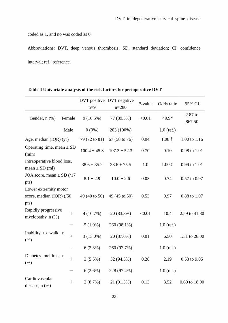
Table 4 Univariate analysis of the risk factors for perioperative DVT
DVT positive n=9
DVT negative n=280
P-value Odds ratio 95% CI
Gender, n (%) Female 9 (10.5%) 77 (89.5%) <0.01 49.9* 2.87 to 867.50
Male 0 (0%) 203 (100%) 1.0 (ref.)
Age, median (IQR) (yr) 79 (72 to 81) 67 (58 to 76) 0.04 1.08† 1.00 to 1.16
Operating time, mean ± SD (min)
100.4 ± 45.3 107.3 ± 52.3 0.70 0.10 0.98 to 1.01
Intraoperative blood loss, mean ± SD (ml)
38.6 ± 35.2 38.6 ± 75.5 1.0 1.00‡ 0.99 to 1.01
JOA score, mean ± SD (/17 pts)
8.1 ± 2.9 10.0 ± 2.6 0.03 0.74 0.57 to 0.97
Lower extremity motor score, median (IQR) (/50 pts)
49 (40 to 50) 49 (45 to 50) 0.53 0.97 0.88 to 1.07
Rapidly progressive myelopathy, n (%)
+ 4 (16.7%) 20 (83.3%) <0.01 10.4 2.59 to 41.80
- 5 (1.9%) 260 (98.1%) 1.0 (ref.)
Inability to walk, n (%)
+ 3 (13.0%) 20 (87.0%) 0.01 6.50 1.51 to 28.00
- 6 (2.3%) 260 (97.7%) 1.0 (ref.)
Diabetes mellitus, n (%)
+ 3 (5.5%) 52 (94.5%) 0.28 2.19 0.53 to 9.05
- 6 (2.6%) 228 (97.4%) 1.0 (ref.)
Cardiovascular disease, n (%)
+ 2 (8.7%) 21 (91.3%) 0.13 3.52 0.69 to 18.00
DVT in degenerative cervical spine disease
24
- 7 (2.6%) 259 (97.4%) 1.0 (ref.)
* modified odds ratio, † Odds ratio per one year increment, ‡ Odds ratio per 1 ml
increment
Gender, female was coded as 1, and male was coded as 0. Rapidly progressive myelopathy,
inability to walk, diabetes mellitus and cardiovascular disease, yes was coded as 1, and no
was coded as 0.
Abbreviations: DVT, deep vein thrombosis; SD, standard deviation; JOA score, Japanese
Orthopaedic Association score; CI, confidence interval; ref., reference.
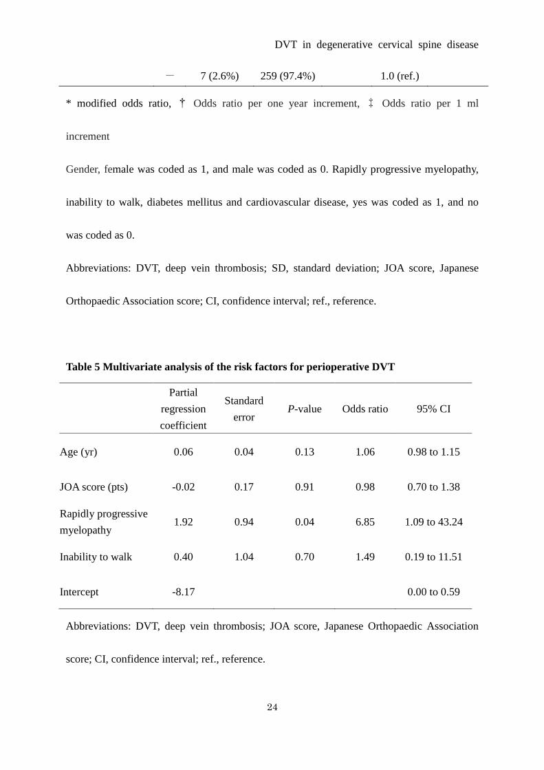
Table 5 Multivariate analysis of the risk factors for perioperative DVT
Partial
regression coefficient
Standard error
P-value Odds ratio 95% CI
Age (yr) 0.06 0.04 0.13 1.06 0.98 to 1.15
JOA score (pts) -0.02 0.17 0.91 0.98 0.70 to 1.38
Rapidly progressive myelopathy
1.92 0.94 0.04 6.85 1.09 to 43.24
Inability to walk 0.40 1.04 0.70 1.49 0.19 to 11.51
Intercept -8.17 0.00 to 0.59
Abbreviations: DVT, deep vein thrombosis; JOA score, Japanese Orthopaedic Association
score; CI, confidence interval; ref., reference.