Embed Size (px)
Citation preview
1521-0111/92/1/88–99$25.00 https://doi.org/10.1124/mol.117.108563MOLECULAR PHARMACOLOGY Mol Pharmacol 92:88–99, July 2017Copyright ª 2017 by The American Society for Pharmacology and Experimental Therapeutics
Rapid Throughput Analysis of GABAA Receptor SubtypeModulators and Blockers Using DiSBAC1(3) Membrane PotentialRed Dye s
Atefeh Mousavi Nik, Brandon Pressly, Vikrant Singh, Shane Antrobus, Susan Hulsizer,Michael A. Rogawski, Heike Wulff, and Isaac N. PessahDepartment of Molecular Biosciences, School of Veterinary Medicine (A.M.N., S.A., S.H., I.N.P.), and Department ofPharmacology (B.P., V.S., M.A.R., H.W.), School of Medicine, University of California Davis, Davis, California; Department ofNeurology, School of Medicine, University of California Davis, Sacramento, California (M.A.R.); and The Medical Investigationof Neurodevelopmental Disorders Institute, Sacramento, California (I.N.P.)
Received February 10, 2017; accepted April 12, 2017
ABSTRACTFluorometric imaging plate reader membrane potential dye(FMP-Red-Dye) is a proprietary tool for basic discovery andhigh-throughput drug screening for G-protein-coupled receptorsand ion channels. We optimized and validated this potentiomet-ric probe to assay functional modulators of heterologousexpressed GABAA receptor (GABAAR) isoforms (synaptica1b3g2, extrasynaptic a4b3d, and b3 homopentomers). High-resolution mass spectrometry identified FMP-Red-Dye as5,59-(1-propen-1-yl-3-ylidene)bis[1,3-dimethyl-2-thio-barbituricacid]. GABAAR-expressing cells equilibrated with FMP-Red-Dyeexhibited depolarized equilibrium membrane potentials com-pared with GABAAR-null cells. The channel blockers picrotoxin,fipronil, and tetramethylenedisulfotetramine, and the competitiveantagonist bicuculline reduced fluorescence near the levels inGABAAR-null cells indicating that FMR-Red-Dye, a barbituratederivative, activates GABAAR-mediated outward Cl2 current inthe absence of GABA. GABA caused concentration-dependentincreases in fluorescence with rank order of potencies among
GABAAR isoforms consistent with results from voltage-clampexperiments (EC50 values for a4b3d, a1b3g2, and b3 homo-pentamers were 6 6 1, 40 6 11, and .18 mM, respectively),whereas GABAAR-null cells were unresponsive. Neuroactivesteroids (NAS) increased fluorescence of GABAAR expressingcells in the absence of GABA and demonstrated positiveallosteric modulation in the presence of GABA, whereas benzo-diazepines only exhibited positive allosteric modulator (PAM)activity. Of 20 NAS tested, allopregnanolone, (3a,5a,20E)-3-hydroxy-13,24-cyclo-18-norcholan-20-ene-21-carbonitrile,eltanolone, 5b-pregnan-3a,21-diol-20-one, and ganaxoloneshowed the highest potency. The FMP-Red-Dye–based assaydescribed here provides a sensitive and quantitative method ofassessing the activity of GABAAR agonists, antagonists, andPAMs on diverse GABAAR isoforms. The assay has a wide rangeof applications, including screening for antiseizure agents andidentifying channel blockers of interest to insecticide discoveryor biosecurity.
IntroductionGABAA receptors (GABAARs) are ligand-gated anion chan-
nels that mediate inhibition in the mammalian centralnervous system by responding to the neurotransmitter GABA(Barnard et al., 1998).GABAAR dysfunctions are associatedwith epilepsy, autism, fragile X syndrome, depression, and
schizophrenia (Verkman and Galietta, 2009; Stafstrom et al.,2012; Rudolph and Möhler, 2014; Braat and Kooy, 2015).GABAARs are also the primary toxicological targets of severalcurrently used insecticides (e.g., fipronil) and pesticides ofhistorical importance that persist in the environment (e.g.,organochlorines) (Casida and Durkin, 2015). Importantly,GABAARs are a major pharmacological target for antiseizuredrugs (Brodie et al., 2016).GABAARs are pentameric anion channels composed of two a
subunits, two b subunits, and one subunit that is designatedas g, d, «, u, or p. There are multiple isoforms of many of thesubunits (six for a, three for b, and three for g), allowing fora high degree of variation depending on brain region anddevelopmental stage. Most synaptic GABAARs contain two asubunits, two b subunits, and one g2 subunit; have a relatively
This work was sponsored by the National Institutes of Health NationalInstitute of Neurologic Disorders and Stoke [UC Davis CounterACT Center ofExcellence Grant U54 NS 011269] and Intellectual and Developmental DisabilitiesResearch Center (IDDRC) Core Center [Grant U54HD079125]; B.P. was supportedby a National Institutes of Health National Institute of General Medical Sciencesfunded Pharmacology Training Program [Grant T32GM099608].
https://doi.org/10.1124/mol.117.108563.s This article has supplemental material available at molpharm.
aspetjournals.org.
ABBREVIATIONS: DiSBAC1(3), 5,59-(1-propen-1-yl-3-ylidene)bis[1,3-dimethyl-2-thio-barbituric acid]; Em, equilibrium membrane; FLIPR, fluoro-metric imaging plate reader; FMP-Red-Dye, fluorometric imaging plate reader membrane potential red dye; GABAAR, GABAA receptor; HEK, humanembryonic kidney; NAS, neuroactive steroids; PAM, positive allosteric modulator; PTX, picrotoxin; TBPS, t-butylbicyclophosphorothionate; TETS,tetramethylenedisulfotetramine; 5b,3a-THDOC, 3a,21-dihydroxy-5b-preganan-20-one; XJ-42, (3a,5a,20E)-3-hydroxy-13,24-cyclo-18-norcholan-20-ene-21-carbonitrile.
88
http://molpharm.aspetjournals.org/content/suppl/2017/04/20/mol.117.108563.DC1Supplemental material to this article can be found at:
at ASPE
T Journals on M
arch 18, 2022m
olpharm.aspetjournals.org
Dow
nloaded from
low affinity for GABA; and mediate fast phasic inhibition(Rissman and Mobley, 2011; Brickley and Mody, 2012). Incontrast, extrasynaptic GABAARs often contain a d subunit,have a higher affinity for GABA, and mediate persistent tonicinhibition (Bettler and Tiao, 2006; Belelli et al., 2009; Brickleyand Mody, 2012). In addition to the endogenous neurotrans-mitter, GABA, which binds to the interface of a and bsubunits, GABAARs possess binding sites for neuroactivesteroids (NAS), benzodiazepines, and barbiturates that allo-sterically enhance GABA-activated Cl2 conductance and insome instances activate Cl2 conductance in the absence ofGABA, thereby exerting anxiolytic, sedative, and antiseizureeffects that are useful for treating anxiety and sleep disordersand epilepsy, as well as serving as general anesthetics (Belelliand Lambert, 2005; Herd et al., 2007). The benzodiazepinebinding site is localized between thea and g subunits, whereasNAS are known to have two binding sites, a potentiation siteon the a subunit and a direct activator site in the a/b interface,both of which need to be occupied for potent channel activation(Akabas, 2004;Campagna-Slater and Weaver, 2007; Hosieet al., 2007; Alvarez and Estrin, 2015). Although the precisesites to which barbiturates bind remain elusive, domainswithin b subunit transmembrane segments TM2 and TM3appear to be critical (Löscher and Rogawski, 2012). Interest-ingly, b3 subunits reconstitute a homopentameric channelthat possessesNAS binding sites (Chen et al., 2012;Miller andAricescu, 2014; Alvarez and Estrin, 2015).Considering the utility of GABAARs as therapeutic targets,
there is substantial interest in the identification of improvedGABAAR modulators. A suitable high-throughput screeningmethod would significantly aid in the identification of suchagents. One potential method to assess GABAAR function in ahigh-throughput manner uses the fluorometric imaging platereader (FLIPR) membrane potential red dye (FMP-Red-Dye)that redistributes across the plasma membrane in a voltage-dependent manner. Cell depolarization results in dye move-ment into the cell and binding to intracellular proteins andhydrophobic sites causing increased florescence signals andvice versa for hyperpolarization. Thus, FMP-Red-Dye allowsmonitoring of membrane potential changes in a bidirectionalfashion independent of ion type. FMP-Red-Dye is convenientin that it does not require cells to be washed of excess dye afterequilibration due to inclusion of a proprietary cell membraneimpermeant red wavelength quencher, which results in a highsignal-to-noise ratio (Fairless et al., 2013). Mennerick et al.(2010) reported that many voltage-sensitive dyes such asDi-4-ANEPPS and DiBAC4(3) can directly activate and/orpotentiate GABAAR currents in a manner similar to NAS andbarbiturates, questioning their usefulness for screeningGABAAR ligands. A previous study reported that FMP-Red-Dye had better performance than FMP-Blue-Dye, and that itproduced results comparable to electrophysiology (Joeschet al., 2008). However, the chemical identity of FMP-Red-Dye is proprietary and the degree to which its constituentsmight influence GABAAR function, and therefore limit thedye’s usefulness in characterizing GABAAR modulators, hasremained unclear.Here, we identify the voltage-sensitive component of FMP-
Red-Dye as 5,59-(1-propen-1-yl-3-ylidene)bis[1,3-dimethyl-2-thio-barbituric acid] [DiSBAC1(3)]; optimize and validate itsuse as a rapid throughput indicator of GABAAR activators,blockers, and positive allosteric modulators (PAMs) using
FLIPR; and implement the assay to screen a small library ofNAS and channel blockers. We show that FMP-Red-Dye canbe used to differentiate GABAAR-mediated responses in celllines that stably or transiently express synaptic (a1b3g2),extrasynaptic (a4b3d), or b3 homomeric GABAAR isoforms.We conclude that the FMP-Red-Dye–based assays providesensitive and quantitative approaches to investigate func-tional drug effects on GABAAR isoforms, whether they aremediated by binding to the GABA recognition site, the NASPAM sites, or convulsant channel blocking sites. The assay isuseful for antiseizure drug screening and identifying novelchannel blockers of interest to insecticide discovery orbiosecurity.
Materials and MethodsReagents
Poly-L-lysine, cytosine arabinoside, picrotoxin (PTX), t-butylbicyclophosphorothionate (TBPS), fipronil, bicuculline, diaze-pam, and GABA were purchased from Sigma Aldrich (St. Louis,MO). Falcon 96-Well Imaging Plates with lids were purchased fromFisher Scientific (Hampton, NH). FLIPR Membrane Potential RedAssay Kit (FMP-Red-Dye) was purchased from Molecular DevicesCorporation (Sunnyvale, CA). Fluo4-AM was purchased from LifeTechnology (Hampton, NH). GS21 supplement was purchasedfrom MTI-Global Stem (Gaithersburg, MD). Org 20599, alphaxalone,eltanolone, progesterone, and midazolam were purchased from Tocris(Pittsburgh, PA). (3a,5a,20E)-3-Hydroxy-13,24-cyclo-18-norcholan-20-ene-21-carbonitrile (XJ-42) (compound 63 in Covey and Jiang,2014) was a generous gift of Dr. Douglas F. Covey (WashingtonUniversity School ofMedicine, St. Louis, MO), 3-[3a-hydroxy-3b-methyl-5a-androstan-17b-yl]-5-(hydroxymethyl)isoxazole (Hogenkamp et al.,2014) was a generous gift of Dr. KelvinW. Gee (University of California,Irvine, Irvine CA). Allopregnanolone was custom synthesized by SAFCPharma (Madison, WI). Dehydroepiandrosterone sulfate, indiplon,and ursodeoxycholic acid (sodium salt) were purchased from Cay-man Chemical (Ann Arbor, MI). Dehydroepiandrosterone acetate,dehydroepiandrosterone, cortisol, epiandrosterone, 20a-dihydropregnenolone,androstenediol, etiocholanolone, androsterone, alphadolone 21-acetate,and tetrahydrocortexone were purchased from Steraloids (Newport,RI). Tetramethylenedisulfotetramine (TETS) was synthesized in thelaboratory of Dr. Bruce Hammock as previously described (Zhao et al.,2014). All reagents were .97% purity.
Heterologous Expression of GABAAR Isoforms
Expression of GABAAR Subunits for Potentiometric Mea-surements with FLIPR FMP-Red-Dye. Our goal was to develop areliable, rapid throughput approach to quantitatively assess theinfluences of blockers, antagonists, and PAMs on the functionalactivity of diverse GABAAR isoforms. To this end, we investigated celllines that stably or transiently express three GABAAR isoforms ofdifferent subunit compositions. A human embryonic kidney (HEK)293 cell line that stably expresses human GABAAR a1b3g2 subunits(CYL3053 PrecisION hGABA-A a1/b3/g2-HEK Recombinant CellLine) was a generous gift of EMD Millipore Corporation, St. Charles,MO. GABAAR a1b3g2 heteropentomeric channels primarily localizeto synaptic sites in mammalian neurons (McCartney et al., 2007). TheGABAAR-null HEK 293 line, which served as the control, was pur-chased from American Type Culture Collection (ATCC-CRL-1573)(Manassas, VA). Upon arrival both cell lineswere expanded inDulbecco’smodified Eagle’s medium/Ham’s F-12 (50/50 mix), 10% fetal bovineserum, 1% nonessential amino acid (Invitrogen, Carlsbad, CA) at 37°Cin 5% CO2 and several bullets were frozen to limit passage numbers.Cells expressing a1b3g2 subunits were kept under selection pressurewith 400 mg/ml genticin, 100 mg/ml hygromycin B (Invitrogen), and
Analysis of GABAAR modulators with potentiometric dye 89
at ASPE
T Journals on M
arch 18, 2022m
olpharm.aspetjournals.org
Dow
nloaded from
0.625mg/ml puromycin (Clontech,Mountain View, CA). GABAAR-nullcells were cultured in 100 U/ml penicillin and 100 mg/ml streptomycin(Gibco,Waltham,MA) tominimize the risk of bacterial contamination.Cell lines were discarded after 15 passages. Cells were passaged when70%–80% confluent and harvested using 0.05% trypsin-EDTA and50,000–60,000 cells/well (in a volume of 100 ml), seeded into poly-L-lysine–coated wells of a 96-well plate, and allowed to recover in theincubator overnight before FMP-Red-Dye loading and measurementsof equilibrium membrane (Em) potential with FLIPR.
A L-tk (mouse connective tissue) cell line expressing the humanGABAAR a4b3d subunit combination under a dexamethasone-inducible promoter was kindly provided by Dr. Trevor Smart (Uni-versity College, London) (Brown et al., 2002). The a4b3d subunitisoform is found at extrasynaptic sites in mammalian brain. The L-tkcell line was cultured in Dulbecco’s modified Eagle’s medium plusglutamine, 4.5 g/l Na pyruvate, and glucose with 10% fetal bovineserum, in the presence of 1 mg/ml genticin and 0.2 mg/ml zeocin toselect a4b3d-positive cells. Dexamethasone (1 mM; Invitrogen) orvehicle was added to the media to induce a4b3d subunit expression orserve as GABAAR-null cells, respectively, when 80%–90% confluent.Cells were induced for 48 hours, passaged using trypsin-EDTA 0.05%,plated at 50,000–60,000 cells/well into 96-well plates, and after24-hour recovery loaded with FMP-Red-Dye and membrane potentialmeasured as described subsequently.
Transient expression of GABAAR b3 homopentamers in HEK293 cells (ATCCCRL-1573) was achievedwith a pcDNA3.1 expressionvector generously provided by Dr. Robert L. Macdonald (VanderbiltUniversity, Nashville, TN). HEK 293 cells were cultured as describedpreviously. Twenty-four hours before transfection, cells were plated on10-cm tissue culture–treated dishes and transfected with plasmidDNA at 70%–90% confluence using TurboFect (Thermo Scientific,Waltham, MA) according to the manufacturer’s instructions. At24-hour post-transfection, cells were dissociated with trypsin,counted, and plated on poly-L-lysine–coated 96-well plates (Falcon,Waltham, MA) at a density of 50,000–60,000 cells/well. All FLIPRexperiments were carried out 48 hours post-transfection.
Expression of GABAAR Subunits for ElectrophysiologicalMeasurements. The human GABAAR a1, b3, and g2 cloned intopcDNA3.1 expression vectors were a gift from Dr. Robert L. Macdon-ald (Vanderbilt University). Fibroblast L929 cells were cultured inDulbecco’s modified Eagle’s medium (Lonza, Portsmouth, NH) sup-plemented with 10% fetal bovine serum, 100 U/ml penicillin, and100 mg/ml streptomycin (Invitrogen) and maintained in humidified95% air and 5% CO2 air at 37°C. Cells were transfected usingFuGENE 6 (Roche, Santa Clara, CA) transfection reagent with anequal amount of each of the subunits in combination with pEGFP-C1.The transfection ratio of total cDNA to transfection reagent was 2:1with equal amounts of a1, b3, and g2 cDNA to reconstitute synaptica1b3g2 GABAAR, or b3 alone to reconstitute homopentamers. Twodays post-transfection, cells were plated on glass coverslips and trans-fected cells were identified using an epifluorescence microscope forelectrophysiological measurements. Electrophysiological recordingsfrom L-tk cells expressing GABAAR a4b3d subunit composition wereobtained with culture and induction methods described previously.
Rapid Throughput Analysis of GABAAR Modulators UsingFLIPR FMP-Red-Dye
Once HEK 293 and L-tk cells were cultured on 96-well plates for24 hours, FMP-Red-Dye was reconstituted to 1� with 100 ml ofLocke’s buffer (NaCl 154 mM, KCl 5.6 mM, CaCl2 2.3 mM, MgCl21 mM, HEPES 8.6 mM, glucose 5.6 mM, glycine 0.1 mM, pH 7.4) asrecommended by the supplier (Molecular Devices Corporation).Growth medium was removed from the wells and cells were loadedwith 100 ml FMP-Red-Dye solution for 30 minutes in the dark at roomtemperature (except for the experiments in Fig. 1 where fluorescencesignals were recorded immediately). Each plate was transferred to theFLIPR Tetra Station; the dye was excited at 510–545 nm, and thefluorescent signals were recorded at 565–625 nm. Baseline recordingswere acquired for 2 minutes at a sampling rate of 1 Hz (400 ms ofillumination per sample). FMP-Red-Dye is a slow-response, potential-sensitive probe and the maximal response is usually seen within
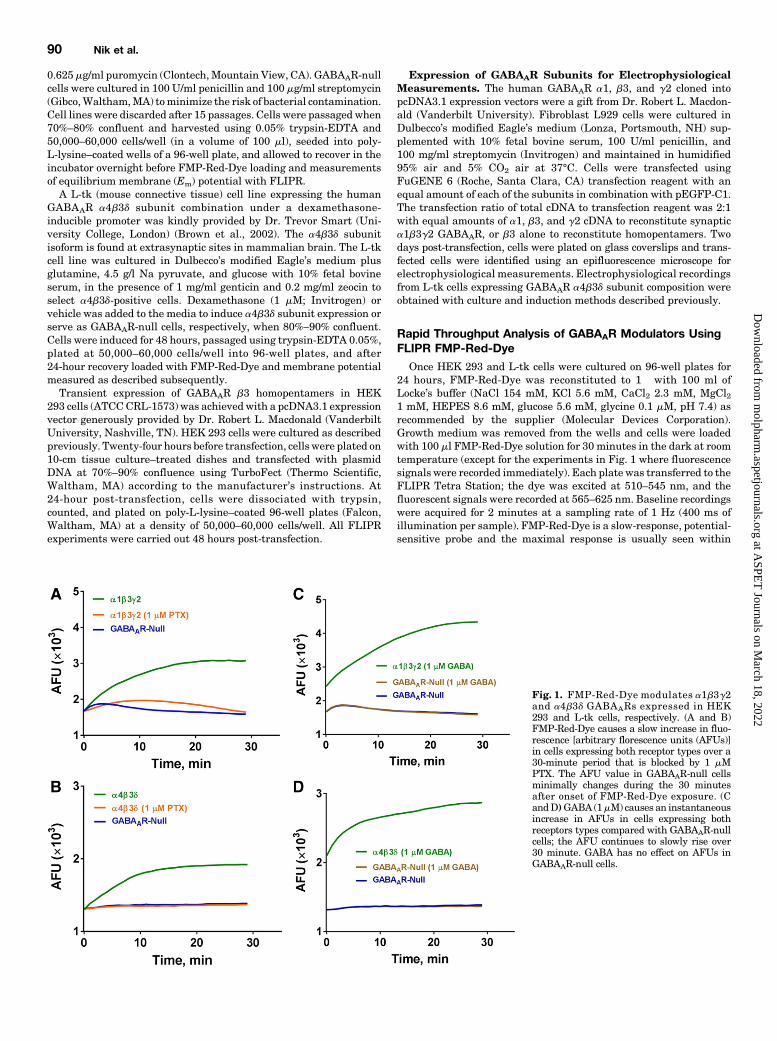
Fig. 1. FMP-Red-Dye modulates a1b3g2and a4b3d GABAARs expressed in HEK293 and L-tk cells, respectively. (A and B)FMP-Red-Dye causes a slow increase in fluo-rescence [arbitrary florescence units (AFUs)]in cells expressing both receptor types over a30-minute period that is blocked by 1 mMPTX. The AFU value in GABAAR-null cellsminimally changes during the 30 minutesafter onset of FMP-Red-Dye exposure. (CandD)GABA (1mM) causes an instantaneousincrease in AFUs in cells expressing bothreceptors types compared with GABAAR-nullcells; the AFU continues to slowly rise over30 minute. GABA has no effect on AFUs inGABAAR-null cells.
90 Nik et al.
at ASPE
T Journals on M
arch 18, 2022m
olpharm.aspetjournals.org
Dow
nloaded from
2minutes after triggering cellular depolarization or hyperpolarization(see Figs. 3 and 4) (Dasheiff, 1985). Cell responses were normalized bycalculation (Fmax2Fmin)/Fmin5DF/F0, whereFmax was themaximumresponse in arbitrary fluorescence units, and Fmin was the baselinearbitrary florescence unit value. Although HEK 293 cells wereunresponsive to vehicle additions, signals from L-tk cells expressinga4b3d subunits exhibited an abrupt but transient drop in fluorescencewith addition of any solution, including Locke’s or vehicle, andtherefore all data from L-tk cells were normalized to vehicle baselineby subtraction.
Electrophysiological Recordings
Whole-cell voltage-clamp and current-clamp recordings were per-formed at room temperature with an EPC-10 amplifier (HEKA,Holliston, MA). Cells were bathed in an external Ringer’s solutionconsisting of 160 mM NaCl, 4.5 mM KCl, 1 mM MgCl2, 2 mM CaCl2,and 10 mMHEPES with pH 7.4 and 311 mOsm. Recording electrodeswere pulled and fire-polished to resistances of 1.8–2.4 MV for voltage-clamp and 3–6 MV for current-clamp experiments. Electrodes werefilledwith an internal solution consisting of 154mMKCl, 2mMCaCl2,1 mM MgCl2, 10 mM HEPES, and 10 mM EGTA with pH 7.3 and308 mOsm. Cells were voltage clamped at 280 mV and controlcurrents were recorded under the application of 1 mM GABA, using agravity-fed fast perfusion system, for 5 seconds followed by a 40–50second wash with external solution. GABA concentration-responserelationships were determined by testing increasing concentrations ofGABA and normalizing GABA currents to the peak response inducedby a saturating concentration of GABA. Normalized currents werefitted using the Hill equation (nH) to determine the EC50 and EC10
values. The EC10 value (1 mM) was used to evaluate the positivemodulatory effects of the neurosteroids. The increases in Cl2 currentelicited in the presence of NAS were compared with the initial EC10
value to determine the fold increase in current. Test solutions of theNAS were freshly prepared immediately before each application ontocells. Membrane potential was recorded on the initial break into thecell while in current-clamp mode. For experiments involving FMP-Red-Dye, cells were preincubation with the dye for 30 minutes beforerecording in the absence or presence of fipronil. Cells were thenexposed to 10 minutes of UV irradiation (460–490 nm) to induce thepreviously reported photodynamic effect of voltage-sensitive dyes(Mennerick et al., 2010).
Data Analysis
GraphPad Prism software (version 6; GraphPad Software, La Jolla,CA) was used for statistical analysis and graphing. The EC50 valueswere determined using nonlinear regression with a four-parameterlogistic equation. Student’s t test or F test (P , 0.05) was applied todetermine statistical differences. For post hoc multiple comparisonsseparate one-way analysis of variance (for EC50 and slope) using Tukey’stest was applied. Data are presented as the mean 6 S.D. as stated. Forelectrophysiology, data analysis was performed using Excel (Microsoft)and Origin 7.0 (OriginLab Corp., Northhampton, MA) software. Datafitting to the Hill equation to obtain the EC50 values was performed withOrigin 7.0. Data are presented as the mean6 S.D.
ResultsOptimization and Evaluation of FMP-Red-Dye Assay
for Functional GABAAR Screening. In the initial exper-iments, HEK 293 cells stably expressing one of the widelyexpressed human synaptic GABAAR isoforms a1b3g2 orGABAAR-null HEK 293 cells were used to test whetherFMP-Red-Dye potentiates GABAAR function in the absence ofGABA as reported for other potentiometric dyes (Mennericket al., 2010). FMP-Red-Dye signals were monitored in real-
time within 1 minute after dye addition to the cell medium.Initial fluorescence signals were similar for both GABAAR-expressing and GABAAR-null HEK 293 cells, but equili-brated to different steady-state fluorescence signals within30 minutes, with a1b3g2-expressing HEK 293 cells in-variably achieving 2-fold higher steady-state fluorescenceintensity than the respective null cells (Fig. 1A). Importantly,inclusion of PTX (1 mM) with dye addition prevented therise in fluorescence in GABAAR-expressing cells (Fig. 1A). Incontrast, inclusion of GABA (1 mM) with the potentiometricdye caused an instantaneous increase in dye signal (within the1 Hz resolution of the recording) followed by a more gradualequilibration of fluorescence to a 3-fold greater signal thanthat achieved in null cells by 30 minutes (Fig. 1C). GABAAR-null cells showed a minimal time-dependent fluctuation influorescence and this responsewas unchanged by the presenceof GABA. Similar results were obtained in studies comparingFMP-Red-Dye responses in L-tk cells expressing the extra-synaptic a4b3dGABAAR isoformwith L-tkGABAAR-null cells(Fig. 1, B and D). These results indicate that by itself FMP-Red-Dye promotes slow depolarization of cells expressingeither synaptic or extrasynaptic GABAAR subunits but failsto cause this effect in the respective null cells. FMP-Red-Dye–induced cell depolarization was prevented by PTX, indicatingthat it requires functional GABAARs. In contrast to the ap-parent slow activation of GABAAR caused by FMP-Red-Dye,GABA induced a rapid activation of the receptors.To further verify the accuracy of our interpretation, FMP-
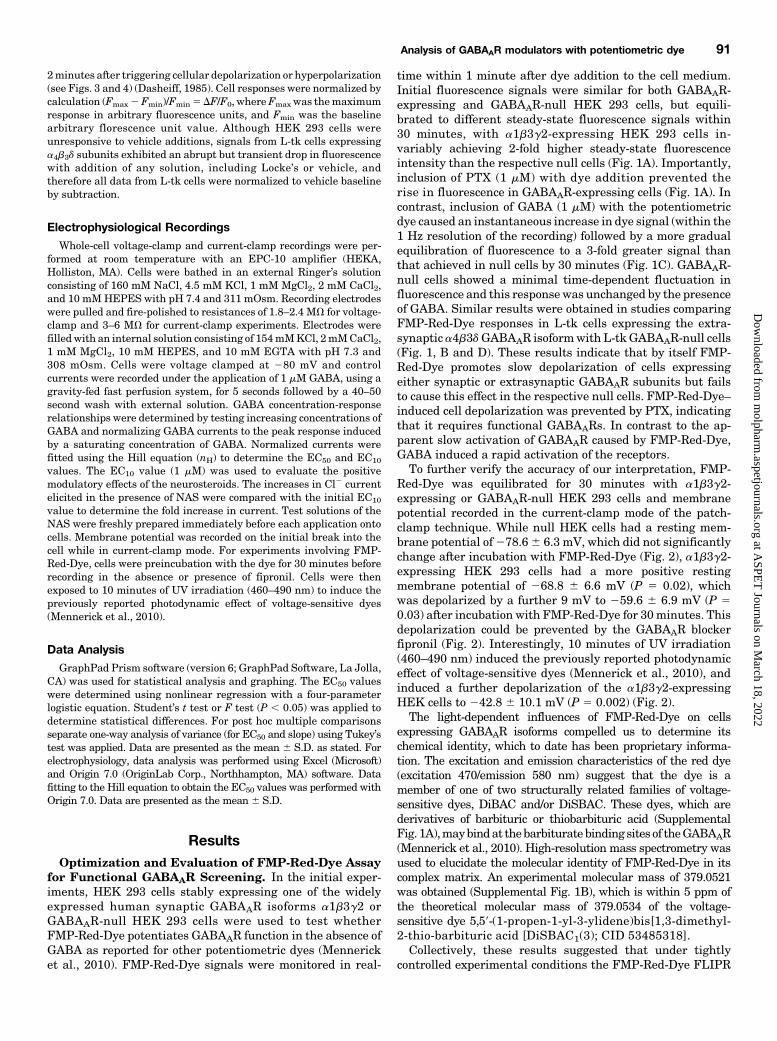
Red-Dye was equilibrated for 30 minutes with a1b3g2-expressing or GABAAR-null HEK 293 cells and membranepotential recorded in the current-clamp mode of the patch-clamp technique. While null HEK cells had a resting mem-brane potential of278.66 6.3 mV, which did not significantlychange after incubation with FMP-Red-Dye (Fig. 2), a1b3g2-expressing HEK 293 cells had a more positive restingmembrane potential of 268.8 6 6.6 mV (P 5 0.02), whichwas depolarized by a further 9 mV to 259.6 6 6.9 mV (P 50.03) after incubation with FMP-Red-Dye for 30minutes. Thisdepolarization could be prevented by the GABAAR blockerfipronil (Fig. 2). Interestingly, 10 minutes of UV irradiation(460–490 nm) induced the previously reported photodynamiceffect of voltage-sensitive dyes (Mennerick et al., 2010), andinduced a further depolarization of the a1b3g2-expressingHEK cells to 242.8 6 10.1 mV (P 5 0.002) (Fig. 2).The light-dependent influences of FMP-Red-Dye on cells
expressing GABAAR isoforms compelled us to determine itschemical identity, which to date has been proprietary informa-tion. The excitation and emission characteristics of the red dye(excitation 470/emission 580 nm) suggest that the dye is amember of one of two structurally related families of voltage-sensitive dyes, DiBAC and/or DiSBAC. These dyes, which arederivatives of barbituric or thiobarbituric acid (SupplementalFig. 1A),maybindat thebarbituratebinding sites of theGABAAR(Mennerick et al., 2010). High-resolution mass spectrometry wasused to elucidate the molecular identity of FMP-Red-Dye in itscomplex matrix. An experimental molecular mass of 379.0521was obtained (Supplemental Fig. 1B), which is within 5 ppm ofthe theoretical molecular mass of 379.0534 of the voltage-sensitive dye 5,59-(1-propen-1-yl-3-ylidene)bis[1,3-dimethyl-2-thio-barbituric acid [DiSBAC1(3); CID 53485318].Collectively, these results suggested that under tightly
controlled experimental conditions the FMP-Red-Dye FLIPR
Analysis of GABAAR modulators with potentiometric dye 91
at ASPE
T Journals on M
arch 18, 2022m
olpharm.aspetjournals.org
Dow
nloaded from
platform could provide a sensitive and quantitative method forinvestigating pharmacological responses of GABAAR isoforms todiverse direct GABAAR activators, GABAAR channel blockers oftoxicological significance, and GABAAR PAMs. To these ends,subsequent experiments were conducted following 30 minutesequilibration of cells seeded in 96-well plates with dye in thedark, followed by 2minutes of baseline recording and 10minutesof response recording after drug addition from a 96-well sourceplate. Excitation and emission were within visible wavelengths(excitation 510–545 nm/emission 565–625 nm), and wells wereexcited by 400ms duration pulsed illumination at a rate of 1 Hz.This protocol minimizes photodynamic influences of theFMP-Red-Dye, which may alter its activity on GABAARs.Differential Potencies of GABA toward Activating
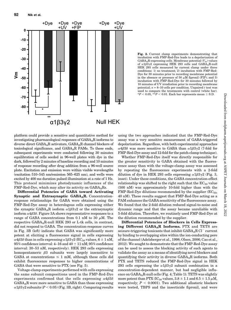
Synaptic and Extrasynaptic GABAAR. Concentration-response relationships for GABA were obtained using theFMP-Red-Dye assay in heterologous cells expressing eitherthe synaptic GABAAR isoform a1b3g2 or the extrasynapticisoform a4b3d. Figure 3A shows representative responses to arange of GABA concentrations from 0.1 nM to 30 mM. Therespective GABAAR-null HEK 293 or L-tk cells, in contrast,did not respond to GABA. The concentration-response curvesin Fig. 3B (left) indicate that GABA was significantly morepotent at eliciting a fluorescence signal in cells expressinga4b3d than in cells expressing a1b3g2 (EC50 values, 66 1 nM,95% confidence interval: 4–16 and 406 11 nM; 95% confidenceinterval: 30–53 nM, respectively). HEK 293 cells expressinghomopentameric b3 subunits were largely insensitive toGABA at concentrations # 1 mM, although these cells didexhibit fluorescence responses to higher concentrations ofGABA that were sensitive to PTX (see Fig. 5).Voltage-clamp experiments performedwith cells expressing
the same subunit compositions used in the FMP-Red-Dyeexperiments confirmed that the cells expressing a4b3dGABAAR were more sensitive to GABA than those expressinga1b3g2 subunits (P, 0.05) (Fig. 3B, right). Comparing results
using the two approaches indicated that the FMP-Red-Dyeassay was a very sensitive measurement of GABA-triggereddepolarization. Regardless, with both experimental approachesa4b3d was more sensitive to GABA than a1b3g2 (7-fold forFMP-Red-Dye assay and12-fold for the patch-clamp technique).Whether FMP-Red-Dye itself was directly responsible for
the greater sensitivity to GABA obtained with the fluores-cence assay than with the voltage-clamp assay was assessedby repeating the fluorescence experiments with a 2-folddilution of dye in HEK 293 cells expressing a1b3g2 (Fig. 3,inset). Under these conditions, the GABA concentration-effectrelationship was shifted to the right such that the EC50 value(500 nM) was approximately 10-fold higher than with theFMP-Red-Dye dilutions recommended by the supplier (EC50,40 nM). These results suggest that FMP-Red-Dye acting as aPAMenhances theGABA sensitivity of the fluorescence assay.We found that the 2-fold dilution reduced signal-to-noise anddynamic range and that the assay became unreliable with5-fold dilution. Therefore, we routinely used FMP-Red-Dye atthe dilution recommended by the supplier.Differential Potencies of Blockers in Cells Express-
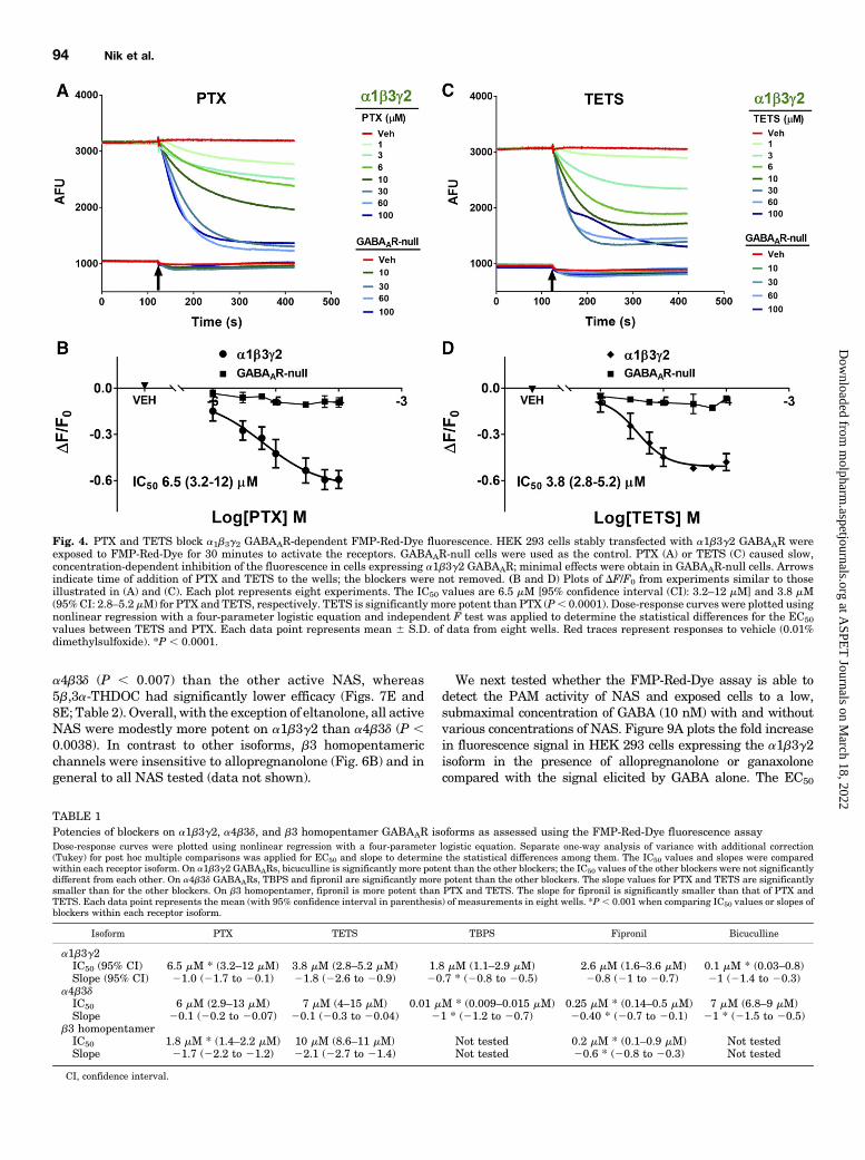
ing Different GABAAR Isoforms. PTX and TETS areseizure-triggering toxicants that inhibit GABAAR Cl2 currentby binding to overlapping sites within the ion-conducting poreof the channel (Adelsberger et al., 1998; Olsen, 2006; Cao et al.,2012).We sought to demonstrate that the FMP-Red-Dye assaycan be used to assess the blocking activity of such agents tovalidate the assay as ameans of identifying novel blockers andquantifying their activity in diverse GABAAR isoforms. BothPTX and TETS reduced the FMP-Red-Dye signal in HEK293 cells expressing the a1b3g2 subunit combination in aconcentration-dependent manner, but had negligible influ-ence onGABAAR-null cells (Fig. 4; Table 1). TETSwas slightlymore potent than PTX (IC50 values, 3.86 1.1 and 6.56 1.5 mM,respectively; P , 0.0001). Two additional allosteric blockerswere tested, TBPS and the insecticide fipronil, and were
Fig. 2. Current clamp experiments demonstrating thatincubation with FMP-Red-Dye leads to a depolarization ofGABAAR-expressing cells. Membrane potential (Vm) valuesof a1b3g2 expressing HEK 293 cells and GABAAR-nullHEK 293 cells measured by current clamp under threeconditions: 1) no treatment; 2) incubation with FMP-Red-Dye for 30 minutes prior to recording membrane potentialin the absence or presence of 50 mM fipronil (FIP); and 3)incubation with FMP-Red-Dye for 30 minutes followed by10 minutes of UV irradiation prior to recording membranepotential; n = 8–10 cells per condition. Unpaired t test wasused to compare the treatments with control (white bar).*P , 0.05, **P , 0.01. Each bar represents mean 6 S.D.
92 Nik et al.
at ASPE
T Journals on M
arch 18, 2022m
olpharm.aspetjournals.org
Dow
nloaded from
shown to have similar potencies to that of TETS (IC50 values,1.8 6 1.2 and 2.6 6 1.1 mM, respectively). The GABAARcompetitive antagonist bicuculline was approximately20-times more potent than TBPS (IC50 value, 0.1 6 0.1 mM;P , 0.0001), although its efficacy at reducing fluorescence tothat in GABAAR-null cells was less complete at the highestconcentration tested (10 mM, data not shown).L-tk cells expressing a4b3d subunits showed a distinct
structure-activity relationship with the blockers. Althoughthe inhibitory potency of PTX was similar to that in cellsexpressinga1b3g2 (IC50 values, 6.061.0mMversus6.561.5mM;P. 0.31), TETSwas 2-fold less potent andTBPSand fipronilwerenearly 200- and 10-fold more potent at the a4b3d isoform than atthe a1b3g2 isoform (Table 1).The divergent potencies exhibited by inhibitors toward cells
expressing different GABAAR isoforms compelled us to de-termine responses of cells expressing only the b3 subunit,which forms homopentamers that lack high-affinity GABAbinding. HEK 293 cells expressing the b3 homomeric isoformexhibited substantially greater fluorescence than the respec-tive GABAAR-null cells (Fig. 5A), as was the case for thea1b3g2 isoform (Fig. 4), suggesting that FMP-Red-Dye is ableto activate b3 homomeric GABAARs as it does the otherisoforms. PTX caused a concentration-dependent inhibition offluorescence (Fig. 5, C and D) that brought the fluorescencenear that in GABAAR-null cells. b3-Expressing cells failed toresponse to GABA at concentrations below 1mM (Fig. 5B), butas in previous voltage-clamp studies (Wooltorton et al., 1997)they did appear to be activated by high (.10 mM) GABA
concentrations. Figure 6A shows that hyperpolarization pro-duced by fipronil is also concentration dependent, with fipronilbeing approximately 10- and 50-fold more potent at restoringEm values to levels near those measured with GABAAR-nullHEK 293 cells than PTX or TETS (Table 1).Detection of Direct Activation and PAM Activity in
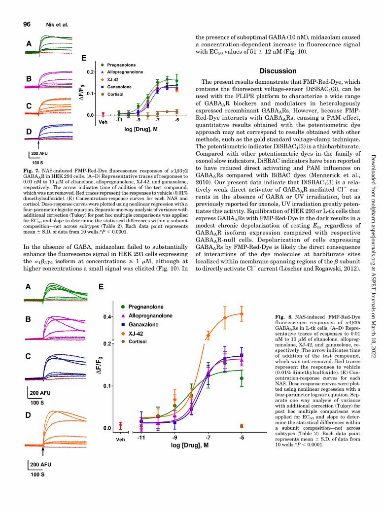
GABAAR Isoforms. NAS have been shown to have GABAARPAM activity at low concentrations and to directly activateGABAARs at higher concentrations (Belelli and Lambert,2005; Wang, 2011). Twenty NAS and related structures weretested for activity on GABAARs in the FMP-Red-Dye assaywith heterologous cells expressing a1b3g2, a4b3d, or b3homomeric isoforms, and the potency and efficacy of activecompounds were quantified (Figs. 7 and 8; Table 2). SeveralNAS caused concentration-dependent increases in fluores-cence in cells expressing the a1b3g2 and a4b3d isoforms withEC50 values below 1 mM, whereas the respective GABAAR-null cells failed to respond to any NAS. The rank order ofpotencies (based on the EC50 values) in cells expressing thea1b3g2 isoformwas:allopregnanolone/eltanolone/XJ-42. ganax-olone/3a,21-dihydroxy-5b-preganan-20-one (5b,3a-THDOC)/alphaxalone . alphadolone 21-acetate .. andosterone. Ofthese, 5b,3a-THDOC and alphadolone 21-acetate showedsignificantly (P, 0.001) lower efficacy (maximum DF/F0) thanthe other activeNAS (Table 2). By contrast, cells expressing thea4b3d isoform showed a different rank order of potencies:eltanolone . allopregnanolone . ganaxolone/5b,3a-THDOC/XJ-42/alphaxalone.. alphadolone 21-acetate. androsterone. XJ-42exhibited significantly greater maximal efficacy in cells expressing
Fig. 3. Comparison of GABA responses in cells expressing GABAAR as assessed with the FMP-Red-Dye technique and by voltage-clamp recording. (A)Both a1b3g2 and a4b3d GABAAR-expressing cells exhibit fluorescence responses of increasing amplitude following exposure to increasingconcentrations of GABA in the range of 0.1 nM to 30 mM. In these experiments, cells were equilibrated with FMP-Red-Dye for 30 minutes. Then,baseline fluorescence was recorded for 2 minutes followed by exposure to vehicle [(VEH); 0.01% dimethylsulfoxide] or GABA. The DF/F0 values weredetermined at the peak of the fluorescence response. The black arrow indicates the time of GABA addition; GABA remained for the duration of therecording. GABAAR-null cells do not respond to GABA. (B, left) Concentration-response curves for GABA based on fluorescence responses reveals thata4b3d GABAARs expressed in L-tk cells are significantly more sensitive to GABA than a1b3g2 GABAARs expressed in HEK 293 cells for EC50 values of6 nM [95% confidence interval (CI): 4–8 nM (nH = 0.7; n = 10)] and 40 nM [95% CI: 33–54 nM (nH = 1.1; n = 10)]. The b3 homopentamers transientlyexpressed in HEK 293 cell are largely insensitive to GABA (EC50 . 1 mM). Dose-response curves were plotted using nonlinear regression with a four-parameter logistic equation and independent F test was applied to determine the statistical differences for the EC50 values and slopes between a1b3g2and a4b3d GABAAR-expressing cell lines. Each data point represents mean 6 S.D. of data from 10 wells. (B, right) Concentration-response curves forGABAactivation ofa1b3g2 receptors inHEK293 cells and a4b3d receptors inL-tk cells fromwhole-cell voltage-clamp recordings for EC50 values of 6.67mM[95%CI: 5.30–8.04mM(nH= 1.8;n= 13)] and 549mM[95%CI: 435–663 nM (nH=1.8;n= 10]Each data point representsmean6S.D. *P, 0.0001 fora1b3g2versus a4b3d.
Analysis of GABAAR modulators with potentiometric dye 93
at ASPE
T Journals on M
arch 18, 2022m
olpharm.aspetjournals.org
Dow
nloaded from
a4b3d (P , 0.007) than the other active NAS, whereas5b,3a-THDOC had significantly lower efficacy (Figs. 7E and8E; Table 2). Overall, with the exception of eltanolone, all activeNAS were modestly more potent on a1b3g2 than a4b3d (P ,0.0038). In contrast to other isoforms, b3 homopentamericchannels were insensitive to allopregnanolone (Fig. 6B) and ingeneral to all NAS tested (data not shown).
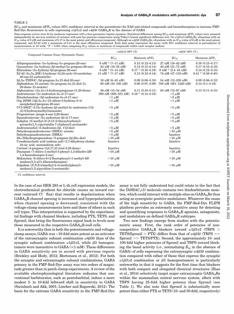
We next tested whether the FMP-Red-Dye assay is able todetect the PAM activity of NAS and exposed cells to a low,submaximal concentration of GABA (10 nM) with and withoutvarious concentrations of NAS. Figure 9A plots the fold increasein fluorescence signal in HEK 293 cells expressing the a1b3g2isoform in the presence of allopregnanolone or ganaxolonecompared with the signal elicited by GABA alone. The EC50
Fig. 4. PTX and TETS block a1b3g2 GABAAR-dependent FMP-Red-Dye fluorescence. HEK 293 cells stably transfected with a1b3g2 GABAAR wereexposed to FMP-Red-Dye for 30 minutes to activate the receptors. GABAAR-null cells were used as the control. PTX (A) or TETS (C) caused slow,concentration-dependent inhibition of the fluorescence in cells expressing a1b3g2 GABAAR; minimal effects were obtain in GABAAR-null cells. Arrowsindicate time of addition of PTX and TETS to the wells; the blockers were not removed. (B and D) Plots of DF/F0 from experiments similar to thoseillustrated in (A) and (C). Each plot represents eight experiments. The IC50 values are 6.5 mM [95% confidence interval (CI): 3.2–12 mM] and 3.8 mM(95%CI: 2.8–5.2 mM) for PTX and TETS, respectively. TETS is significantly more potent than PTX (P, 0.0001). Dose-response curves were plotted usingnonlinear regression with a four-parameter logistic equation and independent F test was applied to determine the statistical differences for the EC50values between TETS and PTX. Each data point represents mean 6 S.D. of data from eight wells. Red traces represent responses to vehicle (0.01%dimethylsulfoxide). *P , 0.0001.
TABLE 1Potencies of blockers on a1b3g2, a4b3d, and b3 homopentamer GABAAR isoforms as assessed using the FMP-Red-Dye fluorescence assayDose-response curves were plotted using nonlinear regression with a four-parameter logistic equation. Separate one-way analysis of variance with additional correction(Tukey) for post hoc multiple comparisons was applied for EC50 and slope to determine the statistical differences among them. The IC50 values and slopes were comparedwithin each receptor isoform. On a1b3g2 GABAARs, bicuculline is significantly more potent than the other blockers; the IC50 values of the other blockers were not significantlydifferent from each other. On a4b3d GABAARs, TBPS and fipronil are significantly more potent than the other blockers. The slope values for PTX and TETS are significantlysmaller than for the other blockers. On b3 homopentamer, fipronil is more potent than PTX and TETS. The slope for fipronil is significantly smaller than that of PTX andTETS. Each data point represents the mean (with 95% confidence interval in parenthesis) of measurements in eight wells. *P , 0.001 when comparing IC50 values or slopes ofblockers within each receptor isoform.
Isoform PTX TETS TBPS Fipronil Bicuculline
a1b3g2IC50 (95% CI) 6.5 mM * (3.2–12 mM) 3.8 mM (2.8–5.2 mM) 1.8 mM (1.1–2.9 mM) 2.6 mM (1.6–3.6 mM) 0.1 mM * (0.03–0.8)Slope (95% CI) 21.0 (21.7 to 20.1) 21.8 (22.6 to 20.9) 20.7 * (20.8 to 20.5) 20.8 (21 to 20.7) 21 (21.4 to 20.3)
a4b3dIC50 6 mM (2.9–13 mM) 7 mM (4–15 mM) 0.01 mM * (0.009–0.015 mM) 0.25 mM * (0.14–0.5 mM) 7 mM (6.8–9 mM)Slope 20.1 (20.2 to 20.07) 20.1 (20.3 to 20.04) 21 * (21.2 to 20.7) 20.40 * (20.7 to 20.1) 21 * (21.5 to 20.5)
b3 homopentamerIC50 1.8 mM * (1.4–2.2 mM) 10 mM (8.6–11 mM) Not tested 0.2 mM * (0.1–0.9 mM) Not testedSlope 21.7 (22.2 to 21.2) 22.1 (22.7 to 21.4) Not tested 20.6 * (20.8 to 20.3) Not tested
CI, confidence interval.
94 Nik et al.
at ASPE
T Journals on M
arch 18, 2022m
olpharm.aspetjournals.org
Dow
nloaded from

values for the two NAS are 1.7 6 1.2 nM (nH 5 1.49) and 20 613 nM (nH 5 1.43), respectively. Because GABAAR channelopening probability increases andmaximizes with increasingGABA concentrations, the relative increase in response by a PAMwill necessarily drop in magnitude as the GABA concentration isincreased. This is demonstrated in the experiment shown inSupplementalFig. 3. The relative enhancementproducedby1nMallopregnanolone in the presence of 1, 10, and 100 nM GABA isgreatest at the lowest GABA concentration, reduced at 10 nMGABA, and there is no further enhancement with 100 nMGABA,
which is a near saturating GABA concentration in the FMP-Red-Dye assay (Fig. 3B). Results from voltage-clamp experimentsdemonstrating thePAMeffectwith transiently expresseda1b3g2GABAARs are shown in Fig. 9B. Addition of allopregnanolone organaxolone in the presence of the GABA EC10 values (1 mM)(Fig. 3B) caused concentration-dependent enhancement of theCl2 current with EC50 values of 71.7 6 14.2 nM (nH 5 1.8) and114.86 15.6 nM (nH 5 2.2), respectively.Finally, we tested whether the FMP-Red-Dye assay could
detect direct or PAM effects of the benzodiazepine midazolam.
Fig. 5. GABA potentiation and PTX inhibition of b3 homopentameric GABAAR-dependent FMP-Red-Dye fluorescence. HEK 293 cells transientlytransfected with b3 homopentameric GABAAR were exposed to FMP-Red-Dye for 30 minutes to activate the receptors. GABAAR-null cells were used asthe control. (A) GABA caused slow, concentration-dependent potentiation of the fluorescence in cells expressing b3 homopentameric GABAAR; minimaleffects were obtained in GABAAR-null cells. (B) PTX caused slow, concentration-dependent inhibition of the fluorescence in cells expressing b3homopentameric GABAAR; minimal effects were obtained in GABAAR-null cells. Arrows indicate time of addition of GABA and PTX, which was notremoved. Red traces represent the responses to vehicle (0.01% dimethylsulfoxide). (B and D) Plots of DF/F0 from experiments similar to those illustratedin (A) and (C). Each data point representsmean6 S.D. of data from 10wells. The EC50 value for GABA could not be determined since the response did notplateau. The IC50 value for PTX is 1.8 mM (95% confidence interval: 1.4–2.2 mM). Dose-response curves were plotted for PTX using nonlinear regressionwith a four-parameter logistic equation.
Fig. 6. Fipronil blocks b3-homomeric GABAARs, while allpregnanolone fails to affect the fluorescence signal even at concentrations $ 10 mM. (A)Fipronil blocks b3 receptors in a concentration-dependent manner but does not have any significant effects on GABAAR-null cells. Dose-response curveswere plotted using nonlinear regression with a four-parameter logistic equation. Each data point represents mean 6 S.D. of data from eight wells. (B)Allopregnanolone does not have any effect even at 10 mM.
Analysis of GABAAR modulators with potentiometric dye 95
at ASPE
T Journals on M
arch 18, 2022m
olpharm.aspetjournals.org
Dow
nloaded from
In the absence of GABA, midazolam failed to substantiallyenhance the fluorescence signal in HEK 293 cells expressingthe a1b3g2 isoform at concentrations # 1 mM, although athigher concentrations a small signal was elicited (Fig. 10). In
the presence of suboptimal GABA (10 nM), midazolam causeda concentration-dependent increase in fluorescence signalwith EC50 values of 51 6 12 nM (Fig. 10).
DiscussionThe present results demonstrate that FMP-Red-Dye, which
contains the fluorescent voltage-sensor DiSBAC1(3), can beused with the FLIPR platform to characterize a wide rangeof GABAAR blockers and modulators in heterologouslyexpressed recombinant GABAARs. However, because FMP-Red-Dye interacts with GABAARs, causing a PAM effect,quantitative results obtained with the potentiometric dyeapproach may not correspond to results obtained with othermethods, such as the gold standard voltage-clamp technique.The potentiometric indicator DiSBAC1(3) is a thiobarbiturate.Compared with other potentiometric dyes in the family ofoxonol slow indicators, DiSBAC indicators have been reportedto have reduced direct activating and PAM influences onGABAARs compared with BiBAC dyes (Mennerick et al.,2010). Our present data indicate that DiSBAC1(3) is a rela-tively weak direct activator of GABAAR-mediated Cl2 cur-rents in the absence of GABA or UV irradiation, but aspreviously reported for oxonols, UV irradiation greatly poten-tiates this activity. Equilibration of HEK 293 or L-tk cells thatexpress GABAARs with FMP-Red-Dye in the dark results in amodest chronic depolarization of resting Em regardless ofGABAAR isoform expression compared with respectiveGABAAR-null cells. Depolarization of cells expressingGABAARs by FMP-Red-Dye is likely the direct consequenceof interactions of the dye molecules at barbiturate siteslocalized within membrane spanning regions of the b subunitto directly activate Cl2 current (Löscher and Rogawski, 2012).
Fig. 7. NAS-induced FMP-Red-Dye fluorescence responses of a1b3g2GABAAR in HEK 293 cells. (A–D) Representative traces of responses to0.01 nM to 10 mM of eltanolone, allopregnanolone, XJ-42, and ganaxolone,respectively. The arrow indicates time of addition of the test compound,whichwas not removed. Red traces represent the responses to vehicle (0.01%dimethylsulfoxide). (E) Concentration-response curves for each NAS andcortisol. Dose-response curveswere plottedusing nonlinear regressionwith afour-parameter logistic equation. Separate one-way analysis of variancewithadditional correction (Tukey) for post hoc multiple comparisons was appliedfor EC50 and slope to determine the statistical differences within a subunitcomposition—not across subtypes (Table 2). Each data point representsmean 6 S.D. of data from 10 wells.*P , 0.0001.
Fig. 8. NAS-induced FMP-Red-Dyefluorescence responses of a4b3dGABAARs in L-tk cells. (A–D) Repre-sentative traces of responses to 0.01nM to 10 mM of eltanolone, allopreg-nanolone, XJ-42, and ganaxolone, re-spectively. The arrow indicates timeof addition of the test compound,which was not removed. Red tracesrepresent the responses to vehicle(0.01% dimethylsulfoxide). (E) Con-centration-response curves for eachNAS. Dose-response curves were plot-ted using nonlinear regression with afour-parameter logistic equation. Sep-arate one way analysis of variancewith additional correction (Tukey) forpost hoc multiple comparisons wasapplied for EC50 and slope to deter-mine the statistical differences withina subunit composition—not acrosssubtypes (Table 2). Each data pointrepresents mean 6 S.D. of data from10 wells.*P , 0.0001.
96 Nik et al.
at ASPE
T Journals on M
arch 18, 2022m
olpharm.aspetjournals.org
Dow
nloaded from
In the case of our HEK 293 or L-tk cell expression models, theelectrochemical gradient for chloride causes an inward cur-rent (outward Cl2 flux) that results in depolarization whenGABAAR channel opening is increased and hyperpolarizationwhen channel opening is decreased, consistent with thevoltage-clamp measurements here and those of others in suchcell types. This interpretation is supported by the experimen-tal findings with channel blockers, including PTX, TETS, andfipronil, that bring the fluorescence signal back to levels nearthose measured in the respective GABAAR-null cells.It is noteworthy that in both the potentiometric and voltage-
clamp assays, GABAwas ∼10-fold more potent as an activatorof the extrasynaptic subunit combination a4b3d than of thesynaptic subunit combination a1b3g2, while b3 homopen-tamers were insensitive to GABA (#1 mM). These differencesin GABA sensitivity are in accord with previous reports(Brickley and Mody, 2012; Mortensen et al., 2012). For boththe synaptic and extrasynaptic subunit combinations, GABApotency in the FMP-Red-Dye assay was two orders of magni-tude greater than in patch-clamp experiments. A review of theavailable electrophysiological literature indicates that con-ventional barbiturates, such as pentobarbital, induce a moremodest 3- to 10-fold leftward shift in sensitivity to GABA(Steinbach and Akk, 2001; Löscher and Rogawski, 2012). Thebasis for the extreme GABA sensitivity in the FMP-Red-Dye
assay is not fully understood but could relate to the fact thatthe DiSBAC1(3) molecule contains two thiobarbiturate moie-ties, which could interact withmutiple sites on GABAARs thusacting as synergistic positive modulators. Whatever the causeof the high sensitivity to GABA, the FMP-Red-Dye FLIPRplatform provides an extremely sensitive assay for detectingand quantifying responses to GABAAR agonists, antagonists,and modulators on defined GABAAR subtypes.Two new findings emerge from studies with the potentio-
metric assay. First, the rank order of potencies of non-competitive GABAAR blockers toward a1b3g2 (TBPS .TETS/fipronil . PTX) differs from that of a4b3d (TBPS ..fipronil .. TETS/PTX). Second, the approximately 10- and100-fold higher potencies of fipronil and TBPS toward block-ing the basal activity (i.e., normalizing Em in the absence ofGABA) of cells expressing the extrasynaptic a4b3d combina-tion compared with either of those that express the synaptica1b3g2 combination or b3 homopentamers is particularlynoteworthy in that it suggests for the first time that blockerswith both compact and elongated chemical structures (Zhaoet al., 2014) selectively target major extrasynaptic GABAARswithin the mammalian central nervous system, albeit withTBPS having 25-fold higher potency than fipronil (seeTable 1). We also note that fipronil is substantially morepotent than either PTX or TETS (10- and 50-fold, respectively)
TABLE 2EC50 and maximum DF/F0 values (95% confidence interval in the parenthesis) for NAS and related compounds and benzodiazepines to increase FMP-Red-Dye flourescence in cells expressing a1b3g2 and a4b3d GABAAR in the absence of GABADose-response curves were fit by nonlinear regression with a four-parameter logistic equation. Statistical differences among EC50 and maximum DF/F0 values were assessedindependently by one-way analysis of variance with post hoc pairwise comparisons using Tukey’s honest significant difference test. On a1b3g2 GABAARs, eltanolone with anIC50 value of 8 nM and maximum of 0.17 is the most potent and efficacious compound. Although on a4b3d GABAARs, eltanolone with an IC50 value of 6 nM is the most potentcompound, and XJ-42 with a maximum of 0.41 is the most efficacious compound. Each data point represents the mean (with 95% confidence interval in parenthesis) ofmeasurements in 10 wells. *P , 0.001 when comparing IC50 values or maximum of compounds within each receptor isoform.
Compound Common Name (Systematic Name)a1b3g2 (95% CI) a4b3d (95% CI )
EC50 Maximum DF/F0 EC50 Maximum DF/F0
Allopregnanolone (3a-hydroxy-5a-pregnan-20-one) 9 nM * (7–11 nM) 0.13 (0.12–0.13) 27 nM (18–42 nM) 0.16 (0.15–0.17)Ganaxolone (3a-hydroxy-3b-methyl-5a-pregnan-20-one) 24 nM (18–32 nM) 0.14 (0.13–0.14) 40 nM (21–73 nM) 0.17 (0.15–0.18)Eltanolone (3a-hydroxy-5b-pregnan-20-one) 8 nM * (5–14 nM) 0.17 * (0.16–0.18) 6 nM * (2.4–16 nM) 0.14 (0.12–0.15)XJ-42 (3a,5a,20E)-3-hydroxy-13,24-cyclo-18-norcholan-
20-ene-21-carbonitrile)11 nM * (7–17 nM) 0.13 (0.12–0.14) 74 nM (47–116 nM) 0.41 * (0.38–0.45)
5b,3a-THDOC (5b-pregnan-3a,21-diol-20-one) 16 nM (6–45 nM) 0.09 (0.08–0.10) 54 nM (12–230 nM) 0.09 (0.06–0.12)Alphadolone 21-acetate (5a-pregnan-3a,21-diol-11,
20-dione 21-acetate)60 nM (33–100 nM) 0.08 (0.07–0.09) 700 nM (381–1220 nM) 0.14 (0.1–0.18)
Alphaxalone ((3a,5a)-3-hydroxypregnane-11,20-dione) 26 nM (13–54 nM) 0.11 (0.10–0.11) 83 nM (72–97 nM) 0.13 (0.11–0.15)Androsterone (5a-androstan-3a-ol-17-one) 300 nM (200–551 nM) 0.16 * (0.14–0.18) .1 mMEtiocholanolone (5b-androstan-3a-ol-17-one) .1 mM .5 mMOrg 20599 ((2b,3a,5a)-21-chloro-3-hydroxy-2-(4-
morpholinyl)pregnan-20-one).1 mM .1 mM
UCI-50027 (3-[3a-hydroxy-3b-methyl-5a-androstan-17b-yl]-5(hydroxymethyl)isoxazole)
.5 mM .5 mM
Progesterone (pregn-4-ene-3,20-dione) .5 mM .5 mMEpiandrosterone (5a-androstan-3b-ol-17-one) .5 mM .5 mMIndiplon (N-methyl-N-[3-[3-(2-thienylcarbonyl)
pyrazolo[1,5-a]pyrimidin-7-yl]phenyl]-acetamide).5 mM .5 mM
Androstenediol (5-Androsten-3b, 17b-diol) .5 mM .5 mMDehydroepiandrosterone (DHEA) acetate .5 mM .10 mMDehydroepiandrosterone (DHEA) .5 mM Inactive20a-Dihydropregnenolone (5-pregnen-3b,20a-diol) .10 mM .10 mMUrsodeoxycholic acid (sodium salt) (3,7-dihydroxy-cholan-
24-oic acid, monosodium salt)Inactive Inactive
Cortisol (4-pregnen-11b,17,21-triol-3,20-dione) Inactive InactiveDiazepam (7-chloro-1-methyl-5-phenyl-1,3-dihydro-2H-
1,4-benzodiazepin-2-one).10 mM .10 mM
Midazolam (8-chloro-6-(2-fluorophenyl)-1-methyl-4H-imidazo[1,5-a][1,4]benzodiazepine)
.10 mM .10 mM
Zolpidem (N,N,6-trimethyl-2-(4-methylphenyl)-imidazo[1,2-a]pyridine-3-acetamide)
.10 mM .10 mM
CI, confidence interval.
Analysis of GABAAR modulators with potentiometric dye 97
at ASPE
T Journals on M
arch 18, 2022m
olpharm.aspetjournals.org
Dow
nloaded from
toward b3 homopentamers, as previously demonstrated inreceptor binding studies (Chen et al., 2006; Zhao et al., 2014).The findings identifying differential potencies of blockerstoward synaptic and extrasynaptic GABAAR subunit combi-nations could help explain outstanding questions with respectto toxicological mechanisms, including the pharmacodynamicbasis for the different seizure-inducing potencies of differentGABAAR blockers. In addition, if it is the case that TBPS andfipronil are generally more active at extrasynaptic GABAARsubunit combinations, these agents may be useful as phar-macological tools for selectively blocking these receptors. Thecompetitive GABAAR antagonist bicuculline was the mostpotent blocker of the a1b3g2 isoform that we studied.However, it failed to completely reduce the fluorescence tothat in GABAAR-null cells, a result fully in accord withprevious voltage-clamp studies where high concentrations of
bicuculline fully block GABA-activated Cl2 current but notbarbiturate-activated current (Rho et al., 1996). Interestingly,bicuculline exhibited the opposite relative selectivity to that ofTBPS and fipronil inasmuch as the a4b3d isoform was lesssensitive than the a1b3g2 isoform.The FMP-Red-Dye potentiometric assay proved to be an
excellent system for characterizing both the directly activat-ing and the PAM effects of NAS. Of the 20 NAS tested, onlyeight activated GABAAR with potencies (EC50 values) below1 mM (Table 2). In general, the structure-activity relationshipthat emerges from the FMP-Red-Dye assay is consistent withprevious reports in the literature (Kokate et al., 1994; Akket al., 2007; Borowicz et al., 2011; Wang, 2011; Reddy andRogawski, 2012). Both synaptic and extrasynaptic GABAARsubtypes respond to NAS (Bianchi and Macdonald, 2003;Maksay et al., 2000). In general, we found comparablepotencies (EC50 values) and efficacies (DF/F0 values) of theactive steroids at the synaptic a1b3g2 isoform and theextrasynaptic a4b3d isoform. In contrast to the other activesteroids that all had similar maximal efficacies, XJ-42, apentacyclic analog of allopregnanolone with a 17,18-fusedcarbonitrile-substituted 6-member carbocyclic ring, had sub-stantially greater efficacy although its potency was in linewith that of the other active steroids. This observationindicates that modifications that replace the 17-acetyl groupof allopreganolone can enhance NAS efficacy, which mayprovide therapeutic advantages. In addition to directly acti-vating GABAARs, at low concentrations NAS act as PAMs toenhance the action of GABA (Kokate et al., 1994). The FMP-Red-Dye assay is able to demonstrate such an effect asillustrated in Fig. 9A, which compares the PAM activity ofallopregnanolone and ganaxolone, two exemplary NAS cur-rently under clinical investigation (Reddy and Rogawski,2012). In experiments with the synaptic a1b3g2 combination,both steroids exhibit substantial PAM activity in the FMP-Red-Dye assay, with allopregnanolone demonstrating modestly
Fig. 9. Allopregnanolone and ganaxolone potentiation of GABA responses of a1b3g2 GABAAR in HEK 293 cells. (A) Concentration-response curves forallopregnanolone and ganaxolone potentiation of FMP-Red Dye responses to 10 nMGABA. The graph plots mean6 S.D. fold increase in peak response inthe presence the NAS compared with the response to 10 nM GABA alone. Each data point is the mean 6 S.D. of measurements of 10 wells. The EC50values for allopregnanolone and ganaxolone are 1.7 nM [95% confidence interval (CI): 1–3.1 nM (nH = 1.5)] and 20 nM [95% CI: 14–55 nM (nH = 1.4)],respectively (P, 0.0001). Dose-response curves were plotted using nonlinear regression with a four-parameter logistic equation and independent F testwas applied to determine the statistical differences for EC50 values and slopes between allopregnanolone and ganaxolone in combination with 10 nMGABA in a1b3g2 GABAAR in HEK 293 cells. Inset shows representative traces with addition of GABA alone and GABA plus allopregnanolone atconcentrations of 1 and 10 nM. (B) Concentration-response curves for allopregnanolone and ganaxolone potentiation of peak inward Cl2 currentresponses to 1 mMGABA (EC10 value) in patch-clamp recordings. Each data point is the mean 6 S.D. of measurements of 3–6 cells. The EC50 values forallopregnanolone and ganaxolone are 71.3 nΜ [95% CI: 57.1–85.5 nM (nH = 1.8)] and 114.8 nΜ [95% CI: 99.2–130.4 nM (nH = 2.2)], respectively. Insetshows representative traces with application of GABA alone and GABA plus allopregnanolone at 100 and 175 nM.
Fig. 10. Effects of midazolam on FMP-Red-Dye fluorescence in cells express-ing a1b3g2 GABAAR. In the absence of GABA, midazolam fails to generatesubstantial fluorescence signals compared with vehicle (Veh) except at highconcentrations (.1 mM). GABA (10 nM) induces a small fluorescence signal.Combination of midazolam and GABA results in potentiation of the signal[EC50, 51 nM (95% confidence interval: 30–83 nM)]. Dose-response curveswereplotted using nonlinear regression with a four-parameter logistic equation.Each data point represents the mean6 S.D. of measurements in eight cells.
98 Nik et al.
at ASPE
T Journals on M
arch 18, 2022m
olpharm.aspetjournals.org
Dow
nloaded from
greater potency than ganaxolone. Similar relative poten-cies were obtained in patch-clamp recordings (Fig. 9B) (seeCarter et al., 1997). Because of the heightened sensitivity toGABAdue to the influence of FMP-Red-Dye, the PAMactivityof NAS was measured in the presence of a low concentrationGABA (10 nM) to prevent saturation evident even at 100 nMGABA (Supplemental Fig. 3). Under these conditions, the FMP-Red-Dye assay generates EC50 and fold-increase values thatindicate substantially greater potency and efficacy than obtainedwith electrophysiological methods, but the relative potencies areconsistent with previous reports using receptor binding andelectrophysiological methods (Carter et al., 1997).
ConclusionsFMP-Red-Dye–based assays provide sensitive and quantita-
tive approaches to investigate functional interactions withGABAAR subtypes mediated through the GABA site, PAM sites,or channel pore sites. FMP-Red-Dye is useful not only fordiscovery of antiseizure drugs but also for identifying novelchannel blockers of interest to insecticide discovery or biosecurity.
Authorship Contributions
Participated in research design: Nik, Hulsizer, Wulff, Pessah.Conducted experiments: Nik, Pressly, Singh, Antrobus.Contributed new reagents or analytic tools: Rogawski, Wulff, Pessah.Performed data analysis: Nik, Pressly, Singh, Hulsizer, Wulff,
Pessah.Wrote or contributed to the writing of the manuscript: Nik, Pressly,
Singh, Antrobus, Hulsizer, Rogawski, Wulff, Pessah.
References
Adelsberger H, Brunswieck S, and Dudel J (1998) Block by picrotoxin of a GABAergicchloride channel expressed on crayfish muscle after axotomy. Eur J Neurosci 10:179–187.
Akabas MH (2004) GABAA receptor structure-function studies: a reexamination inlight of new acetylcholine receptor structures. Int Rev Neurobiol 62:1–43.
Akk G, Covey DF, Evers AS, Steinbach JH, Zorumski CF, and Mennerick S (2007)Mechanisms of neurosteroid interactions with GABAA receptors. Pharmacol Ther116:35–57.
Alvarez LD and Estrin DA (2015) Exploring the molecular basis of neurosteroidbinding to the b3 homopentameric GABAA receptor. J Steroid Biochem Mol Biol154:159–167.
Barnard EA, Skolnick P, Olsen RW, Mohler H, Sieghart W, Biggio G, Braestrup C,Bateson AN, and Langer SZ (1998) International Union of Pharmacology. XV.Subtypes of g-aminobutyric acidA receptors: classification on the basis of subunitstructure and receptor function. Pharmacol Rev 50:291–313.
Belelli D, Harrison NL, Maguire J, Macdonald RL, Walker MC, and Cope DW (2009)Extrasynaptic GABAA receptors: form, pharmacology, and function. J Neurosci 29:12757–12763.
Belelli D and Lambert JJ (2005) Neurosteroids: endogenous regulators of the GABAAreceptor. Nat Rev Neurosci 6:565–575.
Bettler B and Tiao JY (2006) Molecular diversity, trafficking and subcellular locali-zation of GABAB receptors. Pharmacol Ther 110:533–543.
Bianchi MT and Macdonald RL (2003) Neurosteroids shift partial agonist activationof GABAA receptor channels from low- to high-efficacy gating patterns. J Neurosci23:10934–10943.
Borowicz KK, Piskorska B, Banach M, and Czuczwar SJ (2011) Neuroprotectiveactions of neurosteroids. Front Endocrinol (Lausanne) 2:50.
Braat S and Kooy RF (2015) The GABAA receptor as a therapeutic target forneurodevelopmental disorders. Neuron 86:1119–1130.
Brickley SG and Mody I (2012) Extrasynaptic GABAA receptors: their function in theCNS and implications for disease. Neuron 73:23–34.
Brodie JM, Besag F, Ettinger BA, Mula M, Gobbi G, Comai S, Aldenkamp PA,and Steinhoff JB (2016) Epilepsy, Antiepileptic Drugs, and Aggression: AnEvidence-Based Reviews. Pharmacol Rev 68:563–602.
Brown N, Kerby J, Bonnert TP, Whiting PJ, and Wafford KA (2002) Pharmacologicalcharacterization of a novel cell line expressing human a4b3d GABAA receptors. BrJ Pharmacol 136:965–974.
Campagna-Slater V and Weaver DF (2007) Molecular modelling of the GABAA ionchannel protein. J Mol Graph Model 25:721–730.
Cao Z, Hammock BD, McCoy M, Rogawski MA, Lein PJ, and Pessah IN (2012)Tetramethylenedisulfotetramine alters Ca21 dynamics in cultured hippocampalneurons: mitigation by NMDA receptor blockade and GABAA receptor-positivemodulation. Toxicol Sci 130:362–372.
Carter RB, Wood PL, Wieland S, Hawkinson JE, Belelli D, Lambert JJ, White HS,Wolf HH, Mirsadeghi S, Tahir SH, et al. (1997) Characterization of the anticon-vulsant properties of ganaxolone (CCD 1042; 3a-hydroxy-3b-methyl-5a-pregnan-20-one), a selective, high-affinity, steroid modulator of the g-aminobutyric acidAreceptor. J Pharmacol Exp Ther 280:1284–1295.
Casida JE and Durkin KA (2015) Novel GABA receptor pesticide targets. PesticBiochem Physiol 121:22–30.
Chen L, Durkin KA, and Casida JE (2006) Structural model for g-aminobutyric acidreceptor noncompetitive antagonist binding: widely diverse structures fit the samesite. Proc Natl Acad Sci USA 103:5185–5190.
Chen ZW, Manion B, Townsend RR, Reichert DE, Covey DF, Steinbach JH, SieghartW, Fuchs K, and Evers AS (2012) Neurosteroid analog photolabeling of a site in thethird transmembrane domain of the b3 subunit of the GABAA receptor. MolPharmacol 82:408–419.
Covey DF and Jiang X (2014) inventors, Washington University, assignee. Neuro-active 13,24-cyclo-18,21-dinorcholanes and structurally related pentacylic steroids.U.S. Patent 8,759,330. 2014 Jun 24.
Dasheiff RM (1985) A new method of monitoring membrane potential in rat hippo-campal slices using cyanine voltage-sensitive dyes. J Neurosci Methods 13:199–212.
Fairless R, Beck A, Kravchenko M, Williams SK, Wissenbach U, Diem R, and CavaliéA (2013) Membrane potential measurements of isolated neurons using a voltage-sensitive dye. PLoS One 8:e58260.
Herd MB, Belelli D, and Lambert JJ (2007) Neurosteroid modulation of synaptic andextrasynaptic GABAA receptors. Pharmacol Ther 116:20–34.
Hogenkamp DJ, Tran MB, Yoshimura RF, Johnstone TB, Kanner R, and Gee KW(2014) Pharmacological profile of a 17b-heteroaryl-substituted neuroactive steroid.Psychopharmacology (Berl) 231:3517–3524.
Hosie AM, Wilkins ME, and Smart TG (2007) Neurosteroid binding sites on GABAAreceptors. Pharmacol Ther 116:7–19.
Joesch C, Guevarra E, Parel SP, Bergner A, Zbinden P, Konrad D, and Albrecht H(2008) Use of FLIPR membrane potential dyes for validation of high-throughputscreening with the FLIPR and microARCS technologies: identification of ionchannel modulators acting on the GABAA receptor. J Biomol Screen 13:218–228.
Kokate TG, Svensson BE, and Rogawski MA (1994) Anticonvulsant activity of neu-rosteroids: correlation with g-aminobutyric acid-evoked chloride current potentia-tion. J Pharmacol Exp Ther 270:1223–1229.
Löscher W and Rogawski MA (2012) How theories evolved concerning the mechanismof action of barbiturates. Epilepsia 53 (Suppl 8):12–25.
Maksay G, Thompson SA, and Wafford KA (2000) Allosteric modulators affect theefficacy of partial agonists for recombinant GABAA receptors. Br J Pharmacol 129:1794–1800.
McCartney MR, Deeb TZ, Henderson TN, and Hales TG (2007) Tonically activeGABAA receptors in hippocampal pyramidal neurons exhibit constitutive GABA-independent gating. Mol Pharmacol 71:539–548.
Mennerick S, Chisari M, Shu HJ, Taylor A, Vasek M, Eisenman LN, and ZorumskiCF (2010) Diverse voltage-sensitive dyes modulate GABAA receptor function. JNeurosci 30:2871–2879.
Miller PS and Aricescu AR (2014) Crystal structure of a human GABAA receptor.Nature 512:270–275.
Mortensen M, Patel B, and Smart TG (2012) GABA potency at GABAA receptorsfound in synaptic and extrasynaptic zones. Front Cell Neurosci 6:1.
Olsen RW (2006) Picrotoxin-like channel blockers of GABAA receptors. Proc NatlAcad Sci USA 103:6081–6082.
Reddy DS and Rogawski MA (2012) Neurosteroids—endogenous regulators of seizuresusceptibility and role in the treatment of epilepsy, in Jasper’s Basic Mechanismsof the Epilepsies, 4th ed (Noebels JL, Avoli M, Rogawski MA, Olsen RW,and Delgado-Escueta AV eds), National Center for Biotechnology Information,Bethesda, MD.
Rho JM, Donevan SD, and Rogawski MA (1996) Direct activation of GABAA receptorsby barbiturates in cultured rat hippocampal neurons. J Physiol 497:509–522.
Rissman RA and Mobley WC (2011) Implications for treatment: GABAA receptors inaging, Down syndrome and Alzheimer’s disease. J Neurochem 117:613–622.
Rudolph U and Möhler H (2014) GABAA receptor subtypes: Therapeutic potential inDown syndrome, affective disorders, schizophrenia, and autism. Annu Rev Phar-macol Toxicol 54:483–507.
Stafstrom CE, Hagerman P, and Pessah IN (2012) Pathophysiology of epilepsy inautism spectrum disorders, in Jasper’s Basic Mechanisms of the Epilepsies, 4th ed(Noebels JL, Avoli M, Rogawski MA, Olsen RW, and Delgado-Escueta AV eds),National Center for Biotechnology Information (US), Bethesda, MD.
Steinbach JH and Akk G (2001) Modulation of GABAA receptor channel gating bypentobarbital. J Physiol 537:715–733.
Verkman AS and Galietta LJ (2009) Chloride channels as drug targets.Nat Rev DrugDiscov 8:153–171.
Wang M (2011) Neurosteroids and GABA-A Receptor Function. Front Endocrinol(Lausanne) 2:44.
Wooltorton JR, Moss SJ, and Smart TG (1997) Pharmacological and physiologicalcharacterization of murine homomeric b3 GABAA receptors. Eur J Neurosci 9:2225–2235.
Zhao C, Hwang SH, Buchholz BA, Carpenter TS, Lightstone FC, Yang J, HammockBD, and Casida JE (2014) GABAA receptor target of tetramethylenedisulfotetr-amine. Proc Natl Acad Sci USA 111:8607–8612.
Address correspondence to: Isaac N. Pessah, Department of MolecularBiosciences, School of Veterinary Medicine, University of California, Davis,One Shields Avenue, Davis, CA 95616. E-mail: [email protected]
Analysis of GABAAR modulators with potentiometric dye 99
at ASPE
T Journals on M
arch 18, 2022m
olpharm.aspetjournals.org
Dow
nloaded from
![A - Benzodiazepine-Chloride Receptor-Targeted Therapy for ......nisms through GABAA and GABAB receptors [12]. GABA is classified into two main categories: GABAA and GABAB. GABAA and](https://img.pdfslide.us/doc/110x75/60f82a0e0bab2d34196b5ccd/a-benzodiazepine-chloride-receptor-targeted-therapy-for-nisms-through.jpg)