Embed Size (px)
Citation preview
Rapid Sequence InductionAssistant
NH Department of Safety, Division of Fire Standard and Training & Emergency Medical
Service
2011 Edition
Rapid Sequence Intubation
Securing and maintaining an airway is a provider’s highest priority when caring for critically ill or injured patients.
When required, advanced airway interventions
must be performed quickly and efficiently by an experienced individual with the goal of establishing a definitive airway while minimizing any possible complications.
RSI Assistant Program
A Paramedic performing RSI must have a certified RSI Assistant present to help
NH EMS has developed a program for EMT-Basics, Intermediates, and Paramedics to assist with RSI

This class will discuss:
The recognition of airway compromise and management
The proper use of RSI medications
Clinical skills
The goal of developing confidence and competence to successfully and safely perform his/her role in the RSI process in the pre hospital setting.
The first step Decide whether you are ready to take on the
added responsibility of with assisting an RSI. Are you completely comfortable with your basic
airway and backup airway skills or do you need more time to develop them?
Are you intimately familiar with the back up
airways?
If not, then assisting an RSI is not for you.
Once ready, you will need to complete some competencies and meet other minimum requirements:
Successfully complete the RSI in-service Final written exam Completing an Airway Simulation Exam
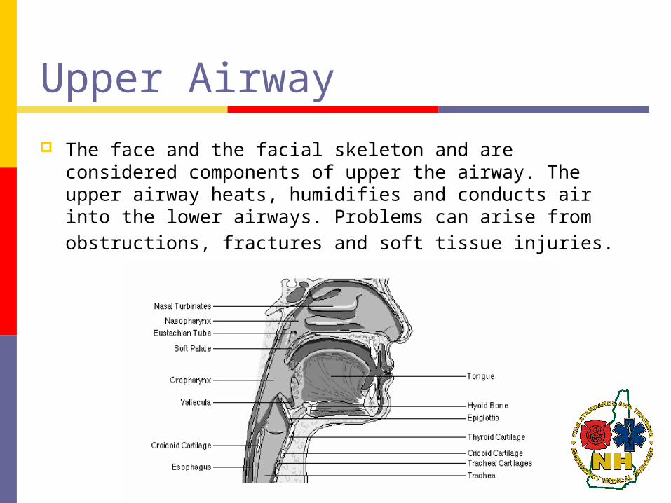
Upper Airway
The face and the facial skeleton and are considered components of upper the airway. The upper airway heats, humidifies and conducts air into the lower airways. Problems can arise from obstructions, fractures and soft tissue injuries.
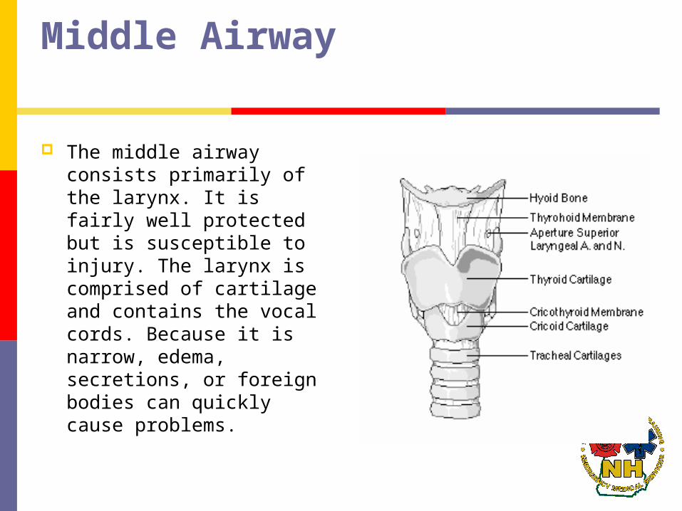
Middle Airway
The middle airway consists primarily of the larynx. It is fairly well protected but is susceptible to injury. The larynx is comprised of cartilage and contains the vocal cords. Because it is narrow, edema, secretions, or foreign bodies can quickly cause problems.

Lower Airways
The lower airway begins at the trachea as it exits the neck and enters the chest. It consists of c-shaped cartilage rings held together by elastic-muscle tissue posteriorly, divides into the right and left mainstem bronchi and continues to the lung tissue.
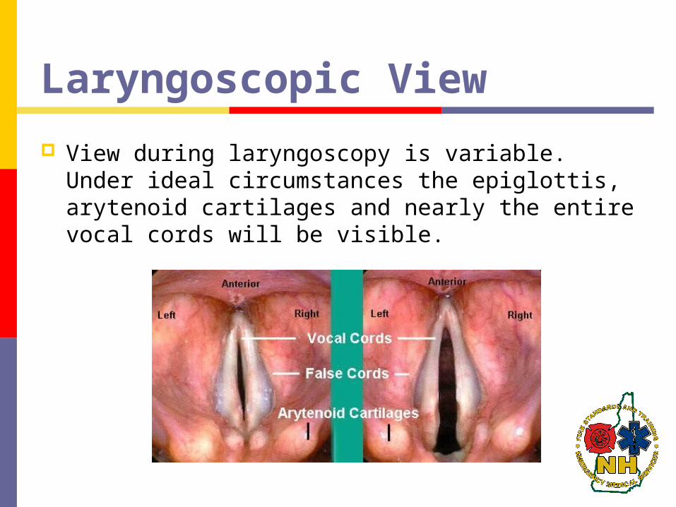
Laryngoscopic View
View during laryngoscopy is variable. Under ideal circumstances the epiglottis, arytenoid cartilages and nearly the entire vocal cords will be visible.
INDICATIONS
One of the basic functions of a provider is to ensure a patent airway. A provider must be able to rapidly identify patients at risk and determine the most appropriate method to manage the airway.
Immediate severe airway compromise in the context of trauma, drug overdose, status epilepticus, etc., where respiratory arrest is imminent.
INDICATIONS
When determining the best method for maintaining an airway, consider the following: Is the patient at risk for a positional obstruction
or aspiration? Is there inadequate oxygenation and/or
ventilation? Is the patient’s condition expected to
deteriorate?
Examples of patients requiring RSI:
Conditions requiring oxygenation/ventilation control or positive pressure ventilation: Traumatic brain injury with ALOC Severe thoracic trauma (flail chest, pulmonary
contusions with hypoxemia) Clinical condition expected to deteriorate
Unconscious or ALOC with potential for or actual airway compromise or vomiting
And patient has…… A clenched jaw An active gag reflex
Contraindications
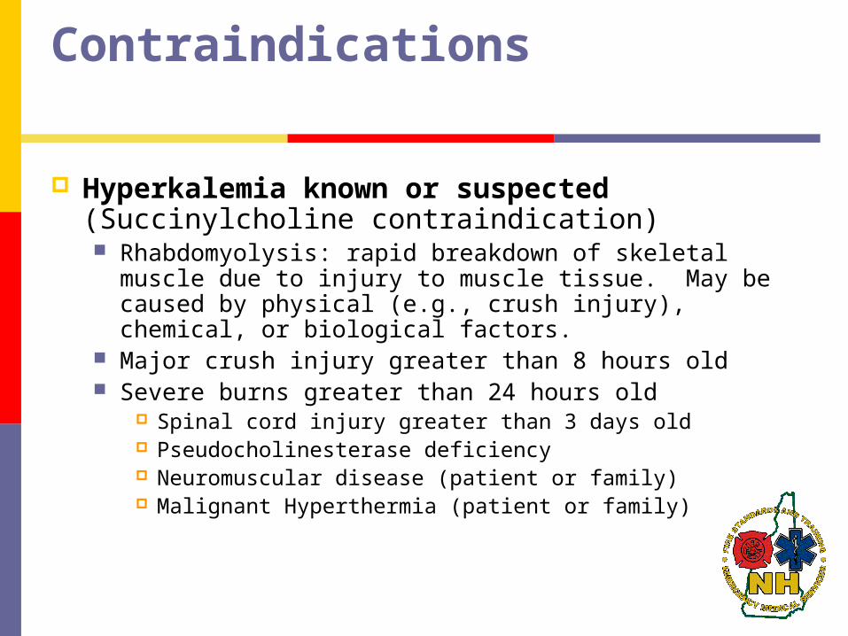
Hyperkalemia known or suspected (Succinylcholine contraindication) Rhabdomyolysis: rapid breakdown of skeletal muscle
due to injury to muscle tissue. May be caused by physical (e.g., crush injury), chemical, or biological factors.
Major crush injury greater than 8 hours old Severe burns greater than 24 hours old
Spinal cord injury greater than 3 days old Pseudocholinesterase deficiency Neuromuscular disease (patient or family) Malignant Hyperthermia (patient or family)
Contraindications
Anticipating an airway that you can’t manage
Penetrating eye injuries
Narrow angle glaucoma
Pregnancy
The medications are “Pregnancy Risk Category – C”
No human studies and animal studies show adverse effect
Transmission to breast milk uncertain – likely – but not a significant concern in an RSI situation
“Because of the higher incidence of difficult intubation in pregnancy (due to increases in size and weight during pregnancy), optimal positioning of the patient becomes more important.

Pregnancy Failure to manage the airway appropriately in a gravid
patient potentially threatens not one life, but two, as maternal complications are the leading cause of fetal insult and death.1 Understanding anatomic and physiologic changes in pregnancy paired with adequate preparation for airway management minimizes this risk.
Although limited data exist concerning drugs for rapid sequence intubation in this population, etomidate and succinylcholine are considered acceptable in the gravid patient. Succinylcholine does not cross the placental barrier, although induction agents do. Etomidate does cross the placental barrier, but has been show to cause less respiratory depression in the newborn than thiopental.10” (acep.org 7/2007)
What is my role?
Basic airway maintenance while the Paramedic prepares their equipment
Positioning, suctioning, oral and nasal airways Pre-oxygenate with 100% O2 via non-rebreather
mask for at least 3-5 minutes Replaces the patient’s functional residual
capacity (FRC) of the lung with oxygen“Nitrogen Washout”
What is my Role? Cont.
Ventilate the patient via the endotracheal tube. Do not push or pull on the tube. Place one hand on the patient’s jaw and hold the tube tightly at the teeth as the Paramedic secures the tube. It is safest to continue to hold onto the tube even with a tube holder in place. This gives you better special orientation relative to the BVM and movement of the tube. Pay attention to the ease of ventilation and immediately tell the Paramedic if this changes at all. A change in ventilatory effort can mean: pneumothorax, displaced tube, equipment failure, and need for more sedation to name but a few.
What is my Role? Cont.
Remove the BVM from the endotracheal tube prior to moving the patient. This is when most tubes become displaced.
Watch the capnography. A change in these numbers can be the result of the previous complications.
Should the Paramedic become incapacitated for any reason or be unable to secure the airway; be prepared to manage the airway with a King Tube or Combitube as well as with BLS maneuvers.
What are the medications and what will I see?
Lidocaine: May be administered 2-3 minutes prior to
paralyzation in certain cases. Asthmatics and traumatic brain injury patients
may benefit from this medication. Lidocaine blunts the gag and cough reflex, acts
as a smooth muscle relaxant, and may prevent transient increases in intercranial pressure associated with intubation.
Dosing is 1.5mg/kg IVP.
What are the medications and what will I see?
Atropine: Utilized in bradycardic patients and in pediatric
intubations. Pediatric patients have a higher
parasympathetic response to intubation. Dosing is 0.5mg IVP.

What are the medications and what will I see?
Etomidate: Sedative that is given prior to paralyzing the
patient. Paralyzation has no affect on sensation or
mentation. There are few things crueler than paralyzing a
conscious patient. This is a quick acting drug (one arm-brain
cycle) and has a short half life.
Etomidate can burn at the IV sight and up the extremity so warn the patient.
Many are asleep though before they complain of it burning.
The dose is 0.3mg/kg IVP; typically 20, 30, or 40mg and lasts 5-6 minutes.
What are the medications and what will I see? Succinylcholine:
Short acting paralytic known as a “Depolarizing Agent”.
This means that all of the body’s cells will fire prior to relaxing and the patient will “Fasciculate”.
This presents as overall twitching.
Succinylcholine The patient will then stop breathing. Remember that this medication does nothing
to sensation and mentation. Hearing, sight, etc are intact dependant upon sedation.
Succinylcholine has a half life of 5-6 minutes also which is why it is the preferred RSI paralytic.
Succinylcholine
Once this medication is given we can not take it back.
We are taking away what airway and
ventilation control they had. We own this responsibility.
Dosing is 1.5mg/kg IVP; typically 75 or 150mg.
What are the medications and what will I see?
Rocuronium: is a “Non-depolarizing Agent” and does not
cause fasciculations. It does however have a 20-40 minute half life. This medication is typically utilized after they
have been intubated or in cases where Succinylcholine is contraindicated.
Dosing is 1mg/kg IVP; typically 50, 100, or 150mg.
What are the medications and what will I see?
Versed (midazolam): A benzodiazepine utilized for post intubation
sedation.
This medication may cause hypotension. Sedation is required immediately after
intubation as the Etomidate will be wearing off.
Versed (midazolam):
Indications for subsequent doses are; tearing, coughing against the tube, rises in heart rate and the capnography numeric.
Increases in movement are late signs.
Dosing is 0.05 to 0.1mg/kg IVP every 5 minutes as needed; typically 2.5 to 10mg.
What are the medications and what will I see?
Fentanyl: Man made narcotic similar to Morphine Fentanyl has less risk of hypotension and
allergic reactions than does morphine. Sedation and paralyzation have no affect on
pain sensors so Fentanyl is important in patients with a pain component such as trauma or head bleeds.
Fentanyl
Fentanyl also works synergistically with versed to assist in sedation.
Dosing is 50mcg every 5-10 minutes as needed
to a maximum of 150mcg.
Further dosing requires medical control.
RSI medications are authorized for RSI Paramedics only!
RAPID SEQUENCE INTUBATION (RSI) 5.4
Paramedic Standing Orders
Prerequisites Required:
This procedure is only to be used by paramedics that are trained and credentialed to perform RSI by NH Bureau of EMS.
Definition:
Near-simultaneous administration of neuromuscular blocking agents and sedative-hypnotic drugs in order to facilitate oral intubation of a patient with the least likelihood of trauma, aspiration, hypoxia, and other physiologic complications.
Procedure: The seven “Ps”
Preparation: The time-frame is limited, but the operator must
have adequate Ambu-mask/oxygen sources, two laryngoscope handles, an assortment of blades, two assistants familiar with the procedure, one or two secure IV’s, rescue airway devices, oxymetry & capnography monitoring, bulb-style tube checker.
Procedure: The seven “Ps”
Preoxygenation: When possible a nonrebreather mask for several
minutes is more effective in performing nitrogen washout and establishing an adequate oxygen reserve during the procedure. In emergent cases, three mask breaths with 100% oxygen may suffice

Procedure: The seven “Ps”
Premedication: Lidocaine (1.5mg/kg) given exactly 2 minutes
before intubation may prevent a rise in ICP for head injured patients.
Atropine should be given to bradycardic adults at
0.5mg IVP.
Procedure: The seven “Ps”
Paralyze: Etomidate (0.3mg/kg IV). Apply cricoid pressure
and maintain until the airway is secure.
Succinylcholine (1.5mg/kg IVP) immediately after Etomidate
Procedure: The seven “Ps”
Pass the tube: Observe for fasciculations approximately 90
seconds after Succinylcholine to indicate imminent paralysis. After paralysis is achieved, follow procedure outlined in section 5.2 to place the endotracheal tube.
Procedure: The seven “Ps”
Proof of placement: Assess for adequate placement by auscultation
(equal breath sounds over the chest and lack of sounds over the epigastrium with bagging), condensation in the ETT, symmetrical chest wall rise and at least one additional method: colorimetric end-tidal CO2 detector, capnography, or esophageal tube detector (note: this device should be used prior to ventilation to be accurate). This should be repeated often, especially after movement of the patient.
Procedure: The seven “Ps”
Post intubation care: The patient may be given incremental doses of
midazolam (0.05-0.10mg/kg IVP) or lorazepam 1 - 2 mg IV as needed for sedation. For continued paralysis, vecuronium 0.1 mg/kg IVP or rocuronium 1 mg/kg IVP may be considered, with on-line medical consultation. Consider wrist restraints.
If done properly, this will permit as much as 3-4 minutes of apnea before hypoxia develops
In emergent cases, three mask breaths with 100% oxygen may have to suffice.
Resist the use of positive pressure ventilation (PPV). Use only if the patient is not ventilating adequately.
PPV leads to gastric distention regurgitation aspiration
If PPV is necessary, utilize cricoid pressure
Cricoid Pressure Use thumb and forefinger to apply pressure directly
backward/posterior over the cricoid cartilage. Also known as “Sellick’s Maneuver” Should be automatic just as Etomidate is administered Maintained until ETT placement is confirmed and tube is
secure (cuff inflated) Used to occlude the esophagus and prevent passive
regurgitation common with Succinylcholine If patient starts to actively vomit – RELEASE! and suction
oropharnyx, otherwise, can lead to esophageal rupture
Cricoid Pressure
Used to occlude the esophagus and prevent passive regurgitation common with Succinylcholine
If patient starts to actively vomit – RELEASE! and suction oropharnyx, otherwise, can lead to esophageal rupture
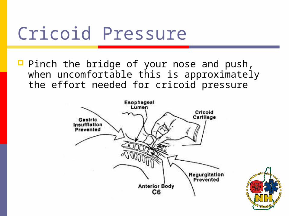
Pinch the bridge of your nose and push, when uncomfortable this is approximately the effort needed for cricoid pressure
Cricoid Pressure
BURP
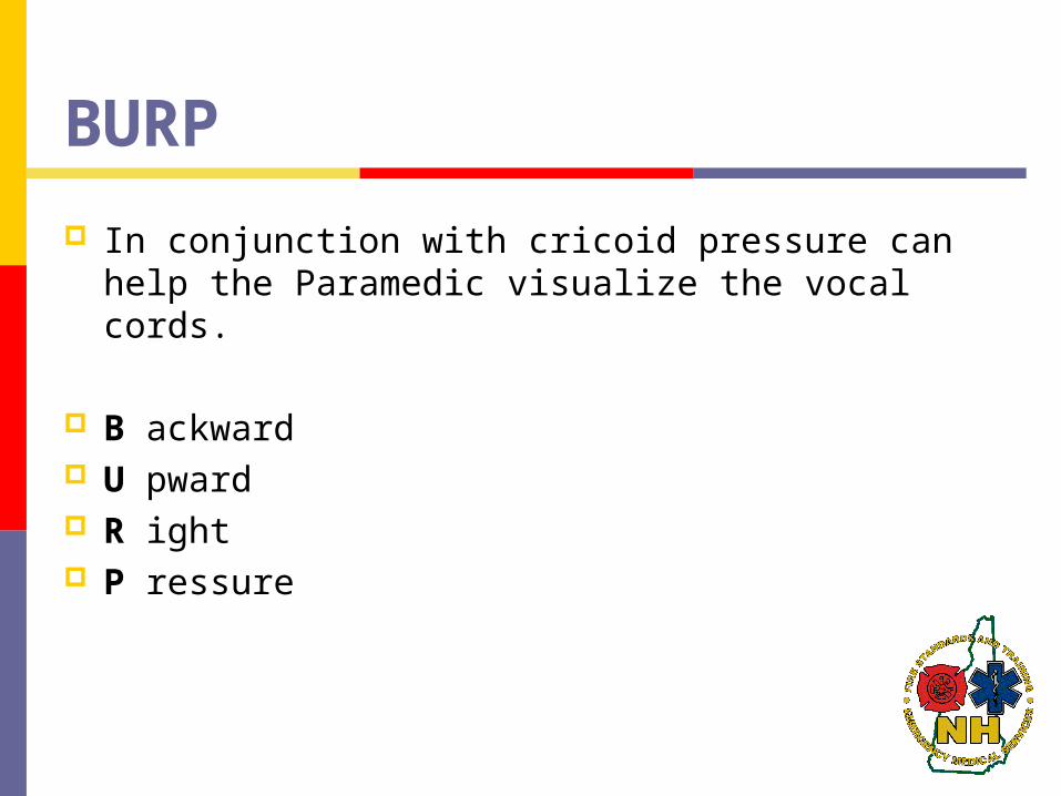
In conjunction with cricoid pressure can help the Paramedic visualize the vocal cords.
B ackward U pward R ight P ressure
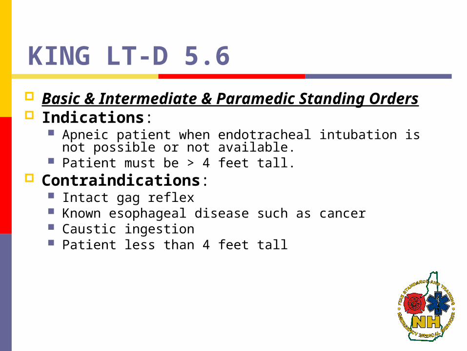
KING LT-D 5.6 Basic & Intermediate & Paramedic Standing
Orders Indications:
Apneic patient when endotracheal intubation is not possible or not available.
Patient must be > 4 feet tall. Contraindications:
Intact gag reflex Known esophageal disease such as cancer Caustic ingestion Patient less than 4 feet tall
KING LT-D 5.6
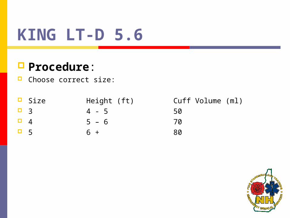
Procedure: Choose correct size:
Size Height (ft) Cuff Volume (ml) 3 4 - 5 50 4 5 – 6 70 5 6 + 80
KING LT-D 5.6
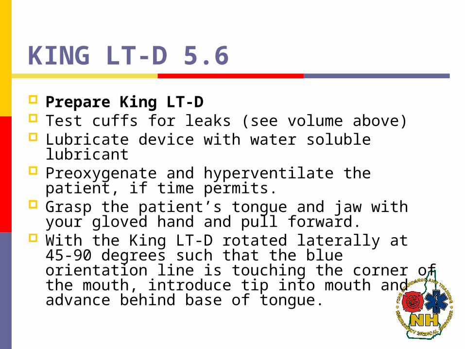
Prepare King LT-D Test cuffs for leaks (see volume above) Lubricate device with water soluble lubricant Preoxygenate and hyperventilate the patient, if
time permits. Grasp the patient’s tongue and jaw with your
gloved hand and pull forward. With the King LT-D rotated laterally at 45-90
degrees such that the blue orientation line is touching the corner of the mouth, introduce tip into mouth and advance behind base of tongue.
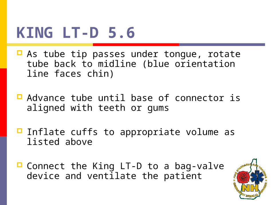
KING LT-D 5.6 As tube tip passes under tongue, rotate tube back
to midline (blue orientation line faces chin)
Advance tube until base of connector is aligned with teeth or gums
Inflate cuffs to appropriate volume as listed above
Connect the King LT-D to a bag-valve device and ventilate the patient

KING LT-D 5.6
Assess for adequate placement by auscultation (equal breath sounds over the chest and lack of sounds over the epigastrium with bagging), condensation in the ETT, symmetrical chest wall rise and at least one additional method: colorimetric end-tidal CO2 detector, capnography, or esophageal tube detector (note: this device should be used prior to ventilation to be accurate).
KING LT-D 5.6
This should be repeated often, especially after movement of the patient.
Secure the device.
COMBITUBE 5.5
Basic & Intermediate & Paramedic standing orders
Indications Apneic patient when endotracheal intubation is
not possible or not available. Standard Combitube: patient must be at least 5
feet tall. Combitube SA(small adult): patient 4 – 5 1/2 feet
tall
COMBITUBE 5.5
Contraindications Intact gag reflex Patients < 4 feet tall Known esophageal disease such as cancer Caustic ingestion Allergy or sensitivity to latex (the pharyngeal
balloon contains latex)
COMBITUBE 5.5 Procedure Prepare Combitube Test balloons Proximal pharyngeal cuff (blue pilot balloon) –
100 ml Distal esophageal cuff (white pilot balloon) – 15
ml Lubricate device with water-soluble lubricant. Preoxygenate and hyperventilate the patient, if
time permits.
COMBITUBE 5.5
Grasp the patient’s tongue and jaw with your gloved hand and pull forward
Gently insert the tube until the teeth (or gums) are between the printed rings
Inflate cuff #1 (blue pilot balloon) with 100 ml of air
Inflate cuff #2 (white pilot balloon) with 15 ml of air
COMBITUBE 5.5 Ventilate taller blue tube (#1) with bag valve
mask Auscultate for breath sounds and sounds over the
epigastrium Look for rise and fall of chest. If breath sounds
are present and epigastric sounds are absent, continue to ventilate through the blue tube. The tube is properly positioned in the esophagus. In the case above you can aspirate stomach contents through the #2 white tube to relieve some gastric distention
COMBITUBE 5.5
If breath sounds are absent and epigastric sounds are present, attempt to ventilate through the shorter white (#2) tube and assess for breath sounds and epigastric sounds. If breath sounds are present and epigastric sounds are absent, continue to ventilate through the white tube (#2); you have intubated the trachea.
COMBITUBE 5.5 In addition to auscultation, confirm tube
placement by using at least one additional method: colorimetric end-tidal CO2 detector, capnography, or esophageal tube detec tor (note: this device should be used prior to ventilation to be accurate).
This should be repeated often, especially after movement of the patient.
Secure the device.
ADVANCED SUCTIONING 5.10
Basic & Intermediate & Paramedic Standing Orders
Indication Obstruction of the airway (secondary to
secretions, blood, and/or any other substance) in a patient currently being assisted by an airway adjunct such as an endotracheal tube, Combitube, tracheostomy tube, or a cricothyrotomy tube
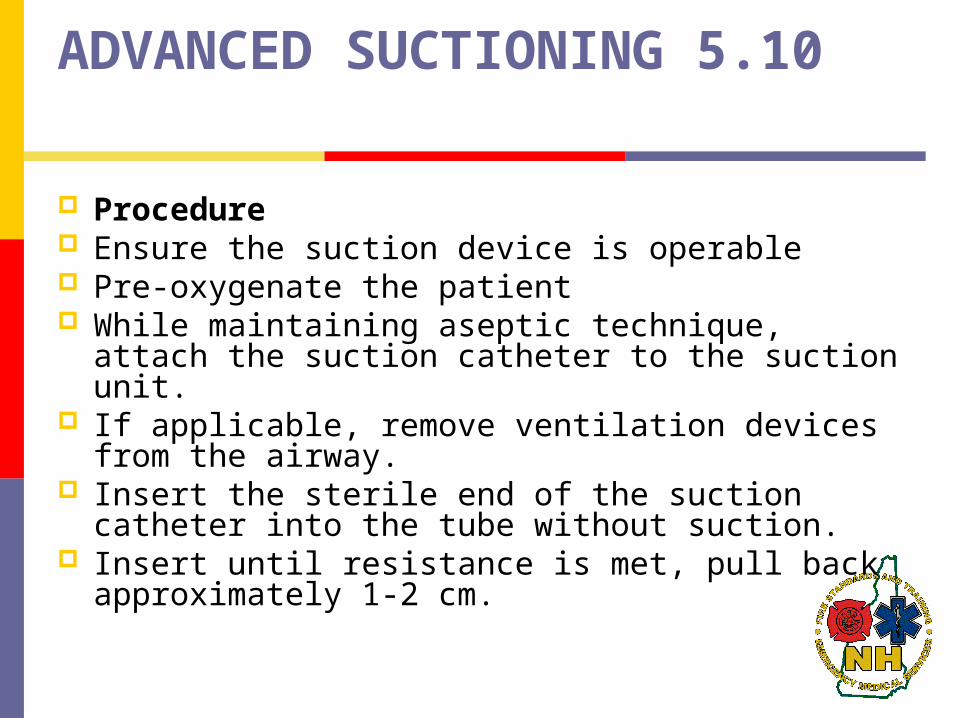
ADVANCED SUCTIONING 5.10
Procedure Ensure the suction device is operable Pre-oxygenate the patient While maintaining aseptic technique, attach the
suction catheter to the suction unit. If applicable, remove ventilation devices from the
airway. Insert the sterile end of the suction catheter into
the tube without suction. Insert until resistance is met, pull back
approximately 1-2 cm.
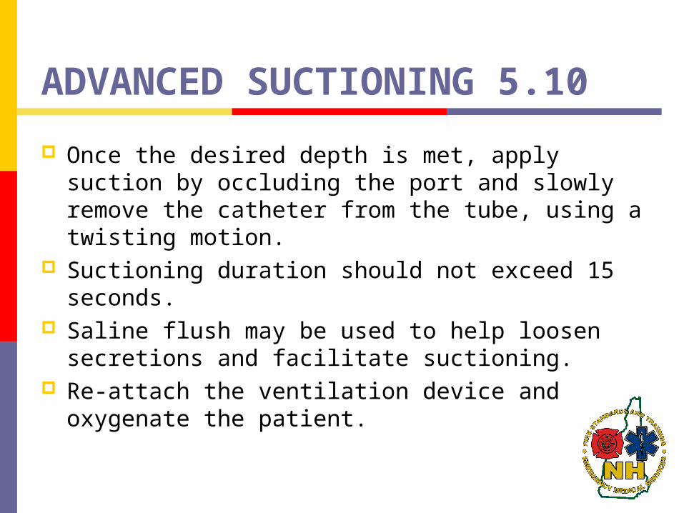
ADVANCED SUCTIONING 5.10
Once the desired depth is met, apply suction by occluding the port and slowly remove the catheter from the tube, using a twisting motion.
Suctioning duration should not exceed 15 seconds.
Saline flush may be used to help loosen secretions and facilitate suctioning.
Re-attach the ventilation device and oxygenate the patient.
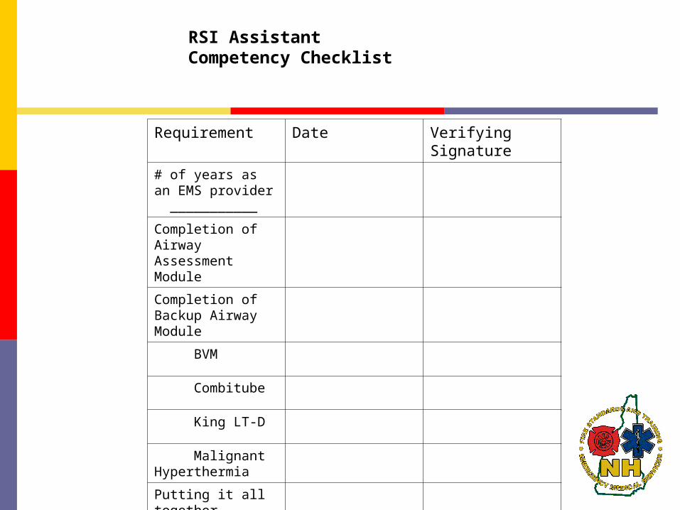
Requirement Date Verifying Signature
# of years as an EMS provider ___________
Completion of Airway Assessment Module
Completion of Backup Airway Module
BVM
Combitube
King LT-D
Malignant Hyperthermia
Putting it all together
Airway Simulation Exam
Signatures Instructor Medical Director
RSI AssistantCompetency Checklist


![Optimality of Universal Bayesian Sequence Prediction for ...[Sol78], and allows for a good prediction. In a sense, this solves the induction In a sense, this solves the induction problem](https://img.pdfslide.us/doc/110x75/605094e690a9227b817bd043/optimality-of-universal-bayesian-sequence-prediction-for-sol78-and-allows.jpg)