Embed Size (px)
Citation preview
Rapid Publication
Epidermolytic Hyperkeratosis (Bullous Congenital Ichthyosiform Erythroderma)Genetic Linkage to Chromosome 12q in the Region of the Type 11 Keratin Gene Cluster
Leena Pulkkinen, Angela M. Christiano, Robert G. Knowlton, and Jouni UittoDepartments of Dermatology, and Biochemistry and Molecular Biology, Jefferson Medical College, and Section of MolecularDermatology, Jefferson Institute of Molecular Medicine, Thomas Jefferson University, Philadelphia, Pennsylvania 19107
Abstract
Epidermolytic hyperkeratosis (EHK) is an autosomal domi-nant genodermatosis characterized by hyperkeratosis and blis-tering of the skin. Histopathology demonstrates suprabasilarblister formation with aggregation of tonofilaments. In thisstudy, we tested the hypothesis that the EHK phenotype islinked to one of the suprabasilar keratins (KRT10 or KRT1)present in the types I and II keratin gene clusters in chromo-somes 17q and 12q, respectively. For this purpose, Southernhybridizations were performed with DNAfrom a large kindredwith EHK, consisting of 11 affected individuals in three genera-tions. Segregation analysis with markers flanking the keratingene clusters demonstrated linkage (Z = 3.61 at 0 = 0) to alocus on 12q, while markers on 17q were excluded. These dataimplicate KRT1, the type II keratin expressed in suprabasilarkeratinocytes, as a candidate gene in this family with EHK. (J.Clin. Invest. 1993. 91:357-361.) Key words: bullous skin dis-eases * genetic linkage analyses * genodermatoses - keratiniza-tion disorders
Introduction
Epidermolytic hyperkeratosis (EHK)' is a heritable keratiniza-tion disorder with a marked tendency for blistering. The preva-lence of this disorder is estimated to be - 1:200,000. (For re-views, see references 1 and 2). The disease is present at birth orshortly thereafter, and the condition can be generalized (bul-lous congenital ichthyosiform erythroderma) or localized, af-fecting primarily the flexural areas of the skin. With time, theskin becomes hyperkeratotic and verrucous, and the blisteringtendency becomes less pronounced, though it can persistthroughout life. The inheritance pattern is autosomal domi-nant, but there is considerable heterogeneity regarding the se-verity of the disease, as well as evidence for variable expressionwithin families. Histopathology of the cutaneous lesions re-
Address reprint requests to Dr. Jouni Uitto, Department of Dermatol-ogy, Jefferson Medical College, 233 South 10th Street, Room450, Phil-adelphia, PA 19107.
Receivedfor publication 29 July 1992 and in revisedform 29 Sep-tember 1992.
1. Abbreviation used in this paper: EHK, epidermolytic hyperkeratosis.
veals that the tissue cleavage occurs intraepidermally throughseparation of abnormal suprabasilar keratinocytes, combinedwith focal hyperkeratosis (3).
Significant progress has recently been made in delineatingthe underlying mutations in various forms of epidermolysisbullosa, a group of heritable blistering diseases (4, 5). In partic-ular, several forms of epidermolysis bullosa simplex have beenshown to result from mutations in the keratin genes expressedin the basal keratinocytes, KRT5 and KRT14 (6-8). Thesecandidate genes were initially suggested by characteristic aggre-gation of tonofilaments of the basal keratinocytes (9). Similarabnormalities have been demonstrated in the suprabasilar kera-tinocytes in EHK, including aggregation of tonofilaments andlysis of suprabasilar keratinocytes ( 10, 11 ). Based on these simi-larities between EHKand epidermolysis bullosa simplex, weused linkage analysis to test the hypothesis that the EHKpheno-type is due to mutations in the suprabasilar keratin genes(KRT 10 or KRT1) present in the types I and II keratin geneclusters on chromosomes 1 7q and 12q, respectively.
Methods
Clinical. Peripheral blood samples were obtained from a family withclinical features of EHK(Fig. 1 ). The affected individuals had a historyof blisters at birth with accompanying erythroderma, and in the adultindividuals, thickened, verrucous skin, primarily on the flexural areason the arms and legs was noted (Fig. 1). The blister formation wasconsiderably more frequent during the summertime, and was inducedby relatively minor trauma. All affected individuals depicted pro-nounced hirsutism on the extremities noticeable as early as 3 yr of age(Fig. 1).
The pattern of transmission of the phenotype was consistent withautosomal dominant inheritance (Fig. 2, top). The proband (IV-9) wasa 3-yr-old female with characteristic clinical features (Fig. 1 ). It was ofinterest to note that the maternal great-grandfather (1-2) of the pro-band was anamnestically affected, yet his 13 siblings were free of thedisease. Therefore, his phenotype may have been the result of a newmutation in this kindred.
Another interesting feature was the occurrence of retinitis pigmen-tosa in this family. However, careful examination of the pedigree indi-cated that the retinitis pigmentosa mutation did not co-segregate withthe EHK phenotype. In fact, the retinitis pigmentosa mutation wasinherited from the maternal grandfather of the proband (11- 10), whilethe EHKmutation was transmitted through the maternal grandmotherof the proband (11-3).
Diagnostic histopathology of the skin performed on patients 111-5and IV-8 revealed prominent hyperkeratosis, zones of compact ortho-keratosis, and reticular alteration with ballooning in the upper spinousand granular layers of the epidermis (Fig. 3). The epidermal cells in theregion of the blister formation were enlarged and vacuolated, and in
Epidermolvtic Hyperkeratosis 357
J. Clin. Invest.© The American Society for Clinical Investigation, Inc.0021-9738/93/01/0357/05 $2.00Volume 91, January 1993, 357-361
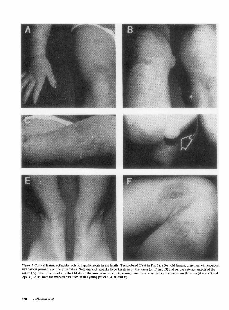
Figure 1. Clinical features of epidermolytic hyperkeratosis in the family. The proband (IV-9 in Fig. 2), a 3-yr-old female, presented with erosionsand blisters primarily on the extremities. Note marked ridgelike hyperkeratosis on the knees (A, B, and D) and on the anterior aspects of theankles (E). The presence of an intact blister of the knee is indicated (D, arrow), and there were extensive erosions on the arms (A and C) andlegs (F). Also, note the marked hirsutism in this young patient (A, B, and F).
358 Pulkkinen et al.
.': :, .,. ",-N-
:PI,:!.,
s,
0s2 e~
(-"\1 0 2B3 4
B31C
21ij21~xj4'5 #i7 i 9 j 0 j2i3L 4ji5AB AC AB BB A1' AC AC BBI AB BC BC
iV tlb2 4 5 j6 7 *e8 0AA AA AS
11-3 111-1 111-2 IV- IV-2 IV-4 111-5 111-6 111-8 111-10 111-11 111-12 111-13 111-14 111-15
.v .....a2::
.X t;fy "
CtL 7is:0-
some areas had completely degenerated to form the blister space. Thesefeatures are diagnostic for EHK(3).
DNAanalysis. DNAwas isolated from peripheral blood and di-gested either with Hinf I or MspI restriction endonuclease, according tothe manufacturer's recommendations (Boehringer-Mannheim Bio-chemicals, Indianapolis, IN). The Southem transfer analyses of therestriction enzyme digests and probe hybridizations were performed asdescribed previously ( 12). The following DNAprobes were used foranalysis: for chromosome 12q, pYNH15 (D 1 2S 17) (13) and WV214
~ p }O - . X ^; *-*-v_-
Figure 3. Histopathology of the skin in the patient (IV-8) with epi-dermolytic hyperkeratosis. Note prominent hyperkeratosis and hy-pergranulosis, and the presence of abnormal keratinocytes with bal-looning degeneration in the spinous and granular layers of the epi-dermis.
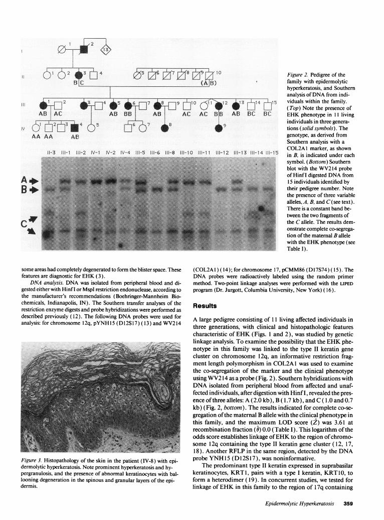
Figure 2. Pedigree of thefamily with epidermolytichyperkeratosis, and Southernanalysis of DNAfrom indi-viduals within the family.(Top) Note the presence ofEHKphenotype in 11 livingindividuals in three genera-tions (solid symbols). Thegenotype, as derived fromSouthern analysis with aCOL2A1 marker, as shownin B, is indicated under eachsymbol. (Bottom) Southernblot with the WV214probeof Hinf I digested DNAfrom15 individuals identified bytheir pedigree number. Notethe presence of three variablealleles, A, B, and C(see text).There is a constant band be-tween the two fragments ofthe Callele. The results dem-onstrate complete co-segrega-tion of the maternal B allelewith the EHKphenotype (seeTable I).
(COL2A 1) ( 14); for chromosome 17, pCMM86(D 1 7S74) (15). TheDNA probes were radioactively labeled using the random primermethod. Two-point linkage analyses were performed with the LIPEDprogram (Dr. Jurgott, Columbia University, NewYork) (16).
Results
A large pedigree consisting of 11 living affected individuals inthree generations, with clinical and histopathologic featurescharacteristic of EHK(Figs. 1 and 2), was studied by geneticlinkage analysis. To examine the possibility that the EHKphe-notype in this family was linked to the type II keratin genecluster on chromosome 12q, an informative restriction frag-ment length polymorphism in COL2A1 was used to examinethe co-segregation of the marker and the clinical phenotypeusing WV214 as a probe (Fig. 2). Southern hybridizations withDNAisolated from peripheral blood from affected and unaf-fected individuals, after digestion with Hinf I, revealed the pres-ence of three alleles: A (2.0 kb), B ( 1.7 kb), and C ( 1.0 and 0.7kb) (Fig. 2, bottom). The results indicated for complete co-se-gregation of the maternal B allele with the clinical phenotype inthis family, and the maximum LOD score (Z) was 3.61 atrecombination fraction (0) 0.0 (Table I). This logarithm of theodds score establishes linkage of EHKto the region of chromo-some 12q containing the type II keratin gene cluster ( 12, 17,18). Another RFLP in the same region, detected by the DNAprobe YNH15 (D l 2S 17), was noninformative.
The predominant type II keratin expressed in suprabasilarkeratinocytes, KRT1, pairs with a type I keratin, KRT10, toform a heterodimer ( 19). In concurrent studies, we tested forlinkage of EHKin this family to the region of 1 7q containing
Epidermolytic Hyperkeratosis 359
(.5 1 1 i. i
B)
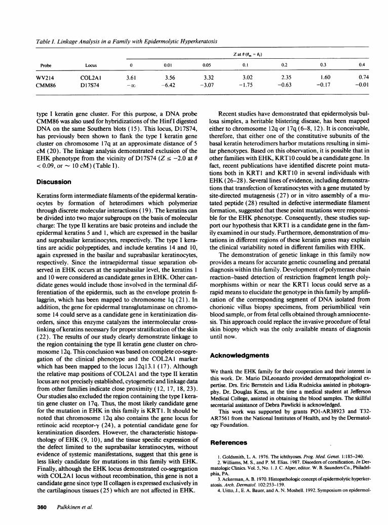
Table I. Linkage Analysis in a Family with Epidermolytic Hyperkeratosis
Z at 6 (Om = of)
Probe Locus 0 0.01 0.05 0.1 0.2 0.3 0.4
WV214 COL2A1 3.61 3.56 3.32 3.02 2.35 1.60 0.74CMM86 D17S74 -oo -6.42 -3.07 -1.75 -0.63 -0. 17 -0.01
type I keratin gene cluster. For this purpose, a DNAprobeCMM86was also used for hybridizations of the Hinf I digestedDNAon the same Southern blots ( 15). This locus, Dl 7S74,has previously been shown to flank the type I keratin genecluster on chromosome 17q at an approximate distance of 5cM (20). The linkage analysis demonstrated exclusion of theEHKphenotype from the vicinity of D17S74 (Z < -2.0 at 0< 0.09, or - 10 cM) (Table I).
Discussion
Keratins form intermediate filaments of the epidermal keratin-ocytes by formation of heterodimers which polymerizethrough discrete molecular interactions ( 19). The keratins canbe divided into two major subgroups on the basis of molecularcharge: The type II keratins are basic proteins and include theepidermal keratins 5 and 1, which are expressed in the basilarand suprabasilar keratinocytes, respectively. The type I kera-tins are acidic polypeptides, and include keratins 14 and 10,again expressed in the basilar and suprabasilar keratinocytes,respectively. Since the intraepidermal tissue separation ob-served in EHKoccurs at the suprabasilar level, the keratins 1and 10 were considered as candidate genes in EHK. Other can-didate genes would include those involved in the terminal dif-ferentiation of the epidermis, such as the envelope protein fi-laggrin, which has been mapped to chromosome Iq (21). Inaddition, the gene for epidermal transglutaminase on chromo-some 14 could serve as a candidate gene in keratinization dis-orders, since this enzyme catalyzes the intermolecular cross-linking of keratins necessary for proper stratification of the skin(22). The results of our study clearly demonstrate linkage tothe region containing the type II keratin gene cluster on chro-mosome 12q. This conclusion was based on complete co-segre-gation of the clinical phenotype and the COL2A1 markerwhich has been mapped to the locus 1 2q 13. 1 (17). Althoughthe relative map positions of COL2A1 and the type II keratinlocus are not precisely established, cytogenetic and linkage datafrom other families indicate close proximity ( 12, 17, 18, 23).Our studies also excluded the region containing the type I kera-tin gene cluster on 17q. Thus, the most likely candidate genefor the mutation in EHKin this family is KRT1. It should benoted that chromosome 12q also contains the gene locus forretinoic acid receptor-y (24), a potential candidate gene forkeratinization disorders. However, the characteristic histopa-thology of EHK(9, 10), and the tissue specific expression ofthe defect limited to the suprabasilar keratinocytes, withoutevidence of systemic manifestations, suggest that this gene isless likely candidate for mutations in this family with EHK.Finally, although the EHKlocus demonstrated co-segregationwith COL2A1 locus without recombination, this gene is not acandidate gene since type II collagen is expressed exclusively inthe cartilaginous tissues (25) which are not affected in EHK.
Recent studies have demonstrated that epidermolysis bul-losa simplex, a heritable blistering disease, has been mappedeither to chromosome 1 2q or 1 7q (6-8, 12). It is conceivable,therefore, that either one of the constitutive subunits of thebasal keratin heterodimers harbor mutations resulting in simi-lar phenotypes. Based on this observation, it is possible that inother families with EHK, KRT1Ocould be a candidate gene. Infact, recent publications have identified discrete point muta-tions both in KRT1 and KRT1O in several individuals withEHK(26-28). Several lines of evidence, including demonstra-tions that transfection of keratinocytes with a gene mutated bysite-directed mutagenesis (27) or in vitro assembly of a mu-tated peptide (28) resulted in defective intermediate filamentformation, suggested that these point mutations were responsi-ble for the EHKphenotype. Consequently, these studies sup-port our hypothesis that KRT1 is a candidate gene in the fam-ily examined in our study. Furthermore, demonstration of mu-tations in different regions of these keratin genes may explainthe clinical variability noted in different families with EHK.
The demonstration of genetic linkage in this family nowprovides a means for accurate genetic counseling and prenataldiagnosis within this family. Development of polymerase chainreaction-based detection of restriction fragment length poly-morphisms within or near the KRT1 locus could serve as arapid means to elucidate the genotype in this family by amplifi-cation of the corresponding segment of DNA isolated fromchorionic villus biopsy specimens, from periumbilical veinblood sample, or from fetal cells obtained through amniocente-sis. This approach could replace the invasive procedure of fetalskin biopsy which was the only available means of diagnosisuntil now.
Acknowledgments
Wethank the EHKfamily for their cooperation and their interest inthis work. Dr. Mario DiLeonardo provided dermatopathological ex-pertise. Drs. Eric Bernstein and Lidia Rudnicka assisted in photogra-phy. Dr. Douglas Kress, at the time a medical student at JeffersonMedical College, assisted in obtaining the blood samples. The skillfulsecretarial assistance of Debra Pawlicki is acknowledged.
This work was supported by grants PO1-AR38923 and T32-AR756 1 from the National Institutes of Health, and by the Dermatol-ogy Foundation.
References
1. Goldsmith, L. A. 1976. The ichthyoses. Prog. Med. Genet. 1:185-240.2. Williams, M. S., and P. M. Elias. 1987. Disorders of comification. In Der-
matologic Clinics. Vol. 5, No. 1. J. C. Alper, editor. W. B. Saunders Co., Philadel-phia, PA.
3. Ackerman, A. B. 1970. Histopathologic concept of epidermolytic hyperker-atosis. Arch. Dermatol. 102:253-159.
4. Uitto, J., E. A. Bauer, and A. N. Moshell. 1992. Symposium on epidermol-
360 Pulkkinen et al.
ysis bullosa: molecular biology and pathology of the cutaneous basement mem-brane zone. J. Invest. Dermatol. 98:391-395.
5. Uitto, J., and A. M. Christiano. 1992. Molecular genetics of the cutaneousbasement membrane zone: perspectives on epidermolysis bullosa and other blis-tering skin diseases. J. Clin. Invest. 90:687-692.
6. Coulombe, P. A., M. E. Hutton, A. Letai, A., A. Hebert, A. P. Paller, and E.Fuchs. 1991. Point mutations in human keratin 14 genes of epidermolysis bullosasimplex patients: genetic and functional analyses. Cell. 66:1301-131 1.
7. Epstein, E. H., Jr. 1992. Molecular genetics of epidermolysis bullosa.Science (Wash. DC) . 256:799-804.
8. Lane, E. B., E. L. Rugg, H. Navsaria, I. M. Leigh, A. H. M. Haegerty, A.Ishida-Yamamoto, and R. A. J. Eady. 1992. A mutation in the conserved helixtermination peptide of keratin 5 in hereditary skin blistering. Nature (Lond.).356:244-246.
9. Ishida-Yamamoto, A., J. A. McGrath, S. J. Chapman, I. M. Leigh, E. B.Lane, and R. A. J. Eady. 1991. Epidermolysis bullosa simplex (Dowling-Mearatype) is a genetic disease characterized by an abnormal keratin-filament networkinvolving keratins K5 and K14. J. Invest. Dermatol. 97:959-968.
10. Holbrook, K. A., B. A. Dale, V. P. Sybert, and R. W. Sagebiel. 1983.Epidermolytic hyperkeratosis: ultrastructure and biochemistry of skin and amni-otic fluid cells from two affected fetuses and a newborn infant. J. Invest. Derma-tol. 80:222-227.
11. Ishida-Yamamoto, A., I. M. Leigh, E. B. Lane, and R. A. J. Eady. 1992.Selective expression of the differentiation specific keratins Kl and KIO in thetonofilament clumps of bullous ichthyosis (epidermolytic hyperkeratosis). J.Invest. Dermatol. 99:19-26.
12. Ryynanen, M., R. G. Knowlton, and J. Uitto. 1991. Mapping of epider-molysis bullosa simplex mutation to chromosome 12. Am. J. Hum. Genet.49:978-984.
13. Nakamura, Y., L. Ballard, P. O'Connell, M. Leppert, G. M. Lathrop,J. -M. Lalouel, and R. White. 1988. Isolation and mapping of a polymorphicDNAsequence pYNH1 5 on chromosome 12q (Dl 2S17). Nucleic Acids Res.16:779.
14. Weaver, E. J., and R. G. Knowlton. 1989. Characterization of a Hinf Ipolymorphism in the type II collagen gene (COL2A 1). Cytogenet. Cell Genet.54:1103.
15. Nakamura, Y., C. Martin, R. Myers, L. Ballard, M. Leppers, P. O'Con-nell, G. M. Lathrop, J.-M. Lalouel, and R. White. 1992. Isolation and mapping ofa polymorphic DNAsequence (pCMM86) on chromosome 17q (Dl 7S74). Nu-cleic Acids Res. 16:5223.
16. Ott, J. 1974. Estimation of the recombination fraction in human pedi-
grees: efficient computation of the likelihood for human linkage studies. Am. J.Hum. Genet. 26:588-597.
17. Takahashi, E., T. Hori, P. O'Connell, M. Leppert, and R. White. 1990.R-banding and nonisotopic in situ hybridization: precise localization of the hu-man type II collagen gene (COL2A1). Hum. Genet. 86:14-16.
18. Lessin, S. R., K. Huebner, M. Isobe, C. M. Croce, and P. M. Steinert.1988. Chromosomal mapping of human keratin genes: evidence of non-linkage.J. Invest. Dermatol. 91:572-578.
19. Coulombe, P. A., M. E. Hutton, R. Vassar, and E. Fuchs. 1991. A functionfor keratins and a commonthread among different types of epidermolysis bullosasimplex diseases. J. Cell Biol. 115:1661-1674.
20. Bonifas, J. M., A. L. Rothman, and E. Epstein. 1991. Linkage of epider-molysis bullosa simplex to probes in the region of keratin gene clusters on chro-mosomes 12q and 17q. J. Invest. Dermatol. 96:550 (Abstract).
21. McKinley-Grant, L. J., W. W. Idler, 1. S. Bernstein, D. A. D. Parry, L.Cannizzaro, C. M. Croce, K. Huebner, S. R. Lessin, and P. M. Steinert. 1989.Characterization of a cDNAclone encoding human filaggrin and localization ofthe gene to chromosome region 1q21. Proc. Natl. Acad. Sci. USA. 86:4848-4852.
22. Polakowska, R. R., R. L. Eddy, T. B. Shows, and L. A. Goldsmith. 1991.Epidermal type I transglutaminase (TgM 1) is assigned to human chromosome14. Cytogenet. Cell. Genet. 56:105-107.
23. Compton, J. G., J. J. DiGiovanna, S. K. Santucci, K. S. Kearns, C. I.Amos, D. L. Abangan, B. P. Korge, 0. W. McBride, P. M. Steinert, and S. J. Bale.1992. Linkage of epidermolytic hyperkeratosis to the type II keratin gene clusteron chromosome 12q. Nature Genet. 1:301-305.
24. Mattei, M.-G., M. Riviere, A. Krust, S. Ingvarsson, B. Vennstrom, M. Q.Islam, G. Levan, P. Kautner, A. Zelent, and P. Chambon. 1991. Chromosomalassignment of retinoic acid receptor (RAR) genes in the human, mouse, and ratgenomes. Genomics. 10:1061-1069.
25. Vuorio, E., and B. deCrombrugghe. 1990. The family of collagen genes.Annu. Rev. Biochem. 39:837-872.
26. Rothnagel, J. A., A. M. Dominey, L. D. Dempsey, M. A. Longley, D. A.Greenhalgh, T. A. Gagne, M. Huber, E. Frenk, D. Hohl, and D. R. Roop. 1992.Mutations in the rod domains of keratins I and 10 in epidermolytic hyperkerato-sis. Science (Wash. DC). 257:1128-1130.
27. Cheng, J., A. J. Syder, Q.-C. Yu, A. Letai, A. S. Paller, and E. Fuchs. 1992.The genetic basis of epidermolytic hyperkeratosis: a disorder of differentiation-specific epidermal keratin genes. Cell. 70:811-819.
28. Chipev, C. C., B. P. Korge, N. Markova, S. J. Bale, J. J. DiGiovanna, J. G.Compton, and P. M. Steinert. 1992. A leucine--.proline mutation in the H1subdomain of keratin 1 causes epidermolytic hyperkeratosis. Cell. 70:821-828.
Epidermolytic Hyperkeratosis 361