Embed Size (px)
Citation preview
RESEARCH LETTER Open Access
Rapid positioning of nasogastric tube byultrasound in COVID-19 patientsAnyu Qian, Shanxiang Xu, Xiao Lu, Luping Tang, Mao Zhang* and Xiao Chen
Keywords: COVID-19, ultrasound, nasogastric tube
Dear editor,It is well known that early enteral nutrition therapy
is one of the basic management for critically illCOVID-19 patients. Nasogastric tube (NGT) is themost common access for enteral nutrition, and thecorrect positioning of NGT is a prerequisite. Other-wise, the malposition may result in severe complica-tions, including asphyxia and pneumonia.The pandemic of COVID-19 caused the overload of
local medical service system, especially intensive careresources, such as ICU beds, ventilators, and intensi-vists. On February 14, a critical care team including 42doctors, 123 nurses, and 6 logistical staffs from the Sec-ond Affiliated Hospital of Zhejiang University (SAHZU)was dispatched to Wuhan to take over a temporaryICU and admitted 61 critically ill patients with COVID-19. The positioning of NGT became one of big chal-lenges. Bedside radiography which is the standardmethod of positioning NGT was not accessible in timein the temporary ICU. The traditional method is aus-cultating for sounds by stethoscope in the epigastriumwhile injecting air into NGT, but it is unreliable [1, 2].Furthermore, the stethoscope is difficult to use due tothe strict personal prevention of medical staff. Measur-ing PH value of gastric juice is an alternative method,but sometimes, it was not available, while monitoring
end-tidal carbon dioxide by NGT can only exclude themalposition of NGT in airway [3, 4]. Some studiesreported the role of ultrasound in positioning NGT [4],especially in settings where X-ray is not readily avail-able, and ultrasound may be useful to detect misplacedgastric tubes [5]. Therefore, we tried to confirm theright place of NGT by ultrasound for these COVID-19patients.We introduced a specific procedure for rapid posi-
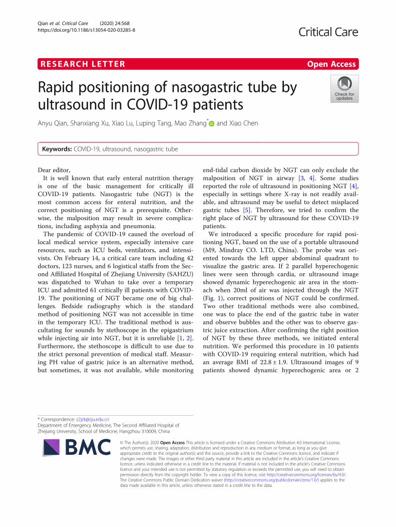
tioning NGT, based on the use of a portable ultrasound(M9, Mindray CO. LTD, China). The probe was ori-ented towards the left upper abdominal quadrant tovisualize the gastric area. If 2 parallel hyperechogeniclines were seen through cardia, or ultrasound imageshowed dynamic hyperechogenic air area in the stom-ach when 20ml of air was injected through the NGT(Fig. 1), correct positions of NGT could be confirmed.Two other traditional methods were also combined,one was to place the end of the gastric tube in waterand observe bubbles and the other was to observe gas-tric juice extraction. After confirming the right positionof NGT by these three methods, we initiated enteralnutrition. We performed this procedure in 10 patientswith COVID-19 requiring enteral nutrition, which hadan average BMI of 22.8 ± 1.9. Ultrasound images of 9patients showed dynamic hyperechogenic area or 2
© The Author(s). 2020 Open Access This article is licensed under a Creative Commons Attribution 4.0 International License,which permits use, sharing, adaptation, distribution and reproduction in any medium or format, as long as you giveappropriate credit to the original author(s) and the source, provide a link to the Creative Commons licence, and indicate ifchanges were made. The images or other third party material in this article are included in the article's Creative Commonslicence, unless indicated otherwise in a credit line to the material. If material is not included in the article's Creative Commonslicence and your intended use is not permitted by statutory regulation or exceeds the permitted use, you will need to obtainpermission directly from the copyright holder. To view a copy of this licence, visit http://creativecommons.org/licenses/by/4.0/.The Creative Commons Public Domain Dedication waiver (http://creativecommons.org/publicdomain/zero/1.0/) applies to thedata made available in this article, unless otherwise stated in a credit line to the data.
* Correspondence: [email protected] of Emergency Medicine, The Second Affiliated Hospital ofZhejiang University, School of Medicine, Hangzhou 310009, China
Qian et al. Critical Care (2020) 24:568 https://doi.org/10.1186/s13054-020-03285-8
parallel hyperechogenic lines in the stomach. In onepatient, we could find neither of these signs, and thenthe NGT was found coiled in the throat and mouth.After it was reset, ultrasound scanning confirmed theright position in stomach.In our opinion, the use of ultrasound is essential in coup-
ling with COVID-19 pandemic, especially lung ultrasound,which has been further confirmed [6]. We introducedanother application of ultrasound during COVID-19 out-breaks, which might help to confirm the position of NGTin this special setting. There were some limitations in ourreport. The BMIs of the cases was not high, so it was easyfor ultrasound observation. However, it might be difficultto find NGT by this way in obesity or flatulence patients,and the experience of operators could also influence theperformance. And the sample of this report was small; alarger study is needed to verify the effectivity and safety ofthis method.
AcknowledgementsWe thank all the physicians and nurses in our medical team to help Wuhanin this epidemic.
Authors’ contributionsAnyu Qian, Shanxiang Xu, Xiao Lu, Luping Tang, and Mao Zhang contributedto the data collection and writing and revision of the paper. Xiao Chencontributed to the revision and polishing the language of the paper. Theauthors read and approved the final manuscript.
FundingNo funding was used.
Availability of data and materialsThe corresponding author had full access to the data and had finalresponsibility for the decision of submitting for publication.
Ethics approval and consent to participateThis study has been approved by the ethics committee in the SecondAffiliated Hospital of Zhejiang University School of Medicine (Hangzhou,China) and performed in accordance with the ethical standards.
Consent for publicationAll authors have read the complete manuscript and have approvedsubmission of the paper.
Competing interestsThe authors declared no conflict of interest.
Received: 31 May 2020 Accepted: 14 September 2020
References1. Metheny NA, Stewart BJ, Mills AC. Blind insertion of feeding tubes in
intensive care units: a national survey. Am J Crit Care. 2012;21:352–60.https://doi.org/10.4037/ajcc2012549.
2. Erzincanli S, Zaybak A, Güler A. Investigation of the efficacy of colorimetriccapnometry method used to verify the correct placement of thenasogastric tube. Intensive Crit Care Nurs. 2017;38:46–52. https://doi.org/10.1016/j.iccn.2016.08.005.
3. Chau JP, Lo SH, Thompson DR, Fernandez R, Griffiths R. Use of end-tidalcarbon dioxide detection to determine correct placement of nasogastrictube: a meta-analysis. Int J Nurs Stud. 2011;48:513–21. https://doi.org/10.1016/j.ijnurstu.2010.12.004.
4. Atalay YO, Aydin R, Ertugrul O, Gul SB, Polat AV, Paksu MS. Does bedsidesonography effectively identify nasogastric tube placements in pediatriccritical care patients? Nutr Clin Pract. 2016;31:805–9. https://doi.org/10.1177/0884533616639401.
5. Tsujimoto H, Tsujimoto Y, Nakata Y, Akazawa M, Kataoka Y. Ultrasonographyfor confirmation of gastric tube placement. Cochrane Database Syst Rev.2017;4(4):CD012083. https://doi.org/10.1002/14651858.CD012083.pub2.
6. Vetrugno L, Bove T, Orso D, Barbariol F, Bassi F, Boero E, Ferrari G, Kong R.Our Italian experience using lung ultrasound for identification, grading andserial follow-up of severity of lung involvement for management of patientswith COVID-19. Echocardiography. 2020;37(4):625–7. https://doi.org/10.1111/echo.14664.
Publisher’s NoteSpringer Nature remains neutral with regard to jurisdictional claims inpublished maps and institutional affiliations.
Fig. 1 Male, 52 years old, with the chief complain “cough for 1 week and chest distress for 2 days.” A gastric tube was put. a Ultrasound showedthe stomach (white arrow) and the liver (white star). b And then 20 ml of air was injected through the NGT; ultrasound showed dynamichyperechogenic air shadow in the stomach (white triangle)
Qian et al. Critical Care (2020) 24:568 Page 2 of 2