Embed Size (px)
Citation preview
Int.J.Curr.Microbiol.App.Sci (2015) 4(11): 470-486
470
Original Research Article
Rapid Diagnosis of Invasive Fungal Infections
Magda M. Azab, Ashwak F. Abo Taleb, Nahla A.E. Mohamed and Farida H. Omran*
Medical Microbiology and Immunology Department, Faculty of Medicine, Zagazig University, Egypt
*Corresponding author
A B S T R A C T
Introduction
The incidence of invasive fungal infections (IFIs) has increased considerably in recent years because of the increasing population with Human immunodeficiency virus (HIV) infection, transplant recipients, cancer patients, and other individuals receiving immuno-suppressive treatments (Badiee et al., 2009).
Despite the availability of new antifungal drugs, the overall survival for immuno-
compromised patients with invasive fungal infections remains too low, with large variations according to underlying disease (Pasqualotto and Denning, 2005). Invasive fungal infections remain an important cause of morbidity and mortality. The most common fungi that cause disease in transplant recipients and other immuno-compromised patients are Candida and Aspergillus species (Erjavec and Verweij, 2002).
ISSN: 2319-7706 Volume 4 Number 11 (2015) pp. 470-486 http://www.ijcmas.com
The incidence of invasive fungal infections (IFIs) has increased considerably in recent years. Invasive fungal infections remain an important cause of morbidity and mortality. New diagnostic approaches have been developed based on non-culture based methods which may allow early diagnosis and treatment of fungal infections The aim of this study is how to rapidly diagnose IFIs in critically ill patient, comparing the conventional diagnostic method (blood culture) with the rapid diagnostic methods (ELISA and PCR). The study was conducted on 79 patients (51 males and 28 females) admitted to ICU units in Zagazig University hospital and suspected clinically to suffer from invasive fungal infections. Twenty milliliters of blood was collected from each patient during febrile episode. The blood samples were subjected to culture, ELISA and PCR. Considering the different methods used for diagnosis of IFIs the present study shows that 19 cases (24.1%) were +ve blood culture. This number increases to 41 (51.9%) by the use of Ag detection by ELISA and further increase to 47 (59.5%) cases out of the examined 79 patients was by panfungal PCR. PCR is superior to conventional methods for the diagnosis of IFIs. In diagnosis of IFI using conventional methods of diagnosis negative result do not rule out infection, so it is essential to use other diagnostic tools. Also, molecular diagnostic tools are promising and display high sensitivity and specificity.
K e y w o r d s
Invasive fungal infection, PCR, ELISA, Diagnosis, Fungi
Int.J.Curr.Microbiol.App.Sci (2015) 4(11): 470-486
471
Although conventional diagnostic tests such as histology, microscopy, and culture remain the cornerstone of proving the presence or absence of fungal disease, their yield is low and, therefore, their impact on clinical decisions to treat patients is limited. Furthermore, cultures become positive at a late stage of infection and delayed therapy is associated with a poor outcome (Maschmeyer et al., 2009).
New diagnostic approaches have been developed based on non-culture based methods, which may allow early diagnosis and treatment of fungal infections (Badiee et al., 2009).
Among the most promising diagnostic techniques are the detection of fungal antigens and molecular biological methods such as Polymerase chain reaction (PCR) (Erjavec and Verweij, 2002).
Galactomannan is a cell wall polysaccharide released by Aspergillus spp. during fungal growth in tissue. A commercially available sandwich enzyme-linked immunosorbent assay (ELISA) detects galactomannan by use of a rat monoclonal antibody (Pasqualotto and Denning, 2005).
Circulating galactomannan could be detected in the serum before diagnosis was made by clinical examination and radiology in approximately 65% of patients and at the same time as conventional diagnosis in 10% of patients. However, in approximately 25% of patients circulating antigen was detected after diagnosis was made (Erjavec and Verweij, 2002). 1,3-Beta-D-glucan is also a component of the cell wall. However, unlike galactomannan, which is Aspergillus specific, the glucan is present in many fungal species and therefore it should be considered as a pan-fungal detection method and a good indicator of systemic fungal
infection, if detectable in blood or other normally sterile body fluids (Cuenca-Estrella et al., 2008).
Several techniques have been commercialized for serum determination of this compound, although the most widely used is Fungitell, which is also an ELISA technique (Cuenca-Estrella et al., 2008).
Mannan is a cell wall surface carbohydrate that circulates during infection with Candida spp., and studies have shown a correlation between detectable mannanemia and tissue invasive in patients with candidaemia (Pasqualotto and Denning, 2005).
The detection of microbial Deoxyribonucleic acid (DNA) by PCR is without doubt one of the most powerful tools for the early diagnosis and identification of human pathogens.
The amplification of gene sequences unique to fungi may allow for early diagnosis of invasive fungal infections and subsequent treatment (Ribeiro et al., 2006).
Although antigen detection has shown variable performance in large clinical studies revealing both benefits and limitations, the availability of standardized protocols and quality assurance provided by commercial kits have allowed their inclusion in the diagnostic criteria for IFD established by the European Organization for Research and Therapy of Cancer and Mycoses Study Group (EORTC/ MSG) Consensus Group. The potential value of nucleic acid-based approaches to the detection and identification of fungal pathogens in immunocompromised patients is indisputable, as shown by a number of studies. However, the lack of standardization and validation across a wide spectrum of diagnostic laboratories has
Int.J.Curr.Microbiol.App.Sci (2015) 4(11): 470-486
472
prevented the inclusion of these tests in the diagnostic criteria of IFD proposed by the EORTC/MSG Consensus Group (Ba ková and Buchta, 2012).
This study was designed to assess the abilities of non-culture based methods (Enzyme linked immunosorbent assay for detection of fungal antigen and PCR using panfungal primer for detection of fungi in blood) in the diagnosis invasive fungal infections and to compare their results with those obtained by conventional methods (Blood culture) in order to devise a method for diagnosis invasive fungal infections which is more rapid and accurate.
Materials and Methods
The cross-sectional study was conducted at Zagazig University Hospitals, Egypt on 79 patients (51 males and 28 females) admitted to ICU units and suspected clinically to suffer from invasive fungal infections. An informed consent was obtained from patients before enrollment. The study gained approval of the institutional committee of Zagazig University.
Cases were defined as possible invasive fungal infection according to modification of EORTC/ MSG Consensus Group definitions (De Pauw et al., 2008).
Inclusion criteria for patients were:
1. Neutropenia (<1.0×10⁹/liter) or prolonged use of corticosteroid or other immuno-suppressant. 2. Fever (body temperature >38.5) for > 96 hours refractory to appropriate broad-spectrum antibacterial treatment. 3. Not on regime for antifungal therapy.
Twenty milliliters of blood was collected
during febrile episode under complete aseptic conditions using a sterile syringe and needle, after preparing the patient skin with alcohol soaked pad.
The blood sample was divided into three parts that was stored as follows:
-First part consists of 16 ml of blood was immediately inoculated bedside patient into two blood culture bottles.
-Second part consists of 2 ml of blood transferred to sterile tube and left to clot then centrifuged; the serum was stored in a sterile plastic aliquot at -20°C till the time of serological testing.
-Third part consists of another 2 ml of blood was put in tube containing EDTA that was stored at -20°C for doing PCR.
These samples were used for:
Culture and identification of yielding fungi
Blood culture was done on conventional blood culture bottles. Subculture was done on SDA with chloramphenicol at 25oC, and SABHI Agar Base with chloramphenicol at 37oC. Culture plates were incubated for at least 4 weeks. Plates were evaluated daily for the first seven days and at least twice per week thereafter.
Yeasts and yeast like colonies were identified by colony morphology, pattern of budding, presence of capsules, germ tube formation, and subculture on Brilliance Candida agar medium (Oxoid, England). Filamentous fungi were identified both macroscopically and microscopically by morphology, color, septation of hyphae, pigmentation, pattern of branching, and fruiting bodies.
Int.J.Curr.Microbiol.App.Sci (2015) 4(11): 470-486
473
Serological test:
Mannan antigen was measured in serum using a commercial sandwich immunoassay, Platelia Candida Ag (BioRad, Marnes La Coquette, France). The test was performed according to the instructions of the manufacturer.
Galactomannan antigen was measured in serum using a commercial sandwich immunoassay, Platelia Asprigellus Ag (BioRad, Marnes La Coquette, France). The test was performed according to the instructions of the manufacturer.
PCR: using panfungal primer for detection of fungi in blood.
DNA extraction (Springer et al., 2012): Extraction of fungal DNA from EDTA whole blood was done using A QIAamp UCP (for ultraclean production) Pure Pathogen Blood Kit (Qiagen, Germany) according to the manufacturer s instructions.
The use of a mechanical disruption using Pathogen Lysis Tubes with large (L) beads to ensure maximal lysis efficiency of the rigid fungal cell wall was recommended QIAGEN.
Blood sample used as negative control was obtained from a healthy volunteer. While positive control used to verify extraction efficiency was prepared by spiking blood sample obtained from healthy volunteer with approximately 150 CFU of C. albicans in a volume of 500 ml.
Amplification (Karakousis et al., 2006 & Ribeiro et al., 2006):
Amplification of fungal DNA was performed using universal primers targeting a highly conserved region of the fungal 18S small-subunit rRNA multicopy gene to
generate a PCR product from fungal DNA of 500 bp. The used primers were 18S-forward primer 5'-ATTGGAGGGCAAGTCTGGTG- 3' and 18S-reverse primer 5'-CCGATCCCTAGTCGGCAT-3' (Biolegio, Netherlands).
Samples were processed at the Medical Mycology in Medical Microbiology and Immunology Department, Faculty of Medicine, Zagazig University, Egypt.
All media included were prepared according to the manufacturer s instructions.
Statistical analysis
All data were collected, tabulated and statistically analyzed using SPSS 18.0 for windows (SPSS Inc., Chicago, IL, USA) & MedCalc 13 for windows (MedCalc Software bvba, Ostend, Belgium). Quantitative data were expressed as the mean ± SD & median (range), and qualitative data were expressed as an absolute frequencies ''number'' & relative frequencies (percentage). Percent of categorical variables were compared using Chi-square test.
Agreement between the three methods was analyzed using and Kappa statistic, = 0.05. Agreement was obtained the Kappa statistic was significant.
The degree of agreement was very good if the kappa value was 0.81 1.00, good if it was 0.61 0.8, moderate if it was 0.41 0.6, fair if it was 0.2 0.4 and poor if it was< 0.2. All tests were two sided. p < 0.05 was considered statistically significant (S), p < 0.01 was considered highly statistically significant (HS), and p 0.05 was considered non statistically significant (NS). Results and Discussion
Int.J.Curr.Microbiol.App.Sci (2015) 4(11): 470-486
474
In this study 79 patients were included, they were 51 males (64.6%) and 28 females (35.4%). Their age ranged from 22 to 69 years with a mean age of 48.1±11.8y, with the highest age group in the age >52 years old.
For the host factors distribution according to (EORTC/MSG), 59.5% of the studied patients were under corticosteroid therapy, 29.1% were neutropenic, while 11.4% of cases were on immune-suppressive therapy. Each patient in the study had one or more risk factors of the following: solid organ cancer (31.7%), hematological malignancy (25.3%), mechanical ventilation (18.9%), autoimmune disease (13.9%), COPD (13.9%), abdominal surgery (11.4%), diabetes mellitus (11.4%), total parenteral nutrition (10.9%), central venous catheter (7.6%), organ transplantation (5.1%) and renal failure with hemodialysis (3%) with statistically significant difference (P<0.001) (Figure 1).
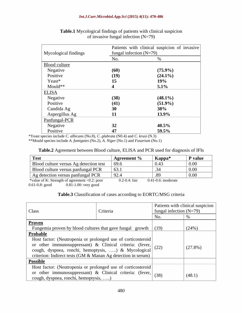
Using conventional methods presented by blood culture 19 samples were positive, of which yeasts accounted for 78.9% and moulds accounted for 21.1%. Yeasts isolated were caused by different Candida species, the majority of Candida infections were caused by C. albicans accounting for 53.3%, whereas non C. albicans species accounted for 46.7% of Candida infections, those include C. glabrata 26.7% and C. krusi 20.0% of yeast infections. The majority of isolated moulds were caused by Aspergillus species, of which A. fumigatus was responsible for 50.0%, while A. niger accounted for 25% of mould infections. Fusarium species was isolated from one blood culture accounting for 25% of mould infections (Table 1).
Using non conventional methods, 37.9% of patients enrolled were positive by PlateliaTM Candida Ag detection test. 13.9% were positive by PlateliaTM Aspergillus Ag detection test and 59.5% were positive by Panfungal PCR, with statistically significant difference (P<0.05) (Table 1 and Figure 2).
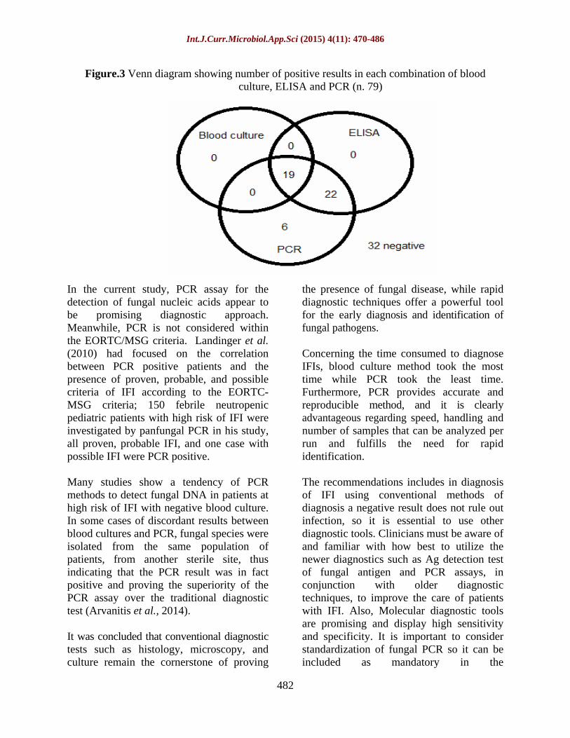
Comparing the results of different methods used for diagnosis of IFIs, 19 cases (24.1%) were +ve blood culture. This number increases to 41 (51.9%) by the use of Ag detection test and further increase to 47 (59.5%) cases out of the examined 79 patients was by panfungal PCR.
Accordingly, ELISA can detect all the blood culture positive cases and 22 of the blood culture negative cases. The agreement between the results of blood culture and Ag detection test of fungal antigen in serum by ELISA was moderate (kappa= 0.43). PCR can detect all the blood culture positive cases and 28 of the blood culture negative cases. The agreement between the results of blood culture and panfungal PCR was fair (kappa= 0.34). At the same time, PCR can detect all the ELISA positive cases and six of the ELISA negative cases. The agreement between the results of Ag detection test of fungal antigen in serum by ELISA and panfungal PCR was very good (kappa= 0.89) (Tables 2 and Figure 3).
Concerning time taken by each method to diagnose IFIs, blood culture method took the most time (up to two weeks) followed by Ag detection (3 4 hours), while PCR took the least time (1 2 hours).
According to EORTC/MSG, 24% of studied patients were proven invasive fungal infection, 27.8 % were probable invasive fungal infection and 48.1% were possible
Int.J.Curr.Microbiol.App.Sci (2015) 4(11): 470-486
475
invasive fungal infection (among them six cases were PCR positive) (Table 3).
Invasive fungal infections constitute a serious threat to an ever-growing population of immunocompromised individuals and other individuals at risk. Traditional diagnostic methods, such as histopathology and culture, which are still considered the gold standards, have low sensitivity, which underscores the need for the development of new means of detecting fungal infectious agents (Arvanitis et al., 2014).
Establishing a definite diagnosis of IFI in immunocompromised patients is particularly challenging and time consuming, but delayed initiation of antifungal treatment increases mortality. In the last few years, diagnostic procedures have been vastly improved, particularly noteworthy is the introduction of newer imaging techniques and non-culture methods, including antigen-based assays, metabolite detection and molecular detection of fungal DNA from body fluid samples (Ruhnke et al., 2012).
In the present study, the highest age group was in the age >52 years old. This is in consistent with the work of Meersseman (2004) & Montagna et al. (2013) where most of cases of invasive fungal infection tend to occur in high age (older than 60 years), and this can be attributed to the increased incidence of invasive mycoses with bipolarity of age in association with diminished immunity and body resistant. On the contrary, Soliman et al. (2012) detected most of invasive fungal infection cases within younger age group, as this study was conducted on patient with hematological malignancy which show higher incidence in this group of age.
As regarding host factors distribution, it was found that 59.5% of the studied patients were under corticosteroid therapy, 29.1%
were neutropenic, while 11.4% of cases were on immune-suppressive therapy. This is in agreement with Meersseman et al. (2004) who found that the highest percentage of studied patients was under corticosteroid therapy, followed by patients who were neutropenic, then those who were on immune-suppressive therapy. Similarly, in the study of Slavin et al. (2010) the highest percentage of studied patients were under corticosteroid therapy while cases that were on immune-suppressive therapy and neutropenic cases nearly were same percentage.
High incidence of IFI with corticosteroids is due to suppression of the immune system
especially the cellular component (Sipsas and Kontoyiannis, 2012). Meersseman et al. (2004) explained that Corticosteroids substantially impair macrophage killing of Aspergillus spores and mononuclear cell killing of Aspergillus hyphae, also threshold steroid level varies according to the type of patients, and emphasized that in the setting of underlying lung disease there is a risk factor for IA at much lower doses.
Distribution of risk factors among the studied patients with clinical suspicion of IFIs revealed a high association of IFIs with solid organ cancer (31.7%) and hematological malignancy (25.3%). This result is consistent with Slavin et al. (2010) who found that the highest percent of studied patients had solid organ cancer followed by those who had hematological malignancy. Also, in the study of Montagna et al (2013), percentage of cases with solid organ cancer was higher than cases of hematological malignancy.
Yeast was found to account for 78.9% and mould for 21.1% of all infections and this is in consistent with Montagna et al. (2013), where yeast accounted for 87.6% and moulds accounted for 12.4% of all infections. These results can be explained as
Int.J.Curr.Microbiol.App.Sci (2015) 4(11): 470-486
476
follows: Unlike invasive candidiasis, which can affect relatively noncompromised patients, invasive disease caused by Aspergillus spp., moulds generally involves severely immunocompromised patients. Also, most of Candida infections are endogenous while Aspergillus infection is mainly exogenous. However, George (2011) detected higher incidence of mould than yeast among all infections.
Yeast was isolated from 19% of studied patients. These results are in agreement with that of Yang et al. (2006) who found positive blood culture for Candida in 13.5% of studied patients. However a lower rate of detection (7.5%) was reported by Abelson et al. (2005).
The majority of Candida infections 53.3% were caused by Candida albicans, whereas 46.7% were caused by non C. albicans species. This result was consistent with Tortorano et al. (2012) study, which showed that Candida albicans accounted for 60% of Candida infections.
On the contrary, Montagna et al. (2013) found that the majority of candida infections 59.8% were caused by non C. albicans species and Candida albicans, accounted for 40.2% of yeast infection, and attributed the reason for this change in the pattern of Candida spp. distribution to some predisposing factors, such as indwelling catheters and parenteral nutrition for C. parapsilosis, cancer and neutropenia for C. tropicalis, and previous exposure to azoles for C. krusei and C. glabrata. However, in nonneutropenic patients, risk factors for C. krusei nosocomial candidemia include recent gastrointestinal surgery and exposure to fluconazole (Playford et al., 2008).
Isolated non C. albicans species included: C. glabrata which formed 26.7% of yeast
infections, and C. krusi which formed 20.0%, of yeast infections. In a study done by Tortorano et al. (2012), it was found that the incidence of C. glabrata was 13.4% and C. krusi was 1.8%. Similarly, in Montagna et al. (2013) study C. glabrata formed 10% of yeast infection.
The clinical significance of the isolation of non albicans Candida spp. is the increased likelihood of resistance to fluconazole and other azoles. In particular, of the commonly isolated nonalbicans Candida spp, fluconazole resistance was noted in 16% of C. glabrata, and 78% of C. krusei (Hachem et al., 2008).
Furthermore, it was found that C. krusei was the fifth most common Candida spp. isolated and accounted for 2.5% in a global antifungal surveillance program (Pfaller et al., 2008). C. krusei, intrinsically resistant to fluconazole, has often been reported in patients with hematologic malignancies with prior fluconazole and antifungal exposure and is associated with the highest mortality of all Candida spp. (Horn et al., 2009).
As regarding moulds which were isolated from 5.1% of studied patients, the majority of the mould infections (75%) were caused by Aspergillus spp, of which A. fumigatus was responsible for 50% and A. niger was responsible for 25% of mould infections. While, Fusarium spp. accounted for 25% of mould infections. However, Girmenia et al. (2001) reported that only 10% of blood cultures were positive in patients with Invasive aspergillosis. These findings were consistent with (Ulrike and Lass-Flörl, 2011) who mentioned that the most common species causing aspergillosis is A. fumigatus, also Jane et al. (2011) who found that A. fumigatus causes 44% of invasive aspergillosis cases. A. fumigatus is more thermotolerant than other disease-causing
Int.J.Curr.Microbiol.App.Sci (2015) 4(11): 470-486
477
species, growing well at 37°C, and several studies suggested that the radial growth and germination rate of aspergilli at 37°C correlate with pathogenicity. Moreover, being resistant to oxidative stress; A. fumigatus detoxifies oxidative threats via glutathione synthesis and oxidoreductase activity (Dagenais and Keller, 2009).
Furthermore, Ulrike and Lass-Flörl (2011) found that the most common species causing aspergillosis is A. fumigatus, accounting for approximately 90% of aspergillus infections. Depending on regional distinctions A. flavus, A. nidulans, A. terreus and A. niger are frequently reported as well, and there is evidence that these non-fumigatus pathogens are increasingly common etiologic agents, in the current study other Aspergillus spp. also recovered from patient samples, where A. niger accounted for 25% of mould infections.
In our study, another filamentous fungus which is Fusarium was found to be responsible for 25% of mould infections. Fusariosis is a life-threatening and increasingly important mycosis in immunocompromised hosts. Risk factors for such infections are skin lesions, burns, use of corticosteroids, prolonged neutropenia and hematological malignancy (Ulrike and Lass-Flörl, 2011). Other studies have reported 3-month mortality rates of 80% in hematopoietic cell transplantation from infections due to Fusarium species (Jane et al., 2011). Nucci and Anaissie (2007), reported that blood cultures are frequently positive in fusariosis and yielded the organism in 41% of the cases. This is possibly due to the fact that Fusarium species produce yeast like structures (adventitious sporulation) that facilitate their dissemination and growth in the blood.
Isolation of etiologic fungal agent by culturing
different clinical specimens is considered one of the methods used for the diagnosis of IFI, provided that the samples are from sterile sites such as blood, tissue or cerebrospinal fluid, in which fungal infection can be documented, although, the sensitivity of blood culture for the diagnosis of fungal infection is controversial. Multiple or repeated blood cultures should be performed to increase the likelihood of detecting candidemia, and filamentous fungi are rarely isolated from blood. So far, culture can be useful to determine the sensitivity of the isolated fungi to antifungal agents and identify resistant species, and are thus needed to optimize patient management. As a result, given the limitations for diagnosis of IFI, negative result on direct or pathologic smears and cultures do not rule out infection, so it is essential to use other methods (Badiee and Hashemizadeh, 2014).
In order to overcome the limitations of conventional diagnostic tests, several studies have focused on the development of non-culture based methods with superior sensitivity and quicker turnaround time, such as the detection of fungal components or metabolites, as an alternative approach for the early detection and identification of the causative fungal species (Yeo and Wong, 2002).
Serological tests for detection of fungal antibodies have been the subject of much study but can be difficult to interpret. For example, circulating antibodies to Candida spp. may occur in normal individuals as a result of commensal colonization of mucosal surfaces, antibody production in the immuno-compromised patient population varies according to immune status and false negative results occur in immunocompromised patients who produce low or undetectable levels of antibodies. However, the antigen detection tests are
Int.J.Curr.Microbiol.App.Sci (2015) 4(11): 470-486
478
more significant for diagnosis because they detect active infection. Antigen is generally detected in serum, yet, it can also be detected in other body fluids, such as urine (Sendid et al., 2002).
Using PlateliaTM Candida Ag detection test, 37.9% patients enrolled were positive. Accordingly Ag detection test detected all of the blood culture positive cases and 25% of the blood culture negative cases.
These results are in agreement with Montagna et al. (2013). Unlike Meersseman et al. (2004) who reported a rate of 40% for antigen detection among patients.
Interestingly, the possible cause of the variability in sensitivity of the serum GM assay is the level and nature of immune suppression of the patient. Classically, Patients with Low neutrophils levels are more likely to have higher serum GM levels than patients with higher neutrophil levels, as when the assay is applied to patients with hematological malignancy or having undergone bone marrow or stem cell transplantation. Also, serum GM assay has been shown to be modestly sensitive in COPD patients with IA and poorly sensitive in solid organ transplantation. The lower incidence of neutropenia in these two patient groups may well account in part for the reduced sensitivity of the serum GM assay. Also, this result could be logical in patients receiving antifungal therapy where antifungal treatment inhibits growth of Aspergillus renders nonviable reducing the load of fungal cells able to shed the GM antigen (Barton, 2013).
The detection of non-cultivable/nonviable cells and circulating free fungal DNA by PCR has been described as an important tool in the early diagnosis of IFI, unlike in culture which needs viable cells. Therefore, diagnostic assays based on in vitro
amplification and detection of fungal DNA has been developed. PCR methods are particularly promising because of their high specificity and sensitivity (Chen et al., 2001).
About 59.5% of enrolled patients were positive by panfungal PCR. However, 24.1% of cases were +ve blood culture. Increased to 51.9% of cases by the use of Ag detection test and further increased to 59.5% of examined cases by panfungal PCR, with statistically significant difference (P<0.05)
These results are coincident with the work of El-Sayed et al. (2012) who detected 57.7% of cases positive by panfungal PCR, consequently 88% of the positive patients and 50% of all were missed by blood culture. Also, another study stated that 64.5% were positive by both culture and PCR, of these samples 19.4% showed no growth of fungi but were positive by PCR (Gurtner et al., 2007). On the other hand, results of Badiee et al. (2009) study revealed that 10.6% of patients had positive cultures for fungal infections whereas 17.7% were positive for fungal infection by panfungal PCR. Ribeiro et al., (2006) study results revealed that 35% of enrolled patients were positive by panfungal PCR.
Khot et al. (2009) mentioned that the ability of panfungal PCR assays to detect a wide spectrum of fungi is clearly an advantage. Conversely, amplification of contaminating fungal nucleic acids present in the environment or laboratory can be a drawback. In general, panfungal PCR assays could be useful when applied to a clinical sample as part of a larger array of assays. For example, panfungal PCR assays could detect new pathogens previously unreported, and identify etiological agents in mixed infections, especially when used with other genus-specific assays.
Int.J.Curr.Microbiol.App.Sci (2015) 4(11): 470-486
479
Blood is the most sterile of the clinical samples used for diagnosis by PCR; therefore, it is more useful for applying panfungal PCR assays because a PCR positive provides more convincing evidence of fungal disease. On the other hand, use of panfungal PCR on nonsterile clinical samples, especially BAL fluid, for the diagnosis of IA may result in suboptimal results
low specificity due to cross-reactivity with commensal fungi such as Candida spp. present in the oral cavity, and low sensitivity because these commensal fungi may compete with the pathogen for amplification (Khot and Fredricks, 2009).
The diagnosis of patients with IFIs in the present study revealed a very good agreement between Ag detection test of fungal antigen in serum by enzyme immunoassay and panfungal PCR. However, there is moderate agreement between the results of blood culture and Ag Detection Test. Additionally there is a fair agreement between the results of blood culture and panfungal PCR. In accordance with our results El masry et al. (2005) also detected a very good agreement (94.4%) between the two non-culture based methods (PCR and ELISA).
Studies by Florent et al. (2006) and White et al. (2006) that compared GM antigen and PCR assays found that when used together the sensitivity of diagnosis can be increased. Accordingly, they concluded that complementary use of these tests may reduce dependence on invasive diagnostic procedures and limit the need for empirical antifungal therapy.
El-Sayed et al. (2012) detected poor agreement between the results of blood culture and panfungal PCR (kappa = 0.10). They conferred the discrepancies in the
results of culture and PCR to that the detection of fungemia by means of fungal blood culture is notoriously difficult and IFIs are diagnosed better by molecular assay as naked DNA can be detected by PCR due to the presence of dead and degrading fungi within circulating phagocytes (Ahmaad et al., 2002). Also, the time factor could offer another explanation for the observed differences in the results of blood culture and PCR. According to Estrella et al. (2009), fungal DNA was detected by PCR much earlier than GM in patients with IFI.
Generally, the diagnosis of infection based on molecular and serologic techniques can provide powerful tools for the early and rapid diagnosis of IFI. As the fungi are common in the environment and opportunistic fungi in immunocompromised patients can cause high morbidity and mortality, the interpretation of positive or negative results with different laboratory methods is difficult for clinicians, so more than one method should be used for early diagnosis (Badiee and Hashemizadeh, 2014).
Concerning time taken by each method to diagnose IFIs, blood culture method took the most time (up to 2 weeks) while PCR took the least time (1 2 hours). Inning et al. (2007) stated that although blood culture technique is still used for routine diagnosis of IFIs, blood cultures are slow and lack the desired sensitivity and specificity.
Invasive fungal infections in our study were defined using EORTC/MSG Consensus Group criteria (De Pauw et al., 2008), and according to these criteria we found that 24% of cases were classified as proven IFI, 27.8 % were probable IFI and 48.1% were possible invasive fungal infection (among them 6 cases were PCR positive).
Int.J.Curr.Microbiol.App.Sci (2015) 4(11): 470-486
480
Table.1 Mycological findings of patients with clinical suspicion of invasive fungal infection (N=79)
Patients with clinical suspicion of invasive fungal infection (N=79) Mycological findings No. %
Blood culture
Negative (60) (75.9%) Positive (19) (24.1%) Yeast* 15 19% Mould** 4 5.1%
ELISA
Negative (38) (48.1%) Positive (41) (51.9%) Candida Ag 30 38% Aspergillus Ag 11 13.9%
Panfungal-PCR
Negative 32 40.5% Positive 47 59.5%
*Yeast species include C. albicans (No.8), C. glabrata (N0.4) and C. krusi (N.3) **Mould species include A. fumigates (No.2), A. Niger (No.1) and Fusarium (No.1)
Table.2 Agreement between Blood culture, ELISA and PCR used for diagnosis of IFIs
Test Agreement % Kappa* P value Blood culture versus Ag detection test 69.6 0.43 0.00 Blood culture versus panfungal PCR 63.1 .34 0.00 Ag detection versus panfungal PCR 92.4 .89 0.00
*value of K: Strength of agreement <0.2: poor 0.2-0.4: fair 0.41-0.6: moderate 0.61-0.8: good 0.81-1.00: very good
Table.3 Classification of cases according to EORTC/MSG criteria
Patients with clinical suspicion fungal infection (N=79) Class Criteria No. %
Proven
Fungemia proven by blood cultures that gave fungal growth (19) (24%) Probable
Host factor: (Neutropenia or prolonged use of corticosteroid or other immunosuppressant) & Clinical criteria: (fever, cough, dyspnea, ronchi, hemoptysis, ..) & Mycological criterion: Indirect tests (GM & Manan Ag detection in serum)
(22) (27.8%)
Possible
Host factor: (Neutropenia or prolonged use of corticosteroid or other immunosuppressant) & Clinical criteria: (fever, cough, dyspnea, ronchi, hemoptysis, ..)
(38) (48.1)

Int.J.Curr.Microbiol.App.Sci (2015) 4(11): 470-486
481
Figure.1 Distribution of risk factors of patients with clinical suspicion of invasive fungal infection (N=79)
Figure.2 PCR using panfungal primer
Lane 1: Molecular size marker (50-1500bp) Lane 2: Negative control Lane 3: Positive control Lane 4: Positive case Lane 5: Negative case Lane 6: Positive case Lane 7: Positive case Lane 8: Negative case
0
5
10
15
20
25
30
35
Hem
atol
ogic
alm
alig
nan
cy
Sol
id o
rgan
can
cer
Org
antr
ansp
lan
tati
on
Ab
dom
inal
surg
ery
Au
toim
mu
ne
dis
ease
Tot
al p
aren
tera
ln
utr
itio
n
Cen
tral
ven
ous
cath
eter CO
PD
Mec
han
ical
ven
tila
tion
Ren
al fa
ilure
wit
hh
emod
ialy
sis
Dia
bet
esm
elit
us
Int.J.Curr.Microbiol.App.Sci (2015) 4(11): 470-486
482
Figure.3 Venn diagram showing number of positive results in each combination of blood culture, ELISA and PCR (n. 79)
In the current study, PCR assay for the detection of fungal nucleic acids appear to be promising diagnostic approach. Meanwhile, PCR is not considered within the EORTC/MSG criteria. Landinger et al. (2010) had focused on the correlation between PCR positive patients and the presence of proven, probable, and possible criteria of IFI according to the EORTC-MSG criteria; 150 febrile neutropenic pediatric patients with high risk of IFI were investigated by panfungal PCR in his study, all proven, probable IFI, and one case with possible IFI were PCR positive.
Many studies show a tendency of PCR methods to detect fungal DNA in patients at high risk of IFI with negative blood culture. In some cases of discordant results between blood cultures and PCR, fungal species were isolated from the same population of patients, from another sterile site, thus indicating that the PCR result was in fact positive and proving the superiority of the PCR assay over the traditional diagnostic test (Arvanitis et al., 2014).
It was concluded that conventional diagnostic tests such as histology, microscopy, and culture remain the cornerstone of proving
the presence of fungal disease, while rapid diagnostic techniques offer a powerful tool for the early diagnosis and identification of fungal pathogens.
Concerning the time consumed to diagnose IFIs, blood culture method took the most time while PCR took the least time. Furthermore, PCR provides accurate and reproducible method, and it is clearly advantageous regarding speed, handling and number of samples that can be analyzed per run and fulfills the need for rapid identification.
The recommendations includes in diagnosis of IFI using conventional methods of diagnosis a negative result does not rule out infection, so it is essential to use other diagnostic tools. Clinicians must be aware of and familiar with how best to utilize the newer diagnostics such as Ag detection test of fungal antigen and PCR assays, in conjunction with older diagnostic techniques, to improve the care of patients with IFI. Also, Molecular diagnostic tools are promising and display high sensitivity and specificity. It is important to consider standardization of fungal PCR so it can be included as mandatory in the
Int.J.Curr.Microbiol.App.Sci (2015) 4(11): 470-486
483
recommendations. Lastly, Future efforts should focus on further improvements of rapid diagnostic techniques, which would allow for the timely application of antifungal therapy and would reduce the use of treatments in inappropriate settings.
Reference
Abelson, J.A., Moore, T., Bruckne, R.D., Deville, J., Nielsen, K. 2005. Related frequency of fungemia in hospitalized pediatric inpatients over 11years at a tertiary care institution. Pediatrics. David Geffen School of Medicine, Los Angeles, California, USA.Jul. 116(1): 61 67.
Ahmaad, S., Khan, Z., Mustafa, A., Khan, Z.U. 2002. Seminested PCR for diagnosis of candidemia: comparison with culture, antigen detection and biochemical methods for species identification. J. Clin. Microbiol., 40: 2483 9.
Arvanitis, M., Anagnostou, T., Fuchs, B. B., Caliendo, A.M., Mylonakis, E. 2014. Molecular and nonmolecular diagnostic methods for invasive fungal infections. Clin. Microbiol. Rev., 27(3): 490.
Badiee, P., Hashemizadeh, Z. 2014. Opportunistic invasive fungal infections: diagnosis & clinical management. Indian J. Med. Res., 139: 195 204.
Badiee, P., Kordbacheh, P., Alborzi, A., Malekhoseini, S., Ramzi, M., Mirhendi, H., Mahmoodi, M., Shakiba E. 2009. Study on invasive fungal infections in mmunocompromised patients to present a suitable early diagnostic procedure. Int. J. Infect. Dis., 13(1): 97 102.
Barton, B.C. 2013. Laboratory diagnosis of invasive aspergillosis: from diagnosis to prediction of outcome. Vol. 2013, Article ID 459405, 29 Pp.
Ba ková, L., Buchta, V. 2012. Laboratory diagnostics of invasive fungal infections:
an overview with emphasis on molecular approach. Folia Microbiol., 57: 421430.
Chen, S.C.A., Ftnillday, C.L., Meyer, W. 2001. A review of nucleic acid-based diagnostic tests for systemic mycosis with an emphasis on polymerase chain based assays. Med. Mycol., 40: 333 357.
Cuenca-Estrella, M., Bernal-Martinez, L., Buitrago, M., Castelli, M.V., Gomez-Lopez, A., Zaragoza, O., Rodriguez-Tudela, J. 2008. Update on the epidemiology and diagnosis of invasive fungal infection. Int. J. Antimicrob. Agents, 32: 43 147.
Dagenais, T.R., Keller, N.P. 2009. Pathogenesis of Aspergillus fumigatus in invasive aspergillosis. Clin. Microbiol. Rev., 22(3): 447 465.
De Pauw, B., Walsh, T.J., Donnelly, J.P., Stevens, D.A., Edwards, J.E., Calandra, T., Pappas, P.G., Maertens, J., Lortholary, O., Kauffman, C.A., Denning, D.W., Patterson, T.F., Maschmeyer, G., Bille, J., Dismukes, W.E., Herbrecht, R., Hope, W.W., Kibbler, C.C., Kullberg, B.J., Marr, K.A.,
Mu oz, P., Odds, F.C., Perfect, J.R., Restrepo, A., Ruhnke, M., Segal, B.H., Sobel, J.D., Sorrell, T.C., Viscoli, C., Wingard,J.R., Zaoutis, T., Bennett, J.E. 2008. Revised definitions of invasive fungal disease from the European Organization for Research and Treatment of Cancer/Invasive Fungal Infections Cooperative Group and the National Institute of Allergy and Infectious Diseases Mycoses Study Group (EORTC/MSG) Consensus Group. Clin. Infect. Dis., 46: 1813 1821.
El Masry, I.S.A.S., Bassim, H.H., Asfour, I.A.H. 2005. Fungal infection in patients with hematological malignancies with special emphasis on molecular diagnosis of Candida. M.D. thesis. Ain Shams University, Egypt. Pp. 155 158.
El-Sayed, Z.A., Hasan, Z.E., Nasr, R.A.R.
Int.J.Curr.Microbiol.App.Sci (2015) 4(11): 470-486
484
2012. Real-Time PCR in the early detection of invasive fungal infection in immunodeficient infants and children. Egypt J. Pediatr. Allergy Immunol., 10(2): 67 74.
Erjavec, Z., Verweij, P.E. 2002. Recent progress in the diagnosis of fungal infections in the immunocompromised host. Drug Resist. Updat., 5(1) 3 10.
Estrella, C., Meije, Y., Pedroche, C., Lopez, A., Buitrago, M., Martinez, L., Grande, C., Juan, RS., Lizasoain, M., Rodriguez-Tudela, J.L., Aguado, J.M. 2009. Value of serial quantification of fungal DNA by a real-time PCR based technique for early diagnosis of invasive aspergillosis in patients with febrile neutropenia. J. Clin. Microbiol., 47(2): 379 84.
Florent, M., Katsahian, S., Vekhoff, A., Vincent, L., Rio, B., Marie, J.P., Bouvet, A., Cornet, M. 2006. Prospective evaluation of a polymerase chain reaction- ELISA targeted to Aspergillus fumigatus and Aspergillus flavus for the early diagnosis of invasive aspergillosis in patients with hematological malignancies. J. Infect. Dis., 193: 741747.
Fujita, S., Alam, F.F., Mustafa, A.S., Khan, Z.U. 2007. Comparative evaluation of mannan and anti-mannan antibodies and Candida species-specific snPCR in patients with candidemia. J. Infect Dis., 4: 7 10.
George, J. 2011. Nosocomial fungal infections epidemiology, infection control, and prevention. Infect. Dis. Clin. N. Am., 25: 201 225.
Girmenia, C., Nucci, M., Martino, P. 2001. Clinical significance of Aspergillus fungaemia in patients with haematological malignancies and invasive aspergillosis. Br. J. Haematol., 114: 93 98.
Gurtner, S., Selitsch, M., Rotter, A., Hirschl, A.M., Willinger, B. 2007. Development of novel real-time PCR assays for detection
and differentiation of 11 medically important Aspergillus and Candida species in clinical specimens. J. Clin. Microbiol., 45: 906 14.
Hachem, R., Hanna, H., Kontoyiannis, D. 2008. The changing epidemiology of invasive candidiasis: Candida glabrata and Candida krusei as the leading causes of candidemia in hematologic malignancy. Cancer, 112(11): 2493 9.
Horn, D., Neofytos, D., Anaissie, E. 2009. Epidemiology and outcomes of candidemia in 2019 patients: data from the prospective antifungal therapy alliance registry. Clin. Infect. Dis., 48(12): 1695703.
Innings, R., Niklas, N., Magnus, I., Herrmann, M. 2007. Multiplex Real-Time PCR targeting the RNase P RNA gene for detection and identification of Candida species. J. Clin. Microbiol., 45(3): 874880.
Jane, K., James, I., Sanjeet, S. 2011. Epidemiology and treatment approaches in management of invasive fungal infections. Clin. Epidemiol., 3: 175 191.
Karakousis, A., Tan, L., Ellis, D., H. Alexiou, H., Wormald, P.J. 2006. An assessment of the efficiency of fungal DNA extraction methods for maximizing the detection of medically important fungi using PCR. J. Microbiol. Methods, 65: 38 48.
Khot, P.D., Fredricks, D.N. 2009. PCR-based diagnosis of human fungal infections. Expert Rev. Anti Infect. Ther., 7(10): 1201 1221.
Khot, P.D., Ko, D.L., Fredricks, D.N. 2009. Sequencing and analysis of fungal rRNA operons for development of broad-range fungal PCR assays. Appl. Environ. Microbiol., 75: 1559 1565.
Landinger, C., Prevner, S., Baskova, L., van Grotel, M., Hartwig, N.G., Dworzak, M., Mann, G., Attarbaschi, A., Kager, L., Peters, C., Matthes-Martin, S.,
Int.J.Curr.Microbiol.App.Sci (2015) 4(11): 470-486
485
Lawitschka, A., van den Heuvel-Eibrink, M.M., Lion, T. 2010. Diagnosis of IFI by a real time panfungal PCR assay in immunocompromised pediatric patients. Leukemia, 24(12): 2032 8.
Maaroufi, Y., Ahariz, N., Husson, M., Crokaert, F. 2004. Comparison of different methods of isolation of DNA of commonly encountered Candida species and its quantitation by using a real-time PCR-based assay. J. Clin. Microbiol., 42: 3159 3163.
Maschmeyer, G., Beinert, T., Buchheidt, D., Cornely, O.A., Einsele, H., Heinz, W., Heussel, C.P., Kahl, C., Kiehl, M., Lorenz, J., Hof, H., Mattiuzzi, M. 2009. Diagnosis and antimicrobial therapy of lung infiltrates in febrile neutropenic patients: Guidelines of the infectious diseases working party of the German Society of Haematology and Oncology. Eur. J. Cancer, 45: 2462 2472.
Meersseman, W., Vandecasteele, S.J., Wilmer, A., Verbeken, E., Peetermans, W.E., Wijngaerden, E.V. 2004. Invasive aspergillosis in critically ill patients without malignancy. Am. J. Respir. Crit. Care Med., 170: 621 625.
Montagna, M.T., Caggiano, G., Lovero, O., De Giglio, O., Coretti, C., Cuna, T., Iatta, R., Giglio, M., Dalfino, L., Bruno, F., Puntillo, F. 2013. Epidemiology of invasive fungal infections in the intensive care unit: results of a multicenter Italian survey (AURORA Project). Infection, 41: 645 653
Nucci, M., Anaissie, E. 2007. Fusarium infections in immunocompromised patients. Clin. Microbiol. Rev., 20(4): 695 704.
Pasqualotto, D.A., Denning D.W. 2005. Diagnosis of invasive fungal infections
Current limitations of classical and new diagnostic methods. Business Briefing: European Oncology Review. Pp. 1 11.
Pfalle,r M., Diekema, D., Gibbs, D. 2008.
Candida krusei, a multidrug-resistant opportunistic fungal pathogen: geographic and temporal trends from the ARTEMIS DISK Antifungal Surveillance Program, 2001 to 2005. J. Clin. Microbiol., 46(2): 515 21.
Pfaller, M.A., Diekema, D.J., Rinaldi, M.G., Barnes, R., Hu, B., Veselov, N. 2005. Results from the ARTEMIS DISK Global antiftingal surveillance study: a 6.5 year analysis of susceptibilities of Candida and other yeast species to fluconazole and voriconazole by standardized disk diffusion testing. J. Clin. Microbiol., 43(12): 5848 5859.
Playford, E., Marriott, Q., Nguyen, S., Chen, D., Ellis, M., Slavin, T., Sorrell, C. 2008. Candidemia in nonneutropenic critically ill patients: risk factors for non-albicans Candida spp. Crit. Care Med., 36: 20342039
Ribeiro, P., Costa, F., Monteiro, A., Caldas, J., Silva, M., Ferreira, G., Veiga, J., Sousa, M.O., Viegas, M.P., Santos, E., Gonçalves, A.J., Sousa, A.B. 2006. Polymerase chain reaction screening for fungemia and/or invasive fungal infections in patients with hematologic malignancies. Support Care Cancer, 14(5): 469 74.
Ruhnke, M., Bo¨hme, A., Buchheidt, D., Cornely, O., Donhuijsen, K., Einsele, H., Enzensberger, R., Hebart, H., Heussel, C. P., Horger, M., Hof, H., Karthaus, M., Kru¨ger, W., Maschmeyer, G., Penack, O., Ritter, J., Schwartz, S. 2012. Diagnosis of invasive fungal infections in hematology and oncology-guidelines from the Infectious Diseases Working Party in Haematology and Oncology of the German Society for Haematology and Oncology (AGIHO). Ann. Oncol., 23: 823 833.
Sendid, B., Tabouret, M., Poirot, J.L., Bonnin, A., Caillot, D., Olian, D. 2002. Combined detection of mannanaemia and antimannan antibodies as a strategy for the diagnosis of
Int.J.Curr.Microbiol.App.Sci (2015) 4(11): 470-486
486
systemic infection caused by pathogenic Candida species. J. Med Microbiol., 51(5): 433 442.
Sipsasa, N.V., Kontoyiannis, D.P. 2012. Invasive fungal infections in patients with cancer in the Intensive Care Unit. Int. J. Antimicrob. Agents, 39: 464 471.
Slavin, M.A., Sorrell, T.C., Marriott, D., Thursky, K.A., Quoc Nguyen, Q., Ellis D.H., Morrissey, C.O., Sharon C.A.C. 2010. Candidaemia in adult cancer patients: risks for fluconazole-resistant isolates and death. J. Antimicrob. Chemother., 65: 1042 51.
Soliman, M.H., Azab, M.M., Abu Taleb, F.M., Mohamed, N.A. 2012. Identification of different types of fungi isolated from patients with haematologic malignancy and from their surrounding exogenous sources. Z.U.M.J., 18(5): 951961.
Springer, J., Schlo nagel, H., Heinz, W., Doedt, T., Soeller, R., Einsele, H., Loefflera, J. 2012. A novel extraction method combining plasma with a whole-blood fraction shows excellent sensitivity and reproducibility for patients at high risk for invasive aspergillosis. J. Clin. Microbiol., 50(8): 2585.
Tortorano, A.M., Dho, G., Prigitano, A., Breda, G., Grancini, A., Emmi, V., Cavanna, C., Marino, G., Morero, S., Ossi, C., Delvecchio, G., Passera, M., Cusumano, V., David, A., Bonaccorso, G., Corona, A., Favaro, M., Vismara, C., Garau, M.G., Falchi, S., Tejada, M.R. 2012. Invasive fungal infections in the intensive care unit: a multicentre, prospective, observational study in Italy (2006 2008). Mycoses, 55(1): 73 79.
Ulrike, B., Lass-Flörl, C. 2011. Epidemiology of Invasive Fungal Infections in the Mediterranean Area. Mediterr. J. Hematol. Infect. Dis., 3; Open Journal System.
Verduyn Lunel, F.M., Donnelly, J.P., van der Lee, H.A., Blijlevens, N.M., Verweij, P.E. 2009. Circulating Candida-specific anti-mannan antibodies precede invasive candidiasis in patients undergoing myelo-ablative chemotherapy. Clin. Microbiol. Infect., 15: 380 386.
White, P.L., Linton, C.J., Perry, M.D., Johnson, E.M., Barnes, R.A. 2006. The evolution and evaluation of a whole blood polymerase chain reaction assay for the detection of invasive aspergillosis in hematology patients in a routine clinical setting. Clin. Infect. Dis., 42: 479 486.
Yang, Y.L., Cheng, H.H., Lo, H. 2006. Distribution and antifungal susceptibility of Candida species isolated from different age populations in Taiwan. Med. Mycol., 44(3): 237 242.
Yeo, S.F., Wong, B. 2002. Current status of nonculture methods for diagnosis of invasive fungal infections. Clin. Microbiol. Rev., 15: 465 484.