Embed Size (px)
Citation preview

Rapid Development of Hepatic MetastasisWith High Incidence Following Orthotopic
Transplantation of Murine Colon 38Carcinoma as Intact Tissue in Syngeneic
C57BL/6 Mice
YASUHIRO FUNAHASHI, MS,* NOZOMU KOYANAGI, MS, JIRO SONODA, DVM,KYOSUKE KITOH, MS, AND KENTARO YOSHIMATSU, PhD
Tsukuba Research Laboratories, Eisai Co. Ltd., Tsukuba, Japan
Background and Objectives:Orthotopic transplantation of human colontumors was a useful method for producing hepatic metastasis in mice. Inmany cases, however, it took about 3 months for evaluation. We examinedan in vivo model of hepatic metastasis for only 4 weeks by conductingorthotopic transplantation of murine Colon 38 tumor using intact tissue insyngeneic mice and determined the efficacy of chemotherapeutic agentsagainst hepatic metastasis.Methods: Twenty milligrams of tumor tissues were prepared from sub-cutaneously (s.c.) growing Colon 38 tumor and orthotopically transplantedon the cecum in C57BL/6 mice. Mice were autopsied about 4 weeks aftertransplantation. Metastases to various organs were detected macroscopi-cally or histochemically and tumor invasion into the cecum was observedhistochemically. In experimental chemotherapy, mice bearing orthotopi-cally transplanted Colon 38 tumor were separated into three equal groupsand were either treated with fluorouracil or cisplatin (CDDP), or untreated.Four weeks after transplantation, activities of both agents against localtumor growth and hepatic metastasis were evaluated.Results:Macroscopic metastases to various organs including the liver, thelung, and the peritoneum were developed during days 28 to 32 afterinoculation. The frequency of hepatic metastasis was 96% (N4 23).Histological examination indicated that the local tumor invaded variouslayers of the cecum and metastasized to the liver and lung hematog-enously. In experimental chemotherapy with fluorouracil and CDDP, onlyfluorouracil decreased the incidence of mice with hepatic metastasis (2/8cases), compared with vehicle treatment (7/8 cases) and the number ofmetastatic nodules in the liver (P 4 0.016), although the inhibition againstlocal growth of CDDP in T/C [45%; mean tumor weight of the test group(T) compared with that of the control group (C)] was similar to that offluorouracil (53%).Conclusions: This model, with its rapid development of hepatic metas-
*Correspondence to: Yasuhiro Funahashi, MS, Cancer Research Unit, Tsukuba Research Laboratories, Eisai Co. Ltd., 1-3, Tokodai 5-chome,Tsukuba-shi, Ibaraki 300-2635, Japan. Fax No.: (81) 298 47 2037. E-mail: [email protected] 3 March 1999
Journal of Surgical Oncology 1999;71:83–90
© 1999 Wiley-Liss, Inc.
tasis in high frequency, should be useful as a screening assay to findanti-metastatic agents for colorectal carcinoma.J. Surg. Oncol. 1999;71:83–90. © 1999 Wiley-Liss, Inc.
KEY WORDS: hepatic metastasis; orthotopic transplantation; murine Colon38 carcinoma; syngeneic mice; intact tissue
INTRODUCTIONAlthough curative excision of the primary tumor is
possible in many colon cancer patients without detect-able metastasis, nevertheless about half of these patientsdie from tumor recurrence or metastasis within a fewyears [1]. The anti-metastatic drugs are expected to in-crease the life span and improve the cure rate of suchpatients. However, the subcutaneous (s.c.) xenograftmodels of human tumors in nude mice used routinely toevaluate anti-cancer agents in preclinical studies are notsuitable to evaluate anti-metastatic activity. Thus, it isdesirable to develop an in vivo model for screening andevaluation of anti-metastatic agents.
Recently, orthotopic transplantation of human tumorsin nude mice has been recognized as a good in vivomodel of human tumor metastasis [2]. Orthotopic trans-plantation was first reported with murine colorectal tu-mor in syngeneic mice using cell suspension [3] and thenwith human tumors including colon carcinoma [4], renalcell carcinoma [5], breast carcinoma [6], bladder carci-noma [7], pancreatic carcinoma [8], and lung cancer [9]in nude mice. Tumor cells inoculated into an orthotopicsite in nude mice where the microenvironment is similarto that of their original organ can develop not only localgrowth but also metastasis. Orthotopic transplantation ofhuman tumor cells using single cell suspension is veryuseful to investigate biological mechanisms of tumorgrowth and the character of tumor cells [10]. However,tumor growth and the incidence of metastasis are rela-tively low, so this model is not convenient for the evalu-ation of drugs.
A significant improvement was achieved by using in-tact tissue of human colon tumor compared with cellsuspension [11], and similar results were obtained withtumors derived from other organs [12]. Furthermore, inorthotopic transplantation using intact tumor tissue ofcancer patients, tumor growth at the orthotopic site isvery fast and there is a good correlation between sites ofmetastasis in clinical patients and nude mice [12]. Inaddition, the effects of an inhibitor of matrix metallopro-teinase and an angiogenesis inhibitor were evaluated inorthotopic transplantation models using intact tumor tis-sue in nude mice [13]. In these reports, the degree ofmetastasis was evaluated not as the number of metastaticnodules, but as the incidence of mice with metastasis,and it took approximately 3 months for the evaluation ofmetastasis.
In nude mice, functional T lymphocytes are defectiveand it is thought that the activity of NK cells influencesthe formation of liver metastasis of xenografted tumors.It was also reported that due to increased NK activity,nude mice were markedly resistant to lung metastasisafter intravenous inoculation of B16F10 melanoma cells,compared with syngeneic C47BL/6 mice [14]. Thus, it israther inconvenient to use existing models of orthotopictransplantation of human tumors in nude mice for screen-ing of anti-metastatic agents, even if intact tissue is em-ployed.
We conducted orthotopic transplantation of murineColon 38 tumor as intact tissue in syngeneic C57BL/6mice. Hepatic metastasis was observed at high incidenceonly 4 weeks after orthotopic transplantation, with mul-tiple metastatic nodules. Furthermore, we examined theeffects of the chemotherapeutic agents fluorouracil andcisplatin against local tumor growth and hepatic metas-tasis of Colon 38 in this model.
MATERIALS AND METHODSDrugs
Fluorouracil was purchased from Kyowa Hakko KogyoCo. Ltd. (Tokyo, Japan) and cisplatin (CDDP) from Bristol-Myers Squibb Co. Ltd. (Tokyo, Japan). Fluorouracil andCDDP were diluted with 0.9% NaCl. Fluororacil was ad-ministered by mouth and CDDP was administered intrave-nously. The oral administration was accomplished by usinga stainless steel gavage tube.
Animals
Female C57BL/6 mice were obtained from CharlesRiver (Atsugi, Japan). They were given food (MF, Ori-ental Yeast Co. Ltd., Tokyo, Japan) and UV-irradiatedwater ad libitum and maintained under specific patho-gen-free conditions. They were used for experimentswhen they were 6–8 weeks old.
Tumor Cells
Murine Colon 38 tumor was supplied by the CancerChemotherapy Center, Japan Foundation for Cancer Re-search (Tokyo, Japan), and maintained by serial s.c. in-oculation in female C57BL/6 mice.
Orthotopic Transplantation of Colon 38Intact Tissue
Orthotopic transplantation of colon cancer intact tissuewas conducted as done previously [11] with a small
84 Funahashi et al.
modification. Briefly, Colon 38 tumor growing s.c. inC57BL/6 mice was resected and the tumor tissues werecut into pieces weighing 25 mg in Hanks balanced saltsolution (HBSS) after aseptic removal of necrotic por-tions. Mice were anesthetized with a 2.5% solution of amixture of 2,2,2-tribromoethanol (Aldrich, Milwaukee,WI) and tert-amylalcohol (1:1; Wako, Osaka, Japan). Anincision was made at the left lower abdomen. Then thececum was gently exposed and one of the tumor pieceswas fixed on the surface of the cecum with a 6-0 DexonII suture (Davis-Geck, Manati, PR). The cecum was re-turned to the abdominal cavity and the incision wasclosed with a Dexon II suture. Mice were killed at dif-ferent days after transplantation or when they becamemoribund. Metastases were analyzed macroscopically.Macroscopic observation of metastasis was performedvisually.
Histological Examination
Samples for histological studies of metastatic noduleswere collected from moribund mice. Tumor invasion intothe cecum and the appearance of tumor nodules in theliver were monitored every week after orthotopic trans-plantation. Collected samples of the liver, lung, and lo-cally growing tumor in the cecum were fixed in formalin.Samples were embedded in paraffin and cross sectionswere cut and stained with hematoxylin-eosin (H&E). Inan experiment to determine the time of appearance oftumor nodules, the liver was cut into 12 blocks of 5 mmwidth and 2 slides were taken from each block. Tumor
emboli were counted under a microscope (24 slides permouse).
Experimental Chemotherapy of OrthotopicallyTransplanted Colon 38
Mice were transplanted orthotopically approximatelywith 25 mg of Colon 38 on day 0 and divided into non-treated and 2 treated groups consisting of 8 animals eachon day 14 after transplantation. Fluorouracil was admin-istered orally daily from day 14 to day 21 at a dose of 30mg/kg. CDDP was administered intravenously as a boluson day 14 at a dose of 7.5 mg/kg. The doses of both drugswere close to the maximum tolerated dose (MTD) in ourexperiments. Mice were killed on day 28 and then thelocally growing tumor and the liver were resected. Theexcised tumors were weighed and the metastatic nodulesin the liver were counted in a blind manner under adissecting microscope after staining the liver with Bouinsolution. Anti-tumor activity was determined by compar-ing the mean tumor weight of the test group (T) with thatof the control group (C) and indicated as a T/C percent-age (T/C × 100).
Statistical Analysis
Kruskal-WallisH-tests were used to evaluate the sig-nificance of differences between experimental groups.Values ofP < 0.05 were considered significant.
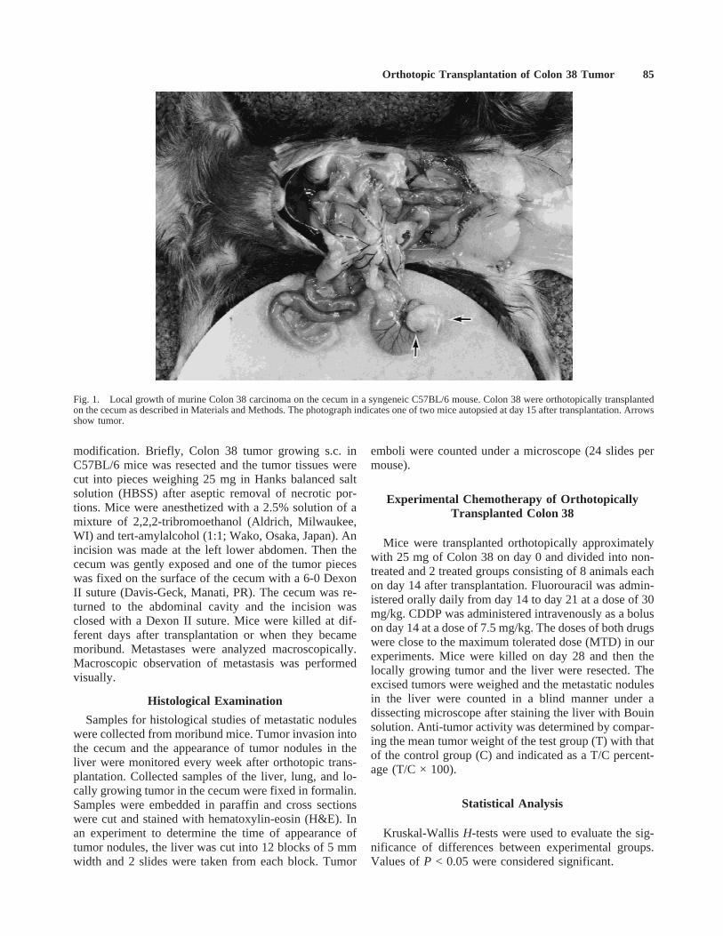
Fig. 1. Local growth of murine Colon 38 carcinoma on the cecum in a syngeneic C57BL/6 mouse. Colon 38 were orthotopically transplantedon the cecum as described in Materials and Methods. The photograph indicates one of two mice autopsied at day 15 after transplantation. Arrowsshow tumor.
Orthotopic Transplantation of Colon 38 Tumor 85
RESULTSLocal Growth and Metastasis of Colon 38 Tumor
Orthotopically Transplanted as Intact Tissuein C57BL/6
Tumor tissue of Colon 38 grown s.c. was inoculated in30 syngeneic mice from which 29 survived the operation.Two mice were killed and autopsied on days 7, 15, and22 after transplantation, respectively. The remainingmice were autopsied between days 28 and 32 after the
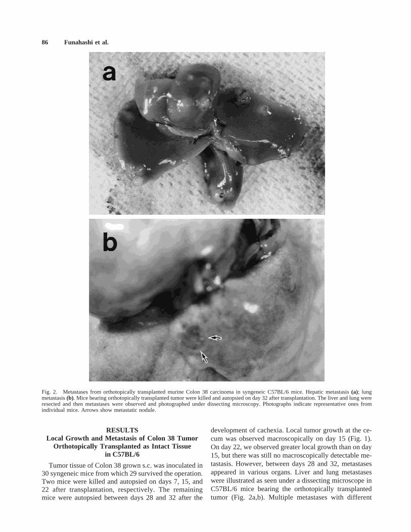
development of cachexia. Local tumor growth at the ce-cum was observed macroscopically on day 15 (Fig. 1).On day 22, we observed greater local growth than on day15, but there was still no macroscopically detectable me-tastasis. However, between days 28 and 32, metastasesappeared in various organs. Liver and lung metastaseswere illustrated as seen under a dissecting microscope inC57BL/6 mice bearing the orthotopically transplantedtumor (Fig. 2a,b). Multiple metastases with different
Fig. 2. Metastases from orthotopically transplanted murine Colon 38 carcinoma in syngeneic C57BL/6 mice. Hepatic metastasis(a); lungmetastasis(b). Mice bearing orthotopically transplanted tumor were killed and autopsied on day 32 after transplantation. The liver and lung wereresected and then metastases were observed and photographed under dissecting microscopy. Photographs indicate representative ones fromindividual mice. Arrows show metastatic nodule.
86 Funahashi et al.
sizes were observed in the liver with a frequency of95.7%, whereas only a few metastases were seen in thelung. Metastases to the peritoneum, mesometrium, ovary,and intestine also developed at relatively low frequency(Table I). All mice evaluated in this experiment showedlocal growth at the cecum, but tumor growth at the mes-enteric lymph node was not detected, although small tu-mors grew at the lymphoid follicle on the cecum.
Tumor Invasion Into the Cecum
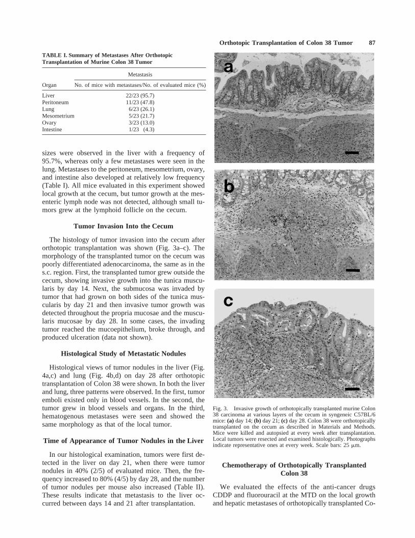
The histology of tumor invasion into the cecum afterorthotopic transplantation was shown (Fig. 3a–c). Themorphology of the transplanted tumor on the cecum waspoorly differentiated adenocarcinoma, the same as in thes.c. region. First, the transplanted tumor grew outside thececum, showing invasive growth into the tunica muscu-laris by day 14. Next, the submucosa was invaded bytumor that had grown on both sides of the tunica mus-cularis by day 21 and then invasive tumor growth wasdetected throughout the propria mucosae and the muscu-laris mucosae by day 28. In some cases, the invadingtumor reached the mucoepithelium, broke through, andproduced ulceration (data not shown).
Histological Study of Metastatic Nodules
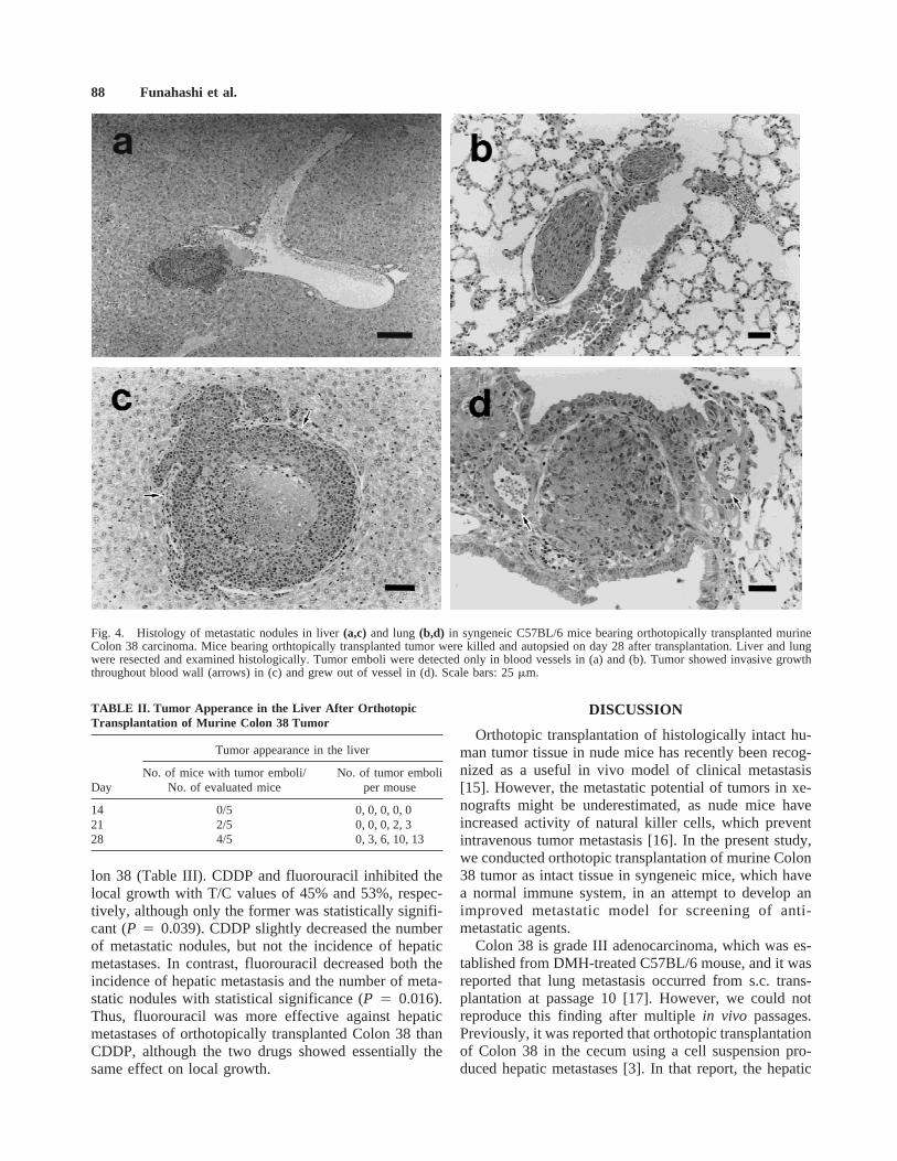
Histological views of tumor nodules in the liver (Fig.4a,c) and lung (Fig. 4b,d) on day 28 after orthotopictransplantation of Colon 38 were shown. In both the liverand lung, three patterns were observed. In the first, tumoremboli existed only in blood vessels. In the second, thetumor grew in blood vessels and organs. In the third,hematogenous metastases were seen and showed thesame morphology as that of the local tumor.
Time of Appearance of Tumor Nodules in the Liver
In our histological examination, tumors were first de-tected in the liver on day 21, when there were tumornodules in 40% (2/5) of evaluated mice. Then, the fre-quency increased to 80% (4/5) by day 28, and the numberof tumor nodules per mouse also increased (Table II).These results indicate that metastasis to the liver oc-curred between days 14 and 21 after transplantation.
Chemotherapy of Orthotopically TransplantedColon 38
We evaluated the effects of the anti-cancer drugsCDDP and fluorouracil at the MTD on the local growthand hepatic metastases of orthotopically transplanted Co-
TABLE I. Summary of Metastases After OrthotopicTransplantation of Murine Colon 38 Tumor
Organ
Metastasis
No. of mice with metastases/No. of evaluated mice (%)
Liver 22/23 (95.7)Peritoneum 11/23 (47.8)Lung 6/23 (26.1)Mesometrium 5/23 (21.7)Ovary 3/23 (13.0)Intestine 1/23 (4.3)
Fig. 3. Invasive growth of orthotopically transplanted murine Colon38 carcinoma at various layers of the cecum in syngeneic C57BL/6mice:(a) day 14;(b) day 21;(c) day 28. Colon 38 were orthotopicallytransplanted on the cecum as described in Materials and Methods.Mice were killed and autopsied at every week after transplantation.Local tumors were resected and examined histologically. Photographsindicate representative ones at every week. Scale bars: 25mm.
Orthotopic Transplantation of Colon 38 Tumor 87
lon 38 (Table III). CDDP and fluorouracil inhibited thelocal growth with T/C values of 45% and 53%, respec-tively, although only the former was statistically signifi-cant (P 4 0.039). CDDP slightly decreased the numberof metastatic nodules, but not the incidence of hepaticmetastases. In contrast, fluorouracil decreased both theincidence of hepatic metastasis and the number of meta-static nodules with statistical significance (P 4 0.016).Thus, fluorouracil was more effective against hepaticmetastases of orthotopically transplanted Colon 38 thanCDDP, although the two drugs showed essentially thesame effect on local growth.
DISCUSSION
Orthotopic transplantation of histologically intact hu-man tumor tissue in nude mice has recently been recog-nized as a useful in vivo model of clinical metastasis[15]. However, the metastatic potential of tumors in xe-nografts might be underestimated, as nude mice haveincreased activity of natural killer cells, which preventintravenous tumor metastasis [16]. In the present study,we conducted orthotopic transplantation of murine Colon38 tumor as intact tissue in syngeneic mice, which havea normal immune system, in an attempt to develop animproved metastatic model for screening of anti-metastatic agents.
Colon 38 is grade III adenocarcinoma, which was es-tablished from DMH-treated C57BL/6 mouse, and it wasreported that lung metastasis occurred from s.c. trans-plantation at passage 10 [17]. However, we could notreproduce this finding after multiplein vivo passages.Previously, it was reported that orthotopic transplantationof Colon 38 in the cecum using a cell suspension pro-duced hepatic metastases [3]. In that report, the hepatic
Fig. 4. Histology of metastatic nodules in liver(a,c) and lung(b,d) in syngeneic C57BL/6 mice bearing orthotopically transplanted murineColon 38 carcinoma. Mice bearing orthtopically transplanted tumor were killed and autopsied on day 28 after transplantation. Liver and lungwere resected and examined histologically. Tumor emboli were detected only in blood vessels in (a) and (b). Tumor showed invasive growththroughout blood wall (arrows) in (c) and grew out of vessel in (d). Scale bars: 25mm.
TABLE II. Tumor Apperance in the Liver After OrthotopicTransplantation of Murine Colon 38 Tumor
Day
Tumor appearance in the liver
No. of mice with tumor emboli/No. of evaluated mice
No. of tumor emboliper mouse
14 0/5 0, 0, 0, 0, 021 2/5 0, 0, 0, 2, 328 4/5 0, 3, 6, 10, 13
88 Funahashi et al.

metastasis was observed in five of nine mice at 10 weeksafter transplantation. However, we found that orthotopictransplantation using histologically intact tissue in syn-geneic mice resulted in metastases to various organswithin only 4 weeks after inoculation. The frequencies ofhepatic metastasis was high (95.7%) and multiple meta-static nodules were observed under a dissecting micro-scope. Thus, we think that this model is superior forevaluating drugs as candidates to prevent hepatic metas-tasis of colorectal cancer.
This model seems to reflect the frequent recurrenceand metastasis of colorectal carcinoma after curative op-eration in clinical patients. Metastasis to other organsincluding peritoneum, lung, mesometrium, ovary, and in-testine occurred in this model, and this resembles thepattern of metastases of colon cancer in about 1,687clinical cases [18]. We observed tumor growth at thelymphoid follicle on the cecum, but mesenteric lymphnode metastasis was not detected between days 28 and 32after transplantation. This was probably because the pe-riod of evaluation was too short to detect mesentericlymph node metastasis, since mice bearing orthotopicallytransplanted Colon 38 die from cachexia at 4–5 weeks.After orthotopic transplantation of Colon 38 using a cellsuspension, mesenteric node metastasis was detected at10 weeks, although the incidence was lower than that ofhepatic metastasis [3]. These results might indicate thatthe hepatic metastasis and mesenteric node metastasiswill have distinct events in these models.
Histological study showed that transplanted Colon 38invaded various layers of the cecum from tunica muscu-laris to muscoepithelium. This seems to occur from day14 to day 21 after transplantation. The first appearance oftumor in the liver was on day 21. These results suggestthat hepatic metastasis is established after local tumorinvasion and tumor growth throughout the tunica mus-cularis and submucosae. In clinical colon cancer, it isthought that metastasis occurs after primary tumor inva-sion into the submucosa from the propria mucosa andgrowth throughout the muscularis mucosae. Thus, wethink that the process of metastasis in this model re-sembles the clinical patients, even though the direction ofinvasion is opposite.
Fluorouracil is generally used for adjuvant chemo-
therapy of colon cancer, although its efficacy is limited.We conducted experimental chemotherapy of orthotopi-cally transplanted Colon 38 using fluorouracil andCDDP. Fluorouracil was effective against local growthand reduced the incidence of hepatic metastasis and thenumber of metastatic nodules. Although CDDP also in-hibited local growth, it was ineffective against hepaticmetastasis. Both fluorouracil and CDDP reduced the in-cidence of metastasis to the peritoneum, which occurredat lower frequency than that to the liver (data not shown).We also evaluated the effect of vincristine, whichshowed no effect on local growth and metastasis (datanot shown). These results might provide the rationale touse fluorouracil for adjuvant chemotherapy for coloncancer.
Experimental chemotherapy using fluorouracil againstanother murine colon carcinoma (Colon 26) cells grow-ing at either orthotopic or ectopic organ in syngeneicmice after inoculation of a tumor cell suspension hasbeen reported [19]. In that case, fluorouracil failed toprevent tumor growth in the liver after inoculationthrough the spleen, though it was effective against thegrowth of tumor s.c. site or at the cecum. The adminis-tration schedule in that study was different from our ex-periment, but the effect of fluorouracil on local growth atthe cecum was almost the same, while there was a dis-crepancy concerning the effect of fluorouracil on tumorgrowth in the liver. We could not examine the effect offluorouracil against hepatic metastasis of Colon 26 fromthe cecum after orthotopic transplantation using intacttissue, since Colon 26 on the cecum resulted in fatalascites within 2 weeks after transplantation (data notshown). The discrepancy of the effect of fluorouracilagainst tumor growth in the liver may be due to thedifference in the route of tumor inoculation or in thecharacter of the tumor cells.
CONCLUSIONS
We have shown that orthotopic transplantation of mu-rine Colon 38 as intact tissue at the cecum of syngeneicmice rapidly resulted in hepatic metastasis with high in-cidence, and the pattern and process of metastasis reflectthose seen in colon cancer in patients. This model shouldbe a useful tool for screening and development of anti-
TABLE III. Anti-tumor Activity of Fluorouracil and CDDP Against Local Growth and Hepatic Metastasis of OrthotopicallyTransplanted Murine Colon 38 Tumor
Drug (dose)Tumor weighta
(mg) T/C (%) P
Hepatic metastasis
No. of mice with metastases/No. of evaluated mice
No. of tumor nodules permouse (median) P
Control 919 ± 482 100 — 7/8 0, 1, 2, 3, 5, 13, 13, 21 (4) —CDDP (7.5 mg/kg) 414 ± 164 45 0.039 6/8 0, 0, 1, 2, 3, 3, 5, 13 (2.5) 0.496Fluorouracil (30 mg/kg) 489 ± 328 53 0.065 2/8 0, 0, 0, 0, 0, 0, 2, 2 (0) 0.016
aMean ± SD.
Orthotopic Transplantation of Colon 38 Tumor 89
metastatic agents, and also for evaluating angiogenesisinhibitors and biological response modifiers that modu-late host-immune system function.
REFERENCES1. Schackert HK, Fidler IJ: Development of an animal model to
study the biology of recurrent colorectal cancer originating frommesenteric lymph system metastases. Int J Cancer 1989;44:177–181.
2. Fidler IJ: Critical factors in the biology of human cancer metas-tasis: Twenty-eighth G.H.A. Clowes Memorial Award Lecture.Cancer Res 1990;50:6130–6139.
3. Tan MH, Holyoke ED, Goldrosen MH: Murine colon adenocar-cinoma: Syngeneic orthotopic transplantation and subsequent he-patic metastases. J Natl Cancer Inst 1977;59:1537–1544.
4. Morikawa K, Walker SM, Nakajima M, et al.: Influence of organenvironment on the growth, selection, and metastasis of humancolon carcinoma cells in nude mice. Cancer Res 1988;48:6863–6871.
5. Naito S, von Eschenbach AC, Giavazzi R, et al.: Growth andmetastasis of tumor cells isolated from a human renal cell carci-noma implanted into different organs of nude mice. Cancer Res1986;46:4109–4115.
6. Price JE, Polyzos A, Zhang R, et al.: Tumorigenicity and metas-tasis of human breast carcinoma cell lines in nude mice. CancerRes 1990;50:717–721.
7. Ahlering TE, Dubeau L, Jones PA: A new in vivo model to studyinvasion and metastasis of human bladder carcinoma. Cancer Res1996;47:6660–6665.
8. Tan MH, Chu TM: Characterization of the tumorigenic and meta-static properties of a human pancreatic tumor cell line (ASPC-1)implanted orthotopically into nude mice. Tumour Biol 1985;6:89–98.
9. McLemore TL, Liu MC, Blacker PC, et al.: Novel intrapulmonarymodel for orthotopic progagation of human lung cancers in athy-mic nude mice. Cancer Res 1987;47:5132–5140.
10. Fidler IJ: Orthotopic implantation of human colon carcinomas intonude mice provides a valuable model for the biology and therapyof metastasis. Cancer Metastasis Rev 1991;10:229–243.
11. Fu X, Besterman JM, Monosov A, et al.: Models of human meta-static colon cancer in nude mice orthotopically constricted byusing histologically intact patient specimens. Proc Natl Acad SciUSA 1991;88:9345–9349.
12. Hoffman RM: Fertile seed and rich soil: The development ofclinically relevant models of human cancer by surgical orthotopicimplantation of intact tissue. In Teicher BA (ed): “Cancer DrugDiscovery and Development 1, Anticancer Drug DevelopmentGuide: Preclinical Screening, Clinical Trials, and Approval.”Totowa, NJ: Humana Press, 1997:127–144.
13. Wang H, Brown PD, Crimmin MJ, et al.: Matrix metalloprotein-ase inhibitor BB-94 (Batimastat) inhibits human colon tumorgrowth and spread in a patient-like orthotopic model in nude mice.Cancer Res 1994;54:4726–4728.
14. Richie JP, McDonald BS, Gittes RF: Resistance to intravenoustumor metastases in the athymic nude mouse: A paradoxic re-sponse. Surgery 1981;90:214–220.
15. Holzman D: Of mice and metastasis: A new for-profit modelemerges. J Natl Cancer Inst 1996;88:396–397.
16. Clark EA, Russell PH, Egghart M, et al.: Characteristics and ge-netic control of NK-cell-mediated cytotoxicity activated by natu-rally acquired infection in the mouse. Int J Cancer 1979;24:688–699.
17. Corbett TH, Griswold DP, Roberts BJ, et al.: Tumor inductionrelationships in development of transplantable cancers of colon inmice for chemotherapy assays, with a note on carcinogen struc-ture. Cancer Res 1975;35:2434–2439.
18. Floyd CE, Stirling CT, Cohn I: Cancer of the colon, rectum andanus: Review of 1,687 cases. Ann Surg 1966;163:829–837.
19. Wilmanns C, Fan D, O’Brian CA, et al.: Orthotopic and ectopicorgan environments differentially influence the sensitivity of mu-rine colon carcinoma cells to doxorubicin and 5-fluoroouracil. IntJ Cancer 1992;52:98–104.
90 Funahashi et al.