Embed Size (px)
Citation preview
University of Groningen
Randomized clinical trial on indirect resin composite and ceramic laminate veneersGresnigt, M M M; Cune, M S; Jansen, K; van der Made, S A M; Özcan, M
Published in:JOURNAL OF DENTISTRY
DOI:10.1016/j.jdent.2019.06.001
IMPORTANT NOTE: You are advised to consult the publisher's version (publisher's PDF) if you wish to cite fromit. Please check the document version below.
Document VersionFinal author's version (accepted by publisher, after peer review)
Publication date:2019
Link to publication in University of Groningen/UMCG research database
Citation for published version (APA):Gresnigt, M. M. M., Cune, M. S., Jansen, K., van der Made, S. A. M., & Özcan, M. (2019). Randomizedclinical trial on indirect resin composite and ceramic laminate veneers: Up to 10-year findings. JOURNALOF DENTISTRY, 86, 102-109. https://doi.org/10.1016/j.jdent.2019.06.001
CopyrightOther than for strictly personal use, it is not permitted to download or to forward/distribute the text or part of it without the consent of theauthor(s) and/or copyright holder(s), unless the work is under an open content license (like Creative Commons).
Take-down policyIf you believe that this document breaches copyright please contact us providing details, and we will remove access to the work immediatelyand investigate your claim.
Downloaded from the University of Groningen/UMCG research database (Pure): http://www.rug.nl/research/portal. For technical reasons thenumber of authors shown on this cover page is limited to 10 maximum.
Download date: 08-08-2020
Accepted Manuscript
Title: Randomized Clinical Trial on Indirect Resin Compositeand Ceramic Laminate Veneers: Up to 10-year Findings
Authors: M.M.M. Gresnigt, M.S. Cune, K. Jansen, S.A.M.van der Made, M. Ozcan
PII: S0300-5712(19)30125-3DOI: https://doi.org/10.1016/j.jdent.2019.06.001Reference: JJOD 3154
To appear in: Journal of Dentistry
Received date: 27 April 2019Revised date: 1 June 2019Accepted date: 3 June 2019
Please cite this article as: Gresnigt MMM, Cune MS, Jansen K, van der MadeSAM, Ozcan M, Randomized Clinical Trial on Indirect Resin Composite andCeramic Laminate Veneers: Up to 10-year Findings, Journal of Dentistry (2019),https://doi.org/10.1016/j.jdent.2019.06.001
This is a PDF file of an unedited manuscript that has been accepted for publication.As a service to our customers we are providing this early version of the manuscript.The manuscript will undergo copyediting, typesetting, and review of the resulting proofbefore it is published in its final form. Please note that during the production processerrors may be discovered which could affect the content, and all legal disclaimers thatapply to the journal pertain.
1
Randomized Clinical Trial on Indirect Resin Composite and Ceramic Laminate Veneers: Up to 10-
year Findings
Randomized Clinical Trial on Indirect Resin Composite and Ceramic Laminate Veneers: Up to 10-
year Findings
M.M.M. Gresnigt1,2*, M.S. Cune1,3,4, K. Jansen1, S.A.M. van der Made5, M. Özcan6
1The University of Groningen, University Medical Center Groningen, Department of Restorative Dentistry and
Biomaterials, Center for Dentistry and Oral Hygiene, The Netherlands; 2Martini Hospital, Department of Special Dental
Care, Groningen, The Netherlands; 3St. Antonius Hospital, Department of Oral and Maxillofacial Surgery and Special
Dental Care, Nieuwegein, the Netherlands; 4University Medical Center Utrecht, department of Oral and Maxillofacial
Surgery and Special Dental Care, Utrecht, the Netherlands; 5Dental laboratory Kwalident, Beilen, The Netherlands;
6University of Zurich, Center for Dental and Oral Medicine, Dental Materials Unit, Clinic for Fixed and Removable
Prosthodontics and Dental Materials Science, Zurich, Switzerland; *corresponding author, [email protected]
Short title: RCT on indirect composite and ceramic laminate veneers
*Corresponding author:
Marco Gresnigt, Department of Restorative Dentistry and Biomaterials, Center for Dentistry and Oral Hygiene, University
Medical Center Groningen, The University of Groningen, Antonius Deusinglaan 1, 9713 AV, Groningen, The Netherlands,
Tel: +31-6-47494611, E-mail address: [email protected].
KEY WORDS: adhesion, ceramic, clinical trial, indirect composite, laminate veneer, randomized
Acknowledgements
The authors extend their gratitude to Ivoclar Vivadent, Liechtenstein and Kuraray, Tokyo, Japan for supplying
some of the materials used in this study. There are no conflicts of interest.
ACCEPTED MANUSCRIP
T
2
Abstract
Objectives
In this randomized split-mouth clinical trial the survival rate and quality of survival of indirect resin composite
and ceramic laminate veneers were evaluated.
Methods
A total of 48 indirect resin composite (Estenia; n=24) and ceramic laminate veneers (IPS Empress Esthetic;
n=24) were placed on maxillary anterior teeth. Veneer preparations with incisal overlap were performed using
a mock up technique. Survival of the restoration was considered the primary outcome measure and reported
using Kaplan-Meier statistics and survival curves compared by means of Log Rank (Mantel-Cox) test. After
luting, restorations were evaluated by calibrated operators at baseline and every year thereafter, using modified
USPHS criteria and compared by means of Mann-Whitney U test.
Results
In total, 6 failures were observed, consisting of debonding (n=3) and fracture (n=3), all in the group of the
indirect resin composite laminate veneers. Cumulative chance on survival after 10 years of the indirect resin
composite and ceramic veneers was 75% (se 3,8%) and 100% respectively (p=0.013). Of the surviving 42
laminate veneers, the variables ‘color match’ (p=0.002), ‘surface roughness’ (p=0.000), ‘fracture of the
restoration’ (p=0.028), and ‘wear of the restoration’ (p=0.014), were significantly less favourable among the
composite laminate veneers as well.
Conclusions
The ceramic veneers on maxillary anterior teeth in this study performed significantly better compared to the
composite indirect laminate veneers after a decade, both in terms of survival rate and in terms of quality of the
surviving restorations.
Clinical Relevance
When indicated, anterior ceramic laminate veneers may be preferred over indirect composite laminate veneers.
ACCEPTED MANUSCRIP
T
3
Clinical Trial Registration Number: NCT03145597
Introduction
Laminate veneer restorations are indicated for different esthetic reasons as a minimal invasive treatment
concept. Based on the literature there is no consent as to which material should be used as the restorative
material, composite or ceramic [1,2]. Some attempts have been made to compare these materials in vivo,
however, no comparison was made in vivo in a split mouth environment with over 8 years of follow up [1,3].
Survival rates of ceramic laminate veneers range between 82-96% after 10-21 years [4–9]. Fracture of
ceramic material (5.6-11%) and marginal defects (12-20%) were the main reasons of failure [4,6,10–14].
Success rates are reported to decrease due to poor marginal quality and discoloration which contained 18-
25% up to 10 years of function.
Indirect composite restorations are easy to cement and repair, have higher flexural modulus, are cost
effective and less abrasive to the antagonistic teeth [15]. Contemporary particulate filler composites (Estenia,
Kuraray Co., Tokyo, Japan) contains up to 92 weight% colloidal silica spheres with 16 weight% superfine
microfillers, grain size of 0.02 µm, and 76 weight% microfillers, grain size of 2 µm in urethane tetramethacrylate
(UTMA) resin matrix. Previous indirect composite resin materials contained merely 50-80 weight% of fillers
[16,17]. In addition, UTMA resin matrix which contains four functional urethane methacrylates resulting in a
higher crosslinking density than other materials [17]. The higher filler content increases both strength and
optical properties, but make the material more brittle as well.
Direct comparison between different material options for laminate veneers were only performed in few studies
with relatively short follow-up periods. Therefore, the Cochrane Collaboration concluded that there is no
evidence as to which material performs better [2]. In an in vivo study by Meijering et al. [1] different materials
were compared for laminate veneers; direct composite, indirect composite and ceramic. Survival rates were
6%, 13% and 0% respectively after a mean follow up period of 1.7 years. Relative failures were not different
among the indirect composite and ceramic restorations. In a split mouth randomised clinical trial with 3 years
of follow up similar failure rates were obtained for indirect resin composite laminate veneers (13%) [3]. Relative
ACCEPTED MANUSCRIP
T
4
failures were seen but not considered significant between the two materials either, except for surface roughness
[3].
Due to aging of dental materials, differences between materials could be expected. Exposure to smoking,
food, acidic beverages, temperature changes, function of the teeth, saliva and biofilm will affect various
materials differently. Although composite materials are known for their degradation, ceramic or the glaze layer
of the ceramic will also deteriorate over time due to acidic influences and functional wear [18,19]. Degradation
of the surface polish or smoothness will not only affect the esthetic appearance, but also biofilm accumulation
[20] and wear of surrounding or opposing teeth [21–23].
The objective of this randomized clinical trial was to evaluate the clinical performance of maxillary anterior
laminate veneers made of particulate filled composite and ceramic in a split-mouth design after a mean
observation period exceeding 8 years of clinical service. Primary outcome parameter was survival of the
restoration, secondary outcome parameter was the quality of survival. The null hypothesis tested was that both
laminate materials would function similarly.
Materials and Methods
Study Design
This is the follow up study of data presented in our previous article.[3] To avoid possible disturbing differences
in case when distinct degrees of tooth discoloration would occur between restorations of different materials, a
modified split mouth design was employed in which the central incisors and the symmetrical other teeth
received the same type of restoration. Randomization was performed using the flip of a coin for the choice of
material. For this observational study the STROBE guidelines were followed.
Inclusion and Exclusion Criteria
Potential candidates were at least 18 years old, able to read and sign the informed consent document,
physically and psychologically able to tolerate conventional restorative procedures, having no high caries risk,
periodontal or pulpal diseases, having teeth with good restorations, require esthetic improvement of at least 2
anterior teeth, not allergic to resin-based materials, not pregnant or nursing, and willing to return for follow-up
ACCEPTED MANUSCRIP
T
5
examinations as outlined by the investigators. Between June-2008 and November-2010, a total of 11 patients
ranging in age between 20 and 69 years (8 female, 3 male, mean age: 54.5 years) could be recruited and
received 48 indirect composite (n=24) and ceramic laminate veneers (n=24). Alternative treatment options were
discussed. All patients provided informed consent as required by the ethical committee of the University Medical
Centre Groningen review board (Clinical Trial identification number: NCT03145597).
Tooth preparation
Treatment planning was performed using digital photos, and stone casts. Shade was determined using different
shade tabs under standard conditions (6500 K, 8 light intensity, Longlife, Aura, The Netherlands) in the dental
laboratory. A wax set-up was made on the plaster model using the mock-up technique [9]. The wax set-up was
used to communicate on the correction of the form and position of the teeth and also to evaluate the
expectations of the patient.
Magnifying microscope (x3.4 - 21.3) (Opmipico, Zeiss, Sliedrecht, The Netherlands) was used for minimal
preparations. Ball-shaped diamond burs (ISO 801 018, Diatech, Altstätten, Switzerland) were used to mark
preparation depths through the set-up. The labial surfaces were axially reduced by 0.3-0.5 mm. Tapered round-
ended diamond burs (ISO 856 018, Diatech) were used for uniform preparations. An incisal overlap of 1-1.5
mm was prepared on all cases. At the cervical area, a shallow chamfer finish line (0.5 mm) was created equi-
or supra-gingival to maintain good periodontal health. A shallow chamfered marginal finish line extended inter-
proximally to hide the restoration margins up to contact area.
All internal angles were smoothed to reduce stress concentration. On the palatal aspect, a right-angled
contour (butt joint) between the incisal edge and the palatal surface was achieved. Impressions were then
made using a polyether impression material (Impregum, 3M ESPE, St. Paul, MN, USA). Temporary veneers
were made chair-side using a spot-etch technique and auto-polymerized bis-acryl (Structur SC, Voco,
Cuxhaven, Germany).
One dental technician made all laminate veneers. Leucite reinforced glass ceramic (IPS Empress Esthetic,
Ivoclar Vivadent, Schaan, Liechtenstein) were processed according to the manufacturer’s instructions using
ACCEPTED MANUSCRIP
T
6
the IPS Empress layering and lost wax technique. After wax-up, a cut-back of 0.2-0.8 mm was performed to
allow for layering of the veneering ceramic.
The indirect composite laminate veneers (Estenia C&B, Kuraray, Tokyo, Japan) were fabricated using the
layering technique following the manufacturer`s instructions. They were heat- (100-110°C for 15 min) and
photo-polymerized (400-515 nm for 270 seconds) using a special polymerization device (Heat-curing-110,
Toesco, Yoshida, Japan) according to the manufacturer’s recommendations.
Both ceramic and resin composite laminate veneers were hand polished using diamond burs and silicone
rubber points (3044HP-30044HP Ceragloss, Edenta, St. Gallen, Switzerland) and diamond pastes with brushes
(Estenia C&B polishing compound and Yeti Diaglaze).
Luting
Form, adaptation and shade match of the restorations were checked clinically using try-in pastes (Variolink
Veneer Try-in Paste, Ivoclar Vivadent).
After cleaning with 99% isopropanol, intaglio surfaces of the laminates were etched with 4.9% hydrofluoric
acid (IPS Ceramic etching gel, Ivoclar Vivadent) for 1 minute, washed thoroughly for 1 minute and dried with
oil-free compressed air. Since etching with hydrofluoric acid leaves a significant amount of crystalline debris
precipitate at the ceramic surface,4 laminate veneers were ultrasonically cleaned in distilled water for 5 minutes.
Thereafter, the adhesive surfaces were silanized (Monobond S, Ivoclar Vivadent) for 1 minute. After
silanization, adhesive resin (ExciTE, Ivoclar Vivadent) was applied, air-thinned but not polymerized.
The intaglio of the indirect composite laminate veneers was tribochemically silica coated (30 µm SiO2, CoJet-
Sand, 3M ESPE) using an intraoral air-abrasion device (Dento-Prep, RØNVIG A/S, Daugaard, Denmark) at a
pressure of 2.5 bar from a distance of approximately 10 mm for 20 seconds. They were then silanized with 3-
methacryloxypropyltrimethoxy silane coupling agent (MPS) (ESPE-Sil, 3M ESPE AG) and waited for its
evaporation for 5 minutes. After silanization, adhesive resin (ExciTE, Ivoclar Vivadent) was applied, air-thinned
but not polymerized.
All teeth to be veneered were isolated using a split-rubberdam technique. Contour strips (Contour-Strip,
Ivoclar Vivadent) were placed interproximal to perform a smooth restoration outline in the approximal-cervical
ACCEPTED MANUSCRIP
T
7
area. The prepared teeth were first cleaned with fluoride-fee pumice (Pumice Flour, Dux, Utrecht, The
Netherlands) using a polishing brush (Polishing brush, Coltène/Whaledent, Altstatten, Switzerland).
Enamel and dentin were etched with 37% H3PO4 (Total Etch, Ivoclar Vivadent, Schaan, Liechtenstein) for
15-30 seconds. After rinsing for 30 seconds and air-drying, the adhesive resin (ExciTE, Ivoclar Vivadent) was
then applied on both the tooth and the restoration surfaces with a microbrush for 15 seconds, air-thinned but
not polymerized.
Laminate veneers were luted using a photo-polymerizing resin composite cement (Variolink Veneer, Ivoclar
Vivadent). Composite was applied to the inner surface of the laminates. After placement, initially, they were
photo-polymerized with an LED lamp (Bluephase 20i, Ivoclar Vivadent) for only 3 s at the buccal surface to
ensure stabilization of the veneer. The light output was at least 800 mW/cm2 in all applications. Gross excess
composite at the margins was removed immediately with the aid of brushes, scalers and dental floss (Oral-B,
Rotterdam, The Netherlands). Application of glycerine gel (Liquid-Strip, Ivoclar Vivadent) at the margins
ensured oxygen inhibition during polymerization. Buccal, oral, and proximal surfaces were further polymerized
for 40 s. After rinsing the glycerine gel, excess material was removed with hand-instruments and finishing burs.
Restoration margins were further polished with silicone polishers (Astropol FP, HP, Ivoclar Vivadent) and
interproximal polishing strips (Soft-Lex Finishing Strips, 3M ESPE) at 7.500-10.000 rpm under water. One
clinician placed all restorations. Finally, the occlusion was checked in protrusive and lateral movements of the
mandible. The goal was to reach anterior guidance and lateral protection in all cases. Patients were given
information on how to clean the restorations and teeth, on diet (no restrictions with food or drinks), no nail biting
and parafunctional habits (providing a night guard).
Evaluation
Restorations were clinically evaluated at baseline and thereafter by two calibrated observers who were blinded
to the objective of this study. Caries, debonding and fracture to failure were considered as absolute failures.
Patients were also questioned about possible post-operative complaints. Both observers evaluated the
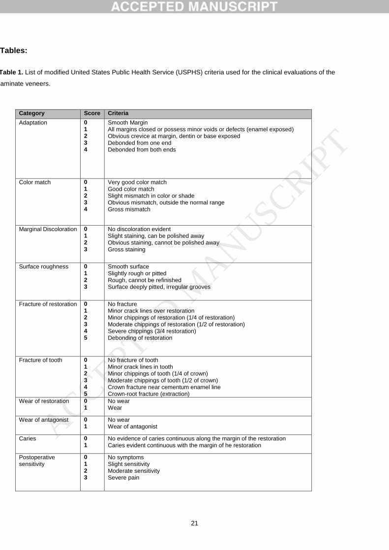
restorations independently, according to the modified United States Public Health Service (USPHS) criteria
ACCEPTED MANUSCRIP
T
8
(Table 1). The restorations were visually inspected with dental mirror and probe. After data collection, in case
of discrepancies in scoring, restorations were evaluated again, a consensus was reached and this was
accepted as the final score. Patients were instructed to call upon any kind of failure. Digital pictures (1:1) were
made after placement of the veneers and during follow-up sessions. In representative cases, an impression
(Ultra-Light and Heavy body Aquasil, Dentsply) was taken from the two laminate veneers after cleansing the
surface with absorbent paper and sodium hypochlorite 0.5%. Impressions were poured with cold mounting
epoxy resin (Epoxy-Cure, Buehler, IL, USA) then sputter-coated with a 3 nm thick layer of gold (80%) /
palladium (20%) (90 s, 45mA; Balzers SCD 030, Balzers, Liechtenstein) and analyzed using cold field emission
Scanning Electron Microscope (SEM) (LyraTC, Tescan, Brno, Czech Republic). Images were made at 15 kV
at a magnification of x22 to x2.500.
Statistical Analysis
Survival analyses were performed with statistical software program (SPSS 23.0; SPSS Inc, Chicago, IL, USA)
using Kaplan-Meier and Log Rank (Mantel-Cox) tests to obtain the overall survival rate in relation to observation
time. A nonparametric test (Mann-Whitney U test) was performed for the qualitative evaluation of the data. An
alpha level of 0.05 for all statistical tests was set.
Results
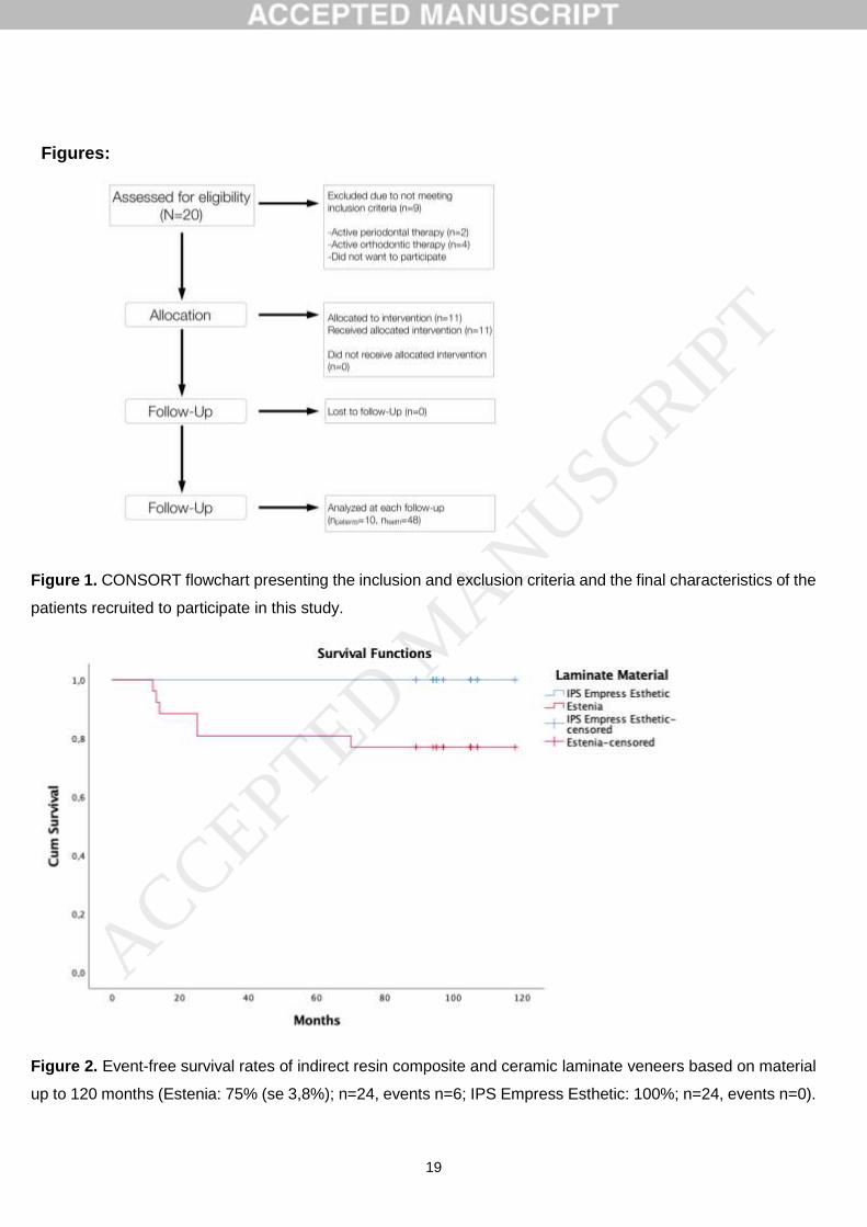
5 Recalls were performed after baseline measurements and no drop-outs occurred, yielding to the evaluation
of 48 indirect laminate veneers (Estenia: n=24; IPS Empress Esthetic: n=24). After including 11 patients, it was
decided to stop the further inclusion of patients due to failures and differences seen in longevity between both
groups. The mean observation time was 97 months with a minimum observation period of 89 months (n=4) and
up to a maximum of 120 months (n=4). The distribution of the location of the restorations was as follows: 20 on
central incisors, 18 on lateral incisors, and 10 on canines. Average treatment time for each restoration was
noted to be approximately 120 minutes, regardless the treatment type. Two patients received occlusal splints
after cementation, indicated because of parafunctional habits.
ACCEPTED MANUSCRIP
T
9
The cumulative chance of survival was 75% (se 3,8%) and 100% for the indirect composite land ceramic
laminate veneers respectively after 10 years (120 months). Survival curves showed a statistically different
distribution (p=0.013) [Kaplan-Meier, Log Rank (Mantel-Cox) (Cl=95%)] (Fig. 2). A total of 6 absolute failures
were observed, all in the in the group of the indirect resin composite veneers in the form of debonding (n=3) or
fracture (n=3). The debondings were a complete adhesive failure between the tooth and the luting cement,
which occurred 11 to 25 months after cementation. Some of the composite remained attached to the inner
surface of the laminate restoration. After cleaning the adhesive surface, the debonded veneers were re-bonded
but were not further evaluated and scored as a failure. All ractures occurred at the incisal area and were
cohesive failures in the indirect composite material. The first fracture occurred on a tooth 11 (figure 5a), 13
months after delivery. The second laminate fracture occurred on a tooth 22 which was sound, 11 months after
delivery. The third fracture occurred 6 years after placement after eating some bread.
Besides absolute failures, success was scored using the USPHS criteria (table 2). Qualitative evaluation
(success) showed some significance differences between laminates made of ceramic and indirect composite
(Table 2). For all of these variables, the ceramic restorations were rated better. Of the 42 laminate veneers,
minor voids and marginal discrepancies and defects were observed in 14 of the composite and 10 of the
ceramic veneers (Adaptation-Score 1-2). Color match was significantly (Mann-Whitney U = 324,
p=0.002)different as the ceramic laminate veneers matched the surrounded teeth, composite restorations did
not match for 8 laminate veneers (p=0.002). Slight staining at the margins was seen more frequent with the
composite laminate veneers (n=12), however not significant (p=0.107). Slightly rough surfaces (Surface
roughness-Score 1) were significantly (Mann-Whitney U = 444, p=0.000) more observed in the resin composite
laminate veneer group (n=18) until the final recall. These rough surfaces also experienced more plaque
adhesion (figure 4b). Internal fractures without intervention were significantly (Mann-Whitney U = 292, p=0.028)
more seen (n=6, p=0.028) in the indirect composite group, chippings of tooth material were more seen in the
composite group as well however this was not significant different (p=0.06). Wear of the restoration was
significantly (Mann-Whitney U = 303, p=0.014) more seen in the indirect composite group (n=7, p=0.014).
Secondary caries, endodontic complications or wear of the antagonist were not observed in any of the cases.
ACCEPTED MANUSCRIP
T
10
In total, 8 teeth showed post-operative sensitivity at baseline, as reported by the patient. All post-operative
sensitivities disappeared after 2 weeks; at the final recall 2 teeth were somewhat sensitive to cold. SEM and
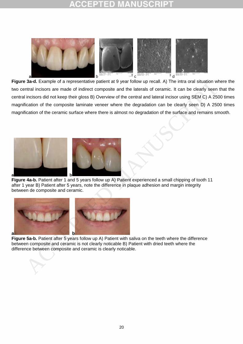
digital pictures were used for surface evaluation as can be seen in figure 3. This particular patient had her
laminate veneers for 9 years and differences in surface change between the two materials can be clearly seen.
Gloss retention was better with the ceramic restorations which is also seen at the SEM analysis. Patients were
not aware of the loss of gloss due to saliva over the restorative materials (figure 4a-b).
Discussion
In this randomized split mouth clinical trial, a comparison of indirect resin composite and ceramic laminate
veneers was performed. This is the first clinical trial on anterior indirect restorations using two different
restorative materials with a mean follow up of more than 8 years. The split mouth study design used
removes a lot of inter-individual variability from the estimates of the treatment effect. The results presented
cover observations up to 120 months of clinical function. In total 90% of the laminates required no
intervention which could be considered as clinically acceptable. However, based on the significant
differences in different aspects of success as well as the differences in survival rates the null hypothesis
that there is no difference between the two restorative materials was rejected. Ceramic veneers performed
significant better than the indirect composite ones.
Six absolute failures occurred in this study of which 3 failed within the first 13 months of the study. The
first failure occurred within the first year following luting and was a delamination of a composite laminate
veneer on a canine where the substrate was predominantly dentin. In the literature, it is suggested that
laminates bonded to large surfaces of dentin have a compromised survival rate and in such a situation
requires an immediate dentin sealing, which was not performed in our study [24–26]. Increased fractures
and chippings were noticed up to 8 times in studies where laminate veneers were made in patients with
bruxing habits [7,27]. In this study, instructions to the patients were given at insertion of the laminate
veneers regarding habits like nail biting and tearing materials with teeth. Two patients were provided with
a hard acrylic resin occlusal appliance as they were suspected nocturnal bruxers. Patients were informed
ACCEPTED MANUSCRIP
T
11
that there was a risk of fracture if compliance was inadequate. Another fracture after 12 months of insertion
is probably related to function during protrusive and lateral excursive movements over teeth. Two
debondings of composite laminate veneers occurred in the same patient (25 months after insertion) where
both central incisors had reveived endodontic treatment prior to our study and the substrate was
predominantly dentin again. All debonded laminate veneers could be rebonded to freshly cut dentin
removing only 0.1mm of dentin, performing a three step dentin bonding adhesive (Optibond FL, Kerr,
Orange USA) and using a direct resin composite (HFO, Micerium, Avegno, Italy) as a cement [28,29]. All
laminate veneers functioned until the end of the study but were scored as failure and were not screened for
follow up evaluations.
Of the qualitative evaluation, most frequently observed differences were the surface degradation and
diminished gloss retention of the indirect resin composite material. All ceramic restorations remained
smooth and their gloss until the final follow up. Both materials were processed in the laboratory and
manufactured following the manufacturers’ instructions by an experienced dental technician. The indirect
composite material was photo- and heat-polymerized and both indirect materials were hand polished.
Increased degradation of the material itself was more prone with the indirect composite material as is seen
in other laboratory and clinical studies [30–33]. Fractures, chippings and wear were frequently seen at the
incisal palatal aspect. This could be related to function and antagonist teeth articulating over these margin-
material surfaces. One internal fracture in a ceramic laminate veneer was observed in the second year of
function. This fracture was not treated or removed, but evaluated and remained stable until the end of the
study.
Marginal quality was evaluated as adaptation of the veneer and discoloration of the margin. In different
studies on ceramic laminate veneers as well as our study these were the mostly observed (adaptation:
56%; discoloration: 44%) qualitative complications [7,8,25,27,34,35] wear or degradation of the luting
composite in the margins leads to discolorations but no caries was observed in any of the patients.
Degradation of the margins was mostly observed in the palatal aspect and sometimes when the cervical
outline was in dentin on the cervico-buccal aspect. Most of the marginal discolorations could be removed
ACCEPTED MANUSCRIP
T
12
by polishing, however this was not performed as patients did not complain and further experimental
evaluation could be performed.
Evaluation of surrounding tissues did not show significant differences in gingival health between the two
materials. Only one patient had 0.5mm of recession at a central incisor (ceramic) and a lateral incisor
(indirect composite), which was probably related to brushing method and not to material properties.
When absolute failures are considered, the clinical performance of indirect resin composite and
ceramic laminate veneers performed better up to 120 months. This finding is different from the first article
which only had data up to 3 years with a mean observation time of 20.3 months.[3] Surface quality changes
were more frequently observed in the composite veneer material that may require more maintenance over
time.
In conclusion, the ceramic veneers on maxillary anterior teeth in this study performed significantly better
compared to the composite indirect laminate veneers after a decade, both in terms of survival rate and in terms
of quality of the surviving restorations.
Compliance with Ethical Standards
Conflict of Interest: All authors declares that they have no conflict of interest.
Funding: The work was supported by the Department of Oral Function and Biomaterials, University Medical
Center, Groningen, the Netherlands.
Ethical approval: All procedures performed in studies involving human participants were in accordance with
the ethical standards of the institutional and/or national research committee and with the 1964 Helsinki
declaration and its later amendments or comparable ethical standards.
Informed consent: Informed consent was obtained from all individual participants included in the study.
Disclosure Statement
ACCEPTED MANUSCRIP
T
13
The authors did not have any commercial interest in any of the materials used in this study and each of the
authors listed below declare no conflict of interest.
ACCEPTED MANUSCRIP
T
14
References
[1] A.. Meijering, N.H.. Creugers, F.J.. Roeters, J. Mulder, Survival of three types
of veneer restorations in a clinical trial: a 2.5-year interim evaluation, J. Dent.
26 (1998) 563–568.
[2] J. Wakiaga, P. Brunton, N. Silikas, A.M. Glenny, Direct versus indirect veneer
restorations for intrinsic dental stains., Cochrane Database Syst. Rev. (2004)
CD004347.
[3] M.M. Gresnigt, W. Kalk, M. Ozcan, Randomized clinical trial of indirect resin
composite and ceramic veneers: up to 3-year follow-up., J. Adhes. Dent. 15
(2013) 181–90.
[4] M.J. Friedman, A 15-year review of porcelain veneer failure--a clinician’s
observations., Compend. Contin. Educ. Dent. 19 (1998) 625–628
[5] M. Fradeani, M. Redemagni, M. Corrado, Porcelain laminate veneers: 6- to 12-
year clinical evaluation--a retrospective study., Int. J. Periodontics Restorative
Dent. 25 (2005) 9–17.
[6] M. Peumans, J. De Munck, S. Fieuws, P. Lambrechts, G. Vanherle, B. Van
Meerbeek, A Prospective Ten-Year Clinical Trial of Porcelain Veneers, J.
Esthet. Restor. Dent. 18 (2006) 110–111.
[7] U.S. Beier, I. Kapferer, D. Burtscher, H. Dumfahrt, Clinical performance of
porcelain laminate veneers for up to 20 years., Int. J. Prosthodont. 25 (2012)
79–85.
[8] D.M. Layton, T.R. Walton, The up to 21-year clinical outcome and survival of
feldspathic porcelain veneers: accounting for clustering., Int. J. Prosthodont. 25
(2012) 604–12.
[9] G. Gurel, S. Morimoto, M.A. Calamita, C. Coachman, N. Sesma, Clinical
performance of porcelain laminate veneers: outcomes of the aesthetic pre-
evaluative temporary (APT) technique., Int. J. Periodontics Restorative Dent.
32 (2012) 625–35.
[10] H. Dumfahrt, H. Schäffer, Porcelain laminate veneers. A retrospective
evaluation after 1 to 10 years of service: Part II--Clinical results., Int. J.
Prosthodont. 13 (2000) 9–18.
[11] S.M. Dunne, B.J. Millar, A longitudinal study of the clinical performance of
porcelain veneers., Br. Dent. J. 175 (1993) 317–21.
[12] M. Fradeani, M. Redemagni, M. Corrado, Porcelain laminate veneers: 6- to 12-
ACCEPTED MANUSCRIP
T
15
year clinical evaluation--a retrospective study., Int. J. Periodontics Restorative
Dent. 25 (2005) 9–17.
[13] P.C. Guess, C.F.J. Stappert, Midterm results of a 5-year prospective clinical
investigation of extended ceramic veneers, Dent. Mater. 24 (2008) 804–813.
[14] F.J. Shaini, a C. Shortall, P.M. Marquis, Clinical performance of porcelain
laminate veneers. A retrospective evaluation over a period of 6.5 years., J. Oral
Rehabil. 24 (1997) 553–559.
[15] N. Krämer, K.-H. Kunzelmann, M. Taschner, A. Mehl, F. Garcia-Godoy, R.
Frankenberger, Antagonist enamel wears more than ceramic inlays., J. Dent.
Res. 85 (2006) 1097–100.
[16] G. Leloup, P.E. Holvoet, S. Bebelman, J. Devaux, Raman scattering
determination of the depth of cure of light-activated composites: Influence of
different clinically relevant parameters, J. Oral Rehabil. 29 (2002) 510–515.
[17] R.O.A. Souza, M. Ozcan, S.M.A. Michida, R.M. de Melo, C.A. Pavanelli, M.A.
Bottino, L.E.S. Soares, A.A. Martin, Conversion degree of indirect resin
composites and effect of thermocycling on their physical properties, J.
Prosthodont. 19 (2010) 218–225.
[18] P.C. Guess, Y. Zhang, V.P. Thompson, Effect of veneering techniques on
damage and reliability of Y-TZP trilayers., Eur. J. Esthet. Dent. 4 (2009) 262–
76.
[19] V.Z.S. Cahuana, M. Ozcan, A.M.M. Mesquita, R.S. Nishioka, E.T. Kimpara,
M.A. Bottino, Surface degradation of glass ceramics after exposure to
acidulated phosphate fluoride., J. Appl. Oral Sci. 18 (2010) 155–165.
[20] S.B.P. Fúcio, F.G. Carvalho, L.C. Sobrinho, M.A.C. Sinhoreti, R.M. Puppin-
Rontani, The influence of 30-day-old Streptococcus mutans biofilm on the
surface of esthetic restorative materials-An in vitro study, J. Dent. 36 (2008)
833–839.
[21] K. Kawai, M. Urano, Adherence of plaque components to different restorative
materials., Oper. Dent. 26 (2001) 396–400.
[22] M. Fuzzi, Z. Zaccheroni, G. Vallania, Scanning electron microscopy and
profilometer evaluation of glazed and polished dental porcelain., Int. J.
Prosthodont. 9 (n.d.) 452–8.
[23] N.C. Lawson, S. Janyavula, S. Syklawer, E.A. McLaren, J.O. Burgess, Wear of
enamel opposing zirconia and lithium disilicate after adjustment, polishing and
ACCEPTED MANUSCRIP
T
16
glazing, J. Dent. 42 (2014) 1586–1591.
[24] P. Magne, Immediate dentin sealing: a fundamental procedure for indirect
bonded restorations., J. Esthet. Restor. Dent. 17 (2005) 144–154.
[25] F.J.T. Burke, P.S.K. Lucarotti, Ten-year outcome of porcelain laminate veneers
placed within the general dental services in England and Wales, J. Dent. 37
(2009) 31–38.
[26] M.M.M. Gresnigt, M.S. Cune, J.G. de Roos, M. Özcan, Effect of immediate and
delayed dentin sealing on the fracture strength, failure type and Weilbull
characteristics of lithiumdisilicate laminate veneers, Dent. Mater. (2016) 1–9.
[27] M.J. Friedman, A 15-year review of porcelain veneer failure--a clinician’s
observations., Compend. Contin. Educ. Dent. 19 (1998) 625–628.
[28] A. Kameyama, K. Bonroy, C. Elsen, A.K. Lührs, Y. Suyama, M. Peumans, B.
Van Meerbeek, J. De Munck, Luting of CAD/CAM ceramic inlays: Direct
composite versus dual-cure luting cement, Biomed. Mater. Eng. 25 (2015)
279–288.
[29] M.M.M. Gresnigt, M. Özcan, M. Carvalho, P. Lazari, M.S. Cune, P. Razavi, P.
Magne, Effect of luting agent on the load to failure and accelerated-fatigue
resistance of lithium disilicate laminate veneers, Dent. Mater. (2017).
[30] J.L. Ferracane, J.K. Hopkin, J.R. Condon, Properties of heat-treated
composites after aging in water., Dent. Mater. 11 (1995) 354–8.
[31] A. Göpferich, Mechanisms of polymer degradation and erosion, Biomaterials.
17 (1996) 103–114.
[32] J.L. Ferracane, H.X. Berge, J.R. Condon, In vitro aging of dental composites in
water--effect of degree of conversion, filler volume, and filler/matrix coupling., J.
Biomed. Mater. Res. 42 (1998) 465–72.
[33] J.L. Drummond, Degradation, fatigue, and failure of resin dental composite
materials., J. Dent. Res. 87 (2008) 710–719.
[34] F.J. Shaini, A.C. Shortall, P.M. Marquis, Clinical performance of porcelain
laminate veneers. A retrospective evaluation over a period of 6.5 years., J. Oral
Rehabil. 24 (1997) 553–559.
[35] M.M.M. Gresnigt, W. Kalk, M. Özcan, Clinical longevity of ceramic laminate
veneers bonded to teeth with and without existing composite restorations up to
40 months, Clin. Oral Investig. 17 (2013) 823-32.
ACCEPTED MANUSCRIP
T
17
ACCEPTED MANUSCRIP
T
18
Legends to figures and tables:
Figures:
Figure 1. CONSORT flowchart presenting the inclusion and exclusion criteria and the final characteristics of
the patients recruited to participate in this study.
Figure 2. Event-free survival rates of indirect resin composite and ceramic laminate veneers based on material
up to 120 months (Estenia: 75% (se 3,8%); n=24, events n=6; IPS Empress Esthetic: 100%; n=24, events n=0).
Figure 3a-d. Example of a representative patient at 9 year follow up recall. A) The intra oral situation where
the two central incisors are made of indirect composite and the laterals of ceramic. It can be clearly seen that
the central incisors did not keep their gloss B) Overview of the central and lateral incisor using SEM C) A 2500
times magnification of the composite laminate veneer where the degradation can be clearly seen D) A 2500
times magnification of the ceramic surface where there is almost no degradation of the surface and remains
smooth.
Figure 4a-b. Patient after 1 and 5 years follow up A) Patient experienced a small chipping of tooth 11 after 1
year B) Patient after 5 years, note the difference in plaque adhesion and margin integrity between de composite
and ceramic.
Figure 5a-b. Patient after 5 years follow up A) Patient with saliva on the teeth where the difference between
composite and ceramic is not clearly noticable B) Patient with dried teeth where the difference between
composite and ceramic is clearly noticable.
Tables:
Table 1. List of modified United States Public Health Service (USPHS) criteria used for the clinical evaluations
of the laminate veneers.
Table 2. Summaries of USPHS evaluations at baseline and final follow-up.
ACCEPTED MANUSCRIP
T
19
Figures:
Figure 1. CONSORT flowchart presenting the inclusion and exclusion criteria and the final characteristics of the
patients recruited to participate in this study.
Figure 2. Event-free survival rates of indirect resin composite and ceramic laminate veneers based on material
up to 120 months (Estenia: 75% (se 3,8%); n=24, events n=6; IPS Empress Esthetic: 100%; n=24, events n=0).
ACCEPTED MANUSCRIP
T
20
a b c d
Figure 3a-d. Example of a representative patient at 9 year follow up recall. A) The intra oral situation where the
two central incisors are made of indirect composite and the laterals of ceramic. It can be clearly seen that the
central incisors did not keep their gloss B) Overview of the central and lateral incisor using SEM C) A 2500 times
magnification of the composite laminate veneer where the degradation can be clearly seen D) A 2500 times
magnification of the ceramic surface where there is almost no degradation of the surface and remains smooth.
a b
Figure 4a-b. Patient after 1 and 5 years follow up A) Patient experienced a small chipping of tooth 11 after 1 year B) Patient after 5 years, note the difference in plaque adhesion and margin integrity between de composite and ceramic.
a b Figure 5a-b. Patient after 5 years follow up A) Patient with saliva on the teeth where the difference between composite and ceramic is not clearly noticable B) Patient with dried teeth where the difference between composite and ceramic is clearly noticable.
ACCEPTED MANUSCRIP
T
21
Tables:
Table 1. List of modified United States Public Health Service (USPHS) criteria used for the clinical evaluations of the
laminate veneers.
Category Score Criteria
Adaptation 0 1 2 3 4
Smooth Margin All margins closed or possess minor voids or defects (enamel exposed) Obvious crevice at margin, dentin or base exposed Debonded from one end Debonded from both ends
Color match 0 1 2 3 4
Very good color match Good color match Slight mismatch in color or shade Obvious mismatch, outside the normal range Gross mismatch
Marginal Discoloration 0 1 2 3
No discoloration evident Slight staining, can be polished away Obvious staining, cannot be polished away Gross staining
Surface roughness 0 1 2 3
Smooth surface Slightly rough or pitted Rough, cannot be refinished Surface deeply pitted, irregular grooves
Fracture of restoration 0 1 2 3 4 5
No fracture Minor crack lines over restoration Minor chippings of restoration (1/4 of restoration) Moderate chippings of restoration (1/2 of restoration) Severe chippings (3/4 restoration) Debonding of restoration
Fracture of tooth 0 1 2 3 4 5
No fracture of tooth Minor crack lines in tooth Minor chippings of tooth (1/4 of crown) Moderate chippings of tooth (1/2 of crown) Crown fracture near cementum enamel line Crown-root fracture (extraction)
Wear of restoration 0 1
No wear Wear
Wear of antagonist 0 1
No wear Wear of antagonist
Caries 0 1
No evidence of caries continuous along the margin of the restoration Caries evident continuous with the margin of he restoration
Postoperative sensitivity
0 1 2 3
No symptoms Slight sensitivity Moderate sensitivity Severe pain
ACCEPTED MANUSCRIP
T
22
Table 2. Summaries of USPHS evaluations at baseline and final follow-up.
Criteria Baseline Estenia IPS Esthetic
(n=24) (n=24)
Final evaluation Estenia IPS Esthetic
(n=18) (n=24)
Adaptation of Restoration
0 1 2 3 4
17 6 1 - -
20 4 - - -
P=0.308 4 10 4 - -
14 10 - - -
P=0.212
Color Match 0 1 2 3 4
9 15 - - -
10 14 - - -
P=0.770 10 3 5 - -
24 - - - -
P=0.002*
Marginal Discoloration
0 1 2 3
24 - - -
24 - - -
P=1 6 8 4 -
17 6 1 -
P=0.107
Surface Roughness 0 1 2 3
18 6 - -
24 - - -
P=0.01* 0 17 1 -
24 - - -
P=0.000*
Fracture of Restoration
0 1 2 3 4 5
24 - - - - -
24 - - - - -
P=1 12 3 3 - - -
23 1 - - - -
P=0.028*
Fracture of Tooth 0 1 2 3 4 5
24 - - - - -
24 - - - - -
P=1 15 - 3 - - -
24 - - - - -
P=0.060
Wear of Restoration 0 1
24 -
24 -
P=1 11 7
23 1
P=0.014*
Wear of Antagonist 0 1
24 -
24 -
P=1 18 -
24 -
P=1
Caries 0 1
24 -
24 -
P=1 18 -
24 -
P=1
Post-operative Sensitivity
0 1 2 3
18 4 2 -
22 2 - -
P=0.125 17 1 - -
23 1 - -
P=1
ACCEPTED MANUSCRIP
T